IN THIS ISSUE Department Otolaryngology - Mount Sinai · Department Otolaryngology Head and Neck...
8
Department of Otolaryngology Head and Neck Surgery Tumor Invades; Surgeons Do Not See Page 3 FEATURED STORY IN THIS ISSUE 2 Message From the Chair 2 Thyroid Cancer in Small Nodes 3 Tumor Invades; Surgeons Do Not 4 HPV Cancer Drug Gets a Trial 5 Resecting Larynx, Restoring Voice 6 Keeping Cancer Dormant 7 Modeling Surgery in Advance www.mountsinai.org SPECIALTY REPORT | WINTER 2017
Transcript of IN THIS ISSUE Department Otolaryngology - Mount Sinai · Department Otolaryngology Head and Neck...

Department of OtolaryngologyHead and Neck Surgery
Tumor Invades; Surgeons Do Not
See Page 3
F E A T U R E D S T O R Y
I N T H I S I S S U E
2 Message From the Chair
2 Thyroid Cancer in Small Nodes
3 Tumor Invades; Surgeons Do Not
4 HPV Cancer Drug Gets a Trial
5 Resecting Larynx, Restoring Voice
6 Keeping Cancer Dormant
7 Modeling Surgery in Advance
www.mountsinai.org
S P E C I A L T Y R E P O R T | W I N T E R 2 0 1 7

2
M E S S A G E F R O M T H E C H A I R
Eric Genden, MDEstablished in 1890 as one of the first ear, nose, and throat services in the country, Mount Sinai’s Department of Otolaryngology – Head and Neck Surgery continues its innovative spirit in all facets of patient care, research, and education. This past year has been an exciting one, replete with
accomplishments and groundbreaking endeavors.
Throughout 2016, our faculty challenged the boundaries of tumor cell dormancy, advanced immunotherapy for patients diagnosed with HPV-related oropharyngeal cancers via a clinical trial with Advaxis, and explored the role of extranodal extension (ENE) in small metastatic lymph nodes. We also forged the use of 3D technology for skull base surgery (Surgical Theater) and oral and maxillofacial surgery (Virtual Surgical Planning). And, Satish Govindaraj, MD, Chief of the Division of Rhinology and Skull Base Surgery and Vice Chair of Clinical A�airs, launched the first ever CME-accredited “On Demand” course on coursera.org, “Acute and Chronic Rhinosinusitis: A Comprehensive Review.”
Additionally, our faculty has expanded, and we have introduced the Mount Sinai Robotics Institute in conjunction with other departments at the Mount Sinai Health System. The Department of Otolaryngology – Head and Neck Surgery is home to the largest robotic surgery program for head and neck oncology in the nation, and we continuously research how to reduce complications and perfect this surgical technique. We also launched our second CME-accredited Coursera course in the fall —“HPV-Related Oral and Throat Cancer: What You Need to Know” —which teaches primary care physicians, otolaryngologists, other health care professionals, and patients the basics of the HPV epidemic and how to diagnose and treat HPV-associated oropharyngeal cancers.
Amid these achievements and this growth trajectory, each member of our team keeps in mind that listening to our patients and e�ectively communicating with our referring physicians remain indispensable tools, trumping any technology. We are unwavering in our commitment to our patients, faculty, and referring physicians and are excited about how technology, bench-to-bedside research, and training will propel this field of medicine into the near and distant future.
Cancer in Small NodesStudy Questions Traditional Diagnostic Rule
A study by Mount Sinai researchers suggests that the traditional
way of gauging the danger of thyroid cancer—based on the size of
metastatic lymph nodes—may not be correct.
The researchers examined extranodal extension (ENE), an established
prognostic indicator of more virulent disease in thyroid cancer
patients. A common presumption in thyroid cancer management is
that small-lymph-node metastases do not indicate aggressive disease,
and that ENE occurs only in metastatic lymph nodes that have reached
a critical size. However, a team of pathologists and researchers, led by
Mark Urken, MD, Chief of the Division of Head and Neck Oncology at
the Department of Otolaryngology – Head and Neck Surgery at Mount
Sinai Beth Israel, found no previous studies that had investigated the
relationship between the size of these lymph nodes and the presence of
ENE. Our multidisciplinary research group conducted a retrospective
study that compared the prevalence of ENE in metastatic nodes with
the diameter of the node in order to clarify the relationship between
metastatic lymph node diameter and the risk of ENE.
A thorough review of the pathology in 1,126 metastatic lymph nodes
from 171 thyroid cancer patients who had been operated on by a single
surgeon at Mount Sinai Beth Israel from 2004 to 2015 was conducted.
We evaluated histological features that included primary tumor
histopathology, number and size of metastatic lymph nodes, and
the presence of ENE in each node. One hundred seventy-three (15.4
percent) of the 1,126 lymph nodes demonstrated ENE. Lymph nodes
manifesting ENE ranged from 1.5 to 44.0 mm in size. Increased lymph
node size had a statistically significant association with ENE status.
However, the results showed that a significant percentage of
even small metastatic lymph nodes exhibited ENE. ENE had been
previously established as a negative prognostic factor associated
with further lymph node metastases, distant metastases,
biochemically incomplete response to therapy, and higher rates
of disease-related death. This study has prompted a reevaluation
of protocols in the Department and nationally, and plans are
under way for future studies to further elucidate the prognostic
significance of small metastatic lymph nodes manifesting ENE
relative to larger nodes with the same adverse feature.
Mark Urken, MD, Chief of the Division of Head and Neck Oncology at the Department of Otolaryngology – Head and Neck Surgery at Mount Sinai Beth Israel
2

3
Tumor Invades; Surgeons Do NotTechnology Aids Removal of Complex Schwannoma
James O’Shaughnessy’s journey to The Mount Sinai
Hospital began with ear problems. He had a persistent
sensation that his right ear was blocked with wax, but it
did not get be�er. He went to Kevin Braat, MD, an ear,
nose, and throat doctor at ENT and Allergy Associates, a
Mount Sinai affiliate on Long Island, who said it looked
like there was fluid trapped behind his eardrum. Dr. Braat
placed a tube through the eardrum to drain the fluid in
order to give James some relief, but suggested he obtain
a referral from his primary care physician to a head and
neck surgeon to further explore the issue.
“A couple of weeks later, I got hit in the head at the
welding shop where I work,” Mr. O’Shaughnessy says, “and
I knew right away it was a severe concussion. The impact
of the rod that hit me was so acute, it created swelling
on my right temple. I went to a hospital where it was
verified that I had a concussion, but they also said I had a
trigeminal schwannoma located near the brain at the base
of my skull.”
The hospital advised Mr. O’Shaughnessy that it was not
urgent to address the trigeminal schwannoma, so it wasn’t
until a few months later that he returned to Dr. Braat, who
placed a more permanent stent in his ear and strongly
suggested that he visit Eric Genden, MD, Chair of the
Department of Otolaryngology – Head and Neck Surgery and
Co-Director of the Skull Base Surgery Center at Mount Sinai.
“I met with Dr. Genden, who relayed to me that this type
of tumor would never stop growing. He said the tumor was
funneling out of the brain and into my sinus cavity, and
the risk was that it could actually expand inside my brain,”
Mr. O’Shaughnessy says.
“Dr. Genden had a great demeanor and was so confident; it
appeared to me that he and his team had done numerous
tumor-removal surgeries. He then introduced me to a skull
base surgeon, Alfred Marc Iloreta, MD, of Mount Sinai’s
Skull Base Surgery Center, who is incredible and easy to
speak with. When I le£, I was calm about having surgery at
Mount Sinai and felt good about my treatment path, which
was a lot less invasive than I expected.”
Highly Complex Tumor, Advanced Technology, and No External Incisions
Mr. O’Shaughnessy’s tumor was large—almost eight
centimeters—and was located in a complex and difficult-
to-access space, surrounded by the blood vessels that
supply the brain and nerves that innervate and control the
eye, as well as the face.
“Traditional approaches to remove this tumor include large
incisions that would expose parts of the brain and even
dismantle and remove the bones of the face and jaw,” Dr.
Iloreta explains. “In his case, we were able to combine our
experience removing tumors using advanced technology,
including optical endoscopes, high-definition cameras, and
intraoperative neuro-navigation, to completely remove his
tumor via a minimally invasive approach.”
Dr. Iloreta employed a novel technology called Surgical
Theater to reconstruct the tumor three dimensionally both
for preoperative planning and for use during the surgery.
Typically, this innovative mapping of a tumor has been
used by neurosurgeons alone or in collaboration with
head and neck surgeons, but this case marked the first
Alfred Marc Iloreta, MD, performs a transnasal
endoscopic surgery to remove patient
James O’Shaughnessy’s trigeminal schwannoma.
After surgery, a patient gets
a nice surprise: waking up to
find no external incision.
F E A T U R E D S T O R Y
Continued on page 6

4
HPV Cancer Drug Gets a TrialTherapy Targets Two Types of Immune Cells
The Head and Neck Cancer Research Program at the
Icahn School of Medicine at Mount Sinai (ISMMS)
is conducting a novel phase II trial to investigate the
treatment of human papillomavirus (HPV)–related head
and neck cancer with Axalimogene filolisbac (AXAL), a
Listeria monocytogenes (Lm)–based immunotherapy
treatment developed by Advaxis. This clinical-stage
biotechnology company is developing multiple
cancer immunotherapies based on its proprietary Lm
Technology™. Mount Sinai was selected by the company
as the initial clinical site because of the Department of
Otolaryngology’s long-standing clinical experience with
TransOral Robotic Surgery (TORS) and with managing
patients with HPV-related cancers, as well as because of
its more than 20 years of research experience.
Bre� Miles, DDS, MD, FACS, a head and neck cancer
robotic surgeon, is the Site Principal Investigator of the
Advaxis Window of Opportunity Trial for ISMMS, and
Andrew Sikora, MD, PhD, is the Study Chairperson and
Co-Director of the Head and Neck Cancer Program at
the Baylor College of Medicine in Houston, Texas (the
coordinating site for the trial). They work closely with
Marshall Posner, MD, Medical Director of the Head and
Neck Oncology Center and Associate Director of the
Center for Personalized Cancer Therapeutics at The Tisch
Cancer Institute. Together they collaborate with a team of
researchers who are currently investigating the safety and
efficacy of the vaccine.
How AXAL Works
AXAL is a live-a�enuated Lm vector system that secretes
an antigen-adjuvant protein (Lm-LLO) targeting HPV. It is
undergoing clinical trials for three potential indications:
phase III for invasive cervical cancer, phase II for head and
neck cancer, and phase II for anal cancer.
Advaxis has two additional immunotherapy products
in human clinical development: ADXS-PSA for prostate
cancer and ADXS-HER2 for HER2-expressing solid tumors.
In addition, Advaxis and Amgen are developing ADXS-
NEO, a preclinical investigational cancer immunotherapy
treatment designed to activate a patient’s immune system
to respond against the unique mutations, or neoepitopes,
identified in his or her tumor. Clinical application is
expected in 2017.
Advaxis Trial Highlights
• AXAL is administered prior to standard-of-care TORS
resection of the cancer in appropriate patients.
• The Mount Sinai Health System has enrolled eight study
patients and three observational control patients.
• While it is very early in analysis of this trial, the
preliminary data suggest systemic and intratumoral
immune activation and enhanced antitumor immunity.
The FDA-funded trial of AXAL will continue to accrue
patients at ISMMS while a second trial site is being
added at the Baylor College of Medicine. Once a sufficient
number of patients has been accrued and specimens have
been collected, the study team will have an unparalleled
opportunity to understand immune mechanisms activated
by the immunotherapy and its potential to become a
therapeutic approach for patients with HPV-related head
and neck cancers.
Investigating immunotherapy as a supplement to robotic surgery for head and neck cancers.
(Left) An HPV-related tumor is outlined in red under fluorescent microscopy. (Right) Red-cell infiltration after administration of the Advaxis vaccine. These cells represent infiltrating tumor-immune lymphocytes.

5
Resecting Larynx, Restoring VoiceComplex Procedure Rebuilds Glottis and Trachea
Anna Balint lost her voice, but not from straining it. She
fell in her home and broke her neck, and subsequently
underwent surgery at a local hospital. During the
procedure, an anesthesiologist damaged her voice box,
and she awoke unable to speak a£er a tracheostomy. For
months, Mrs. Balint communicated by writing on paper
everything she wanted to say.
In March 2016, she was referred to Mark Courey, MD,
Chief of the Division of Laryngology for the Mount Sinai
Health System. He told her that she would require surgery,
but he was confident he could restore her voice.
“When Mrs. Balint came to see me, she had a complicated
multilevel airway stenosis involving her posterior glo�is,
subglo�is, and trachea,” Dr. Courey notes. “At the level of
the subglo�is, the obstruction was complete. I performed
a primary cricotracheal resection. I removed the involved
anterior portion of her cricoid ring and the top three
tracheal rings. Then I resected the scar from the posterior
commissure and advanced a flap of posterior tracheal wall
tissue to cover the exposed cricoarytenoid joints.”
Dr. Courey cut the tracheal rings in a step-like manner
and used them to reconstruct the anterior cricoid
ring. “Because I was uncertain of how successfully the
advancement flap would relieve the posterior glo�is
stenosis, I created a permanent tracheostomy below my
anastomotic line. The skin was advanced down to the
trachea to create a skin-lined tract. This also had the
benefit of taking tension off of her tracheal suture line.”
As soon as Mrs. Balint was out of surgery, she started
to speak. Two months later, Dr. Courey removed her
tracheotomy tube and closed her stoma. “I was free and so
happy I could speak to my family,” Mrs. Balint recalls. “Dr.
Courey is the only one who knew how to correct my voice. I
am thrilled that I found him.”
Inspire Therapy at Mount Sinai
The Inspire Therapy implant is o�ered at Mount Sinai Beth Israel and The Mount Sinai Hospital as an alternate treatment for those with sleep apnea who cannot comply with CPAP. Peter Schectman, a patient who received the first implant at The Mount Sinai Hospital, had an excellent outcome—resolved snoring and respiratory events—with one of the lowest therapeutic amplitudes on record, just weeks after starting to use the device. “I have truly benefited from the Sleep Surgery Program at Mount Sinai,” he said.
Anna Balint with Mark Courey, MD
6
time Surgical Theater was used solely by head and neck surgeons
in the operating room, according to its manufacturer. “Before even
meeting Mr. O’Shaughnessy, we were able to model and simulate the
approach to the tumor and felt very confident that we would remove
the entire mass with minimal impact.”
Because of where the tumor was located, Dr. Genden initially told
Mr. O’Shaughnessy that the surgeons might need to make incisions
in both his gum line and outside his jaw. “To my surprise, when I
awoke, I had no external incision at all,” Mr. O’Shaughnessy recalls.
Following the eight-and-a-half-hour surgery on April 29, 2016, he
felt his face and said, “I don’t even feel the external incision.” That is
because there wasn’t one. The surgeons operated inside his mouth
and alongside his jaw from the inside, without ever making the
external incision. Mr. O’Shaughnessy stayed in the hospital only
on Friday night and Saturday, and then on Sunday he was able to
return home to continue his recovery.
“The nurses in the recovery room—I have never met people like that
in my life,” Mr. O’Shaughnessy says. “They were incredible. To watch
them and the help they gave me was amazing. They were so nice and
always had smiles on their faces.”
“As for Drs. Genden and Iloreta, I don’t even know where to begin,”
he adds. “I never felt stress speaking with them because they were
so confident and easygoing about the surgery. The fact that they
can focus for that many hours and use this incredible technology
is amazing.”
Upon arriving home, Mr. O’Shaughnessy felt fine and took
painkillers for three weeks. “I had some bleeding for a few days, but
I was talking and swallowing fine. The first days were filled with
a lot of soup, shakes, and so£ foods,” he recalls. It took about four
weeks for him to return to his daily routines. Now he is back to
work, exercising four days a week, and paddleboarding.
“This whole journey was a wakeup call, because I was working long
hours, six days a week. I certainly look at life differently now. I don’t
sweat the small stuff anymore, and I am so much happier with life!
I can’t praise or recommend The Mount Sinai Hospital enough. The
doctors, nurses, and friendly staff were all amazing! I have a feeling
of euphoria and am absolutely grateful to Mount Sinai for it.”
Keeping Cancer DormantTherapy Would Reprogram Cells Left After Treatment
Cancer cells frequently remain in the body a£er initial treatment for
the disease. To prevent recurrence, we need to identify drugs that
will prevent these cells from ever growing again.
Julio Aguirre-Ghiso, PhD, Professor of Medicine (Hematology and
Oncology), Otolaryngology, and Oncological Sciences, has identified
a nuclear receptor as a master regulator of a program of cancer-cell
dormancy. This gene, named NR2F1, allows cancer cells to enter
dormancy and remain in this state for later awakening, which
results in lethal metastatic growth.
Continuing this work, the Aguirre-Ghiso team and Maria Soledad Sosa,
PhD, an Assistant Professor of Medicine (Hematology and Oncology)
at the Icahn School of Medicine at Mount Sinai, have identified a new
drug that activates NR2F1. In doing so, Dr. Aguirre-Ghiso and his team
were the first to use data from dormancy models to design a therapy
for cancer that taps into the mechanisms of dormancy.
The Aguirre-Ghiso team proposed that the reprogramming of cancer
cells into a dormant state could be a way to prevent recurrences.
They discovered specific agonists that activate NR2F1, which induces
cancer cell dormancy.
Combining the NR2F1 agonist with a DNA demethylating agent
(5-aza-C) that allows for NR2F1 expression and with agonists of
the retinoic acid receptors (AM580, AM80, or BML) might allow
restoration of dormancy. This depends on the production by dormant
cells of BMP7 and TGFb2 signals that are strong dormancy inducers.
Combining the NR2F1 agonists with MEK1/2 inhibitors might further
enforce dormancy induction. The goal is to treat patients with such
drugs in the adjuvant se�ing to prevent recurrences by inducing the
reprogramming of cancer cells into dormancy or making the cells
function as they do in normal tissue.
The Partnership for New York City has selected this research for the
Partnership Fund’s BioAccelerate NYC Prize. This is an exceptional
opportunity to expand these researchers’ discovery and translate
their findings into clinical applications.
Tumor Invades continued from page 3
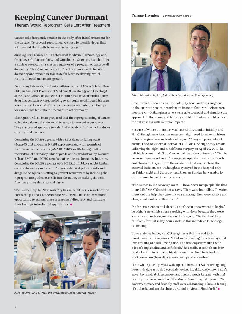
Alfred Marc Iloreta, MD, left, with patient James O’Shaughnessy
Julio Aguirre-Ghiso, PhD, and graduate student Kathryn Harper

7
Modeling Surgery in AdvanceImaging and 3D Help Doctors Plan and Patients Understand
For patients requiring complex correction of craniofacial
and oncologic deformities and orthognathic surgeries,
precise treatment planning is of the utmost importance. At
Mount Sinai’s Division of Oral and Maxillofacial Surgery,
we utilize a digital X-ray system and cone beam technology
to determine exact diagnoses and obtain three-dimensional
models for surgical treatment planning. This radiographic
technology then enables our surgeons to use virtual surgical
planning (VSP) to plan various procedures. Additionally,
we use 3D printed models of the jaw, particularly for jaw-
reconstruction procedures, which help us adapt plates and
contour the bone flaps in the operating room.
Since 2008, Mount Sinai has been at the forefront of
employing VSP—closely collaborating with a Belgian
company, Materialise NV, that provides specialized
planning so£ware and 3D physical models used in
surgical planning, as well as the medical know-how and
input to devise surgical workflows. Our team is one of
only a handful across the nation planning advanced
oral and maxillofacial surgery cases in daily practice.
We use Mimics® Innovation Suite, a computer program
made by Materialise that helps surgeons accurately
plan a procedure using a computer-generated model.
This technology can be applied to the placement of
dental implants, advanced jaw-repositioning procedures
(orthognathic surgery), repair of congenital deformities,
and jaw reconstruction requiring sophisticated bone
gra£ing and microvascular transplants. The use of VSP
is becoming routine for complex oral and maxillofacial
surgeries at the Mount Sinai Health System.
Upon obtaining each patient’s imaging, the physician
creates a cephalometric analysis using the so£ware
program, and 3D virtual models are rendered. The goal
is to simulate the planned surgical movements on the
3D model, based on a preop analysis and individualized
problem list. The so£ware is quite intuitive and user
friendly, showing the segmented objects in different
colors. A£er the planned movements are made on the 3D
computer rendering, plastic surgical cu�ing guides are
fabricated using a 3D printer, allowing us to execute the
plan in the OR. We use occlusal guides during surgery to
ensure the precise repositioning of the jaws.
In the case of mandibular reconstruction, a 3D virtual
model of the jaw is created. Using a virtual resection
plane tool, a new object is then cra£ed consisting of
the jaw segment that needs to be removed due to the
tumor invasion. Then we use the CT scan of the patient’s
fibula—from which a segment of bone will be used in jaw
reconstruction—to create precise cuts to restore the jaw to
the same form as prior to surgery. Again, plastic resection
and cu�ing guides are printed and used in the OR to
execute the planned surgical procedure.
Patients appreciate the communication and clarity offered
with VSP, because they can visualize exactly what is going
to happen during surgery. “Physicians can draw a sketch
on a piece of paper to explain the procedure to patients,”
says Daniel Buchbinder, MD, DDS, Chief of the Division of
Oral and Maxillofacial Surgery at the Mount Sinai Health
System. “But when we show patients a 3D rendering of their
own jaw with the tumor and the proposed surgical plan for
removal, it puts the entire procedure into perspective.”
Other patient benefits include:
• More individualized care
• Less operating time (VSP eliminates up to 45 minutes in
reconstruction cases and several hours of orthognathic
preop planning)
• Fewer intraoperative “surprises”
• More predictable and be�er outcomes, because the
guides enable surgeons to double-check the planned
operative movements.
Led by Vincent Carrao, MD, DDS, Chief of the Division
of Oral and Maxillofacial Surgery at The Mount Sinai
Hospital, our team conducted an outcomes study
comparing the actual amount of maxillary anterior
movement achieved in the OR to the VSP measurements
that were predicted preoperatively. A retrospective
analysis of 20 orthognathic cases over 18 months revealed
a level of accuracy with a standard deviation for all of
the procedures at 0.27 mm from the predicted value,
confirming the accuracy of this method of surgical
planning and the be�erment of outcomes using it.
Daniel Buchbinder, MD, DDS
Plastic replicas made with 3D printers let surgeons decide precisely where to cut.
22 PERCENTREDUCTION IN PATIENT SAFETY
INDICATOR INPATIENT HARM EVENTS( M e d i c a r e p a t i e n t s )
43 PERCENTREDUCTION IN C. DIFFICILE INFECTIONS
4 MILLION ANNUAL PATIENT VISITS
$ 290 MILLIONIN TOTAL NIH FUNDING
THE MOUNT SINAI HOSPITAL
R ANKED No. 15 NATIONALLY BY U.S. NEWS &
WORLD REPORT
RANKED No. 1AMONG PRIVATE MEDICAL SCHOOLS IN TOTAL RESEARCH DOLLARS PER
PRINCIPAL INVESTIGATOR
Icahn School of Medicineat Mount Sinai
Mount Sinai Beth Israel
Mount Sinai Brooklyn
The Mount Sinai Hospital
Mount Sinai Queens
Mount Sinai St. Luke’s
Mount Sinai West
New York Eye and EarInfi rmary of Mount Sinai
Con
cept
/Des
ign:
Suk
a N
Y/s
ukac
reat
ive.
com
For you. For life.
Q U I C K F A C T S
M O U N T S I N A I H E A LT H S Y S T E M
2 0 1 6 O U T C O M E S H I G H L I G H T S
2 0 1 6 D E P A R T M E N T A L H I G H L I G H T S
I C A H N S C H O O L O F M E D I C I N E A T M O U N T S I N A I
7,100+PHYSICIANS
The Mount Sinai Hospital5 East 98th Street, 8th Floor New York, NY 10029 212-241-9410
Department of Otolaryngology – Head and Neck Surgery Mount Sinai Beth Israel10 Union Square East New York, NY 10003 212-844-8450
New York Eye and Ear Infirmary of Mount Sinai 310 East 14th Street New York, NY 10003 212-979-4472
Rapid Referrals Physician Access Services800-TO-SINAI
Onlinewww.mountsinai.org/ent
LARGESTHEAD AND NECK ROBOTIC SURGERY
PROGRAM IN THE NATION
RANKED No. 1IN NEW YORK FOR OTOLARYNGOLOGY
BY U.S. NEWS & WORLD REPORT