Otolaryngology Advances
16
Otolaryngology Advances In December 2005, Marshall Strome, M.D., Chairman of the Head and Neck In- stitute, performed the first robotic-assisted transoral CO2 laser supraglottic laryngec- tomy in a 74-year-old woman. The sur- gery, which involved removal of most of the larynx above the vocal folds, was completed without encountering any sig- nificant technical difficulty and was con- sidered successful in completely resecting the malignancy. The patient experienced minimal mor- bidity after the procedure. As in traditional endoscopic surgery, a tracheotomy was not necessary; she was able to speak al- most immediately after the surgery, and she was able to swallow without difficulty by postoperative day 5. Follow-up endo- scopic examination after one month re- vealed no evidence of residual tumor. “This new technology is intriguing and yet challenging, and I believe it holds great promise for the future,” Dr. Strome says. “However, issues relating to instrumenta- tion, setup time, cost and patient selection all need careful consideration.” Partnering with industry The landmark procedure became possi- ble as the result of a unique collaboration between Cleveland Clinic head and neck surgeons, Intuitive Surgical – manufacturer of the da Vinci robotic surgical system, and OmniGuide, which developed the specialized fiber delivery system for the CO2 laser. “The first work with robotic-assisted en- doscopic laryngeal surgery was undertak- en by researchers at the University of Pennsylvania, but was performed using monopolar cautery for dissection,” explains C. Arturo Solares, M.D., otolaryngology resident. “The laser offers a major advan- tage over that technology because the laser beam is much more precise and causes much less collateral tissue injury. However, in order for the surgery to be performed with the CO2 laser, it was first necessary to develop a fiber to carry the laser beam that would be coupled to the robot.” Rapid transition into the clinic The Cleveland Clinic team began its initial feasibility studies of robotic-assisted transoral CO2 laser laryngectomy just last fall. The team began working in an edentu- lous cadaver to develop the necessary skills and then moved on to a live canine model. “Safety was a major outcome measure in those studies and showed the laser dis- section could be performed successfully and without causing major bleeding,” Dr. Solares says. Benefits and remaining challenges The robotic-assisted procedure has po- tential advantages relative to traditional transoral endoscopic surgery. Although the setup in the operating room took longer in the first robotic-assisted case, operating time was reduced. Improved surgeon con- trol and precision account for the differ- ence, as well as enhanced maneuverability of the laser beam. Cleveland Clinic Surgical Team Performs Groundbreaking Robotic-Assisted Transoral Laser Supraglottic Laryngectomy FALL 2006 A PHYSICIAN’S NEWSLETTER Latest Research on Immunosuppression of Squamous Cell Tumors p. 3 Improved Diagnosis and Treatment of Laryngeal Reflux p. 6 Fully Endoscopic Procedure for Pituitary Tumors p. 10 Auditory/Verbal Therapy for Kids with Cochlear Implants p. 12 Continued on page 2 Transoral CO2 laser surgery, in conjunction with cryoablation, for early- stage laryngeal carcinoma has demonstrated benefits of decreased mor - bidity and improved postoperative phonatory function compared with alter- native treatment techniques. Robotic-assisted surgical techniques have the potential to be employed by more surgeons so that a broader population of patients can attain the benefits of a minimally invasive procedure.
Transcript of Otolaryngology Advances
Otolaryngology Advances
In December 2005, Marshall Strome, M.D., Chairman of the Head and Neck In-stitute, performed the first robotic-assisted transoral CO2 laser supraglottic laryngec-tomy in a 74-year-old woman. The sur-gery, which involved removal of most of the larynx above the vocal folds, was completed without encountering any sig-nificant technical difficulty and was con-sidered successful in completely resecting the malignancy.
The patient experienced minimal mor-bidity after the procedure. As in traditional endoscopic surgery, a tracheotomy was not necessary; she was able to speak al-most immediately after the surgery, and she was able to swallow without difficulty by postoperative day 5. Follow-up endo-scopic examination after one month re-vealed no evidence of residual tumor.
“This new technology is intriguing and yet challenging, and I believe it holds great promise for the future,” Dr. Strome says.
“However, issues relating to instrumenta-tion, setup time, cost and patient selection all need careful consideration.”
Partnering with industryThe landmark procedure became possi-
ble as the result of a unique collaboration between Cleveland Clinic head and neck surgeons, Intuitive Surgical – manufacturer of the da Vinci robotic surgical system, and OmniGuide, which developed the specialized fiber delivery system for the CO2 laser.
“The first work with robotic-assisted en-doscopic laryngeal surgery was undertak-en by researchers at the University of Pennsylvania, but was performed using monopolar cautery for dissection,” explains C. Arturo Solares, M.D., otolaryngology resident. “The laser offers a major advan-tage over that technology because the laser beam is much more precise and
causes much less collateral tissue injury. However, in order for the surgery to be performed with the CO2 laser, it was first necessary to develop a fiber to carry the laser beam that would be coupled to the robot.”
Rapid transition into the clinic The Cleveland Clinic team began its
initial feasibility studies of robotic-assisted transoral CO2 laser laryngectomy just last fall. The team began working in an edentu-lous cadaver to develop the necessary skills and then moved on to a live canine model.
“Safety was a major outcome measure in those studies and showed the laser dis-section could be performed successfully and without causing major bleeding,” Dr. Solares says.
Benefits and remaining challengesThe robotic-assisted procedure has po-
tential advantages relative to traditional transoral endoscopic surgery. Although the setup in the operating room took longer in the first robotic-assisted case, operating time was reduced. Improved surgeon con-trol and precision account for the differ-ence, as well as enhanced maneuverability of the laser beam.
Cleveland Clinic Surgical Team Performs Groundbreaking Robotic-Assisted Transoral Laser Supraglottic Laryngectomy
F a l l 2 0 0 6
a P h y s i c i a n ’ s n e w s l e t t e R
Latest Research on Immunosuppression of Squamous Cell Tumorsp. 3
Improved Diagnosis and Treatment of Laryngeal Reflux p. 6
Fully Endoscopic Procedure for Pituitary Tumors p. 10
Auditory/Verbal Therapy for Kids with Cochlear Implants p. 12
Continued on page 2
Transoral CO2 laser surgery, in conjunction with cryoablation, for early-stage laryngeal carcinoma has demonstrated benefits of decreased mor-bidity and improved postoperative phonatory function compared with alter-native treatment techniques. Robotic-assisted surgical techniques have the potential to be employed by more surgeons so that a broader population of patients can attain the benefits of a minimally invasive procedure.
Dear Colleague: By all measurable parameters—fiscal, academic and research—this has been the Head and Neck Insti-tute’s finest year. It is with this in mind that I share with you that this will be my last year as chairman.
Personally and professionally, my tenure has been fulfilling. I have been more than fortunate to have shared this time with outstanding professionals in the institute. The bonds developed with residents will last a lifetime. So many of our support personnel have made the days pleasurable, and I am person-ally and professionally indebted to all of the afore-mentioned. Saying thank you doesn’t begin to ex-press my gratitude.
One of our faculty said that these were the golden years for so many of us. It is my hope that the years ahead for the Head and Neck Institute will hold con-tinued innovative research and cutting-edge medicine for the betterment of those we serve—our patients.
Sincerely,
Marshall Strome, M.D., M.S., F.A.C.S.ChairmanHead and Neck Institute
FRoM The ChAiRMAn
“In the traditional endoscopic surgery, the beam is delivered to the tissue in a straight line from the laser tip that is located about 16 inches away from the target, and the surgeon needs to manipulate the tissue in order to align the site of intended dissection with the beam. Given the three-dimensional structure of the larynx, that is complex and challeng-ing,” Dr. Solares says. “The robotic setup allows multiplanar tissue transsection at any angle, and by enhancing the surgeon’s capabilities for dissection, the operation can be performed faster with much less tissue manipulation.”
Another advantage of the robotic-assisted procedure is that it offers visualization supe-rior to that available through the microscope used for traditional endoscopic surgery with a broader and crisper, 3-D field of view. The surgeon benefits with better depth perception and an improved ability to identify surgical landmarks.
Eventually, the development of the robotic-assisted technology could also allow more surgeons to perform transoral partial laryn-gectomy.
“Traditional endoscopic CO2 laser laryngec-tomy has a very steep learning curve and while excellent results can be achieved in the
hands of experts, their numbers are few. The robot will allow more surgeons to master endoscopic techniques,” Dr. Solares said.
There are still challenges to be overcome. Thus far, two other patients have agreed to undergo the robotic-assisted procedure at Cleveland Clinic. However, due to various anatomic- and tumor-related factors, expo-sure of the larynx was inadequate for the robotic arms and the procedure could not be completed using that approach.
Dr. Strome explains, “Adequate exposure is critical for the safety of this operation, and not every patient is a candidate for this proce-dure. With the technology that is currently available, the procedure can be performed for supraglottic tongue base and lateral pharyn-geal wall tumors, but jaw size and position of the larynx are also important determinants in case selection because it is difficult to attain adequate visualization through the transoral approach if the jaw is too short and the larynx lies anteriorly.”
Dr. Solares adds, “Presumably, some of these obstacles will be overcome in the future as the robotic technology continues to evolve and the arms become smaller and more maneuverable.”
Robotic-Assisted Transoral Laser LaryngectomyContinued
� Head and Neck Institute
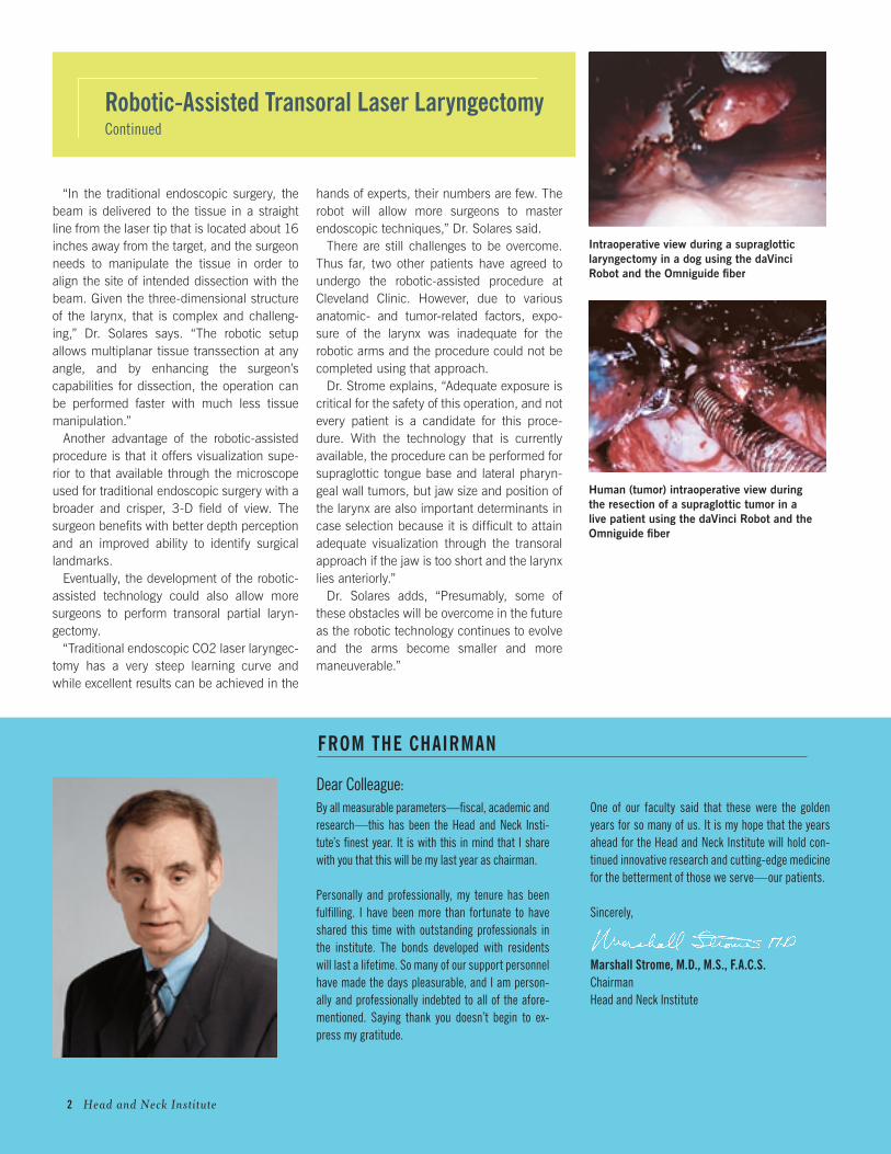
intraoperative view during a supraglottic laryngectomy in a dog using the daVinci Robot and the Omniguide fiber
Human (tumor) intraoperative view during the resection of a supraglottic tumor in a live patient using the daVinci Robot and the Omniguide fiber
In 1998, Cleveland Clinic made history when Marshall Strome, M.D., and a team of surgeons here performed the first total larynx transplant. The patient continues to do well with excellent voicing. However, despite the quality of life benefits provided by that surgery, the risks associated with immunosuppressive therapy needed to maintain graft survival have prevented the procedure from having more widespread use.
“Now eight years post transplantation with normal voicing, our human transplant patient attests to the viability of the procedure,” Dr. Strome says. “Needed are newer regimens that incorporate antiproliferative immunosuppressants to affect transplantation in the face of malignancy. That gap is closing.”
Dr. Strome and colleagues have been doing research aimed at identifying an immunosuppressive regimen that offers a more favorable risk:benefit profile and, in particular, one that would not potentiate tumor development.
“The need for long-term treatment with conventional immunosuppressive therapy remains a barrier to routine performance of laryngeal transplantation in the setting of malignancy as those regimens are associated with significant toxicity and a high risk of recurrent or second malignancy,” explains Samir S. Khariwala, M.D., who is on the research team.
Their research has focused on the novel agent everolimus (Certican, Novartis), an orally administered derivative of rapamycin. Everolimus is approved in Europe for use in cardiac and renal transplant recipients and is undergoing FDA approval regimens for the same indications. As potential immunosuppressive therapy to prevent graft rejection after laryngeal transplantation, everolimus offers both potent immunosuppressive and antitumor activities.
In an initial study performed in a rat model of laryngeal transplantation, Cleveland Clinic researchers demonstrated that a short-course intermittent regimen combining everolimus plus a T-cell depleting monoclonal antibody was effective in maintaining 100% graft survival after 6 months. The treatment protocol involved an initial 7-day course of everolimus 5 mg/kg/day, followed 90 days later by a second 5-day course of the same daily dose.
Research Findings with immunosuppressive/Antitumor Agent May open the Door to Routine Larynx Transplant Following Laryngectomy
Subsequently, a study was done to investigate the inhibitory effect of everolimus against SCC, and the results of that investigation were recently published [Khariwala SS, et al. The Laryngoscope, 2006;116:814-20].
“Available studies evaluating the antitumor activity of everolimus had demonstrated its efficacy in preventing the development of post-transplant lymphoproliferative disorders and inhibiting various other types of tumors in vivo and in vitro. However, its activity against SCC has not been evaluated previously,” Dr. Khariwala says.
Researchers studied 40 mice for growth of intradermal tumors after intradermal injection with a murine SCC line and evaluated a second group of 40 animals for the establishment of pulmonary metastases after intravenous inoculation with the same tumor cell line. The animals in each of those two groups were further subdivided into four subgroups to receive everolimus 1 mg/kg twice a day, everolimus 0.5 mg/kg twice a day, cyclosporine 7.5 mg/kg daily, or no treatment. Everolimus treatment was initiated on the day after tumor inoculation, administered by gavage, and continued for 17 days in the animals being studied for pulmonary metastases and 21 days in the intradermal group.
Compared with both no treatment and cyclosporine, both doses of everolimus significantly inhibited intradermal tumor growth and significantly decreased the number of pulmonary metastases that developed. The two doses of everolimus had comparable efficacy for limiting intradermal tumor growth while a dose-response effect was observed in the drug’s activity for inhibiting establishment of the pulmonary metastases.
“The finding that everolimus actually inhibited tumor development was thrilling because it suggests patients undergoing laryngectomy for SCC can receive a larynx transplant and everolimus immunosuppressive therapy without exposing themselves to increased risk for tumor recurrence,” Dr. Khariwala says.
FRoM The MeDiCAL eDiToRTom Abelson, M.D.
This issue of Otolaryngology Advances showcases the latest medical and technological advances from Cleveland Clinic’s Head and Neck Institute. In it you will find information on our landmark robotic-as-sisted transoral CO2 laser partial laryngectomy, performed by Marshall Strome, M.D. You also can read about the latest findings related to the immuno-suppressive agent everolimus, which has the poten-tial to improve laryngeal transplant outcomes in the future.
We provide findings from our recent studies on the dual laser/cryoablation procedure, while delving into our efforts to better diagnose and treat laryngopha-ryngeal reflux. Read about the Head and Neck Insti-tute’s recently established Hearing Implant Program, which is one of a very few centers offering auditory-verbal therapy for speech and language rehabilita-tion for pediatric implant patients. And review the Tinnitus Management Clinic’s new treatment option – neuronomics.
The Head and Neck Institute has had another note-worthy year, developing a new test for detecting su-perior canal dehiscence and expanding rhinologists’ use of CT-MR fusion software. We continue to use our ever-expanding patient population to expand our knowledge base and to share this knowledge with the worldwide otolaryngology community.
We hope you enjoy this edition of Otolaryngology Advances. We look forward to continuing our collab-oration with you in the future.
Head and Neck Institute �
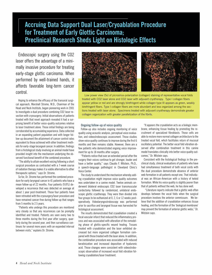
Low power view (5x) of picrosirius-polarization (collagen) staining of representative vocal folds treated with CO2 laser alone and CO2 laser with adjuvant cryotherapy. Type I collagen fibers appear yellow or red and are strongly birefringent while collagen type III appears as green, weakly birefringent fibers. Type I collagen fibers are more abundant and less organized among the sec-tions treated with laser alone. Specimens treated with adjuvant cryotherapy demonstrate greater collagen organization with greater parallelization of the fibrils.
� Head and Neck Institute
Endoscopic surgery using the CO2 laser offers the advantage of a mini-mally invasive procedure for treating early-stage glottic carcinoma. When performed by well-trained hands, it affords favorable long-term cancer control.
Hoping to enhance the efficacy of the transoral surgi-cal approach, Marshall Strome, M.D., Chairman of the Head and Neck Institute, began pioneering work in 2001 to investigate a dual procedure combining CO2 laser re-section with cryosurgery. Initial observations of patients treated with that novel approach revealed it had a sur-prising benefit of better voice-quality outcomes relative to laser treatment alone. Those initial findings are being corroborated by accumulating experience. Data collected in an expanding patient population and with longer fol-low-up document the attainment of cancer control rates equivalent to those achieved with other treatment meth-ods for early-stage laryngeal cancer. In addition, findings from a histological study involving an animal model have provided insight into the mechanism underlying the ob-served functional benefit of the combined procedure.
“The ability to attain excellent voicing following a short surgical procedure as contrasted with a 7-week course of radiation therapy makes it a viable alternative to other therapeutic options,” says Dr. Strome.
So far, Dr. Strome has performed the combined proce-dure for early laryngeal cancer in 41 patients who have a mean follow-up of 22 months. Four patients (9.8%) de-veloped a recurrence that was detected on average at about 1 year post-treatment. Those four patients have been retreated with the cryosurgery/laser procedure and have remained cancer-free during follow-up that ranges from 5 months to 2.5 years.
“Patients who undergo this procedure are monitored very closely so that any recurrences can be promptly identified and treated. Patients are seen every two to three months during the first year after surgery, quar-terly during the second year, and then surveillance con-tinues for several more years with an expanded interval between visits,” explains Dr. Strome.
Accruing Data Support Dual Laser/Cryoablation Procedure for Treatment of early Glottic Carcinoma; Preclinical Research Sheds Light on histologic effects
ongoing follow-up of voice qualityFollow-up also includes ongoing monitoring of voice
quality using acoustic analysis, perceptual voice evalua-tion, and videostroboscopic assessment. Those studies show voice quality continues to improve during the first 6 months and then remains stable. However, there are a few patients who demonstrated ongoing voice improve-ment for up to 18 months after surgery.
“Patients notice that over an extended period after the surgery their voices continue to get stronger, louder and have a better quality,” says Claudio F. Milstein, Ph.D., a speech-language pathologist in Cleveland Clinic’s Voice Center.
The study to understand the mechanism whereby add-ing cryoablation might improve voice quality outcomes was undertaken in a canine model. Twelve animals un-derwent bilateral endoscopic CO2 laser transmuscular cordectomy followed by randomized, unilateral endo-scopic glottic cryotherapy. They were then divided into four groups to be euthanized at 0, 2, 6 or 12 weeks post-operatively. Videolaryngostroboscopy was performed prior to sacrifice and laryngeal tissue was harvested for histological assessment.
The results demonstrated that cryoablation created a focal vascular infarct that reduced the inflammatory pro-cess and was associated with alteration of the remodel-ing phase of glottis-specific wound healing. Tissues treated with cryoablation and the laser exhibited de-creased but more organized collagen formation com-pared with those treated with the laser alone. In addition, the combination procedure was associated with reduced keratinization and increased deposition of hyaluronic acid. Those changes were consistent with videostrobo-scopic observation of increased vocal fold vibration fol-lowing cryoablation/laser treatment.
“It appears the cryoablation acts as a biologic mem-brane, enhancing tissue healing by promoting the re-cruitment of specialized fibroblasts. Those cells are able to restore more normal collagen architecture to the treated vocal fold, which facilitates return of mucosal oscillatory potential. The better vocal fold vibration ob-served after combination treatment in this canine model translates clinically into better voice quality out-comes,” Dr. Milstein says.
Consistent with the histological findings in the pre-clinical study, clinical evaluations of patients who have had simultaneous treatment of both vocal cords with the dual procedure demonstrate absence of anterior web formation in all patients except one. That individu-al was an African-American with a history of keloid formation. While his voice quality is slightly poorer than that of patients without the web, he has done well.
“Literature reports indicate that a glottic web often develops between the vocal folds when the surgical procedure involves the anterior commissure. We be-lieve that the addition of cryoablation enhances tissue healing, and the formation of the ‘biological membrane’ may prevent the formation of anterior glottic webs,” Dr. Milstein says.
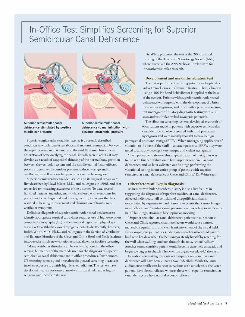
Superior semicircular canal dehiscence is a recently described condition in which there is an abnormal anatomic connection between the superior semicircular canal and the middle cranial fossa due to absorption of bone overlying the canal. Usually seen in adults, it may develop as a result of congenital thinning of the normal bony partition between the vestibular system and the middle cranial fossa. Affected patients present with sound- or pressure-induced vertigo and/or oscillopsia, as well as a low-frequency conductive hearing loss.
Superior semicircular canal dehiscence and its surgical repair were first described by Lloyd Minor, M.D., and colleagues in 1998, and that report led to increasing awareness of the disorder. To date, several hundred patients, including many who suffered with symptoms for years, have been diagnosed and undergone surgical repair that has resulted in hearing improvement and elimination of troublesome vestibular symptoms.
Definitive diagnosis of superior semicircular canal dehiscence to identify appropriate surgical candidates requires use of high-resolution computed tomography (CT) of the temporal region and physiologic testing with vestibular evoked myogenic potentials. Recently, however, Judith White, M.D., Ph.D., and colleagues in the Section of Vestibular and Balance Disorders of the Cleveland Clinic Head and Neck Institute introduced a simple new vibration test that allows for in-office screening.
“Many vestibular disorders can be easily diagnosed in the office setting, but neither of the methods used for the diagnosis of superior semicircular canal dehiscence are in-office procedures. Furthermore, CT scanning is not a good procedure for general screening because it involves exposure to a fairly high level of radiation. The test we have developed is easily performed, involves minimal risk, and is highly sensitive and specific,” she says.
Dr. White presented the test at the 2006 annual meeting of the American Neurotology Society (ANS) where it received the ANS Nicholas Torok Award for innovative vestibular research.
Development and use of the vibration testThe test is performed by fitting patients with optical or
video Frenzel lenses to eliminate fixation. Then, vibration using a 100 Hz hand-held vibrator is applied at the base of the occiput. Patients with superior semicircular canal dehiscence will respond with the development of a brisk torsional nystagumus, and those with a positive screening test undergo confirmatory diagnostic testing with a CT scan and vestibular evoked myogenic potentials.
The vibration screening test was developed as a result of observations made in patients with superior semicircular canal dehiscence who presented with mild positional nystagmus and were initially thought to have benign
paroxysmal positional vertigo (BPPV). When undergoing application of vibration to the base of the skull in an attempt to treat BPPV, they were noted to abruptly develop a very unique and violent nystagmus.
“Each patient who showed this atypical pattern of nystagmus was found with further evaluation to have superior semicircular canal dehiscence, and we later validated our findings performing the vibrational testing in our entire group of patients with superior semicircular canal dehiscence at Cleveland Clinic,” Dr. White says.
Other factors still key in diagnosisAs in most vestibular disorders, history is also a key feature in
suggesting the diagnosis of superior semicircular canal dehiscence. Affected individuals will complain of disequilibrium that is exacerbated by exposure to loud noises or to events that cause changes in middle ear and/or intracranial pressure, such as riding in an elevator in tall buildings, straining, hiccupping or sneezing.
“Superior semicircular canal dehiscence patients in our cohort at Cleveland Clinic reported that these factors would cause nausea, marked disequilibrium and even frank movement of the visual field. For example, one patient is a kindergarten teacher who would have to hold onto her desk when the bell rang or steady herself by reaching for the wall when walking students through the noisy school hallway. Another sound-sensitive patient would become extremely unsteady and begin to stagger in church whenever the organ was played,” she says.
In audiometry testing, patients with superior semicircular canal dehiscence will have bone curves above 0 decibels. While the same audiometric profile can be seen in patients with otosclerosis, the latter patients have absent reflexes, whereas those with superior semicircular canal dehiscence have normal acoustic reflexes.
Superior semicircular canal dehiscence stimulated by positive middle ear pressure
Superior semicircular canal dehiscence – canal inhibition with elevated intracranial pressure
Head and Neck Institute �
In-Office Test Simplifies Screening for Superior Semicircular Canal Dehiscence
LPR is a distinct form of reflux During the past 7 years, Douglas M. Hicks, Ph.D., and Claudio
F. Milstein, Ph.D., speech-language pathologists at the Voice Cen-ter of the Cleveland Clinic Head and Neck Institute, in collabora-tion with a group of colleagues from otolaryngology, gastroenterol-ogy and general surgery, have undertaken a series of research studies to gain a better understanding of the issues surrounding LPR in order to gain insight into its true nature, effective evaluation and optimal treatment. The findings of their studies underscore foremost that while LPR and the more widely recognized disorder gastroesophageal reflux (GER) are related by having gastric reflux as a common cause, LPR and GER are clinically distinct entities with respect to presentation, methods used for diagnosis and regi-mens for treatment.
“Educating patients, practitioners and the decision-makers for prescription medication insurance reimbursement about those dif-ferences is critical for improving the care of patients who suffer from LPR,” says Dr. Hicks.
Patients may have both GER and LPR or either of those entities alone. One reason why LPR may be overlooked in the differential diagnosis is failure to understand that it can develop in the ab-sence of GER.
“The delicate tissues in the larynx and throat are much more susceptible to irritation from the gastric juices and can be injured by even a small amount and very intermittent exposure that would not adversely affect the esophagus,” explains Dr. Milstein.
Querying patients about reflux in obtaining a medical history also can cause LPR to be ruled out prematurely because patients tend to associate that problem only with the classic symptoms of GER.
“A physician might first ask patients who present with head and neck symptoms if they smoke or drink, and then if they experience reflux. However, influenced by popular advertising campaigns for GER treatments, patients may respond in the negative because they associate the term reflux with symptoms such as gastric discomfort, heartburn, bloating and indigestion. This scenario illustrates the need to increase public awareness about LPR and to educate physicians on potential pitfalls in medical history taking for LPR evaluation,” says Dr. Hicks.
Researching methods of diagnosisWhen LPR is suspected, methods such as esophagogastroduo-
denoscopy, manometry, barium swallow and ambulatory pH monitoring, which are standard for diagnosing GER, offer poor efficacy for identifying LPR. Instead, laryngoscopic examination of the hypopharynx and larynx, which can be performed in office by a specially trained speech pathologist or otolaryngologist, represents the most sensitive and specific test for diagnosing LPR.
As part of their research, Dr. Hicks, Dr. Milstein and their colleagues have aimed to characterize specific findings that could improve the endoscopic diagnosis of LPR. Although no consensus on a subset of exam findings yet exists, promising results are emerging, says Dr. Hicks.
“One point is that findings of generalized erythema and edema, which have historically been used as the basis for diagnosing LPR, have poor specificity. In contrast, abnormalities of the vocal folds, arytenoid cartilage, and post-cricoid wall seem to have diagnostic value and are viewed as a core set of signs,” he notes.
Laryngopharyngeal reflux (LPR) is caused by exposure of the laryngeal and hypopharyngeal mucosa to noxious refluxed gastric juices. Also known as atypical reflux or silent reflux, LPR is manifested by a complex of head and neck symptoms, including hoarseness, coughing, throat clearing, sore throat and globus sensation that can have a significant detrimental impact on quality of life. Unfortunately, proper diagnosis and successful treat-ment is often impeded by lack of awareness and misconceptions about LPR among patients, physicians and prescription medication insurance providers.
� Head and Neck Institute
Researchers Take Aim On Improving Diagnosis And Treatment Of LPR
Researching treatment approachesOther studies have been designed to gain insight into optimal
treatment. Those investigations show that, in contrast to GER that is effectively treated with a proton pump inhibitor (PPI) administered once daily for several weeks, LPR requires more intensive treatment with twice daily PPI dosing continued for 3 to 4 months. Research also indicates that combination therapy with an H2-antagonist at night adds no benefit, and demonstrates that, as opposed to GER treatment where onset of improvement may be noticed within 1 to 2 weeks after starting a PPI, it can take 1 or 2 months before patients find their symptoms of LPR beginning to resolve. These differences are important to recog-nize so that patients are prescribed an adequate dose and duration of PPI therapy as well as thoroughly counseled about the timing to expected onset of improvement.
“Failure to understand the differences in PPI treatment between GER and LPR can lead to premature termination of potentially effective medication. We believe a 3- to 4-month treatment trial should be allowed before starting to consider other causes of their symptoms,” Dr. Hicks says.
Thorough patient counseling about the nature of LPR, the rationale for treatment with PPIs, and the importance of medication compliance is also critical for effective treatment.
“For example, patients who do not understand why they are being treated with a PPI may even fail to fill their prescription if they think their physician has not correctly diagnosed their condition,” notes Dr. Milstein.
“It is also important to emphasize the need to take the PPI twice daily without fail because any missed dose can allow exposure of the laryngeal and hypopharyngeal tissues to the irritating gastric juices that cause LPR, and may negate benefits achieved through weeks of medication compliance,” Dr. Hicks says.
After completing the initial course of twice-daily PPI treatment, an attempt should be made to reduce the dose and maintain the recovery with lifestyle and dietary modifications. That approach also contrasts with the management philosophy for GER held by some gastroenterologists who emphasize disease control strictly by medication.
“We implement aggressive intervention to allow tissue recovery, but once that outcome is achieved, we believe proper diet and lifestyle are important for maintaining tissue health. Once LPR has been effectively treated, the intensity of care may be tapered so that it might even involve judicious diet and lifestyle without any medication,” Dr. Hicks said.
Cleveland Clinic researchers also are making the prescription medication insurance industry a target of their educational efforts because authorization failure remains an obstacle to effective treatment.
“Allowance for PPI dispensing is based on dosing principles for GER treatment that are much different than those applying to LPR. The decision-makers in the insurance industry need to understand that information and change their policies about PPI coverage. Progress is being made in this realm,” Dr. Milstein says.
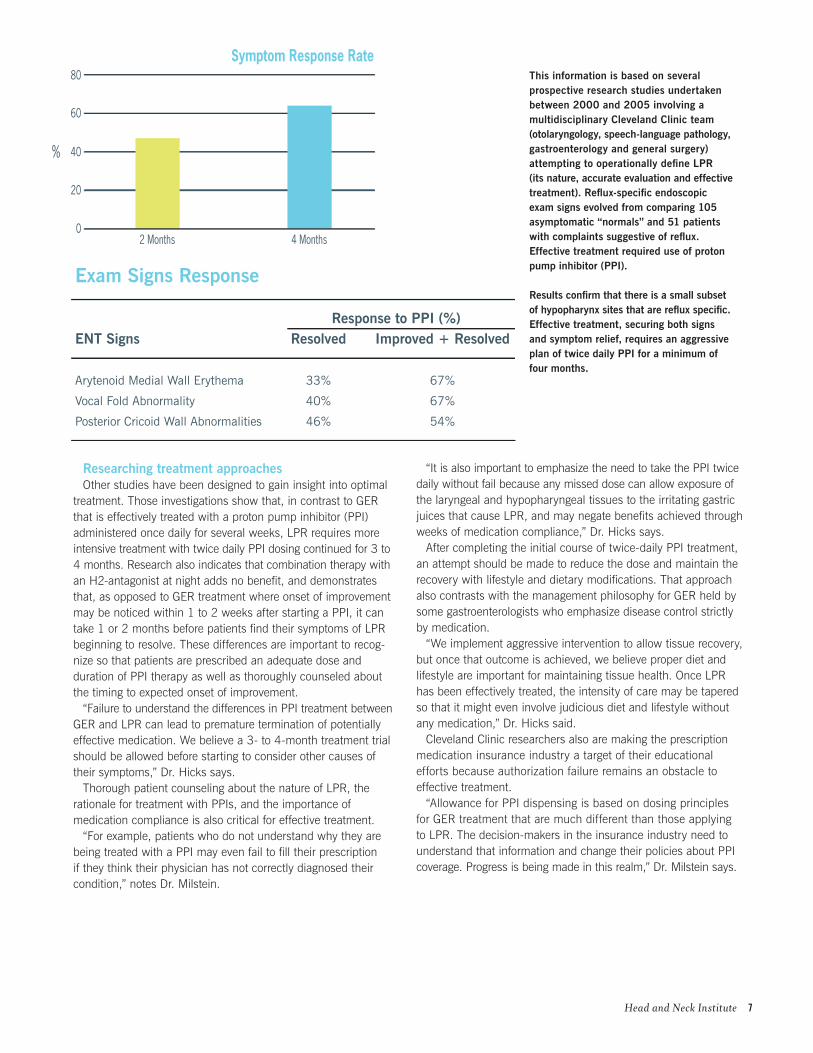
Exam Signs Response
Response to PPI (%)ENT Signs Resolved Improved + Resolved
Arytenoid Medial Wall Erythema 33% 67%
Vocal Fold Abnormality 40% 67%
Posterior Cricoid Wall Abnormalities 46% 54%
0
80
20
40
60
2 Months
Symptom Response Rate
4 Months
%
This information is based on several prospective research studies undertaken between 2000 and 2005 involving a multidisciplinary Cleveland Clinic team (otolaryngology, speech-language pathology, gastroenterology and general surgery) attempting to operationally define LPR (its nature, accurate evaluation and effective treatment). Reflux-specific endoscopic exam signs evolved from comparing 105 asymptomatic “normals” and 51 patients with complaints suggestive of reflux. Effective treatment required use of proton pump inhibitor (PPI).
Results confirm that there is a small subset of hypopharynx sites that are reflux specific. Effective treatment, securing both signs and symptom relief, requires an aggressive plan of twice daily PPI for a minimum of four months.
Head and Neck Institute �
In 1996, the International Society for Computer-Aided Surgery was established to explore the surgical applications of semi-conductor-based technologies. Over the past decade, the use of computer-based systems within otolaryngology has grown dramatically as their performance and use-fulness have increased. Since 2001, the Section of Nasal and Sinus Disorders has developed a comprehensive program that incorporates computer-based technologies in its endeavors for patient care, physician education and research.
Martin J. Citardi, M.D., F.A.C.S., Section Head of Nasal and Sinus Disorders, ex-plains, “Our program includes intraoperative surgical navigation as well as other efforts that seek to expand the usefulness and applications of this technology.” Pete S. Batra, M.D., Assistant Professor of Surgery, adds “This technology is part of what we do every day—in the office and in the op-erating room. It also has become a plat-form for research and education.”
Components of this program for computer-aided surgery include:
Physician education. “We have a huge library of archived patient and cadav-eric CT scan data which can be used for the education of our residents,” Dr. Batra says. At teaching conferences, cases from this database may be reviewed easily and quickly. Furthermore, image review at the workstation facilitates better understanding, because residents and fellows can “click and scroll” through the images, rather than statically studying a few select images.
Research. “Our research program in computer-aided surgery is unique, since it has both a dry lab for the study of the surgical navigation and a clinical component for the study of relevant surgical anatomy,” says Dr. Citardi. The section has presented data on registration protocols, surgical navigation’s critical initial step which correlates the preoperative imaging with intraoperative anatomy. Furthermore, the section has published computer-enabled reviews of CT anatomy of the frontal sinus and sphenoid sinus.
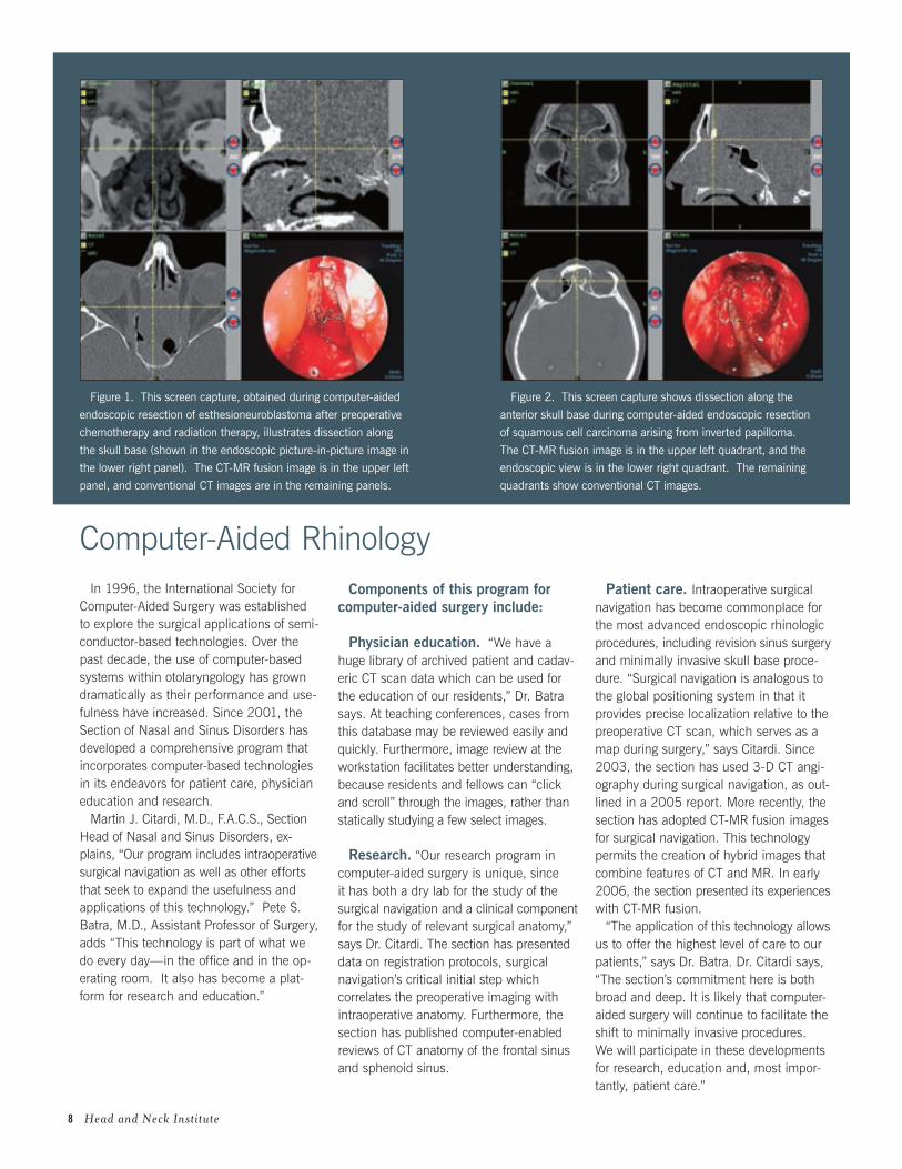
Patient care. Intraoperative surgical navigation has become commonplace for the most advanced endoscopic rhinologic procedures, including revision sinus surgery and minimally invasive skull base proce-dure. “Surgical navigation is analogous to the global positioning system in that it provides precise localization relative to the preoperative CT scan, which serves as a map during surgery,” says Citardi. Since 2003, the section has used 3-D CT angi-ography during surgical navigation, as out-lined in a 2005 report. More recently, the section has adopted CT-MR fusion images for surgical navigation. This technology permits the creation of hybrid images that combine features of CT and MR. In early 2006, the section presented its experiences with CT-MR fusion.
“The application of this technology allows us to offer the highest level of care to our patients,” says Dr. Batra. Dr. Citardi says, “The section’s commitment here is both broad and deep. It is likely that computer-aided surgery will continue to facilitate the shift to minimally invasive procedures. We will participate in these developments for research, education and, most impor-tantly, patient care.”
Computer-Aided Rhinology
Figure 2. This screen capture shows dissection along the
anterior skull base during computer-aided endoscopic resection
of squamous cell carcinoma arising from inverted papilloma.
The CT-MR fusion image is in the upper left quadrant, and the
endoscopic view is in the lower right quadrant. The remaining
quadrants show conventional CT images.
Figure 1. This screen capture, obtained during computer-aided
endoscopic resection of esthesioneuroblastoma after preoperative
chemotherapy and radiation therapy, illustrates dissection along
the skull base (shown in the endoscopic picture-in-picture image in
the lower right panel). The CT-MR fusion image is in the upper left
panel, and conventional CT images are in the remaining panels.
� Head and Neck Institute
Free-flap microsurgical techniques have revolutionized recon-structive surgery and enabled improved functional and cosmetic outcomes for patients requiring aggressive head and neck ablative procedures. While this type of surgery is no longer new and is being performed at institutions across the United States, recent comparative studies demonstrate that the outcomes achieved by facial aesthetics and reconstructive surgeons at the Cleveland Clinic Head and Neck Institute are excellent.
Analyses based on several surveys involving major academic institutions indicate that free-flap surgery adds an average of 4.15 hours to the time for conventional reconstructive surgery. At Cleveland Clinic, free-flap surgery adds an average of only 1.58 hours to the procedure, making it a viable option for a broad pop-ulation of patients.
This efficiency has been achieved without compromise in out-comes. There have been no flap failures among patients operated on at Cleveland Clinic since 2001 – well below the national aver-age of 6 percent failure rate.
Cutting-edge innovations demonstrated by novel method for TMJ reconstruction
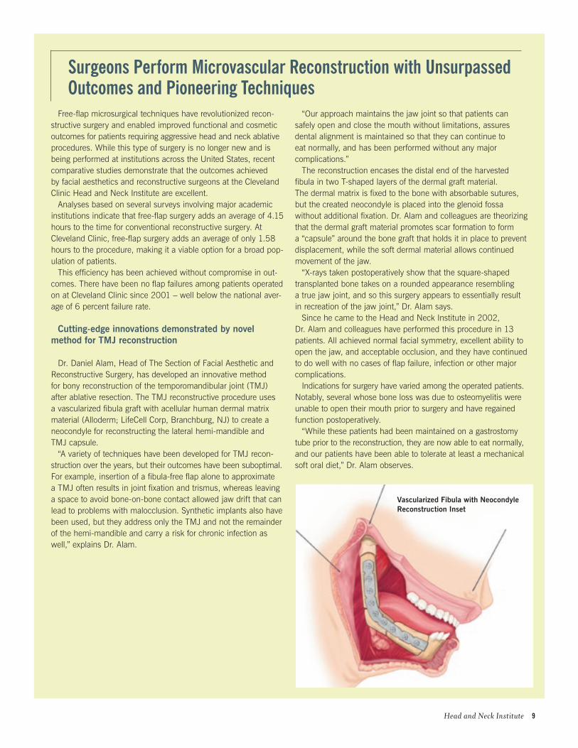
Dr. Daniel Alam, Head of The Section of Facial Aesthetic and Reconstructive Surgery, has developed an innovative method for bony reconstruction of the temporomandibular joint (TMJ) after ablative resection. The TMJ reconstructive procedure uses a vascularized fibula graft with acellular human dermal matrix material (Alloderm; LifeCell Corp, Branchburg, NJ) to create a neocondyle for reconstructing the lateral hemi-mandible and TMJ capsule.
“A variety of techniques have been developed for TMJ recon-struction over the years, but their outcomes have been suboptimal. For example, insertion of a fibula-free flap alone to approximate a TMJ often results in joint fixation and trismus, whereas leaving a space to avoid bone-on-bone contact allowed jaw drift that can lead to problems with malocclusion. Synthetic implants also have been used, but they address only the TMJ and not the remainder of the hemi-mandible and carry a risk for chronic infection as well,” explains Dr. Alam.
“Our approach maintains the jaw joint so that patients can safely open and close the mouth without limitations, assures dental alignment is maintained so that they can continue to eat normally, and has been performed without any major complications.”
The reconstruction encases the distal end of the harvested fibula in two T-shaped layers of the dermal graft material. The dermal matrix is fixed to the bone with absorbable sutures, but the created neocondyle is placed into the glenoid fossa without additional fixation. Dr. Alam and colleagues are theorizing that the dermal graft material promotes scar formation to form a “capsule” around the bone graft that holds it in place to prevent displacement, while the soft dermal material allows continued movement of the jaw.
“X-rays taken postoperatively show that the square-shaped transplanted bone takes on a rounded appearance resembling a true jaw joint, and so this surgery appears to essentially result in recreation of the jaw joint,” Dr. Alam says.
Since he came to the Head and Neck Institute in 2002, Dr. Alam and colleagues have performed this procedure in 13 patients. All achieved normal facial symmetry, excellent ability to open the jaw, and acceptable occlusion, and they have continued to do well with no cases of flap failure, infection or other major complications.
Indications for surgery have varied among the operated patients. Notably, several whose bone loss was due to osteomyelitis were unable to open their mouth prior to surgery and have regained function postoperatively.
“While these patients had been maintained on a gastrostomy tube prior to the reconstruction, they are now able to eat normally, and our patients have been able to tolerate at least a mechanical soft oral diet,” Dr. Alam observes.
Surgeons Perform Microvascular Reconstruction with Unsurpassed outcomes and Pioneering Techniques
Head and Neck Institute �
Vascularized Fibula with Neocondyle Reconstruction inset
Endoscopic transsphenoidal surgery has advanced over the past decade thanks to the collaborative efforts of pioneering otolaryngologists and neurosurgeons worldwide. The minimally invasive technique is now becoming standard for surgical resection in most patients with non-functioning pituitary tumors.
Cleveland Clinic otolaryngologist Robert Lorenz, M.D., and neurosurgeon Robert Weil, M.D., have performed endoscopic pituitary surgery since the beginning of 2005. Based on the favorable efficacy and safety results achieved and the benefits of the minimally invasive procedure relative to open surgery performed through a transnasal or sublabial approach, the endoscopic technique is now being used routinely in the management of patients with intrasellar pituitary tumors causing visual field deficits.
improved outcomesAt the Panamanian Society of Otolaryngology meeting
this year, Dr. Lorenz presented analyses of data collected prospectively from the first 50 consecutive cases of endoscopic pituitary surgery, and the outcomes were excellent, he said.
“Our review showed that the endoscopic approach is associated with a reduction in postoperative morbidity, and a much faster recovery period compared with the open microsurgical approach,” notes Dr. Lorenz.
A decrease in surgical time is accounted for by shortening of the ENT portion of the procedure. An average of only 45 minutes is now needed from the time Dr. Lorenz begins the surgery with introduction of the endoscope into the nostril until he reaches the dura and Dr. Weil performs the resection of the tumor. Total length of hospital stay has been reduced from an aver-age of 3 days with the open procedure to 2 days after endoscopic surgery.
“However, we have many patients who are dis-charged the next day,” Dr. Lorenz says.
Patient recovery better, fasterThere is no need for nasal packing after the surgery,
and because the endoscopic procedure is less trau-matic, patients experience less pain, minimal cosmetic changes and a faster overall recovery. Swelling in the nasal area is generally mild, and some patients are even pain-free and do not require any analgesic medication. Full recovery is usually achieved within one week compared with 3 to 4 weeks after the open procedure.
“In addition, the endoscopic surgery preserves the normal sphenoid sinus function since the reconstruc-tion tends to be confined within the sella turcica. And there are no problems with numbness to the incisor teeth with this technique since it does not disrupt the incisive canal,” Dr. Lorenz says.
He notes that while he has been concerned about postoperative epistaxis from the sphenopalatine artery, no such bleeding complications have been encountered so far, possibly due to close attention to specifically cauterizing the artery prior to the sphenoidotomy.
The experience of the Cleveland Clinic surgeons demonstrated that there is a learning curve for devel-oping expertise with the endoscopic approach, and modifications were introduced over time to improve outcomes. For example, in order to reduce formation of synechiae within the nose, splints are now placed in the nasal cavity postoperatively and removed when the patient returns at one week. A gentle nasal debride-ment is also performed in the office at the one-week post-op visit and repeated at four weeks after surgery.
“We have found that good nasal hygiene with saline douches is also a key to success with this surgery,” Dr. Lorenz says.
The endoscopic technique is used primarily when the indication for hypophysectomy is decompression of optic chiasm. It is not currently used in patients with Cushing’s syndrome where the goal of surgery is to achieve a cure by removing all of the secretory elements of the tumor. However, indications for endoscopic transsphenoidal surgery are expanding and the approach is being applied for the removal of intracranial tumors in the sella region.
Cleveland Clinic offers Fully endoscopic Procedure to Patients with Pituitary Tumors
10 Head and Neck Institute
Outcomes Data AvailableThe latest edition of outcomes data from the Cleveland Clinic Head and Neck Institute is available. Our outcomes booklet also offers summary reviews of medical and surgical trends and approaches. Charts, graphs and data illustrate the scope and volume of procedures performed in our institute each year. To view outcomes booklets for the Head and Neck Institute, as well as many other Cleveland Clinic medical and surgical disciplines, visit clevelandclinic.org/quality.
Tinnitus Management Clinic Offers Neuronomics as New Treatment Option
Audiologists Craig Newman, Ph.D., and Sharon Sandridge, Ph.D., are co-directors of the Tinnitus Management Clinic (TMC), which recently became one of just 5 centers in the country initially selected to offer the Neuronomics Tinnitus Treatment (NTT), an innovative form of acoustic therapy developed and introduced by a company in Australia.
Currently, the TMC is the leading center for enrollment of patients into the NTT program. Since September 2005, 12 patients have been enrolled into this therapy, with several completing the 6-month acoustic training program. All are benefiting as documented by reductions in tinnitus handicap and distress assessed using standardized questionnaires as well as with improvement in sound tolerance.
“Unfortunately, patients with tinnitus are often told they will have to live with their condition if it is not amenable to medical or surgical intervention. While that approach may be satisfactory for mild tinnitus, it often is not for new tinnitus. Fortunately there are a number of therapeutic options that can be offered to provide relief, and the NTT program seems to be a valuable new addition,” says Dr. Newman. “Not only are we one of the few centers in the country approved to provide this treatment, but our clinic takes a more comprehensive approach to intervention, coupling sound therapy with counseling to address the psychoemotional component of tinnitus in order to optimize the relief obtained.”
The NTT approachIn the NTT program, patients listen to a musical signal
delivered through high-fidelity earphones from a custom-programmed, lightweight processor. The acoustic signal is spectrally shaped based on the individual’s hearing and tinnitus profile and able to present a very high frequency signal up to 12.5 KHz. The processor is loaded with four tracks of the specially selected music, of which two are Baroque style and two are New Wave.
The NTT program consists of 3 phases, beginning with the pre-conditioning stage, which lasts 2 months and is designed to provide a high level of interaction between the acoustic signal and tinnitus perception. During this period, patients are instructed to listen to their treatment for at least 2 to 3 hours a day at times when their tinnitus is most disturbing. The music they hear contains an embedded shower sound that helps provide instant relief during the initial phase of treatment.
“Music therapy causes progressive relaxation and provides patients a way to escape from their tinnitus while the shower sound affords a masking effect,” Dr. Newman explains.
Active treatment stageThe next 4 months constitutes the active treatment stage during
which the acoustic signal is modified through omission of the embedded shower sound so as to deliver an intermittent or lower level of interaction with the tinnitus perception. Patients continue
to listen to the treatment for at least 2 to 3 hours a day during the times of the most distressing tinnitus, but may be able to reduce the daily treatment dose over time as their tinnitus awareness and disturbance improves. Further, patients begin to notice relief from their tinnitus during the times when they are not using the treatment.
“During this phase of NTT patients can still hear the tinnitus while listening to the treatment and, by doing so, are able to begin a habituation or desensitization process so that they are less aware and disturbed by it,” Dr. Newman explains.
Dr. Sandridge adds, “When patients are unable to hear their tinnitus during the pre-conditioning stage, they can de-stress and are better able to sleep. Therefore, when they enter the active treatment stage, they are better able to cope. Even though they can hear their tinnitus, because it is coupled with the pleasant music, the brain no longer fixates on the tinnitus nor identifies it as a threatening sound.”
Maintenance stageAfter 6 months, patients enter the maintenance stage in which
they may use the device on an as-needed basis if their tinnitus flares.Results of some preliminary studies suggest NTT may have
some advantages compared with other sound therapies in terms of the onset and magnitude of relief obtained, as well as patient satisfaction.
“Patients who have used this device usually achieve an acceptable level of relief after 6 months, whereas it can take 12 to 18 months to reach maximum benefit using other forms of sound therapy. In addition, other programs require listening for about 8 hours each day,” Dr. Newman says.
“Patients generally find the music of the NTT program much more pleasant than the white noise presented in other forms of sound therapy,” Dr. Sandridge notes.
Prior to enrollment in the TMC, all patients are seen by an otolaryngologist to rule out any tinnitus-causing condition that would require surgical or medical treatment, and they undergo a complete audiological evaluation. Their care in the TMC begins with a shared medical appointment that gathers approximately 8 patients together for group educational counseling. The topics discussed include causes of tinnitus, coping strategies and treatment options.
“We have found that information is power for patients with tinnitus and, for many individuals, the education and coping strategies they receive in the group session can be sufficient and obviate the need to return for additional treatment,” Dr. Newman says.
TMC patients who become candidates for sound therapy are presented with the range of therapeutic options and are given an opportunity to listen to the various forms of sound therapy to determine which might be most agreeable to them.
Head and Neck Institute 11
“Auditory-verbal therapy teaches spoken language development by tapping into listening and, arguably, is the most effective means for capitalizing on the potential of children with cochlear implants to acquire speech and language,” says Donald M. Goldberg, Ph.D.
Dr. Goldberg is co-director of the HIP, and holds certifications in audiology, speech-language pathology, and auditory-verbal therapy. He has been working as an auditory-verbal therapist for more than 25 years. Dr. Goldberg joined the Cleveland Clinic staff one year ago. He has been an interventionist for hundreds of children and has achieved results that are very gratifying for him as well as for the patients and their families.
“Even the most significantly hearing-impaired persons have some residual hearing that can be stimulated. In the past, that usable hearing was predominantly trained with hearing aids. However, the advent of cochlear implantation has truly brought sound to these individuals and made the sky the limit as far as their potential for spoken language development,” he says.
The vast majority of the patients Dr. Goldberg works with in his pediatric therapy program are infants through preschool age. Dr. Goldberg’s youngest therapy patient began when she was 3 months old. Children become surgically eligible for cochlear implantation once they reach their first birthday.
Coaching both children and parentsAuditory-verbal therapy involves working one-on-
one with the child and parent(s) because intensive efforts at home are critical to success. Dr. Goldberg says his role is as a coach who acts to counsel parents on how they can stimulate listening in their child and teach listening for comprehension and spoken language development.
“If I do my job right, I work myself out of it relatively quickly, and as the children are coming into this therapy at an increasingly younger age, the necessary duration is continually shortening,” Dr. Goldberg says.
The therapy follows normal child development sequences to teach auditory skills and promote speech production, language development, vocabulary, grammar and the pragmatics of language. The initial
focus is on developing basic listening. Early on, awareness of sound and learning to listen are taught using repeated pointing to the ear and use of the command “listen.” Responding enthusiastically to any vocalization effort by the child is another key principle of the therapy because it transfers the message that talking is very positive. “Learning to listen” toys are an integral component of the demonstration-based approach to early auditory-verbal rehabilitation. For example, saying “moo” for a cow and “choo-choo” for a toy train.
“Toys are important for stimulating interest in babies, and I’m proud to say I probably have some of the best toy collections in the world,” Dr. Goldberg says.
When cochlear implantation is performed for 1- to 2-year-olds, it is expected that the child can be discharged from auditory-verbal therapy within the first 3 or 4 years of life, and most children can be dismissed by the time they are ready to enter kindergarten.
“There are always variables in patient care at home, but most positive outcomes are certainly achievable when all the necessary elements are in place,” Dr. Goldberg says.
outcomes of this therapyPeer-reviewed studies show that with proper
teaching and good family support, graduates of auditory-verbal therapy blend successfully into mainstream society and become the “anonymous deaf.” However, in order to further document the value of this approach in children who have undergone cochlear implantation, Dr. Goldberg is currently seeking external funding to perform neuroradiographic imaging of such individuals. Such studies aim to explore the hypothesis that those children will have brain anatomy and physiology findings similar to those of matched controls born with normal hearing. A current cohort of five sets of twins, of which one is deaf and the other is not, is the intended target population for this initial research.
“We are hoping that the findings of this research will provide objective data to further validate the clinical efficacy of what we are doing,” Dr. Goldberg says.
“Even the most significantly hearing-impaired persons have some residual hearing that can be stimulated. The advent of cochlear implantation has truly brought sound to these individuals and made the sky the limit as far as their potential for spoken language development.”
1� Head and Neck Institute
Auditory-Verbal Therapy Key for opening Doorway to Maximum Spoken Language Potential in Pediatric Cochlear implant Patients
Cochlear implants have opened up a new and wonderful world of sound to very young children with congenital or acquired profound hearing impairment. Cleveland Clinic’s long-standing cochlear implant experience has evolved into the recently established Hearing Implant Program (HIP) at Hillcrest Hospital offering comprehensive pediatric services. The HIP is one of only a handful of centers in the United States providing implant patients with auditory-verbal therapy for speech and language rehabilitation in addition to traditional audiologic care.
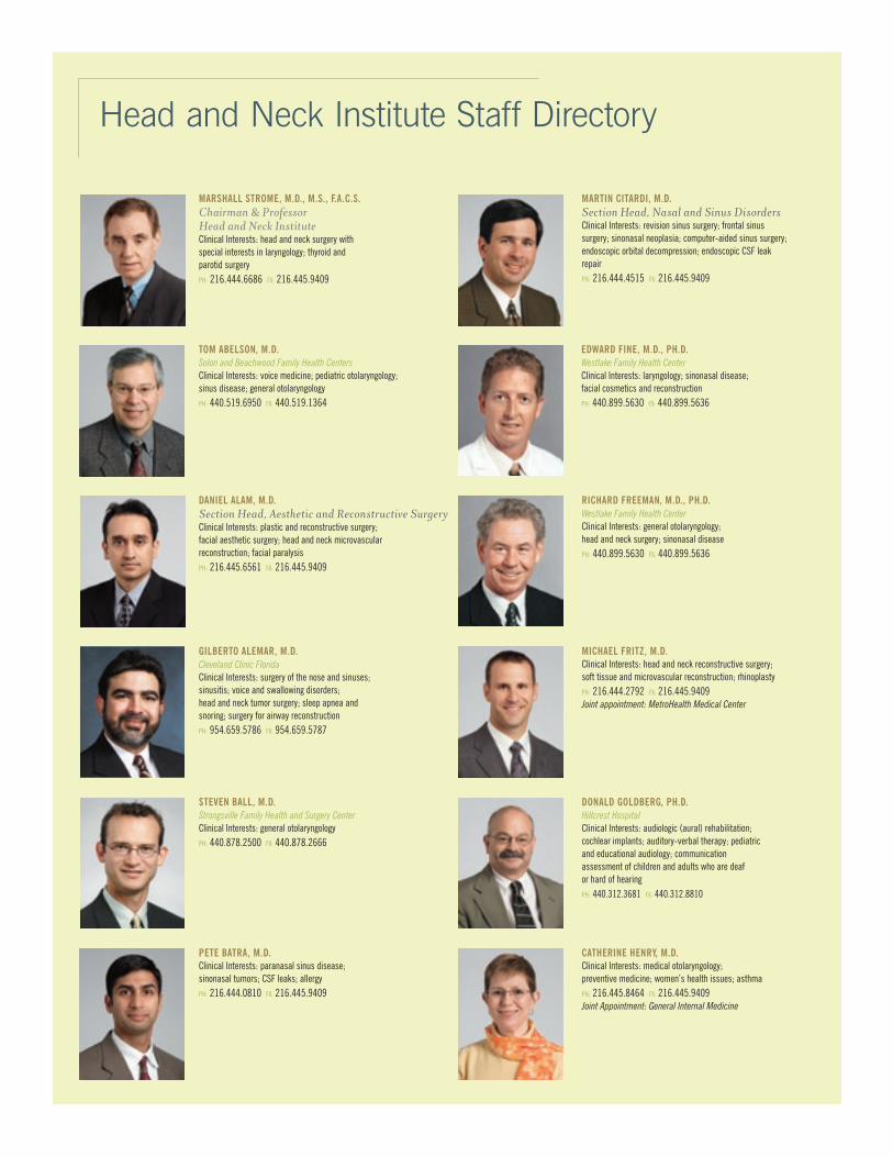
Head and Neck Institute Staff Directory
MARShALL STRoMe, M.D., M.S., F.A.C.S.Chairman & ProfessorHead and Neck InstituteClinical Interests: head and neck surgery with special interests in laryngology; thyroid and parotid surgeryPH: 216.444.6686 FX: 216.445.9409
ToM AbeLSon, M.D.Solon and Beachwood Family Health CentersClinical Interests: voice medicine; pediatric otolaryngology; sinus disease; general otolaryngologyPH: 440.519.6950 FX: 440.519.1364
eDwARD Fine, M.D., Ph.D.Westlake Family Health CenterClinical Interests: laryngology; sinonasal disease; facial cosmetics and reconstructionPH: 440.899.5630 FX: 440.899.5636
DAnieL ALAM, M.D.Section Head, Aesthetic and Reconstructive SurgeryClinical Interests: plastic and reconstructive surgery; facial aesthetic surgery; head and neck microvascular reconstruction; facial paralysisPH: 216.445.6561 FX: 216.445.9409
RiChARD FReeMAn, M.D., Ph.D.Westlake Family Health CenterClinical Interests: general otolaryngology; head and neck surgery; sinonasal diseasePH: 440.899.5630 FX: 440.899.5636
GiLbeRTo ALeMAR, M.D.Cleveland Clinic FloridaClinical Interests: surgery of the nose and sinuses; sinusitis; voice and swallowing disorders; head and neck tumor surgery; sleep apnea and snoring; surgery for airway reconstructionPH: 954.659.5786 FX: 954.659.5787
MiChAeL FRiTz, M.D.Clinical Interests: head and neck reconstructive surgery; soft tissue and microvascular reconstruction; rhinoplastyPH: 216.444.2792 FX: 216.445.9409Joint appointment: MetroHealth Medical Center
STeVen bALL, M.D.Strongsville Family Health and Surgery CenterClinical Interests: general otolaryngologyPH: 440.878.2500 FX: 440.878.2666
DonALD GoLDbeRG, Ph.D.Hillcrest HospitalClinical Interests: audiologic (aural) rehabilitation; cochlear implants; auditory-verbal therapy; pediatric and educational audiology; communicationassessment of children and adults who are deaf or hard of hearingPH: 440.312.3681 FX: 440.312.8810
PeTe bATRA, M.D.Clinical Interests: paranasal sinus disease; sinonasal tumors; CSF leaks; allergyPH: 216.444.0810 FX: 216.445.9409
MARTin CiTARDi, M.D.Section Head, Nasal and Sinus DisordersClinical Interests: revision sinus surgery; frontal sinus surgery; sinonasal neoplasia; computer-aided sinus surgery; endoscopic orbital decompression; endoscopic CSF leak repairPH: 216.444.4515 FX: 216.445.9409
CATheRine henRy, M.D.Clinical Interests: medical otolaryngology; preventive medicine; women’s health issues; asthmaPH: 216.445.8464 FX: 216.445.9409Joint Appointment: General Internal Medicine
SUyU ShU, Ph.D.Research Interests: cellular immunology; cancer immunotherapy; molecular biologyPH: 216.445.3800 FX: 216.445.3805Joint Appointment: Director, Center for Surgery Research
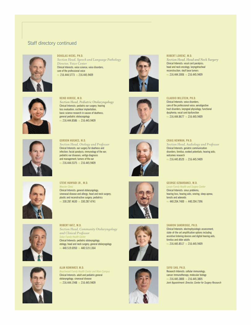
DoUGLAS hiCKS, Ph.D.Section Head, Speech and Language PathologyDirector, Voice CenterClinical Interests: voice science; voice disorders; care of the professional voicePH: 216.444.5773 FX: 216.445.9409
RobeRT LoRenz, M.D.Section Head, Head and Neck SurgeryClinical Interests: vocal cord paralysis; head and neck oncology; laryngotracheal reconstruction; skull base tumorsPH: 216.444.3006 FX: 216.445.9409
KeiKo hiRoSe, M.D. Section Head, Pediatric OtolaryngologyClinical Interests: pediatric ear surgery; hearing loss evaluation; cochlear implantation; basic science research in causes of deafness; general pediatric otolaryngologyPH: 216.444.8586 FX: 216.445.9409
CLAUDio MiLSTein, Ph.D.Clinical Interests: voice disorders; care of the professional voice; aerodigestive tract disorders; laryngeal physiology; functional dysphonia; vocal cord dysfunctionPH: 216.444.8677 FX: 216.445.9409
GoRDon hUGheS, M.D.Section Head, Otology and ProfessorClinical Interests: ear surgery for deafness and infection; facial paralysis; immunology of the ear; pediatric ear diseases; vertigo diagnosis and management; tumors of the earPH: 216.444.5375 FX: 216.445.9409
CRAiG newMAn, Ph.D.Section Head, Audiology and ProfessorClinical Interests: geriatric communication disorders; tinnitus; evoked potentials; hearing aids; outcomes researchPH: 216.445.8520 FX: 216.445.9409
STeVe hUnyADi JR., M.D.Wooster ClinicClinical Interests: general otolaryngology; sinonasal disease and allergy; head and neck surgery; plastic and reconstructive surgery; pediatricsPH: 330.287.4630 FX: 330.287.4741
GeoRGe ozbARDAKCi, M.D.Lorain Family Health and Surgery CenterClinical Interests: sinus problems; hearing loss; hearing aids; snoring; sleep apnea; tonsils and adenoidsPH: 440.204.7400 FX: 440.204.7396
RobeRT KATz, M.D.Section Head, Community Otolaryngologyand Clinical ProfessorSolon Family Health CenterClinical Interests: pediatric otolaryngology; otology; head and neck surgery; general otolaryngologyPH: 440.519.6950 FX: 440.519.1364
ALAn KoMinKSy, M.D.Beachwood Family Health Center and Main CampusClinical Interests: adult and pediatric general otolaryngology; sinonasal diseasePH: 216.444.1948 FX: 216.445.9409
ShARon SAnDRiDGe, Ph.D.Clinical Interests: electrophysiologic assessment; state-of-the-art amplification options including assistive listening devices and digital hearing aids; tinnitus and older adultsPH: 216.445.8517 FX: 216.445.9409
Staff directory continued
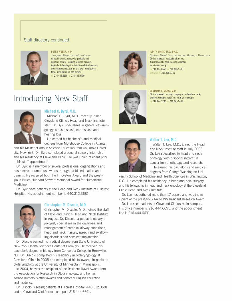
Introducing New Staff Michael C. byrd, M.D.
Michael C. Byrd, M.D., recently joined Cleveland Clinic’s Head and Neck Institute staff. Dr. Byrd specializes in general otolaryn-gology, sinus disease, ear disease and hearing loss.
He earned his bachelor’s and medical degrees from Morehouse College in Atlanta,
and his Master of Arts in Science Education from Columbia Univer-sity, New York. Dr. Byrd completed a general surgery internship and his residency at Cleveland Clinic. He was Chief Resident prior to his staff appointment.
Dr. Byrd is a member of several professional organizations and has received numerous awards throughout his education and training. He received both the Innovators Award and the presti-gious Bruce Hubbard Stewart Memorial Award for Humanistic Medicine.
Dr. Byrd sees patients at the Head and Neck Institute at Hillcrest Hospital. His appointment number is 440.312.3681.
Christopher M. Discolo, M.D. Christopher M. Discolo, M.D., joined the staff of Cleveland Clinic’s Head and Neck Institute in August. Dr. Discolo, a pediatric otolaryn-gologist, specializes in the diagnosis and management of complex airway conditions, head and neck masses, speech and swallow-ing disorders and cochlear implantation.
Dr. Discolo earned his medical degree from State University of New York Health Sciences Center at Brooklyn. He received his bachelor’s degree in biology from Concordia College in Bronxville, N.Y. Dr. Discolo completed his residency in otolaryngology at Cleveland Clinic in 2005 and completed his fellowship in pediatric otolaryngology at the University of Minnesota in Minneapolis.
In 2004, he was the recipient of the Resident Travel Award from the Association for Research in Otolaryngology, and he has earned numerous other awards and honors during his education and residency.
Dr. Discolo is seeing patients at Hillcrest Hospital, 440.312.3681, and at Cleveland Clinic’s main campus, 216.444.6691.
walter T. Lee, M.D.Walter T. Lee, M.D., joined the Head
and Neck Institute staff in July 2006. Dr. Lee specializes in head and neck oncology with a special interest in cancer immunotherapy and research.
He earned his bachelor’s and medical degrees from George Washington Uni-
versity School of Medicine and Health Sciences in Washington, D.C. He completed his residency in head and neck surgery and his fellowship in head and neck oncology at the Cleveland Clinic Head and Neck Institute.
Dr. Lee has authored more than 17 papers and was the re-cipient of the prestigious AAO-HNS Resident Research Award.
Dr. Lee sees patients at Cleveland Clinic’s main campus. His office number is 216.444.6695, and the appointment line is 216.444.6691.
JUDiTh whiTe, M.D., Ph.D.Section Head, Vestibular and Balance DisordersClinical Interests: vestibular disorders; dizziness and balance; hearing problems; ear disease; vertigo PH: 216.444.8552 FX: 216.445.9409BEACHWOOD PH: 216.839.3740
PeTeR webeR, M.D.Program Director and ProfessorClinical Interests: surgery for pediatric and adult ear disease including cochlear implants; implantable hearing aids; infectious cholesteatomas; acoustic neuromas; ear tumors; skull bone lesions; facial nerve disorders and vertigoPH: 216.444.6696 FX: 216.445.9409
benJAMin G. wooD, M.D.Clinical Interests: oncologic surgery of the head and neck; skull base surgery; nasal/paranasal sinus surgeryPH: 216.444.5700 FX: 216.445.9409
Staff directory continued
Otolaryngology Advances offers information from Cleveland Clinic otolaryngologists, speech pathologists and audiolo-gists about state-of-the-art medical, surgical and rehabili-tative techniques.
PLeASe DiReCT CoRReSPonDenCe To:
Head and Neck Institute / A71The Cleveland Clinic Foundation9500 Euclid AvenueCleveland, OH 44195
Tom Abelson, M.D.Medical Editor
Dino RhodesMarketing Manager
Christine HarrellManaging Editor
Irwin KriegerArt Director
Established in 1921, The Cleveland Clinic Foundation provides state-of-the-art care in a multispecialty academic medical center that integrates clinical and hospital care with research and education in a private, not-for-profit group practice. Otolaryngology and Communicative Disorders services are offered at the main campus as well as at Cleveland Clinic family health centers throughout Greater Cleveland.
Otolaryngology Advances is written for physicians and should be relied upon for medical education purposes only. It does not provide a complete overview of the topics covered, and should not replace the independent judgment of a physician about the appropriateness or risks of a procedure for a given patient.
© The Cleveland Clinic Foundation 9/2006
The Cleveland Clinic Foundation Head and Neck Institute / A719500 Euclid AvenueCleveland, OH 44195
Visit Us Online To learn more about Cleveland Clinic’s Head and Neck Institute, including information about our research and educational programs, visit our Web site: clevelandclinic.org/headandneck.
Online Access to Your Patient’s Treatment ProgressWhether you are referring from near or far, our new e-Cleveland Clinic service, Dr.Connect, can streamline communication from Cleveland Clinic physicians to your office. This new online tool offers you secure access to your patient’s treatment progress at Cleveland Clinic. With one-click convenience, you can track your patient’s care using the secure Dr.Connect Web site. To establish a Dr.Connect account, visit eclevelandclinic.org or e-mail [email protected].
Cleveland Clinic excels in Latest U.S.news Rankings head and neck institute Ranked Among Top 10 in the nation
Cleveland Clinic is ranked among the top three hospitals in the nation in the 2006 U.S.News & World Report’s physician poll. The magazine’s “America’s Best Hospitals” survey ranks our otolaryngology program among the top 10 in the nation. Cleveland Clinic is named among the nation’s best in all 16 specialties surveyed, and better than any other Ohio hospital in 14 of them. For more details, visit clevelandclinic.org.
Otolaryngology AdvancesF a l l 2 0 0 6