Implantation Serine Proteinases heterodimerize and are critical in hatching and implantation
11
BioMed Central Page 1 of 11 (page number not for citation purposes) BMC Developmental Biology Open Access Research article Implantation Serine Proteinases heterodimerize and are critical in hatching and implantation Navneet Sharma, Shiying Liu, Lin Tang, Jackie Irwin, Guoliang Meng and Derrick E Rancourt* Address: Department of Biochemistry & Molecular Biology, Faculty of Medicine, University of Calgary, Heritage Medical Research Building, 3330 Hospital Dr. NW, Calgary AB, T2N 4N1, Canada Email: Navneet Sharma - [email protected]; Shiying Liu - [email protected]; Lin Tang - [email protected]; Jackie Irwin - [email protected]; Guoliang Meng - [email protected]; Derrick E Rancourt* - [email protected] * Corresponding author Abstract Background: We have recently reported the expression of murine Implantation Serine Proteinase genes in pre-implantation embryos (ISP1) and uterus (ISP1 and ISP2). These proteinases belong to the S1 proteinase family and are similar to mast cell tryptases, which function as multimers. Results: Here, we report the purification and initial characterization of ISP1 and 2 with respect to their physico-chemical properties and physiological function. In addition to being co-expressed in uterus, we show that ISP1 and ISP2 are also co-expressed in the pre-implantation embryo. Together, they form a heterodimer with an approximate molecular weight of 63 kD. This complex is the active form of the enzyme, which we have further characterized as being trypsin-like, based on substrate and inhibitor specificities. In addition to having a role in embryo hatching and outgrowth, we demonstrate that ISP enzyme is localized to the site of embryo invasion during implantation and that its activity is important for successful implantation in vivo. Conclusion: On the basis of similarities in structural, chemical, and functional properties, we suggest that this ISP enzyme complex represents the classical hatching enzyme, strypsin. Our results demonstrate a critical role for ISP in embryo hatching and implantation. Background Embryo implantation is a necessary stage in fetal develop- ment: in order to gain access to nutrients and gas exchange, the embryo attaches to the uterine epithelium and invades into the endometrium. It is a multi-step proc- ess that consists of: the hatching of the blastocyst from the zona pellucida, adhesion of the blastocyst to the uterine epithelium, stromal invasion and reorganization. This process is orchestrated through the coordinated, recipro- cal interaction between the embryo and uterus and is mediated via a variety of molecules including steroid hor- mones, cytokines, adhesion molecules, proteinases and their inhibitors [1-4]. Proteinases of different classes have been hypothesized to give the blastocyst its invasive character and/or participate in the remodelling of the uterine stroma. Alfonso et al [5] have reported that cysteine proteinases play a critical role Published: 11 December 2006 BMC Developmental Biology 2006, 6:61 doi:10.1186/1471-213X-6-61 Received: 07 July 2006 Accepted: 11 December 2006 This article is available from: http://www.biomedcentral.com/1471-213X/6/61 © 2006 Sharma et al; licensee BioMed Central Ltd. This is an Open Access article distributed under the terms of the Creative Commons Attribution License (http://creativecommons.org/licenses/by/2.0 ), which permits unrestricted use, distribution, and reproduction in any medium, provided the original work is properly cited.
-
Upload
navneet-sharma -
Category
Documents
-
view
213 -
download
0
Transcript of Implantation Serine Proteinases heterodimerize and are critical in hatching and implantation

BioMed CentralBMC Developmental Biology
ss
Open AcceResearch articleImplantation Serine Proteinases heterodimerize and are critical in hatching and implantationNavneet Sharma, Shiying Liu, Lin Tang, Jackie Irwin, Guoliang Meng and Derrick E Rancourt*Address: Department of Biochemistry & Molecular Biology, Faculty of Medicine, University of Calgary, Heritage Medical Research Building, 3330 Hospital Dr. NW, Calgary AB, T2N 4N1, Canada
Email: Navneet Sharma - [email protected]; Shiying Liu - [email protected]; Lin Tang - [email protected]; Jackie Irwin - [email protected]; Guoliang Meng - [email protected]; Derrick E Rancourt* - [email protected]
* Corresponding author
AbstractBackground: We have recently reported the expression of murine Implantation SerineProteinase genes in pre-implantation embryos (ISP1) and uterus (ISP1 and ISP2). These proteinasesbelong to the S1 proteinase family and are similar to mast cell tryptases, which function asmultimers.
Results: Here, we report the purification and initial characterization of ISP1 and 2 with respect totheir physico-chemical properties and physiological function. In addition to being co-expressed inuterus, we show that ISP1 and ISP2 are also co-expressed in the pre-implantation embryo.Together, they form a heterodimer with an approximate molecular weight of 63 kD. This complexis the active form of the enzyme, which we have further characterized as being trypsin-like, basedon substrate and inhibitor specificities. In addition to having a role in embryo hatching andoutgrowth, we demonstrate that ISP enzyme is localized to the site of embryo invasion duringimplantation and that its activity is important for successful implantation in vivo.
Conclusion: On the basis of similarities in structural, chemical, and functional properties, wesuggest that this ISP enzyme complex represents the classical hatching enzyme, strypsin. Ourresults demonstrate a critical role for ISP in embryo hatching and implantation.
BackgroundEmbryo implantation is a necessary stage in fetal develop-ment: in order to gain access to nutrients and gasexchange, the embryo attaches to the uterine epitheliumand invades into the endometrium. It is a multi-step proc-ess that consists of: the hatching of the blastocyst from thezona pellucida, adhesion of the blastocyst to the uterineepithelium, stromal invasion and reorganization. Thisprocess is orchestrated through the coordinated, recipro-
cal interaction between the embryo and uterus and ismediated via a variety of molecules including steroid hor-mones, cytokines, adhesion molecules, proteinases andtheir inhibitors [1-4].
Proteinases of different classes have been hypothesized togive the blastocyst its invasive character and/or participatein the remodelling of the uterine stroma. Alfonso et al [5]have reported that cysteine proteinases play a critical role
Published: 11 December 2006
BMC Developmental Biology 2006, 6:61 doi:10.1186/1471-213X-6-61
Received: 07 July 2006Accepted: 11 December 2006
This article is available from: http://www.biomedcentral.com/1471-213X/6/61
© 2006 Sharma et al; licensee BioMed Central Ltd. This is an Open Access article distributed under the terms of the Creative Commons Attribution License (http://creativecommons.org/licenses/by/2.0), which permits unrestricted use, distribution, and reproduction in any medium, provided the original work is properly cited.
Page 1 of 11(page number not for citation purposes)

BMC Developmental Biology 2006, 6:61 http://www.biomedcentral.com/1471-213X/6/61
in implantation, and there have been several reportsimplicating matrix metalloproteinases (MMPs) in thisprocess [6-8]. Different serine proteinases are also knownto be expressed in a finely regulated pattern duringimplantation, including urokinase-type plasminogen acti-vator (uPA) and proprotein convertase SPC5/6 [9,10].However, the targeted disruption of several murine protei-nase genes, presumed essential for implantation, has indi-cated that many are dispensable, suggesting that otherdistinct proteinases may be involved [1,11].
In order to discover additional serine proteinases withpotential involvement in implantation, we identified twonovel implantation serine proteinase genes (ISP1 and 2).These were found to be co-expressed in mouse uterineendometrium throughout the peri-implantation periodand tandemly organized within a bed of tryptase genes onmouse chromosome 17A3.3 [12]. ISP1 gene expressionwas first detected in pre-implantation embryos [13]. Anti-sense disruption of ISP1 gene expression preventedembryo hatching and outgrowth in vitro [13]. Both ISP1and 2 gene expression was also detected in the uterineendometrial gland, under the positive influence of proges-terone [14,15]. Using immunoblotting, both ISP proteinswere detected in the uterine fluid on day 4 of pregnancy,just prior to the commencement of implantation [16].This appearance of protein in the glands and uterine fluidseems to be negatively regulated by estrogen, such thatboth ISP proteins appear in the uterine fluid shortly afterthe estrogen spike synchronizes uterine-embryo receptiv-ity and the commencement of implantation [16]. Interest-ingly, antibodies directed against ISP2 protein haverecently been found to abrogate implantation, suggestingan important role for the ISPs in implantation [17].
Mast cell tryptases are known to exist in multimeric form[18]. Since the ISPs are co-expressed in endometrialglands, we previously hypothesized that they exist as het-ero-tetramers, a theory that was supported by proteinmodelling studies [15]. In this study, we have purified aheterodimeric 63 kD ISP enzyme complex from day fourpregnant mouse uterus, which is comprised of ISP1 (30kD) and ISP2 (33 kD) monomers. The same enzyme com-plex was detected in uterine fluid and pre-implantationembryos. Enzyme kinetic studies have demonstrated theaffiliation of ISP enzyme complex with S1 proteinases,having trypsin-like substrate specificity. Immunohisto-chemistry suggests the ISP enzyme complex localizes tothe site of embryo invasion during implantation. Gabex-ate mesylate, a potent tryptase inhibitor, was found toinhibit ISP activity, and arrest hatching and outgrowth ofembryos in vitro, and implantation in utero. These resultsdemonstrate that ISP enzyme complex plays a critical rolein initiating murine implantation.
ResultsCharacterization and Purification of the ISP1-ISP2 enzyme complexWe have characterized the expression of the ISPs in uter-ine tissue, uterine fluid and blastocysts. Uterine tissuehomogenate and intra-uterine fluid from CD1 mice at theperi-implantation period were probed for the presence ofISPs using mAbs (Fig. 1A and 1B). Under denaturing con-ditions, monomers of both ISP1 (30 kD) and ISP2 (33kD) proteins were found in highly enriched uterine tissuehomogenates and uterine fluid. In addition to the mono-mers, an upper band (~63 kD), presumably a complex ofISP1 and ISP2 was also obtained when either anti-ISP1 oranti-ISP2 mAb were used to probe (Fig. 1). This 63 kDband exhibits proteolytic activity in gel when N-BenzoylArginine p-Nitroanilide (BAPNA) is used as a substrate(Fig. 1C). The other two bands obtained (Fig. 1) wereidentified as IgG light (27 kD) and heavy (54 kD) chainsby mass spectrometry analysis. The existence of IgGs inthe uterine tissue homogenates and uterine fluid can beexplained by the vascular nature of pre-implantationuteri, such that blood serum products are found in highabundance. A similar enzyme complex was also detectedin mouse blastocysts in western blots using denaturingconditions (Fig. 2A and 2B). As we had previously notdetected ISP2 expression in the embryo, we used RT-PCRto confirm that ISP2 was indeed expressed in the preim-plantation embryo (Fig. 2C). Following upon our previ-ous observation of ISP1 and ISP2 co-expression in uterineglands [15] and now embryos (this study), as well as thishigher molecular weight complex in the gels, we used co-immunopreciptation to investigate the possibility that ISPmonomers formed hetero-dimeric complexes. Co-IPswere performed by cross-linking anti-ISP mAbs to acti-vated agarose beads followed by affinity purification ofthe respective proteins from the uterine tissue homoge-nates. The bound antibodies were able to precipitate theircognate protein targets (Fig. 3C), as well as their corre-sponding opposite ISP (Fig. 3D). These results lend cre-dence to the idea that ISP1 and ISP2 hetero-dimerize toform a higher molecular weight enzyme.
The native ISP enzyme complex was purified from mouseuterine tissue homogenates (uteri collected at 4.5 dpc –6.5 dpc stage of pregnancy) by ammonium sulphate pre-cipitation of homogenized tissue, followed by multi-stepcolumn chromatography. Several different methods ofseparation were tested to serve as the first chromatographystep. These included an inhibitor affinity method usingBenzamidine-sepharose (Fig. 3A), ion exchange chroma-tography (DEAE sepharose and CM-sepharose), andhydrophobic interaction chromatography. DEAE sepha-rose gave the best results in terms of yield and was thenfollowed by a two-step gel filtration using Superdex-200and Superdex-75 (Fig. 3B). The purity of the enzyme com-
Page 2 of 11(page number not for citation purposes)

BMC Developmental Biology 2006, 6:61 http://www.biomedcentral.com/1471-213X/6/61
plex was confirmed by native gel electrophoresis afterpurification (Additional file 1. Fig. 1a). The purified frac-tion is enzymatically active and exhibits enzyme activityin gel. The native size of the complex was found to beapproximately 60 kD when analysed by gel filtrationusing molecular weight standards (Additional file 1. Fig.1b). Additionally, the identity of the subunits was con-firmed by mass spectrometry analysis.
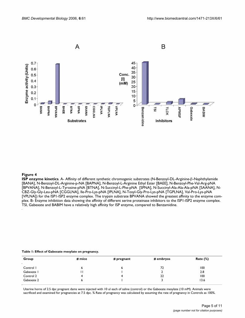
Enzyme KineticsTo determine the substrate specificity of the ISP complexand to classify it into a subclass of the S1 family of serineproteinases, enzyme assays were carried out using differ-ent p-nitroanilide linked peptide (1–3 mer) substrates.These included chymotrypsin-like (Tyr/Phe at cleavage
(P1) site), trypsin-like (Arg/Lys at P1 position), elastase-like (Ala at P1 position), and subtilisin-like (Leu at P1position) substrates. The results obtained are shown infigure 4A. In order to compare affinity of specific sub-strates and derive Michaelis Menton constant (Km) val-ues, the kinetic curves were plotted (substrate conc. [S] vs.rate of reaction [v]). A trypsin-specific substrate, N-ben-zoyl Phe-Val-Arg p-nitroanilide, showed the best affinitytowards the ISP enzyme complex.
Many general serine proteinase inhibitors were tested inorder to identify suitable ISP inhibitors when BAPNA wasused as a substrate. These included Benzamidine hydro-chloride, 4-(Amidino-Phenyl)-Methane-Sulfonyl Fluo-ride (APMSF), L-1 Chloro-3- [4-tosyl-amido]-7-amino-2-heptanone. HCl (TLCK), Trypsin Soybean Inhibitor (TSI),Bis (5-amidino-2-benzimadozolyl) methane (BABIM),Gabexate mesylate, Nafamostat, and the kunitz family ser-
Characterization of blastocyst ISP expressionFigure 2Characterization of blastocyst ISP expression. A and B- Western blot analysis showing the expression of the ISP1- ISP2 enzyme complex from 3.5 dpc blastocysts when probed with anti-ISP1 (A) and anti-ISP2 (B) mAbs. C- RT-PCR from pre-implantation embryos. Lanes 1–4 and 6–10 represent samples amplified using ISP1 and ISP2 primers, respectively: lanes 4 and 6: non-pregnant uterus cDNA (negative control); lanes 3 and 7: 2.5 dpc embryo cDNA; lanes 2 and 8: 3.5 dpc embryo cDNA; lanes 1 and 9 represent 4.5 dpc embryo cDNA. Lane 5 is the 1 kb plus DNA ladder.
Characterization of uterine ISP expressionFigure 1Characterization of uterine ISP expression. A- West-ern blot results showing the expression of the ISP1-ISP2 complex from mice uterine homogenates (lane 1) and intra-uterine fluid (lane 2) collected at the peri-implantation period (4.5 dpc – 5.5 dpc) probed with anti-ISP1 monoclonal anti-body, B- Western blot results showing the expression of the ISP1-ISP2 complex from mice uterine homogenates (lane 1) and intra-uterine fluid (lane 2) collected at the peri-implanta-tion period (4.5 d pc – 6.5 d pc) probed with anti-ISP2 mono-clonal antibody, C- A zymogram showing in-gel enzyme activity of ISP1-ISP2 enzyme complex, carried out by using 5 mM BAPNA as substrate (lane 1- uterine tissue homogenate, lane 2- uterine fluid).
Page 3 of 11(page number not for citation purposes)

BMC Developmental Biology 2006, 6:61 http://www.biomedcentral.com/1471-213X/6/61
pins Uterine Plasmin-Trypsin Inhibitor (UPTI) and Secre-tary Leukocyte proteinase inhibitor (SLPI).
Only inhibitors specific for trypsin-like serine proteinaseswere able to inhibit the ISPs (Fig. 4B and Additional file 2.Table 1). IC50 (concentration of an inhibitor capable ofinhibiting the enzyme activity by 50%) values wereobtained by plotting inhibition data, which were extrapo-lated into Cheng and Prusoff's [19] equation to derive thedissociation constant (Ki).
Hatching and Implantation studiesPreviously we have determined that both embryo hatch-ing and outgrowth in vitro were inhibited by antisense oli-gonucleotides, which abrogated ISP1 gene expression[13]. In further immunohistochemistry experiments, we
have determined that both ISP1 and ISP2 localize to theembryo-uterine boundary during implantation (Fig. 5A, Band 5C).
In order to define a physiological role for the ISPs, theeffect of inhibiting ISP activity upon embryo hatching,outgrowth (Fig. 6 and 7) and implantation (Table 1) wasinvestigated. Blastocysts were first cultured in the presenceor absence of anti-ISP mAbs or selected inhibitors. Differ-ent concentrations of anti-ISP antibodies and inhibitors(Benzamidine, Gabexate and BABIM) were tried in orderto eliminate their toxic concentrations from influencingresults (Additional file 2. Tables 2 and 3). Figure 7A dem-onstrates the effect of two different concentrations ofGabexate on the hatching of blastocysts in comparison tocontrols. Here, an incremental decrease in the rate of
Purification of the uterine ISP enzyme complexFigure 3Purification of the uterine ISP enzyme complex. A- FPLC chromatogram displaying the affinity purification of ISP1-ISP2 complex from mouse uter-ine homogenates using a Benzamidine Sepharose column. The inset shows the Western Blot results of this partially purified fraction (lane 1: ISP1-ISP2 complex probed with anti-ISP1 antibody, lane 2: ISP1-ISP2 complex probed with anti-ISP2 antibody). B- FPLC chromatogram displaying the separation of ISP1-ISP2 complex using a Superdex™-200 column. The inset shows the Western blot results of the fraction purified from mouse uterine homogenates by ion exchange chromatography (DEAE-Sepharose) followed by gel filtration using Superdex™-200 and Superdex™-75 columns (lane 1: ISP1-ISP2 complex probed with anti-ISP1 antibody, lane 2: ISP1-ISP2 complex probed with anti-ISP2 antibody). C- Affinity purification of ISPs by immunoprecipitation (1- eluted ISP1 fraction pulled down by anti-ISP1 antibody covalently bound to acrylamide activated agarose beads in a chromatography column, 2 – eluted ISP2 fraction pulled down by anti-ISP2 antibody covalently bound to acrylamide activated agarose beads in a chromatography column). D- Co-immunopre-cipitation of ISPs (1- Fraction from section C1 probed with anti-ISP2 antibody, 2- Fraction from section C2 probed with anti-ISP1 antibody.
Page 4 of 11(page number not for citation purposes)
BMC Developmental Biology 2006, 6:61 http://www.biomedcentral.com/1471-213X/6/61
Page 5 of 11(page number not for citation purposes)
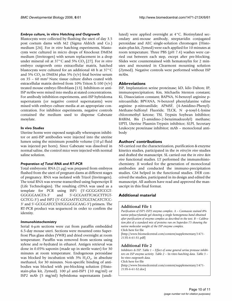
Table 1: Effect of Gabexate mesylate on pregnancy.
Group # mice # pregnant # embryos Rate (%)
Control 1 6 6 72 100Gabexate 1 11 1 2 2.8Control 2 4 4 22 100Gabexate 2 6 1 3 13.6
Uterine horns of 2.5 dpc pregnant dams were injected with 10 ul each of saline (control) or the Gabexate mesylate (10 mM). Animals were sacrificed and examined for pregnancies at 7.5 dpc. % Rate of pregnancy was calculated by assuming the rate of pregnancy in Controls as 100%.
ISP enzyme kineticsFigure 4ISP enzyme kinetics. A- Affinity of different synthetic chromogenic substrates (N-Benzoyl-DL-Arginine-β-Naphthylamide [BANA], N-Benzoyl-DL-Arginine-p-NA [BAPNA], N-Benzoyl-L-Arginine Ethyl Ester [BAEE], N-Benzoyl-Phe-Val-Arg-pNA [BPVANA], N-Benzoyl-L-Tyrosine-pNA [BTNA], N-Succinyl-L-Phe-pNA [SPNA], N-Succinyl-Ala-Ala-Ala-pNA [SAANA], N-CBZ-Gly-Gly-Leu-pNA [CGGLNA], Ile-Pro-Lys-pNA [IPLNA], N-Tosyl-Gly-Pro-Lys-pNA [TGPLNA], Val-Pro-Lys-pNA [VPLNA]) for the ISP1-ISP2 enzyme complex. The trypsin substrate BPVANA showed the greatest affinity to the enzyme com-plex. B- Enzyme inhibition data showing the affinity of different serine proteinase inhibitors to the ISP1-ISP2 enzyme complex. TSI, Gabexate and BABIM have a relatively high affinity for ISP enzyme, compared to Benzamidine.

BMC Developmental Biology 2006, 6:61 http://www.biomedcentral.com/1471-213X/6/61
hatching is noticeable with increasing Gabexate concen-tration. Although kinetic studies showed that BABIM wasa more effective inhibitor (Ki = 0.204 mM) than Gabexate(Ki = 0.49 mM), limited quantities of this inhibitor pre-vented a significant in vitro hatching/outgrowth analysis.Similarly, the effect of varying concentrations of anti-ISP1and anti-ISP2 mAbs was studied both individually andtogether (Fig. 7C, E and 7G). The results obtained clearlyshow that anti-ISP mAbs can efficiently arrest blastocysthatching compared to controls (Fig. 6A and 6B).
Implantation was simulated in vitro by growth of thehatched blastocysts in culture plates coated with the extra-cellular matrix derived from mouse embryonic fibrob-lasts, which can be invaded by outgrowing embryos (Fig.6C and 6D). Different concentrations of anti-ISP antibod-ies and Gabexate were tried as shown in figures 7B, D, Fand 7H. The effect of Gabexate on blastocyst outgrowth isalso incremental with increasing concentration of Gabex-ate (Fig. 7B). Individually, anti-ISP1 and anti-ISP2 mAbsdid not seem to have a significant effect on embryo out-growth (Fig. 7D and 7F). However, when administeredtogether, anti-ISP antibodies led to an arrest/delay inembryo outgrowth (Fig. 7H). Gabexate also had a signifi-cant effect on embryo implantation in vivo. When admin-istered topically to the uterus by injecting into the lumenat 2.5 dpc, the rate of pregnancy drops to very low in com-parison to controls (Table 1). A similar result was not
observed for individual or combined mAbs, possiblyowing to the abundance of ISP enzyme that occurs withinthe uterine fluid.
Discussion and ConclusionWe have previously demonstrated that the ISPs are tan-demly linked genes, which are co-regulated in the uterineendometrial gland during the peri-implantation period[12,15]. ISP gene expression leads to the secretion of bothISP1 and ISP2 into uterine fluid at the time of hatchingand initiation of implantation [16]. In this report, weshow that the ISPs are also co-expressed in pre-implanta-tion embryos, consistent with the idea that they may alsobe co-regulated in the pre-implantation embryo. Theseobservations are consistent with our observation that ISP1and ISP2 form a hetero-dimeric complex. Upon purifica-tion of ISP enzyme complex from pregnant uterus andconfirmation by immunoblotting and mass spectrometry,we determined its molecular weight to be ~63 kD, withthe ISP1 and ISP2 monomers being ~30 and 33 kD,respectively. These molecular weights are consistent withour previous study, where we characterized the appear-ance of ISP monomers in uterine fluid during the peri-implantation period [16]. However, our results contradicta previous report of ISP2 as having a molecular weight of48 kD [17].
Inhibition of embryo hatching and outgrowthFigure 6Inhibition of embryo hatching and outgrowth. A, C- Control mAb-treated blastocysts showing the normal hatch-ing and outgrowth, respectively. B, D- ISP1/2 mAB-treated blastocysts showing arrested hatching and outgrowth respec-tively.
ISP protein localization at the site of implantationFigure 5ISP protein localization at the site of implantation. A, B and C- Immunohistochemistry analysis showing ISP1 and ISP2 protein at the 4.5 dpc implantation site. A- Section stained with secondary antibody only; B- Section stained with anti-ISP1 mAb and secondary antibody; C- Section stained with anti-ISP2 mAb and secondary antibody.
Page 6 of 11(page number not for citation purposes)

BMC Developmental Biology 2006, 6:61 http://www.biomedcentral.com/1471-213X/6/61
Page 7 of 11(page number not for citation purposes)
Effect of inhibitors and antibodies on hatching and invasionFigure 7Effect of inhibitors and antibodies on hatching and invasion. In vitro hatching and outgrowth of blastocysts when grown in culture medium supplemented with different concentrations of Gabexate mesylate, anti-ISP1/-ISP2 mAbs (blue: embryos per set; purple: hatched after 24 h/outgrown after 48 h; white: hatched after 48 h/outgrown after 72 h). A – Effect of Gabexate on hatching (P value – 0.03). B – Effect of Gabexate on outgrowth (P value – 0.006). C – Effect of anti-ISP1 mAb on hatching (P value – 0.03). D – Effect of anti-ISP1 mAb on outgrowth (P value – not significant). E – Effect of anti ISP2 mAb on hatching of blastocysts (P value at 24 hrs. – 0.02; at 48 hrs. – not significant). F – Effect of anti-ISP2 mAb on the outgrowth of hatched blas-tocysts (P value – not significant). G – Effect of anti-ISP1/ISP2 mAb on the hatching of blastocysts (P value – 0.003). H – Effect of anti-ISP1/ISP2 mAb on the outgrowth of hatched blastocysts (P value – 0.006).

BMC Developmental Biology 2006, 6:61 http://www.biomedcentral.com/1471-213X/6/61
We have suggested that the ISPs are members of the mastcell tryptase family [13,14] and have demonstrated thatthe ISP genes reside in a tryptase cluster on mouse chro-mosome 17A3.3 [12]. In this study we have used syntheticsubstrates to classify ISP enzyme complex based on itsenzymatic activity. Consistent with our previous sugges-tions, we have sub-classified ISP enzyme complex as beingtrypsin-like since; it does not show any reactivity with syn-thetic serine proteinase substrates specific for chymot-rypsin, subtilisin or elastase. Hence, ISP enzyme can onlycleave peptides that contain arginine or lysine at the P1position, with arginine being the preferred choice.Amongst the substrates tested, BPVANA has the highestaffinity towards the enzyme (Km = 0.06 mM) demonstrat-ing preference of a tripeptide having valine at P2 positionand phenylalanine at P3 position over the other peptidesubstrates. Currently, work is in progress to further definethe substrate preferences of ISP using phage display pep-tide mimetics.
The ISP genes are most closely related to their neighboursMMCP6 and 7, which have previously been shown to existas homo-tetrameric enzymes [18]. Based on previous insilico molecular modelling, we have postulated that ISP1and ISP2 exist as hetero-tetramers [15]. Following on ourfinding that the ISPs do not seem to tetramerize in vitro,we have examined the properties of tryptases, which pro-mote tetramer formation. Although the ISPs possess thehydrophobic domain containing eight Trp residuesimportant for tetramer formation [20], they lack two ofthe three surface-exposed His residues that are necessaryfor MMCP6 tetramerization [21]. Interestingly, in thisregard, at least one other tryptase in this cluster, tryptase γ,has been speculated to form only dimers [22]. Althoughwe demonstrate at this time that ISP1 and ISP2 form het-erodimers, we do not know if this is exclusive, or whetherthe ISPs can also homodimerize. Our observation thatISP1 and ISP2 mAbs can block embryo hatching and/oroutgrowth, only when applied in combination, suggeststhe possibility that homo-dimers not only exist, but canalso function in embryo hatching and invasion.
Previously, we have reported that the antisense abroga-tion of ISP1 expression in blastocysts resulted in the inhi-bition of hatching from the zona pellucida and in vitrooutgrowth into extracellular matrix [14]. We have sug-gested that embryonic ISP enzyme represents the murinehatching enzyme strypsin, which is secreted from blasto-cysts and predicted to be ~74 kD based on autoradiogra-phy [24]. In this study, several general trypsin inhibitorsincluding Benzamidine, APMSF, TLCK, Gabexatemesylate, BABIM and TSI showed an inhibitory influenceon ISP activity. Amongst them TSI exhibited the highestaffinity for the ISP enzyme complex (Ki = 2.18 µM). Ourresults agree with the previous characterization of stypsin,
which demonstrated that trypsin inhibitors had a signifi-cant inhibitory effect on blastocyst hatching. Like ISP, TSIwas the strongest inhibitor of strypsin activity. Likestrypsin, ISP demonstrates the highest affinity towardsinhibitors of 'trypsin like' activity; and doesn't catalyse thecleavage of chymotrypsin or elastase substrates.
In this study, we have confirmed a role for ISP in embryohatching and outgrowth. When exposed to gabexatemesylate, a synthetic inhibitor of tryptases [25] and ISP(this study), both in vitro embryo hatching and outgrowthwere almost completely inhibited. Similarly, administra-tion of anti-ISP1 and anti-ISP2 monoclonal antibodiesalso led to a delay in hatching and outgrowth in vitro.Together these results suggest that ISP plays a vital roleduring embryo implantation, likely at the level of zona pel-lucida lysis and the initiation of invasion into uterineendometrium.
Interestingly, immunohistochemistry of implantingembryos shows that ISP enzyme complex surrounds theinvading embryo, highlighting their importance inimplantation. At this time, it is not clear whether this peri-implantation ISP staining is strictly from the embryo orwhether ISP complex that resides in uterine fluid is alsorecruited to sites of embryo invasion. However, inhibitionof ISP activity in vivo via the topical application of gabex-ate had a significant negative effect on implantation.Although we cannot rule out the involvement of otherproteinases in the process of hatching and implantation atthis time, ISP appears to be an essential link in the cascadeof events. Presently, we are disrupting the ISP genes togenetically confirm their role in hatching and implanta-tion. We are also characterizing the substrates and proteintargets of ISP in order to better understand the process ofimplantation. This could in turn lead to the design ofmore specific synthetic inhibitors of ISP and the develop-ment of new and more potent contraceptives.
MethodsSubstrates and Inhibitorsp-Nitroanilide substrates, Benzamidine Hydrochloride,TLCK (N -p-tosyl-L-lysine chloromethyl ketone), APMSF(4-Amidino-Phenyl)-Methane-Sulfonyl Fluoride),Trypsin Soybean Inhibitor (TSI) and Gabexate mesylatewere obtained from Sigma Aldrich and Co. Dr. George H.Caughey, University of California, San Francisco kindlysupplied BABIM.
Kunitz type UPTI like recombinant inhibitors were kindlyprovided by Dr. Jonathan Green (University of Missouri).
Handling of miceCD1 mice were obtained at the age of 6–8 weeks fromCharles River Canada (St. Constant, Quebec) and main-
Page 8 of 11(page number not for citation purposes)

BMC Developmental Biology 2006, 6:61 http://www.biomedcentral.com/1471-213X/6/61
tained in a standard laboratory animal facility with con-trolled temperature (20°C), humidity and light cycle(lights on between 0700 and 1900 hr). The maintenanceand treatment of animals was in full compliance with theStandard laboratory animal care protocols approved bythe University of Calgary Animal Care Committee. Toobtain natural pregnancies, female mice were paired withadult males and checked daily for the presence of vaginalcopulatory plugs, as an indication of mating. The day ofplug detection is designated as day 0.5 post coetum. Forcollection of uteri and embryos, pregnant dams were sac-rificed on a specific day by cervical dislocation.
Preparations of Monoclonal AntibodiesAnti-mouse ISP monoclonal antibodies (mAbs) were pre-pared by Immuno-Precise Antibodies Ltd., Victoria, Can-ada. Antibodies were raised against unique fragments of(His)6 tagged ISP1 and ISP2 expressed in E. coli and puri-fied using NiNTA chromatographic columns, as outlinedpreviously [16]. The effective protein concentration ofmonoclonal antibodies used in subsequent experimentswas 10 mg/ml (anti-ISP1) and 5 mg/ml (anti-ISP2).
Purification of ISP1-ISP2 complexUteri were placed in 25 mM Tris.Cl buffer (pH-8.0) andhomogenized using a mechanical homogenizer (Kine-matica). The homogenate was centrifuged at 10000 rpm(Beckman JA17 rotor) for 30 min. The supernatantobtained was subjected to fractionation by varying con-centrations of ammonium sulphate. The precipitated frac-tions were re-suspended and subjected to columnchromatography using the Duo Flow FPLC system (Bio-Rad). The purification involved sequential ion exchange(DEAE Sepharose), desalting (Sephadex G-25) and gel fil-tration (Superdex-200 and Superdex-75) chromatogra-phy. Affinity chromatography was also carried out usingHiTrap™ Benzamidine column (M/S Amersham).
ImmunoprecipitationInstead of traditional immunoprecipitation (IP) by usingProtein A/Protein G beads, antibody affinity columnswere made with the Amino Link Plus kit from Pierce. ISP1and ISP2 monoclonal antibodies were covalently boundto activated agarose beads by Schiff base chemistry andthe beads were packed into columns. These were thenused to pull the ISPs from uterine tissue homogenates,which were then eluted using low pH conditions (25 mMTris.Cl pH 3.0). The various fractions obtained duringpurification were analysed for enzyme activity as per theenzyme assay protocol described below. Traditionalimmunoprecipitation was also performed using Protein Gbeads.
ImmunoblottingDuring protein purification, fractions were analysed byPAGE (10%, 12% and 4% – 16% gradient gels) and elec-trophoresed under denaturing or non-denaturing condi-tions. Equal amounts of protein were loaded ontodifferent lanes in the same gel. The proteins were thentransferred to nitrocellulose membrane and probed withanti-ISP monoclonal antibodies. A horseradish peroxi-dase (HRP)-labelled anti-mouse IgG antibody (Amer-sham) was used at a 1:10,000 dilution for detection onBioMax film™ (Kodak).
ZymographyIn-gel enzyme assays were run electrophoretically asdescribed in the above section. Non-reduced protein sam-ples were loaded onto a 10% SDS polyacrylamide gel.Electrophoresis was performed at 100 V for 1 hr. Subse-quently, the gel was washed twice with 4% Triton- X-100made up in distilled water for 30 min. The gel was thenrinsed thoroughly with distilled water and incubated in25 mM Tris.Cl (pH 8.0) buffer having 5 mM N-BenzoylArginine p-Nitroanilide (BAPNA) and 5 mM EDTA, over-night at 37°C. The gel was again rinsed thoroughly withdistilled water on the following day in order to removeany precipitation and imaged in the visible range of wave-length to detect any bands on an otherwise transparentgel.
Enzyme Assay and KineticsChromogenic assays were carried out using the p-Nitroanilide conjugated peptidic substrates. The reactionconditions used were: 4 mM BAPNA (substrate), in 25mM Tris.Cl (pH 8.0) and 10 mM EDTA reaction buffer, ina total reaction volume of 0.5 mL. The reactions were car-ried out at room temperature in a glass cuvette (1 cm pathlength), and scanned at wavelength maxima of 405 nmusing a spectrophotometer. 1 Unit of enzyme is defined asenzyme activity sufficient for the breakdown of 1 µM ofsubstrate/ml/min. The affinity of ISP complex with vari-ous substrates was characterized by measuring enzymeactivity at differing substrate concentrations and plottingenzyme activity [V] vs. substrate concentration [S]. TheKm was calculated using the Michaelis Menton equation(Km = [S] at 1/2 Vmax).
The inhibition of proteolytic activity of ISP enzyme com-plex exhibited by known serine proteinase inhibitors wasdetermined by determining IC50 (inhibitor concentrationcapable of 50% inhibition of enzyme activity). IC50 valueswere derived by plotting enzyme activity (V) vs. inhibitorconcentration [I]. Dissociation constants (Ki) values fordifferent inhibitors were determined by using the Chengand Prusoff [19] equation (Ki = IC50/1 + [S]/Km).
Page 9 of 11(page number not for citation purposes)
BMC Developmental Biology 2006, 6:61 http://www.biomedcentral.com/1471-213X/6/61
Embryo culture, in vitro Hatching and OutgrowthBlastocysts were collected by flushing the uteri of day 3.5post coetum dams with M2 (Sigma Aldrich and Co.)medium [26]. For in vitro hatching experiments, blasto-cysts were cultured in micro drops of Knockout DMEMmedium (Invitrogen) with serum replacement in a dropunder mineral oil at 37°C and 5% CO2 [27]. For in vitroembryo outgrowth onto extracellular matrix, hatchedblastocysts were cultured for an additional 48 h at 37°Cand 5% CO2 in DMEM plus 5% (v/v) fetal bovine serumon 35 – 60 mm2 Nunc tissue culture dishes coated withextracellular matrix derived from 10% Triton X-100 (v/v)treated mouse embryo fibroblasts [13]. Inhibitors or anti-ISP mAbs were mixed into media at stated concentrations.For antibody inhibition experiments, anti-ISP hybridomasupernatants (or negative control supernatants) weremixed with embryo culture media at an appropriate con-centration. For inhibitor experiments, negative controlscontained the medium used to dispense Gabexatemesylate.
In vivo StudiesUterine horns were exposed surgically whereupon inhibi-tor or anti-ISP antibodies were injected into the uterinelumen using the minimum possible volume (10 µl fluidwas injected per horn). Since Gabexate was dissolved innormal saline, the control mice were injected with normalsaline solution.
Preparation of Total RNA and RT-PCRTotal embryonic RNA (1 µg) was prepared from embryosflushed from the uteri of pregnant dams at different stagesof pregnancy. RNA was isolated with Trizol (Invitrogen).The total RNA was reverse-transcribed using Superscript II(Life Technologies). The resulting cDNA was used as atemplate for PCR using ISP1 (5'-GCGGATCCGT-GGGGGAAGTA-3' and 5'-GCGAATTCAGCTTTGT-GCTCG-3') and ISP2 (5'-GCGAATTCGTGGTACATCTCC-3' and 5'-GCGGATCCTATGGGGGCAAG-3') primers. TheRT-PCR product was sequenced in order to confirm theidentity.
ImmunohistochemistrySerial 6-µm sections were cut from paraffin embedded6.5-day mouse uteri. Sections were mounted onto Super-frost Plus glass slides (VWR) and dried overnight at roomtemperature. Paraffin was removed from sections usingxylene and re-hydrated in ethanol. Antigen retrieval wasdone in 0.05% saponin (made up in sterile water) for 30minutes at room temperature. Endogenous peroxidasewas blocked by incubation with 3% H2O2, in absolutemethanol, for 30 minutes. Non-specific binding of anti-bodies was blocked with pre-blocking solution (Histo-stain-plus kit, Zymed). 100 µl anti-ISP1 (10 mg/ml) orISP2 mAb (5 mg/ml) hybridoma supernatants (undi-
luted) were applied overnight at 4°C. Biotinylated sec-ondary anti-mouse antibody, streptavidin conjugatedperoxidase and AEC single-solution chromogen (Histo-stain-plus kit, Zymed) were each applied for 10 minutes atroom temperature. Three PBS (pH 7.4) washes were car-ried out between each step, except after pre-blocking.Slides were counterstained with hematoxylin for 2 min-utes and mounted in Clearmont mounting solution(Zymed). Negative controls were performed without ISPmAbs.
AbbreviationsISP, Implantation serine proteinase; kD, kilo Dalton; IP,immunoprecipitation; Km, Michaelis Menton constant;Ki, Dissociation constant; BAPNA, N-benzoyl arginine p-nitroanilide; BPVANA, N-benzoyl phenylalanine valinearginine p-nitroanilide; APMSF, (4-Amidino-Phenyl)-Methane-Sulfonyl Fluoride; TLCK, N -p-tosyl-L-lysinechloromethyl ketone; TSI, Trypsin Soybean Inhibitor;BABIM, Bis (5-amidino-2-benzimadozolyl) methane;UPTI, Uterine Plasmin-Trypsin inhibitor; SLPI, SecretaryLeukocyte proteinase inhibitor; mAb – monoclonal anti-body
Authors' contributionsNS carried out the characterization, purification & enzymekinetics studies, participated in the in vitro/in vivo studiesand drafted the manuscript. SL carried out the in vitro &invivo functional studies. LT performed the immunohisto-chemistry. JI worked for the generation of monoclonalantibodies and conducted the immuno-precipitationstudies. GM helped in the functional studies. DER con-ceived the studies, participated in its design and edited themanuscript. All authors have read and approved the man-uscript in this final format.
Additional material
Additional File 1Purification of ISP1-ISP2 enzyme complex. A – Coomassie stained 8% native polyacrylamide gel showing a single homogenous band obtained after purification of enzyme complex as described in the text. B – Calibra-tion plot of a standard mix of proteins run on Superdex-75 showing the native molecular weight of the ISP enzyme complex.Click here for file[http://www.biomedcentral.com/content/supplementary/1471-213X-6-61-S1.pdf]
Additional File 2Inhibitors & ISP. Table 1 – Effect of some general serine protease inhibi-tors on ISP enzyme activity. Table 2 – In vitro hatching data. Table 3 – In vitro outgrowth data.Click here for file[http://www.biomedcentral.com/content/supplementary/1471-213X-6-61-S2.doc]
Page 10 of 11(page number not for citation purposes)

BMC Developmental Biology 2006, 6:61 http://www.biomedcentral.com/1471-213X/6/61
Publish with BioMed Central and every scientist can read your work free of charge
"BioMed Central will be the most significant development for disseminating the results of biomedical research in our lifetime."
Sir Paul Nurse, Cancer Research UK
Your research papers will be:
available free of charge to the entire biomedical community
peer reviewed and published immediately upon acceptance
cited in PubMed and archived on PubMed Central
yours — you keep the copyright
Submit your manuscript here:http://www.biomedcentral.com/info/publishing_adv.asp
BioMedcentral
AcknowledgementsWe appreciate the generous help of Dr. George Caughey (University of California, San Francisco) and Dr. Jonathan A. Green (University of Mis-souri) in providing us with BABIM and kunitz type UPTI like recombinant inhibitors respectively. We acknowledge the continuing support of Cana-dian Institute of Health Research (CIHR) in funding this project. Lin Tang was supported by the Alberta Heritage Foundation for Medical Research. Both Lin Tang and Guoliang Meng were supported by a CIHR Training Grant in Maternal and Child Health. Derrick E. Rancourt is a Senior Scholar of the Alberta Heritage Foundation for Medical Research.
References1. Salamonsen LA: Role of proteases in implantation. Rev Reprod
1999, 4:11-22.2. Carson DD, Bagchi I, Dey SK, Enders AC, Fazleabas AT, Lessey BA,
Yoshinaga K: Embryo Implantation. Develop Biol 2000,223(2):217-237.
3. Kimber SJ: Molecular interactions at the maternal-embryonicinterface during the early phase of implantation. Semin ReprodMed 2000, 18(3):237-53.
4. Paria BC, Wang H, Dey SK: Endocannabinoid signaling in syn-chronizing embryo development and uterine receptivity forimplantation. Chem Phys Lipids 2002, 121(1–2):201-10.
5. Alfonso S, Romagnano L, Babiarz B: The expression and functionof cystatin C and cathepsin B and cathepsin L during mouseembryo implantation and placentation. Development 1997,124:3415-3425.
6. Alexander CM, Hansell EJ, Behrendtsen O, Flannery ML, Kishnani NS,Hawkes SP, Werb Z: Expression and function of matrix metal-loproteinases and their inhibitors at the maternal-embry-onic boundary during mouse embryo implantation.Development 1996, 122(6):1723-1736.
7. Das SK, Yano S, Wang J, Edwards DR, Nagase H, Dey SK: Expres-sion of matrix metalloproteinases and tissue inhibitors ofmetalloproteinases in the mouse uterus during the peri-implantation period. Dev Genet 1997, 21:44-54.
8. Bany BM, Harvey MB, Schultz GA: Expression of matrix metallo-proteinases 2 and 9 in the mouse uterus during implantationand oil-induced decidualization. J Reprod Fertil 2000,120:125-134.
9. Teesalu T, Blasi F, Talarico D: Embryo implantation in mouse:fetomaternal coordination. Mechan Dev 1996, 56:103-116.
10. Rancourt SL, Rancourt DE: Murine subtilisin-like proteinaseSPC6 is expressed during embryonic implantation, somi-togenesis, and skeletal formation. Dev Genet 1997, 21:75-81.
11. Salamonsen LA, Nie G: Proteases at the endometrial-trophob-last interface: their role in implantation. Rev Endoc Metab Dis-ord 2002, 3(2):133-43.
12. O'Sullivan CM, Tang L, Xu H, Liu S, Rancourt DE: Origin of themurine implantation serine proteinase subfamily. Mol ReprodDev 2004, 69(2):126-36.
13. O'Sullivan CM, Rancourt SL, Liu SY, Rancourt DE: A novel murinetryptase involved in blastocyst hatching and outgrowth.Reproduction 2001, 122:61-71.
14. O'Sullivan CM, Liu SY, Rancourt SL, Rancourt DE: Regulation of thestrypsin-related proteinase ISP2 by progesterone inendometrial gland epithelium during implantation in mice.Reproduction 2001, 122:235-244.
15. O'Sullivan CM, Liu S, Karpinka JB, Rancourt DE: Embryonic hatch-ing enzyme strypsin/ISP1 is expressed with ISP2 in endome-trial glands during implantation. Mol Reprod Dev 2002,62:328-334.
16. O'Sullivan CM, Ungarian JL, Singh K, Liu S, Hance J, Rancourt DE:Uterine secretion of ISP1 & 2 tryptases is regulated by pro-gesterone and estrogen during pregnancy and the endome-trial cycle. Mol Reprod Dev 2004, 69(3):252-259.
17. Huang ZP, Yu H, Yang ZM, Shen WX, Wang J, Shen QX: Uterineexpression of implantation serine proteinase 2 during theimplantation period and in vivo inhibitory effect of its anti-body on embryo implantation in mice. Reprod Fertil2004:379-384.
18. Sommerhoff CP, Wolfram B, Matschiner G, Bergner A, Fritz H: Thehuman mast cell tryptase tetramer: a fascinating riddlesolved by structure. Biochim Biophys 2000, 1477:75-89.
19. Cheng Y, Prusoff WH: Relationship between the inhibition con-stant (K1) and the concentration of inhibitor, which causes50 per cent inhibition (I50) of an enzymatic reaction. BiochemPharmacol 1973, 22:3099-3108.
20. Huang C, Morales G, Vagi A, Chanasyk K, Ferrazzi M, Burklow C, QiuWT, Feyfant E, Sali A, Stevens RL: Formation of enzymaticallyactive, homotypic, and heterotypic tetramers of mousemast cell tryptases. Dependence on a conserved Trp-richdomain on the surface. J Biol Chem 2000, 275:351-8.
21. Hallgren J, Backstrom S, Estrada S, Thuveson M, Pejler G: Histidinesare critical for heparin-dependent activation of mast celltryptase. J Immunol 2004, 173:1868-75.
22. Wong GW, Tang Y, Feyfant E, Sali A, Li L, Li Y, Huang C, Friend DS,Krilis SA, Stevens RL: Identification of a new member of thetryptase family of mouse and human mast cell proteaseswhich possesses a novel COOH-terminal hydrophobicextension. J Biol Chem 1999, 274(43):30784-30793.
23. Wong GW, Yasuda S, Morokawa N, Li L, Stevens RL: Mouse chro-mosome 17A3.3 contains 13 genes that encode functionaltryptic-like serine proteases with distinct tissue and cellexpression patterns. J Biol Chem 2004, 279(4):2438-52.
24. Perona RM, Wassarman PM: Mouse blastocysts hatch in vitro byusing a trypsin-like proteinase associated with cells of muraltrophectoderm. Develop Biol 1986, 114:42-52.
25. Erba F, Fiorucci L, Pascarella S, Menegatti E, Ascenzi P, Ascoli F:Selective inhibition of human mast cell tryptase by gabexatemesylate, an antiproteinase drug. Biochem Pharmacol 2001,61(3):271-27.
26. Hogan B, Beddington R, Constantini F, Lacy E: In Manipulating themouse embryo: A Laboratory Manual. Cold Spring HarborPress, New York; 1994.
27. Erbach GT, Lawitts JA, Papaioannou VE, Biggers JD: Differentialgrowth of the mouse preimplantation embryo in chemicallydefined media. Biol Reprod 1994, 50:1027-1033.
Page 11 of 11(page number not for citation purposes)
http://www.ncbi.nlm.nih.gov/entrez/query.fcgi?cmd=Retrieve&db=PubMed&dopt=Abstract&list_uids=9310336
http://www.ncbi.nlm.nih.gov/entrez/query.fcgi?cmd=Retrieve&db=PubMed&dopt=Abstract&list_uids=9310336
http://www.ncbi.nlm.nih.gov/entrez/query.fcgi?cmd=Retrieve&db=PubMed&dopt=Abstract&list_uids=9310336
http://www.ncbi.nlm.nih.gov/entrez/query.fcgi?cmd=Retrieve&db=PubMed&dopt=Abstract&list_uids=8674412
http://www.ncbi.nlm.nih.gov/entrez/query.fcgi?cmd=Retrieve&db=PubMed&dopt=Abstract&list_uids=8674412
http://www.ncbi.nlm.nih.gov/entrez/query.fcgi?cmd=Retrieve&db=PubMed&dopt=Abstract&list_uids=9291579
http://www.ncbi.nlm.nih.gov/entrez/query.fcgi?cmd=Retrieve&db=PubMed&dopt=Abstract&list_uids=9291579
http://www.ncbi.nlm.nih.gov/entrez/query.fcgi?cmd=Retrieve&db=PubMed&dopt=Abstract&list_uids=9291579
http://www.ncbi.nlm.nih.gov/entrez/query.fcgi?cmd=Retrieve&db=PubMed&dopt=Abstract&list_uids=9291583
http://www.ncbi.nlm.nih.gov/entrez/query.fcgi?cmd=Retrieve&db=PubMed&dopt=Abstract&list_uids=9291583
http://www.ncbi.nlm.nih.gov/entrez/query.fcgi?cmd=Retrieve&db=PubMed&dopt=Abstract&list_uids=9291583
http://www.ncbi.nlm.nih.gov/entrez/query.fcgi?cmd=Retrieve&db=PubMed&dopt=Abstract&list_uids=4202581
http://www.ncbi.nlm.nih.gov/entrez/query.fcgi?cmd=Retrieve&db=PubMed&dopt=Abstract&list_uids=4202581
http://www.ncbi.nlm.nih.gov/entrez/query.fcgi?cmd=Retrieve&db=PubMed&dopt=Abstract&list_uids=4202581
http://www.ncbi.nlm.nih.gov/entrez/query.fcgi?cmd=Retrieve&db=PubMed&dopt=Abstract&list_uids=3514318
http://www.ncbi.nlm.nih.gov/entrez/query.fcgi?cmd=Retrieve&db=PubMed&dopt=Abstract&list_uids=3514318
http://www.ncbi.nlm.nih.gov/entrez/query.fcgi?cmd=Retrieve&db=PubMed&dopt=Abstract&list_uids=3514318
http://www.ncbi.nlm.nih.gov/entrez/query.fcgi?cmd=Retrieve&db=PubMed&dopt=Abstract&list_uids=8025158
http://www.ncbi.nlm.nih.gov/entrez/query.fcgi?cmd=Retrieve&db=PubMed&dopt=Abstract&list_uids=8025158