Impact of Notches on Complete Denture - UNIWA
16
European Scientific Journal August 2019 edition Vol.15, No.24 ISSN: 1857 – 7881 (Print) e - ISSN 1857- 7431 433 The Impact of Notches on the Fracture Strength of Complete Upper Dentures: A Novel Biomechanical Approach Prombonas Anthony, PhD Department of Biomedical Sciences, Sector of Dental Technology, University of West Attica, Athens, Greece Nikolas A. Poulis, PhD Centre for Dental Sciences, Faculty of Health and Wellbeing, University of Bolton, Bolton, United Kingdom Stavros A. Yannikakis, PhD Department of Biomedical Sciences, Sector of Dental Technology, University of West Attica, Athens, Greece Doi:10.19044/esj.2019.v15n24p433 URL:http://dx.doi.org/10.19044/esj.2019.v15n24p433 Abstract This paper focuses on introducing a new biomechanical method for estimating the fracture strength of complete upper dentures (CUDs) and evaluating which notch mode has the greatest impact on their strength reduction. Forty identical CUDs were constructed according to a previously applied methodology, and it was divided into four groups of ten specimens. This is dependent on the location and size of the notches in the labial region of the denture. The upper grip of a universal testing machine was replaced by a newly designed and constructed loading element for simulating the intraoral denture loading conditions. The fracture load and the deflection at fracture were measured, while the fracture energy was calculated (product of the load to the deflection) under compression. The measurement of fracture energy through the present novel method leads to more precise outcomes. The highest impact on the reduction of CUDs strength was for the combination group 4 with both the midline (incisal) diastema and deepened labial fraenal notch. Nevertheless, it gradually reduced for group 2 with midline (incisal) diastema, group 3 with deep labial fraenal notch, and group 1 with initial-shallow labial fraenal notch (P<0.001). The conditions for groups 2 and 4 strongly require reinforcement of the denture bases. Keywords: Complete upper dentures, Fracture energy, Fracture strength, Incisal diastema, Labial fraenal notch
Transcript of Impact of Notches on Complete Denture - UNIWA
European Scientific Journal August 2019 edition Vol.15, No.24 ISSN: 1857 – 7881 (Print) e - ISSN 1857- 7431
433
The Impact of Notches on the Fracture Strength of
Complete Upper Dentures: A Novel Biomechanical
Approach
Prombonas Anthony, PhD Department of Biomedical Sciences, Sector of Dental Technology,
University of West Attica, Athens, Greece
Nikolas A. Poulis, PhD Centre for Dental Sciences, Faculty of Health and Wellbeing,
University of Bolton, Bolton, United Kingdom
Stavros A. Yannikakis, PhD Department of Biomedical Sciences, Sector of Dental Technology,
University of West Attica, Athens, Greece
Doi:10.19044/esj.2019.v15n24p433 URL:http://dx.doi.org/10.19044/esj.2019.v15n24p433
Abstract
This paper focuses on introducing a new biomechanical method for
estimating the fracture strength of complete upper dentures (CUDs) and
evaluating which notch mode has the greatest impact on their strength
reduction. Forty identical CUDs were constructed according to a previously
applied methodology, and it was divided into four groups of ten specimens.
This is dependent on the location and size of the notches in the labial region
of the denture. The upper grip of a universal testing machine was replaced by
a newly designed and constructed loading element for simulating the intraoral
denture loading conditions. The fracture load and the deflection at fracture
were measured, while the fracture energy was calculated (product of the load
to the deflection) under compression. The measurement of fracture energy
through the present novel method leads to more precise outcomes. The highest
impact on the reduction of CUDs strength was for the combination group 4
with both the midline (incisal) diastema and deepened labial fraenal notch.
Nevertheless, it gradually reduced for group 2 with midline (incisal) diastema,
group 3 with deep labial fraenal notch, and group 1 with initial-shallow labial
fraenal notch (P<0.001). The conditions for groups 2 and 4 strongly require
reinforcement of the denture bases.
Keywords: Complete upper dentures, Fracture energy, Fracture strength,
Incisal diastema, Labial fraenal notch
European Scientific Journal August 2019 edition Vol.15, No.24 ISSN: 1857 – 7881 (Print) e - ISSN 1857- 7431
434
Introduction
The fracture of complete upper dentures (CUDs) is still an unsolved
problem for clinicians (Polyzois et al., 1996; Morris et al., 1985; Saraf et al.,
2013; Shimizu et al., 2005).
The deformation of such prostheses leading to fractures has its origin
from a complex mechanical phenomenon which is dependent on many factors.
The viscoelasticity of edentulous mucosa and its non-uniform thickness, the
absence of an exact axis of symmetry, the variation in values and magnitude
of loading, the thickness diversity in CUDs, and the torsional deformation of
the CUDs during function in coexistence with the bending deflection are some
of the mechanical factors that make the study of this issue difficult (Lambrecht
& Kydd, 1962; Reddy et al., 2013; Prombonas et al., 2013).
The CUD base is subjected to various stresses during function, namely
compressive, tensile, shear, and torsion that may lead to fracture. In order to
withstand these stresses, the denture base material must possess good
mechanical properties, and one of the most important is the fracture strength
(Polyzois et al., 1996; Morris et al., 1985; Saraf et al., 2013; Shimizu et al.,
2005).
Consequently, a method of assessing the effect of notches on the
strength of CUDs is to measure the denture fracture strength. Many
researchers have used a simplified method for studying the fracture strength
of CUDs. They measured the fracture strength of the denture acrylic base
without artificial teeth. The load is applied on the tissue surface of the denture
base midline, in the area that corresponds to the premolar and first molar
region (Polyzois et al., 1996; Morris et al., 1985; Saraf et al., 2013; Shimizu
et al., 2005; Reddy et al., 2013; Sowmya et al., 2013). However, this method
presents several weaknesses (Polyzois et al., 1996; Saraf et al., 2013).
Βrittle materials that are normally used to fabricate dentures, such as
polymethylmethacrylate (PMMA), exhibit greater compression strength (150
MPa) than tensile strength (80.4 MPa) and shear strength (122 MPa).
Furthermore, they are sensitive to the presence of surface notches (O΄Brien,
2002).
Despite wide clinical success to date, there has been limited fundamental
understanding of the biomechanical consequences induced by surface notches
which, among all the above mentioned factors, have a significant effect on the
mechanical behavior of denture (Saraf et al., 2013; Morris et al., 1985;
Polyzois et al., 1996; Shimizu et al., 2005; Hedzelek & Gaidus, 2006; Seo et
al., 2006; Reddy et al., 2013; Sowmya et al., 2013; Al-Kadi et al., 2015).
Although numerous studies refers to the contribution of surface notches to the
failure of CUDs, there is considerable disagreement among researchers. In
particular, some researchers stated that CUD fractures occurs due to the
presence of labial fraenal notches (Morris et al., 1985; Lambrecht & Kydd,
European Scientific Journal August 2019 edition Vol.15, No.24 ISSN: 1857 – 7881 (Print) e - ISSN 1857- 7431
435
1962; Cilingir et al., 2013; Farmer, 1983; Lamb et al., 1985; Hirajima et al.,
2009; Hill et al., 1983), while others occurs due to both labial fraenal notches
and midline (incisal) diastemas (Takamiya et al., 2012; Beyli & Fraunhofer,
1981; Matthews & Wain, 1956). The explanation given is that these features
lead to eventual fracture by stress concentration (Dhiman & Chowdhury,
2009; Nejatidanesh et al., 2009; Stafford & Griffiths, 1979; Glantz & Stafford,
1983; Vallittu, 1996).
According to a survey of 489 questionnaires concerning complete
denture repairs, the existence of midline (incisal) diastema, deep fraenal notch,
and the combination of these two notches affected more than half of the
dentures in need of repair. Although the prevalence of midline fracture of
dentures with midline (incisal) diastema was higher than those with no
diastema, there was no statistical significant difference. Thus, the existence of
deep fraenal notch is strongly related to the prevalence of midline fracture
(Zissis et al., 1997).
None of the mentioned studies have studied the impact of each one of
these notches on the reduction of the fracture strength of CUDs.
Therefore, this paper focuses on introducing a new biomechanical
method for measuring the fracture load and fracture energy of CUDs by
simulating the intraoral functional loading mechanism to assess their fracture
strength. The scope is also an answer to the controversy about which notch
mode has the greatest impact on the fracture strength reduction of the CUD.
Furthermore, this study provides a basis for understanding the local
biomechanical responses of notches on fracture strength of CUDs, which is
considered as a further approach to the overall understanding of the
biomechanical behavior of CUDs.
Materials and Methods
Specimen Fabrication
Forty identical CUDs were constructed. Two commercial edentulous
jaw molds were used (one for the maxilla and one for the mandible)
(Edentulous molds, size 55, Columbia Dentoform, Long Island, New York,
USA) to fabricate the dentures-specimens of the present study, according to a
previously applied methodology and standard procedures (Prombonas et al.,
2012; Prombonas et al., 2013; Zarb et al., 1997). The polymerized CUDs’
teeth were made of the same material used to construct the denture bases. The
acrylic dentures were finished according to standard finishing procedures for
acrylic resin denture bases (Zarb et al., 1997). During the grinding and
polishing of the acrylic resin dentures, their thickness was measured at seven
points on both the labial (lateral incisors’ region) and buccal flanges (first
molars’ region), as well as on the palatal midline, namely the second molar
region, the first premolar region, and the anterior region. These measurements
European Scientific Journal August 2019 edition Vol.15, No.24 ISSN: 1857 – 7881 (Print) e - ISSN 1857- 7431
436
were made using an analog thickness gauge with 0.1 mm precision (K series,
Schmidt Control Instruments, Waldkraiburg, Germany) to ensure that the
bases of all the denture specimens had the same thickness (3.0 ±0.1 mm)
(Prombonas et al., 2012; Prombonas et al., 2013).
Fracture Strength Testing: The New Method
Construction of the Solid Mandibular Acrylic Cast
The mandibular waxed denture was duplicated by taking an impression
using a silicone putty (Bonasil Putty DMP Dental Industry S.A. Markopoulo
Industrial Zone, Greece). Self-curing resin (Paladur, Heraeus Kulzer, Hanau,
Frankfurt, Germany) was poured into the lower denture impression producing
an acrylic resin mandibular cast.
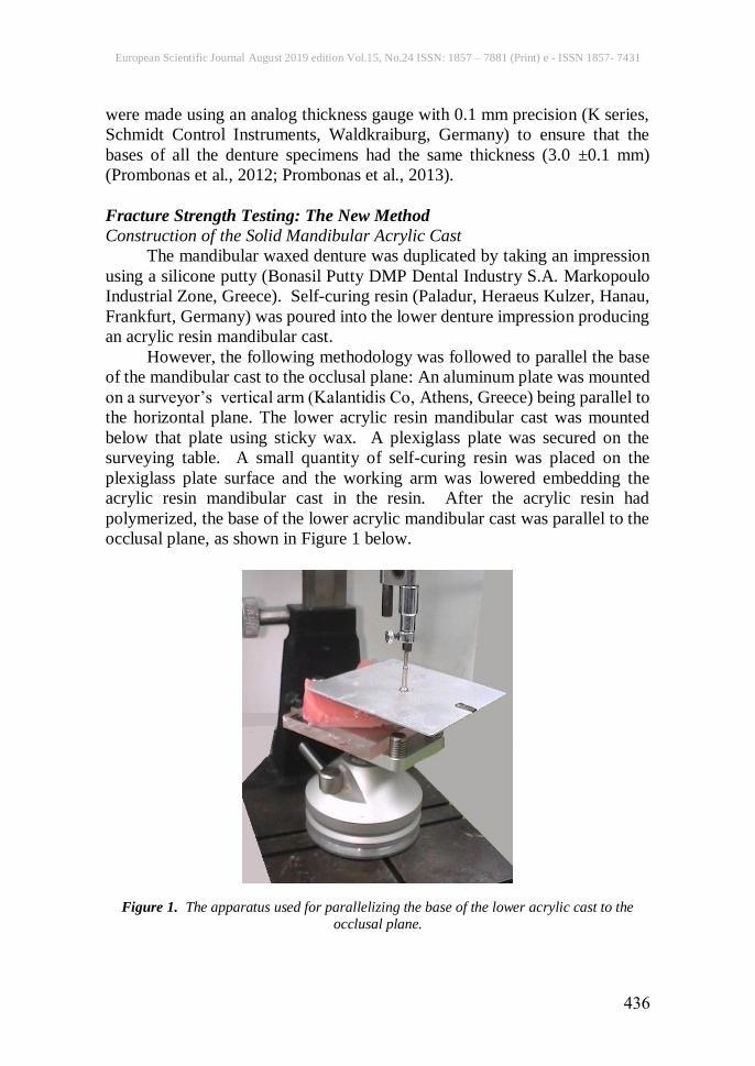
However, the following methodology was followed to parallel the base
of the mandibular cast to the occlusal plane: An aluminum plate was mounted
on a surveyor’s vertical arm (Kalantidis Co, Athens, Greece) being parallel to
the horizontal plane. The lower acrylic resin mandibular cast was mounted
below that plate using sticky wax. A plexiglass plate was secured on the
surveying table. A small quantity of self-curing resin was placed on the
plexiglass plate surface and the working arm was lowered embedding the
acrylic resin mandibular cast in the resin. After the acrylic resin had
polymerized, the base of the lower acrylic mandibular cast was parallel to the
occlusal plane, as shown in Figure 1 below.
Figure 1. The apparatus used for parallelizing the base of the lower acrylic cast to the
occlusal plane.
European Scientific Journal August 2019 edition Vol.15, No.24 ISSN: 1857 – 7881 (Print) e - ISSN 1857- 7431
437
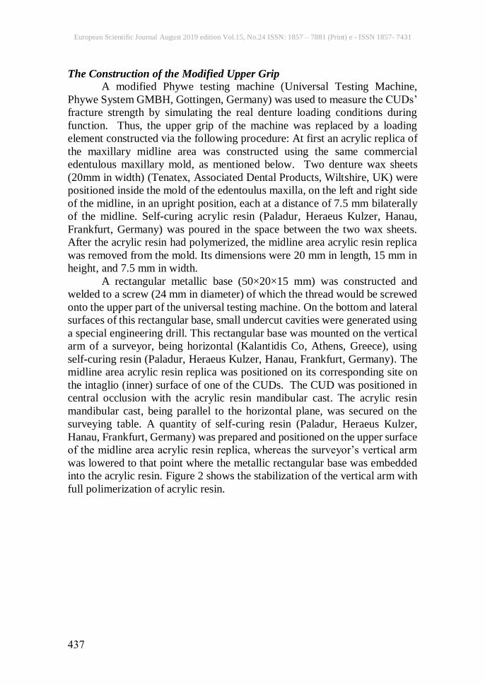
The Construction of the Modified Upper Grip
A modified Phywe testing machine (Universal Testing Machine,
Phywe System GMBH, Gottingen, Germany) was used to measure the CUDs’
fracture strength by simulating the real denture loading conditions during
function. Thus, the upper grip of the machine was replaced by a loading
element constructed via the following procedure: At first an acrylic replica of
the maxillary midline area was constructed using the same commercial
edentulous maxillary mold, as mentioned below. Two denture wax sheets
(20mm in width) (Tenatex, Associated Dental Products, Wiltshire, UK) were
positioned inside the mold of the edentoulus maxilla, on the left and right side
of the midline, in an upright position, each at a distance of 7.5 mm bilaterally
of the midline. Self-curing acrylic resin (Paladur, Heraeus Kulzer, Hanau,
Frankfurt, Germany) was poured in the space between the two wax sheets.
After the acrylic resin had polymerized, the midline area acrylic resin replica
was removed from the mold. Its dimensions were 20 mm in length, 15 mm in
height, and 7.5 mm in width.
A rectangular metallic base (50×20×15 mm) was constructed and
welded to a screw (24 mm in diameter) of which the thread would be screwed
onto the upper part of the universal testing machine. On the bottom and lateral
surfaces of this rectangular base, small undercut cavities were generated using
a special engineering drill. This rectangular base was mounted on the vertical
arm of a surveyor, being horizontal (Kalantidis Co, Athens, Greece), using
self-curing resin (Paladur, Heraeus Kulzer, Hanau, Frankfurt, Germany). The
midline area acrylic resin replica was positioned on its corresponding site on
the intaglio (inner) surface of one of the CUDs. The CUD was positioned in
central occlusion with the acrylic resin mandibular cast. The acrylic resin
mandibular cast, being parallel to the horizontal plane, was secured on the
surveying table. A quantity of self-curing resin (Paladur, Heraeus Kulzer,
Hanau, Frankfurt, Germany) was prepared and positioned on the upper surface
of the midline area acrylic resin replica, whereas the surveyor’s vertical arm
was lowered to that point where the metallic rectangular base was embedded
into the acrylic resin. Figure 2 shows the stabilization of the vertical arm with
full polimerization of acrylic resin.
European Scientific Journal August 2019 edition Vol.15, No.24 ISSN: 1857 – 7881 (Print) e - ISSN 1857- 7431
438
Figure 2. The construction of the modified upper grip of the universal testing machine. The incorporation of the rectangular metallic base into the acrylic replica of the midline area.
While the midline area acrylic resin replica was in place, the rest of the
inner (intaglio) surface of the denture was filled in with silicone putty (Bonasil
Putty DMP Dental Industry S.A. Markopoulo Industrial Zone, Greece).
Following polymerization, these two silicone matrices adhered to the midline
area acrylic resin replica using cyanoacrylate adhesive (Three Bond,
Minamiosawa, Hachiojishi, Tokyo, Japan). The resultant hybrid cast,
constituting of acrylic in the region along the midline and silicone in the
remaining areas, was the upper loading element of the specimens. This loading
element was screwed onto the upper part of the universal testing machine as
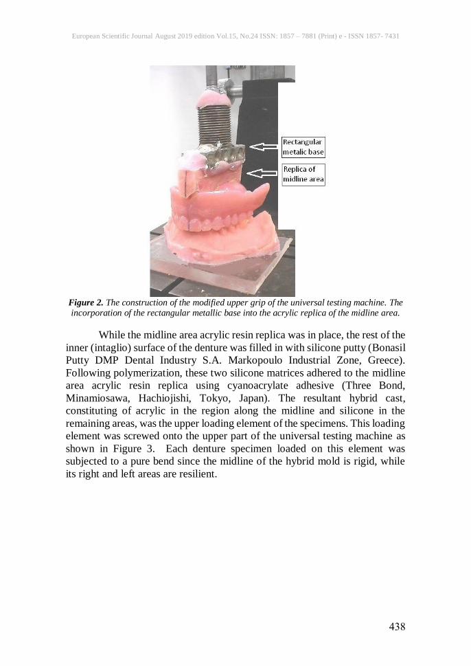
shown in Figure 3. Each denture specimen loaded on this element was
subjected to a pure bend since the midline of the hybrid mold is rigid, while
its right and left areas are resilient.
European Scientific Journal August 2019 edition Vol.15, No.24 ISSN: 1857 – 7881 (Print) e - ISSN 1857- 7431
439
Figure 3. The resultant hybrid cast (the upper loading element of the specimens), made of
acrylic resin and silicone putty, screwed onto the upper part of the universal testing machine.
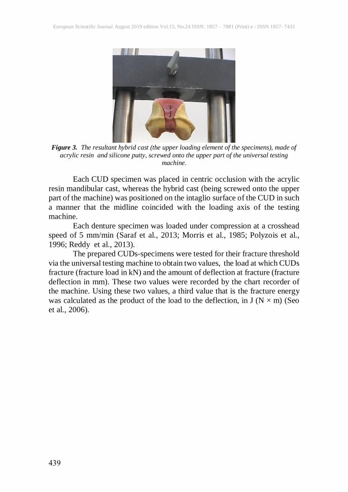
Each CUD specimen was placed in centric occlusion with the acrylic
resin mandibular cast, whereas the hybrid cast (being screwed onto the upper
part of the machine) was positioned on the intaglio surface of the CUD in such
a manner that the midline coincided with the loading axis of the testing
machine.
Each denture specimen was loaded under compression at a crosshead
speed of 5 mm/min (Saraf et al., 2013; Morris et al., 1985; Polyzois et al.,
1996; Reddy et al., 2013).
The prepared CUDs-specimens were tested for their fracture threshold
via the universal testing machine to obtain two values, the load at which CUDs
fracture (fracture load in kN) and the amount of deflection at fracture (fracture
deflection in mm). These two values were recorded by the chart recorder of
the machine. Using these two values, a third value that is the fracture energy
was calculated as the product of the load to the deflection, in J (N × m) (Seo
et al., 2006).
European Scientific Journal August 2019 edition Vol.15, No.24 ISSN: 1857 – 7881 (Print) e - ISSN 1857- 7431
440
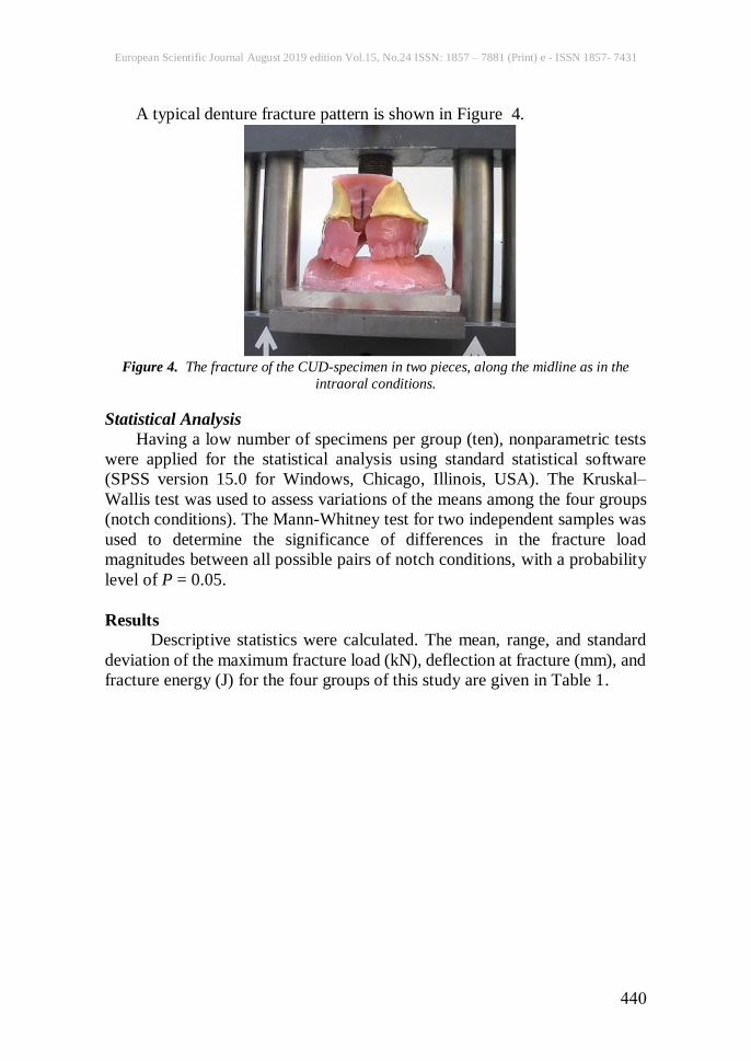
A typical denture fracture pattern is shown in Figure 4.
Figure 4. The fracture of the CUD-specimen in two pieces, along the midline as in the
intraoral conditions.
Statistical Analysis
Having a low number of specimens per group (ten), nonparametric tests
were applied for the statistical analysis using standard statistical software
(SPSS version 15.0 for Windows, Chicago, Illinois, USA). The Kruskal–
Wallis test was used to assess variations of the means among the four groups
(notch conditions). The Mann-Whitney test for two independent samples was
used to determine the significance of differences in the fracture load
magnitudes between all possible pairs of notch conditions, with a probability
level of P = 0.05.
Results
Descriptive statistics were calculated. The mean, range, and standard
deviation of the maximum fracture load (kN), deflection at fracture (mm), and
fracture energy (J) for the four groups of this study are given in Table 1.

European Scientific Journal August 2019 edition Vol.15, No.24 ISSN: 1857 – 7881 (Print) e - ISSN 1857- 7431
441
Table 1. Results of fracture load, fracture energy and deflection at fracture of complete
upper dentures-specimens (n =10)
GROUP 1 GROUP 2 GROUP 3 GROUP 4 FRACTURE LOAD (kN)
Mean 4.15 2.27 2.73 1.94 Range 0.40 1.20 1.65 1.35 St. Deviation 0.13 0.39 0.51 0.55 FRACTURE ENERGY (J)
Mean 7.91 3.16 4.56 2.56 Range 3.06 1.80 3.30 3.44 St. Deviation 1.02 0.59 1.10 1.19 DEFLECTION AT FRACTURE (mm)
Mean 1.90 1.39 1.66 1.26 Range 0.56 0.52 0.53 0.82 St. Deviation 0.19 0.14 0.17 0.27
Highly significant variance of means was found among the four groups
for fracture load, fracture energy, and deflection at fracture (Kruskal–Wallis
test, P<0.001).
It is clear from Table 1 that group 1 (complete denture with shallow
initial fraenal notch) showed the highest mean value in fracture load (fracture
strength) as well as in fracture energy, compared to group 2 (incisal diastema
between central incisors), group 3 (deep labial fraenal notch), and group 4
(combination of deep labial fraenal notch and diastema) (statistical
significance regarding all the above mentioned pairwise comparisons
P<0.001).
The results revealed statistically significant differences in means for
fracture energy between the groups 2 and 3 (P=0.005) as well as 3 and 4
(P=0.002) but not between groups 2 and 4 (P=0.218). As far as it concerns
the differences in means for fracture load, these were statistically significant
only between groups 3 and 4 (P=0.007). Νo statistically significant difference
was found for the fracture load between the groups 2 and 3 as well as 2 and 4
(P = 0.063 and P = 0.123 respectively).
Group 4 (combination of midline (incisal) diastema and labial fraenal
notch deepening) had the lowest mean values among the four groups of
notches, for both fracture load and fracture energy. These mean values
presented a statistically significant difference when compared to those of
group 3, but did not present a statistically significant difference when
compared to group 2.
European Scientific Journal August 2019 edition Vol.15, No.24 ISSN: 1857 – 7881 (Print) e - ISSN 1857- 7431
442
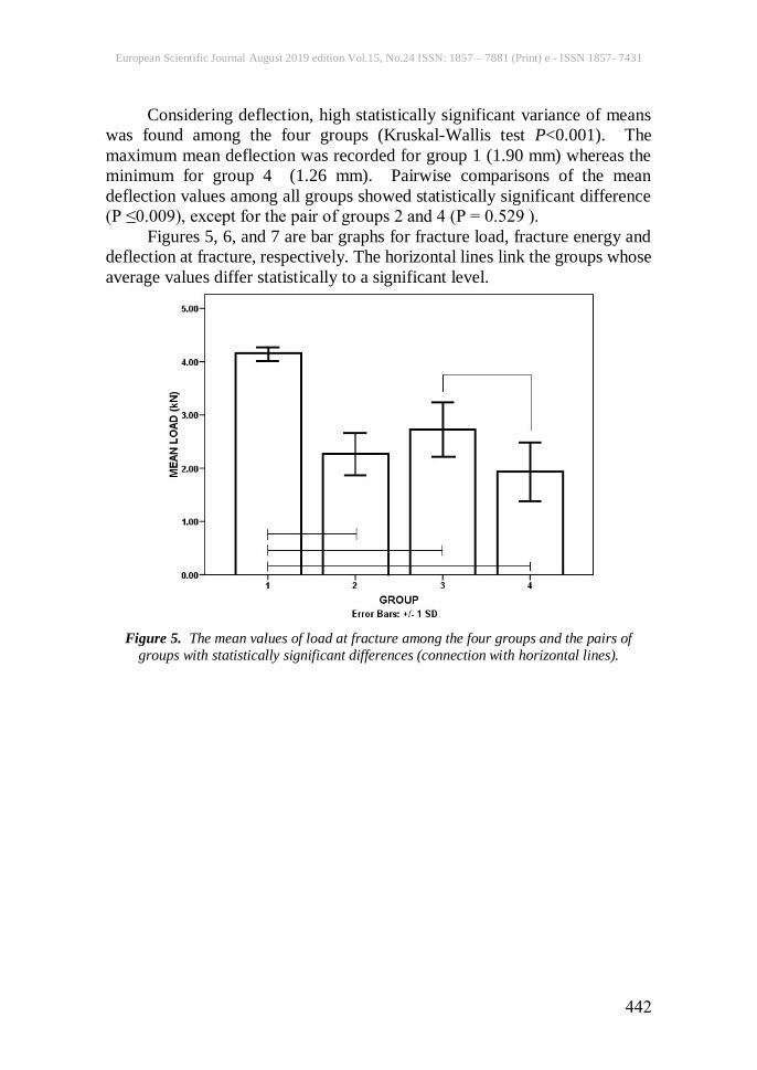
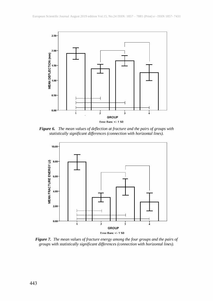
Considering deflection, high statistically significant variance of means
was found among the four groups (Kruskal-Wallis test P<0.001). Τhe
maximum mean deflection was recorded for group 1 (1.90 mm) whereas the
minimum for group 4 (1.26 mm). Pairwise comparisons of the mean
deflection values among all groups showed statistically significant difference
(P ≤0.009), except for the pair of groups 2 and 4 (P = 0.529 ).
Figures 5, 6, and 7 are bar graphs for fracture load, fracture energy and
deflection at fracture, respectively. The horizontal lines link the groups whose
average values differ statistically to a significant level.
Figure 5. The mean values of load at fracture among the four groups and the pairs of
groups with statistically significant differences (connection with horizontal lines).
European Scientific Journal August 2019 edition Vol.15, No.24 ISSN: 1857 – 7881 (Print) e - ISSN 1857- 7431
443
Figure 6. The mean values of deflection at fracture and the pairs of groups with
statistically significant differences (connection with horizontal lines).
Figure 7. The mean values of fracture energy among the four groups and the pairs of
groups with statistically significant differences (connection with horizontal lines).
European Scientific Journal August 2019 edition Vol.15, No.24 ISSN: 1857 – 7881 (Print) e - ISSN 1857- 7431
444
Discussion
In the present study, the fracture strength, deflection at fracture, and
fracture energy of CUDs were measured.
Regarding the latter, some researchers have used the formula “fracture
energy = ½ × fracture load × deflection”, in kgr/cm units (Morris et al., 1985;
Reddy et al., 2013; Sowmya et al., 2013). Other researchers avoided
calculating the fracture energy, estimating the fracture strength based only on
the fracture load measurement (Saraf et al., 2013; Polyzois et al., 1996;
Shimizu et al., 2005; Hedzelek & Gaidus, 2006; Al-Kadi et al., 2015). In the
present study, the fracture energy was calculated as the product of fracture
load and deflection at fracture, as mentioned by previous studies (Seo et al.,
2006).
Measurement of CUDs’ fracture energy by this novel biomechanical
methodology presented in our study (simulation of intraoral loading
mechanism) has not been reported to the available literature. Therefore, direct
comparison cannot be made. Nevertheless, according to the available
literature, the measured fracture load ranged between 0.69 and 1.16 kN (Saraf
et al., 2013), 0.79 and 2.01 kN (Shimizu et al., 2005), 0.7 and 0.9 kN (Polyzois
et al., 1996), 0.7 and 1.3 kN (Seo et al., 2006), or 0.56 and 1.89 kN (Al-Kadi
et al., 2015). These values are typically lower than those measured in this
study, given that the load was applied at one particular point resulting in high
load concentration and leading to rapid failure of the denture.
Other researchers have also stated that measuring the deflection and
fracture strength of identical plain acrylic denture bases (with no teeth) by
loading them on an arbitrary point of the intaglio surface (methodology used
in previous studies) had several weaknesses (Saraf et al., 2013; Polyzois et al.,
1996). This is verified by the mode of fracture observed in some of these
studies, where the bases fractured in three pieces (Morris et al., 1985; Sowmya
et al., 2013). This fracture mode is never (or extremely rarely) encountered in
the functional fracture (due to fatigue) of CUDs in the oral cavity.
The CUD presents uniqueness as a loadable structure due to some
distinct features like: lack of symmetry, fluctuating cross-section, complexity
of applied loads during normal function in both magnitude and direction, and
non-uniformity of load distribution throughout the specimen (Lambrecht &
Kydd, 1962; Prombonas et al., 2012; Prombonas et al., 2013; Reddy et al.,
2013).
In order to simulate the above mentioned functional load conditions of
CUDs, a new biomechanical approach for measuring the fracture strength of
CUDs was applied in this study. This explains why all CUDs-specimens of
the present study were fractured in two pieces along the midline, as it happens
when fractured in the oral cavity. An additional reason that the specific design
of the midline area acrylic resin replica was implemented in the study was to
European Scientific Journal August 2019 edition Vol.15, No.24 ISSN: 1857 – 7881 (Print) e - ISSN 1857- 7431
445
simulate the rigidity of the palatal raphe compared to the flexibility that the
rest of the palatal area presents (Zarb et al., 1997).
It was shown that the presence of notches (group 1 compared to groups
2, 3, and 4) drastically reduced the fracture load and fracture energy, as well
as the deflection at fracture of the CUDs-specimens.
Among the groups 2 and 3, group 2 (after generating a midline (incisal)
diastema (7mm) by grinding in between the two central incisors) showed
lower fracture load and fracture energy compared to group 3 (deepening the
initial labial fraenal notch by 2 mm to a total notch length of 7 mm). However,
this difference was statistically significant only for fracture energy. According
to previous work, the impact of notches in the fracture of CUDs was not clear,
namely the frequency of midline fracture of dentures with incisal diastema was
higher than those without diastema, with no statistical significance, whereas
the existence of deep labial fraenal notch was strongly related to the frequency
of midline fracture (Zissis et al., 1997).
From the present study, it can be assumed that the fracture energy
measurement (as the product of two physical quantities), compared to fracture
load, is more sensitive on estimating the statistical differences, leading to more
precise outcomes.
Furthermore, by considering groups 1, 2 and 3, it was shown that the
presence of incisal diastema (group 2) lowers the fracture strength to a
significant level compared to the presence of a labial fraenal notch regardless
of whether it is shallow or deep. This leads to the assumption that the incisal
diastema has the greatest impact on reducing the fracture strength of CUDs,
compared to the other two types of notches (groups 1 and 3). Furthermore,
this finding is in agreement with a previous study where it has been shown that
it is less likely that a failure crack in a CUD will be initiated from the fraenal
notch region. This is due to the compressive nature of the principal stresses
and the low value of the maximum shear stress in this region (Prombonas et
al., 2012).
The combination of midline (incisal) diastema and labial fraenal notch
deepening (group 4) had the lowest mean values among the four groups of
notches, for both fracture load and fracture energy. These mean values
presented a statistically significant difference when compared to those of
group 3 but did not present a statistically significant difference when
compared to group 2. The above finding showed that among the three notch
conditions (groups 2, 3 and 4), the incisal diastema generation (group 2)
decreases the fracture strength to a statistically significant low level (compared
to group 3) which is close to the fracture strength level shown when combining
the deepening of the labial fraenal notch and the midline (incisal) diastema
generation (group 4). These two conditions (groups 2 and 4) strongly require
reinforcement of the denture bases.
European Scientific Journal August 2019 edition Vol.15, No.24 ISSN: 1857 – 7881 (Print) e - ISSN 1857- 7431
446
Answering to the scope of the present study, concerning the impact of
each one of the notch conditions on the reduction of the fracture strength of
the CUD, the highest was for the combination of the labial fraenal notch
deepening and the midline (incisal) diastema generation (group 4). Also, it
gradually reduced for the midline (incisal) diastema generation (group 2), the
labial fraenal notch deepening (group 3), and the shallow (initial) fraenal notch
(group 1). The above findings are also an answer to the existing controversy
of which notch mode, among the incisal diastema and the fraenal notch,
presents the highest impact on the reduction of CUDs strength.
The current findings reveal the necessity of CUD reinforcement in cases
that involve deep labial fraenal notch but especially in the case of midline
(incisal) diastema or its combination with deep labial fraenal notch.
Conclusion
The new biomechanical approach applied in this study for measuring the
fracture strength of the CUD is an excellent simulation of the denture load in
the oral cavity. In estimating statistically the differences in fracture strength of
CUD, the measurement of fracture energy through the present novel
biomechanical method is more sensitive than the measurement of fracture
load, leading to more precise outcomes.
The midline (incisal) diastema and the combination of a deep labial
fraenal notch with a midline (incisal) diastema lowers the strength
significantly compared to the shallow labial fraenal notch. Thus, such
conditions require reinforcement of the denture bases.
References:
1. Ali Al-Kadi, F.K., Abdulkareem, J.F. & Al-jmoor, C.A. (2015).
Fracture strength of palatal denture base constructed from different
acrylic denture base materials. European Scientific Journal; 11:354-
364.
2. Beyli, M.S. & von Fraunhofer, JA. (1981). An analysis of causes of
fracture of acrylic resin dentures. J. Prosthet. Dent.; 46:238-241.
3. Cilingir, A., Bilhan, H., Baysal, G., Sunbuloglu, E. & Bozdag, E.
(2013). The impact of frenulum height on strains in maxillary denture
bases. J. Adv. Prosthodont; 5:409-415.
4. Dhiman, R.K. & Chowdhury, S.R. (2009). Midline Fractures in Single
Maxillary Complete Acrylic vs Flexible Dentures. Med. J. Armed
Forces India; 65:141-145.
5. Farmer, J.B. (1983). Preventive prosthodontics: Maxillary denture
fracture. J. Prosthet. Dent.; 50:172-175.
6. Glantz, P.O. & Stafford, G.D. (1983). Clinical deformation of
maxillary complete dentures. J. Dent.; 11:224–230.
European Scientific Journal August 2019 edition Vol.15, No.24 ISSN: 1857 – 7881 (Print) e - ISSN 1857- 7431
447
7. Hedzelek, W. & Gajdus, P. (2006). Comparison of mechanical strength
of palatal denture bases made from various plastic materials. Int. J.
Prosthodont; 19:193-194.
8. Hill, E.G., Bates, J.F., Lewis, T.T. & Rees, N. (1983). Fracture
toughness of acrylic denture base. Biomaterials; 4:112-120.
9. Hirajima, Y., Takahash, H. & Minakuchi, S. (2009). Influence of a
denture strengthener on the deformation of a maxillary complete
denture. Dent. Mater. J.; 28:507–512.
10. Lamb, D.J., Ellis, B. & van Noort, R. (1985). The fracture topography
of acrylic dentures broken in service. Biomaterials; 6:110-112.
11. Lambrecht, J.R. & Kydd, W.L. (1962). A functional stress analysis of
the maxillary complete denture base. J. Prosthet. Dent; 12:865-872.
12. Matthews, E. & Wain, E.A. (1956). Stresses in denture bases. Br. Dent.
J.; 100:167-171.
13. Morris, J.C., Khan, Z. & von Fraunhofer, J.A. (1985). Palatal shape
and the flexural strength of maxillary denture bases. J. Prosthet. Dent.;
53:670-673.
14. Nejatidanesh, F., Peimannia, E. & Savabi, O. (2009). Effect of labial
frenum notch size and palatal vault depth on stress concentration in a
maxillary complete denture: a finite element study. J. Contemp. Dent.
Pract.; 10:59-66.
15. O’Brien, W.J. (2002). Dental materials and their selection, 3d edn.
Chicago: Quintessence Publications Co. Inc; p.p. 1–17. 352. 369. 375.
16. Polyzois, G.L., Andreopoulos, A.G. & Lagouvardos, P.E. (1996).
Acrylic resin denture repair with adhesive resin and metal wires:
effects on strength parameters. J. Prosthet. Dent.; 75 :381-87.
17. Prombonas, A.E., Vlissidis, D.S., Paralika, M.A. & Poulis, N.A.
(2012). The stress state of the fraenal notch region in complete upper
dentures. Med. Eng. Phys.; 34 :1477–1482.
18. Prombonas, A.E., Paralika, M.A. & Poulis, N.A. (2013). Investigation
of the torsional deformation of the complete upper denture: A pilot
study. J. Biomed. Sci. Eng.; 6:443-448.
19. Reddy, B.M., Himabindu, M., Padmaja, B.I., Sunil, M. & Reddy, N.R.
(2013). Palatal vault depth influence on the flexural strength of two
heat cure acrylic denture base resins: an in vit30ro study. J. Contemp.
Dent. Pract.; 14:1131-1136.
20. Saraf, V.R., Shetty, V., Anandakrishna, G.N. & Rairam, S.G. (2013).
Comparison of mechanical strength of palatal denture bases made from
three commercially available high impact acrylic resin denture base
materials in different palatal vault configuration – An invitro study. J.
Oral Health Res.; 4:1-7.
European Scientific Journal August 2019 edition Vol.15, No.24 ISSN: 1857 – 7881 (Print) e - ISSN 1857- 7431
448
21. Seo, R.S., Murata, H., Hong, G., Vergani, C.E. & Hamada, T. (2006).
Influence of thermal and mechanical stresses on the strength of intact
and relined denture bases. J. Prosthet. Dent. 2006 ; 96:59-67.
22. Shimizu, H., Tsue, F., Obukuro, M., Kido, H., Takahashi, Y. &
Ohmura, H. (2005). Fracture strength of metal-based complete
maxillary dentures with a newly designed metal framework. Int. Chin.
J .Dent.; 5:35-38.
23. Sowmya, S., Dhakshaini, M.R., Gujjari, A.K., Ravi, M.B. &
Raghavendra Swamy, K.N. (2013). Palatal Vault Depth Influence on
the Flexural Strength of Two Heat Cure Acrylic Denture Base Resins
- An In vitro Comparative Study. World Appl. Sci. J.; 21:1290-1294.
24. Stafford, G.D. & Griffiths, D.W. (1979). Investigation of the strain
produced in maxillary complete dentures in function. J. Oral Rehabil.;
6:241–256.
25. Takamiya, A.S., Monteiro, D.R., Marra, J., Compagnoni, M.A. &
Barbosa, DB. (2012). Complete denture wearing and fractures among
edentulous patients treated in university clinics. Gerodontology;
29:728–734.
26. Vallittu, P.K. (1996). Fracture surface characteristics of damaged
acrylic-resin-based dentures as analysed by SEM-replica technique. J.
Oral Rehabil.; 23:524–529.
27. Zarb, G.A., Bolender, C.I., & Carlsson, G.E. (1997). Boucher’s
prosthodontic treatment for edentulous patients, 11th edn. St. Louis:
C.V. Mosby; p.p. 332.346.
28. Zissis, A.J., Polyzois, G.L. & Yannikakis, S.A. (1997). Repairs in
complete dentures: results of a survey. Quintessence Dent. Technol.;
20:149-155.