Imaging human brain anatomy, function and … cerebral physiology compatible with MR imaging of ......
56
Transcript of Imaging human brain anatomy, function and … cerebral physiology compatible with MR imaging of ......


1992 First papers introducing FUNCTIONAL IMAGING in the HUMAN BRAIN (fMRI) appeared in Press
2012 We celebrated two-decades of fMRI
TWO MAJOR INVESTMENTS in INSTRUMENTATION
• UMinn: 4 Tesla HUMAN capable MR INSTRUMENT
• MGH: 1.5Tesla but Ultrafast Imaging Capable Instrument

Functional Imaging with Magnetic Resonance
Ogawa et al. Proc Natl Acad Sci USA (1992) 89, 5951-5955

BEHAVIOR NEURONAL
or ACTIVITY
PERCEPTION
The different SCALES of the BRAIN’s FUNCTIONAL ORGANIZATION

Scales of the Brain
Global networks
Silent Word Generation

Scales of the Brain

Scales of the Brain
Orientation Domains in the Primary Visual Cortex
~4 mm
Monkey Cortex Optical Imaging

Tanaka, Annu Rev Neurosci, 1996
Object selective columns in IT

Whisker Barrels

Scales of the Brain
Orientation Domains in the Primary Visual Cortex
~4 mm
Monkey Cortex Optical Imaging

FIRST QUESTION:
Is cerebral physiology compatible with MR imaging of
functional activity at columnar level??

DEOXY-Hemoglobin (Paramagnetic)

INCREASED NEURONAL ACTIVITY
INCREASE in REGIONAL BLOOD FLOW (& Volume)
LOWER DEOXYHEMOGLOBIN CONTENT per unit volume in the BRAIN if Cerebral Oxygen Consumption (CMRO2) does not
increase commensurately

SPECIFICTY of the METABOLIC and HEMODYNAMIC RESPONSES
Site of Elevated Neuronal Activity
blood supply
Increased Perfusion

9.4 Tesla/ 31 cm Bore for animal model studies (~1994)

ISO-ORIENTATION DOMAINS in the CAT VISUAL Area 18: CBFSINGLE Orientation, SINGLE CONDITION
T. Duong, S-G. Kim et al., PNAS (2001): 98; 10904-10909

Duong, T.Q., D.S. Kim, K. Ugurbil, and S.G. Kim: Localized cerebral blood flow response at submillimeter columnar resolution. Proc Natl Acad Sci U S A, 2001. 98(19): p. 10904-10909.
CBF is regulated at he level of Cortical
Columns
“brain waters the entire garden for the sake of a thirsty flower”X “the brain really waters the thirsty flower while it sprinkles generously around it”

From Kwong et al 1992 PNAS paper
From Ogawa et al 1992 PNAS Paper.

Cortical surface
ULTRAHIGH FIELDS
(7 Tesla or higher)
White Matter
Image From: Reine-De La Torre et al. The Anatomical Record 251:87-96 (1998)

AND of MR detected Mapping Signals and Physiologic Changes induced by Neuronal activity
K. Uludağ, B. Müller-Bierl, K. Uğurbil Neuroimage (2009) 48(1): p. 150-65.

7 Tesla/90 cm bore ~1999

GE BOLD fMRI 4 vs 7 Tesla, as a function of TE (ms)
4 TESLA 15 22.5 34 51 76
7 TESLA 10 15 22.5 34 51
Yacoub E, et al. Magn Reson Med 2001;45(4):588-594

Orientation Domains in the Primary Visual Cortex
Monkey Human Optical Imaging fMRI (SE, 7 Tesla)
~4 mm ~4 mm
Yacoub, Harel, Uğurbil PNAS 2008

Ocular Dominance (ODC) and Orientation maps in Human V1 (7 Tesla)
Yacoub, Harel, UğurbilYacoub, Shmuel, et al. PNAS (2008) 105(30): 10607-12Neuroimage (2007) 37(4): 1161-77
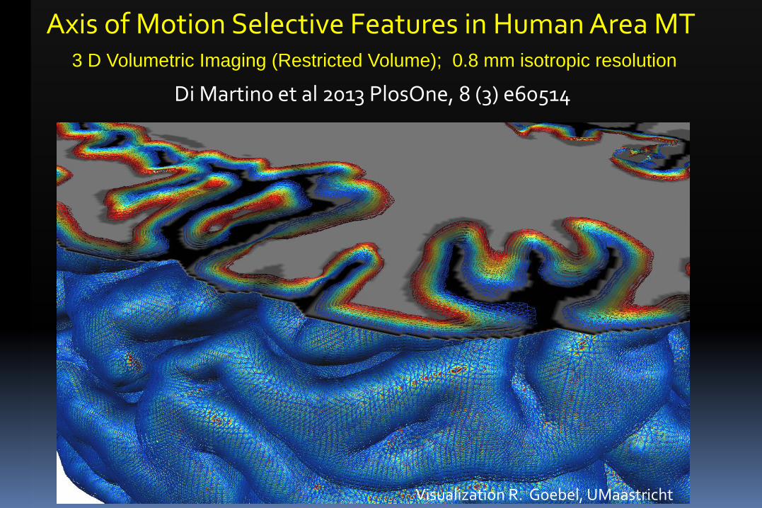
Axis of Motion Selective Features in Human Area MT 3 D Volumetric Imaging (Restricted Volume); 0.8 mm isotropic resolution
Di Martino et al 2013 PlosOne, 8 (3) e60514
Visualization R. Goebel, UMaastricht


Axis of Motion Selective Features in Human Area MT
Zimmermann, et al PLoS ONE 6(12): e28716. (2011)

Tuning Curves in Human Area MT for Axis of Motion
Di Martino et al 2013 PlosOne, 8 (3) e60514 (CMRR & UMaastricht)
28

Tonotopic Mapping in Human Primary Auditory Cortex

Sub millimeter predominantly T2 weighted functional responses in human area A1.
High (200)
Low (6400)
Frequency
(Hz)
F. De Martino, E. Yacoub , E. Formisano et al.
Medial
Lateral
Heschl’s
gyrus

Frequency preference orthogonal to the brain surface (tonotopic columns).
Cortical Ribbon
High (200)
Frequency
(Hz)
Low (6400)
F. De Martino, E. Yacoub , E. Formisano et al.
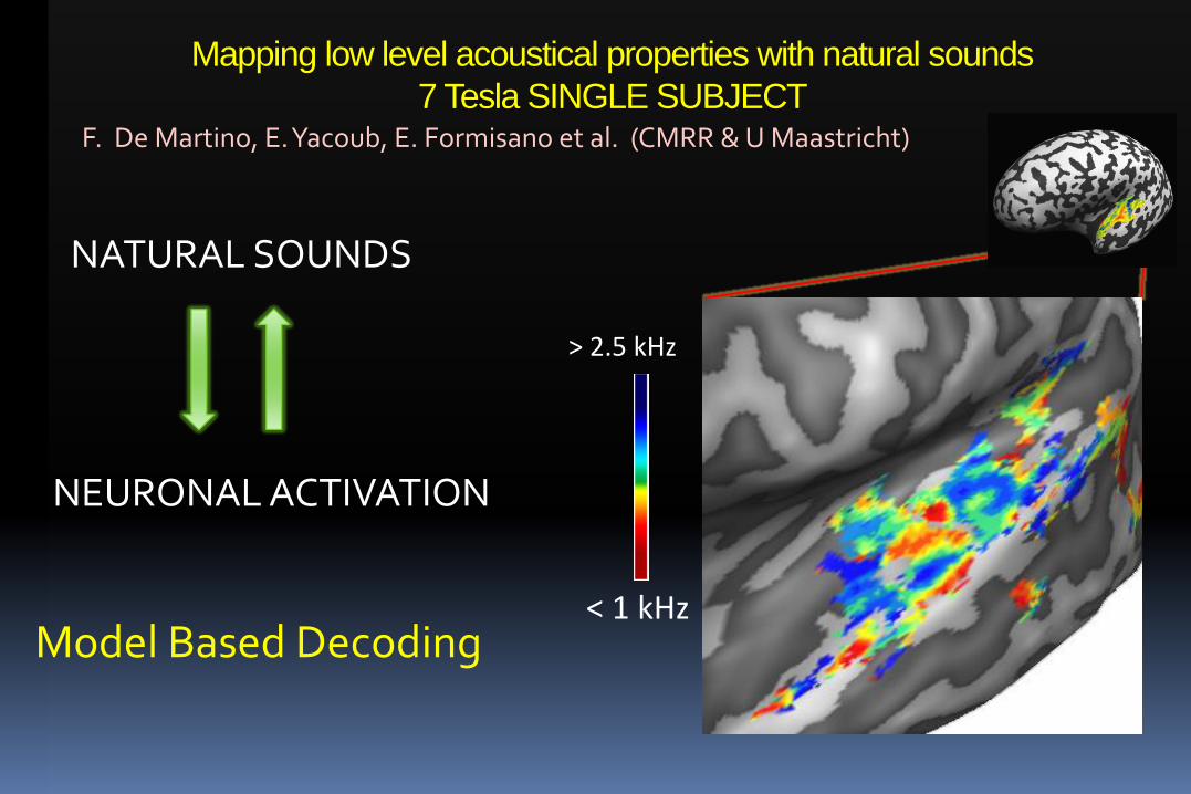
Mapping low level acoustical properties with natural sounds 7 Tesla SINGLE SUBJECT
F. De Martino, E. Yacoub, E. Formisano et al. (CMRR & U Maastricht)
NATURAL SOUNDS
> 2.5 kHz
NEURO NAL ACTIVATION
< 1 kHz Model Based Decoding

Principal Investigators: David C. Van Essen and Kamil Ugurbil
33
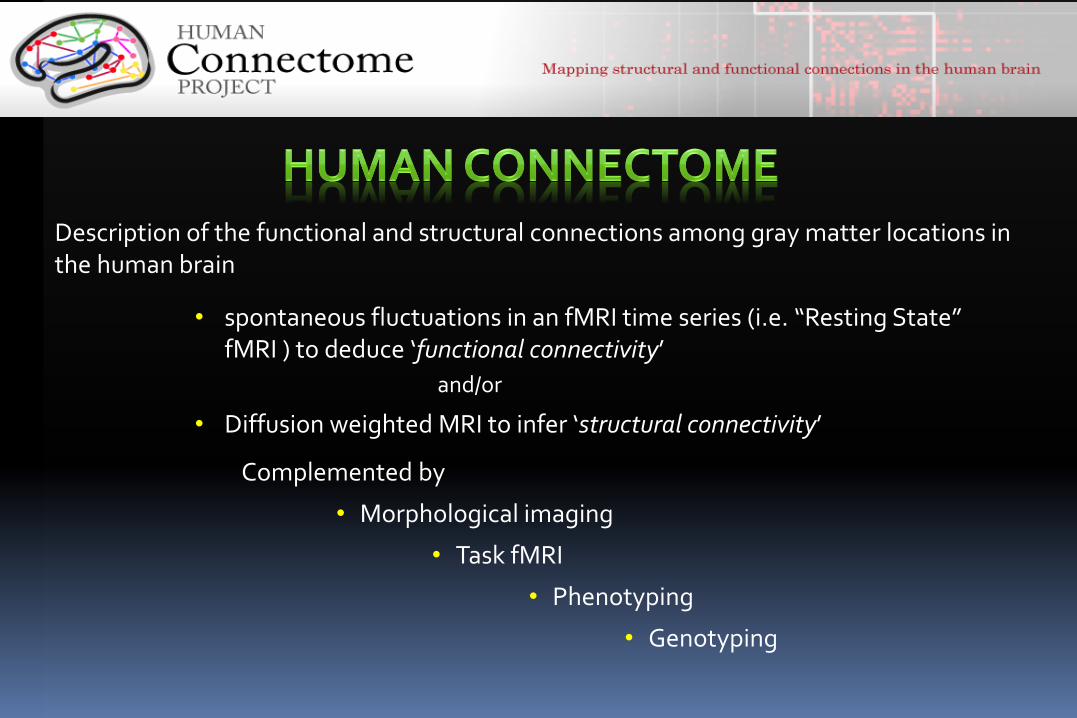
Description of the functional and structural connections among gray matter locations in the human brain
• spontaneous fluctuations in an fMRI time series (i.e. “Resting State” fMRI ) to deduce ‘functional connectivity’
and/or
• Diffusion weighted MRI to infer ‘structural connectivity’
Complemented by
• Morphological imaging
• Task fMRI
• Phenotyping
• Genotyping

Resting-State Networks (RSNs) SLIDE COURTESY of STEVEN SMITH OXFORD UNIVERSITY
Time
Spatial patterns of correlated temporal dynamics, resembling activation maps


In vivo labeling of axonal fibers using Diffusion Tensor Imaging (DTI)

• HIGH SPATIAL RESOLUTION over the WHOLE BRAIN !!!!!
MAXIMIZE SNR
FASTER DATA ACQUISITION SPEED without SIGNIFICANTLY SACRIFICING SNR

Conventional Multi-slice Imaging
Whole Volume TR x Time per slice = Nslice

IMPROVED IMAGING STRATEGIES
SliceAccelerated, Simultaneous Multi Slice, Multiband Imaging
• Larkman et al JMRI 2001 (leg)
• Moeller, Yacoub, Auerbach, Ugurbil ISMRM 2008; # 236
• Moeller et al. Magn Reson Med, 2010; 63(5): p. 1144-53
• Setsompop et al. Magn Reson Med, 2012; 67, 1210-1224.
Excite multiple slices simultaneously
Use Parallel Imaging and Multichannel receive coil array to unalias simultaneously acquired images

INSTRUMENTATION
3 TESLA equipped with 100 mT/m Gradients
CONVENTIONAL INSTRUMENTS operate with ~40 mT/m Gradients
7 Tesla

PROBABILISTIC TRACTOGRAPHY: medial to lateral : cortico-thalamic, cortico-bulbar, cortico-spinal and cortico-striatal projections. Images are shown in radiological view.
Sotiropoulos, S. N., Jbabdi, S., Xu, J., Andersson, J. L., Moeller, S., Auerbach, et al. & - for the WU-Minn HCP Consortium.

STRUCTURAL Connectivity (Group average 9 subjects)
FUNCTIONAL Connectivity (Group average 9 subjects)
43

TFMs from HCP Phase II, released Data (Higher spatial (2 vs 3 mm iso.) and temporal
resolution (0.7 vs 0.8 s )
44

Intracortical myelin Content TOF angiography at 7 Tesla at 0.4 mm isotropic
S. Schmitter et al (CMRR)(CMRR and UMaastricht)
7 Tesla High Resolution fMRI 7 Tesla CMRR De Martino et al (CMRR and U Maastricht) Henry, T.R., et al Radiology, 2011; 261(1): p. 199-209.

Bilateral Hip Imaging 7 T
7T Kidney Angiography (CMRR) Metzger, G. J. (2013) Magn Reson Med 69, 114-126.
Ellermann et al. NMR in Biomed 2012; 25, 1202-8
7 Tesla (CMRR) 7T Kidney PERFUSION Metzger et al. (CMRR)
C. Snyder, L. DelaBarre, T. Vaughan et al.

Localized Human Brain 1H Spectrum at 7 Tesla

Three-way correlation in a 26 month AD mouse C. R. Jack, Jr., M. Garwood, et all MRM 2004. 52(6): p. 1263-71.
in vivo
ex vivo
Histology (thioflavin S stained)
9;4 T MRI resolution: 60 μm x 60 μm x 120 μm

9.4 Tesla/31 cm bore ~1994 7 Tesla/90 cm bore ~1999
• INCESSENT advances in Instrumentation
intricately tied to Discoveries/Improvements
in Methods of Image acquisition and
reconstruction.

10.5T System
Field / diameter 10.5T/88cm Conductor NbTi / 433 km Manufacturer Siemens / Agilent Temperature 3K Temporal Stability 0.03 ppm/hr Size 4.1 x 3.2 m Spatial Homogeneity< 0.07 ppm/25cm dsv Weight 110 tons Stored Energy 280 MJ Delivery End of November

Completed Magnet former before assembly

A New Factory Floor
80 Ton cold mass (magnet in helium can) being loaded into the cryostat using a specially commissioned 10 Ton “H” beam.


CENTER for MAGNETIC RESONANCE RESEARCH
(CMRR)

PULSE SEQUENCE, IMAGE RECONSTRUCTION and EVALUATION
Steen Moeller Eddie Auerbach Gordon Xu Essa Yacoub An (Joseph) Vu Dingxin Wang
DIFFUSION IMAGING
Christophe Lenglet
PARALLEL TRANSMIT PULSE DESIGN
UMinn
Sebastian Pierre Francois Xiaoping Wu Schmitter Van de Moortele

Tim Behrens Stam Sotiropoulos Saad Jbabdi Jesper Andersson
DIFFUSION IMAGE ACQUISITION, PROCESSING and ANALYSIS
RESTING STATE ANALYSIS
Steve Smith Karla Miller
Matt Glaser David Van Essen