Imaging Elastographic Properties of Soft Tissues Using ... Elastographic Properties of Soft Tissues...
31
Imaging Elastographic Properties of Soft Tissues Using Ultrasound Tomy Varghese Department of Medical Physics Department of Biomedical Engineering University of Wisconsin-Madison Madison, WI -53706 Presentation Outline Motivation Definitions & Basic Principles Young’s Modulus Contrast For Strain Imaging Methods for Strain Imaging Elastography Palpation Imaging Physiological Stimuli Sonoelasticity Imaging Radiation Fore Imaging Clinical Ultrasound Systems
Transcript of Imaging Elastographic Properties of Soft Tissues Using ... Elastographic Properties of Soft Tissues...

Imaging Elastographic Properties of Soft Tissues Using Ultrasound
Tomy Varghese
Department of Medical PhysicsDepartment of Biomedical EngineeringUniversity of Wisconsin-Madison
Madison, WI -53706
Presentation Outline
Motivation
Definitions & Basic Principles
Young’s Modulus Contrast For Strain Imaging
Methods for Strain ImagingElastographyPalpation ImagingPhysiological StimuliSonoelasticity ImagingRadiation Fore Imaging
Clinical Ultrasound Systems

Motivation
Most pathological changes are associated with changes in tissue stiffness.
Palpation is an effective method for lesion detection.
Many cancers (breast, prostate) are isoechoic, and hence difficult to detect by ultrasound.
DefinitionsStress is defined as force per unit area.
Shear stress has the same units as normal stress but represents a stress that acts parallel to the surface (cross section).
Strain is the change in length per unit length. Computed as (Lf - L0) / L0 where Lf is the final length and L0 is the initial length.
Strain Rate, specifies how quickly (or slowly) a material is being deformed or loaded, i.e. the amount of strain that occurs in a unit of time. Since strain is dimensionless, units are 1/time.

Definitions (cont)
Young's Modulus is the constant of proportionality between stress and strain. Units are the same as stress (i.e., force per unit area) and the most commonly used are psi, Pa (Pascal), kPa, and MPa.
Poisson's Ratio, is the ratio of lateral strain to longitudinal strain. Typical range of values is between zero and 0.5.
Basic Principles
Pre-compressionRF A-lines
Post-compressionRF A-lines
Applied Stress
Theoretical Stress Distribution
Local Cross CorrelationAnalysis
Absolute Axial StressEstimation
Ylo c a llo c a l
=
S tr e s s
S t r a in
ESTIMATION OF STRAIN ESTIMATION OF STRESS

Strain ImagingQuasi Static Methods (Ophir et al. 1991, O’Donnell et al. 1991)
Dynamic Methods (Parker et al. 1990, Krouskop et al. 1987, Sandrin et al. 1999)
Radiation Force (Walker 1999, Fatemi & Greenleaf 1999, Nightingale et al. 2002, Lizzi et al. 2003)
Stress ImagingMechanical or Tactile Imaging (Sarvazyan et al. 1998, Wellman et al. 2001)
Computational modelsFinite Element ModelingUsing Surface Pressure Information
Modulus ImagingIterative Modulus Reconstruction (Kallel et al. 1995)
Direct Methods (Solving PDE’s) (Emelianov et al. 2000, Sumi et al. 1995)
Finite Element Inversion (Zhu et al. 2003)
Young’s Modulus As the Contrast Mechanism

Background
Tissue Elasticity Imaging methods are based on imaging differences in stiffness or Young’s Modulus between normal and abnormal tissue conditions.Literature reports on stiffness variations between different tissue types is limited.However, these results demonstrate significant stiffness variations between normal and pathological tissue.
Measurements of breast tissues in-vitro*
*Krouskop TA, Wheeler TM, Kallel F, Garra BS, Hall T., Elastic moduli of breast and prostate tissues under compression, Ultrason Imaging, 1998; 20(4): 260-74.
Tissue Type NumberOf
Patients
Tissue Stiffness (kPa)20% Pre-compression20%/sec Strain Rate
Normal Fat 40 20 ± 6Normal Glandular 31 57 ± 19Fibrous 21 233 ± 59Ductal Tumor 23 301 ± 58Infiltrating DuctalTumor
32 490 ± 112

Basic data on Breast tissues
Tissue Type Tissue Stiffness (kPa)
5% precompression 20% precompression
Strain rate Strain rate
1%/sec 10%/sec 40%/sec 2%/sec 20%/sec 80%/sec
Normal fat (n=40) 18 ± 7 19 ± 7 22 ± 9 20 ± 8 20 ± 6 23 ± 5
Normal glandular (n=31) 28 ± 14 33 ± 11 35 ± 14 48 ± 15 57 ± 19 66 ± 17
Fibrous (n=21) 97 ± 33 107 ± 32 118 ± 83 220 ± 88 233 ± 59 245 ± 83
Ductal tumor (n=23) 22 ± 8 25 ± 4 26 ± 5 291 ± 67 301 ± 58 307 ± 78
Infilt.ductal tumor (n=32)106 ± 32 93 ± 33 112 ± 43 558 ± 180490 ± 112460 ± 178
*Krouskop TA, Wheeler TM, Kallel F, Garra BS, Hall T., Elastic moduli of breast and prostate tissues under compression, Ultrason Imaging, 1998; 20(4): 260-74.
ELF 3220 at UW-Madison

Experimental Methods
Tissue samples prepared as cylinders 20 mm diameter x ~5 mm heightSamples compressed to 4 % strain at varying frequenciesLoad response measured with 50 Lb load cellThermal lesions prepared by RF ablation (70°, 90° C for 10 minutes)
Experimental ResultsIn Vitro Canine Liver – Comparison
10-1 100 101 102 103102
103
104
105
106
E"E'
E*
E*
E'
Mod
ulus
(P
a)
Frequency (Hz)
E"
NormalLesion

Experimental ResultsIn Vitro Canine Liver – Comparison, Model Fits
100 101 102 103
0.16 1.59 15.92 159.16
102
103
104
105
106
|E*(
ω)| (
Pa)
Angular Frequency ω (s-1)
f (Hz)
Lesion
Normal
Normal
E0 = 2.54 x 10³ Pa
E1 = 3 x 10³ Pa secα
α = 0.154
Lesion
E0 = 2.66 x 10⁴ Pa
E1 = 2.2 x 10³ Pa secα
α = 0.555
Discussion
Pathology generally exhibits large elastic contrast with normal background.Reliable small elastic contrast exists among normal soft-tissue components Results show that the complex modulus is dependent on frequency, but is explained well by fractional derivative Kelvin-Voigt modelHigh frequency results may be better represented by measuring shear modulus

Methods for Strain Imaging
Methods for Strain ImagingMechanical Stimuli used for perturbation
Quasi-Static Methods (Ophir et al., O’Donnell et al.)
Quasi Static Compression (Elastography)Palpation Imaging (Hall TJ) or Freehand Compression
Low frequency Oscillatory Compression (Ermert H)
Dynamic MethodsLow frequency Vibration (Sonoelasticity imaging)Physiological Stimuli
Radiation Force Based Techniques (static & dynamic)Imaging Modality Utilized
UltrasoundMagnetic Resonance ImagingOptical Coherence Tomography
Time Domain or Frequency Domain Processing

Quasi-Static MethodsAlgorithms
Cross-Correlation (Ophir et al. 1991)
Phase shift correlation (O’Donnell et al. 1991)
Phase-root seeking (Pesavento et al. 2000)
Block-matching methods (Chaturvedi et al. 1998; Bohs et al. 1995)
Decorrelation methods (Bamber et al. 1995; Varghese & Ophir 1996)
Envelope processing (Varghese & Ophir 1998)
Envelope + RF (Shiina et al. 1995; Alam et al. 1997)
Quasi-Static Methods
Elastography(cross-correlation)

Definition
Elastography:
An imaging technique whereby local tissue strains are measured from differential ultrasonic speckle displacements. These displacements are generated
by a weak, quasi-static stress field.
The resultant axial-strain, lateral-strain, modulus or Poisson’s ratio images are all referred to as
Elastograms.
ELASTOGRAPHYPre-Compression Post-Compression
Array TransducerCompressor
12.….100 12.……100Scan Lines

RF Signal Compression: 2% applied strain
Notice that a small compression (strain) of the tissue results in a small compression of the signal
(similar to frequency modulation)
Pre-CompressionPost-Compression
Basic Principles: Estimation of Strain
Pre-compression RF line
Post-compression RF line
2τ
1τ T∆
TStrain
∆−= 12
ττ

Elastography Data Acquisition System
The Elastographic Process
IntrinsicTissue
Elastic ModulusProperties
StrainDistribution
the response
Imaging Analysis
Elastogram
Statistical Analysis
(Strain Filter)
ContrastTransfer Efficiency
I
II
III
TissueCompression
the stimulus

The Elastographic Process
Input Tissue
Modulus
ElastogramOutput
Parameters
eE
ContrastTransfer Efficiency
IStatisticalAnalysis
(Strain Filter)
IIImage
Analysis(Elastogram)
III
e
Young’s ModulusDistribution
Ideal StrainDistribution
Estimated Strain(Elastogram)
In-vivo Applications of Elastography
Breast Imaging (Garra et al. 1997; Hiltawsky et al. 2001; Hall et al. 2003)
Prostate Imaging (Lorenz et al. 2000; Souchon et al. 2002, 2003)
Thyroid Imaging (Meixner et al. 2002)
Liver Imaging (Varghese et al. 2002; Merritt et al. 2002; Kolen et al. 2002)
Treatment Monitoring (Varghese et al. 2002; Merritt et al. 2002; Souchon et al. 2003)
Intravascular Strain Imaging (de Korte et al. 2000; 2002a; 2002b; 2003)
Cardiac Elastography (Konofagou et al. 2000; Varghese et al. 2002)
Deep Vein Thrombosis (Emelianov et al. 2002)
Kidney Transplant Monitoring (Emelianov et al. 2002)

Breast ImagingCancers are statistically significantly darker (stiffer) than benign fibroadenomas and other benign lesions.
The transverse dimension of cancers is larger on the elastogramsthan their size estimates on the sonogram.
Freehand and real-time implementation on an US scanner.
*Garra, B.S., Céspedes, E.I., Ophir, J., Spratt R.S., Zuurbier R.A., Magnant, C.M., Pennanen, M.F., Elastography of breast lesions: Initial clinical results, Radiology, 1997, 202, 79-86.
*Ophir, J., Alam, K.,Garra, B., Kallel, F., Konofagou, E., Krouskop, T., Varghese T., Elastography: Ultrasonic estimation and imaging of the elastic properties of tissues, Invited Paper & Review, Proc. Inst. Mech. Eng., Part H J Engg. Med., Vol. 213, pp. 203-233,1999.
*Hiltawsky KM, Kruger M, Starke C, Heuser L, Ermert H, and Jensen A, Freehand ultrasound elastography of breast lesions: clinical results, Ultrasound Med Biol. 27 (11), 1461-1469., 2001
*Hall TJ, Zhu Y, and Spalding CS, In vivo real-time freehand palpation imaging, Ultrasound Med Biol. 29 (3), 427-435., 2003 .
Breast tumors in-vivo (1999)
Fibroadenoma
Infiltrating DuctalCarcinoma
Sonograms Elastogram
Prostate Imaging
Elastograms obtained using a balloon to provide the compressive force.
The transducer probe also has been used as the compressor.
Real-time implementation has been demonstrated.
*Lorenz A, Ermert H, Sommerfeld HJ, Garcia-Schurmann M, Senge T, and Philippou S, [Ultrasound elastography of the prostate. A new technique for tumor detection], Ultraschall Med. 21 (1), 8-15., 2000.
*Souchon R, Soualmi L, Bertrand M, Chapelon JY, Kallel F, and Ophir J, Ultrasonic elastography using sector scan imaging and a radial compression, Ultrasonics. 40 (1-8), 867-871., 2002 .
*Souchon R, Rouviere O, Gelet A, Detti V, Srinivasan S, Ophir J, and Chapelon JY, Visualisation of HIFU lesions using elastography of the human prostate in vivo: preliminary results, Ultrasound Med Biol. 29 (7), 1007-1015., 2003.
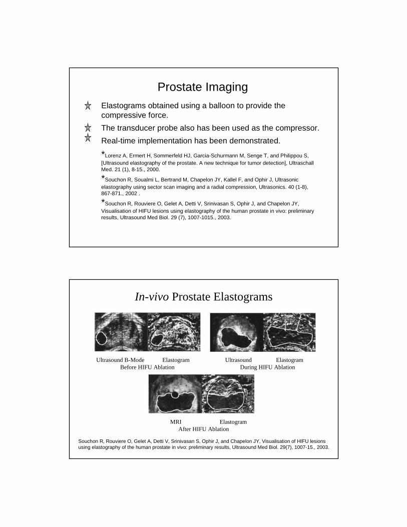
In-vivo Prostate Elastograms
Ultrasound B-Mode Elastogram Ultrasound ElastogramBefore HIFU Ablation During HIFU Ablation
MRI ElastogramAfter HIFU Ablation
Souchon R, Rouviere O, Gelet A, Detti V, Srinivasan S, Ophir J, and Chapelon JY, Visualisation of HIFU lesions using elastography of the human prostate in vivo: preliminary results, Ultrasound Med Biol. 29(7), 1007-15., 2003.

Treatment MonitoringRF ablated lesion in-vivo in an animal model
• Use the RF electrode as the compressor• Use compression induced due to diaphragmatic stimuli
HIFU lesions in prostate imaged using a balloon to provide the compressive stimuli.
Using a stepper motor controlled compression on an open chest animal model on RF ablated lesions.*Varghese T, Zagzebski JA, and Lee FT, Jr., Elastographic imaging of thermal lesions in the liver in vivo following radiofrequency ablation: preliminary results, Ultrasound Med Biol. 28 (11-12), 1467-1473., 2002.
* Souchon R, Rouviere O, Gelet A, Detti V, Srinivasan S, Ophir J, and Chapelon JY, Visualisation of HIFU lesions using elastography of the human prostate in vivo: preliminary results, Ultrasound Med Biol. 29 (7), 1007-1015., 2003.
*Merritt CR, Forsberg F, Liu J, and Kallel F, In-vivo elastography in animal models: Feasibility studies, (abstract), J. Ultrasound Med.. 21 S98, 2002.
In Vivo Study 75 kg Female Yorkshire Pig
Approved RARC Protocol
Liver tissue exposed by laparatomy
RITA 1500 electrosurgical device for ablation
50W; 100oC; 10min
Acuson scanner with 12 Bit Gage Board at 50 MHz sampling rate to acquire RF echo signals at 2 f/s.
Liver removed, sliced following procedure

In-vivo Elastograms Using Respiratory Motion
t = 51 Seconds t = 3.78 Minutes t= 8.13 Minutes t = 12.03 Minutes
In-vivo Elastograms Using the Ablation Probe
Ultrasound B-scan Elastogram Pathology Image
Ultrasound B-scan Elastogram Pathology Image

Thyroid Imaging
Freehand and real-time elastographic imaging of thyroid
Tumor slippage is believed to occur in many benign masses, while slippage appears to be absent in malignant masses.
*Meixner D, Hangiandreou NJ, Charboneau JW, Hall TJ, Zhu Y, and Farrell MA, Initial Clinical Experience with Real-time Ultrasound Strain Imaging of the Thyroid, (abstract) RSNA. 225 713, 2002
* Wilson et al. (personal communication).
Quasi-Static Methods
Palpation Imaging(block matching)

Palpation Imaging Systemn Implemented on the Siemens Elegra
- Acquisition, Processing, and Display
n Uses Any Linear Array Transducer
n Displays B-mode and Strain Images Side-by-Side in Real-Time (~7fps)
n Performed with Freehand Scanning- Technique almost identical to standard breast
sonography with compression
n Only modification to the Elegra (other than our software) is a small compressor plate attached to the array
Fibroadenoma(Example from Charing Cross Data)
n Typical Strain Patterns in Fibroadenoma- Compression-dependent strain image contrast- Smooth boundaries- Lesion area comparable to that in B-mode images

Invasive Ductal Carcinoma (Example from Charing Cross Data)
n Typical Strain Patterns in IDC- High negative contrast (dark) lesion regardless of
compression- Lesion area larger than seen in B-mode image
Relative Size of Lesions
Invasive Ductal Carcinoma
Fibroadenoma

Combined KUMC and Charing Cross Results
Results of ROC (1 observer)
99.7%100%100%NPV
8.6%38%56.9%PPV
94%67.8%75.5%Specificity
69%98.4%100%Sensitivity
---0.7290.930ROC Area
1st Screen Mammo
(Baines, et al)
B-mode Sono (Stavros, et al.)
Area Ratio

Dynamic Methods
Sonoelasticity Imaging
Sonoelasticity Imaging
DefinitionSonoelasticity imaging is a method for assessing the
stiffness, or elastic constants, of tissues. This is a hybrid imaging technique which uses Doppler ultrasound to map out, or image, the local vibrations within tissues or structures which are excited by externally applied oscillations at low frequencies (10-1000 Hz typically.) The concept is that stiff tumors surrounded by soft tissues will present abnormal vibration amplitudes and can therefore be detected.
Robert M. Lerner and Kevin J. Parker, University of Rochester, 1986.

Sonoelasticity Imaging
Robert M. Lerner and Kevin J. Parker, University of Rochester, 1986.
Dynamic Methods
Physiological Stimuli

Cardiac Elastography
Cardiac-elastography can provide a 2-D quantitative and translation independent measure of myocardial strain.
Cardiac elastography provides strain information with excellent spatial resolution (dependent on window length and overlap), with the same temporal resolution provided by 1-D SRI.
Motion artifacts due to the translation and rotation of the heart are minimized with the high frame rates available.*Konofagou EE, D'Hooge J, and Ophir J, Myocardial elastography--a feasibility study in vivo, Ultrasound Med Biol. 28 (4), 475-482., 2002.
* Varghese T, Zagzebski JA, Rahko P, and Breburda CS, Ultrasonic imaging of myocardial strain using cardiac elastography, Ultrason Imaging. 25 (1), 1-16., 2003.
End Systole End Diastole
Short Axis ViewPatient with Coronary Artery Disease
Diastasis

Intravascular ElastographyReproducible intravascular elastograms in-vivo are obtained near end-diastole.
Elastograms can also be obtained using a balloon to provide the compressive force.
*deKorte, C. L., Van der Steen, A. F. W., Céspedes, E.I., Pasterkamp G., Carlier S G., Mastik F., SchoneveldA H., Serruys P W., and Bom, N, Characterization of plaque components and vulnerability with intravascular ultrasound elastography, Phys. Med. Biol., 25, 1465-1475, 2000.
* Shapo, B.M., Crowe, J.R., Skovoroda, A.R., Eberle, M.J., Cohn, N.A., and O'Donnell, M., Displacement and strain imaging of coronary arteries with intraluminal ultrasound, IEEE Trans. Ultrason. Ferroel. Freq. Cont., 43(2), 234-246, 1996.
*de Korte CL, Carlier SG, Mastik F, Doyley MM, van der Steen AF, Serruys PW, and Bom N, Morphological and mechanical information of coronary arteries obtained with intravascular elastography; feasibility study in vivo, Eur Heart J. 23 (5), 405-413., 2002
*de Korte CL, Sierevogel MJ, Mastik F, Strijder C, Schaar JA, Velema E, Pasterkamp G, Serruys PW, and van der Steen AF, Identification of atherosclerotic plaque components with intravascular ultrasound elastography in vivo: a Yucatan pig study, Circulation. 105 (14), 1627-1630., 2002.
*de Korte CL, Schaar JA, Mastik F, Serruys PW, and van der Steen AF, Intravascular elastography: from bench to bedside, J Interv Cardiol. 16 (3), 253-259., 2003
In-vivo Intravascular ElastogramsCoronary Artery
de Korte CL, Carlier SG, Mastik F, Doyley MM, van der Steen AF, Serruys PW, and Bom N, Morphological and mechanical information of coronary arteries obtained with intravascular elastography; feasibility study in vivo, Eur Heart J. 23 (5), 405-413., 2002

Radiation ForceImaging
Radiation Force Based ImagingQuasi-static methods
Acoustic Radiation Force Imaging (ARFI)Duke University (Trahey GE, Nightingale K)University of Virginia (Walker W)
Radiation Force ImagingRiverside Research Institute (Muratore R, Lizzi F)Brigham & Women's Hospital (Hyunen K, Konofagou E)
Dynamic MethodsVibro-acoustography
Mayo Clinic (Greenleaf J, Fatemi M)
Supersonic Shear ImagingTransient Elastography
Laboratoire Ondes et Acoustique (Fink M, Catheline S)
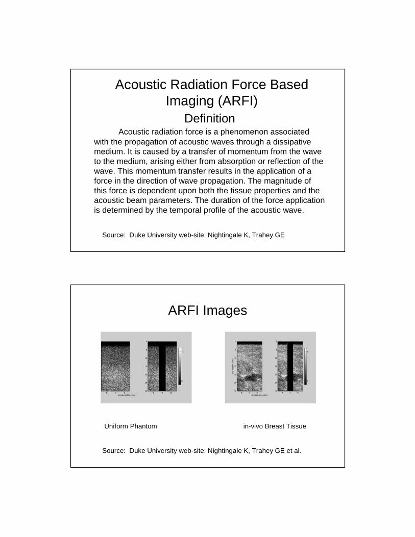
Acoustic Radiation Force Based Imaging (ARFI)
DefinitionAcoustic radiation force is a phenomenon associated
with the propagation of acoustic waves through a dissipative medium. It is caused by a transfer of momentum from the wave to the medium, arising either from absorption or reflection of the wave. This momentum transfer results in the application of a force in the direction of wave propagation. The magnitude of this force is dependent upon both the tissue properties and the acoustic beam parameters. The duration of the force application is determined by the temporal profile of the acoustic wave.
Source: Duke University web-site: Nightingale K, Trahey GE
ARFI Images
Uniform Phantom in-vivo Breast Tissue
Source: Duke University web-site: Nightingale K, Trahey GE et al.

Clinical Systems For Strain Imaging
Siemens SONOLINE® Antares Platform
nGeneric Description- 192 element beamformer- 100-base-T network port- CD writer onboard
nUltrasound Research Interface
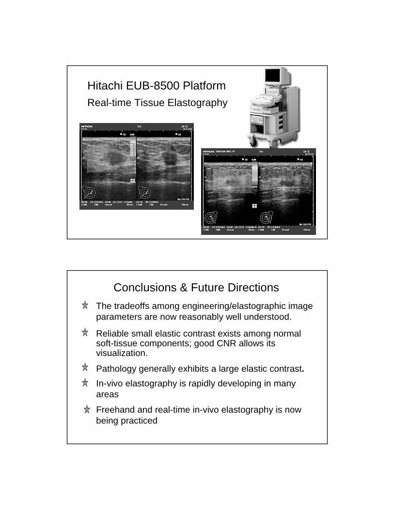
Hitachi EUB-8500 PlatformReal-time Tissue Elastography
Conclusions & Future Directions
The tradeoffs among engineering/elastographic image parameters are now reasonably well understood.
Reliable small elastic contrast exists among normal soft-tissue components; good CNR allows its visualization.
Pathology generally exhibits a large elastic contrast.
In-vivo elastography is rapidly developing in many areas
Freehand and real-time in-vivo elastography is now being practiced