Il Glaucoma OLTRE…! Tricase 16/05/14. Structure and function: not only glaucoma 2.
26
Il Glaucoma OLTRE…! Il Glaucoma OLTRE…! Tricase 16/05/14
-
Upload
daniela-hawkins -
Category
Documents
-
view
215 -
download
1
Transcript of Il Glaucoma OLTRE…! Tricase 16/05/14. Structure and function: not only glaucoma 2.

Il Glaucoma OLTRE…!Il Glaucoma OLTRE…!
Tricase 16/05/14

Structure and function: not only glaucomaStructure and function: not only glaucoma
2

Glaucoma Continuum by R. WeinrebGlaucoma Continuum by R. Weinreb
3
R. Weinreb et al. A. J. Ophthalmol 2004; 138; 458-467

4Adapted from Medeiros FA, Lisboa R, Weinreb RN, et al. A combined index of structure and function for staging glaucomatous damage. Arch Ophthalmol. 2012; 130 (5)
5000/9000 Retinal Ganglion Cells/Year5000/9000 Retinal Ganglion Cells/Year
RGCRGCss & MD CV & MD CV HFAHFA

5Felipe A. Medeiros, Linda M. Zangwill, Christopher Bowd, Kaweh Mansouri, and Robert N. Weinreb Investigative Ophthalmology & Visual Science, October 2012, Vol. 53, No. 11
-- At At early stagesearly stages of damage of damage (high RGC counts), (high RGC counts), changes changes in estimatedin estimated RGCRGC counts counts correspond to relatively correspond to relatively smaller changes in MDsmaller changes in MD (continuous line) (continuous line) andand relativelyrelatively larger changes inlarger changes in average RNFL average RNFL thickness thickness (dashed line).(dashed line).
-- At At advancedadvanced stagesstages of of damage damage (low RGC counts), (low RGC counts), changes in estimatedchanges in estimated RGCRGC counts correspond to counts correspond to relatively relatively large changes in large changes in MDMD, butbut onlyonly small small changes in average RNFLchanges in average RNFL thickness.thickness.
MD CV HFAMD CV HFA
Thickness Thickness μμm HD-OCT Cirrusm HD-OCT Cirrus
Estimated Estimated RCGRCGss count count (x10.000 cells)(x10.000 cells)
RNFLRNFL Average Thickness Average Thickness (µm)(µm)
MD MD Mean Deviation CV Mean Deviation CV (dB)(dB)
CSFICSFI Combined Combined
Structure Function Structure Function Index Index
6
Felipe A. Medeiros Renato Lisboa Robert N. Weinreb Christopher A. Girkin Jeffrey M. Liebmann Linda M. Zangwill Arch Ophthalmol. 2012
Douglas GR, Drance SM, Schulzer M. A correlation of fields and discs in open angle glaucoma. Can J. O. 1974

Inner and outer retinaInner and outer retina
RGCsRGCsMLIMLI
IPLIPLMüllerMüller CellsCells
GCCGCC

Ganglion Cell Analysis Report for Cirrus Ganglion Cell Analysis Report for Cirrus
8
5000/9000 RCG/Year
6 quadranti6 quadranti 90% RCG parve 50% in macula90% RCG parve 50% in macula

Structural and functional recovery in juvenile open angle Structural and functional recovery in juvenile open angle glaucoma after trabeculectomyglaucoma after trabeculectomy C K S Leung, J Woo, M K Tsang and K K Tse
Fundus photographs, OCT optic nerve head scans (vertical cut) and Humphrey visual field pattern deviation plots of the left eye obtained the day before trabeculectomy (a) and 1 week postoperatively (b). The red lines on the fundus photographs indicate the location of the OCT scans in the middle panel. Eye (Lond). 2006 Jan;20(1):132-4Eye (Lond). 2006 Jan;20(1):132-4 9
RREE
ccoovveerryy
??
RREEVVEERRSSIIBBLLEE
??
C/D = 0,726C/D = 0,726
C/D = 0,089C/D = 0,089

Preoperative and postoperative optic nerve images of the left eye of a 18-year old (A, B) and an 81-year old woman (C, D), where the IOPs were lowered from 25 to 6 mmHg and from 20 to 7 mmHg, respectively. The reversal of the backward bowing of the lamina cribrosa is clearly noticeable. Images were taken by enhanced depth imaging SD-OCT. Dashed lines indicate the plane of Bruch’s membrane opening BMO. (da Weinreb et al. 2012)
18-year old Pre 18-Year old Post
81-year old Pre 81-year old Post

IOP Elevation Reduces the Waviness of the Load IOP Elevation Reduces the Waviness of the Load Bearing Collagen Fibers in the Lamina Cribrosa Bearing Collagen Fibers in the Lamina Cribrosa
Ian A. Sigal et alIan A. Sigal et al. . ARVO 2013 Annual Meeting AbstractsARVO 2013 Annual Meeting Abstracts
11
Collagen fibers with and without crimpCollagen fibers with and without crimp

Finite Element Modeling of the Lamina Cribrosa of the Finite Element Modeling of the Lamina Cribrosa of the Optic Nerve Head in GlaucomaOptic Nerve Head in Glaucoma
Devers Eye Institute / National Institute of Health Optic Nerve Head Research Laboratory directed by dr. Claude Burgoyne (Portland Oregon)
12Struttura FrattaleStruttura Frattale

13
Racial Differences in Mechanical Strain in the Posterior Human ScleraRacial Differences in Mechanical Strain in the Posterior Human ScleraM. A. Fazio 1-2, R. Grytz 1, L. Bruno 2, J. S. Morris 3, C. A. Girkin 2, J. Crawford C. Downs 2.
1 Ophthalmology, The University of Alabama in Birmingham, Birmingham, AL;2 Mechanical Engineering, University of Calabria, Cosenza, Italy;
3 Department of Biostatistics, The University of Texas MD Anderson Cancer Center, Houston, TX. ARVO 2013 Annual Meeting Abstracts
Baltimore Eye Survey : African 4 times higher risk Caucasian

14
• Zeiss Cirrus & Humphrey con FORUMFORUM
• Heidelberg Spectralis & HEP con HEYEXHEYEX
• Optovue & Octopus Bundle Haag-Streit Bundle Haag-Streit
* *
Piattaforme Multimediali & Combo ReportPiattaforme Multimediali & Combo Report

Map representing the relationshiprelationship betweenbetween Standard Automated Perimetry Standard Automated Perimetry visual field sectors and sections of the peripapillary OCT scan circleOCT scan circle. This map is based on the work of work of Garway-HeathGarway-Heath et al et al and shows the correspondence between areas of the between areas of the visual field and peripapillary retinal nerve fiber layer visual field and peripapillary retinal nerve fiber layer due to the anatomical configuration of the retinal nerve fiber bundles.First Release : Presented in part at the Glaucoma Society (UK & Eire) Annual Meeting, London, England, November
1998
Garway-Heath, Moorfields Eye Hospital London

Six corresponding regions of neuroretinal rim area (A), peripapillary retinal nerve fiber layer (B), and visual field (C), used to measure the structure–function relationship (based on structure–function map introduced by Garway-Heath et al.) Nilforushan N et al. Invest Ophthalmol Vis Sci. 2012 May
16
ST + SN : 80°+ST + SN : 80°+
Nasal : 110° +Nasal : 110° +
IN + IT : 80° +IN + IT : 80° +
Temporal : 90° =Temporal : 90° =
A = Rim AreaA = Rim Area
B = RNFLB = RNFL
C = CV C = CV
Rim / RNFL: 360°Rim / RNFL: 360°

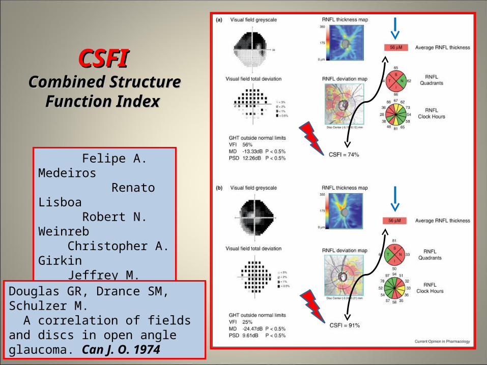
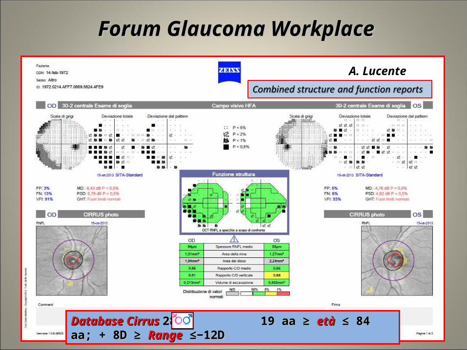
Combined structure and function reportsCombined structure and function reports
A. Lucente
Database HFA Database HFA : 422 18aa ≥ : 422 18aa ≥ età età ≤ 89 aa; + 5D ≥ ≤ 89 aa; + 5D ≥ RangeRange ≤ - 5D ≤ - 5D
Forum Glaucoma Workplace Forum Glaucoma Workplace
18
A. Lucente
Forum Glaucoma WorkplaceForum Glaucoma Workplace
Database Cirrus Database Cirrus 284 19 aa ≥ 284 19 aa ≥ etàetà ≤ 84 aa; + 8D ≥ ≤ 84 aa; + 8D ≥ RangeRange ≤−12D ≤−12D
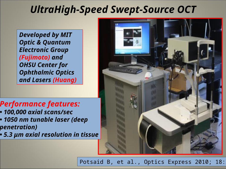
UltraHigh-Speed Swept-Source OCT
19
Developed by MIT Optic & QuantumElectronic Group (Fujimoto) andOHSU Center for Ophthalmic Opticsand Lasers (Huang)
Performance features:• 100,000 axial scans/sec• 1050 nm tunable laser (deeppenetration)• 5.3 μm axial resolution in tissue
Potsaid B, et al., Optics Express 2010; 18:20029

dr Amedeo Lucente
Immagini ad alta definizione: HD 5 Line Raster OS

21

SS-OCT Zeiss SS-OCT Zeiss Key factors: lamina cribrosa Key factors: lamina cribrosa
22

SSADASSADA SSplit-SSpectrum AAmplitude DDecorrelation AAlgorithm
23
Full Spectrum
Split Spectrum
Less Background Noise
Clear vessels
Jia Y, Tan O, Tokayer J, Potsaid B, Wang Y, Liu JJ, Kraus MF, Subhash H, Fujimoto JG, Hornegger J, Huang D. Split-spectrum amplitude-decorrelation angiography with optical coherence tomography. Optics Express 2012; 20:4710
Improves signal to noise ratio of flow detection
FAZ

24www.amedeolucente.it

25
« When you can measure what you speaking about and express it in numbers you know something about it; but when you cannot express it in numbers, your knowledge is of a meagre and unsatisfactory kind»
«Possiamo conoscere qualcosa dell’oggetto di cui stiamo parlando solo se possiamo eseguirvi misurazioni, per descriverlo mediante numeri;altrimenti la nostra conoscenza è scarsa e insoddisfacente»
Lord William Thomson Kelvin Lord William Thomson Kelvin (1824-1907(1824-1907))

Thanks for your Thanks for your attentionattention
26