GLAUCOMA NUTS AND BOLTS & SECONDARY GLAUCOMAS … · GLAUCOMA NUTS AND BOLTS & SECONDARY GLAUCOMAS....
119
Jason Ahee, MD Marcos Reyes, MD Dixie Ophthalmic Specialists GLAUCOMA NUTS AND BOLTS & SECONDARY GLAUCOMAS
Transcript of GLAUCOMA NUTS AND BOLTS & SECONDARY GLAUCOMAS … · GLAUCOMA NUTS AND BOLTS & SECONDARY GLAUCOMAS....

Jason Ahee, MD
Marcos Reyes, MD
Dixie Ophthalmic
Specialists
GLAUCOMA
NUTS AND BOLTS &
SECONDARY
GLAUCOMAS
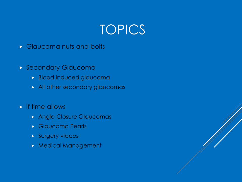
TOPICS Glaucoma nuts and bolts
Secondary Glaucoma
Blood induced glaucoma
All other secondary glaucomas
If time allows
Angle Closure Glaucomas
Glaucoma Pearls
Surgery videos
Medical Management

DISCLOSURES
None!
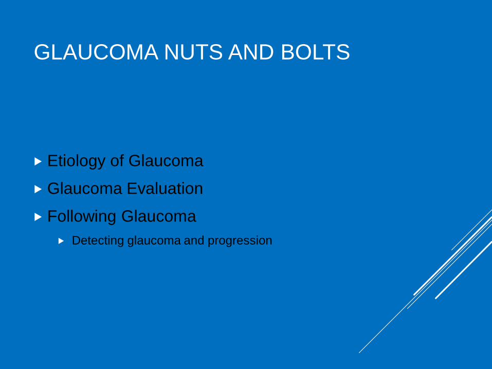
GLAUCOMA NUTS AND BOLTS
Etiology of Glaucoma
Glaucoma Evaluation
Following Glaucoma
Detecting glaucoma and progression
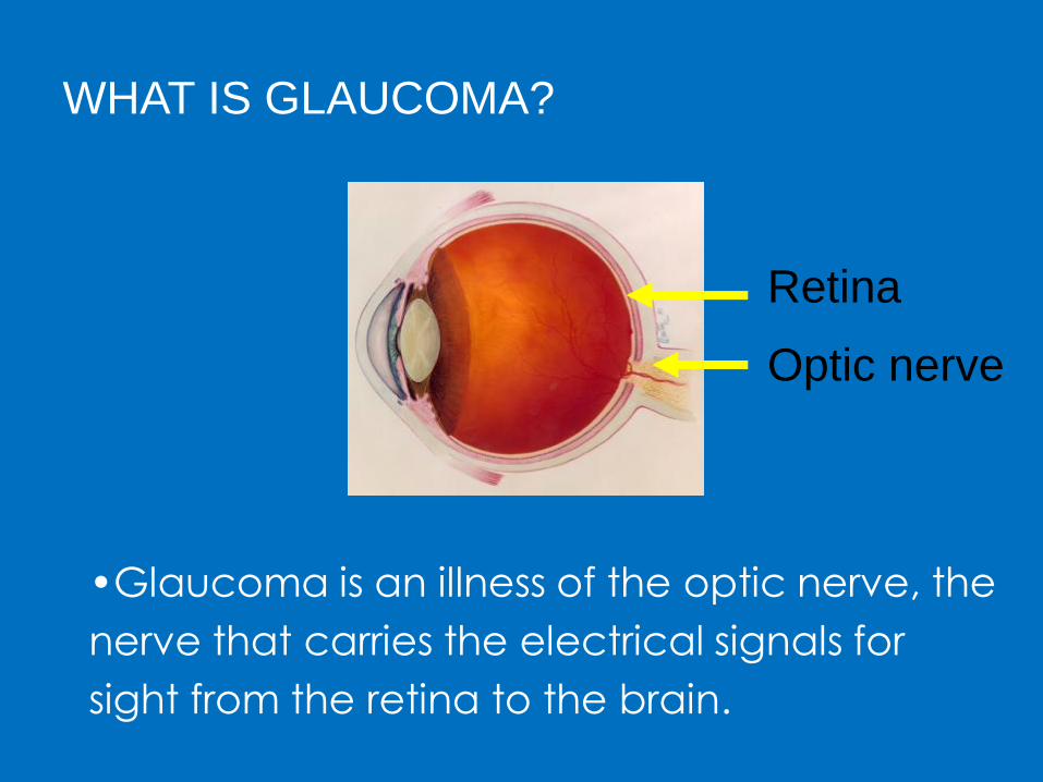
WHAT IS GLAUCOMA?
•Glaucoma is an illness of the optic nerve, the
nerve that carries the electrical signals for
sight from the retina to the brain.
Retina
Optic nerve
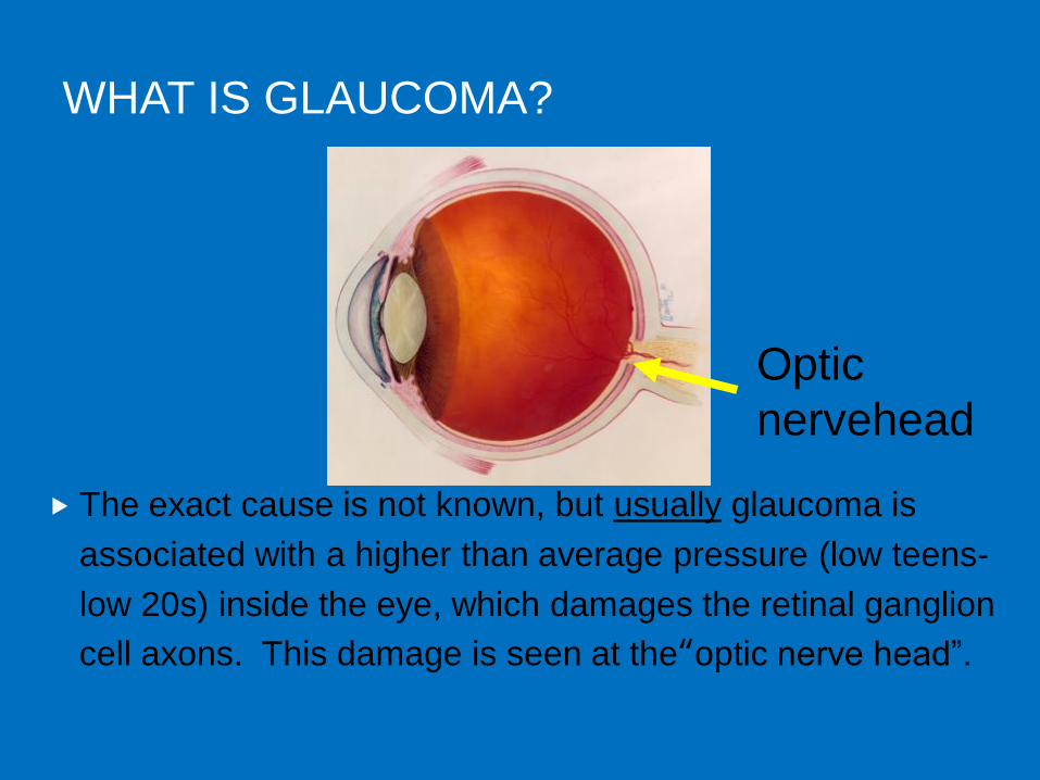
WHAT IS GLAUCOMA?
The exact cause is not known, but usually glaucoma is
associated with a higher than average pressure (low teens-
low 20s) inside the eye, which damages the retinal ganglion
cell axons. This damage is seen at the“optic nerve head‖.
Optic
nervehead

WHAT DAMAGES THE OPTIC NERVE IN
GLAUCOMA?
Pattern of Loss tells us
the site of damage is at
the optic nervehead
Likely mechanism in
high pressure glaucoma
is a pressure-dependent
blockade of axoplasmic
transport
Douglas Anderson 1974 Blockade of material (white) at the optic nerve
head in a monkey with high eye pressure

WHAT IS GLAUCOMA?
There are many types of glaucoma, each named for
special features.
POAG or Primary Open Angle Glaucoma is a diagnosis of exclusion
(i.e. look for other causes before labeling it POAG.
Most are chronic conditions which can be treated but not
cured, by lowering the eye pressure to prevent additional
loss of sight.

GLAUCOMA EVALUATION
Current Medications
Compliance
Vision
Pressure ( Applanation is gold standard )
Pachymetry
Gonioscopy
Anterior segment exam
Optic Nerve cupping – structure
Visual Field ( with BCVA ) - function
Imaging (OCT or equivalent) - structure

GLAUCOMA EVALUATION
Gonio LOTS of normal angles, not just narrow ones.
You need to know what does NORMAL looks like
Look for narrow angles.
―You can’t see what you’re not
looking for‖
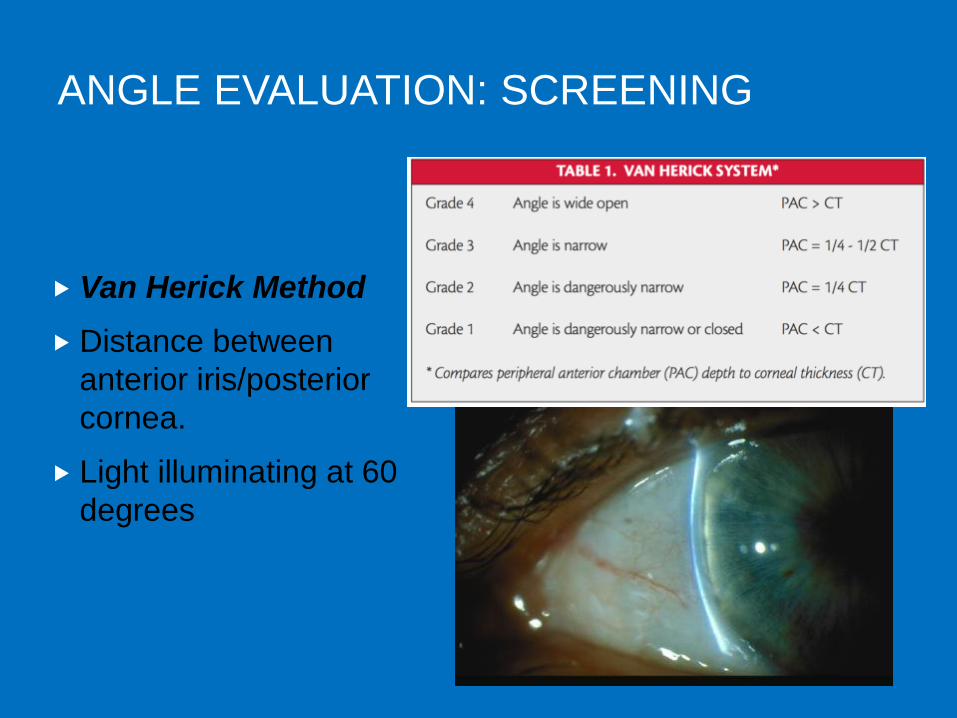
ANGLE EVALUATION: SCREENING
Van Herick Method
Distance between
anterior iris/posterior
cornea.
Light illuminating at 60
degrees

ANGLE EVALUATION: GONIOSCOPY
Gonioscopy
Direct (surgical):
Koeppe, Barkan,
Wurst, Richardson
Indirect: Zeiss,
Goldman, Sussman
Total internal reflection
– makes direct evaluation impossible
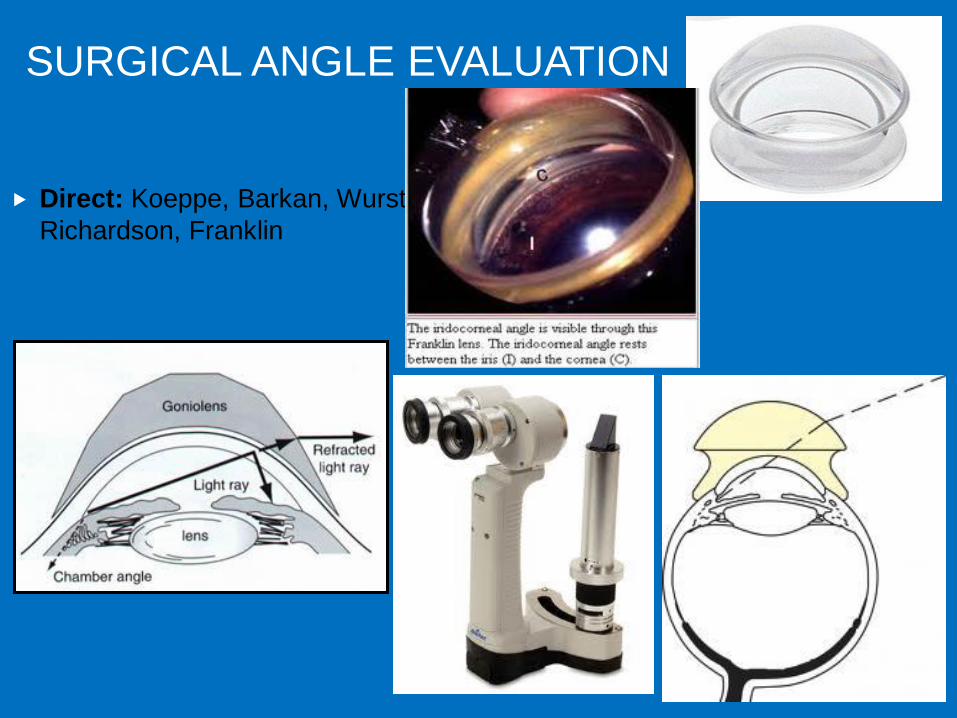
SURGICAL ANGLE EVALUATION
Direct: Koeppe, Barkan, Wurst,
Richardson, Franklin
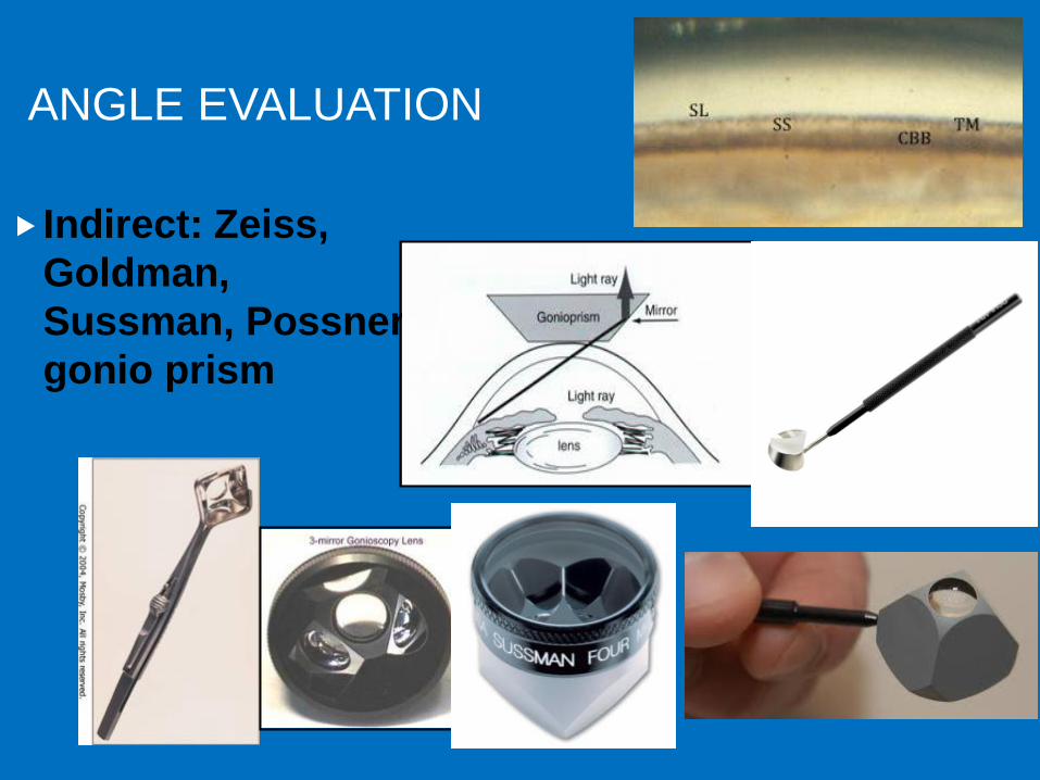
ANGLE EVALUATION
Indirect: Zeiss,
Goldman,
Sussman, Possner,
gonio prism

GOLDMANN THREE MIRROR LENS-COUPLING AGENT REQUIRED BUT BEST VIEW OF ANGLE

ANGLE EVALUATION:
SHAFFER SYSTEM
Shaffer System
Angle between iris/TM
Visible angle structures
TM pigmentation

Angle evaluation:
Shaffer System

ANGLE EVALUATION:
SCHEIE SYSTEM 1957

ANGLE EVALUATION:
SCHEIE SYSTEM 1957

ANGLE EVALUATION:
SPAETH SYSTEM
Spaeth System
Iris/cornea angle
Configuration
Iris insertion
pigmentation

ANGLE EVALUATION:
SPAETH SYSTEM

ANGULAR APPROACH
(NOT IRIS RECESS)
Assess by means of tangential lines
One line is tangential to the inner surface of the trabecular meshwork
The other line is tangential to the middle third of the anterior iris surface.
The angle formed by these two lines defines the angular approach 0-50 degrees
This angle does not identify the angle of the iris recess itself, but rather the angular approach of the iris to this recess.

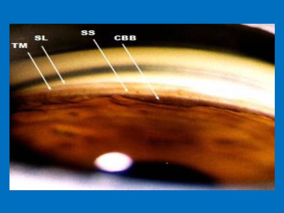
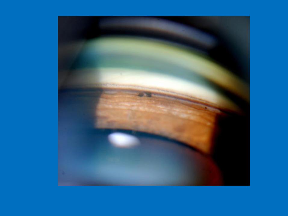
ANGLE EVALUATION:
STATE WHAT YOU SEE
SL
ATM
PTM
SS
CB
Iris config
Pigment


GONIOSCOPY
PEARLS/PITFALLS
Examine the least narrow or the eye without pathology first.
Why? To know what normal is!
Start with the Inferior Angle (superior mirror)
The most open - best way to see all structures
Most pigmented TM - easily identified structures

GONIOSCOPY
PEARLS/PITFALLS
Iris Contour
Steep insertion (aka narrow approach)
○ Have the patient look into the examining mirror or tip the lens towards the angle being examined to look over the hill and into the angle.
○ In general, if one can readily see over the hill and into the angle without indenting, the angle is not occludable. But if unsure. LPI or re-gonio in the future after discussing angle closure warnings. Which are…?
Regular
Concave – Queer formation – not politically correct.
Corneal Wedge technique helps to identify Schwalbe’s line, in lightly pigmented eyes, young people
http://www.gonioscopy.org/index.php?option=com_k2&view=item&layout=item&id=99&Itemid=665

GLAUCOMA EVALUATION
Structure – objective measurement
-Optic nerve head (cupping, thinning, notching)
- follow the vessel curve, NOT the
color of the rim.
-Nerve fiber layer
Goal!
Differentiate
structural changes secondary
to glaucoma FROM retinal and
other optic nerve pathology
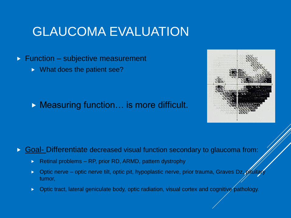
GLAUCOMA EVALUATION
Function – subjective measurement
What does the patient see?
Measuring function… is more difficult.
Goal- Differentiate decreased visual function secondary to glaucoma from:
Retinal problems – RP, prior RD, ARMD, pattern dystrophy
Optic nerve – optic nerve tilt, optic pit, hypoplastic nerve, prior trauma, Graves Dz, pituitary
tumor,
Optic tract, lateral geniculate body, optic radiation, visual cortex and cognitive pathology.

GLAUCOMA EVALUATION –
FOLLOW UP
Which to follow…Structure or Function?
or Both?

IN ESTABLISHED GLAUCOMA WHICH
DEMONSTRATES PROGRESSION FIRST, LOSS OF
STRUCTURE OR OF LOSS OF FUNCTION?
Artes PH, Chauhan BC. Longitudinal changes in the visual field and optic disc in glaucoma. Prog Retin Eye Res. 2005;24:333-54 (May, 2005).
Why? Possibilities…
Structural changes 1st but no functional changes – perhaps in the nerve fiber
layer ‘area of loss’ there are ‘redundant cells’ covering the same area
allowing function in that area to continue.
Functional changes 1st but no structural changes – perhaps the nerves are
‘malfunctioning’ prior to actually dying and undergoing apoptosis. Maybe the
variability in HVF is not only patient performance. Maybe it is the intermitent
functioning of the cells.

EVALUATING STRUCTURE Obsolete way:
Optic disc drawings—inaccurate, not done 50% of time
Optic disc photography—very qualitative, only semi-quantitative
Optical Coherence Tomography (laser interferometer)
true structure - 5 micron resolution (Cirrus OCT)
(Technique offers the huge advantage that it can also
be used to monitor macular disease)
GDx (Laser Polarimetry)
optical rotation, generally proportional to structure
Heidelberg Retinal Tomography (confocal reflectance from surface),
measures shape rather than tissue

EVALUATING FUNCTION
Obsolete ways:
Tangent screen
Goldmann Kinetic Perimetry
Standard Automated Perimetry
Humphery Visual Field
Less Common ways:
Frequency Doubling Perimetry (FDP)
Short-wavelength Perimetry (SWAP)
Pattern Electro Retinogram (ERG)
High Pass Resolution Perimetry (HRP)
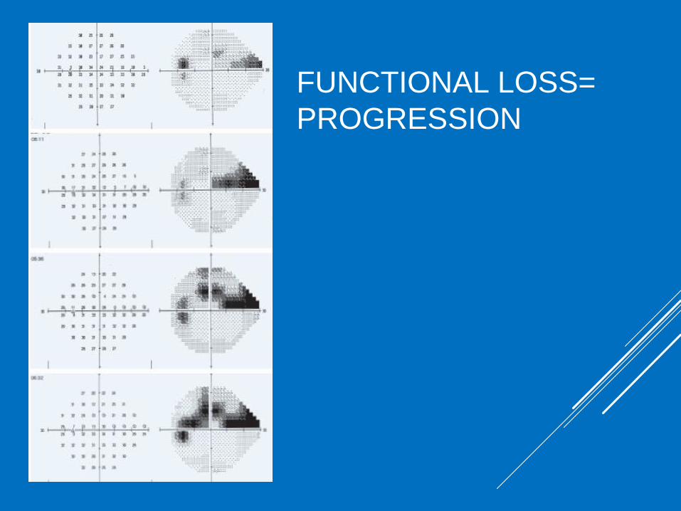
FUNCTIONAL LOSS=
PROGRESSION
FOLLOWING GLAUCOMA Central questions:
Is the patient compliant with current regimen?
Is the glaucoma stable?
Is more therapy needed?
Central Answers:
Compliance can’t be measured but we know its BAD!!
IOP increasing – make sure to use goldmann applanation
VF worsening – repeat to verify loss versus bad test day.
Optic nerve worsening
OCT – repeat because there is some inter-visit variability
Photos – OCT attempts to measure the ONH, cupping, etc. but it can interpret to data wrong, especially in abnormal nerves.

Secondary Glaucomas
• Blood induced glaucomas
• Pseudoexfoliation
• Pigmentary
• Phacolytic
• Lens Particle
• Intraocular Tumors
• Fuchs Heterochromic iridocyclitis
• Possner-Schlazmanns Syndrome
• Uvietic
• Elevated EVP
• Trauma
• Neovascular
• Corneal endothelial
• Angle closure glaucomas

Blood Induced
Secondary Glaucomas

Blunt Trauma
• 70% less than 20 years old, Male 3:1
• Sports injuries up to 60%
• Increased IOP,secondary hemorrhages 4-35%
• Corneal blood staining 2%
• Poor visual acuity with rebleed and increased
IOP

Blunt Trauma Associations
• Hyphema
• Iris sphincter tear
• Iridodialysis
• Cyclodialysis
• Trabecular Tear
• Inflammation
• Zonular rupture
• Ruptured Globe


Hyphema occurs from injury to vessels of
peripheral iris or anterior ciliary body
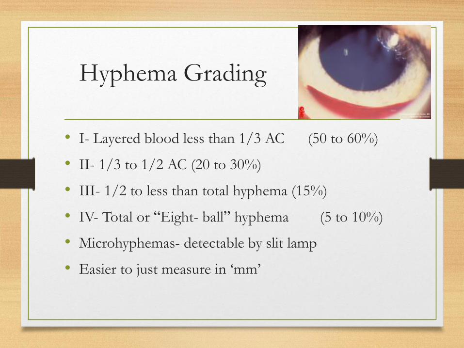
Hyphema Grading
• I- Layered blood less than 1/3 AC (50 to 60%)
• II- 1/3 to 1/2 AC (20 to 30%)
• III- 1/2 to less than total hyphema (15%)
• IV- Total or “Eight- ball” hyphema (5 to 10%)
• Microhyphemas- detectable by slit lamp
• Easier to just measure in ‘mm’

Complications- Secondary Glaucoma
• Leads to corneal blood staining
• Optic Atrophy
• Mean duration- 6 days
• Greater than 24 mmHg in 32% of patients
• Higher IOP and longer duration with total or near
total hyphemas

Glaucoma- Mechanism
• Mechanical obstruction of TM with RBCs and fibrin
• +/- pupillary block from clot
• Long term- damage to TM/ Outflow system

Corneal Blood Staining
• Stromal infiltration with hemosiderin
• Tiny yellowish granules appear in posterior 1/3 of corneal stroma (Precedes gross staining by 24-36 hrs)
• Usually result of prolonged IOP elevation
• Clearing occurs peripherally- Amblyopia

Secondary Hemorrhage
• Clot lysis and retraction 2 to 5 days after injury
• Maximal rebleed in this period
• Highest incidence in grade III and IV, children less than age 6, blacks ( 24% vs 4% for whites)
• May be more severe than initial bleed
• Incidence of rebleed 4 to 35%
• Worse visual prognosis

Hyphema Complications
• Angle recession 30 to 85%- Gonioscopy
• 6-10% angle recession glaucoma
• Posterior synechiae
• Peripheral Anterior synechiae
• Optic atrophy
• Traumatic mydriasis
Hyphema Management
• Most can be managed as outpatients
• Consider admission in high risk cases or
uncooperative patients
Patient Orders
• Quiet activities, may walk in hall
• Unilateral shield, no patch
• No reading, TV okay
• Elevate HOB 30 degrees
• Tylenol prn pain, no Aspirin
• Atropine 1% tid, +/-Pred Forte qid
• Sickle cell test if Black or Mediterranean
• If hospitalized consider Amicar – fibrinolysis inhibitor (very controversial)

Glaucoma Management
• Non sickle cell disease/trait:
• Beta Blockers Alpha agonists Topical CAIs Oral CAIs IV Mannitol Surgery
• Avoid miotics – increased inflammation

Glaucoma Management
• Sickle cell disease or trait:
• Start with beta blockers
• Use caution with all other agents
• Topical and oral CAIs may reduce AC pH and induce
sickling
• Alpha agonists may affect (constrict) iris vasculature
• Consider surgery earlier

After Discharge
• 3 to 4 weeks
• Gonioscopy
• Retinal exam
• Yearly follow up due to late glaucoma
• Glaucoma more frequent with > 270 degree angle
recession and is uncommon with less than 180
degrees
Possible Surgical Criteria ?
(controversial)
• IOP>60mmHg x 48 hrs
• IOP>50mmHg x 3 days
• IOP>35mmHG x 7 days
• IOP>24 x 6 days with >50% hyphema
• Total hyphema x 4 days
• Sickle trait or disease IOP >35 for >24 hours
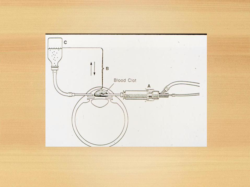
Surgical Techniques
• Limbal paracentesis
• Limbal irrigation/aspiration
• Clot extraction
• Vitrector methods –
• Avoid instrumentation in AC if possible


Sickle Cell- Vicious Cycles
• Increased IOP --> decreased perfusion of nerve and retina --> sludging/sickling --> infarction of neuronal tissue
• Decreased perfusion of anterior segment --> hypoxia in AC --> further sickling --> TM obstruction --> increased IOP
• Hyperosmotics --> hemoconcentration --> more sludging/sickling
• Diamox --> decreased pH --> more sickling

Hyphema after surgery
• Early- Usually clears quickly
• Late- Look for vessels on inner aspect of incision
Ghost Cell
Glaucoma

Etiology
• Obstruction to aqueous outflow secondary to
degenerated RBCs
• Occurs weeks to months after hemorrhage

Clinical
• Clearing hyphema,
increasing IOP 40 to
70mmHg
• Khaki colored cells in
aqueous
• Pseudohypopyon, khaki
colored TM

Histology
• Degenerating RBCs with
Heinz bodies
• Aggregates of
hemoglobin on vitreous
strands
• Ghost cells are less
pliable than fresh RBCs
Differential Diagnosis
• Uveitis
• Endophthalmitis
• No KPs seen with ghost cells
Management
• Medical
• AC washout- may need to be repeated
• Vitrectomy – these cells usually come from old
vitreous hemorrhages

Hemolytic Glaucoma
• Clinical- Similar to Ghost cell but with reddish-
brown appearance
• Mechanism- Free hemoglobin, RBCs and
macrophages filled with debris that blocks the TM

Hemosiderotic Glaucoma
• Clinical- prolonged history of recurrent intraocular
hemorrhage
• Slow onset- weeks to years later
• Associated with retinal degeneration, cataract,
heterochromia, iron staining of cornea, ciliary body
degeneration
Histology
• Breakdown of hemoglobin to globin, bilirubin and
iron
• Iron causes degenerative changes in TM

Spontaneous Hemorrhage
• Intraocular tumors
• JXG
• Melanoma
• Retinoblastoma
• Other
• Neovascularization of
the iris
• Blood dyscrasias

Secondary Open-Angle
Glaucoma
Pseudoexfoliation
Pigment dispersion
Lens induced
Tumor related
Uveitis
Elevated episcleral venous pressure

Secondary OAG
(Pseudoexfoliation)
Vignette: 50 y/o white (Scandanavian) female referred
optometry for asymmetric IOP or c/d OR 50 y/o with
dense 4+ cataract, small pupil, phacodonesis, and end-
stage glaucomatous optic neuropathy referred by your
previous resident colleague for cataract extraction

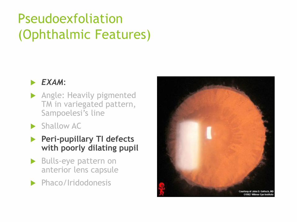
Pseudoexfoliation
(Ophthalmic Features)
EXAM:
Angle: Heavily pigmented TM in variegated pattern, Sampoelesi’s line
Shallow AC
Peri-pupillary TI defects with poorly dilating pupil
Bulls-eye pattern on anterior lens capsule
Phaco/Iridodonesis

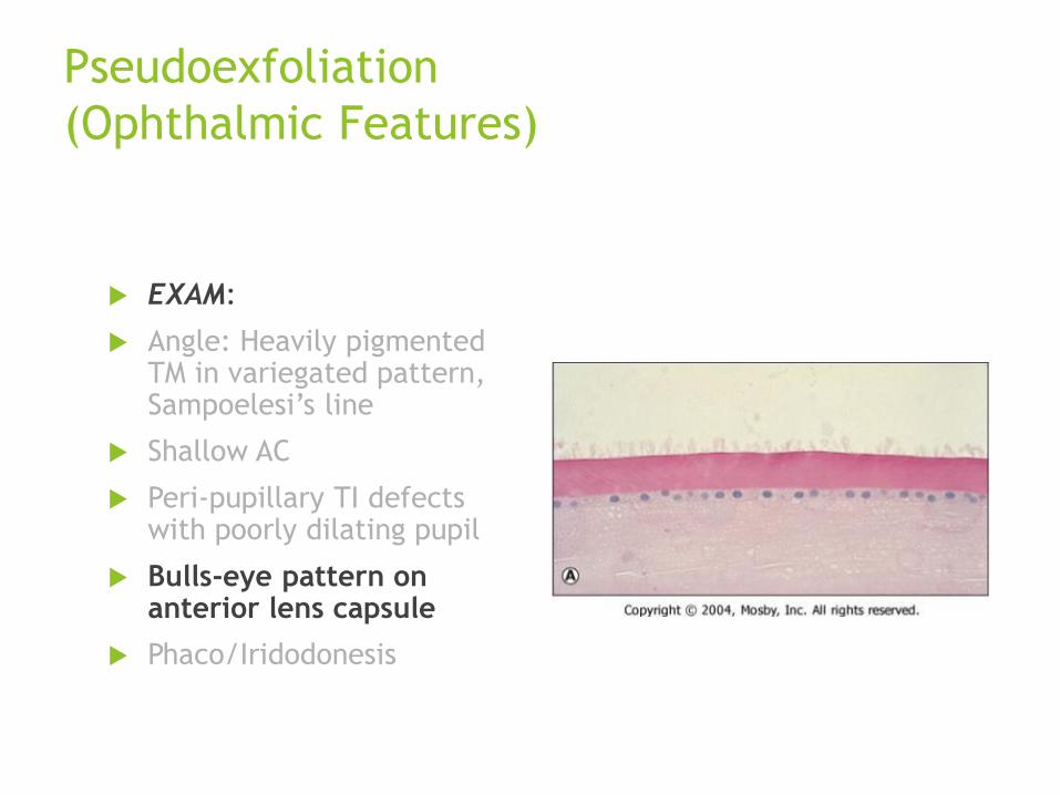
Pseudoexfoliation
(Ophthalmic Features)
EXAM:
Angle: Heavily pigmented TM in variegated pattern, Sampoelesi’s line
Shallow AC
Peri-pupillary TI defects with poorly dilating pupil
Bulls-eye pattern on anterior lens capsule
Phaco/Iridodonesis
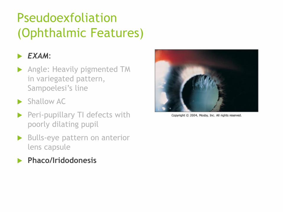
Pseudoexfoliation
(Ophthalmic Features)
EXAM:
Angle: Heavily pigmented TM in variegated pattern, Sampoelesi’s line
Shallow AC
Peri-pupillary TI defects with poorly dilating pupil
Bulls-eye pattern on anterior lens capsule
Phaco/Iridodonesis

Pseudoexfoliation
(Ophthalmic Features)
EXAM:
Angle: Heavily pigmented TM in variegated pattern, Sampoelesi’s line
Shallow AC
Peri-pupillary TI defects with poorly dilating pupil
Bulls-eye pattern on anterior lens capsule
Phaco/Iridodonesis
Pseudoexfoliation
(Ophthalmic Features)
EXAM:
Angle: Heavily pigmented TM in variegated pattern, Sampoelesi’s line
Shallow AC
Peri-pupillary TI defects with poorly dilating pupil
Bulls-eye pattern on anterior lens capsule
Phaco/Iridodonesis
Pseudoexfoliation
(Ophthalmic Features)
EXAM:
Angle: Heavily pigmented TM
in variegated pattern,
Sampoelesi’s line
Shallow AC
Peri-pupillary TI defects with
poorly dilating pupil
Bulls-eye pattern on anterior
lens capsule
Phaco/Iridodonesis

Pseudoexfoliation
(Ophthalmic Features)
Cause: Distinctive fibrillar material deposition throught body (angleTM damage/obstruction, endothelium, lens capsule, ZONULES)
Common in: Strongly age related (>50 yrs), Scandanavian, unilateral or bilateral (asymmetric),
Treatment:
Higher IOP than POAG, worse pronosis
ALT very effective, but short lived (PXE>PDS>POAG>NTG)
May need TAE (increased inflammation)
Higher rate of vitreous loss with cataract surgery due to zonular weakness

Laser Trabeculoplasty
Argon Laser Trabeculoplasty
Placement of laser burns at junction of pigmented/non-pigmented TM
Effective at reducing IOP in PXE, PDS, POAG, NTG
Contraindicated in inflammatory glaucoma, angle recession, lack of effect in fellow eye
GLT: As affective as Timolol at 2 years
Selective Laser Trabeculoplasty
Non-destructive laser procedure
Energy directed at pigmented cells of TM
Less energy, broader field of treatment
SLT ALT
Secondary OAG
(Pigment Dispersion Syndrome)
Vignette: 32 year-old ophthalmology residents presents
c/o mild eye pain in OD during exercise.

Pigment Dispersion Syndrome
(Ophthalmic Features)
EXAM:
Krukenberg spindles
Heavily pigmented TM (Homogenous)
Mid-peripheral iris TI defects
Anterior/Posterior lens deposits

Pigment Dispersion Syndrome
(Ophthalmic Features)
EXAM:
Krukenberg spindles
Heavily pigmented TM (Homogenous)
Mid-peripheral iris TI
defects
Anterior/Posterior lens
deposits

Pigment Dispersion Syndrome
(Ophthalmic Features)
EXAM:
Krukenberg spindles
Heavily pigmented TM (Homogenous)
Mid-peripheral iris TI defects
Anterior/Posterior lens deposits

Pigment Dispersion Syndrome
(Ophthalmic Features)
EXAM:
Krukenberg spindles
Heavily pigmented TM (Homogenous)
Mid-peripheral iris TI defects
Anterior/Posterior lens deposits
Pigment Dispersion Syndrome
(Ophthalmic Features)
Cause: Liberation of iris pigment due to a “reverse
pupillary block”
Occurs in: Young, myopic, type A males
IOP may increase with exercise
Treatment:
LPI to eliminate iris/zonular touch
May need chronic IOP meds/TAE (20-50% progress to
pigmentary glaucoma)

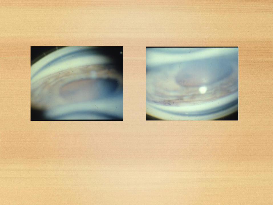
Pigment Dispersion Syndrome
(Ophthalmic Features)
Ultrasound biomicroscopy pre/post LPI

Secondary OAG
(Lens Induced—Phacolytic)
Vignette: 65 y/o with h/o poor Va OD x 5 years
(painless, progressive) presents with acute onset of
decreased vision, pain, photophobia.
Secondary OAG
(Lens Induced—Phacolytic)
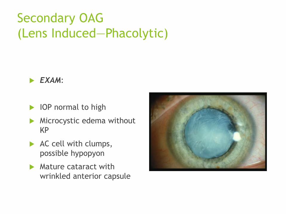
EXAM:
IOP normal to high
Microcystic edema without
KP
AC cell with clumps,
possible hypopyon
Mature cataract with
wrinkled anterior capsule

Secondary OAG
(Lens Induced—Phacolytic)
EXAM:
IOP normal to high
Microcystic edema without
KP
AC cell with clumps,
possible hypopyon
Mature cataract with
wrinkled anterior capsule

Secondary OAG
(Lens Induced—Phacolytic)
Cause:
Leakage of HMW lens protein through capsule of
hypermature cataract
Engorged Mø engulfed w/ HMWP clog TM
Treatment:
Treat with IOP agents, topical steroids
Definitive treatment cataract extraction

Secondary OAG
(Lens-Particle Glaucoma)
Vignette: 45 y/o s/p phaco with IOL POW#1 with c/o
pain, photophobia, headache, nausea, vomiting, and
decreased vision. Cataract extraction was uneventful
but a significant amount of sub-incisional cortex was
left in capsular bag.

Lens Particle Glaucoma
(Ophthalmic Features)
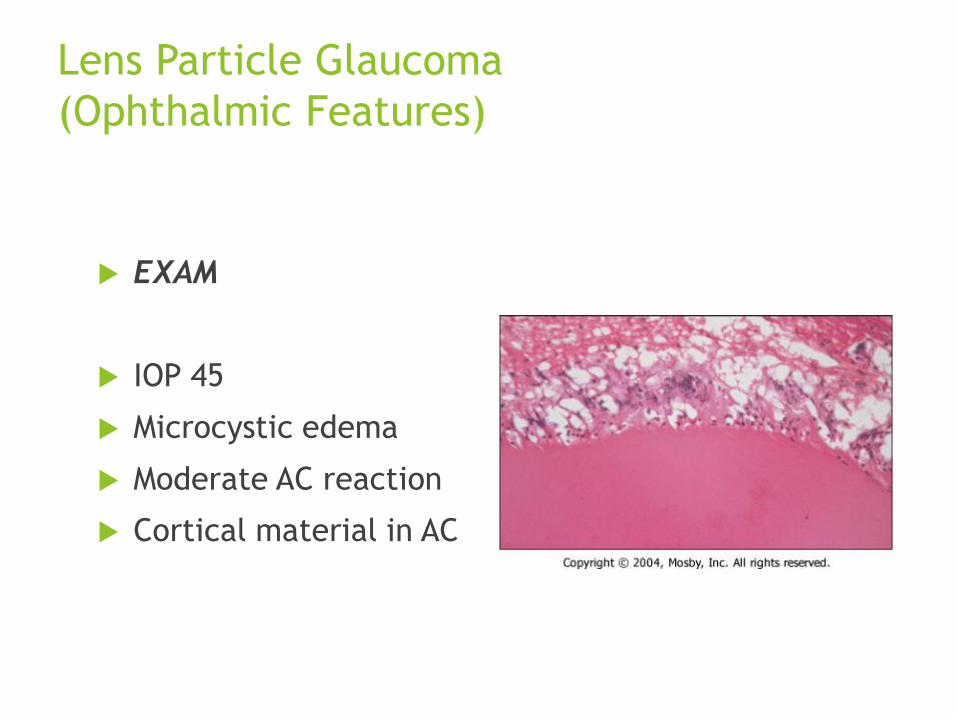
EXAM
IOP 45
Microcystic edema
Moderate AC reaction
Cortical material in AC
Lens Particle Glaucoma
(Ophthalmic Features)
EXAM
IOP 45
Microcystic edema
Moderate AC reaction
Cortical material in AC

Lens Particle Glaucoma
(Ophthalmic Features)
Cause:
Incomplete removal of lens material
Obstruction of TM by lens material (IOP rise dependent on TM function)
Occurs in: Post cataract patients following CE, capsulotomy, trauma(weeks to years).
Treatment:
IOP control (topical/systemic), topical steroids initially
AC washout (+/- tube shunt)

Lens Particle Glaucoma
(Ophthalmic Features)
Phacoanaphylaxis rare
variant of lens induced
uveitis/glaucoma. Caused
by sensitization of lens
protein after
surgery/trauma with
secondary granulomatous
uveitis (has KP)

Secondary OAC
(Intraocular tumors)
Causes of glaucoma:
Direct invasion of angle—most common
Deposition of tumor cells/debris/WBC on TM
Intraocular hemorrhage
Angle closure (CB rotation, Iris neovascularization
Adult tumors: Melanoma, metastatic, lymphoma,
leukemia
Childhood tumors: Rb, JXG, Dyktioma

Secondary OAG
(Uveitic Glaucoma--Fuchs)
Vignette: 45 year-old male referred to you by
optometry for evaluation of cataract OS. While taking a
history the patient reports some doc treated him in the
past with a milky colored drop off and on for a couple of
years. He denies ever having significant pain,
photophobia, but does report he now has difficult
driving at night due to glare.
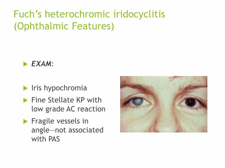
Fuch’s heterochromic iridocyclitis
(Ophthalmic Features)
EXAM:
Iris hypochromia
Fine Stellate KP with
low grade AC reaction
Fragile vessels in
angle—not associated
with PAS

Fuch’s heterochromic iridocyclitis
(Ophthalmic Features)
EXAM:
Iris hypochromia
Fine Stellate KP with
low grade AC reaction
Fragile vessels in
angle—not associated
with PAS

Fuch’s heterochromic iridocyclitis
(Ophthalmic Features)
Cause: Idiopathic, unilateral, chronic iritis with low-grade AC reaction with small, stellate KP
Occurs in: Middle aged men
Treatment:
Inflamation: Treat cell not flare (less response to PF)
IOP control:
Possible avoidance of prostaglandin analogs due to association with uveitis. (Note: brimonidine tartate has been reported to induce granulomatous uveitis in the literature)
OAG occurs in ~15% of patients—IOP does not correspond to degree of cell.
Cataract: Eye needs to be quiet x 3 months for CE
Secondary OAG
(Uveitic Glaucoma—
Glaucomaocyclitis crisis)
Vignette: 45 year-old man with h/o recurrent episodes
of unilateral blurred vision with mild eye pain presents
c/o similar symptoms. He has never sought treatment
for previous episodes due to funding issues. Now
insured, he seeks your medical attention immediately
with this current episode.

Glaucomatocyclitic crisis
(Ophthalmic Features)
EXAM:
IOP: 50 OD, 14 OS
Iritis with fine KP OD
Open angle with KP on
TM OD
C/D 0.9 AND 0.25

Glaucomatocyclitic crisis
(Ophthalmic Features)
Cause:
Idiopathic (possibly prostalandin-mediated).
Iritis generally resolved without treatment and IOP normalizes inbetween abouts.
May develop OAG after increasing number of attacks
Common in: Middle-aged
Treatment: Treat active Dz. May need chronic IOP control

Uveitic Glaucoma
(Iridocyclitis)
During acute iridocyclitis IOP may be low due ciliary body shut-down
IOP may be high if trabecular function reduced (HZV associated with high IOP)
Angle may become occluded by fibirn, debris, white cells
Angle closure possible from PAS or seclusio pupillae
Treat with topical steroids and aqueous suppressants (? Prostaglandins). If filtration required, tube shunt better option. PI for Seclusio pupillae

Secondary OAG
(Elevated EVP)
Vignette: 54 year-old white male referred by family
medicine for evaluation of chronic red eye. Patient had
a h/o MVA 6 months ago and sustained head trauma.
Patient has been treated unsuccessfully with topical
anti-histamines, antibiotics, steroids, and vaso-
constrictors.
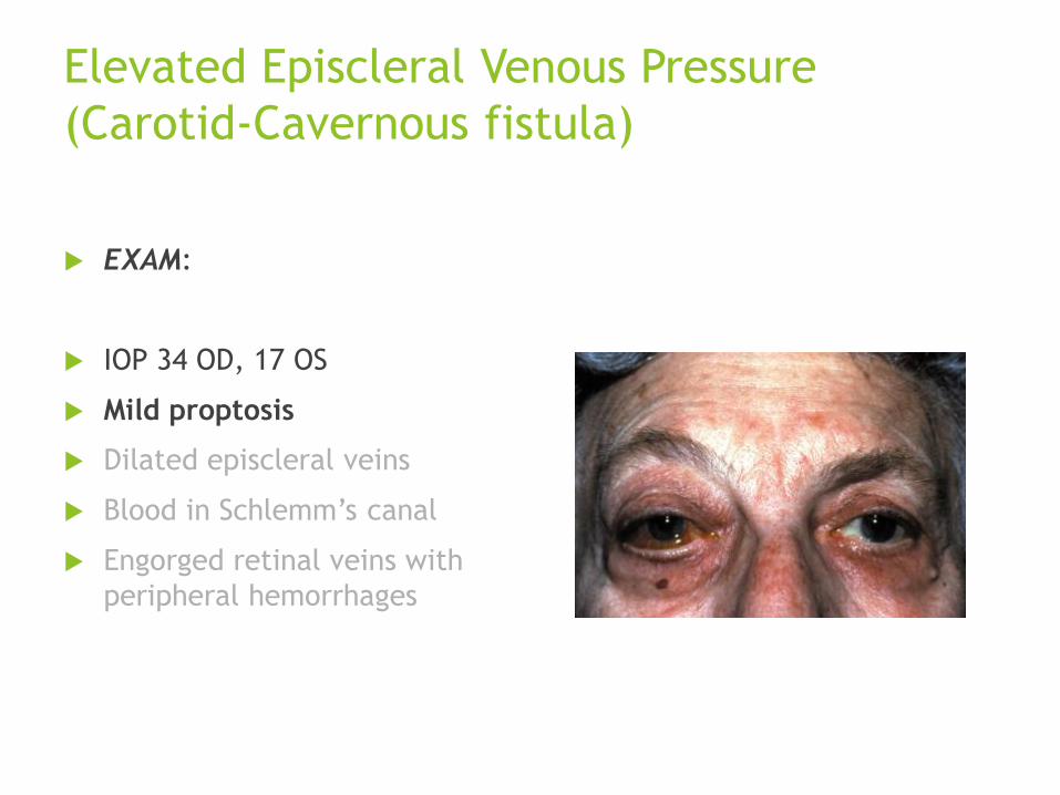
Elevated Episcleral Venous Pressure
(Carotid-Cavernous fistula)
EXAM:
IOP 34 OD, 17 OS
Mild proptosis
Dilated episcleral veins
Blood in Schlemm’s canal
Engorged retinal veins with
peripheral hemorrhages
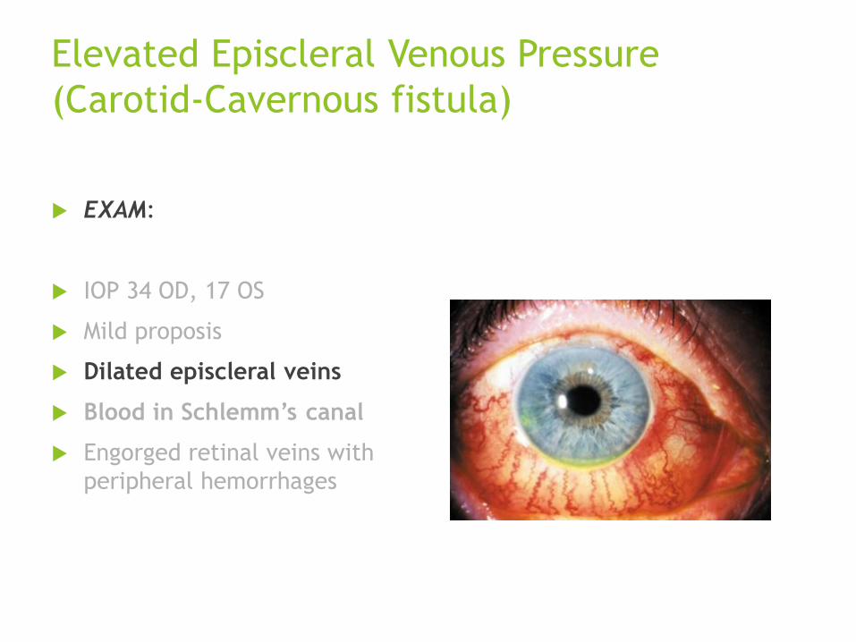
Elevated Episcleral Venous Pressure
(Carotid-Cavernous fistula)
EXAM:
IOP 34 OD, 17 OS
Mild proposis
Dilated episcleral veins
Blood in Schlemm’s canal
Engorged retinal veins with
peripheral hemorrhages

Elevated Episcleral Venous Pressure
(Carotid-Cavernous fistula)
EXAM:
IOP 34 OD, 17 OS
Mild proptosis
Dilated episcleral veins
Blood in Schlemm’s canal
Engorged retinal veins with
peripheral hemorrhages

Secondary OAG
(Elevated EVP)
Cause:
AV Malformations: CC fistula, orbital varix, Sturge-Weber Syndrome
Venous Obstruction: Thyroid ophthalmopathy, retrobulbar tumor
Superior vena cava syndrome
Treatment:
Aqueous suppressants better than meds that increase TM outflow
Filtering surgery (tube-shunt) may be complicagted by uveal effusions or suprachoroidal hemorrhage--sclerostomies

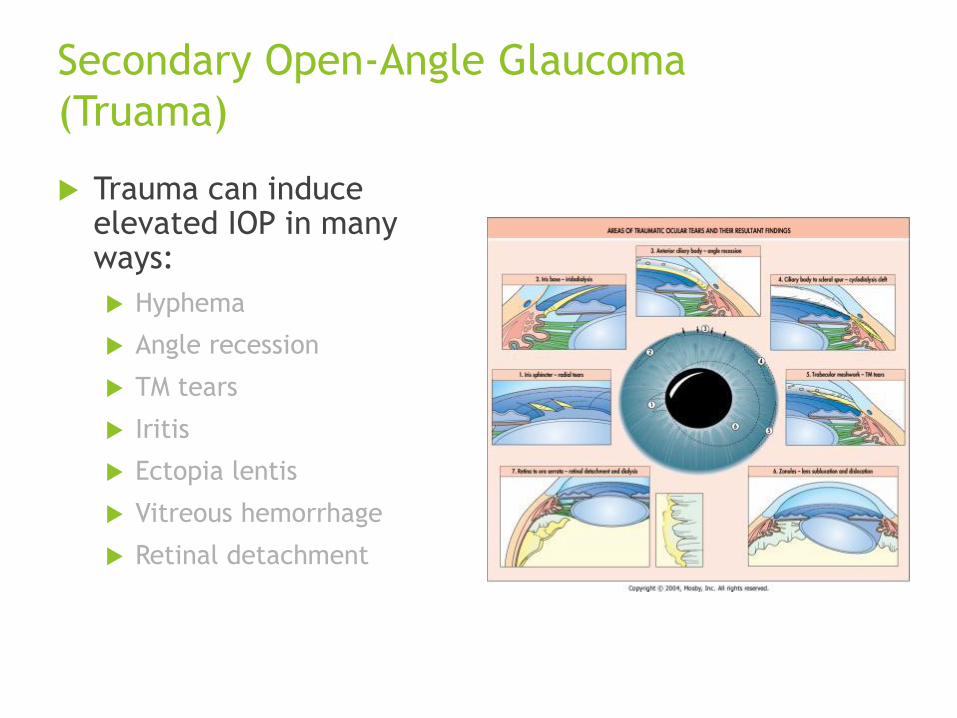
Secondary Open-Angle Glaucoma
(Truama)
Trauma can induce elevated IOP in many ways:
Hyphema
Angle recession
TM tears
Iritis
Ectopia lentis
Vitreous hemorrhage
Retinal detachment
Secondary Open-Angle Glaucoma
(Truama)
Trauma can induce elevated IOP in many ways:
Hyphema
Angle recession
TM tears
Iritis
Ectopia lentis
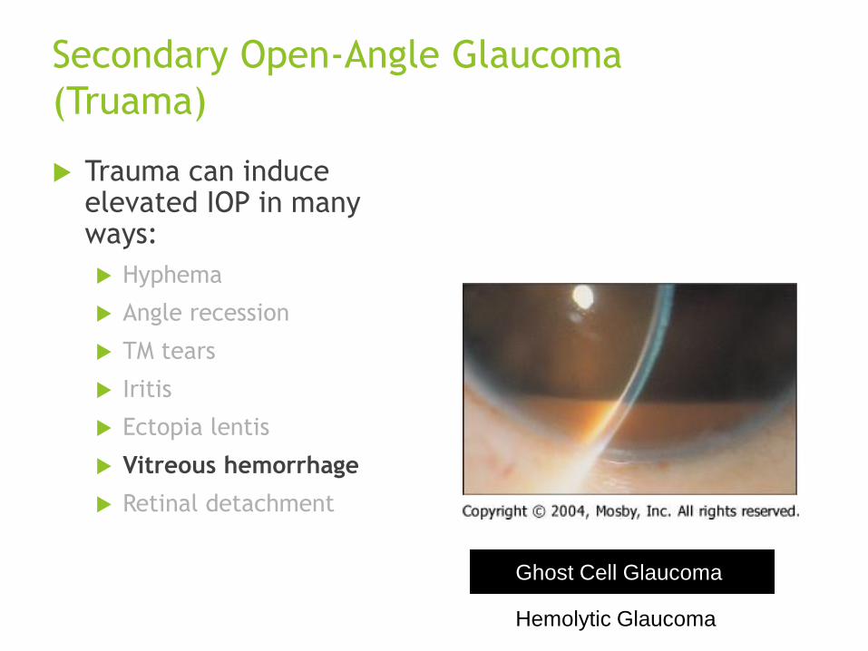
Vitreous hemorrhage
Retinal detachment

Secondary Open-Angle Glaucoma
(Truama)
Trauma can induce elevated IOP in many ways:
Hyphema
Angle recession
TM tears
Iritis
Ectopia lentis
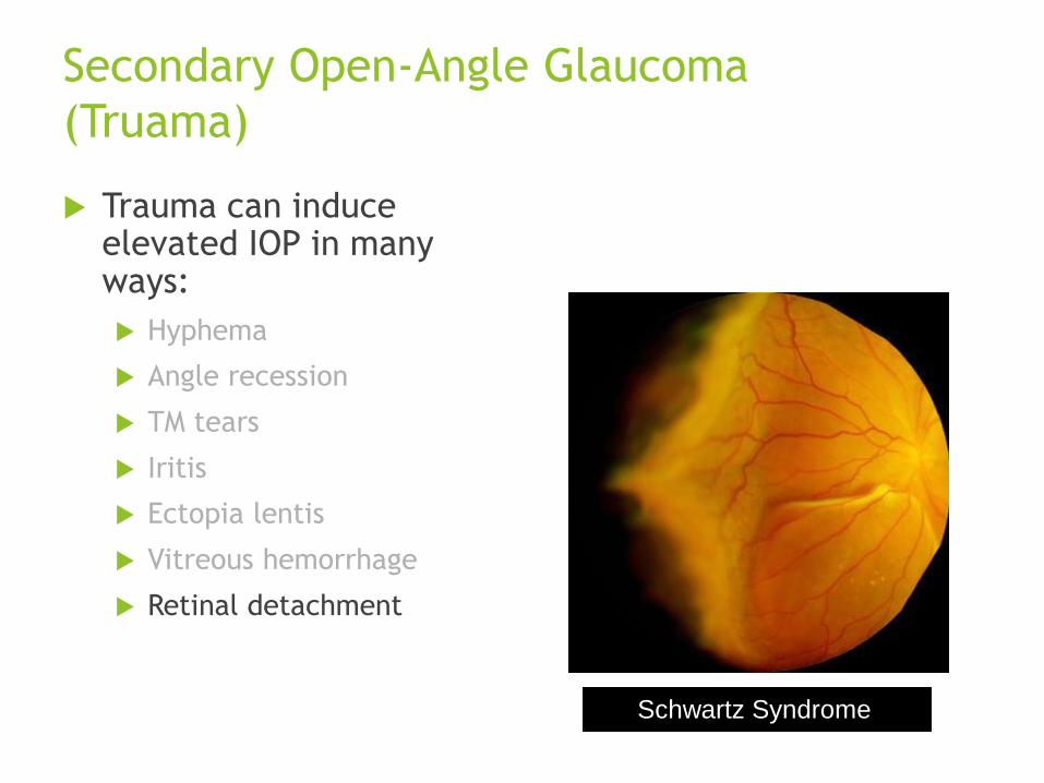
Vitreous hemorrhage
Retinal detachment

Secondary Open-Angle Glaucoma
(Truama)
Trauma can induce elevated IOP in many ways:
Hyphema
Angle recession
TM tears
Iritis
Ectopia lentis
Vitreous hemorrhage
Retinal detachment
Secondary Open-Angle Glaucoma
(Truama)
Trauma can induce elevated IOP in many ways:
Hyphema
Angle recession
TM tears
Iritis
Ectopia lentis
Vitreous hemorrhage
Retinal detachment
Ghost Cell Glaucoma
Hemolytic Glaucoma
Secondary Open-Angle Glaucoma
(Truama)
Trauma can induce elevated IOP in many ways:
Hyphema
Angle recession
TM tears
Iritis
Ectopia lentis
Vitreous hemorrhage
Retinal detachment
Schwartz Syndrome

Thank you for
your attention