Identifying and Referring Patients with Suspected Cancer Dr Nick Pendleton.
69
Identifying and Identifying and Referring Patients with Referring Patients with Suspected Cancer Suspected Cancer Dr Nick Pendleton Dr Nick Pendleton
-
Upload
darby-canon -
Category
Documents
-
view
224 -
download
1
Transcript of Identifying and Referring Patients with Suspected Cancer Dr Nick Pendleton.

Identifying and Referring Identifying and Referring Patients with Suspected CancerPatients with Suspected Cancer
Dr Nick PendletonDr Nick Pendleton

NICE Clinical Knowledge NICE Clinical Knowledge Summaries (CKS)Summaries (CKS)
• Cancer – suspected (NICE referral advice)
http://cks.nice.org.uk/#specialityTabnt

Referral timelines
• Immediate: an acute admission or referral occurring within a few hours, or even more quickly if necessary
• Urgent: the patient is seen within the national target for urgent referrals (currently 2 weeks)
• Non-urgent: all other referrals

About this presentation:About this presentation:
• The scenarios in this slide presention are based wholly or partly on real patients who have presented to GP surgeries. They have been anonymised for use as a teaching tool for GPs in Training. For realism the patients have been given fictional names, ages and professions.

Lesley Summers - 31Lesley Summers - 31
• Whilst I’m here can you check this mole on my arm?

A B C D E RuleA B C D E Rule
ASYMMETRY
IRREGULAR BORDER
COLOUR – gaining, losing(?), multiple colours
Diameter greater than 6mm (1/4 inch)
Evolving


Mr Simpson 53, Company DirectorMr Simpson 53, Company Director
• « I try and stay away from Doctors if I can but my wife has made this appointment! »
• « What is your wife worried about!? »
• « I have this lump on my leg… Its getting a bit bigger and its quite sore »


Can I ask you some questions about it?
• « How long has it been there? »• « About 3 months or so »
• « Is there any history of an injury? »• « Yes, come to think about it, I knocked my
leg with an axe whilst chopping logs about 4 months ago »

What are the worrying features of What are the worrying features of a palpable lump?a palpable lump?
• Refer urgently as suspected soft tissue sarcoma if:
• Greater than about 5 cm in diameter• Deep to fascia, fixed or immobile• Painful• Increasing in size• A recurrence after previous excision• If there is any doubt about the need for referral,
discussion with a local specialist should be undertaken

Mr Simpson was referred (2WW)
• CT showed an homogenous mass with capsule formation. US scan appearances resembled a multi-locular cyst. The mass was excised.
• Histology – necrotic debris, fibrin and blood clots.
• Fortunately it was not a Sarcoma.• “A case of chronic expanding hematoma in the tensor
fascia lata”
• http://escholarship.org/uc/item/6wg5260x

Ricky, 15Ricky, 15
« Coach said I should come and see you about my left leg –It’s interfering with my training. I play a lot of sport including football 3 times a week »

Tell me more about it..Tell me more about it..
• I don’t remember injuring it, but I’ve not been able to run on it for a few weeks now
• It is sore and tender to press on• It hurts even when I’m not walking about• It’s more sore this week than a few weeks
ago• On examination: he’s limping, there is a
bony and tender swelling below the knee

What is the Differential Diagnosis?What is the Differential Diagnosis?
• Osgood-Schlatter’s Disease?
• A Primary Bone Tumour?
• Osteosarcoma most commonly presents between 10 and 24 years old
• This is an age when a lot of people take part in sports

What should you do next?What should you do next?
• Patients with increasing, unexplained or persistent bone pain or tenderness, particularly pain at rest (and especially if not in the joint), or an unexplained limp should be investigated urgently ?Bone Tumour
• CKS Guidance recommends an immediate Xray and then if bone tumour is a possibility – refer urgently (2WW)

OSTEOSARCOMA (MALIGNANT BONE TUMOUR)

Mr Jones, 46, SalesmanMr Jones, 46, Salesman
• Blood results done as part of health screen:LFTs• ALP slightly raised 25% above normal• ALT raised 50% above normal• Other bloods and LFTs normal• Not on any medications, PMH nil, non-smoker

Review appointmentReview appointment
• Alcohol intake 60 -70 units a week
• ‘Don’t worry I will curb my drinking doctor – its just become a habit to open a bottle of wine after work with my wife’
• Plan: recheck LFTs in 4-6 weeks (NB. the guidance says 6 months)

Review appointment 2Review appointment 2
• Alcohol intake 20 units a week
• ‘We have also started healthy eating and exercising doctor!
• LFT results: ALT still raised 50% above normal, ALP slightly better but still close to 25% above normal

Ultrasound ReportUltrasound Report
• There is a hyperechoic mass with in one lobe of the liver. It is not possible to say whether this is a benign cyst or a sinister lesion. Referral for urgent MRI is indicated.

Telephone EncounterTelephone Encounter
• Hello Mr Jones – I am ringing about your Ultrasound report, is now a good time to talk?
• No, sorry Doctor – we have just had a telephone call to say my mother has passed away in the nursing home. I don’t want to discuss anything at the moment. I’ll come and see you at the surgery soon. Goodbye.
• What do you do next?

Mrs Gladys Parker, 72Mrs Gladys Parker, 72
• Dysphagia and weight loss. Gastroscopy 1 month ago normal.
• Came with daughter. My mum is still losing weight and can’t swallow properly. The Doctor we saw last week gave her some ensure drinks but something’s not right!

Re-referral for gastroscopyRe-referral for gastroscopy
Report: There is a circumferential stricture seen with the appearances of an advanced oesophageal carcinoma…
The patient died 4 weeks later

Letter to Endoscopy UnitLetter to Endoscopy Unit
Dear Sister X
I would like to enquire whether it is possible for a tumour of this advanced stage to appear with in this short time scale and do you have any video footage of the previous exam?

Response from GI ConsultantResponse from GI Consultant
Thank you for your letter. No I do not think this lesion could have arisen in this short time scale. I think it was missed during the first examination. We will be exploring this with the endoscopist. We do not currently video the examinations.

A Cutaneous Horn – 25% will have A Cutaneous Horn – 25% will have SCC at the baseSCC at the base

Mr Chandra, 46, IT DeveloperMr Chandra, 46, IT Developer
• I have been passing blood from my back passage every time I go to the toilet for the last 3 days
• No change in bowel habit• Its bright red• Its after a motion• It’s not painful

ExaminationExamination
• Abdomen examination normal, no mass
• PR examination normal
• What would you do next?

WHAT DOES THE CKS GUIDANCE SAY?WHAT DOES THE CKS GUIDANCE SAY?
• In patients 40 years of age and older, reporting rectal bleeding with a change of bowel habit towards looser stools and/or increased stool frequency persisting for 6 weeks or more, an urgent referral should be made.
• In patients 60 years of age and older, with rectal bleeding persisting for 6 weeks or more without a change in bowel habit and without anal symptoms, an urgent referral should be made .

Mr Chandra, 46, IT DeveloperMr Chandra, 46, IT Developer
• I have been passing blood from my back passage every time I go to the toilet for the last 3 days
• No change in bowel habit• Its bright red.• Its after a motion• It’s not painful

WHAT DOES THE CKS GUIDANCE SAY?WHAT DOES THE CKS GUIDANCE SAY?
• In patients with equivocal symptoms who are not unduly anxious, it is reasonable to use a period of 'treat, watch and wait' as a method of management
• In men of any age with unexplained iron deficiency anaemia and a haemoglobin of 11 g/100 mL or below, an urgent referral should be made
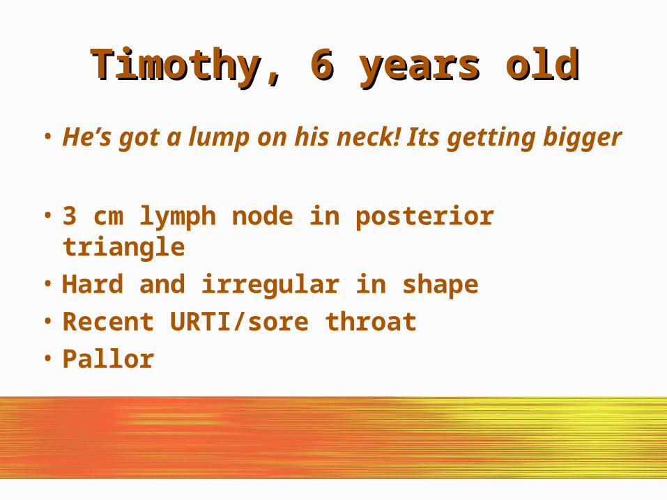
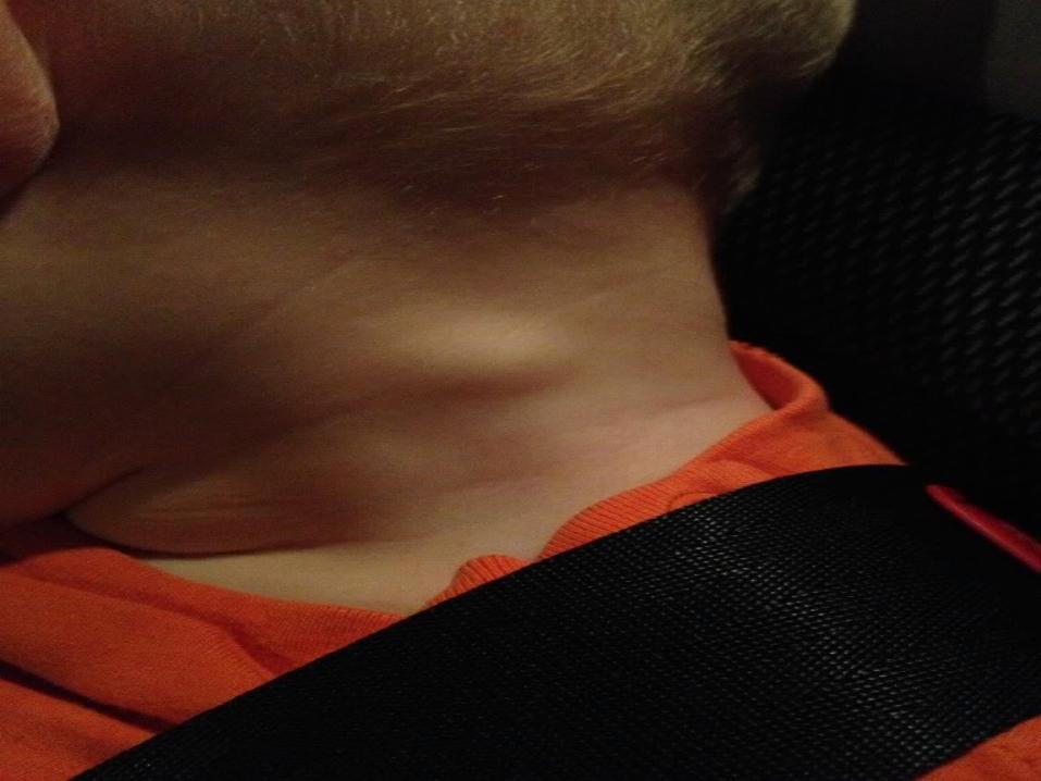
Timothy, 6 years oldTimothy, 6 years old
• He’s got a lump on his neck! Its getting bigger
• 3 cm lymph node in posterior triangle• Hard and irregular in shape• Recent URTI/sore throat• Pallor

Causes of Neck Swelling in ChildrenCauses of Neck Swelling in Children
LYMPHADENOPATHY (enlarged lymph nodes)
• LOCAL• SYSTEMIC
LYMPHADENITIS (inflamed lymph nodes) or ABSCESS
NON-LYMPHADENOMATOUS NECK SWELLINGS
BMJ 2012;344:e3171

LYMPHADENOPATHY (enlarged lymph nodes)
• LOCAL• Viral or bacterial upper respiratory tract
• Ear infection, Oropharyngeal infection
• Headlice infestation, Dental abscess
• Cat scratch disease (gram –ve bacteria Bartonella Henselae or Quintana)
• SYSTEMIC• Malignancy (lymphoma or leukaemia)
• Viral infections (Epstein-Barr virus, cytomegalovirus, rubella)
• Kawasaki disease
• Mycobacterial infection (tuberculous or non-tuberculous), Sarcoidosis
• Systemic lupus erythematosus
• Juvenile idiopathic arthritis
BMJ 2012;344:e3171
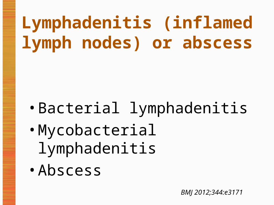
Lymphadenitis (inflamed lymph nodes) or abscess
• Bacterial lymphadenitis
• Mycobacterial lymphadenitis
• Abscess
BMJ 2012;344:e3171

Non-lymphadenomatous neck swellings
• Cystic hygroma
• Sternocleidomastoid swelling
• Thyroid gland enlargement
• Thyroglossal cyst
• Dermoid cyst
• Branchial cyst
• MumpsBMJ 2012;344:e3171
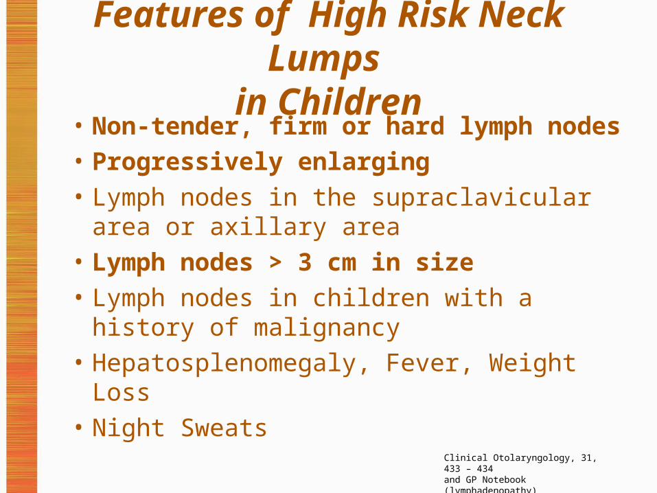
Features of High Risk Neck Lumps in Children
• Non-tender, firm or hard lymph nodes• Progressively enlarging• Lymph nodes in the supraclavicular area or
axillary area• Lymph nodes > 3 cm in size• Lymph nodes in children with a history of
malignancy• Hepatosplenomegaly, Fever, Weight Loss• Night Sweats
Clinical Otolaryngology, 31, 433 – 434and GP Notebook (lymphadenopathy)

Timothy, 6 years oldTimothy, 6 years old
• He’s got a lump on his neck!• 3 cm lymph node in posterior triangle• Hard and irregular in shape• Recent URTI/sore throat, Pallor
• Clearly fits urgent referral criteria for a suspicious neck lump

Mrs Sullivan, 50, unemployedMrs Sullivan, 50, unemployed
• I’ve got this ringing in my left ear!• I can’t hear as well either• I sometimes have a spinning sensation in my
head

“IN MY RIGHT EAR”

“IN FRONT”

Weber without lateralization
Weber lateralizes leftWeber lateralizes right
Rinne both ears AC>BC
Normal/bilateral sensorineural loss
Sensorineural loss in right
Sensorineural loss in left
Rinne left BC>ACConductive loss in left
Combined loss : conductive and sensorineural loss in left
Rinne right BC>AC
Combined loss : conductive and sensorineural loss in right
Conductive loss in right
Rinne both ears BC>AC
Conductive loss in both ears
Combined loss in right and conductive loss on left
Combined loss in left and conductive loss on right
AC = Air Conduction BC = Bone Conduction

Mr Sullivan, 50, unemployedMr Sullivan, 50, unemployed
• I’ve got this ringing in my left ear!• I can’t hear as well either• I sometimes have a spinning sensation in my
head• Examination: sensorineural hearing loss• Diagnosis – small acoustic neuroma (tumour of
vestibulocochlear nerve)
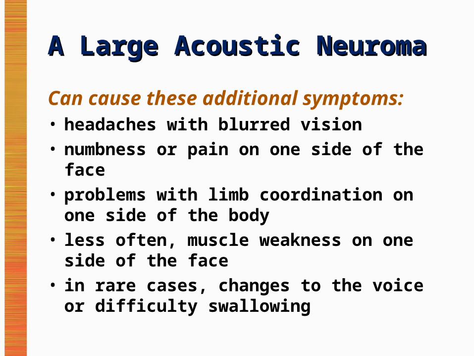
A Large Acoustic NeuromaA Large Acoustic Neuroma
Can cause these additional symptoms:• headaches with blurred vision • numbness or pain on one side of the face • problems with limb coordination on one side of
the body • less often, muscle weakness on one side of the
face • in rare cases, changes to the voice or difficulty
swallowing

Mrs Simpson, 52Mrs Simpson, 52« I am fed up with this, just look at
my belly its massive, I feel bloated, but I’ve got no appetite and when I do eat I’ve either got diarrhoea or can’t go at all. Also I keep having to urinate, I feel tired and my back hurts! »

OVARIAN CANCER
VERSUS
IRRITABLE BOWEL SYNDROME
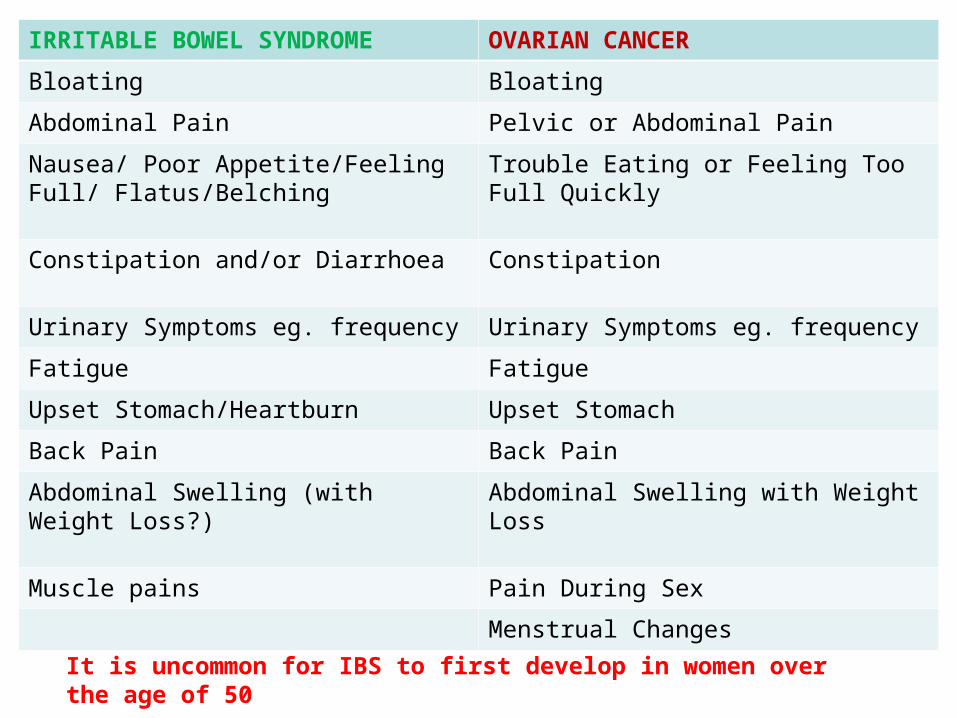
IRRITABLE BOWEL SYNDROME OVARIAN CANCER
Bloating Bloating
Abdominal Pain Pelvic or Abdominal Pain
Nausea/ Poor Appetite/Feeling Full/ Flatus/Belching
Trouble Eating or Feeling Too Full Quickly
Constipation and/or Diarrhoea Constipation
Urinary Symptoms eg. frequency Urinary Symptoms eg. frequency
Fatigue Fatigue
Upset Stomach/Heartburn Upset Stomach
Back Pain Back Pain
Abdominal Swelling (with Weight Loss?) Abdominal Swelling with Weight Loss
Muscle pains Pain During Sex
Menstrual Changes
It is uncommon for IBS to first develop in women over the age of 50

Investigating Ovarian Cancer Investigating Ovarian Cancer Symptoms in Primary CareSymptoms in Primary Care
• Measure serum CA125 in primary care in women with symptoms that suggest ovarian cancer
• If serum CA125 is 35 IU/ml or greater, arrange an ultrasound scan of the abdomen and pelvis
• For any woman who has normal serum CA125 (less than 35 IU/ml), or CA125 of 35 IU/ml or greater but a normal ultrasound: assess her carefully for other clinical causes of her symptoms and investigate if appropriate
NICE CG 122 - OVARIAN CANCER

Sally Smith, 39, SecretarySally Smith, 39, Secretary
« My Sister is 45 and having treatment for breast cancer and I want to know if I am at risk »
« My Aunt died from Ovarian cancer 2 years ago »

What is a Significant Family History?What is a Significant Family History?• One first-degree female relative diagnosed with breast cancer at
younger than age 40 years• One first-degree male relative diagnosed with breast cancer at any
age• One first-degree relative with bilateral breast cancer where the first
primary was diagnosed at younger than age 50 years• Two first-degree relatives, or one first-degree and one second-
degree relative, diagnosed with breast cancer at any age• One first-degree or second-degree relative diagnosed with breast
cancer at any age and one first-degree or second-degree relative diagnosed with ovarian cancer at any age (one of these should be a first-degree relative)
• Three first-degree or second-degree relatives diagnosed with breast cancer at any age
http://www.patient.co.uk/doctor/familial-breast-cancer

Alternative ScenarioAlternative Scenario• Mother had breast cancer aged 50. No other
family history.
• Offer information and reassurance, secondary care referral not indicated unless the family history contains:
• Bilateral breast cancer, Male breast cancer• Ovarian cancer, Jewish ancestry• Sarcoma in a relative younger than age 45 years• Glioma or childhood adrenal cortical carcinomas• Complicated patterns of multiple cancers at a young age• Paternal history of breast cancer (two or more relatives on the father's side of
the family)
http://www.patient.co.uk/doctor/familial-breast-cancer

Mr Jenkinson 71 Mr Jenkinson 71 • Telephone call: « I cannot tolerate this
shoulder pain any longer. Surely I need an X-ray or something. The Drs have said there would be no point as it would just confirm arthritis, but it is getting worse and my arm is loosing muscle and strength! »
• XRAY request: 6 months of right shoulder pain now needing morphine

PANCOAST TUMOUR AT RIGHT APEX
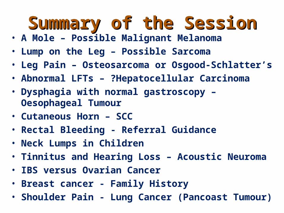
Summary of the SessionSummary of the Session• A Mole – Possible Malignant Melanoma• Lump on the Leg – Possible Sarcoma• Leg Pain – Osteosarcoma or Osgood-Schlatter’s• Abnormal LFTs – ?Hepatocellular Carcinoma• Dysphagia with normal gastroscopy – Oesophageal Tumour• Cutaneous Horn – SCC• Rectal Bleeding - Referral Guidance• Neck Lumps in Children• Tinnitus and Hearing Loss – Acoustic Neuroma• IBS versus Ovarian Cancer• Breast cancer - Family History• Shoulder Pain - Lung Cancer (Pancoast Tumour)

Identifying and Referring Identifying and Referring Patients with Suspected CancerPatients with Suspected Cancer
CLINICAL RECORD REVIEWCLINICAL RECORD REVIEW

Tony Frazer 36, National Account Tony Frazer 36, National Account Manager (Sales)Manager (Sales)
• July 2013• Dr A on-call• Telephone triage encounter:• Haematemesis fresh and dried (coffee
bean) blood• Abnormal weight loss, 3 stone in 7/12

Same day appointment with Dr BSame day appointment with Dr B• Heamatemesis after drinking
excessive alcohol and vomiting• 2 stone weight loss in 7 months• Exam normal, weight 65kg (75kg Sept 12)
• Needs 2WW referral, upper GI poss mallory weiss tear but in combination with weight loss need to r/o malignancy.

14 August – Dr C14 August – Dr C• Gastroscopy normal, h.pylori -ve• Very tired• Intermittent diarrhoea• No appetite, weight 63kg• Mood OK – but a lot of stress in last year
• Blood tests requested to exclude coeliac• Start omeprazole 20mg bd
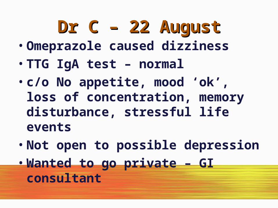
Dr C – 22 AugustDr C – 22 August• Omeprazole caused dizziness• TTG IgA test – normal• c/o No appetite, mood ‘ok’, loss of
concentration, memory disturbance, stressful life events
• Not open to possible depression• Wanted to go private – GI consultant

22ndnd October October• Continues to lose weight - wt 59Kg• Consuming 2000 calories in food from
McDonalds and 2500 calories in supplements• Upper GI consultant suggested the cause of
his weight loss is depression and suggested starting him on mirtazapine (and arranges CT)
• Patient thinks this is wrong as he has a great life and everything to feel good about.

Weight ChartWeight Chart

25 September – Dr D25 September – Dr D• CT scan was normal• Now feels too weak and tired to work• Weight stable• Feels frustrated and down in mood• TATT, sleeping lots, buying own high
calorie supplements• Awaiting further GI consultant review.
See in 3 weeks

25 November – Dr C25 November – Dr C• Gaining weight• Taking mirtazapine• Has seen consultant again who suggests
Chronic Fatigue Syndrome (CFS) is the possible diagnosis
• Referred CFS Specialist for opinion• In the meantime wants to try hydrotherapy
to get some fitness back

Weight ChartWeight Chart
7 February 20147 February 2014
• Diagnosis of CFS confirmed by specialist
• 16 September 2014 – making progress with CFS therapy and a return to work is possible in early 2015

About this presentation:About this presentation:
• The scenarios in this slide presention are based wholly or partly on real patients who have presented to GP surgeries. They have been anonymised for use as a teaching tool for GPs in Training. For realism the patients have been given fictional names, ages and professions.