Hyperuricemia deficientmice - PNASprimateevolution predisposesmanto hyperuricemia, ameta-bolic...
5
Proc. Natl. Acad. Sci. USA Vol. 91, pp. 742-746, January 1994 Medical Sciences Hyperuricemia and urate nephropathy in urate oxidase- deficient mice (gene targeting/animal model) XIANGWEI WU*t, MAKI WAKAMIYA*, SUKESHI VAISHNAV*t, ROBERT GESKE§, CHARLES MONTGOMERY, JR.§, PAMELA JONES*t, ALLAN BRADLEY**, AND C. THOMAS CASKEY*$ *Institute for Molecular Genetics, tHoward Hughes Medical Institute, and Center of Comparative Medicine, Baylor College of Medicine, Houston, TX 77030 Contributed by C. Thomas Caskey, October 1, 1993 ABSTRACT Urate oxidase, or uricase (EC 1.7.3.3), is a purine metabolic enzyme that catalyzes the conversion of uric acid to aUantoin in most mammals except humans and certain other primates. The loss of urate oxidase in the human during primate evolution predisposes man to hyperuricemia, a meta- bolic disturbance that can lead to gouty arthritis and renal stones. To create a mouse model for hyperuricemia and gout, and to address the question of whether urate oxidase is essential in lower mammalian species, we have disrupted the urate oxidase gene in the mouse by homologous recombination in embryonic stem ceUls. Unlike the human situation, urate oxi- dase deficiency in mice causes pronounced hyperuricemia and urate nephropathy. More than half of the mutant mice died before 4 weeks of age, indicating that urate oxidase is essential in mice. These mutant mice may also serve as animal models for hyperuricemia and its related nephropathy in humans. The end product of purine metabolism varies among species (1-3). Most mammals excrete allantoin because they have an enzyme, urate oxidase (EC 1.7.3.3), which degrades the sparingly soluble uric acid to a more soluble allantoin. Urate oxidase was lost in humans and certain other primates by deleterious mutations in the urate oxidase gene (4). As a result, uric acid is the end product of purine metabolism in these species and uric acid levels approach the saturation point (2, 3). Renal excretion of uric acid is therefore essential. Elevated uric acid levels are associated with several met- abolic disorders such as gouty arthritis and renal stones in humans (5, 6). Unlike most other mammals, humans are predisposed to the development of these diseases through lack of urate oxidase. The degradation of uric acid by urate oxidase leads to very low levels of uric acid in most other mammals and makes them unsuitable as experimental models for human hyperuricemia. The generation of urate oxidase- deficient mice by homologous recombination in mouse em- bryonic stem (ES) cells may provide a good animal model system to study this common disease and to develop more effective treatments. Although it is clear that urate oxidase was lost in humans and a few other species by genetic mutations during the evolution of primates (4), the biological function and the evolutionary significance of this loss remain unclear. It has been debated whether this loss provided evolutionary advan- tages or was simply an evolutionary accident (7-10). To determine whether urate oxidase is an essential enzyme in lower mammals and to develop a model for human hyperu- ricemia, we generated mice with a targeted mutation at the urate oxidase locus by gene targeting in ES cells. This mutation resulted in a complete loss of urate oxidase activity and a 10-fold increase in the serum uric acid level. These The publication costs of this article were defrayed in part by page charge payment. This article must therefore be hereby marked "advertisement" in accordance with 18 U.S.C. §1734 solely to indicate this fact. mutant mice developed pronounced hyperuricemia and ob- structive nephropathy resulting from uric acid crystals. The accumulation of uric acid also led to early death in 65% of the mutant mice, which may be preventable with the adminis- tration of allopurinol, a xanthine oxidase inhibitor. MATERIALS AND METHODS Plasmid Construction and ES Cell Culture. Genomic clones of the mouse urate oxidase gene were isolated from a C57BL/6J mouse genomic library by using a mouse cDNA probe, and the gene structure was characterized (X.W., un- published results). A gene replacement-type vector, pUTK, was constructed as follows. A 6.6-kb Bgl II fragment contain- ing exons 2-4 of the mouse gene was cloned into the BamHI site of the pBluescript SK vector (Stratagene). A pMClneo cassette (11), containing a neomycin-resistance gene (neo), was inserted into the Xho I site in exon 3, and a herpes simplex virus (HSV) thymidine kinase gene (tk) was cloned into a Sal I site at one end of the urate oxidase sequence (12). AB1 ES cells (13) were used, and the procedures for culturing ES cells were adopted from Robertson (14). DNA Electroporation and Identification of Targeted Clones. pUTK DNA was digested with the restriction enzyme Xho I to release the plasmid sequence prior to electroporation. The DNA was introduced into ES cells by electroporation (13, 15) and the cells were seeded in 24-well plates. The neomycin analogue G418 (effective concentration, 150 jug/ml) and 1-(2-deoxy-2-fluoro-,-D-arabinofuranosyl)-5-iodouracil (0.2 AM) were added 24 hr later; cells were maintained in selec- tion medium for 10-11 days. Colonies from each well (be- tween 10 and 20) were pooled by treatment with trypsin. An aliquot (about 1/10th) of the cells was used for polymerase chain reaction (PCR) analysis and the remaining cells were stored in Dulbecco's modified Eagle's medium with 10% fetal bovine serum and 10% dimethyl sulfoxide (freezing medium) at -80°C. Individual positive clones were purified from PCR-positive pools by serial dilution, and successful gene- targeting events were confirmed by Southern analysis. Assay for Urate Oxidase Activity and Uric Acid. A crude extract enriched for urate oxidase was prepared from liver (16). Urate oxidase activity was determined spectrometrically by measuring the rate of consumption of uric acid (17). Uric acid and allantoin levels were measured as described (18, 19). Histology. Tissues were fixed with either neutral buffered 10% formalin (Surgipath Medical, Richmond, IL), absolute ethanol, or Zenker's solution. After fixation, the tissues were trimmed, dehydrated, and infiltrated with paraffin wax. Sec- tions of the tissue were stained with hematoxylin and eosin. Additional sections were taken of ethanol-fixed tissues. After Abbreviations: ES cell, embryonic stem cell; neo, neomycin- resistance gene; tk, thymidine kinase gene. tPresent address: Department of Molecular Biology, Princeton Uni- versity, Princeton, NJ 08544. 742 Downloaded by guest on July 15, 2020
Transcript of Hyperuricemia deficientmice - PNASprimateevolution predisposesmanto hyperuricemia, ameta-bolic...
Proc. Natl. Acad. Sci. USAVol. 91, pp. 742-746, January 1994Medical Sciences
Hyperuricemia and urate nephropathy in urate oxidase-deficient mice
(gene targeting/animal model)
XIANGWEI WU*t, MAKI WAKAMIYA*, SUKESHI VAISHNAV*t, ROBERT GESKE§, CHARLES MONTGOMERY, JR.§,PAMELA JONES*t, ALLAN BRADLEY**, AND C. THOMAS CASKEY*$*Institute for Molecular Genetics, tHoward Hughes Medical Institute, and Center of Comparative Medicine, Baylor College of Medicine, Houston, TX 77030
Contributed by C. Thomas Caskey, October 1, 1993
ABSTRACT Urate oxidase, or uricase (EC 1.7.3.3), is apurine metabolic enzyme that catalyzes the conversion of uricacid to aUantoin in most mammals except humans and certainother primates. The loss of urate oxidase in the human duringprimate evolution predisposes man to hyperuricemia, a meta-bolic disturbance that can lead to gouty arthritis and renalstones. To create a mouse model for hyperuricemia and gout,and to address the question ofwhether urate oxidase is essentialin lower mammalian species, we have disrupted the urateoxidase gene in the mouse by homologous recombination inembryonic stem ceUls. Unlike the human situation, urate oxi-dase deficiency in mice causes pronounced hyperuricemia andurate nephropathy. More than half of the mutant mice diedbefore 4 weeks of age, indicating that urate oxidase is essentialin mice. These mutant mice may also serve as animal models forhyperuricemia and its related nephropathy in humans.
The end product of purine metabolism varies among species(1-3). Most mammals excrete allantoin because they have anenzyme, urate oxidase (EC 1.7.3.3), which degrades thesparingly soluble uric acid to a more soluble allantoin. Urateoxidase was lost in humans and certain other primates bydeleterious mutations in the urate oxidase gene (4). As aresult, uric acid is the end product of purine metabolism inthese species and uric acid levels approach the saturationpoint (2, 3). Renal excretion ofuric acid is therefore essential.
Elevated uric acid levels are associated with several met-abolic disorders such as gouty arthritis and renal stones inhumans (5, 6). Unlike most other mammals, humans arepredisposed to the development of these diseases throughlack of urate oxidase. The degradation of uric acid by urateoxidase leads to very low levels of uric acid in most othermammals and makes them unsuitable as experimental modelsfor human hyperuricemia. The generation of urate oxidase-deficient mice by homologous recombination in mouse em-bryonic stem (ES) cells may provide a good animal modelsystem to study this common disease and to develop moreeffective treatments.Although it is clear that urate oxidase was lost in humans
and a few other species by genetic mutations during theevolution of primates (4), the biological function and theevolutionary significance of this loss remain unclear. It hasbeen debated whether this loss provided evolutionary advan-tages or was simply an evolutionary accident (7-10). Todetermine whether urate oxidase is an essential enzyme inlower mammals and to develop a model for human hyperu-ricemia, we generated mice with a targeted mutation at theurate oxidase locus by gene targeting in ES cells. Thismutation resulted in a complete loss of urate oxidase activityand a 10-fold increase in the serum uric acid level. These
The publication costs of this article were defrayed in part by page chargepayment. This article must therefore be hereby marked "advertisement"in accordance with 18 U.S.C. §1734 solely to indicate this fact.
mutant mice developed pronounced hyperuricemia and ob-structive nephropathy resulting from uric acid crystals. Theaccumulation of uric acid also led to early death in 65% of themutant mice, which may be preventable with the adminis-tration of allopurinol, a xanthine oxidase inhibitor.
MATERIALS AND METHODSPlasmid Construction and ES Cell Culture. Genomic clones
of the mouse urate oxidase gene were isolated from aC57BL/6J mouse genomic library by using a mouse cDNAprobe, and the gene structure was characterized (X.W., un-published results). A gene replacement-type vector, pUTK,was constructed as follows. A 6.6-kb Bgl II fragment contain-ing exons 2-4 of the mouse gene was cloned into the BamHIsite of the pBluescript SK vector (Stratagene). A pMClneocassette (11), containing a neomycin-resistance gene (neo),was inserted into the Xho I site in exon 3, and a herpes simplexvirus (HSV) thymidine kinase gene (tk) was cloned into a SalI site at one end of the urate oxidase sequence (12). AB1 EScells (13) were used, and the procedures for culturing ES cellswere adopted from Robertson (14).DNA Electroporation and Identification of Targeted Clones.
pUTK DNA was digested with the restriction enzyme Xho Ito release the plasmid sequence prior to electroporation. TheDNA was introduced into ES cells by electroporation (13, 15)and the cells were seeded in 24-well plates. The neomycinanalogue G418 (effective concentration, 150 jug/ml) and1-(2-deoxy-2-fluoro-,-D-arabinofuranosyl)-5-iodouracil (0.2AM) were added 24 hr later; cells were maintained in selec-tion medium for 10-11 days. Colonies from each well (be-tween 10 and 20) were pooled by treatment with trypsin. Analiquot (about 1/10th) of the cells was used for polymerasechain reaction (PCR) analysis and the remaining cells werestored in Dulbecco's modified Eagle's medium with 10% fetalbovine serum and 10% dimethyl sulfoxide (freezing medium)at -80°C. Individual positive clones were purified fromPCR-positive pools by serial dilution, and successful gene-targeting events were confirmed by Southern analysis.
Assay for Urate Oxidase Activity and Uric Acid. A crudeextract enriched for urate oxidase was prepared from liver(16). Urate oxidase activity was determined spectrometricallyby measuring the rate of consumption of uric acid (17). Uricacid and allantoin levels were measured as described (18, 19).
Histology. Tissues were fixed with either neutral buffered10% formalin (Surgipath Medical, Richmond, IL), absoluteethanol, or Zenker's solution. After fixation, the tissues weretrimmed, dehydrated, and infiltrated with paraffin wax. Sec-tions of the tissue were stained with hematoxylin and eosin.Additional sections were taken of ethanol-fixed tissues. After
Abbreviations: ES cell, embryonic stem cell; neo, neomycin-resistance gene; tk, thymidine kinase gene.tPresent address: Department of Molecular Biology, Princeton Uni-versity, Princeton, NJ 08544.
742
Dow
nloa
ded
by g
uest
on
July
15,
202
0
Proc. Natl. Acad. Sci. USA 91 (1994) 743
paraffin removal these sections were placed in absolute eth-anol and stained with an ethanolic eosin solution. Uric aciddeposits were either stained with Gomori's silver method orviewed under polarized light to assess anisotropism (20).
Administration of Allopurinol. Allopurinol (Sigma) at var-ious concentrations was administrated continuously in thedrinking water to homozygous targeted mutants, includinghomozygous breeding pairs.
RESULTSTargeted Disruption of Urate Oxidase Gene in Mice. A
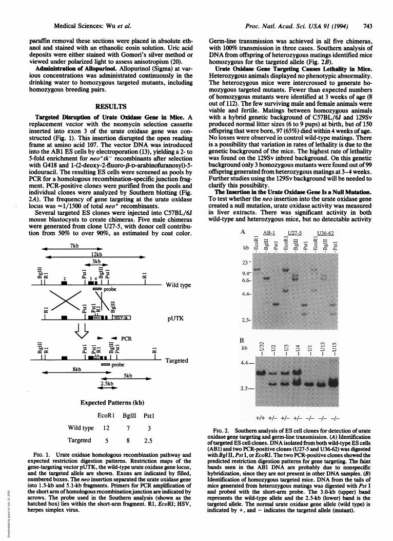
replacement vector with the neomycin selection cassetteinserted into exon 3 of the urate oxidase gene was con-structed (Fig. 1). This insertion disrupted the open readingframe at amino acid 107. The vector DNA was introducedinto the AB1 ES cells by electroporation (13), yielding a 2- to5-fold enrichment for neo+tk- recombinants after selectionwith G418 and 1-(2-deoxy-2-fluoro-,fD-arabinofuranosyl)-5-iodouracil. The resulting ES cells were screened as pools byPCR for a homologous recombination-specific junction frag-ment. PCR-positive clones were purified from the pools andindividual clones were analyzed by Southern blotting (Fig.2A). The frequency of gene targeting at the urate oxidaselocus was -1/1500 of total neo+ recombinants.
Several targeted ES clones were injected into C57BL/6Jmouse blastocysts to create chimeras. Five male chimeraswere generated from clone U27-5, with donor cell contribu-tion from 50% to over 90%o, as estimated by coat color.
7kb12kb
3kbo.(Am- .mnne
~Ir ~ 34
probe
I o I HSV--k I
_ ~~~~~ ~~- PCR
II * Iu~uIneo, |
probe8kb k
2kb5b2.5kb
Wild type
Germ-line transmission was achieved in all five chimeras,with 100% transmission in three cases. Southern analysis ofDNA from offspring of heterozygous matings identified micehomozygous for the targeted allele (Fig. 2B).
Urate Oxidase Gene Targeting Causes Lethality in Mice.Heterozygous animals displayed no phenotypic abnormality.The heterozygous mice were intercrossed to generate ho-mozygous targeted mutants. Fewer than expected numbersof homozygous mutants were identified at 3 weeks of age (8out of 112). The few surviving male and female animals wereviable and fertile. Matings between homozygous animalswith a hybrid genetic background of C57BL/6J and 129Svproduced normal litter sizes (6 to 9 pups) at birth, but of 150offspring that were born, 97 (65%) died within 4 weeks ofage.No losses were observed in control wild-type matings. Thereis a possibility that variation in rates of lethality is due to thegenetic background of the mice. The highest rate of lethalitywas found on the 129Sv inbred background. On this geneticbackground only 3 homozygous mutants were found out of99offspring generated from heterozygous matings at 3-4 weeks.Further studies using the 129Sv background will be needed toclarify this possibility.The Insertion in the Urate Oxidase Gene Is a Null Mutation.
To test whether the neo insertion into the urate oxidase genecreated a null mutation, urate oxidase activity was measuredin liver extracts. There was significant activity in bothwild-type and heterozygous mice, but no detectable activity
A AB-1
kb=kb v~ m XL
I I
U27-5O lam nV. .
CL
u36-62l-q
1,4 = ".
C>- ..
23 -
9.46.6-
4.4-
pUTK 2.3-
Bkb
Targeted
Int_~:) ~: D: D >:I I I I I
4.4-
2.3-
Expected Patterns (kb)
EcoR I BglII Pstl
Wild type 12 7 3
Targeted 5 8 2.5
FIG. 1. Urate oxidase homologous recombination pathway andexpected restriction digestion patterns. Restriction maps of thegene-targeting vector pUTK, the wild-type urate oxidase gene locus,and the targeted allele are shown. Exons are indicated by filled,numbered boxes. The neo insertion separated the urate oxidase geneinto 1.5-kb and 5.1-kb fragments. Primers for PCR amplification ofthe short arm ofhomologous recombinationjunction are indicated byarrows. The probe used in the Southern analysis (shown as thehatched box) lies within the short-arm fragment. Rl, EcoRI; HSV,herpes simplex virus.
+1 +1- +1- +1--Il- -Il- -Il-FIG. 2. Southern analysis of ES cell clones for detection of urate
oxidase gene targeting and germ-line transmission. (A) Identificationoftargeted ES cell clones. DNA isolated from both wild-type ES cells(ABl) and two PCR-positive clones (U27-5 and U36-62) was digestedwithBgl II, Pst I, orEcoRI. The two PCR-positive clones showed thepredicted restriction digestion patterns for gene targeting. The faintbands seen in the AB1 DNA are probably due to nonspecifichybridization, since they are not present in other DNA samples. (B)Identification of homozygous targeted mice. DNA from the tails ofmice generated from heterozygous matings was digested with Pst Iand probed with the short-arm probe. The 3.0-kb (upper) bandrepresents the wild-type allele and the 2.5-kb (lower) band is thetargeted allele. The normal urate oxidase gene allele (wild type) isindicated by +, and - indicates the targeted allele (mutant).
Medical Sciences: Wu et al.
0-b4a -rn NO
Dow
nloa
ded
by g
uest
on
July
15,
202
0
Proc. Natl. Acad. Sci. USA 91 (1994)
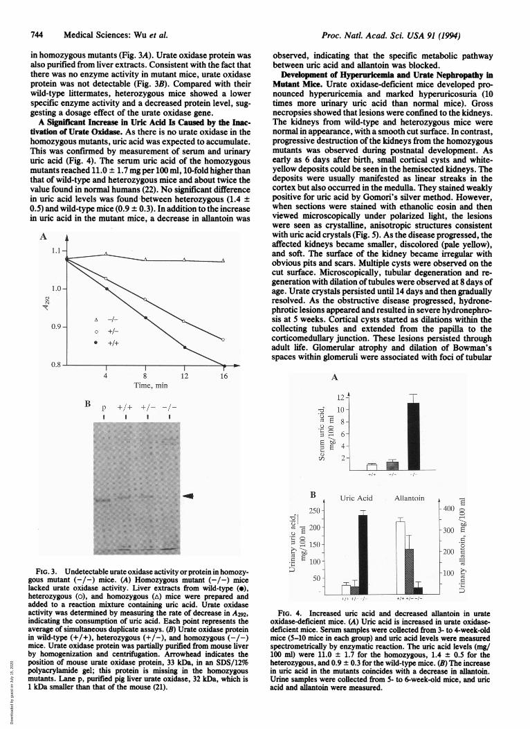
in homozygous mutants (Fig. 3A). Urate oxidase protein wasalso purified from liver extracts. Consistent with the fact thatthere was no enzyme activity in mutant mice, urate oxidaseprotein was not detectable (Fig. 3B). Compared with theirwild-type littermates, heterozygous mice showed a lowerspecific enzyme activity and a decreased protein level, sug-gesting a dosage effect of the urate oxidase gene.A Significant Increase in Uric Acid Is Caused by the Inac-
tivation of Urate Oxidase. As there is no urate oxidase in thehomozygous mutants, uric acid was expected to accumulate.This was confirmed by measurement of serum and urinaryuric acid (Fig. 4). The serum uric acid of the homozygousmutants reached 11.0 ± 1.7 mg per 100 ml, 10-fold higher thanthat of wild-type and heterozygous mice and about twice thevalue found in normal humans (22). No significant differencein uric acid levels was found between heterozygous (1.4 ±0.5) and wild-type mice (0.9 ± 0.3). In addition to the increasein uric acid in the mutant mice, a decrease in allantoin was
A
Time, min
B p I/± I/- -I-I I 1
observed, indicating that the specific metabolic pathwaybetween uric acid and allantoin was blocked.Development of Hyperuricemia and Urate Nephropathy in
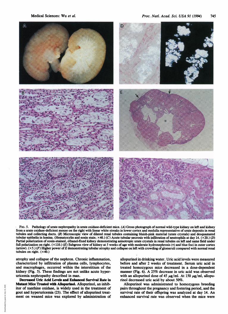
Mutant Mice. Urate oxidase-deficient mice developed pro-nounced hyperuricemia and marked hyperuricosuria (10times more urinary uric acid than normal mice). Grossnecropsies showed that lesions were confined to the kidneys.The kidneys from wild-type and heterozygous mice werenormal in appearance, with a smooth cut surface. In contrast,progressive destruction of the kidneys from the homozygousmutants was observed during postnatal development. Asearly as 6 days after birth, small cortical cysts and white-yellow deposits could be seen in the hemisected kidneys. Thedeposits were usually manifested as linear streaks in thecortex but also occurred in the medulla. They stained weaklypositive for uric acid by Gomori's silver method. However,when sections were stained with ethanolic eosin and thenviewed microscopically under polarized light, the lesionswere seen as crystalline, anisotropic structures consistentwith uric acid crystals (Fig. 5). As the disease progressed, theaffected kidneys became smaller, discolored (pale yellow),and soft. The surface of the kidney became irregular withobvious pits and scars. Multiple cysts were observed on thecut surface. Microscopically, tubular degeneration and re-generation with dilation of tubules were observed at 8 days ofage. Urate crystals persisted until 14 days and then graduallyresolved. As the obstructive disease progressed, hydrone-phrotic lesions appeared and resulted in severe hydronephro-sis at 5 weeks. Cortical cysts started as dilations within thecollecting tubules and extended from the papilla to thecorticomedullary junction. These lesions persisted throughadult life. Glomerular atrophy and dilation of Bowman'sspaces within glomeruli were associated with foci of tubular
A
12 -
,,^ 10-
.u-
8
E 6
4
ci: 2
.i
m C-
._ tovs
r-c;i:FIG. 3. Undetectable urate oxidase activity or protein in homozy-
gous mutant (-/-) mice. (A) Homozygous mutant (-/-) micelacked urate oxidase activity. Liver extracts from wild-type (e),heterozygous (o), and homozygous (A) mice were prepared andadded to a reaction mixture containing uric acid. Urate oxidaseactivity was determined by measuring the rate of decrease in A292,indicating the consumption of uric acid. Each point represents theaverage of simultaneous duplicate assays. (B) Urate oxidase proteinin wild-type (+/+), heterozygous (+/-), and homozygous (-/-)mice. Urate oxidase protein was partially purified from mouse liverby homogenization and centrifugation. Arrowhead indicates theposition of mouse urate oxidase protein, 33 kDa, in an SDS/12%polyacrylamide gel; this protein is missing in the homozygousmutants. Lane p, purified pig liver urate oxidase, 32 kDa, which is1 kDa smaller than that of the mouse (21).
B
250 -
200 -
150 -
50 -
50 -
/1~ /1 /I
Uric Acid Allantoin E400 c
-300 Erv
._
-20010-100 2~CZ
a
+1I+ ,-/ I
FIG. 4. Increased uric acid and decreased allantoin in urateoxidase-deficient mice. (A) Uric acid is increased in urate oxidase-deficient mice. Serum samples were collected from 3- to 4-week-oldmice (5-10 mice in each group) and uric acid levels were measuredspectrometrically by enzymatic reaction. The uric acid levels (mg/100 ml) were 11.0 ± 1.7 for the homozygous, 1.4 ± 0.5 for theheterozygous, and 0.9 ± 0.3 for the wild-type mice. (B) The increasein uric acid in the mutants coincides with a decrease in allantoin.Urine samples were collected from 5- to 6-week-old mice, and uricacid and allantoin were measured.
744 Medical Sciences: Wu et al.
Dow
nloa
ded
by g
uest
on
July
15,
202
0
Proc. Natl. Acad. Sci. USA 91 (1994) 745
D
AJ7~~~%
-*r lb-
*,iv,~~~~~~~~~~~~~~~~~~4
IP
~~~~~Q .f-n
V~~~~~~~~~~~~~~~~~~~~~~~~~~~~~~~~~1:
FIG. 5. Pathology of urate nephropathy in urate oxidase-deficient mice. (A) Gross photograph of normal wild-type kidney on left and kidneyfrom a urate oxidase-deficient mouse on the right with linear white streaks in lower cortex and medulla representative of urate deposits in renaltubules and collecting ducts. (B) Microscopic view of dilated renal tubules containing bluish-pink material (urate crystals) and desquamatedtubular epithelia in lumina. (Hematoxylin and eosin stain; x40.) (C) Acute tubular necrosis with infitration of neutrophils at day 14. (x20.) (D)Partial polarization of eosin-stained, ethanol-fixed kidney demonstrating anisotropic urate crystals in renal tubules on left and same field underfull polarization on right. (x 110.) (E) Subgross view of kidney at 5 weeks of age with moderate hydronephrosis (*) and blue foci in outer cortex(arrow). (x5.) (F) Higher power ofE demonstrating tubular atrophy and collapse on left with crowding ofglomeruli compared with normal renaltubules on right. (x40.)
atrophy and collapse of the nephron. Chronic inflammation,characterized by infiltration of plasma cells, lymphocytes,and macrophages, occurred within the interstitium of thekidney (Fig. 5). These findings are not unlike acute hyper-uricemia nephropathy described in man.
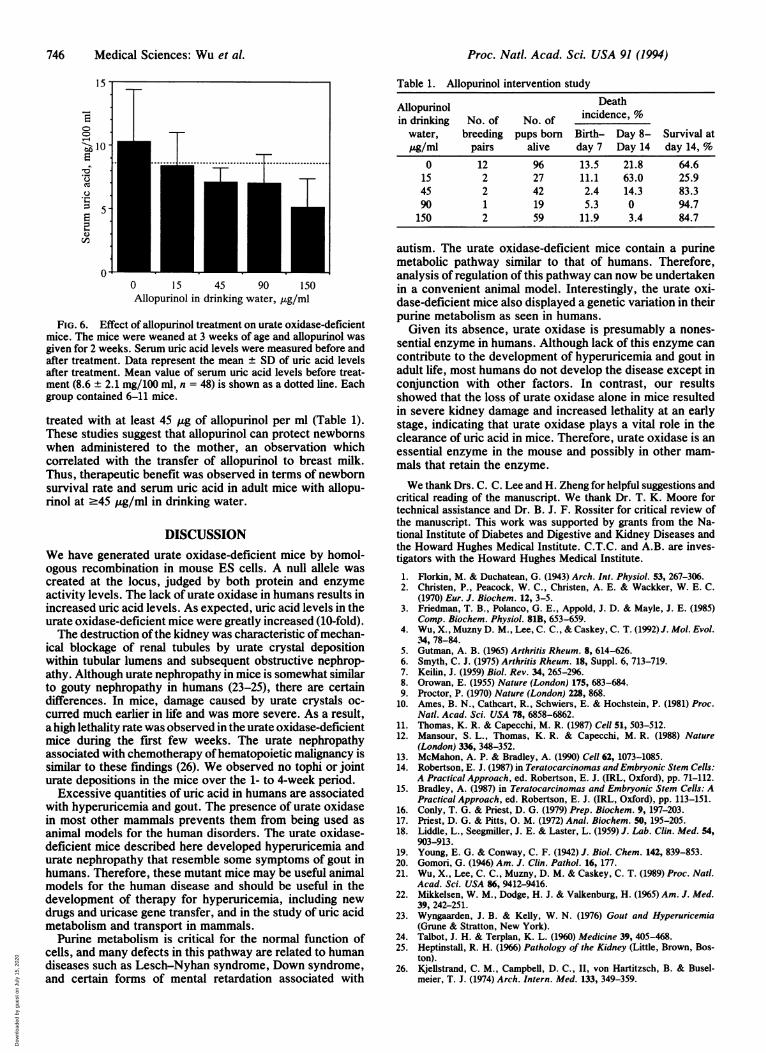
Decreased Uric Acid Levels and Enhanced Survival Rate inMutant Mice Treated with Aflopurinol. Allopurinol, an inhib-itor of xanthine oxidase, is widely used in the treatment ofgout and hyperuricemia (23). The effect of allopurinol treat-ment on weaned mice was explored by administration of
allopurinol in drinking water. Uric acid levels were measuredbefore and after 2 weeks of treatment. Serum uric acid intreated homozygous mice decreased in a dose-dependentmanner (Fig. 6). A 25% decrease in uric acid was observedwith an allopurinol dose of 45 ,ug/ml. At 150 ,g/ml, allopu-rinol decreased uric acid by about 50%.
Allopurinol was administrated to homozygous breedingpairs throughout the pregnancy and fostering period, and thesurvival rate of their offspring was analyzed at day 14. Anenhanced survival rate was observed when the mice were
Medical Sciences: Wu et al.
Dow
nloa
ded
by g
uest
on
July
15,
202
0
Proc. Natl. Acad. Sci. USA 91 (1994)
15-
r-6
U- 1
0* I..................
J0 15 45 90 150Allopurinol in drinking water, ,g/ml
FIG. 6. Effect of allopurinol treatment on urate oxidase-deficientmice. The mice were weaned at 3 weeks of age and allopurinol wasgiven for 2 weeks. Serum uric acid levels were measured before andafter treatment. Data represent the mean ± SD of uric acid levelsafter treatment. Mean value of serum uric acid levels before treat-ment (8.6 ± 2.1 mg/100 ml, n = 48) is shown as a dotted line. Eachgroup contained 6-11 mice.
treated with at least 45 ug of allopurinol per ml (Table 1).These studies suggest that allopurinol can protect newbornswhen administered to the mother, an observation whichcorrelated with the transfer of allopurinol to breast milk.Thus, therapeutic benefit was observed in terms of newbornsurvival rate and serum uric acid in adult mice with allopu-rinol at .45 Mg/ml in drinking water.
DISCUSSIONWe have generated urate oxidase-deficient mice by homol-ogous recombination in mouse ES cells. A null allele wascreated at the locus, judged by both protein and enzymeactivity levels. The lack of urate oxidase in humans results inincreased uric acid levels. As expected, uric acid levels in theurate oxidase-deficient mice were greatly increased (10-fold).The destruction ofthe kidney was characteristic of mechan-
ical blockage of renal tubules by urate crystal depositionwithin tubular lumens and subsequent obstructive nephrop-athy. Although urate nephropathy in mice is somewhat similarto gouty nephropathy in humans (23-25), there are certaindifferences. In mice, damage caused by urate crystals oc-curred much earlier in life and was more severe. As a result,a high lethality rate was observed in the urate oxidase-deficientmice during the first few weeks. The urate nephropathyassociated with chemotherapy ofhematopoietic malignancy issimilar to these findings (26). We observed no tophi or jointurate depositions in the mice over the 1- to 4-week period.
Excessive quantities of uric acid in humans are associatedwith hyperuricemia and gout. The presence of urate oxidasein most other mammals prevents them from being used asanimal models for the human disorders. The urate oxidase-deficient mice described here developed hyperuricemia andurate nephropathy that resemble some symptoms of gout inhumans. Therefore, these mutant mice may be useful animalmodels for the human disease and should be useful in thedevelopment of therapy for hyperuricemia, including newdrugs and uricase gene transfer, and in the study of uric acidmetabolism and transport in mammals.
Purine metabolism is critical for the normal function ofcells, and many defects in this pathway are related to humandiseases such as Lesch-Nyhan syndrome, Down syndrome,and certain forms of mental retardation associated with
Table 1. Allopurinol intervention study
Allopurinol Deathin drinking No. of No. of incidence, %
water, breeding pups born Birth- Day 8- Survival atpg/ml pairs alive day 7 Day 14 day 14, %
0 12 96 13.5 21.8 64.615 2 27 11.1 63.0 25.945 2 42 2.4 14.3 83.390 1 19 5.3 0 94.7150 2 59 11.9 3.4 84.7
autism. The urate oxidase-deficient mice contain a purinemetabolic pathway similar to that of humans. Therefore,analysis of regulation of this pathway can now be undertakenin a convenient animal model. Interestingly, the urate oxi-dase-deficient mice also displayed a genetic variation in theirpurine metabolism as seen in humans.Given its absence, urate oxidase is presumably a nones-
sential enzyme in humans. Although lack of this enzyme cancontribute to the development of hyperuricemia and gout inadult life, most humans do not develop the disease except inconjunction with other factors. In contrast, our resultsshowed that the loss of urate oxidase alone in mice resultedin severe kidney damage and increased lethality at an earlystage, indicating that urate oxidase plays a vital role in theclearance of uric acid in mice. Therefore, urate oxidase is anessential enzyme in the mouse and possibly in other mam-mals that retain the enzyme.
We thank Drs. C. C. Lee and H. Zheng for helpful suggestions andcritical reading of the manuscript. We thank Dr. T. K. Moore fortechnical assistance and Dr. B. J. F. Rossiter for critical review ofthe manuscript. This work was supported by grants from the Na-tional Institute of Diabetes and Digestive and Kidney Diseases andthe Howard Hughes Medical Institute. C.T.C. and A.B. are inves-tigators with the Howard Hughes Medical Institute.1.2.
3.
4.
5.6.7.8.9.
10.
11.12.
13.14.
15.
16.17.18.
19.20.21.
22.
23.
24.25.
26.
Florkin, M. & Duchatean, G. (1943) Arch. Int. Physiol. 53, 267-306.Christen, P., Peacock, W. C., Christen, A. E. & Wackker, W. E. C.(1970) Eur. J. Biochem. 12, 3-5.Friedman, T. B., Polanco, G. E., Appold, J. D. & Mayle, J. E. (1985)Comp. Biochem. Physiol. 81B, 653-659.Wu, X., Muzny D. M., Lee, C. C., & Caskey, C. T. (1992) J. Mol. Evol.34, 78-84.Gutman, A. B. (1965) Arthritis Rheum. 8, 614-626.Smyth, C. J. (1975) Arthritis Rheum. 18, Suppl. 6, 713-719.Keilin, J. (1959) Biol. Rev. 34, 265-296.Orowan, E. (1955) Nature (London) 175, 683-684.Proctor, P. (1970) Nature (London) 228, 868.Ames, B. N., Cathcart, R., Schwiers, E. & Hochstein, P. (1981) Proc.Natl. Acad. Sci. USA 78, 6858-6862.Thomas, K. R. & Capecchi, M. R. (1987) Cell 51, 503-512.Mansour, S. L., Thomas, K. R. & Capecchi, M. R. (1988) Nature(London) 336, 348-352.McMahon, A. P. & Bradley, A. (1990) Cell 62, 1073-1085.Robertson, E. J. (1987) in Teratocarcinomas and Embryonic Stem Cells:A Practical Approach, ed. Robertson, E. J. (IRL, Oxford), pp. 71-112.Bradley, A. (1987) in Teratocarcinomas and Embryonic Stem Cells: APractical Approach, ed. Robertson, E. J. (IRL, Oxford), pp. 113-151.Conly, T. G. & Priest, D. G. (1979) Prep. Biochem. 9, 197-203.Priest, D. G. & Pitts, 0. M. (1972) Anal. Biochem. 50, 195-205.Liddle, L., Seegmilier, J. E. & Laster, L. (1959) J. Lab. Clin. Med. 54,903-913.Young, E. G. & Conway, C. F. (1942) J. Biol. Chem. 142, 839-853.Gomori, G. (1946) Am. J. Clin. Pathol. 16, 177.Wu, X., Lee, C. C., Muzny, D. M. & Caskey, C. T. (1989) Proc. Natl.Acad. Sci. USA 86, 9412-9416.Mikkelsen, W. M., Dodge, H. J. & Valkenburg, H. (1965) Am. J. Med.39, 242-251.Wyngaarden, J. B. & Kelly, W. N. (1976) Gout and Hyperuricemia(Grune & Stratton, New York).Talbot, J. H. & Terplan, K. L. (1960) Medicine 39, 405-468.Heptinstall, R. H. (1966) Pathology of the Kidney (Little, Brown, Bos-ton).Kjellstrand, C. M., Campbell, D. C., II, von Hartitzsch, B. & Busel-meier, T. J. (1974) Arch. Intern. Med. 133, 349-359.
746 Medical Sciences: Wu et al.
Dow
nloa
ded
by g
uest
on
July
15,
202
0