HYMENOLEPIDID CESTODES FROM GREBES (AVES, PODICIPEDIDAE) IN
18
Vestnik zoologii, 35(6): 13—31, 2001 © G. P. Vasileva, V. V. Kornyushin, T. Genov, 2001 UDC 595.121 : 598.233(477) HYMENOLEPIDID CESTODES FROM GREBES (AVES, PODICIPEDIDAE) IN UKRAINE: THE GENUS CONFLUARIA G. P. Vasileva 1 , V. V. Kornyushin 2 , T. Genov 1 1 Central Laboratory of General Ecology, Bulgarian Academy of Sciences, 2, Gagarin Str., 1113 Sofia, Bulgaria 2 Schmalhausen Institute of Zoology, vul. B. Khmelnits’kogo, 15, Kyiv-30, MSP, 01601 Ukraine Accepted 28 July 2000 Hymenolepidid Cestodes from Grebes (Aves, Podicipedidae) in Ukraine: the Genus Confluaria. Va- sileva G. P., Kornyushin V. V., Genov T. – Redescriptions of 5 species of the hymenolepidid cestodes from the genus Confluaria Ablasov in Spasskaja, 1966 recorded from grebes (Podicipedidae) in Ukraine are presented: Confluaria podicipina from Podiceps nigricollis; Confluaria furcifera from P. grisegena, P. cristatus and P. nigricollis; Confluaria pseudofurcifera (new geographical record) from P. cristatus; Con- fluaria capillaris from P. grisegena and P. nigricollis; Confluaria multistriata from Tachybaptus ruficollis. Confluaria krabbei sp. n. from T. ruficollis is described. It is differentiated from C. multistriata by pos- sessing rostellar hooks with epiphyseal thickenings comprising both handle and guard and by the shorter cirrus-sac which does not reach the mid-line of the proglottis. K e y w o r d s : Cestodes, Hymenolepididae, Confluaria, grebes, Ukraine. Öåñòîäû-ãèìåíîëåïèäèäû ïîãàíîê (Aves, Podicipedidae) Óêðàèíû: ðîä Confluaria. Âàñèëåâà Ã. Ï., Êîðíþøèí Â. Â., Ãåíîâ Ò. – Äàíû ïåðåîïèñàíèÿ 5 âèäîâ ãèìåíîëåïèäèä ðîäà Confluaria Ab- lasov in Spasskaja, 1966, çàðåãèñòðèðîâàííûõ ó ïîãàíîê â Óêðàèíå: C. podicipina îò Podiceps nigri- collis; C. furcifera îò P. nigricollis, P. grisågena è P. cristatus; C. pseudofurcifera (ðåãèñòðèðóåòñÿ â Óê- ðàèíå âïåðâûå) îò P. cristatus; C. capillaris îò P. grisågena, P. nigricollis; C. multistriata îò Tachy- baptes ruficollis. Îïèñàí òàêæå íîâûé âèä Confluaria krabbei sp. n. îò T. ruficollis, êîòîðûé äèôôå- ðåíöèðóåòñÿ îò C. multistriata ïî íàëè÷èþ íà êðþ÷üÿõ õîáîòêà áîëüøèõ ýïèôèçàðíûõ óòîëùå- íèé, îõâàòûâàþùèõ êàê ðóêîÿòêó, òàê è îòðîñòîê êîðíÿ, à òàêæå ìåíüøåé äëèíå áóðñû öèððó- ñà, íå äîñòèãàþùåé ñðåäíåé ëèíèè ÷ëåíèêà. Êëþ÷åâûå ñëîâà: öåñòîäû, Hymenolepididae, Confluaria, ïîãàíêè, Óêðàèíà. Introduction In our previous publication (Vasileva et al., 2001), the species of the genera Dollfusilepis Vasileva, Georgiev & Genov, 1998 and Parafimbriaria Voge & Read, 1954 were redescribed on the basis of cestode specimens from grebes (Aves, Podicipedidae) in Ukraine. The further examinations on the hymenolepidid cestodes from grebes in the Parasitological Collection of the I. I. Schmalhausen Institute of Zoology of the National Academy of Sciences of Ukraine (SIZK) resulted in the identification of 6 species of the genus Confluaria Ablasov in Spasskaya, 1966, including one new species. Their description is the aim of the present publication. Material and methods Specimens of Confluaria spp. from the Parasitological Collection of the SIZK had been isolated from 34 specimens of four grebe species: 12 Podiceps cristatus (L.), 10 P. grisegena (Boddaert), 10 P. nigricol- lis (Brehm) and 2 Tachybaptus ruficollis (Pallas). The birds were captured in the period 1949—1995 from various regions of Ukraine by L. A. Smogorzhevskaya, V. V. Kornyushin, I. V. Grushchinskaya. Localities: Yagotyn (lake Supoy), Kyiv (mouth of Desna river), Pology-Yanenky, Tatsenky (river Dniper) – Kyiv Re- gion; Prokhorovka, Kaniv, Khreshchatyc (river Dniper), Trakhtemirov (Kaniv Reservoir), Zhovnino (Kremenchug Reservoir) – Cherkassy Region; Bugayevka (Kremenchug Reservoir) – Poltava Region; Vy- shchetarasovka (river Dniper) – Dnepropetrovsk Region; districts Potievka, Solenoozerny, Yagorlytsky Kute Black-Sea Biosphere Resreve (BSBR) – Kherson Region; Vilkovo (Danube Delta) – Odessa Region; Min- ino (Crimean canal), Olenevka (cape Tarchankut) – Crimea. Details of hosts, localities and collection numbers are given in the text for each species. Methods were described in the previous publications (Vasileva et al., 2001). The index length of the cirrus-sac/maximum width of the proglottis (L CS /W P ), calculated for well-relaxed mature proglottides (taken from specimens in exact dorso-ventral position only), was used. The index length of the vagina/length of the
Transcript of HYMENOLEPIDID CESTODES FROM GREBES (AVES, PODICIPEDIDAE) IN

Vestnik zoologii, 35(6): 13—31, 2001 © G. P. Vasileva, V. V. Kornyushin, T. Genov, 2001
UDC 595.121 : 598.233(477)
HYMENOLEPIDID CESTODES FROM GREBES (AVES, PODICIPEDIDAE) IN UKRAINE: THE GENUS CONFLUARIA
G. P. Vasileva1, V. V. Kornyushin2, T. Genov1
1Central Laboratory of General Ecology, Bulgarian Academy of Sciences, 2, Gagarin Str., 1113 Sofia, Bulgaria 2 Schmalhausen Institute of Zoology, vul. B. Khmelnits’kogo, 15, Kyiv-30, MSP, 01601 Ukraine
Accepted 28 July 2000
Hymenolepidid Cestodes from Grebes (Aves, Podicipedidae) in Ukraine: the Genus Confluaria. Va-sileva G. P., Kornyushin V. V., Genov T. – Redescriptions of 5 species of the hymenolepidid cestodes from the genus Confluaria Ablasov in Spasskaja, 1966 recorded from grebes (Podicipedidae) in Ukraine are presented: Confluaria podicipina from Podiceps nigricollis; Confluaria furcifera from P. grisegena, P. cristatus and P. nigricollis; Confluaria pseudofurcifera (new geographical record) from P. cristatus; Con-fluaria capillaris from P. grisegena and P. nigricollis; Confluaria multistriata from Tachybaptus ruficollis. Confluaria krabbei sp. n. from T. ruficollis is described. It is differentiated from C. multistriata by pos-sessing rostellar hooks with epiphyseal thickenings comprising both handle and guard and by the shorter cirrus-sac which does not reach the mid-line of the proglottis.
Key wo rd s : Cestodes, Hymenolepididae, Confluaria, grebes, Ukraine.
Öåñòîäû-ãèìåíîëåïèäèäû ïîãàíîê (Aves, Podicipedidae) Óêðàèíû: ðîä Confluaria. Âàñèëåâà Ã. Ï., Êîðíþøèí Â. Â., Ãåíîâ Ò. – Äàíû ïåðåîïèñàíèÿ 5 âèäîâ ãèìåíîëåïèäèä ðîäà Confluaria Ab-lasov in Spasskaja, 1966, çàðåãèñòðèðîâàííûõ ó ïîãàíîê â Óêðàèíå: C. podicipina îò Podiceps nigri-collis; C. furcifera îò P. nigricollis, P. grisågena è P. cristatus; C. pseudofurcifera (ðåãèñòðèðóåòñÿ â Óê-ðàèíå âïåðâûå) îò P. cristatus; C. capillaris îò P. grisågena, P. nigricollis; C. multistriata îò Tachy-baptes ruficollis. Îïèñàí òàêæå íîâûé âèä Confluaria krabbei sp. n. îò T. ruficollis, êîòîðûé äèôôå-ðåíöèðóåòñÿ îò C. multistriata ïî íàëè÷èþ íà êðþ÷üÿõ õîáîòêà áîëüøèõ ýïèôèçàðíûõ óòîëùå-íèé, îõâàòûâàþùèõ êàê ðóêîÿòêó, òàê è îòðîñòîê êîðíÿ, à òàêæå ìåíüøåé äëèíå áóðñû öèððó-ñà, íå äîñòèãàþùåé ñðåäíåé ëèíèè ÷ëåíèêà.
Êëþ÷åâûå ñëîâà : öåñòîäû, Hymenolepididae, Confluaria, ïîãàíêè, Óêðàèíà.
Introduction
In our previous publication (Vasileva et al., 2001), the species of the genera Dollfusilepis Vasileva, Georgiev & Genov, 1998 and Parafimbriaria Voge & Read, 1954 were redescribed on the basis of cestode specimens from grebes (Aves, Podicipedidae) in Ukraine. The further examinations on the hymenolepidid cestodes from grebes in the Parasitological Collection of the I. I. Schmalhausen Institute of Zoology of the National Academy of Sciences of Ukraine (SIZK) resulted in the identification of 6 species of the genus Confluaria Ablasov in Spasskaya, 1966, including one new species. Their description is the aim of the present publication.
Material and methods
Specimens of Confluaria spp. from the Parasitological Collection of the SIZK had been isolated from 34 specimens of four grebe species: 12 Podiceps cristatus (L.), 10 P. grisegena (Boddaert), 10 P. nigricol- lis (Brehm) and 2 Tachybaptus ruficollis (Pallas). The birds were captured in the period 1949—1995 from various regions of Ukraine by L. A. Smogorzhevskaya, V. V. Kornyushin, I. V. Grushchinskaya. Localities: Yagotyn (lake Supoy), Kyiv (mouth of Desna river), Pology-Yanenky, Tatsenky (river Dniper) – Kyiv Re-gion; Prokhorovka, Kaniv, Khreshchatyc (river Dniper), Trakhtemirov (Kaniv Reservoir), Zhovnino (Kremenchug Reservoir) – Cherkassy Region; Bugayevka (Kremenchug Reservoir) – Poltava Region; Vy-shchetarasovka (river Dniper) – Dnepropetrovsk Region; districts Potievka, Solenoozerny, Yagorlytsky Kute Black-Sea Biosphere Resreve (BSBR) – Kherson Region; Vilkovo (Danube Delta) – Odessa Region; Min-ino (Crimean canal), Olenevka (cape Tarchankut) – Crimea. Details of hosts, localities and collection numbers are given in the text for each species.
Methods were described in the previous publications (Vasileva et al., 2001). The index length of the cirrus-sac/maximum width of the proglottis (LCS/WP), calculated for well-relaxed mature proglottides (taken from specimens in exact dorso-ventral position only), was used. The index length of the vagina/length of the

G. P. Vasileva, V. V. Kornyushin, T. Genov 14
cirrus-sac (LV/LCS), calculated for mature proglottides, was used for distinguishing the two closely related species Confluaria multistriata and C. krabbei.
The metrical data are given as the range, the mean in parentheses and the number of measurements taken (n). The measurements are given in micrometers unless otherwise stated.
Results
Confluaria Ablasov in Spasskaya, 1966
Syn. Colymbilepis Spasskaya, 1966; Dimorphocanthus Maksimova, 1989
Remarks . Recently, the Palaearctic species of the genus Confluaria were a subject of a taxonomic re-vision (Vasileva et al., 1999 a, 1999 b, 2000). These studies revealed the validity of C. capillaris (Rudolphi, 1810) (Syn. C. capillaroides (Fuhrmann, 1906)), C. multistriata (Rudolphi, 1810), C. japonica (Yamaguti, 1935), C. furcifera (Krabbe, 1869) and C. podicipina (Szymanski, 1905) (Syn. C. spasskii Ablasov in Spasskaya, 1966). In addition, C. pseudofurcifera Vasileva, Georgiev & Genov, 2000, a specific parasite of Podiceps cristatus, was described. A key to the species was proposed and the previous records of Confluaria spp. from Palaearctic grebes were re-evaluated. This revision also confirmed the synonymy of the genera Colymbilepis and Dimorphocanthus with Confluaria, as previously proposed by Czaplinski (in Czaplinski & Vaucher, 1994).
The present study recorded from Ukrainian grebes 5 species of Confluaria recently redescribed by Va-sileva et al. (1999 a, 1999 b, 2000). In addition, the present material provided a basis for the description of C. krabbei sp. n. from Tachybaptus ruficollis (Syn. Confluaria sp. Vasileva, Georgiev & Genov, 1999 b).
Confluaria podicipina (Szymanski, 1905) Spasskaya, 1966
Syn. Confluaria spasskii Ablasov in Spasskaya, 1966
Spec imen s s t ud i ed . From P. nigricollis, Ukraine: Coll. Nos 49—1, 50—2, Zhovnino, 06.06.1974, 18 mature specimens and about 15 fragments of strobila, stained whole-mounts (14 slides), 6 scoleces mounted in Berlese medium and 6 mature specimens and 12 fragments of strobila, stained whole-mounts (4 slides). Coll. No 68—5, Bugayevka, 13.07.1974, 4 immature specimens and about 10 fragments of strobila, stained whole-mounts (4 slides), all mentioned by Grushchinskaya (1978) as Confluaria spasskii. Coll. No 24—1, Solenooz-erny (BSBR), 15.09.1960, 1 immature specimen, stained whole-mount. Coll. No 186—3, Yagorlytsky Kut (BSBR), 02.11.1963, 1 scolex mounted in Berlese medium and Coll. No 137—1, Yagotyn, 10.10.1952, Coll. No 51—2, Yagorlytsky Kut (BSBR), 12.12.1962, Coll. Nos 714—1, 715—2, Minino, 20.08.1969, Coll. No 54—3, Zhovnino, 08.07.1974 – fragments of strobila or separated scoleces.
Redescription (based on specimens from Coll. No 49—1; fig. 1—6; for some meas-urements see tab. 1). Strobila band-like, with maximum width at level of pregravid proglottides. Scolex (fig. 1) with anterior conical protrusion and maximum width at middle of suckers. Suckers round, with well-developed musculature. Rostellum powerful, mushroom-like, with well-developed musculature; intensely staining glandular cells situ-ated in it. Rostellar sheath with well-developed musculature of walls, passing beyond posterior margins of suckers; intensely stained glandular masses present in it. Rostellum with crown of 10 hooks; each hook (fig. 2) consists of refractive particle (with well de-veloped blade, shorter guard and handle) and long epiphyseal thickening of both handle and guard. Measurements of rostellar hooks: length of refractive particle 21—24 (23, n=24), length of blade 11—14 (13, n=24), length of base 41—51 (46, n=24). Proglottides (fig. 3—5) craspedote, much wider than long. Genital pores unilateral, situated almost in middle of lateral proglottis margin. Genital atrium (fig. 6) deep, cylindrical, surrounded with intensely stained cells. Ventral and dorsal osmoregulatory canals without transverse anastomoses. Diameter of osmoregulatory canals: ventral 18—31 (25, n=14), dorsal 10—15 (13, n=14). Genital ducts dorsal to osmoregulatory canals.
Strobila protandrous. Testes (fig. 3) three, small, compact; situated dorsally to female glands or to their primordia, usually in median field; testes arranged in triangle, rarely in row. External seminal vesicle (fig. 3—6) oval or elliptical. Cirrus-sac (fig. 3—6) elongate, cylindrical, thin-walled, usually crossing poral osmoregulatory canals; in hermaphroditic mature proglottides cirrus-sac does not extend to mid-line of proglot-tis. Internal seminal vesicle elongate, occupying almost 1/2 of cirrus-sac. Evaginated cirrus (fig. 3, 6) long, curved, whip-shaped; armed with minute needle-shaped spines arranged in 7—8 longitudinal rows; distal part of evaginated cirrus with sparse spines.

Hymenolepidid Cestodes from Grebes in Ukraine: the Genus Confluaria … 15
Vitellarium (fig. 4) compact, median, elliptical. Ovary (fig. 4) median, with three compact lobes. Seminal receptacle (fig. 3—6) oval or elliptical, situated almost at mid-dle of proglottis, dorsally to ovary. Copulatory vagina (fig. 6) long, tubular, thick-walled, opening and passing ventrally to cirrus-sac. Conductive part curved, slender, surrounded with intensely stained cells.
Fig. 1—6. Confluaria podicipina, specimens from P. nigricollis: 1 – scolex; 2 – rostellar hooks; 3 – male ma-ture proglottis; 4 – hermaphroditic mature proglottis; 5 – pregravid proglottis; 6 – terminal genital ducts in hermaphroditic mature proglottis. Scale-bars: 1, 5 – 100 μm; 2—4, 6 – 50 μm.
Ðèñ. 1—6. Confluaria podicipina, ýêçåìïëÿðû îò P. nigricollis: 1 – cêîëåêñ; 2 – êðþ÷üÿ õîáîòêà; 3 – ìóæ-ñêîé ÷ëåíèê; 4 – ãåðìàôðîäèòíûé ÷ëåíèê; 5 – íå âïîëíå çðåëûé ÷ëåíèê; 6 – êîíöåâûå ó÷àñòêè ïî-ëîâûõ ïðîòîêîâ â ãåðìàôðîäèòíîì ÷ëåíèêå. Ìàñøòàáíàÿ ëèíåéêà: 1, 5 – 100 ìêì; 2—4, 6 – 50 ìêì.
G. P. Vasileva, V. V. Kornyushin, T. Genov 16
Developing uterus (fig. 5) sac-like, transversely elongated, crossing dorsally osmo-regulatory canals. Proglottides with fully-developed uterus and ripe eggs not available.
Rema rk s . The Ukrainian specimens of C. podicipina are in agreement with the main morphological and metrical characters of this species as described by previous authors (Vasileva et al., 2000) (tab. 1). The specimens studied have been reported as Confluaria spasskii too (Grushchinskaya, 1978). The present study confirms that this material belongs to C. podicipina.
The host range of C. podicipina includes P. auritus (L.), P. cristatus, P. nigricollis and T. ruficollis, type location – Dublyany, Lvov Region, Ukraine. Its geographical range except Ukraine (Szymanski, 1905 and present study), includes Poland, Slovak Republic, Czech Republic, Romania, Bulgaria, Kazakhstan (Macko, 1962; Rysavy, 1961; Rysavy & Sitko, 1992; Chiriac, 1960; Vasileva et al., 2000). In the Parasitological Col-lection of the SIZK, specimens collected by A. Eminov from P. nigricollis in Turkmenistan are preserved (Coll. Nos 2, 136). Their study revealed the affiliation of this material to C. podicipina. The remaining re-cords (Vasileva et al., 2000) have been published without descriptions or illustrations and need further con-firmation.
Confluaria furcifera (Krabbe, 1869) Spasskaya, 1966
Spec imen s s t u d i ed . From P. cristatus: Coll. No 31—2, Khreshchatyk, 11.09.1950, one slide with mature fragment of strobila, stained whole mount.
From P. grisegena: Coll. Nos 570—10, 573—13, 567—7, Potievka (BSBR), 10.09.1988, about 20 frag-ments of strobila, stained whole-mounts (8 slides), 13 scoleces mounted in Berlese medium. Coll. N 856—14, Potievka (BSBR), 17.08.1989, about 5 fragments of strobila, stained whole-mounts (2 slides). Coll. No 29—2, Vyschetarasovka, 15.05.1953, 5 immature specimens, stained whole-mounts (5 slides), 5 scoleces mounted in Berlese medium and Coll. Nos 2—2, 7—4, Olenevka, 04. and 07.08.1958, Coll. Nos 88—1, 518—3 Yagorlytsky Kut (BSBR), 01.04.1963 and 28.11.1964, fragments of strobila or scoleces.
From P. nigricollis: Coll. Nos 49—1, 54—3, Zhovnino, 06.06.1974, 2 immature specimens and about 7 fragments of strobila, stained whole-mounts (3 slides). 08.07.1974, 2 immature specimens and 3 fragments of strobila, stained whole-mounts (one slide). Coll. No 24—1, Solenoozerny (BSBR), 15.09.1960, 3 fragments of strobila, stained whole-mounts (2 slides). Coll. No 137—1, Yagotyn, 10.10.1952, 2 immature specimens and 3 fragments of strobila, stained whole-mounts (one slide). Coll. No 51—2 Yagorlytsky Kut (BSBR), 02.12.1962, Coll. No 55—4 Zhovnino, 08.07.1974, fragments of strobila or scoleces.
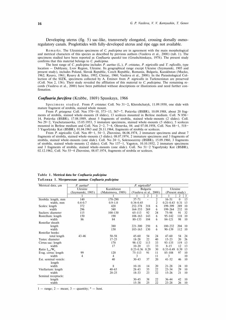
Tab l e 1 . Metrical data for Confluaria podicipina
Òàáëèöà 1 . Ìåòðè÷åñêèå äàííûå Confluaria podicipina
P. auritus* P. nigricollis* Ukraine
(Szymanski, 1905)Kazakhstan
(Maksimova, 1989) Bulgaria
(Vasileva et al., 2000) Ukraine
(Present study)
Metrical data, μm
1 1 1 2 3 1 2 3 Strobila: length, mm 140 170-290 37-71 - 2 16-51 0 13
width, mm 0.4-0.7 0.9-1.0 0.34-0.45 2 0.21-0.43 0.31 13Scolex: length 372 420 252-378 318 6 199-399 289 10
width 296 340 164-353 269 6 199-264 232 10Suckers: diameter 115 100-130 65-113 92 24 75-98 91 32Rostellum: length 150 190 108-163 143 6 95-142 118 10
width 86 84 80-133 104 6 84-121 98 10Rostellar sheath
length 380 221-300 250 6 180-232 204 10width 150 103-163 130 6 90-139 112 10
Rostellar hooks: total length 43-46 50-58 45-60 54 24 47-60 54 24
Testes: diameter 17-25 18-28 22 40 15-23 20 26Cirrus-sac: length 170 98-132 113 33 93-135 119 13
width 17 10-20 13 33 8-15 12 13Ratio Lcs/Wp 0.23-0.36 0.29 30 0.33-0.49 0.39 13Evag. cirrus: length 99 120 75-113 91 11 85-108 97 10
width 4 4 3 11 3 10Ext. seminal vesicle: 40 30-43 37 20 41-52 46 10
length width 8 10-18 14 20 21-28 24 10
Vitellarium: length 40-65 28-43 35 22 23-34 29 10width 20-25 18-33 23 22 15-26 21 10
Seminal receptacle: length 30-45 38 22 36-44 42 10width 15-38 25 22 23-28 26 10
1 – range; 2 – mean; 3 – quantity; * – host.

Hymenolepidid Cestodes from Grebes in Ukraine: the Genus Confluaria … 17
From T. ruficollis: Coll. Nos 4—1, 9, Pology Yanenky, 22.08.1953, 20 fragments of strobila, stained whole-mounts (7 slides).
Redescr ip t ion (based on specimens from P. grisegena, Coll. Nos 570—10, 567—7, 573—1; fig. 7—12; for some measurements see tab. 2). Strobila band-like, slender. Scolex (fig. 7) oval, with conically protruded anterior part and maximum width at level of suckers. Suckers oval, with weakly-developed musculature. Rostellum conical, with well-developed musculature; intensely stained cells situated in it. Rostellar sheath thin-walled, passing beyond posterior margins of suckers; intensely stained masses present in it. Rostellar hooks 10; each hook (fig. 8) consisting of refractive particle (with long
Fig. 7—12. Confluaria furcifera, specimens from P. grisegena: 7 – scolex; 8 – rostellar hooks; 9 – male mature proglottis; 10 – hermaphroditic mature proglottis; 11 – pregravid proglottis; 12 – terminal genital ducts in hermaphroditic mature proglottis. Scale-bars: 7, 9, 10 – 100 μm; 8 – 20 μm; 11 – 200 μm; 12 – 50 μm.
Ðèñ. 7—12. Confluaria furcifera, ýêçåìïëÿðû îò P. grisegena: 7 – ñêîëåêñ; 8 – êðþ÷üÿ õîáîòêà; 9 – ìóæ-ñêîé ÷ëåíèê; 10 – ãåðìàôðîäèòíûé ÷ëåíèê; 11 – íå âïîëíå çðåëûé ÷ëåíèê; 12 – êîíöåâûå ó÷àñòêè ïîëîâûõ ïðîòîêîâ â ãåðìàôðîäèòíîì ÷ëåíèêå. Ìàñøòàáíàÿ ëèíåéêà: 7, 9, 10 – 100 ìêì; 8 – 20 ìêì; 11 – 200 ìêì; 12 – 50 ìêì.

G. P. Vasileva, V. V. Kornyushin, T. Genov 18
blade and shorter guard and handle) and epiphyseal thickening of handle. Measure-ments of hooks: length of refractive particle 18—21 (19, n=28), length of blade 11—12 (11, n=28), length of base 16—19 (18, n=28). Proglottides (fig. 9—11) craspedote, wider than long. Genital pores unilateral, situated almost in middle of lateral proglottis margin. Genital atrium (fig. 12) cylindrical, surrounded by intensely stained cells. Ven-tral and dorsal osmoregulatory canals without transverse anastomoses; diameter of ven-tral osmoregulatory canals 28—39 (34, n=10), diameter of dorsal osmoregulatory canals 10—21 (15, n=10). Genital ducts dorsal to osmoregulatory canals.
Testes (fig. 9) three, compact, situated dorsally to female glands; arranged in one row or in shallow triangle. External seminal vesicle (fig. 9—12) elongate, elliptical. Cir-rus-sac (fig. 9—12) elongate, thin-walled, usually crossing poral osmoregulatory canals, sometimes reaching mid-line of proglottis. Internal seminal vesicle elongate, usually occupies more than 1/2 of cirrus-sac. Evaginated cirrus (fig. 9, 12) cylindrical, armed basally with dense, needle-shaped spines; distal part of evaginated cirrus armed with sparse, rosethorn-shaped spines; extremity of fully-evaginated cirrus unarmed.
Vitellarium (fig. 10) compact, elliptical, median. Ovary (fig. 10) with three com-pact lobes, dorsal to vitellarium, median. Seminal receptacle (fig. 9—12) oval, ventral to male genital ducts and dorsal to ovary. Copulatory vagina (fig. 12) refractive, tubu-lar, surrounded by thin cellular sleeve; opening and passing ventrally to cirrus-sac. Conductive part of vagina coiled, thin-walled, surrounded by intensely stained cells.
Developing uterus (fig. 11) sac-like, transversely elongate, crossing dorsally osmo-regulatory canals. Proglottides with fully-developed uterus and ripe eggs not available.
Observat ions on spec imens from P. nigricollis (Coll. Nos 49—1, 54—3). The morphology of these specimens is presented in figures 13—18. The metrical data are included in table 2. Additional measurements: Rostellar hooks: length of refractive par-ticle 15—16 (15, n=5), length of blade 8—9 (9, n=5), length of base 15—18 (16, n=5). Diameter of osmoregulatory canals: ventral 23—31 (28, n=10), dorsal 5—10 (8, n=10).
Rema rk s . The present results on the morphology of C. furcifera correspond to the data of the previ-ous descriptions (Vasileva et al., 2000). The comparative study on C. furcifera from P. grisegena and P. nigri-collis demonstrates some metrical differences between cestodes found in the two host species (tab. 2). The specimens from P. grisegena possess larger rostellar hooks (24—28 μm) whilst those from P. nigricollis have rostellar hooks 22—24 μm long (tab. 2) from both Bulgaria and Ukraine. The cestodes from P. grisegena have also a larger cirrus-sac compared to those from P. nigricollis. In spite of these differences, the main morpho-logical characters of C. furcifera from these two hosts are very similar (fig. 7—12, fig. 13—18), especially con-cerning the shape of the rostellar hooks, the ratio LCS/WP, the shape and the armament of the evaginated cirrus, the shape and the structure of the vagina which is characterised by a refractive copulatory part. Therefore, the metrical differences of C. furcifera from P. grisegena and P. nigricollis can be regarded as in-traspecific variations.
The specimens of C. furcifera from P. cristatus and T. ruficollis consists of fragments of strobila in dif-ferent stages of development. Their morphological and metrical characters (tab. 2) are in close correspon-dence with those of the specimens from P. grisegena.
Vasileva et al. (2000) redescribed C. furcifera from P. grisegena (syntypes), P. nigricollis and T. ruficollis from Bulgaria. In addition to these birds, the host range of this species includes also Podiceps auritus and Podilymbus podiceps (L.). Its geographical range includes Denmark, Germany, Bulgaria, Russia (Kam-chatka), Canada (Vasileva et al., 2000) and Ukraine (present study). Several previous records described cestodes differing in their morphology from the present results (Brglez, 1981; Vasileva et al., 2000). Many authors mentioned this species without publishing any documentations (Vasileva et al., 2000). In view of the presence of another similar species, Confluaria pseudofurcifera Vasileva, Georgiev & Genov, 2000, these re-cords need further confirmation.
Confluaria pseudofurcifera Vasileva, Georgiev & Genov, 2000
Syn. Hymenolepis furcigera (Krabbe, 1869) of Joyeux & Baer (1950); H. furcifera (Krabbe, 1869) of Ja-recka (1958) and Korpaczewska (1960); Confluaria furcifera (Krabbe, 1869) of Galkin (1986) and Rysavy & Sitko (1995)

Hymenolepidid Cestodes from Grebes in Ukraine: the Genus Confluaria … 19
Spec imen s s t ud i ed . From P. cristatus: Coll. No 1—1, Khreshchatyk, 19.10.1952, 1 immature specimen and 12 fragments of strobila, stained and mounted in Canada balsam (3 slides), 1 scolex mounted in Berlese medium. Coll. Nos 9—4, 11—5, Khreshchatik, 29.10.1952, about 20 fragments of strobila, stained and mounted in Canada balsam (5 slides). Coll. No 31—2, 11.09.1950, Coll. No 33—3, Kaniv, 17.09.1950, 2
Fig. 13—18. Confluaria furcifera, specimens from P. nigricollis: 13 – scolex; 14 – rostellar hooks; 15 – male mature proglottis; 16 – hermaphroditic mature proglottis; 17 – pregravid proglottis; 18 – terminal genital ducts in hermaphroditic mature proglottis. Scale-bars: 13, 15—17 – 100 μm; 14 – 20 μm; 18 – 50 μm.
Ðèñ. 13—18. Confluaria furcifera, ýêçåìïëÿðû îò P. nigricollis: 13 – ñêîëåêñ; 14 – êðþ÷üÿ õîáîòêà; 15–ìóæñêîé ÷ëåíèê; 16 – ãåðìàôðîäèòíûé ÷ëåíèê; 17 – íå âïîëíå çðåëûé ÷ëåíèê; 18 – êîíöåâûå ó÷à-ñòêè ïîëîâûõ ïðîòîêîâ â ãåðìàôðîäèòíîì ÷ëåíèêå. Ìàñøòàáíàÿ ëèíåéêà: 13, 15—17 – 100 ìêì; 14 –20 ìêì; 18 – 50 ìêì.

G. P. Vasileva, V. V. Kornyushin, T. Genov 20
scoleces mounted in Berlese medium. Coll. No 97—1, Prohorovka, 06.09.1949, 10 fragments of strobila, stained and mounted in Canada balsam (3 slides). Coll. No 28—1, Vyshchetarasovka, 15.05.1953, 4 frag-ments of strobila, stained and mounted in Canada balsam (1 slide). Coll. No 291—2, Potievka (BSBR), 29.07.1987, 3 mature specimens stained whole-mounts (5 slides), 1 specimen and 4 fragments of strobila mounted in Berlese medium (2 slides). Coll. No 1031—1, Vilkovo, 30.07.1995, 1 immature specimen mounted in Berlese medium. Coll. No 37—4, Khreshchatyk, 22.09.1950, Coll. No 1—1, Kyiv, 01.04.1953, Coll Nos 70—1, 71—2, Zhovnino, 15 and 16.07.1974 – fragments of strobila or scoleces.
Redescr ip t ion (based on specimens from Coll. No 291—2; fig. 19—24; for measurements see tab. 3). Strobila band-like. Scolex (fig. 19) with conically tapering anterior part; maximum width at about middle of suckers. Neck long. Suckers round, unarmed, with well-developed musculature. Rostellum with well-developed muscula-ture of walls; intensely staining glandular cells situated centrally. Rostellar sheath with weakly-developed musculature of walls, passing beyond posterior margins of suckers; intensely stained glandular masses present in it. Rostellum with crown of 10 hooks; each hook (fig. 20) consisting of refractive particle (with long straight blade, short handle and slightly larger guard), and large epiphyseal thickening of both handle and guard. Measurements of hooks: length of refractive particle 20—24 (22, n=11), length of blade 13—15 (14, n=11), length of base 23—27 (25, n=11). Proglottides (fig. 21—23) craspedote, much wider than long. Genital pores unilateral, situated almost at middle of lateral proglottis margin. Genital atrium (fig. 24) deep, thick-walled, cylin-drical, surrounded by intensely stained cells. Ventral and dorsal osmoregulatory canals without transverse anastomoses; diameter of dorsal osmoregulatory canals 10—31 (16,
Tab l e 3 . Metrical data for Confluaria pseudofurcifera from P. cristatus
Òàáëèöà 3 . Ìåòðè÷åñêèå äàííûå Confluaria pseudofurcifera îò P. cristatus
Switzerland (Vasileva et al., 2000)
Bulgaria (Vasileva et al., 2000)
Ukraine (Present study) Metrical data, μm
1 2 3 1 2 3 1 2 3 Strobila: length, mm 42-59 - 2 25-32 29 4 221-234 - 2 width, mm 0.4-0.7 - 2 0.4-0.5 0.5 4 0.8-1.0 - 2 Scolex: length 168-219 186 4 152-215 188 6 180-238 173 3 width 137-170 159 4 145-179 162 6 160-180 173 3 Suckers: diameter 57-72 65 16 52-72 62 24 64-75 71 12 Rostellum: length 108-116 112 4 81-90 89 6 103-124 114 3 width 62-64 64 4 50-66 60 6 52-64 60 3 Rostellar sheath length 149-168 160 4 137-175 156 6 147-186 163 3 width 72-82 78 4 68-86 77 6 82-90 87 3 Rostellar hooks total length 31-37 35 16 32-37 34 10 31-36 34 11 Testes: diameter 44-49 46 20 36-62 44 30 41-64 56 20 Cirrus-sac: length 180-257 224 20 180-212 200 20 267-302 285 10 width 26-32 27 20 18-23 19 20 10-13 12 10 Ratio Lcs/Wp 0.29-0.38 0.35 20 0.36-0.42 0.39 20 0.32-0.36 0.34 10 Evag. cirrus: 13-23 19 10 8-15 10 12 15-18 17 10 lenght of basal part width of basal part 13-18 14 10 10-21 14 12 15-18 16 10 length of distal part 13-23 16 10 21-31 26 12 23-31 28 10 width of distal part 3 - 10 3 - 12 3 - 10 Ext. seminal vesicle: lenght 110-180 152 13 52-103 79 17 77-121 102 10 width 31-57 47 13 21-52 37 17 36-77 54 10 Vitellarium: length 59-67 64 10 59-75 68 15 57-77 64 10 width 31-39 34 10 23-36 29 15 31-39 34 10 Seminal receptacle (full): 103-180 139 15 137-173 153 10 103-173 137 10 length width 31-62 45 15 44-77 62 10 41-62 50 10 Copulatory vagina length 31-36 33 8 31-36 33 15 36-41 38 6 width 3-8 5 8 3-8 5 15 3-8 5 6
1 – range; 2 – mean; 3 – quantity.

Hymenolepidid Cestodes from Grebes in Ukraine: the Genus Confluaria … 21
n=10); diameter of ventral osmoregulatory canals 21—52 (29, n=10). Genital ducts dorsal to osmoregulatory canals.
Strobila protandrous. Testes (fig. 21) three, oval, compact, situated usually in trian-gle, rarely in one row; dorsal to female primordia. External seminal vesicle (fig. 21—23) elliptical, situated dorsal to female glands and seminal receptacle. Cirrus-sac (fig. 21—23) elongate, thin-walled; intensely stained cells surrounding ejaculatory duct; in “male” proglottides, cirrus-sac crossing poral osmoregulatory canals, ratio LCS/WP 0.47—0.53 (0.49, n=10); in mature hermaphroditic proglottides, cirrus-sac shorter and never reach-ing mid-line of proglottis. Internal seminal vesicle (fig. 21—23) large, elongate, occupy-
Fig. 19—24. Confluaria pseudofurcifera, specimens from P. cristatus: 19 – scolex; 20 – rostellar hooks; 21 –male mature proglottis; 22 – hermaphroditic mature proglottis; 23 – pregravid proglottis; 24 – terminal geni-tal ducts. Scale-bars: 19, 21, 22 – 100 μm; 20 – 30 μm; 23 – 300 μm; 24 – 50 μm.
Ðèñ. 19—24. Confluaria pseudofurcifera, ýêçåìïëÿðû îò P. cristatus: 19 – cêîëåêñ; 20 – êðþ÷üÿ õîáîòêà; 2 – ìóæñêîé ÷ëåíèê; 22 – ãåðìàôðîäèòíûé ÷ëåíèê; 23 – íå âïîëíå çðåëûé ÷ëåíèê; 24 – êîíöåâûå ó÷àñòêè ïîëîâûõ ïðîòîêîâ. Ìàñøòàáíàÿ ëèíåéêà: 19, 21, 22 – 100 ìêì; 20 – 30 ìêì; 23 – 300 ìêì; 24 – 50 ìêì.

G. P. Vasileva, V. V. Kornyushin, T. Genov 22
ing almost all cirrus-sac. Evaginated cirrus (fig. 24) with enlarged conical basal part armed with rosethorn-shaped spines and thin, cylindrical, unarmed distal part.
Vitellarium (fig. 21) compact, elliptical, median. Ovary (fig. 21) with three com-pact lobes, median, dorsal to vitellarium. Seminal receptacle (fig. 21, 22) elongate, sac-like, voluminous; situated dorsally to ovary. Copulatory part of vagina (fig. 24) funnel-shaped, thick-walled, with thin cellular sleeve; opening and passing ventrally or postero-ventrally to cirrus-sac. Conductive part of vagina (fig. 24) thin-walled, with thin cellular sleeve.
Uterus (fig. 23) sac-like, transversely elongate, crossing osmoregulatory canals dorsally to them, fills complete proglottis with further maturation. Eggs with oval em-bryophore, with diameter 23—28 (25, n=8); oncosphere round, with diameter 15— 18 (16, n=8); length of embryonic hooks 8—10 (8, n=10).
Remarks . The present results on C. pseudofurcifera, including the metrical data (tab. 3), are in agreement with the original description (Vasileva et al., 2000). C. pseudofurcifera is a specific parasite of P. cristatus (confirmed also by the present results). On the basis of the re-examination of specimens or the re-evaluation of published descriptions or illustrations, Vasileva et al. (2000) recognised some previous re-cords of C. furcifera from P. cristatus in Europe as referring to C. pseudofurcifera (see above). The geographi-cal range of this species includes Switzerland, Poland, Russia (Kaliningrad Region), Czech Republic, Bul-garia (Rysavy, Sitko, 1995; Vasileva et al., 2000) and Ukraine (present study).
Confluaria capillaris (Rudolphi, 1810) Spasskaya, 1966
Syn. Confluaria capillaroides (Fuhrmann, 1906) Spasskaya, 1966
Tab l e 4. Metrical data for Confluaria capillaris
Òàáëèöà 4. Ìåòðè÷åñêèå äàííûå Confluaria capillaris
Germany ? (Vasileva et al.,
1999 a)**
Brasil (Vasileva et al.,
1999 a)***
Bulgaria (Vasileva et al.,
1999 a)
Ukraine (Present study)
Podiceps auritus* Tachybaptus dominicus* P. grisegena* P. grisegena* Metrical data, μm
1 2 3 1 2 3 1 2 3 1 2 3Strobila: length, mm 13-19 16 3 29 - 1 - - - - - -
width, mm 0.1 - 3 0.4 - 1 - - - - - - Scolex length 196 - 1 157-186 168 3 135-182 153 5 - - - width 180 - 1 103-142 121 3 109-127 118 5 - - - Suckers: diameter 59-62 - 2 46-52 49 12 44-56 48 20 - - - Rostellum length - - - 49-64 55 3 51-64 59 5 - - - width - - - 36-39 38 3 29-38 34 5 - - - Rostellar sheath: length - - - 116-137 123 3 100-136 122 5 - - - width - - - 34-57 46 3 45-53 48 5 - - - Rostellar hooks: total length 18-21 19 5 20-23 21 7 19 - 7 - - - Testes: diameter 26-31 28 5 21-34 26 40 - - - 18-23 21 20Cirrus-sac length 77-103 89 10 82-106 94 20 - - - 90-111 104 16 width 10-18 13 10 13-21 16 20 - - - 13-18 14 16Ratio LCS/WP 0.25-0.44 0.34 10 0.30-0.47 0.39 20 - - - 0.21-0.27 0.24 16Ev. cirrus: length 18-26 20 7 21-28 24 20 - - - 18-39 28 11 width 3 - 7 3 - 20 - - - 3 - 11External seminal vesicle: length 26-36 30 5 21-31 26 20 - - - 39-49 43 10 width 18-21 20 5 10-21 17 20 - - - 18-39 25 10Vitellarium: length 20-27 24 10 31-39 35 10 - - - 31-44 38 11 width 15-20 17 10 23-28 26 10 - - - 26-36 29 11Seminal receptacle: length - - - 28-44 38 20 - - - 31-44 38 11 width - - - 23-28 25 20 - - - 23-34 28 11
1 – range; 2 – mean; 3 – quantity; * – host; ** – syntypes of C. capillaris (Rudolphi, 1810); *** – syn-types of C. capillaroides (Fuhrmann, 1906).

Hymenolepidid Cestodes from Grebes in Ukraine: the Genus Confluaria … 23
Spec imens s tud ied . From P. grisegena: Coll. No 12—1, Vyshchetarasovka, 10.05.1953, 24 frag-ments of strobila, stained whole-mounts (5 slides), scoleces not available. Coll. No 572—14, Potievka (BSBR), 10.09.1988, 1 specimen, mounted in Berlese medium.
From P. nigricollis: Coll. Nos 54—3, 55—4, Zhovnino, 06.06. and 08.07.1974, Coll. No 68—5, Bugayevka, 13.07.74 – fragments of strobila.
Redescr ip t ion (fig. 25—28; for some measurements see tab. 4). Strobila slender. Proglottides (fig. 25—27) craspedote, wider than long. Genital pores open at about middle of lateral proglottis margin. Genital atrium (fig. 28) cylindrical, surrounded by intensely stained cells. Ventral and dorsal osmoregulatory canals without transverse anastomoses; diameter of ventral osmoregulatory canals 8—28 (17, n=15), diameter of dorsal osmoregulatory canals 3—15 (7, n=15). Genital ducts dorsal to osmoregulatory canals.
Testes (fig. 25) three, compact, arranged in flattened triangle, one poral or over-lapping primordium of vitellarium; two testes antiporal. External seminal vesicle (fig. 25—28) elliptical or oval, situated dorsally to female glands. Cirrus-sac (fig. 25—28) with comparatively thick walls, elongate, with rounded antiporal and tapering poral end; reaching poral osmoregulatory canals, in “male” mature proglottides crossing them; never reaching mid-line of proglottis. Internal seminal vesicle (fig. 28) occupy-
Fig. 25—28. Confluaria capillaris, specimens from P. grisegena: 25 – male mature proglottides; 26 – hermaph-roditic mature proglottis; 27 – pregravid proglottis; 28 – terminal genital ducts. Scale-bars: 25—27 – 100 μm; 28 – 50 μm.
Ðèñ. 25—28. Confluaria capillaris, ýêçåìïëÿðû îò P. grisegena: 25 – ìóæñêèå ÷ëåíèêè; 26 – ãåðìàôðîäèò-íûå ÷ëåíèêè; 27 – íå âïîëíå çðåëûé ÷ëåíèê; 28 – êîíöåâûå ó÷àñòêè ïîëîâûõ ïðîòîêîâ. Ìàñøòàáíàÿ ëèíåéêà: 25—27 – 100 ìêì; 28 – 50 ìêì.

G. P. Vasileva, V. V. Kornyushin, T. Genov 24
ing almost all cirrus-sac. Evaginated cirrus (fig. 28) thin, conical, armed with minute needle-shaped spines situated more densely at base and lacking at cirrus tip.
Vitellarium (fig. 26) elliptical or oval, compact, median. Ovary (fig. 26) with three compact oval lobes, situated centrally, dorsal to vitellarium. Seminal receptacle (fig. 26—28) oval, situated dorsally to ovary. Vagina (fig. 28) with funnel-shaped, thick-walled copulatory part, opening and passing ventrally to cirrus-sac. Conductive part of vagina slender, curved, surrounded by intensely stained cells.
Uterus (fig. 27) sac-like, transversely elongate, situated dorsally to osmoregulatory canals and ventrally to cirrus-sac. Fully-developed uterus fills entire proglottis. Eggs with thin-walled, oval embryophore with diameter 23—26 (25, n=10); oncosphere round, with diameter 13—18 (16, n=10); embryonic hooks 5—8 (7, n=10) long.
Rema rk s . The present results on the morphology, including metrical data (tab. 4), correspond well to the recent redescription of this species (Vasileva et al., 1999 a). The cirrus-sac and the evaginated cirrus of specimens from Ukraine are similar in shape and size to those of the types and specimens from Bulgaria. Although there are no scoleces in the material from Ukraine, on the basis of the similarities in the strobilar morphology, we regard these specimens from P. grisegena as belonging to C. capillaris.
The comparative study of types of C. capillaris and Hymenolepis capillaroides (Fuhrmann, 1906) re-vealed the position of C. capillaroides as a junior synonym of C. capillaris (Vasileva et al., 1999 a). The host range of C. capillaris includes P. auritus, P. cristatus, P. grisegena, P. nigricollis and T. dominicus. On the basis of the well-documented previous records, its geographical range includes Germany (?), Iceland, Czech Republic, Slovak Republic, Bulgaria, Kazakhstan and Brasil (Vasileva et al., 1999 a). The present study adds also Ukraine to this list. All the other previous records need confirmation because they do not include either descriptions or illustrations.
Confluaria multistriata (Rudolphi, 1810) Spasskaya, 1966
Syn. Confluaria podicipina (Szymanski, 1905) of Galkin (1986)
Spec imens s tud ied . From T. ruficollis: Coll. No 235, Tatsenky, 12.08.1976, 6 mature specimens and about 15 fragments of strobila, stained whole-mounts (4 slides), 7 scoleces, mounted in Berlese me-dium. Coll. Nos 4—1, Pology Yanenky, 22.08.1953, 1 mature specimen and about 25 fragments of strobila, stained whole-mounts (10 slides).
Redescr ip t ion. (fig. 29—34; for some measurements see tab. 5). Strobila band-like, with maximum width at gravid proglottides. Scolex (fig. 29) oval, with conically protruded anterior part and maximum width at level of suckers. Suckers round, muscu-lar, unarmed. Rostellum powerful, with well-developed musculature; intensely stained cells present in it. Rostellar sheath sac-like, with weakly-developed musculature of walls; intensely stained glandular masses situated in it. Rostellum provided with crown of 10 hooks. Each hook (fig. 30) with refractive particle and epiphyseal thickening of handle. Measurements of rostellar hooks: length of refractive particle 45—51 (49, n=18), length of blade 18—21 (19, n=18), length of base 36—40 (38, n=18). Proglot-tides craspedote. Genital pores unilateral, opened almost in middle of lateral margin of mature proglottides. Genital atrium (fig. 31—34) deep, cylindrical, surrounded by in-tensely stained cells. Ventral and dorsal osmoregulatory canals without transverse anas-tomoses; diameter of dorsal osmoregulatory canals 5—13 (8, n=12), diameter of ventral 15—52 (26, n=12). Genital ducts dorsal to osmoregulatory canals.
Testes (fig. 31) three, compact, situated in flattened triangle, rarely in one row; usually antiporal testis disposed slightly anteriorly. External seminal vesicle (fig. 31—34) elliptical or oval, often overlapping antiporal end of cirrus-sac; situated anteriorly and dorsally to female glands. Cirrus-sac (fig. 31—34) elongated, thick-walled; crossing poral osmoregulatory canals, usually crossing mid-line of proglottis. Internal seminal vesicle elongate, occupies more than 1/2 of cirrus-sac. Evaginated cirrus (fig. 31—34) cylindrical, distally enlarged, armed with sparse rosethorn-shaped spines on distal part and dense fine spines at basal part; fully-evaginated cirrus with conical terminal por-tion without armament.

Hymenolepidid Cestodes from Grebes in Ukraine: the Genus Confluaria … 25
Vitellarium (fig. 32) compact, median. Ovary (fig. 32) median, with three com-pact transversely elongate lobes. Seminal receptacle (fig. 34) oval or elliptical, situated dorsally to ovary. Copulatory part of vagina (fig. 31—34) tubular, thick-walled, sur-rounded by thin cellular sheath, opening and passing ventrally or postero-ventrally to cirrus-sac; length of copulatory vagina almost 1/2 of length of cirrus-sac. Ratio LY/LCS
Fig. 29—34. Confluaria multistriata, specimens from T. ruficollis: 29 – scolex; 30 – rostellar hooks; 31 – male mature proglottis; 32 – hermaphroditic mature proglottis; 33 – gravid proglottis; 34 – terminal genital ducts. Scale-bars: 29, 33 – 200 μm; 31, 32 – 100 μm; 30, 34 – 50 μm.
Ðèñ. 29—34. Confluaria multistriata, ýêçåìïëÿðû îò T. ruficollis: 29 – ñêîëåêñ; 30 – êðþ÷üÿ õîáîòêà; 31 –ìóæñêîé ÷ëåíèê; 32 – ãåðìàôðîäèòíûå ÷ëåíèêè; 33 – çðåëûé ÷ëåíèê; 34 – êîíöåâûå ó÷àñòêè ïîëî-âûõ ïðîòîêîâ. Ìàñøòàáíàÿ ëèíåéêà: 29, 33 – 200 ìêì; 31, 32 – 100 ìêì; 30, 34 – 50 ìêì.

G. P. Vasileva, V. V. Kornyushin, T. Genov 26
measures: 0.45—0.51 (0.49, n=20) for “male” proglottides, 0.45—0.51 (0.48, n=20) for mature hermaphroditic proglottides and 0.44—0.52 (0.48, n=20) for pregravid and gravid proglottides. Conductive part thin, coiled.
Uterus (fig. 33) sac-like, usually divided into two transversely elongate lobes; crossing dorsally osmoregulatory canals. Fully developed uterus occupying almost en-tire proglottis. Eggs with thin-walled outer shell; embryophore oval or elliptical, with diameter 34—39 (36, n=10); oncosphere oval, with diameter 28—31 (30, n=10); em-bryonic hooks 10—13 (13, n=10) long.
Rema rk s . The present material corresponds to the previous descriptions of this species from the same host (Vasileva et al., 1999 b). The metrical data are also similar (tab. 5). The specimens from Ukraine possess a larger scolex, and testes, external seminal vesicle and longer cirrus-sac and vagina, especially in comparison with cestodes described from Bulgaria (Vasileva et al., 1999 b). These variations in the metrical data could be explained by the differences in the development of the strobila of the cestodes from these two localities. The cestodes from Ukraine have gravid strobila, with fully-developed uteri and ripe eggs whilst those from Bulgarian Black Sea coast terminate with proglottides with a developing uterus.
According to Vasileva et al. (2000), the ratio LCS/WP was defined as “almost constant in all mature proglottides” in Confluaria spp., with the only exception C. pseudofurcifera. In the present study, we exam-ined more abundant material of C. multistriata than previously available. In Ukrainian specimens of this species, LCS/WP varies from 0.56—0.71 (0.60, n=20) for “male” to 0.48—0.53 (0.52, n=20) for hermaphro-ditic proglottides, i. e., it is not constant in proglottides at the different degree of maturation. Therefore, C. multistriata can also be characterised as having a varying ratio LCS/WP.
The host range of C. multistriata includes Tachybaptus ruficollis ruficollis, T. ruficollis capensis, Podiceps cristatus, P. nigricollis and P. auritus. Its geographical range is Switzerland, France, Czech Republic, Spain, Russia (Kaliningrad Region), Bulgaria, Tadjikistan, India (Illescas-Gomez & Lopez-Roman, 1980; Vasileva et al., 1999 b; Singh, 1959) and Ukraine (present study).
Tab l e 5 . Metrical data for Confluaria multistriata from Tachybaptus ruficollis
Òàáëèöà 5 . Ìåòðè÷åñêèå äàííûå Confluaria multistriata îò Tachybaptus ruficollis
Locality unknown (Vasileva et al.,
1999 b)*
Bulgaria (Vasileva et al., 1999 b)
France (Vasileva et al.,
1999 b)
Ukraine (Present study) Metrical data, μm
1 2 3 1 2 3 1 2 3 1 2 3 Strobila: length, mm - - - 16-27 22 9 24 - 1 10-15 12 4 width, mm - - - 0.26-0.38 0.32 9 0.5 - 1 0.4-0.7 0.5 4 Scolex length - - - 227-272 260 9 321-386 356 5 289-366 348 6 width - - - 159-218 181 9 302-366 323 5 257-347 308 6 Suckers: diameter - - - 53-64 58 36 77-99 87 20 101-124 112 24Rostellum length - - - 109-136 126 9 137-180 156 5 142-173 160 6 width - - - 64-73 67 9 77-108 94 5 99-113 104 6 Rostellar sheath : length - - - 182-245 213 9 209-289 254 5 237-289 259 6 width - - - 86-123 102 9 119-160 138 5 141-178 157 6 Rostellar hooks: total length - - - 51-53 53 29 47-51 48 17 49-56 53 18Testes diameter 26-31 26 15 18-25 23 40 41-52 47 20 44-59 54 24Cirrus-sac length 186-283 235 20 163-180 170 20 231-283 257 20 296-347 320 20 width 26-32 28 20 16-20 18 20 31-39 36 20 39-51 45 20Ratio Lcs/Wp 0.57-0.67 0.61 20 0.56-0.69 0.64 30 0.54-0.70 0.62 20 0.48-0.53 0.52 20Evag. cirrus: length 106-126 116 15 113-127 119 20 119-147 131 10 142-162 153 10 width 10-13 13 15 9-13 11 20 13-15 15 10 15 10 Ext. seminal vesicle: length 52-67 62 15 44-55 51 20 64-88 77 15 77-103 84 12 width 36-44 40 15 27-33 31 20 46-64 54 15 44-70 61 12Vitellarium: length 36-39 38 10 22-27 25 20 44-49 46 10 46-59 51 10 width 26-31 28 10 16-24 19 20 31-39 34 10 36-44 39 10Seminal receptacle: length 39-52 46 15 45-60 54 20 54-72 63 10 52-77 67 10 width 26-39 30 15 24-35 28 20 31-52 38 10 39-46 42 10Cop vagina: length 121-137 129 15 102-127 112 20 129-152 138 10 147-168 153 10 width 8 - 15 9-13 10 20 8-10 9 10 8-13 10 10
1 – range; 2 – mean; 3 – quantity; * – material from Rudolphi’s collection.

Hymenolepidid Cestodes from Grebes in Ukraine: the Genus Confluaria … 27
Confluaria krabbei Vasileva, Kornyushin et Genov, sp. n.
Syn. Confluaria sp. Vasileva, Georgiev & Genov, 1999 b
Spec imen s s t ud i ed . From T. ruficollis: Holotype: BMNH 2000.11.15.1, Pology Yanenky, Pere-yaslav – Khmel’nytsky rayon, Kyiv Region, 22.08.1953, one slide containing one mature specimen, stained whole-mount (from Coll. No 4—1 (9). Paratypes: Parasitological Collection, Institute of Zoology, Kyiv, Coll. No 4—1 (9), Pology Yanenky, 22.08.1953, 2 mature specimens, stained whole-mounts (2 slides).
Descr ip t ion (fig. 35—40; for some measurements see tab. 6). Strobila band-like, with strong musculature and maximum width at gravid proglottides. Scolex (fig. 35) round, with maximum width at level just posterior to suckers. Suckers round, muscu-lar, unarmed. Rostellum conical, with well-developed musculature; intensely stained cells present in it. Rostellar sheath sac-like, with weakly-developed musculature of walls; intensely stained glandular masses and four retractor muscles present in it. Ros-tellum provided with crown of 10 hooks. Each hook (fig. 36) with almost diorchoid refractive particle and epiphyseal thickening of both handle and guard. Measurements of rostellar hooks: length of refractive particle 42—44 (43, n=4), length of blade 17—19 (18, n=4), length of base 36 (n=4). Proglottides (fig. 37—39) craspedote, with well expressed velum, much wider than long in every stage of development. Genital pores unilateral, open in anterior 1/3—1/2 of lateral proglottis margin. Genital atrium (fig. 37—40) very deep, cylindrical or slightly funnel-shaped; its bottom surrounded by in-tensely stained cells. Ventral and dorsal osmoregulatory canals without transverse anas-tomoses; diameter of dorsal osmoregulatory canals 8—13 (10, n=20), diameter of ven-tral osmoregulatory canals 34—62 (50, n=20). Genital ducts dorsal to osmoregulatory canals.
Testes (fig. 37) three, compact, elliptical; situated usually in one row, rarely in flattened triangle. External seminal vesicle (fig. 37—40) elliptical or oval, connected
Tab l e 6 . Metrical data for Confluaria krabbei from Tachybaptus ruficollis
Òàáëèöà 6 . Ìåòðè÷åñêèå äàííûå Confluaria krabbei îò Tachybaptus ruficollis Kaliningrad
(Vasileva et al., 1999 b) Ukraine
(Present study)
Metrical data, μm 1 2 3 1 2 3
Strobila: length, mm 54 - 1 44-82 - 2 width, mm 0.5 - 1 1.1-1.2 - 2
Scolex: length 238 - 1 334-354 - 2 width 302 - 1 289-296 - 2
Suckers: diameter 82-88 85 4 100-108 104 8 Rostellum: length 134 - 1 186-193 - 2
width 88 - 1 95-111 - 2 Rostellar sheath: length 198 - 1 232-264 - 2
width 116 - 1 147-160 - 2 Rostellar hooks: total length 49-50 - 2 47-49 48 4 Testes: length - - - 54-72 64 20
width 34-39 37 20 31-44 37 20 Cirrus-sac: length 180-225 197 10 231-302 266 20
width 32-45 39 10 51-64 58 20 Ratio LCS/WP 0.36-0.47 0.41 10 0.28-0.39 0.34 20 Evaginated cirrus: length 95-129 114 10 162-188 173 20
width 13-15 14 10 15-18 17 20 External seminal vesicle: length 52-77 60 10 98-129 114 20
width 39-59 44 10 59-80 68 20 Vitellarium: length 31-39 35 10 52-70 59 20
width 23-26 25 10 26-36 31 20 Seminal receptacle: length 36-54 45 10 95-121 110 20
width 21-34 26 10 31-52 41 20 Copulatory vagina: length 116-134 127 10 157-174 166 20
width 8-10 9 10 8-10 9 20
1 – range; 2 – mean; 3 – quantity.

G. P. Vasileva, V. V. Kornyushin, T. Genov 28
with cirrus-sac by long isthmus. Cirrus-sac (fig. 37—40) elongate, with thick muscular walls; never reaching mid-line of proglottis. Internal seminal vesicle elongate, occupy-ing 1/2—1/3 of cirrus-sac. Evaginated cirrus (fig. 37—40) cylindrical, distally enlarged, armed with sparse, rosethorn-shaped spines on distal portion and dense, fine spines at basal portion. Fully-evaginated cirrus (fig. 37, 38, 40) terminating with conical un-armed tip with length 13—21 (16, n=20) and maximum width 8 (n=20).
Vitellarium (fig. 38) compact, elliptical, median. Ovary (fig. 38) median, with three transversely elongate lobes. Seminal receptacle (fig. 38, 40) elliptical, situated dorsally to ovary. Copulatory vagina (fig. 40) well-differentiated, tubular, thick-walled; poral part surrounded by thick sleeve of intensely stained cells; antiporal part sur-
Fig. 35—40. Confluaria krabbei, specimens from T. ruficollis: 35 – scolex; 36 – rostellar hooks; 37 – male mature proglottis; 38 – hermaphroditic mature proglottis; 39 – gravid proglottis; 40 – terminal genital ducts. Scale-bars: 35, 38, 39 – 200 μm; 37, 40 – 100 μm; 36 – 50 μm.
Ðèñ. 35—40. Confluaria krabbei, ýêçåìïëÿðû îò T. ruficollis: 35 – cêîëåêñ; 36 – êðþ÷üÿ õîáîòêà; 37 –ìóæñêîé ÷ëåíèê; 38 – ãåðìàôðîäèòíûå ÷ëåíèêè; 39 – çðåëûé ÷ëåíèê; 40 – êîíöåâûå ó÷àñòêè ïîëî-âûõ ïðîòîêîâ. Ìàñøòàáíàÿ ëèíåéêà: 35, 38, 39 – 200 ìêì; 37, 40 – 100 ìêì; 36 – 50 ìêì.
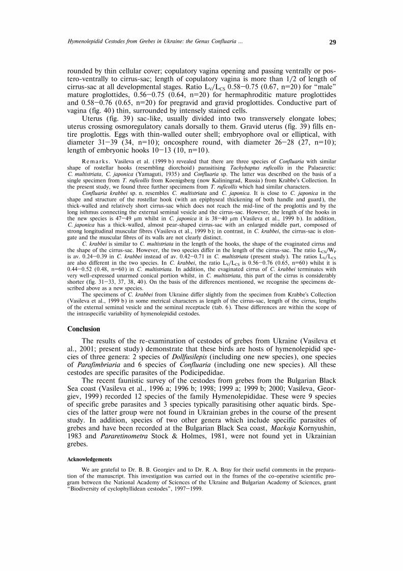
Hymenolepidid Cestodes from Grebes in Ukraine: the Genus Confluaria … 29
rounded by thin cellular cover; copulatory vagina opening and passing ventrally or pos-tero-ventrally to cirrus-sac; length of copulatory vagina is more than 1/2 of length of cirrus-sac at all developmental stages. Ratio LV/LCS 0.58—0.75 (0.67, n=20) for “male” mature proglottides, 0.56—0.75 (0.64, n=20) for hermaphroditic mature proglottides and 0.58—0.76 (0.65, n=20) for pregravid and gravid proglottides. Conductive part of vagina (fig. 40) thin, surrounded by intensely stained cells.
Uterus (fig. 39) sac-like, usually divided into two transversely elongate lobes; uterus crossing osmoregulatory canals dorsally to them. Gravid uterus (fig. 39) fills en-tire proglottis. Eggs with thin-walled outer shell; embryophore oval or elliptical, with diameter 31—39 (34, n=10); oncosphere round, with diameter 26—28 (27, n=10); length of embryonic hooks 10—13 (10, n=10).
Rema rk s . Vasileva et al. (1999 b) revealed that there are three species of Confluaria with similar shape of rostellar hooks (resembling diorchoid) parasitising Tachybaptus ruficollis in the Palaearctic: C. multistriata, C. japonica (Yamaguti, 1935) and Confluaria sp. The latter was described on the basis of a single specimen from T. ruficollis from Koenigsberg (now Kaliningrad, Russia) from Krabbe's Collection. In the present study, we found three further specimens from T. ruficollis which had similar characters.
Confluaria krabbei sp. n. resembles C. multistriata and C. japonica. It is close to C. japonica in the shape and structure of the rostellar hook (with an epiphyseal thickening of both handle and guard), the thick-walled and relatively short cirrus-sac which does not reach the mid-line of the proglottis and by the long isthmus connecting the external seminal vesicle and the cirrus-sac. However, the length of the hooks in the new species is 47—49 μm whilst in C. japonica it is 38—40 μm (Vasileva et al., 1999 b). In addition, C. japonica has a thick-walled, almost pear-shaped cirrus-sac with an enlarged middle part, composed of strong longitudinal muscular fibres (Vasileva et al., 1999 b); in contrast, in C. krabbei, the cirrus-sac is elon-gate and the muscular fibres of its walls are not clearly distinct.
C. krabbei is similar to C. multistriata in the length of the hooks, the shape of the evaginated cirrus and the shape of the cirrus-sac. However, the two species differ in the length of the cirrus-sac. The ratio LCS/WP is av. 0.24—0.39 in C. krabbei instead of av. 0.42—0.71 in C. multistriata (present study). The ratios LV/LCS are also different in the two species. In C. krabbei, the ratio LV/LCS is 0.56—0.76 (0.65, n=60) whilst it is 0.44—0.52 (0.48, n=60) in C. multistriata. In addition, the evaginated cirrus of C. krabbei terminates with very well-expressed unarmed conical portion whilst, in C. multistriata, this part of the cirrus is considerably shorter (fig. 31—33, 37, 38, 40). On the basis of the differences mentioned, we recognise the specimens de-scribed above as a new species.
The specimens of C. krabbei from Ukraine differ slightly from the specimen from Krabbe's Collection (Vasileva et al., 1999 b) in some metrical characters as length of the cirrus-sac, length of the cirrus, lengths of the external seminal vesicle and the seminal receptacle (tab. 6). These differences are within the scope of the intraspecific variability of hymenolepidid cestodes.
Conclusion
The results of the re-examination of cestodes of grebes from Ukraine (Vasileva et al., 2001; present study) demonstrate that these birds are hosts of hymenolepidid spe-cies of three genera: 2 species of Dollfusilepis (including one new species), one species of Parafimbriaria and 6 species of Confluaria (including one new species). All these cestodes are specific parasites of the Podicipedidae.
The recent faunistic survey of the cestodes from grebes from the Bulgarian Black Sea coast (Vasileva et al., 1996 a; 1996 b; 1998; 1999 a; 1999 b; 2000; Vasileva, Geor-giev, 1999) recorded 12 species of the family Hymenolepididae. These were 9 species of specific grebe parasites and 3 species typically parasitising other aquatic birds. Spe-cies of the latter group were not found in Ukrainian grebes in the course of the present study. In addition, species of two other genera which include specific parasites of grebes and have been recorded at the Bulgarian Black Sea coast, Mackoja Kornyushin, 1983 and Pararetinometra Stock & Holmes, 1981, were not found yet in Ukrainian grebes.
Acknowledgements
We are grateful to Dr. B. B. Georgiev and to Dr. R. A. Bray for their useful comments in the prepara-tion of the manuscript. This investigation was carried out in the frames of the co-operative scientific pro-gram between the National Academy of Sciences of the Ukraine and Bulgarian Academy of Sciences, grant “Biodiversity of cyclophyllidean cestodes”, 1997—1999.

G. P. Vasileva, V. V. Kornyushin, T. Genov 30
Brglez J. Zajedavci pri pticah v Sloveniji. Cestoda, Nematoda, Acanthocephala // Zb. Biotehn. Fak. Univ. Ljubl. Veterin. – 1981. – Supl. 5. – 410 p.
Chiriac E. To the knowledge of taeniid worms (Cestodes) of birds of the People's Republic of Roumania // Rev. Roum. Biol. – 1960. – 5. – P. 373—391.
Czaplinski B., Vaucher C. Family Hymenolepididae Ariola, 1899 // Keys to the cestode parasites of vertebrates / Eds L. F. Khalil, A. Jones, R. A. Bray – Wallingford: CAB International, 1994. – P. 595—663.
Grushchinskaya I. V. The cestode and nematode fauna of fish-eating birds at the Kremenchug Reservoir // Probl. Gidroparasitol. / Ed. A. P. Markevich – Kyiv: Nauk. Dumka, 1978. – P. 25—29. [In Russian].
Illescas-Gomez P., Lopez-Roman R. Dubininolepis multistriata (Rudolphi, 1810) Spasski, Spasskaya, 1954; parasito del Podiceps ruficollis Pallas // Revista Iber. Parasitologia. – 1980. – 40. – P. 223—230.
Macko J. K. Ploske cervy a ich vyznam u heibezneysich volne zitucich vtakov na vychodnom Slovensku // Sborn. Vychodoslovensk. Muz. Kosiciach. – 1962. – 2—3 (Ser. A). – P. 129—154.
Rysavy B. Tasemnice vodniho ptactva z Rybnicni Oblasti Jiznich Cech. I. Hymenolepididae Fuhrmann 1907 // Ceskoslovenska Parasitologie. – 1961. – 8. – P. 325—363.
Rysavy B., Sitko J. Tapeworms (Cestoda) of birds from Moravia (Czech and Slovak Federal Republik) // Prirodovedne Prace ustavu Ceskoslovenske Akad. Ved v Brne. – 1992. – 26. – P. 1—93.
Rysavy B., Sitko J. New findings of tapeworms (Cestoda) of birds from Moravia and synopsis of bird cesto-des from Czech Republic // Prirodovedne Prace Ustavu Ceskoslovenske Akademie Ved v Brne. – 1995. – 29 (5). – P. 1—66.
Singh K. P. Some avian cestodes from India. III. Species belonging to family Hymenolepididae // Ind. J. Helminthol. – 1959. – 11. – P. 43—62.
Szymanski M. Przyczynek do helmintologii // Rozprawy Wydzialu Matematyczno-Przyrodnieczego Akademii Umiejetnosci w Krakowie. – 1905. – 44 (Ser. B). – P. 342—345.
Vasileva G. P., Georgiev B. B., Genov T. Pararetinometra lateralacantha Stock and Holmes, 1981 (Cestoda, Hymenolepididae): the first record in the Palaearctic Region and comments on its morphology and taxonomy // Canad. J. Zool. – 1996 a. – 74. – P. 110—117.
Vasileva G. P., Georgiev B. B., Genov T. Redescription and new records of Mackoja podirufi (Macko, 1962), with an amended diagnosis of Mackoja Kornyushin, 1983 (Cestoda: Hymenolepididae) // Systematic Parasitology. – 1996 b. – 34. – P. 171—177.
Vasileva G. P., Georgiev B. B., Genov T. Palaearctic species of the genus Confluaria Ablasov (Cestoda, Hy-menolepididae): redescription and synonymy of C. capillaris (Rudolphi) // Systematic Parasitol. – 1999 a. – 43. – P. 49—57.
Vasileva G. P., Georgiev B. B., Genov T. Palaearctic species of the genus Confluaria Ablasov (Cestoda, Hy-menolepididae): redescriptions of C. multistriata (Rudolphi, 1810) and C. japonica (Yamaguti, 1935), and a description of Confluaria sp. // Systematic Parasitol. – 1999 b. – 44. – P. 87—103.
Vasileva G. P., Georgiev B. B., Genov T. Palaearctic species of the genus Confluaria Ablasov (Cestoda, Hy-menolepididae): redescriptions of C. podicipina (Szymanski, 1905) and C. furcifera (Krabbe, 1869), description of C. pseudofurcifera n. sp., a key and final comments // Systematic Parasitol. – 2000. – 45. – P. 109—130.
Vasileva G. P., Kornyushin V. V., Genov T. Hymenolepidid cestodes from grebes (Aves: Podicipedidae) in Ukraine: the genera Dollfusilepis Vasileva, Georgiev & Genov, 1998 and Parafimbriaria Voge & Read, 1954 // Vestn. zoologii. – 2001. – 35, N 2. – P. 3—14.