Human Self-reactive T Cell Clones Expressing Identical T Cell
10
Human Self-reactive T Cell Clones Expressing Identical T Cell Receptor [3 Chains Differ in Their Ability to Recognize a Cryptic Self-Epitope By Sonia Quaratino, Marc Feldmann, Colin M. Dayan, Oreste Acuto,* and Marco Londei From the Mathilda & TerenceKennedy Institute of Rheumatology, Sunley Division, 1, London W6 8L W, United Kingdom; and *Laboratory of Molecular Immunology, Pasteur Institute, 75724 Paris, France SulTlmary Recognition of self-antigens by T lymphocytes is a central event in autoimmunity. Under- standing of the molecular interactions between T cell receptors (TCR) and self-epitopes may explain how T cells escape thymic education and initiate an autoimmune reaction. We have studied five human in vivo activated T cell clones specific for the region 535-551 of human thyroid peroxidase (TPO) established from a Graves' patient. Three clones (37, 72, and 73) ex- pressed identical TCR [3 and et chains rearranging V[3 1.1 and Vet 15.1, and were considered sister clones. Clone 43 differed from clone 37 and its sisters in the Jet region only. Clone NP-7 expressed V[3 6.5 but rearranged two in-frame TCR et chain, both using the Vet 22,1 segment. Fine epitope mapping using nested peptides showed that clones using identical TCR [3 chains, identical Vet, but a different Jot recognized distinct, nonoverlapping epitopes in the TPO 535- 551 region. This finding shows that a different Jet region alone leads to a heterogeneous pattern of recognition. This indicates that the "restricted" TCR V region usage sometimes found in autoimmune diseases may not always correspond to identical epitope recognition. To confirm that clones 37 (and its sisters) and 43 recognize different epitopes, the T cell clones were stimu- lated with a TPO-transfected autologous Epstein-Barr virus (EBV) cell line (TPO-EBV) that presents TPO epitopes after endogenous processing. Only clone 37 and its sisters recognizes the TPO-EBV cell line, suggesting that the epitope recognized by clone 43 is not presented upon endogenous processing. We have shown that thyroid epithelial cells (TEC), the only cells that produce TPO, express HLA class II molecules in Graves' disease and can act as antigen- presenting cells, presenting TPO after endogenous processing to autoantigen-reactive T cell clones. We tested, therefore, whether autologous TEC induced the same pattern of stimulation as TPO-EBV; T cell clone 37 recognizes the TEC, whereas it is stimulated poorly by the TPO loaded to autologous peripheral blood mononuclear cells (PBMC). Clone 43, which fails to recognize the TPO-EBV, also fails to recognize the TEC, but is activated by exogenous TPO presented by autologous PBMC. These results show that exogenous versus endogenous pro- cessing in vivo generates a different TPO epitope repertoire, producing a "cryptic" epitope (epitope not always available for recognition). Our findings define a route by which human self-reactive T cells may escape thymic selection and become activated in vivo, thus possibly leading to autoimmunity. A major unsolved question is how self-reactive T cells escape thymic selection to reach the periphery, and how under certain conditions they become activated, lead- ing to autoimmune diseases. A number of hypotheses have been proposed to explain these events, and recently it has become apparent that the overall affinity/avidity of the TCR-peptide-MHC complex dictates thymic selection (1). For instance, such affinity/avidity may be reduced by two TCR expressed on the same T cell surface (2, 3). Al- ternatively, self-peptides with extremely low affinity for their genetic restriction elements might not form a stable enough complex with MHC in the thymus, and thus per- mit self-reactive T cells to escape negative selection (4, and Quaratino et al., manuscript in preparation). Another hy- pothesis is based o~a the concept that different epitope rep- ertoires may be processed and presented under different conditions or by distinct APC (5-7). Thus, the immune system may not usually see some im- portant self-epitopes, which are considered to be cryptic since they are hidden during the essential phases of thymic 349 J. Exp. Med. The Rockefeller University Press 0022-1007/96/02/349/10 $2.00 Volume 183 February 1996 349-358
Transcript of Human Self-reactive T Cell Clones Expressing Identical T Cell
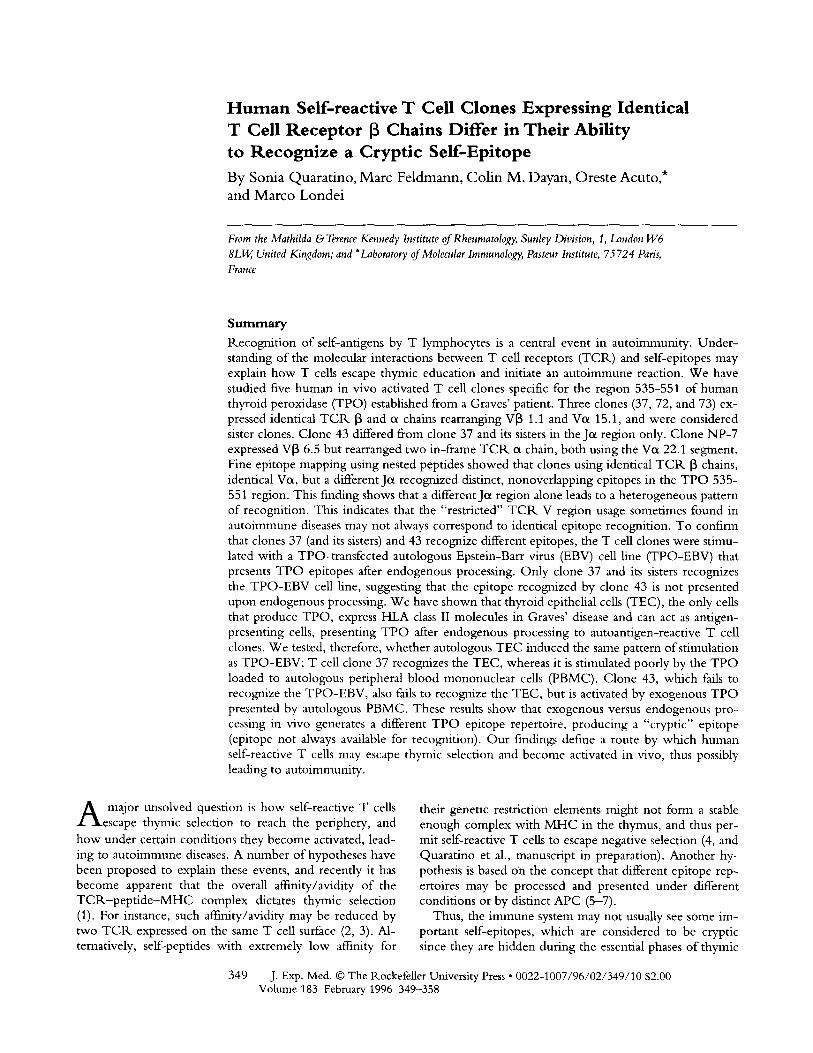
Human Self-reactive T Cell Clones Expressing Identical T Cell Receptor [3 Chains Differ in Their Ability to Recognize a Cryptic Self-Epitope By Sonia Quaratino, Marc Feldmann, Colin M. Dayan, Oreste Acuto,* and Marco Londei
From the Mathilda & Terence Kennedy Institute of Rheumatology, Sunley Division, 1, London W6 8 L W, United Kingdom; and *Laboratory of Molecular Immunology, Pasteur Institute, 75724 Paris, France
SulTlmary Recognition of self-antigens by T lymphocytes is a central event in autoimmunity. Under- standing of the molecular interactions between T cell receptors (TCR) and self-epitopes may explain how T cells escape thymic education and initiate an autoimmune reaction. We have studied five human in vivo activated T cell clones specific for the region 535-551 of human thyroid peroxidase (TPO) established from a Graves' patient. Three clones (37, 72, and 73) ex- pressed identical T C R [3 and et chains rearranging V[3 1.1 and Vet 15.1, and were considered sister clones. Clone 43 differed from clone 37 and its sisters in the Jet region only. Clone NP-7 expressed V[3 6.5 but rearranged two in-frame T C R et chain, both using the Vet 22,1 segment. Fine epitope mapping using nested peptides showed that clones using identical T C R [3 chains, identical Vet, but a different Jot recognized distinct, nonoverlapping epitopes in the TPO 535- 551 region. This finding shows that a different Jet region alone leads to a heterogeneous pattern of recognition. This indicates that the "restricted" T C R V region usage sometimes found in autoimmune diseases may not always correspond to identical epitope recognition. To confirm that clones 37 (and its sisters) and 43 recognize different epitopes, the T cell clones were stimu- lated with a TPO-transfected autologous Epstein-Barr virus (EBV) cell line (TPO-EBV) that presents TPO epitopes after endogenous processing. Only clone 37 and its sisters recognizes the TPO-EBV cell line, suggesting that the epitope recognized by clone 43 is not presented upon endogenous processing. We have shown that thyroid epithelial cells (TEC), the only cells that produce TPO, express HLA class II molecules in Graves' disease and can act as antigen- presenting cells, presenting TPO after endogenous processing to autoantigen-reactive T cell clones. We tested, therefore, whether autologous TEC induced the same pattern of stimulation as TPO-EBV; T cell clone 37 recognizes the TEC, whereas it is stimulated poorly by the TPO loaded to autologous peripheral blood mononuclear cells (PBMC). Clone 43, which fails to recognize the TPO-EBV, also fails to recognize the TEC, but is activated by exogenous TPO presented by autologous PBMC. These results show that exogenous versus endogenous pro- cessing in vivo generates a different TPO epitope repertoire, producing a "cryptic" epitope (epitope not always available for recognition). Our findings define a route by which human self-reactive T cells may escape thymic selection and become activated in vivo, thus possibly leading to autoimmunity.
A major unsolved question is how self-reactive T cells escape thymic selection to reach the periphery, and
how under certain conditions they become activated, lead- ing to autoimmune diseases. A number of hypotheses have been proposed to explain these events, and recently it has become apparent that the overall affinity/avidity of the TCR-pep t ide -MHC complex dictates thymic selection (1). For instance, such affinity/avidity may be reduced by two T C R expressed on the same T cell surface (2, 3). Al- ternatively, self-peptides with extremely low affinity for
their genetic restriction elements might not form a stable enough complex with MHC in the thymus, and thus per- mit self-reactive T cells to escape negative selection (4, and Quaratino et al., manuscript in preparation). Another hy- pothesis is based o~a the concept that different epitope rep- ertoires may be processed and presented under different conditions or by distinct APC (5-7).
Thus, the immune system may not usually see some im- portant self-epitopes, which are considered to be cryptic since they are hidden during the essential phases of thymic
349 J. Exp. Med. �9 The Rockefeller University Press �9 0022-1007/96/02/349/10 $2.00 Volume 183 February 1996 349-358

education. I f these cryptic epitopes are displayed in the pe- riphery, then autoimmuni ty could ensue (7, 8). The factors leading to this differential display are not totally defined, but it is now known that different arrays o f epitopes can be generated i f proteins (such as viral proteins) are processed by the endogenous or exogenous pathway (9, 10). H o w - ever, this has not yet been shown in human autoimmunity.
Ultimately, whatever is the processing pathway, the ini- tiating event o f an immune response is the recognit ion o f the peptide fragment exposed in the NtHC binding groove by the T C R . Many studies have at tempted to characterize the T C R usage, epitope recognition, and HLA restriction o f human autoreactive T cells. It has been reported that myelin basic protein (MBP)l-specific T cells have biased T C R usage in humans (11, 12) and in mice (13), and may dominantly recognize a specific region o f MBP in the con- text o f the same HLA molecule (14). In these studies, how- ever, there is no evidence that the T cells studied were ei- ther activated in vivo, or were involved in the disease process. This is because o f the technical limitations o f work with human pathologic material. In Graves' disease (GD) we have been able to overcome some o f these limitations, as the cells studied were activated in vivo expressing the IL-2 receptor, and were cloned wi thout exposure to antigen (15, 16). Hence, these cells may have greater relevance to disease than T cells cloned with antigen from PBMC, for which there is no conclusive evidence of disease relevance, since similar clones can be generated from healthy individ- uals. Using this approach we found that a region o f thyroid peroxidase (TPO) amino acids 535-551 was recognized by a significant propor t ion (16%) o f in vivo-act ivated T cell clones (16). Thus we focused our attention on these clones. W e describe be low the TCR. sequence expressed by these T cell clones, the core epitope recognized, and the H L A element o f genetic restriction. Since thyroid epithelial cells (TEC) can act as APC in GD and present autoantigens to T cells, after endogenous processing (15-17), we used a TPO-transfected EBV cell line as well as the autologous TEC to stimulate the T cell clones and compared them with T P O presented after exogenous processing by PBMC. The differences observed between exogenous and endogenous processing demonstrate the existence o f T cells recognizing a cryptic epitope, an epitope otherwise hidden, which can be a strong T cell stimulator. The results de- scribed here suggest that cryptic self-determinants gener- ated by atypical APC may play an important role in the pathogenesis of autoinmmne diseases.
Materials and Methods
T Cell Clones. T cell clones were established from Graves' thyroid infiltrate in absence of antigen, according to our pub- lished procedure (15). Briefly, activated thyroid-infiltrating T lymphocytes were cultured in the presence of IL-2 (20 ng/ml,
~Abbreviations used in this paper: AnPCR, anchored PCR; G1), Graves' dis- ease; MBP, myelin basic protein; TEC, thyroid epithelial cell; TPO, human thyroid peroxidase.
kindly donated by Hoffmann-La Roche, Nutley, NJ) for 1 wk m RPMI 1640/10% human serum and then expanded for a further week with irradiated (4,500 rad) autologous peripheral blood leu- kocytes, OKT3 anti-CD3 mAb (30 ng/ml), and IL-2. Cells were cloned by limiting dilution in absence of any antigen as described (15, 16). The T cell clones were stimulated with irradiated feeder cells and PHA or with OKT3 coated to plastic and cultured in media consisting of RPMI 1640, 10% pooled human serum, 100 U/ml penicillin, and 50 Ixg/ml streptomycin. Every 4-5 d, ex- panding T cell clones were fed with 10 ng/ml oflL-2.
Antigens. Peptide TPO 535-551, corresponding to amino acids 535-551 of the human TPO molecule (LDPLIRGLLARPA- KLQ), and eight overlapping "nested" synthetic peptides of 12 amino acids spanning from residues 534 to 552 of human TPO were gifts from Anergen (Redwood City, CA) and Chiron Mim- otopes (Clayton, Australia).
T Cell Proliferation Assay. The TPO peptides were used to define the minimum epitope specificity of each T cell clone. The T cell clones were tested at least 2 wk after the last TCR stimula- tion and 5 d after the last addition of IL-2. The reactivity to the TPO 534-552 synthetic overlapping peptides was assessed by coculturing 104 T cells from each T cell clone with 3 • 104 glu- taraldehyde-fixed, autologous EBV-transformed PBMC as APC (16). The autologous EBV-transformed cell line was pulsed with or without 10 txg/ml of each of the peptides used in the assay for 1 h at 37~ before incubation with T cells. All T cell clones were tested at least twice, and all the experiments showed a similar pro- file of responsiveness.
T cell clones were also tested using autologous PBMC as APC. 3 • 104 PBMC isolated from heparinized whole blood by buoy- ant density centrifugation (Lymphoprep; Nycomed Pharma, Oslo, Norway) were irradiated (4,500 rad) and incubated with 25 fxg/ml of recombinant human TPO expressed in Chinese ham- ster ovary (CHO) cells for 2 h before adding the T cell clones.
When autologous TEC were used, they were obtained after enzymatic digestion of the thyroid and used in proliferative assays without addition of antigen as described (15). Briefly, TEC were irradiated as above and plated at 3 • 104/well and used to stinm- late the T cell clones plated at 104/well. All the proliferative assays were performed in triplicate for 72 h in a flat- or round-bottoln 96-well microtiter plate and the cells were pulsed with [3Hlthy- midine (1 IxCi) during the last 8 h of culture.
TCR GenetkRestriction. Two homozygous EBV-transfonned B cell lines expressing identical DP2 or DQ6 with the autologous APC were selected from the 10th HLA Workshop to define whether the element of genetic restriction was DP2 or DQ6. EBV cell line 9036 expressed DPBI*02012 and DQB1*0502/ DQA*0102, whereas cell hne 9013 expressed DPB1*0402 and DQB1*0602/DQA*0102. 3 • 104 gluteraldehyde-fixed EBVs were challenged with or without the TPO peptide 535-551 for 1 h before adding the T cell clones. The experiment was performed in triplicate, and the cells were pulsed with [~H]thymidine during the last 8 h of the 72-h culture.
PCR Amplification and Sequence Analysis ~y- TCR-oe and -[3 eDNA. Total RNA was extracted from 3-5 X 10 ~' cells by a modified guanidine-isothiocyanate-phenol-chloroform method and first-strand eDNA was synthesized by a standard oligo(dT) method, using ~-'5 Ixg of total RNA (18). To elicit maximum TCR expression before RNA extraction and eliminate possible contamination by feeder cell RNA, resting T cells (more than 14 d after last stimulation) were stimulated with immobilized OKT3 for 24 h before harvesting for RNA preparation, eDNA encoding the TCR o~ and [3 chains were then amplified using the anchored
350 Restricted TCR Recognize Multiple Human SelfEpitopes

PCR method (AnPCR) (19). A poly(dG) tail sequence was added to the single-strand cDNA with terminal deoxynucleotidyl trans- ferase in cobalt buffer (Boehringer-Mannheim Corp., Indianapolis, IN) and 0.1 mM dGTP for 30 rain at 37~ AnPCR was performed using oligonucleotide primers complementary to the poly(dG) (5'-CACTCGAGCGGCCGCGTCGACCCCCCCCCC-3') and to either the 5' end of ot constant region (5'-GCGAATTCA- GATCTTAGGCAGACAGACTTG TCACTGG-3') or the 5' end of 13 constant region (5'-GCTCTAGAGTCG ACGGCT- GCTCAGGTCAGTATCTGGAGT-3'). Each DNA reaction mix- ture containing 15 mM MgC12 and 50 pmol of each of the two oligo primers was subjected to 25 cycles of amplification in a thermal cycler (Perkin-Elmer Cetus Corp., Norwalk, CT) at the following temperatures: 93~ for 15 s, 55~ for 1 rain, 60~ for 15 s, and 72~ for 2 rain. A final 10-rain extension was per- formed at 72~ to ensure fully duplexed DNA for optimal liga- tion efficiency. PCR products (~0.6-0.7 kb) were digested with appropriate restriction enzymes and isolated from agarose gels, subcloned into PUC19 vector, and sequenced according to the dideoxynucleotide termination method (20). Sequencing was performed on both strands using the forward and reverse univer- sal M13 primers. Several independent colonies (usually five to eight) were sequenced to confirm the clonal nature of the T cell clones, to exclude base misincorporations due to PCR errors (21), and to detect eventual functional rearrangements of both the ci loci in a single clone (19, 22). Sequences obtained were com- pared to those of EMBL/GenBank/DDBJ using the DNAStar | software.
Different RNA preparations of the tested clones were analyzed for 2 yr. They were also tested using a set of VI3 and Vet family- specific primers (23-25) as described (18). The amplified products were purified from unincorporated primers and nucleotides and directly sequenced using a Dye deoxy terminator cycle sequenc- ing kit (Applied Biosystems, Inc., a division of Perkin-Elmer Ce- tus Corp., Foster City, CA) in an automatic sequencer (model 373 DNA Sequencing System; Applied Biosystems). Each clone amplified only one V[3 or Vet product, and was sequenced using 5 pmol of the V[3 or Vot primers, respectively. PCR amplification was also performed incorporating 10 I.LCi of [32p]dCTP, to detect eventual amplified products imperceptible after ethidium bromide staining and UV visualization. The gels were dried and exposed for 24 h on X-omat film (Eastman Kodak Co., Rochester, NY).
TPO Transfection of Autologous EBV. The autologous TPO- transfected EBV-transformed B cell line was a kind gift of Dr. R. Mullins (The Mathilda & Terence Kennedy Institute of Rheu- matology). The entire cDNA encoding the human TPO (3.1 kb, gift of Dr. B. Rapaport, University of California, San Francisco, CA) was ligated into the Asp718-HindlII sites of the vector pREP 4 (lnvitrogen, San Diego, CA), containing the Rous sar- coma virus enhancer/promoter and the hygromycin-resistance gene. Successfully transfected cells were selected by growth in hy- gromycin-containing medium at a final concentration of 200 ~g/ ml in RPMI 1640 containing 10% FCS (26). An autologous EBV-transformed B cell line transfected with the vector pREP- CAT, encoding the CAT protein, was used as control in the pro- liferative experiments.
Results
A panel o f self-reactive T cell clones specific for the 535- 551 region o f the human T P O antigen was raised from the thyroid infiltrate o f a patient with autoimmune thyroiditis
351 Quaratino et al.
(GD). These clones were established in the absence o f antigen (15, 16), reducing the bias o f in vitro antigen- driven expansion, and enabling analysis o f a spontaneous in vivo autoantigen-reactive T cell response. The antigen specificity o f all the clones was determined by their respon- siveness to exogenous TPO, processed and presented by autologous PBMC (16). The epitope specificity o f some of the TPO-specific T cell clones was further characterized, and the T P O region 535-551 was recognized by 6 out o f 81 clones (16). Five o f these clones have been analyzed in detail in this study.
T C R Analysis. We asked whether the similar peptide specificity o f the clones reflected a T C R sequence similar- ity, and whether there were any conserved amino acids in "motifs" at particular positions o f their ix or 13 chains. The c D N A corresponding to the T C R ix and 13 chains o f the five T cell clones was amplified by the A n P C R technique and sequenced (19). The nucleotide and predicted protein sequences spanning the junctional V(D)J regions o f the T C R 13 and ix chains o f the five clones are shown in Fig. 1, A and B, respectively. Clone NP-7 expressed V[3 6.5/D[3 2.1/J[32.5, whereas clones 72, 73, 37, and 43 expressed an identical T C R [3 chain, V[3 1.1a/D[3 2.1/J[3 2.1 (Fig. 1 A). A common mot i f was observed in the C D R 3 of these [3 chains (Fig. 1 A): at position 101, a Glu was flanked on both sides by polar uncharged residues (Asn-Gln and Gln- Thr). Alignment of the sequences showed that the overall length o f the presumptive T C R C D R 3 loops o f clones bearing identical specificity differed in length by only one amino acid.
We detected two "productive" ix chains in clone NP-7 (Fig. 1 B): productive rearrangement o f both ix chain loci has been observed in up to 30% of both human and murine T cells (19, 22); two ix chains can be expressed on the sur- face o f a single cell, pairing with the only 13 chain available (2, 3). The motif Gly-Asn (residues 95-96) observed in the two C D R 3 of clone NP-7 was also detected in the C D R 3 of T cell clones 37, 72, and 73. The sequences o f the ix chains o f clones 72, 73, and 37 were identical, rearranging Vix 15.1/Jix AC24. Since clones 37, 72, and 73 express identical TC1K ix and 13 chains at the nucleotide level, they are sister clones.
A first A n P C R analysis o f clone 43 indicated the pres- ence o f a T C R ix chain identical to one o f the two ix chains of clone NP-7. To exclude cross-contaminations that occurred in manipulating clone 43, T cells were sub- cloned by limiting dilution. We analyzed the T C R of only a single subclone that functionally behaved in an identical way to the original one. The expression of only a single V[3 and Vix was confirmed using a panel o f 22 VI3 and 24 Vix family-specific primers (Fig. 2). Direct sequencing of the V~ amplified product confirmed the A n P C R analysis. The direct sequencing o f the only Vix amplified (Vix 15) indi- cated that Jot I G R Ja08 was used instead o f Jix AC24, used by clone 37 and its sisters (Fig. 1 B). N o Vix 22 product was amplified, indicating that the previous result was due to an A n P C R or cellular contamination. W e also used the same panel o f Vix and V[3 family-specific primers to test

A
Clone Vl~ NDN Jl~ CI~
101 A S S S G L A E N E Q Y F G P G T R L L V L
72 GOCAGCAGC TCA GGACTAGCG G ~ AATGAG CAG TAC Trc GGG CCA GGC ACG CGG CTC CTG GTG CTA
A S S S G L A E N E Q Y F G P G T R L L V L 73 QCCAGCAGC TCA GGACTA GCG C-~8 AAT C-~G CAG TAC TTC GGG CCA GGC ACG CGG CTC CTG GTG CTA
A S S S G L A E N E Q Y F G P G T R L L V L 3 7 GIOCAGCAGC TCA C.-GA CTA GCG G ~ AAT GAG CAG TAC "TTC GGG (~A GGC ACG CGG CTC CTG GTG CTA
A S S S G L A E N E Q Y F G P G T R L L V L 4 3 GCCAGCAGC TCA GGA CTA GOG C-~8 AAT GAG CAG TACTTC GGG OCA GGC ACG OGG CTC CTG G'rG CTA
A S T T T S R Y Q E T Q Y F G P G T R L L V L NP7 GCCAGCACC ACC ACCT~r CGG TAC CAA GAG ACC CAG TAC "rrc ~_.~ CCA GGC ACG CC~ CTC CTG GTG CTC
E GaG V~ 1.1a/DI3 2.1/J1~2.1
E GaG V~, 1.1a/D~ 2.1/J~ 2.1
E GaG V~ 1.1a/D[~ 2.1/J~ 2.1
E GaG V~ 1.1a/D[3 2.1/J~2.1
E G ~ V~ 6.5/D~ 2.1/J~ 2.5
B C l o n e Vc~ J(x C(x
F C A A R G F G N F N K F Y F G S G T K L N V K P N I
37 TrCFGTGC.A G O C O C - - - ~ G G G T F C G C - : ~ A A C T I ' C A , ' a , C A , ~ , T I T T A C T T T G C - ~ T C T G G G ~ ~ T ~ A ~ T ATC Vu 15.1/J(~AC24
F C A A R G F G N F N K F Y F G S G T K L N V K P N I
72 TI'CTGTC,-,-,-,-,-,-,-,-,~A G C ~ O G G G G G ' I " F C G G G A / ~ C T T C / ~ , C A A A T I ' r T A C T T I G C ~ T C T C _ . ~ G ~ ~ T ~ A ~ T Arc Vet 15.1/J~ AC24
F C A A R G F G N F N K F Y F G S G T K L N V K P N I
73 T I ' C T G T ~ GCCCC-~GGGTTCGGGAACTTCAACAAATFrTACTFFGGATCT~ACCAAACTC~TGTA~A~T ATC V(x 15.1/Jc( AC24
F C A E S S S G G Y N K L I F G A G T R L A V H P Y I
43 TFCTGTGCA GAGAGTTCTTCTGGTGGCTACAATAAGCTGATrTTFGC-~GCAGGGAOCAGGCTG~GTA~ATAT ATC Ve 15.1/IGRJc(08
F C A L S G N N D K L I F G T G T R L Q V F P N I
NP-7 T r C T G T G C " F CTGAGTGGAAACAACGACAAGCTCATCTTTGGGACTGGGACCAGATTACAAGTCTTr~A~T ArC Vc~ 22.11Jet 1
F C A P Q (3 N Y G Q N F V F G P G T R L S V L P Y I
TI'CTGTGCT OCCCAGGGGAACTATGGTCAGAATTB'GTCTITGGTCCCGC,~AOCAGATIGT(X;GTGCTGCCCTAT ArC V~t 22.1/J(x 33
Figure 1. Nucleotide and amino acid sequence and alignment of (A) TCR [3 V-(D)-J regions and (B) TCR 0~ chain V-J regions. For each clone are indicated the names of the corresponding VI3, J[3, vct, and fix genes. Only the last nine nucleotides encoding for the last three amino acid residues of each V segment are shown, followed by the junctional regions. The random TdT addition nucleotides 5' and 3' to the diversity region are underlined. Amino acids are indicated by the single letter code, numerated according to Chothia et al. (36). Amino acids in bold are the common motifs in the CDR3 re- gion. Several sequences from 5 to 10 bacterial colonies were performed for each chain, and repeated analysis using mRNA obtained several months apart gave identical results. (A) In the CDR3 of all chains at position 101 corresponds a Glu residue (E), which is flanked at positions 100 and 102 by polar but uncharged amino acids Ash (N), Gln (Q), or Thr (7). JI3 2.1 was used by all the clones, except clone NP-7, which encoded for the full germline JI3 2.5. (B) The CDR3 of the oL chains of clone NP-7 revealed a common motif Gly-Asn (GN) entirely template dependent in Vct22/J0~33, whereas the gly was encoded by the N-nucleotide addition and the Asn was template dependent in V(x22/J~l. Snrfilar GN motif was also shared by the CDR3 of the sister clones 37, 72, and 73.J regions were named accordingly to reported sequences; J(xC24 (50), IGP, Ja08 (25), Jcd (51), andJc133 (52). These sequence data are available from EMBL/GenBank/DDBJ under accession numbers: X84687, X89751, X89752, X89753, X89754, X89809, and X89860.
clones 37, 72, 73, and N P - 7 ; the direct sequenc ing o f the only amplif ied Vo~ and V[3 products gave identical results to the A n P C R . analysis.
Fine Epitope Mapping of TPO 535-551-specific T Cell Clones. T h e fine specificity o f ant igen recogni t ion a m o n g the T cell clones was examined using a nested set o f eight over lapping 12-arn ino acid synthetic peptides spanning res- idues 534-552 o f the T P O molecu le ( G L D P L I R G L L A R - P, ,PAKLQV), each advancing one amino acid in the se-
quence . W e expec ted that the unusual and except ional ly restricted TCI:L usage (that is, TCP,. chain sharing) migh t p roduce an identical or very similar pat tern o f recogni t ion . O n the contrary, as shown in Fig. 3, three distinct profiles o f recogni t ion o f the T P O 535-551 reg ion were identified. T P O 536-547 and 539-550 peptides were recognized by clones 37, 72, and 73, but were no t recognized by c lone 43, wh ich was specific for the 541-552 reg ion o f the T P O molecule . C l o n e N P - 7 recognized the reg ion T P O 537-
352 Restricted T C k Recognize Multiple Human SelfEpitopes

552, but unlike clones 37, 72, and 73, failed to recognize T P O 536-547.
T Cell Clones Recognizing TPO 535-551 in Association with HLA-DQ. In a previous report (16) we excluded the D R antigens as the restricting element, but could not de- fine whether D Q 6 and /o r DP2 molecules were active ge- netic restriction elements. Since it has been reported that nonself as well as self-epitopes may be recognized in the context o f two different HLA molecules in the same indi- vidual (27-29), it was o f interest to fully characterize the M H C restriction o f the clones. The T cell clones were cocultured with fixed EBV-transformed B cell lines o f known M H C haplotypes, with or wi thout exogenous pep- tide 535-551. The EBV-transformed B cell lines were se- lected to be homozygous either for D Q 6 or for DP2, dif- fering for all the other HLA antigens with the autologous APC. The results (Fig. 4) indicate that none o f the clones
Figure 2. T cell clone 43 was analyzed by RT-PCR incorpo- rating [32p]dCTP, with a panel of V[3 and V0t family-specific primers. PCR products ranged from ,'-'170 to 300 bp and from 300 to 500 bp for the [3 and a chain, respectively. Products were run on 2% agarose gel and visualized at UV light. The gel was then dried and exposed for 24 h on autoradiograph. This re- sult is representative of three dif- ferent experiments.
responded to the T P O peptide 535-551 when presented on EBV 9036, which expressed DPB1*02012, whereas all clones recognized the T P O peptide 535-551 in the context o f the DQBI*0602/DQA*0102 molecule (EBV 9013). Fur- ther studies o f biotinylated peptide binding have shown that DQBI*0602/DQAI*0102 binds the T P O peptide 535-551 whereas Dtk2 and D R 3 do not (Quaratino, S., unpublished observations). Inhibitory experiments performed using anti- D Q , (SVLB4) ant i -DP (B7/21), and a n t i - D R (Ta114.1) mAbs confirmed that all the T cell clones are restricted by D Q molecules (data not shown). Thus, the epitope microhet- erogeneity we observed was not due to presentation o f the same peptide in the context o f different HLA molecules.
Recognition of the Autologous TPO-transfected EBV Cell Line. tkecendy we reported that clone 37 recognizes an autologous EBV cell line transfected with the cDNA encod- ing T P O and expressing T P O at the cell surface (26). W e
353 Quaratino et al.
Figure 3. Fine specificity of TPO 535-551--specific T cell clones 37, 72, 73, 43, and NP-7. Eight overlapping synthetic pep- tides of 12 amino acids spanning from residues 534 to 552 of hu- man TPO were used to define the minimum epitope of each T cell clone. All T cell clones were tested at least twice, and all the experiments showed a similar profile of responsiveness.

Figure 4. HLA genetic re- striction of T cell clones 37, 72, 73, 43, and NP-7. Homozygous EBV-transformed B cell lines 9036 and 9013 expressing identi- cal DP2 or DQ6, respectively, with the autologous APC were chosen to define if the genetic restriction element was DP2 or DQ6. The EBV cell line 9036 expressed DPBI*02012 and DQB*0502/DQA*0102, whereas the 9013 cell line expressed DPBI*0402 and DQBl*0602/ DQA*0102. EBV 9036 1)P2 + failed to present the TPO 535- 551 peptide to all the clones, whereas EBV 9013 DQ6 ~ pre- sented the peptide to all five clones with comparable effi- ciency to the autologous EBV cell line.
compared the capacity o f this TPO-transfected autologous EBV cell line to stimulate clones 37, 43, and NP-7 . Clones 43 and NP-7 did not recognize the autologous T P O - E B V cells, whereas in the same experiment, clone 37 did (Fig. 5). As expected, all the clones responded to the exogenous peptide 535-551 presented by the autologous EBV line (untransfected) and they had previously responded, al- though to a different extent, to exogenous T P O presented by PBL (16). These results imply that the TPO-transfected EBV cells presented on the cell surface the epitope recog- nized by clone 37, but not the epitopes recognized by clone 43 or clone NP-7 . These data demonstrate that clones 37 and 43, which differ in the T C R Jet region only, recognize two completely different epitopes, only one o f which is presented after endogenous processing. Compared with the results shown in Fig. 3, only a peptide identical or very similar to T P O 536-547 was presented, upon endoge- nous presentation, in the region 535-551.
Recognition of the Autologous TEC. Although the results obtained with the autologous TPO-transfected EBV cell line were intriguing, there was still the possibility that the EBV cells endogenously processing T P O were generating a different epitope repertoire than TEC. To evaluate the po- tential in vivo situation, we repeated the experiment using the autologous T E C to stimulate the T cell clones in the absence o f exogenous antigen. As shown in Fig. 6, clone 43 did not respond to TEC, whereas it did respond to the exogenous T P O presented by PBMC. In the same experi- ment, clone 37, on the contrary, expressed a marginal (if any) response to exogenous T P O processed and presented by P B M C whereas it was well stimulated by TEC, con- firming that different T P O epitopes are displayed upon ex- ogenous versus endogenous processing. This epitope can also be considered as cryptic, since it is an epitope available for recognition only under restricted conditions, in this case by the TEC expressing class II acting as nonclassical APC.
EBV + "17
EBV-CAT + T '
EBV-TPO + T
EBV + TPO 535-551 + T '
Clone37 Clone 43 Clone NP-7
0 5 10 15 20 5 10 t5 0 25 50 75 100
3H-thymidine incorporation c.p.m. ( x 10"3)
354 Restricted TCR Recognize Multiple Human SelfEpitopes
Figure 5. Recognition of au- tologous TPO-transfected EBV cell line by T cell clones 37, 43, and NP-7. Only clone 37 recog- nized the endogenously pro- cessed TPO. Clones 43 and NP-7 recognized the peptide TPO 535-551 added exogenously, but failed to recognize the endoge- nously processed TPO. These results suggested that an epitope corresponding to the peptide 536-547 was presented upon en- dogenous processing. Results shown are from single experi- ments, and they are representa- tive of those seen in three differ- ent assays.

PBMC + T
PBMC + micros + T
PBMC + TPO micros + T 1 l
TEC + T
T E e only
0 1000 2000 3000 4000
3H-thymidine incorporation (c.p.m.)
Figure 6. Recognition of endogenously and exogenously processed TPO by T cell clones 37 and 43. (Striped bars) Clone 37; (empty bars) clone 43. Autologous PBMC were used to present purified microsomes of CHO cells untransfected or transfected with TPO (16). Autologous TEC induced proliferation of clone 37 only, whereas exogenously processed TPO by PBMC was recognized by clone 43 and not by clone 37.
D i s c u s s i o n
The analysis o f self-reactive T cells has been one of the major targets o f research in autoimmunity during the last few years. These studies have aimed to define the three specific elements involved in self-recognition; much effort has been placed into the characterization of immunodomi - nant self-epitopes and T C R usage (11-13, 30). The aim of these experiments is to understand the general features common to all autoimmune diseases. Many attempts have been made to determine whether self-reactive T cells have a restricted T C R Vet or V[3 usage (11, 24, 31-33), since restricted T C R usage or detection of conserved motifs could suggest dominant self-epitope recognition (34). There is evidence that the C D R 3 of the T C R is directly involved in epitope recognition (35-37), and this has led to the notion that T cells expressing conserved C D R 3 motifs are prone to recognize identical epitopes (38, 39).
Studies of T cell clones specific for MBP (13, 33) and sperm whale myoglobin (40) have demonstrated that mul- tiple combinations o f T C R Vot/Jet paired with an identical or similar T C R [3 chain may yield a nearly identical epi- tope specificity. It is interesting to note that a conserved C D R 3 mot i f was observed in T cell clones from Lewis rats with experimental acute encephalomyelitis reactive to the MBP peptide 87-99 (38). The finding that this conserved mot i f was also observed in a large proport ion of T C R [3 transcripts o f T lymphocytes infiltrating plaques from multiple sclerosis patients was thus considered to be strong evidence that infiltrating T cells recognized the same MBP peptide (34).
Our report demonstrates that T cell clones sharing the same T C R [3 and Vet but differing in the Jet are able to rec- ognize contiguous but different epitopes of the same self- molecule. Thus, even with restricted T C R V region usage, multiple epitope recognition still occurs. It is plausible to envisage that T cell clones may share identical [3 chains at
the nucleotide level, since the [3 locus rearranges before et (41); thus two different clones from a common precursor may rearrange distinct et chains and emerge from the thy- mus. The T C R sequencing of other TPO-specific T cell clones established from the same individual showed that different et and 13 chains are used (data not shown).
Regarding clone NP-7 in which we detected two et chain messages, it has b e e n hypothesized that clones ex- pressing two o~ chains may be involved in the pathogenesis o f autoimmune diseases (2, 3). Since the overall expression of the self-reactive T C R is significantly reduced by the presence of a second T C R , such a decreased expression may reduce the specific avidity of these cells for self, thus allowing their positive selection in the thymus (3). Another aspect o f double T C R expression is that in clones express- ing two functional T C R , one of which may recognize self, the activation o f the T cell via engagement of the other re- ceptor may lead to an "unwanted" activation against self, leading to autoimmunity (2, 3). Although we do not know whether the two (x chains are expressed at the cell surface, clone NP-7 may be useful to test this hypothesis, by sepa- rately cotransfecting the T C R et with the T C R [3 chain in a suitable TCR-nega t ive cell (42). The marked sequence similarity suggests that antigenic pressure, rather than a ran- dora event, selected its two et chains, and indicates that both chains might be involved in self-recognition. If this is the case, and no second specificity is found for clone NP-7, it would suggest that the two expressed T C R , to escape thymic selection, are both o f low affinity for self (3).
Different hypotheses have been evoked to explain pe- ripheral activation of self-reactive T cells. It has been sug- gested that a bystander or unwanted activation of self-reac- tive T cells could be induced by superantigens engaging T cells in a V[3-specific fashion (23) or via molecular mimicry (43); recently, it has been demonstrated that T cells specific for a self-epitope may recognize peptides of viral or bacterial origin (44, 45). A theory that can simultaneously address peripheral activation and lack of central tolerance is based on the existence of a "cryptic self-epitope repertoire:" epitopes hidden during thymic education may become evi- dent in the periphery upon restricted conditions such as different antigen loading or type of cells acting as APC (6, 7). That a cryptic repertoire exists has been described in several animal models (46); this theory has also been useful to explain the process ofep i tope spreading (8), and recently has been shown in a human system (47).
A central finding of our study is that two different types of APC or antigen loading produce functional differences in the self-peptides displayed in their HLA grooves. It indi- cates that cryptic epitopes of a disease-related autoantigen may be generated, and more importantly recognized by some, but not all, in vivo-activated, antigen-specific T cell clones (Figs. 5 and 6). Our results highlight another re- markable feature of the cryptic (that is, only presented upon endogenous processing) epitope. Indeed, the close analysis o f the data reported in Fig. 3 indicates that peptide 536-547, which closely corresponds to the natural cryptic
355 Quaratino et al.

epitope presented upon endogenous processing is, at equiv- alent molar concentration o f peptide T P O 535-551, ex- tremely efficient in activating the sister clones 37, 72, and 73. The epitope recognized by the T cell clone 37 was studied in detail using truncated peptides at the N H 2 and C O O H termini o f peptides 536-547 and 539-550, to ex- plain the peculiar pattern o f responsiveness observed in Fig. 3; the results are detailed elsewhere (48). It has been sug- gested that nonprofessional APC such as class II--expressing TEC, could act as APC (5), and via endogenous process- ing, display a self-epitope profile different than the one dis- played after antigen uptake and by reprocessing APC in PBMC (46). A few years ago, we demonstrated that TEC could be specifically recognized by self-reactive T cells (15). Subsequently, we have shown that many TPO-specific T cell clones were also stimulated by autologous thyrocytes (16). More recently, we have demosntrated, using T P O - and thyroid-stimulating hormone receptor-transfected EBV cell lines, that processing and presentation o f membrane- associated self-antigens can occur (26, 49). The results re-
ported in this study demonstrate that a cryptic epitope is displayed by TEC and recognized in vivo by autoreactive T cells (Fig. 6). We have also found other T cells at the site o f the disease that recognized the autoantigen only when presented by professional APC, as clone 43 recognized T P O presented by P B M C and not by TEC. We do not know which o f the two types o f self-reactive T cells may initiate the autoimmune process, but we favor the hypoth- esis that the primary event is the recognition o f the self- processed epitope, once the thyroid epithelial cells acquire antigen-presenting capability (5, 15, 17). That recognition o f the cryptic epitope may represent a primary event is also supported by the evidence that this epitope is extremely ef- ficient in activating the sister clones 37, 72, and 73, com- pared to peptide 535-551. Cells such as clones 43 and NP-7 may intervene when the autoantigen, released after tissue damage, is processed by professional APC, and may corre- spond to a second phase o f seK-recognition, that may rep- resent epitope spreading in human autoimmunity.
We thank Dr. Marc Bonneville for critical reading of the manuscript. We also thank Dr. R. Mullins for the gift of the TPO-transfected EBV cell line, Dr. B. Rapaport for the gift of TPO-CHO cells, Dr. G. Lom- bardi for the gift of the HLA typed cells, Dr. S. Sharma of Anergen, and Dr. M. Geysen of Chiron Mimo- topes for synthesis of the peptides related to TPO 535-551.
This work was supported by the Wellcome Trust, the Arthritis and Rheumatism Council, and the Associa- zione Italiana Sclerosi Multipla.
Address correspondence to Dr. Sonia Quaratino, The Mathilda & Terence Kennedy Institute of Rheuma- tology, Surtley Division, 1 Lurgan Avenue, London W6 8LW, UK. C. M. Dayan's present address is De- partment of Medicine, University of Bristol, University Walk, Bristol B581TD, UK.
Received for publication 12January 1995 and in revised form 20 September 1995.
References 1. Hogquist, K.A., S.C. Jameson, W.R. Heath, J.L. Howard,
MJ. Bevan, and F.R.. Carbone. 1994. T cell receptor antago- nist peptides induce positive selection. Cell. 76:17-27.
2. Padovan, E., G. Casorati, P. Dellabona, S. Meyer, M. Brock- haus, and A. Lanzavecchia. 1993. Expression of two T cell receptor ix chains: dual receptor T cells. Science (Wash. DC). 262:422-424.
3. Heath, W.R., andJ.F.A.P. Miller. 1993. Expression of two ix chains on the surface of T cells in T cell receptor transgenic mice.J. Exp. Med. 178:1807-1811.
4. Fairchild, P.J., Ik. Wildgoose, E. Atherton, S. Webb, and D.C. Wraith. 1993. An autoantigenic T cell epitope forms unstable complexes with class II MHC: a novel route for es- cape from tolerance induction. Int. Immunol. 5:1151-1158.
5. Bottazzo, G.F., R. Pujol-Borrell, T. Hanafusa, and M. Feld- mann. 1983. Hypothesis: role of aberrant HLA-DR. expres- sion and antigen presentation in the induction of endocrine autoimmunity. Lancet. ii: 1115-1119.
6. Sercarz, E.E., P.V. Lehmann, A. Ametani, G. Benichou, A. Miller, and K. Moudgil. 1993. Dominance and crypticity of
T cell antigenic determinants. Annu. Rev. lmmunol. 11:729-766. 7. Lanzavecchia, A. 1995. How can cryptic epitopes trigger au-
toimmunity?J. Exp. Med. 181:1945-1948. 8. Moudgil, K.D., and E.E. Sercarz. 1993. Dominant determi-
nants in hen eggwhite lysozyme correspond to the cryptic determinants within its self-homologue, mouse lysozyme: implications in shaping of the T cell repertoire and autoim- munity.J. Exp. Med. 178:2131-2138.
9. Stille, C.J., L.J. Thomas, V.E. Teyes, and R.E. Humphreys. 1987. Hydrophobic strip-of-helix algorithm for selection of T cell-presented peptides. Mot. Immunol. 24:1021-1027.
10. Rothbard, J.B., and W.R. Taylor. 1988. A sequence pattern common to T cell epitopes. Eur. Mol. Biol. Organ.J. 7:93-100.
11. Kotzin, B.L., S. Karuturi, Y.K. Chou, J. Lafferty, J.M. For- rester, M. Better, G.E. Nedwin, H. Offner, and A.A. Van- denbark. 1991. Preferential T cell receptor 13 chain variable gene use in myelin basic protein-reactive T cell clones from patients with multiple sclerosis. Proc. Natl. Acad. Sci. USA. 88:9161-9165.
12. Wucherpfennig, K.W., K. Ota, N. Endo, J.C. Seidman, A.
356 Restricted TCR Recognize Multiple Human SelfEpitopes

Rosenzweig, H.L. Weiner, and D.A. Hailer. 1990. Shared human T cell receptor V~3 usage to immunodominant re- gions of myelin basic protein. Science (Wash. DC). 248:1016- 1019.
13. Acha-Orbea, H., D.J. Mitchell, L. Timmermann, D.C. Wraith, G.S. Tausch, M.K. Waldor, S.S. Zamvill, H.O. McDevitt, and L. Steinman. 1988. Limited heterogeneity of T cell receptors from lymphocytes mediating autoimmune encephalomyelitis allows specific immune intervention. Cell. 54:263-273.
14. Wucherpfennig, K.W., A. Sette, S. Southwood, C. Oseroff, M. Matsui, J.L. Strominger, and D.A. Hailer. 1994. Struc- tural requirements for binding of an immunodominant my- elin basic protein peptide to DR2 isotypes and for its recog- nition by human T cell clones.J. Exp. Med. 179:279-290.
15. Londei, M., G.F. Bottazzo, and M. Feldmann. 1985. Human T cell clones from autoimmune thyroid glands: specific recog- nition of autologous thyroid cells. Science (Wash. DC). 228: 85-89.
16. Dayan, C.M., M. Londei, A.E. Corcoran, B. Grubeck-Loe- benstein, R.F.L James, B. Rapaport, and M. Feldmann. 1991. Autoantigen recognition by thyroid infiltrating T cells in Graves' disease. Proc. Natl. Acad. Sci. USA. 88:7415-7419.
17. Londei, M., J.R. Lamb, G.F. Bottazzo, and M. Feldmann. 1984. Epithelial cells expressing aberrant MHC class lI deter- minants can present antigen to cloned human T cells. Nature (Lond.). 312:639-641.
18. Quaratino, S., G. Murison, R.E. Knyba, A. Verhoef, and M. Londei. 1991. Human CD4- CD8- + c~[3 T cells express a functional T C R and can be activated by superantigens.J. Im- munol. 10:3319-3323.
19. Boitel, B., M. Ermonaval, P. Panina-Bordignon, R.A. Mari- uzza, A. Lanzavecchia, and O. Acuto. 1992. Preferential VI3 gene usage and lack of junctional sequence conservation among human T cell receptors specific for a tetanus toxin peptide: evidence for a dominant role of a germline-encoded V region in antigen/major histocompatibility complex recog- nition.J. Exp. Med. 175:765-777.
20. Sanger, F., S. Nicklen, and A.R. Coulston. 1977. DNA se- quencing with chain-terminating inhibitor. Proc. Natl. Acad. Sci. USA. 74:5463-5467.
21. Tindall, K.R., and R.A. Kunkel. 1988. Fidelity of DNA syn- thesis by the Thermus aquaticus DNA polymerase. Biochemis- try. 27:6008-6011.
22. Casanova, J.L., P. Romero, C. Widmann, P. Kourilsky, and J.L. Maryanski. 1991. T cell receptor genes in a series of class I major histocompatibility complex-restricted cytotoxic T lymphocyte clones specific for a Plasmodium berghei nonapep- tide: implications for T cell allelic exclusion and antigen-spe- cific repertoire.J. Exp. Med. 174:1371-1383.
23. Choi, Y., B. Kotzin, L. Herron, J. Callahan, P. Marrack, and J. Kappler. 1989. Interaction o f Staphylococcus aureus toxin su- perantigens with human T cells. Proc. Natl. Acad. Sci. USA. 86:8941-8945.
24. Oksenberg, J.R., S. Stuart, A.B. Begovich, R.B. Bell, H.A. Erlich, L. Steinman, and C.C. Bernard. 1990. Limited heter- ogeneity of rearranged T-cell receptor V alpha transcripts in brains of multiple sclerosis patients [published erratum ap- pears in Nature (Lond.). Vol. 353, Sept. 5, 1991, p. 94]. Nature (Lond.). 345:344-346.
25. Roman-Roman, S., L. Ferradini, J. Azocar, C. Genevee, T. Hercend, and F. Triebel. 1991. Studies of the human T cell receptor alpha/beta variable region genes. Identification of 7
additional V alpha subfamilies and 14 J alpha gene segments. Eur. J. Immunol. 21:927-933.
26. Mullins, R.J., J. Chernajovsky, C. Dayan, M. Londei, and M. Feldrnann. 1994. Transfection of thyroid autoantigens into EBV-transformed B cell lines. J. Immunol. 152:5572-5580.
27. Panina-Bordignon, P., A. Tan, A. Termijtelen, S. Demotz, G. Corradin, and A. Lanzavecchia. 1989. Universally immu- nogenic T cell epitopes: promiscuous binding to human MHC class II and promiscuous recognition by T cells. Eur. J. Immunol. 19:2237-2242.
28. Valli, A., A. Sette, L. Kappos, C. Oseroff, J. Sidney, G. Mie- scher, M. Hochberger, E.D. Albert, and L. Adorini. 1993. Binding of myelin basic protein peptides to human histocom- patibility leukocyte antigen class II molecules and their rec- ognition by T cells from multiple sclerosis patients. J. Clin. Invest. 91:616-628.
29. Martin, R., M.D. Howell, D. Jaraquemada, M. Flerlage, J. Richert, S. Brostoff, E.O. Long, D.E. McFarlin, and H.F. McFarland. 1991. A myelin basic protein peptide is recog- nized by cytotoxic T cells in the context of four HLA-DR types associated with multiple sclerosis.J. Exp. Med. 173:19-24.
30. Jenkins, R.N., A. Nikaein, A. Zimmermann, K. Meek, and P.E. Lipsky. 1993. T cell receptor V[3 gene bias in rheuma- toid arthritis.J. Clin. Invest. 92:2688-2701.
31. Nitta, T., J.R. Oksenberg, N.A. Rao, and L. Steinman. 1990. Predominant expression o fT cell receptor V alpha 7 in tumor-infiltrating lymphocytes of uveal melanoma. Science (Wash. DC). 249:672-674.
32. Oksenberg, J.R., M. Sherritt, A.B. Begovich, H.A. Erlich, C.C. Bernard, S.L. CavaUi, and L. Steinman. 1989. T-cell re- ceptor V alpha and C alpha alleles associated with multiple and rnyasthenia gravis. Proc. Natl. Acad. Sci. USA. 86:988-992.
33. Wucherpfennig, K.W., J. Zhang, C. Witek, M. Matsui, Y. Modabber, K. Ota, and D.A. Hailer. 1994. Clonal expansion and persistence of human T cells specific for an immuno- dominant myelin basic protein peptide. J. Immunol. 152: 5581-5592.
34. Oksenberg, J.R., M.A. Panzara, A.B. Begovich, D. Mitchell, H.A. Erlich, R.S. Murray, R. Shimonkevitz, M. Sherritt, J. Rothbard, C.C.A. Bernard, and L. Steinman. 1993. Selection for T cell receptor V~3-DI3-JI3 gene rearrangements with specificity for a myelin basic protein peptide in brain lesions of multiple sclerosis. Nature (Lond.). 362:68-70.
35. Davis, M.M., and P.J. Bjorkman. 1988. T cell antigen recep- tor genes and T cell recognition. Nature (Lond.). 334:395-402.
36. Chothia, C., D.R. Boswell, and A.M. Lesk. 1988. The out- line structure of the T cell ot[3 receptor. EMBO (Eur. Mol. Biol. Organ.)J. 7:2745-2755.
37. Jorgensen, J.L., U. Esser, B. Fazekas de St. Groth, P.A. Reay, and M.M. Davis. 1992. Mapping T cell receptor peptide contacts by variant peptide immunization of single chain transgenics. Nature (Lond.). 355:224-230.
38. Gold, D.P., M. Vainiene, B. Celnik, S. Wiley, C. Gibbs, G.A. Hashim, A.A. Vandenbark, and H. Offner. 1992. Char- acterization of the immune response to a secondary encepha- litogenic epitope of basic protein in Lewis rats. 2. Biased T cell receptor V beta expression predominates in spinal cord infiltrating T cells.J. Immunol. 148:1712-1717.
39. Bowness, P., P.A.H. Moss, S. Rowland-Jones, J.I. Bell, and A.J. McMichael. 1993. Conservation o fT cell receptor usage by HLA B27-restricted influenza-specific cytotoxic T lyrn- phocytes suggests a general pattern for antigen-specific major histocornpatibility complex class I-restricted responses. J. Ira-
357 Quaratino et al.

munol. 23:1417-1421. 40. Danska, J.S., A.M. Livingstone, V. Paragas, T. Ishihara, and
C.G. Fathman. 1990. The presumptive CDR.3 regions of both T cell receptor ot and [3 chains determine T cell specific- ity for myoglobin peptides.or. Exp. Med. 172:27-33.
41. yon Boehmer, H. 1990. Developmental biology ofT cells in T cell receptor transgenic mice. Annu. Reu. Immunol. 8:531-556.
42. Blank, U., B. Boitel, D. Mege, M. Ermonval, and O. Acuto. 1993. Analysis of tetanus toxin peptide/DR recognition by human T cell receptors reconstituted into a murine T cell hy- bridoma. Eur. J. Immunol. 23:3057-3065.
43. Fujinami, R.S., and M.B. Oldstone. 1989. Molecular mim- icry as a mechanism for virus-induced autoimmunity. Immn- nol. Res. 8:3-15.
44. Wucherpfennig, K.W., andJ.L. Strominger. 1995. Molecular mimicry in T cell-mediated autoimmunity: viral peptides ac- tivate human T cell clones specific for myelin basic protein. Cell. 80:695-705.
45. Shimoda, S., M. Nakamura, H. lshibashi, K. Hayashida, and Y. Niho. 1995. HLA DRB4 0101-restricted immunodomi- nant T cell autoepitope of pyruvate dehydrogenase complex in primary biliary cirrhosis: evidence of molecular mimicry in human autoimmune diseases.0 r. Exp. Med. 181:1835-1845.
46. Lehmann, P.V., E.E. Sercarz, T. Forsthuber, C.M. Dayan, and G. Gammon. 1993. Determinant spreading and the dy- namics of the autoimmune T-cell repertoire. Immunol. Today. 14:203-208.
47. Salemi, S., A.P. Caporossi, L. Boffa, M.G. Longobardi, and
V. Barnaba. 1995. HIV-gp 120 activates autoreactive CD4- specific T cell responses by unveiling of hidden CD4 peptides during processing.J. Exp. Med. 181:2253-2257.
48. Quaratino, S., CJ. Thorpe, pJ. Travers, and M. Londei. 1995. Similar antigenic surfaces, rather than sequence homol- ogy, dictate T-cell epkope molecular mimicry. Proc. ,Natl. Acad. Sd. USA. 92:10398-10402.
49. Mullins, R., S. Cohen, L. Webb, Y. Chernajovsky, C. Dayan, M. Londei, and M. Feldmann. 1995. Identification of thyroid stimulating hormone receptor-specific T cells in Graves' disease thyroid using autoantigen-transfected Ep- stein-Barr virus-transformed B cell lines, or. Clin. Invest. 96: 30--37.
50. Klein, M.H., P. Concannon, M. Everett, L.D. Kim, T. Hunkapiller, and L.E. Hood. 1987. Diversity and structure of human T cell receptor alpha-chain variable region genes. Proc. Natl. Acad. Sci. USA. 84:6884-6888.
51. Yoshikai, Y., N. Kimura, B. Toyonaga, and T.W. Mak. 1986. Sequences and repertoire of human T cell receptor ~x chain variable region genes in mature T lymphocytes. J. Exp. ivied. 164:90-103.
52. Davey, M.P., V.L. Bertnerss, K. Nakahara, J.P. Johnson, O.W. McBride, T.A. Waldmann, and I.R. Kirsch. 1988. Juxtaposition of the T cell receptor alpha chain locus (14gl 1) and a region (14g32) of potential importance in leukemogen- esis by a 14; 14 translocation in a patient with T cell chronic lymphocytic leukemia and ataxia telangiectasia. Proc. Natl. Acad. Sd. USA. 85:9287-9291.
358 Restricted TCR Recognize Multiple Human SelfEpitopes