Huh-7 Human Liver Cancer Cells: A Model System to ... · man hepatoma Huh-7 cells and its...
26
Journal of Cancer Therapy, 2013, 4, 606-631 http://dx.doi.org/10.4236/jct.2013.42078 Published Online April 2013 (http://www.scirp.org/journal/jct) Huh-7 Human Liver Cancer Cells: A Model System to Understand Hepatocellular Carcinoma and Therapy Anna C. Krelle 1 , Arinze S. Okoli 2 , George L. Mendz 1 1 School of Medicine, The University of Notre Dame Australia, Sydney, Australia; 2 GenØK—Centre for Biosafety, University of Tromsø, Tromsø, Norway. Email: [email protected] Received December 27 th , 2012; revised January 29 th , 2013; accepted February 6 th , 2013 Copyright © 2013 Anna C. Krelle et al. This is an open access article distributed under the Creative Commons Attribution License, which permits unrestricted use, distribution, and reproduction in any medium, provided the original work is properly cited. ABSTRACT In the last decades, the use of in vitro systems in liver research has grown exponentially. Important reasons promoting this work are the high throughput and ease of genetic manipulations afforded by these experiments relative to in vivo experiments. Thousands of investigations of hepatocellular carcinoma have been performed employing the human hepatoma Huh-7 cell line. The extensive body of knowledge produced attests to the importance and value of this in vitro cell system to study the characteristics of hepatomas and the potential of natural and synthetic compounds to prevent and eliminate this liver cancer. The necessarily brief summary provided here attempts to summarise some of the most recent achievements and limitations of investigations with Huh-7 cells and derivatives. Keywords: Huh-7 Cells; Hepatoma; Anti-Cancer Therapies 1. Introduction In general, cirrhosis of any aetiology is the major risk factor for hepatocellular carcinoma (HCC). Major causes of cirrhotic liver are alcohol, hepatitis B virus (HBV) and hepatitis C virus (HCV). About 85% of the global HCC cases are attributable to infections with HBV or HCV (with a small proportion the result of joint infections) [1]. Unlike for HBV, presently there is no vaccine against HCV infection; thus, many current investigations on HCC are focused on understanding the life cycle of HCV in order to elucidate therapeutic and vaccine targets aga- inst the disease. Hence, the availability of a suitable cell culture system to enable the study of the replication cycle of HCV is an important prerequisite to understand HCC and to devise strategies for prophylactic and therapeutic interventions. Such model system should mimic ade- quately the in vivo hepatic environment encountered by the virus, as well as allow full viral replication. The mo- del system also should be suitable for studies, both in- dependent and interactive, of other non-viral aetiologies of HCC, and for evaluating anti-HCC drugs and vac- cines. Hepatitis B and C viruses are poorly propagated in vi- tro in cell culture systems [2]. Both viruses have limited hosts, infecting only humans and chimpanzees. For this reason, most cell culture models including primary he- patocytes and established cell lines can at best demon- strate infectivity by the virus but are not suitable for studies of HCC requiring complete viral life cycle be- cause of low levels of viral replication. HCV propagation systems have been devised based on transfecting the hu- man hepatoma cell lines Huh-7, HepG2 and IMY-N9 with genomic HCV RNA derived from cloned viral ge- nomes [3,4]; the efficiency of colony-formation in Hep- G2 and IMY-9 was 10-1000-fold lower compared to that of Huh-7 cells [4-6]. Nonetheless, these systems solve, at least in part, the problem of non HCV replication in cell culture models. 2. Huh-7 Hepatoma Cell Line Huh-7 is a well-established and differentiated hepatocyte- derived cellular carcinoma cell line that was originnally taken from a liver tumour in a 57-year old Japanese male in 1982 [7]. Besides Huh-7, other common heaptoma cell lines used in HCC-related studies include HepG2, Huh-6 [8], PLC/PRF/5 [9,10], Hep3B [11], Huh-1 [12], Huh-4 [12], etc. Huh-7 cells can be propagated in a chemically defined medium containing trace amounts of selenium in place of serum [7], unlike other established human heap- toma cell lines with differentiated functions of the liver Copyright © 2013 SciRes. JCT
-
Upload
truongkhue -
Category
Documents
-
view
214 -
download
0
Transcript of Huh-7 Human Liver Cancer Cells: A Model System to ... · man hepatoma Huh-7 cells and its...

Journal of Cancer Therapy, 2013, 4, 606-631 http://dx.doi.org/10.4236/jct.2013.42078 Published Online April 2013 (http://www.scirp.org/journal/jct)
Huh-7 Human Liver Cancer Cells: A Model System to Understand Hepatocellular Carcinoma and Therapy
Anna C. Krelle1, Arinze S. Okoli2, George L. Mendz1
1School of Medicine, The University of Notre Dame Australia, Sydney, Australia; 2GenØK—Centre for Biosafety, University of Tromsø, Tromsø, Norway. Email: [email protected] Received December 27th, 2012; revised January 29th, 2013; accepted February 6th, 2013 Copyright © 2013 Anna C. Krelle et al. This is an open access article distributed under the Creative Commons Attribution License, which permits unrestricted use, distribution, and reproduction in any medium, provided the original work is properly cited.
ABSTRACT
In the last decades, the use of in vitro systems in liver research has grown exponentially. Important reasons promoting this work are the high throughput and ease of genetic manipulations afforded by these experiments relative to in vivo experiments. Thousands of investigations of hepatocellular carcinoma have been performed employing the human hepatoma Huh-7 cell line. The extensive body of knowledge produced attests to the importance and value of this in vitro cell system to study the characteristics of hepatomas and the potential of natural and synthetic compounds to prevent and eliminate this liver cancer. The necessarily brief summary provided here attempts to summarise some of the most recent achievements and limitations of investigations with Huh-7 cells and derivatives. Keywords: Huh-7 Cells; Hepatoma; Anti-Cancer Therapies
1. Introduction
In general, cirrhosis of any aetiology is the major risk factor for hepatocellular carcinoma (HCC). Major causes of cirrhotic liver are alcohol, hepatitis B virus (HBV) and hepatitis C virus (HCV). About 85% of the global HCC cases are attributable to infections with HBV or HCV (with a small proportion the result of joint infections) [1]. Unlike for HBV, presently there is no vaccine against HCV infection; thus, many current investigations on HCC are focused on understanding the life cycle of HCV in order to elucidate therapeutic and vaccine targets aga- inst the disease. Hence, the availability of a suitable cell culture system to enable the study of the replication cycle of HCV is an important prerequisite to understand HCC and to devise strategies for prophylactic and therapeutic interventions. Such model system should mimic ade- quately the in vivo hepatic environment encountered by the virus, as well as allow full viral replication. The mo- del system also should be suitable for studies, both in- dependent and interactive, of other non-viral aetiologies of HCC, and for evaluating anti-HCC drugs and vac- cines.
Hepatitis B and C viruses are poorly propagated in vi- tro in cell culture systems [2]. Both viruses have limited hosts, infecting only humans and chimpanzees. For this
reason, most cell culture models including primary he- patocytes and established cell lines can at best demon- strate infectivity by the virus but are not suitable for studies of HCC requiring complete viral life cycle be- cause of low levels of viral replication. HCV propagation systems have been devised based on transfecting the hu- man hepatoma cell lines Huh-7, HepG2 and IMY-N9 with genomic HCV RNA derived from cloned viral ge- nomes [3,4]; the efficiency of colony-formation in Hep- G2 and IMY-9 was 10-1000-fold lower compared to that of Huh-7 cells [4-6]. Nonetheless, these systems solve, at least in part, the problem of non HCV replication in cell culture models.
2. Huh-7 Hepatoma Cell Line
Huh-7 is a well-established and differentiated hepatocyte- derived cellular carcinoma cell line that was originnally taken from a liver tumour in a 57-year old Japanese male in 1982 [7]. Besides Huh-7, other common heaptoma cell lines used in HCC-related studies include HepG2, Huh-6 [8], PLC/PRF/5 [9,10], Hep3B [11], Huh-1 [12], Huh-4 [12], etc. Huh-7 cells can be propagated in a chemically defined medium containing trace amounts of selenium in place of serum [7], unlike other established human heap- toma cell lines with differentiated functions of the liver
Copyright © 2013 SciRes. JCT

Huh-7 Human Liver Cancer Cells: A Model System to Understand Hepatocellular Carcinoma and Therapy 607
such as Huh-6, PLC/PRF/5, Hep3B, Huh-1 and Huh-4 that require the addition of serum. It was found that Huh- 7 cells secrete the mitogen heaptoma-derived growth fac- tor that promotes cell growth allowing them not to de- pend on growth factors found in serum [13]. Accordingly, together with its permissiveness to HCV genomic repli- cation, the ability to propagate Huh-7 cells in a culture medium containing defined components broadens its scope from that of a model for the investigation of onco- genesis to one suitable also for the elucidation of regu- latory mechanisms of gene expression; thus, the proper- ties of these hepatoma cells allow systematic studies of in vitro effects of various compounds on their growth and metabolism.
The criteria informing the choice of cell line(s) are highly variable, and there are no final rules to guide the decision on which cell line has relevance for a particular study. The choice of what hepatoma cell line to use de- pends largely on the aspect of the cancer under investiga- tion. Generally, studies that require the presence of HCV replicons are conducted with Huh-7 cells and HepG2 cells [14]; studies requiring the presence of HBV are more commonly conducted with HepG2 and Hep3B [15]. The latter cell lines were established from liver tumour biopsies obtained during extended lobectomies of a 15- year old Caucasian male from Argentina, and an 18-year old black male from the USA, respectively [11]. The PLC/PRF/5 cell line was derived from a patient with primary liver cancer whose blood had circulating hepati- tis B surface antigen [6,9]. In contrast to the PLC/PRF/5 cell line, HepG2 and Hep3B have the capacity to synthe- size many human plasma proteins, including both albu- min and alpha-fetoprotein [16]. In comparison to HepG2 and Huh-7, Hep3B has been described to express less dio- xin-responsive genes, indicating a lower detoxification activity [17]. Epigenetic studies suggested also a more ag- gressive hepatocellular carcinoma phenotype of Hep3B compared to HepG2 or Huh7 cells [18].
Studies evaluating anti-HCC drug metabolism are commonly conducted in HepG2, although it has re-cently been proposed that Huh-7 could be an alternative to the limited available primary human hepatocytes and frequently used HepG2 cell line [19]. Huh-6 and Huh-4 both also produce alpha-fetoprotein and albumin in cell culture [12], but unlike the Huh-7, do not allow replica-tion of HCV genomic replicon and, as stated earlier, cannot be propagated in purely chemically defined me-dia, thus limiting their use in HCC related studies. Hu-man hepatoma Huh-7 cells and its derivatives are thus suitable model system to study HCC and other liver diseases, for drug targeting studies, studies of liver me-tabolism and toxicity of xenobiotics, and for the detec-tion of cytoprotective, anti-genotoxic and co-genotoxic agents [20].
3. Effects of Microbial Carcinogens on Huh-7 Cells
3.1. Hepatitis B Virus
Several viruses have been associated with human cancers. For example, human papilloma virus is a cause of cervi- cal, skin and oropharynx cancers; and Epstein-Barr virus is involved in nasopharyngeal carcinoma, adult cell leu- kemia, head and neck cancers, Burkitt lymphoma. In the liver, persistent HBV or HCV infection trigger molecular events that can lead to HCC.
Hepatitis B virus is one of the most important aetio- logical agents implicated in causing HCC. The virus has a unique life cycle that produces large viral loads during active replication but is not directly cytopathic, and liver injury appears to be mostly caused by repeated attempts of the host’s immune responses to control the infection by inducing inflammation, apoptosis and regeneration; thus, fostering the accumulation of genetic and epige- netic alterations. Two of the key events in the life cycle of HBV are the generation of a covalently closed circular (ccc)DNA transcriptional template, and the reverse tran- scription of the viral pregenomic RNA to form progeny HBV DNA genomes [21]. Notwithstanding its clinical sigificance, there is only a limited understanding of the molecular cellular and environmental mechanisms that in chronic HBV infections lead to chronic hepatitis, cirrho- sis and HCC [22]. The unique strategies employed by HBV to replicate and the immunological mechanisms that prevent its eradication allow the virus to persist wi- thin infected hepatocytes, and current therapeutic regi- mens only can suppress efficiently viral replication. De- velopment of more effective therapies to achieve sus- tained viral control and virus eradication is hindered by the slow progress in understanding specific molecualar events of HBV replication [23].
Hepatocytes have evolved strategies for sensing the presence of infection and for cellular defence, but until recently, knowledge of their innate immune responses was limited. Three hepatoma cell lines, including Huh-7 cells, were employed to characterise gene transcription and signalling of Toll-like receptors (TLR), which are important cellular mediators of microbial recognition. Huh-7 cells express mRNA for all TLR with the excep- tion of TLR8, as well as a limited a repertoire of TLR signalling proteins suggesting that hepatocytes may them- selves play an active role in innate immune responses to viruses such as HBV. However, the absence of responses to ligands of specific TLR proteins suggests that not all TLR mRNA may translate into proteins. Nonetheless, the expression of functional TLR2 and selective expression of TLR3 indicated that this cell line is suitable for in- vestigating the interactions between HBV and hepatocyte- expressed pattern recognition receptors [24]. Besides TLR2
Copyright © 2013 SciRes. JCT
Huh-7 Human Liver Cancer Cells: A Model System to Understand Hepatocellular Carcinoma and Therapy 608
receptors, Huh-7 cells express functional interleukin-1 receptors whose stimulation lead to a signalling cascade expressing the pro-inflammatory cytokines tumour ne- crosis factor-alpha (TNF-α) and IL-8 via a nuclear fac- tor-kappaB (NF-κB)-dependent pathway that inhibits HBV DNA replication [25]. Thus, TNF-α is produced not only by immune cells but also by hepatocytes. It plays a pivotal role in the inflammatory processes of viral infection and hepatocyte death, and many of its biolo- gical effects, including ability to induce cell death are mediated by the transcription factor NF-κB.
The synthesis and maintenance of a covalently ccc- DNA form of the viral genome in the nucleus of host cells is required for establishing an infection with HBV. The Huh-7 cell line was transformed to express high levels of the virus and facilitate the investigation of the synthesis of cccDNA and other aspects of HBV repli- cation [26]. Persistence of HBV infection requires ccc- DNA formation and amplification. This nuclear form of HBV is synthesised intracellularly by the repair of the viral polymerase-linked relaxed circular (rc) DNA ge- nomes present in infecting virions; additional copies of cccDNA are derived from newly synthesised rcDNA mo- lecules by intracellular amplification. The regulation of the synthesis of cccDNA by the envelope proteins of HBV was investigated in Huh-7 cells; an increase in the level of cccDNA was measured by ablation of the ex- pression of envelope proteins and a decrease in its level by restoration of the expresion of the proteins [27]. Com- pared to amplification of hepatitis B viruses in other spe- cies, the rate of cccDNA formation during HBV infection in vivo is significantly less efficient in humans. A study of the rates of HBV rcDNA to cccDNA conversion in Huh-7 cells found that nuclear import, complete versus partial release from the capsid, and complete versus par- tial removal of the covalently bound polymerase con- tribute to limiting cccDNA formation [28]. This lesser efficiency may be due to limitations in the cellular ac- tivities promoting the conversion in this cell line, or that inhibitory factors are overexpressed. [28]. Nuclear ccc- DNA accumulates as a stable episome organized into mi- nichromosomes by histone and nonhistone proteins. The relationships between viral replication and HBV chro- matin assembly, transcription, and interaction with viral and cellular regulatory proteins were investigated by transfection of linear HBV DNA into human hepatoma Huh-7 cells. It was found that HBV replication is regu- lated by the acetylation status of the cccDNA-bound H3/H4 histones. Inhibitors of Class I histone deacety- lases induce an increase of both cccDNA-bound acety- lated H4 and HBV replication. Histone hypoacetylation and histone deacetylase 1 recruitment onto the cccDNA in liver tissue correlated with low HBV viremia in he- patitis B patients [29].
The limited induction of innate immune responses that follow HBV infection may be due to its ability to evade innate immune recognition. One of the four overlapping open reading frames of the HBV genome encodes the core protein and a non-structural protein known as se- creted e-antigen (HBeAg) that shares a significant ho- mology with the core protein. Using Huh-7 cells it was found that HBeAg down-regulates genes involved in cell signalling, RNA transport and processing, cytosol-nu- clear trafficking and innate immune responses. The e- antigen is not required for viral replication, but displays immune-modulating functions and hence contributes to viral persistence [30].
Another HBV open reading frame, the X gene, en- codes the HBx protein, a multifunctional regulator mo- dulating transcription of several genes involved in signal transduction pathways, cell cycle progress, protein deg- radation, apoptosis, and genetic stability by directly or indirectly interacting with host transcription factors [31]. Isolates of human HCC tissues with integrated HBV DNA possess truncated open reading frames coding for viral polymerase and the core antigen and only encode fully two gene products: the HBx and the surface (HBs) protein. Hence, HBx and HBs proteins represent the two potential candidates involved in HBV hepatocarcino- genesis [32]. Moreover, HBx-defective virus cannot initi- ate infection in vivo [22], and some mutations in the X gene modulate HBV DNA levels [33]. Although HBx has been extensively investigated on account of its a cri- tical role in neoplastic transformation of hepatocytes in liver infected with HBV, the molecular mechanism(s) involved in HBx-induced HCC are not yet fully under- stood [22]. Huh-7 cells have been used as an in vitro system to elucidate some of these HBx mechanisms. One of the cell’s earliest responses to HCV infection is the production of interferons (IFN). These cytokines estab- lish an antiviral state in the cell and bridge the innate and adaptive immune systems [34]. HBx protein inhibits ac- tivation of interferon-β [35], and also activates NF-κB [36]; considering that this factor regulates many genes that are involved in various cell functions and growth control, its inappropriate activation can lead to inflam- mation and tumorigenesis. Thus, transactivation of NF- κB by HBx contributes to the pathogenesis of HBV. The potential effects of HBx on the progression of HCC was demonstrated by its up-regulation of path- ways that promote proliferation and migration of he- patoma cells [37].
Frequently, reduction of HBV viremia is observed in patients co-infected with HBV and HCV. Double in- fection leads to more severe liver disease activity and progression, and at the same time to a reciprocal re- plicative suppression suggesting the existence of viral in- terferences [23]. To study mechanisms of HBV/HCV
Copyright © 2013 SciRes. JCT
Huh-7 Human Liver Cancer Cells: A Model System to Understand Hepatocellular Carcinoma and Therapy 609
viral interference, Huh-7 cell lines inducibly replicating HBV were generated using a tetracycline-regulated gene expression system [38]. Transfection of these cells with HCV replicons or infection with cell culture-derived HCV indicated that both viruses replicated without overt interference suggesting that the reciprocal replicative sup- pression observed in co-infected patients arises proba- bly from indirect immune-mediate mechanisms [38].
3.2. Hepatitis C Virus
Cellular responses activated by HCV include endo- plas- mic reticulum (ER) stress/unfolded protein response (UPR), autophagy, apoptosis and cell cycle arrest [39]. These responses are implicated in the pathogenesis of liver disease; persistent HCV infection leads chronic- infected hepatocytes progressively to develop hepatos- teatosis, liver cirrhosis, and tumourigenesis of hepatoce- llular carcinoma.
In vitro and in vivo studies served to characterise HCV- regulated ER stress and UPR. The ER is an important organelle for fatty acid synthesis, cholesterol metabolism and protein folding; multiple mechanisms have been pro- posed for ER stress-induced hepaticsteatosis. Huh-7 cells were employed to establish functional effects of HCV on the ER and UPR; e.g. activation of 5’ mRNA capde- pendent and cap-independent initiation of translation [40, 41], modulation of intracellular NF-κB signaling [42], supression of antiviral innate response [41], and pro- motion of viral replication [43,44].
The intracellular process of autophagy targets cyto- plasmic components to lysosomes for degradation. It plays a role in the regulation of lipid metabolism and in malignant transformations. Autophagy is reduced in a variety of tumours; inhibition of tumour supressor genes can lead to supression of autophagy, and can promote survival of cancer cells. Cancer cells may exploit the autophagy pathway to counteract a variety of cellular responses and ensure cell survival in the development of tumours [39]. Some viruses, including HCV induce au- tophagy to benefit their life cycle. Huh-7 cells were used to understand better HCV-activated autophagy in viral replication [43], supression of anti-viral innate immunity [44], protection of host cells from viral infection-induced cell death [45], mitochondria-mediated cytopathic effects [46], lipid metabolism and regulation of lipid storage [47], and dysregulation of glucose homeostasis and in- duction of insulin resistance [48].
An important strategy of host cells to protect them- selves against viral infection is to trigger virus-infected cell death via apoptotic processes. Apoptosis is induced through two major pathways; the mitochondria-mediated intrinsic pathway and the TNF family-induced extrinsic pathway. Mitochondrial apoptosis is initially triggered by various stress signals that are integrated in the mito-
chondria and produce a release of cytochrome c that results in caspase cascade activation. The extrinsic cell death pathway is triggered by the TNF superfamily of cytokines that activates signaling pathways for cell sur- vival, apoptosis, inflammatory responses and cellular differentiation [39]. HCV activates apoptotic cell death via both pathways [49,50]; at variance with this, some viral proteins inhibit apoptosis in cells [51,52]. Studies with Huh-7 cells showed that some HCV proteins sen- sitised the cells to apoptosis [53] and promoted apoptosis via the intrinsic pathway [54] or the extrinsic pathway [55]; other HVC components inhibited apoptotic signaling [56] or extrinsic apoptosis [51]. Evidence suggests that apoptosis has a potential role in the pathogenesis of chro- nic liver disease by inducing liver inflammation, liver injury, and liver fibrosis. Dysregulation of apoptosis may promote hepatocarcinogenesis by altering expression of pro-apoptotic molecules and introducing the cell survival signal, thus promoting unregulated cell proliferation and finally leading to development of hepatocellular carci- noma [39].
Cyclin-dependent kinase (CDK) complexes exert tight control of progression of the cell cycle. The activities of these kinases are positively regulated by cyclins and negatively regulated by CDK inhibitors (CKI). The role of CKI inhibitors in cell cycle progression is to coor- dinate internal and external signals and control prolife- ration at several checkpoints [57]. Although HCV virus infection often leads to cell cycle arrest through altering the activity of some of its regulators [58], it is suggested that the HCV core protein promotes cell proliferation by repressing the expression of tumor suppressor p53 and CKI p21, decreasing the abundance of the retinoblastoma (RB) gene product, and enhancing the transcription fac- tor E2F transactivation of S-phase genes [59]. Inves- tigations with Huh-7 cells helped to elucidate how HCV interferes with RB-mediated cell cycle progression [60], and modulates host gene expression by regulating RB abundance [61]. The tumour suppresor function of RB is exercised by tightly regulating cell proliferation and apoptosis, HCV-induced disruption of the RB-E2F-re- gulatory pathway in infected cells may promote uncon- trolled hepatocellular proliferation, an initial factor lead- ing to development of liver cancer [39]. In addition, Huh- 7 cells were employed also to show that HCV infection induces chromosomal polypolyploidy and impairs mi- totic checkpoint [62,63]; this chromosomal instability and defect in mitosis could provide an alternative me- chanism to promote hepatocar-cinogenesis. Studies in HCV-infected hepatocytes show that the infection also interferes with host DNA damage/repair responses [64]; inhibition of this pathway may allow infected cells to tolerate massive mutations, leading to tumourigenesis. Overexpression of the HCV protein NS3/4A in Huh-7
Copyright © 2013 SciRes. JCT

Huh-7 Human Liver Cancer Cells: A Model System to Understand Hepatocellular Carcinoma and Therapy 610
cells demonstrated how HCV interferes with DNA repair and potentiates chromosomal instability [65].
3.3. Bacterial Agents
Several non-viral carcinogens have been associated with HCC including Aspergillus aflatoxin B1, haemochro- matosis, and fatty liver disease related to diabetes and obesity, but their frequencies of association with the liver cancer are lower than with HBV or HCV. Many of the chronic carriers of HBV or HCV do not develop cirrhotic liver, and only a subset of patients suffering from viral- induced liver cirrhosis eventually progress to HCC. These data suggest that in the presence of HBV or HCV, some co-factors may contribute to hepatocarcinogenesis.
Increasing evidence shows that the bacterium Helico- bacter pylori, a group 1 carcinogen [66], together with other Helicobacter bacteria could play a role in human HCC [67]. Associations between various of species of this genus and HCC have been proposed because their nucleic acids have been found in tissue biopsies of HCV-induced HCC patients [67], as well as in patients without other risk factors [68]. Hence, a number of stud- ies have been conducted employing the Huh-7 in vitro model system to elucidate the mechanisms by which Helicobacter species may cause or potentiate the events leading to neoplasia. Several strains of H. pylori and one strain of H. hepaticus form podosomes in Huh-7 cells [69]. Podosomes are actin-based protrusive organelles arranged into peculiar dot-like structures, and consist of a densely packed F-actin core and actin-regulatory proteins such as vinculins, integrins and signaling proteins [70]. They have been described in responses of cells to envi- ronmental stresses such as ischemia or nutrient depriva- tion, where they are likely to manifest a defense mecha- nism aimed at reducing mechanical or metabolic stress [71]. It was suggested that the formation of podosomes by Huh-7 cells might mirror loss of tissue integrity and bacterial infection in diseased livers [69]. Similar results were obtained in primary hepatocyte cultures indicating that Huh-7 effectively mirrored the behavior of heapto- cytes, in this in vitro context, mimicking a pathological state [69].
To elucidate potential mechanisms leading to the deve- lopment of HCC in the presence of Helicobacter spp., a global proteomic approach was employed to investigate the differential protein expression in co-cultures of He- licobacter bilis with Huh-7 cells harbouring the HCV sub-genomic replicon, and replicon cured-Huh-7 cells [72]. The analyses revealed the effects of the bacterium on the expression of Huh-7 proteins in the presence of HCV replicon and when the replicon had been removed from the cells through treatment with IFN-α. The hum- mingbird morphology was observed in both the trans- fected- and cured-Huh-7 cells co-cultured with the bacte-
rium, and no significant decline in cell proliferation was observed between them, suggesting that neither the pre- sence of the HCV-replicon nor its inactivation by IFN-α treatment affected differently the morphology and growth- response of the liver cells. At the molecular level, modu- lation of several proteins that participate in differrent biological processes were observed including stress re- sponse proteins, metabolic enzymes, protein translation, modification and degradation, cell proliferation and struc- ture, as well as tumor-related proteins [72]. Several me- tabolic enzymes expressed in the transfected-Huh-7 cells in response to H. bilis were not expressed in the cured- Huh-7 cells from which the replicon has been eliminated, or in the parental Huh-7 cells not transfected with the replicon [73]. This finding suggested an interaction be- tween the effects of the replicon and H. bilis on me- tabolic pathways of the hepatoma cells. The differences in the expression of metabolic proteins between the pa- rental and cured Huh-7 cells suggests an effect of IFN-α treatment of the cured cells and/or a permanent effect of the virus on the cells. The latter would explain the dif- ferent modulation of protein expression in response to H. bilis of cured and parental Huh-7 cells. These findings are in agreement with the results of a study showing variations in protein expression between parental Huh-7 cells, transfected with an HCV replicon and cured cells [74].
4. Natural Products as Chemopre-Ventive Agents against HCC
Current treatments for hepatocellular carcinoma, in- cluding chemotherapy, hepatectomy and liver transplant, do not always offer optimal outcomes for patients. Liver cancer is often not detected until late in the progression of the disease, and thus chemotherapy can prove a poor treatment option and can induce severe side effects. Likewise, hepatectomy and liver transplantation can be curative, but they have high recurrence rates and poor subsequent survival rates [75].
Natural products are non-toxic natural extracts or compounds that compared with synthetic materials, ge- nerally produce less side effects [75]; thus, potentially they provide an alternative or adjunct therapeutic option for patients with cancer. Many natural compounds have been the focus of research to determine their anti-cancer properties and mechanisms of action. From these studies, new chemopreventive agents have been found that may be developed for the treatment of HCC.
4.1. Apoptosis-Inducing Compounds
Apoptosis, also known as “programmed cell death”, is the method of many anti-cancer treatments; apoptosis signal transduction can occur via extrinsic or intrinsic
Copyright © 2013 SciRes. JCT

Huh-7 Human Liver Cancer Cells: A Model System to Understand Hepatocellular Carcinoma and Therapy 611
pathways. A significant proportion of recent research into natural products as chemopreventive agents has focused on natural products that exhibit their anticancer effects through the induction of apoptosis.
4.1.1. Flavonoids Flavonoids are a group of natural products that have been investigated as agents for the prevention and treatment of HCC. Commonly, they are found in fruits and vegetables, and are known to possess anti-allergic, anti-inflammatory, antioxidant and anticancer properties [75]. Like many other natural compounds, flavonoids do not cause signifi- cant toxicity or side effects, a property that enhances their potential as therapeutic agents against cancers, in- cluding HCC [75]. Flavonoids can act as anticancer agents through the induction of apoptosis. However, little is known about the exact mode of action inducing their apoptotic activity. Results of recent studies using the Huh-7 cell line provided a deeper understanding of the exact mechanisms of these compounds in inducing apop- tosis (Table 1).
Apigenin (APG) is a flavonoid found in orange, tea, chamomile, onion and wheat sprouts, that exhibits anti- oxidant and anti-inflammatory effects [76]. The effects of APG on Huh-7 cells, were investigated in a recent study of APG-induced apoptosis which focused on the changes in the levels of the intracellular protein vimentin and type I collagen; vimentin has a central role in main- taining cell shape and supporting and anchoring the position of the organelles in the cytosol. They were chosen in the study of APG owing to its significant in- volvement in cell migration, adhesion and signalling, and apoptotic processes [76]. In particular, vimentin methy- lation has been proposed as biomarker of upper gastro- intestinal neoplasias [77]. APG-treated Huh-7 cells had decreased expression of vimentin, consistent with in- creased apoptosis of these cells. Also, the levels of the angiogenic proteins vascular endothelial growth factor (VEGF) and matrix metalloproteinase-8 (MMP-8) were decreased. These results on Huh-7 cells indicate that a central effect of APG is to lower expression of proteins essential for cell structural integrity and function such as vimentin and type I collagen, as well as of proteins that promote vascularisation and tumour growth.
Studies with the flavonoid Aloe-emodin (AE), a hy- droxyanthraquinone that can be extracted from Aloe vera leaves, established that DNA damage and apoptosis were induced in Huh-7 cells treated with it. Specifically, two apoptotic pathway signalling proteins calpain-2 (CAPN2) and ubiquitin-protein ligase E3A (UBE3A), were down- regulated. Like for APG, the mechanism of apoptosis for AE has been identified, and could serve as the basis for further research into its development as a chemopre- ventive drug for the treatment of HCC [78].
The flavonoid luteolin is found in fruits and vegetables, and also in tea. It possesses anti-inflammatory, anti- oxidant and anti-proliferative properties. This natural pro- duct exhibits anticancer apoptotic effects on Huh-7 cells through production of reactive oxygen species (ROS) and changes in the expression of levels of the proteins pe- roxiredoxin 6 (PRDX6) and prohibitin (PHB) [79]. The conclusion of this work was that PRDX6 and PHB are key targets of luteolin, and that the flavonoid induced apoptosis in Huh-7 cells through effects involving in- tracellular ROS [79].
Fisetin is a flavonoid found in strawberries, apples and grapes; it exhibits anti-inflammatory, anti-platelet, anti- oxidant, and anticancer properties. Specifically, fisetin increased intracellular levels of ROS, contributing to induction of apoptosis in cells via the caspase cascade through the release of cytochrome c. This flavonoid has effects also on the expression of proteins involved in apoptosis; in Huh-7 cells it causes down-regulation of the baculoviral IAP repeat-containing protein 8 (BIRC8) and the apoptosis regulator Bcl-W (Bcl2L2) [80]. Thus, the mode of action of fisetin in Huh-7 cell line is by in- ducing apoptosis through the dysregulation of apop- totic proteins and increasing intracellular levels of ROS. These properties make it a key flavonoid to be further studied for its potential as a chemopreventive agent in HCC.
A natural product extract from the stem bark of the African tree Pycnanthus angolensis contains several iso- flavones: irilone, tectorigenine, formononetin, genistein, 2-hydroxybiochanin A, a mixture of biochanin A and prunetin, and 4’,7-dihydroxy-2’-methoxyisoflavan, as well as the flavonone liquiritigenin. The study was under- taken to determine whether these compounds possess cytotoxic properties on Huh-7 cells; the results indicated that the flavonoids had apoptotic effects on the hepatoma cells through induction of caspase-3 activity [81]. Further studies will serve to determine in greater detail the spe- cific mechanism of action of each flavonoid and its po- tential as chemopreventive agent.
The flavonoids described in this section induce apop- tosis via diverse pathways. Recent research has made clearer the cytotoxic mechanisms of many of these com- pounds, but further investigations will help to achieve a complete understanding of their mode of action. Not- withstanding the knowledge gap, current data provide evidence for their potential as chemopreventive agents in the treatment of HCC.
4.1.2. Non-Flavonoid Compounds In addition to flavonoids, recently, other natural com- pounds have been investigated for their potential as chemopreventive agents in the treatment of HCC. A tudy of Pulsatilla koreana extract (PKE), a traditional s
Copyright © 2013 SciRes. JCT
Huh-7 Human Liver Cancer Cells: A Model System to Understand Hepatocellular Carcinoma and Therapy
Copyright © 2013 SciRes. JCT
612
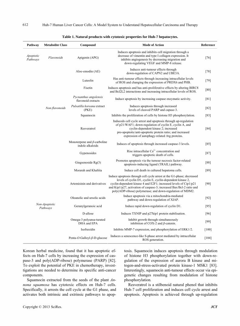
Table 1. Natural products with cytotoxic properties for Huh-7 hepatocytes.
Pathway Metabolite Class Compound Mode of Action Reference
Apoptotic Pathways
Flavonoids Apigenin (APG)
Induces apoptosis and inhibits cell migration through a decrease of vimentin and type I collagen expression. It
inhibits angiogenesis by decreasing migration and down-regulating VEGF and MMP-8 release.
[76]
Aloe-emodin (AE) Induces anti-tumour effects through
down-regulation of CAPN2 and UBE3A. [78]
Luteolin Has anti-tumour effects through increasing intracellular levels
of ROS and changing the expression of PRDX6 and PHB. [79]
Fisetin
Induces apoptosis and has anti-proliferative effects by altering BIRC8 and Bcl2L2 interactions and increasing intracellular levels of ROS.
[80]
Pycnanthus angolensis flavonoid extracts
Induce apoptosis by increasing caspase enzymatic activity. [81]
Non-flavonoids Pulsatilla koreana extract
(PKE) Induces apoptosis through increased
levels of cleaved PARP and capase-3. [82]
Squamocin Inhibits the proliferation of cells by histone H3 phosphorylation. [83]
Resveratrol
Induceds cell cycle arrest and apoptosis through up-regulation of p21/WAF1; down-regulation of cyclin E, cyclin A, and
cyclin-dependent kinase 2; increased pro-apoptotic/anti-apoptotic protein ratio; and increased
expression of autophagy-related Atg proteins.
[84]
Monoterpene and β-carboline indole alkaloids
Induces of apoptosis through increased caspase-3 levels. [85]
Gypenosides Rise intracellular Ca2+ concentration and
triggers apoptotic death of cells. [87]
Gingsenoside Rg(3) Promotes apoptosis via the tumour necrosis factor-related
apoptosis-inducing ligand (TRAIL) pathway. [88]
Morarah and Khaltita Induce cell death in cultured hepatoma cells. [89]
Artemisinin and derivatives
Induce apoptosis through cell cycle arrest at the G1-phase; decreased levels of cyclin D1, cyclin E, cyclin-dependent kinase 2,
cyclin-dependent kinase 4 and E2F1; increased levels of Cip1/p21 and Kip1/p27, activation of caspase-3; increased Bax/Bcl-2 ratio and
poly(ADP-ribose) polymerase; and down-regulation of MDM2.
[90]
Oleanolic and ursolic acidsInduce apoptosis via a mitochondria-mediated
pathway and down-regulation of XIAP. [92]
Non-Apoptotic Pathways
Geranylgeranoic acid Induce rapid down-regulation of cyclin D1. [95]
D-allose Induces TXNIP and p27kip1 protein stabilisation. [96]
Omega-3 polyunsa-turated DHA and EPA
Inhibit growth through simultaneously inhibition of COX-2 and β-catenin.
[99]
Isofraxidin Inhibits MMP-7 expression, and phosphorylation of ERK1/2. [100]
Penta-O-Galloyl-β-D-glucoseInduces a senescence-like S-phase arrest mediated by intracellular
ROS generation. [104]
Korean herbal medicine, found that it has apoptotic ef- fects on Huh-7 cells by increasing the expression of cas- pase-3 and poly(ADP-ribose) polymerase (PARP) [82]. To exploit the potential of PKE in chemotherapy, invest- tigations are needed to determine its specific anti-cancer components.
Squamocin extracted from the seeds of the plant An- nona squamosa has cytotoxic effects on Huh-7 cells. Specifically, it arrests the cell cycle at the G1 phase, and activates both intrinsic and extrinsic pathways to apop-
tosis. Squamocin induces apoptosis through modulation of histone H3 phosphorylation together with down-re- gulation of the expression of aurora B kinase and mi- togen-and-stress-activated protein kinase-1 MSK1 [83]. Interestingly, squamocin anti-tumour effects occur via epi- genetic changes resulting from modulation of histone phosphorylation.
Resveratrol is a stilbenoid natural phenol that inhibits Huh-7 cell proliferation and induces cell cycle arrest and apoptosis. Apoptosis is achieved through up-regulation

Huh-7 Human Liver Cancer Cells: A Model System to Understand Hepatocellular Carcinoma and Therapy 613
of cyclin-dependent kinase inhibitor 1 (p21/WAF1) and down-regulation of cyclin E, cyclin A, and cyclin-de- pendent kinase 2. The study showed that resveratrol in- creases the ratio of pro-apoptotic/anti-apoptotic proteins, is associated with the mitochondrial membrane depolari- zation and the increase in caspase activity, and increases expression of the autophagy-related proteins Atg5, Atg7, Atg9, and Atg12 [84].
Monoterpene and β-carboline indole alkaloids are iso- lated from the methanol extract of leaves of Tabernae- montana elegans. Huh-7 cells treated with these alka- loids showed increased caspase-3 activity and subsequent induction of apoptosis. The work identified the cory- nanthe-type monoterpene alkaloids tabernaemontanine and vobasine to have the best potential as chemopre- ventive agents against HCC [85].
The triterpenoids saponins gypenosides derived from Gynostemma pentaphyllum are widely employed in tra- ditional Chinese medicine. They have cytotoxic pro- perties and a study undertaken to understand their effects on Huh-7 cells found that they induce apoptosis through a mitochondria-dependent caspase-9 activation cascade [86]. A more detailed investigation of the mechanisms leading to apoptosis in gypenoside-treated cells showed that the saponins induced the generation of ROS, dis- ruption of the mito-chondrial membrane potential, in- activation of ERK, increased miotchondrial Bax and de- creased mitochondrial Bcl-2 levels, and cause a rapid rise in intracellular Ca2+ levels. It was concluded that the increase in Ca2+ levels acts as a trigger for apoptosis of these cells [87].
Ginsenosides are pharmacologically active steroidal saponins of ginseng, natural herbal extract of the root of the medicinal plant Panax ginseng. The 20S-ginsenoside Rg(3) promoted apoptosis of Huh-7 cells via the tumour necrosis factor-related apoptosis-inducing ligand (TRAIL) pathway. Rg(3) up-regulates tumour necrosis factor re- ceptor member 10b (TNFRSF10B, DR5) expression at the transcriptional level that is mediated bythe transcrip- tion factor CCAAT-enhance-binding protein (C/EBP) homologous protein (CHOP) an endoplasmic reticulum stress response protein. Since Rg(3) is well tolerated, its properties support its further devlopment as novel anti- cancer therapeutic agent in combined therapy with TRAIL [88].
The properties of the natural herbs Morarah and Khaltita were studied to determine their effects on Huh-7 cells. Extracts of these herbs induced apoptosis in the cells in culture showing anticancer effects in vitro and potential as chemopreventive agents against HCC. Fur- ther investigations need to take place in order to establish their mechanism of action in induction of apoptosis and possible side effects [89].
Artemisinin is a natural sesquiterpene lactone from the
plant Artemesia annua that traditionally has been used in the treatment of malaria. A recent study showed that some derivatives, including dihydroartemisinin, arteme- ther and artesunate, have also anticancer capabilities [90]. These artemisinin derivatives exert anticancer properties through induction of apoptosis. A recent study em- ploying Huh-7 cells was undertaken to better understand the mode of action of these products in their induction of apoptosis, It established that they cause cell-cycle arrest at the G1-phase; they decrease the levels of cyclin D1, cyclin E, cyclin-dependent kinase 2, cyclin-dependent kinase 4, and E2F1; and they increase the levels of cyclin-dependent kinase inhibitors 1 (Cip1/p21) and 7 (Kip1/p27). In addition, the derivatives cause an increase the Bax/Bcl-2 ratio and up-regulation of caspase-3, an elevation of PARP levels, and down-regulation of the double minute oncogene MDM2 [90]. The PARP family of proteins is found in the nucleus, and the proteins are involved in DNA repair and programmed cell death; they are activated in cells experiencing stress and/or DNA damage. PARP stimulate the release of mitochondrial apoptosis-inducing factor that initiates caspase-indepen- dent apoptosis and is an intrinsic regulator of apoptosis [91]. All these effects contribute to their cell death ef- fects on Huh-7 cells and support their potential use as chemopreventive agents against HCC.
The naturally occurring triterpenoid oleanolic acid (OA) and pentacyclic triterpene ursolic acid (UA) com- monly found in plants and herbs posses hepatoprotective, anti-inflammatory and anti-cancer activities. OA and UA inhibit the growth of Huh-7 cells by stopping cell-cycle progression at the G1 phase. Treatment of this hepatoma cell line produced a loss of mitochondria membrane po- tential, and changed the ratio of expression levels of pro- and anti-apoptotic proteins. In the treated Huh-7 cells mitochondrial cytochrome c is released to the cytosol, caspase-3 and caspase-9 are activated followed by PARP cleavage. In addition, treated cell suppressed the activity of NF-κB and modulated the expression of X-linked apoptotic protein (XIAP). The data indicated that OA and UA induced apoptosis of Huh-7 cells via a mitochon- dria-mediated pathway and down-regulation of XIAP [92].
4.2. Compounds Affecting Non-Apoptotic Pathways
Apoptosis is a common mechanism of action of many natural products, but many others exert their effects through various cellular pathways. Geranylgeranoic acid (GGA) is a polyprenoic natural product found in the Chinese herb Schisandra chinensis [93]. A recent study of the effects of GGA and its derivatives showed that it causes an initiation of autophagy on Huh-7 cells, but blocks maturation of autolysomes and consequently the
Copyright © 2013 SciRes. JCT
Huh-7 Human Liver Cancer Cells: A Model System to Understand Hepatocellular Carcinoma and Therapy 614
late stages of autophagy [94]. Another study with these hepatoma cells showed in GGA-treated cells suppression of cyclin D1 protein, subsequent decrease in the phos- phorylation and nuclear translocation of retino-blastoma protein (RB), and nuclear accumulation of RB. Such ac- tivity disrupts cell growth and cell-cycle progression, making it a potential agent for the treatment of HCC [95].
Another compound that disrupts the cell cycle is D- allose, which has been the focus of a recent study of its effects on various cancer cells. In particular on Huh-7 cells GCA causes G1 cell-cycle arrest, as well as up- regulation of gene expression of the thioredoxin inter- acting protein (TXNIP) and stabilisation of p27kip1, that cause also cell-cycle arrest [96].
Prostaglandins (PG) are mediators of inflammation and have an important role in hepatocarcinogenesis [97]. The are derived from arachidonic acid, an omega-6 po- lyunsaturated fatty acids (ω-6 PUFA), by the action of cyclooxygenase enzymes (COX). Elevated levels of PG and increased expression of the inducible cyclooxy- genase-2 (COX-2) have been measured in HCC. The Wnt/beta-catenin (β-catenin) pathway ialso is implicated in hepatic carcino-genesis [98], and elevated levels of β-catenin are found in hepatomas. In contrast, there is considerable evidence that omega-3 poly-unsaturated fat- ty acids (ω-3 PUFA) prevent carcinogenesis. A study of the effects on Huh-7 cells of the ω-3 PUFA docosa- hexaenoic acid (DHA) and eicosapentaenoic acid (EPA) showed that they inhibit COX-2 and β-catenin. Treat- ment of Huh-7 cell cultures with DHA and EPA causes cleavage of poly(ADP-ribose) polymerase, and activation of caspase-3 and caspase-9. These effects suggest some ω-3 PUFA acids as potential agents for the treatment of HCC [99].
Isofraxidin (7-hydroxy-6,8-dimethoxy-2H-1-benzopyran- 2-one), is a coumarin component extracted from the stem bark of the Chinese herb Acanthopanax senticosus. Re- cent studies of its effect on Huh-7 cells show that it has anti-cancer effects and is a potential future thera- peutic agent for HCC. Specifically, isofraxidin inhibits meta- lloproteinase-7 (MMP-7) expression, and also the phos- phorylation of extracellular-signal-regulated kinases 1 and 2 (ERK1/2); the functional outcome is to inhibit in vitro cell invasion [100].
Cellular senescence is a process that limits the prolife- ration (growth) of normal human cells in culture. It is an essentially irreversible growth arrest [101]. Penta-1,2, 3,4,6-O-galloyl-beta-D-glucose (PGG) is a naturally oc- curring gallotannin polyphenolic compound found in herbs such as Rhus chinensis and Paeonia suffructicosa capable of inducing S-phase and G1 cell-cycle arrests [102,103]. PGG-induced senescence-like S-phase arrest in Huh-7 human hepatoma cells; the senescence-like re-
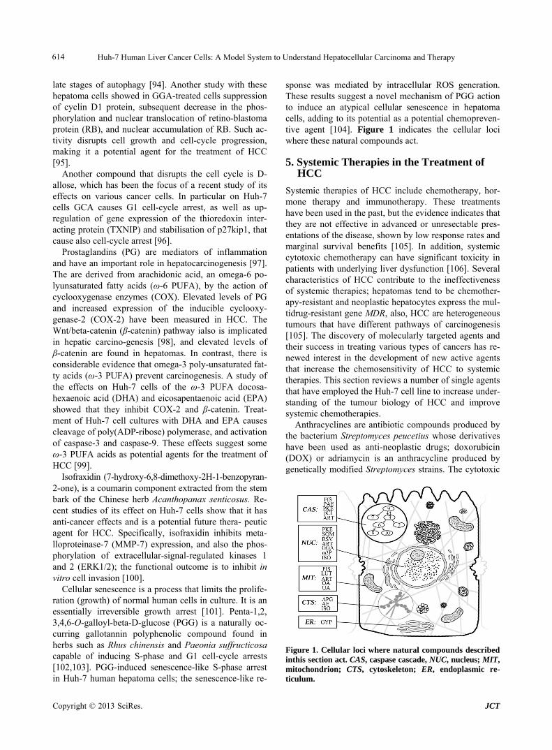
sponse was mediated by intracellular ROS generation. These results suggest a novel mechanism of PGG action to induce an atypical cellular senescence in hepatoma cells, adding to its potential as a potential chemopreven- tive agent [104]. Figure 1 indicates the cellular loci where these natural compounds act.
5. Systemic Therapies in the Treatment of HCC
Systemic therapies of HCC include chemotherapy, hor- mone therapy and immunotherapy. These treatments have been used in the past, but the evidence indicates that they are not effective in advanced or unresectable pres- entations of the disease, shown by low response rates and marginal survival benefits [105]. In addition, systemic cytotoxic chemotherapy can have significant toxicity in patients with underlying liver dysfunction [106]. Several characteristics of HCC contribute to the ineffectiveness of systemic therapies; hepatomas tend to be chemother- apy-resistant and neoplastic hepatocytes express the mul- tidrug-resistant gene MDR, also, HCC are heterogeneous tumours that have different pathways of carcinogenesis [105]. The discovery of molecularly targeted agents and their success in treating various types of cancers has re- newed interest in the development of new active agents that increase the chemosensitivity of HCC to systemic therapies. This section reviews a number of single agents that have employed the Huh-7 cell line to increase under- standing of the tumour biology of HCC and improve systemic chemotherapies.
Anthracyclines are antibiotic compounds produced by the bacterium Streptomyces peucetius whose derivatives have been used as anti-neoplastic drugs; doxorubicin (DOX) or adriamycin is an anthracycline produced by genetically modified Streptomyces strains. The cytotoxic
Figure 1. Cellular loci where natural compounds described inthis section act. CAS, caspase cascade, NUC, nucleus; MIT, mitochondrion; CTS, cytoskeleton; ER, endoplasmic re- ticulum.
Copyright © 2013 SciRes. JCT
Huh-7 Human Liver Cancer Cells: A Model System to Understand Hepatocellular Carcinoma and Therapy 615
activity of DOX stems from inhibition of macromolecu- lar biosynthesis at the transcription level through inter- calation with DNA and stopping topoisomerase II. It has been used commonly to treat HCC, but as a single agent has not proved effective owing to tumour resistance, and its toxicity to normal cells. Thus, investigations were directed at discovering new therapeutic agents that can enhance the effectiveness of doxorubicin treatment. A reason for HCC resistance to DOX is the ability of neo-plastic cells to undergo epithelial-mesenchymal transition (EMT) during tumour progression; a process by which epithelial cells down-modulate cell-adhesion, change their polarity and cytoskeleton, become isolated and motile and develop resistance to programmed cell death; in he- patoma cells, activation of signal transducer and activator of transcription 3 (STAT3) contributes to the process of EMT [107]. A study of the novel STAT3 inhibitor NSC 74859 using Huh-7 cells showed that co-administered with DXR it has synergistic anti-tumour effects enhanc-ing DOX cytotoxicity [108].
Cancer stem cells (CSC) are defined as a minor popu- lation that possess a prominent ability to generate new tumours, and are capable to self-renew and generate dif- ferentiated progenies like normal stem cells [109]; they are key factors in tumour progression and growth. A side population of Huh-7 cells considered representative of HCC stem cells was used to identify molecular signalling pathways in liver CSC, and to suggest new therapeutic strategies based on the results [110]. The study esta- blished that in the CSC side population of Huh-7 cells hypoxic conditions promoted cell proliferation and growth owing to overexpression of cytochrome P450 2C9 (CYP2- C9), a protein localised in the endoplasmic reticulum and that metabolises many xenobiotics. Also, in the CSC sub- population STAT3 was activated by interleukin-6 was more significantly than in non-CSC cells, and plays a role in resistance to DOX. Treatment with sulfaphe- nazole, an inhibitor of CYP2C9 inhibitor, or AG490, an inhibitor of STAT3, sensitised the CSC population to DOX, suggesting that co-administration of the inhibitors with DOX may be a potential new therapeutic strategy [110].
Nutlin-3 is cis-imidazole analogue that inhibits inter- actions between the tumour suppressors p53 and p73 and HDM2, the human analogue of the murine double minute (MDM2) oncoprotein. Recent investigations using Huh-7 cells showed that Nutlin-3 enhances the chemosensitivity to DOX through inhibition of p73-HDM2 binding with subsequent activation of the apoptotic pathway. Such re- sult suggests that Nutlin-3 may be a potential HCC ther-apy when co-administered with DOX [111].
The effects of some chemotherapeutic agents are en- hanced by co-administration with natural products. A study investigated the benefits caffeine-assisted chemo-
therapy in the treatment of HCC with cis-diammine- dichloroplatinum-II (cisplatin). Caffeine could increase the anti-tumour effect of cisplatin or other DNA-da- maging agents because it inhibits DNA repair. Cisplatin is an agent used in the treatment of HCC, but its ad- ministration alone is not optimal [200]. Huh-7 cells were treated with caffeine and cisplatin and the co-adminis- tration enhances the anti-tumour effects of cisplatin [112]. Expression of the MDR gene is mechanism by which HCC cells become resistant to many of the available chemotherapy drugs. Recent work found that coadminis- tration of Kampo drugs, traditional Japanese medicines, has potential to overcoming drug resistance of HCC to paclitaxel (PTX), a mitotic inhibitor synthesised by fungi in the bark of Taxus brevifolia, the Pacific yew tree. Us- ing the Huh-7 paclitaxel-resistant variant Huh-7/PTX that expresses high levels of the multidrug resistance per- meability glycoprotein 1 (MDR-1), 26 kinds Kampo me- dicines were tested and two of them, takushato and go- reisan, increased the sensitivity of HuH-7/PTX cells to paclitaxel. These compounds contain the triterpenes Ali- sol A, Alisol B, and Alisol B acetate that increase resis- tance to paclitaxel by preventing drug efflux by MDR-1 without affecting its expression levels. This study iden- tified Kampo drugs as potentially effective when co- administered with the chemotherapy drug paclitaxel [113].
The uptake of chemotherapy drugs and their subse- quent therapeutic effects can be enhanced by the use of nanocarriers. A nanoparticle from an amphiphilic block copolymer composed of conventional monomethoxy (po- lyethylene glycol)-poly (D,L-lactide-co-glycolide)-poly (L-lysine) (mPEG-PLGA-b-PLL) was used for delivery of DOX or of small interfering RNA-negative (siRNA) into Huh-7 cells. The study shows increased cellular up- take of the drugs when they were delivered into the cell via the nanoparticle carrier. The findings suggest the po- tential of these carriers to deliver chemotherapy agents to HCC with subsequent enhanced cellular uptake and the- rapeutic results [114].
Another study investigated the delivery to Huh-7 cells of mitoxantrone (MX), an anthracenedione anti-neoplas- tic agent that disrupts DNA synthesis and DNA repair. Dual-functional liposomes (LPG) with the synthetic po- lymeric nano-biomaterial (Gal-P123) that targets cancer cells and reverses multidrug resistance were loaded with MX. Delivery via LPG enhanced uptake of MX by Huh- 7 cells, and inhibited drug efflux, thus improving sig- nificantly the cytotoxic therapeutic efficacy of MX [115].
To increase the effectiveness of cisplatin treatment, further knowledge has been required of its pro-apoptotic mechanisms. The mitogen-activated protein kinase (MAPK) pathway plays a key role in cell response to cisplatin. A
Copyright © 2013 SciRes. JCT

Huh-7 Human Liver Cancer Cells: A Model System to Understand Hepatocellular Carcinoma and Therapy 616
recent study determined the individual contribution of each kinase of this pathway on cisplatininduced death. ERK1 has a greater role than ERK2 in cisplatin-induced pro-apoptotic signal in HCC cells and it was up-regulated by ERK2 inhibition. The results of this study provide the basis for more targeted cisplatin therapy suggesting that its efficacy of could be increased by targeting ERK2 [116].
Recent studies show that gene therapy can enhance the effects of some chemotherapeutic agents. A study dem- onstrated that transfection of the sodium iodine sym- porter (NIS) or the mutant Herpes-simplex virus type1 sr39 thymidine kinase (HSV1-sr39tk) gene into Huh-7 cells enhances intracellular accumulation of therapeutic radionuclides and guanosine nucleoside analogue pro- drugs. The work demonstrates the potential of combi- nation gene therapy using NIS and HSV1-sr39tk follow- ed by radioiodine treatment and chemotherapy to treat human hepatocellular carcinoma cells [117].
6. Targeted Therapies in the Treatment of HCC
Dysfunction of many different signaling pathways occurs in the pathogenesis of hepatocellular carcinoma [118]. Also, the etiology of hepatocellular carcinoma is com- plex and diverse; hence, improved understanding of these pathways is required [119]. Unlike systemic therapies, targeted therapies aim at specific aberrant pathways [120]. Thus, a better of understanding of the biology of neo- plastic hepatocytes will facilitate the development of new, targeted therapeutic agents that overcome drug resistance and minimise side effects, with the ultimate aim of im- proving patient outcomes [119], for this reason targeted therapy for HCC is currently under intensive investiga- tion [120]. The Huh-7 cell line is extensively used in current investigations to develop of therapies for HCC, and this section attempts to provide an overview of this research. Table 2 sumamrises the targeted therapies dis- cussed in this section.
6.1. Sorafenib Combination Therapies
Presently, the mainstay of targeted therapy against HCC is sorafenib, a bi-aryl urea which interferes with tyrosine protein kinases; it inhibits cancer cell growth signaling and angiogenesis and promotes apoptosis [121]. The story of this drug has proven the concept that targeted therapy brings benefits to patients with HCC, although at present these remain limited [120] because sorafenib like other therapeutic agents has a number of limitations, in- cluding the development of resistance against it and side effects [122]. A number of investigations have been un- dertaken to identify sorafenib combination therapies that would improve its efficacy.
To investigate the role of the signal transducers and
activators of transcription 3 (STAT3) in sorafenib in- hibittion of HCC, Huh-7 cells were treated with this drug or SC-1, one of its derivatives that is structurally similar but without kinase inhibition activity; the treated heap- toma cells showed growth inhibition and apoptosis. The STAT3 signaling pathway regulates proteins such as myeloid cell leukemia sequence 1 (Mcl-1), survivin and cyclin D1 that are involved in cell growth, survival, dif- ferentiation and anti-apoptosis; STAT3 is down-regu- lated by the protein tyrosine phosphatase SHP-1. The re- sults showed that SC-1 and sorafenib up-regulated SHP-1 activity and down-regulated STAT3, thereby inhibiting HCC cell growth and proliferation. This work suggests that for sorafenib-treated patients, STAT3 could poten- tially be used as a biomarker for determining HCC prog- nosis [123]. In a study with Huh-7 cells, it was deter- mined that sorafenib or SC-49, one of its derivatives de- void of kinase inhibition activity, in combination with CS-1008, a novel anti-human death receptor 5 antibody that induces apoptosis, down-regulate the phosphoryla- tion of STAT3 and subsequently reduce the protein le- vels of STAT3-regulated proteins. The mechanism of action of the combination therapy was STAT3 inactiva- tion through increasing the activity of the modulator of signaling cascades SHP-1 [124].
The effects of co-administration of sorafenib and CI- 1040, an inhibitor of mitogen-activated protein kinase/ extracellular signal-regulated kinase (ERK) kinase (MEK) were investigated in Huh-7 cells. Combination of sora- fenib and CI-1040 synergistically inhibited ERK phos- phorylation and cell growth and induced apoptosis. Ver- tical blockade of signaling through the protooncogene serine/threonine-protein kinase (Raf)/MEK/ERK was achieved by the co-administration of these agents, im-proving the anti-tumour effects of sorafenib in HCC [125].
Programmed cell death can be stimulated by tumour necrosis factor (TNF)-related apoptosis-inducing ligand (TRAIL) [126]; hence, TRAIL is a potential target for anti-cancer drugs. However, hepatomas often display re- sistance to TRAIL-induced apoptosis; specifically, Huh-7 cells show significant resistance to TRAIL-induced apop- tosis. These hepatoma cells were treated with sorafenib and/or LBY135, an agonistic death receptor 5 (DR5) an- tibody, and the cell responses were investigated in terms of signal transduction and apoptosis. Besides down- regulating phospho-STAT3, and subsequently reducing the expression levels of STAT3-related proteins, sora- fenib in combination with LBY135 significantly su- pressed xenograft tumour growth, suggesting that it sen- sitises hepatoma cells resistant to TRAIL-induced apop- tosis [127]. This study provides a positive foundation for future applications of sorafenib as an agent to help over- come TRAIL resistance in HCC [127].
Copyright © 2013 SciRes. JCT

Huh-7 Human Liver Cancer Cells: A Model System to Understand Hepatocellular Carcinoma and Therapy
Copyright © 2013 SciRes. JCT
617
Table 2. Targeted therapies for HCC that have been investigated using Huh7-cells.
Therapy Mechanism/pathway targeted Therapy method of action Reference
Sorafenib combination therapies
SC-1 STAT3 Inhibition of HCC cell growth and proliferation through up-regulation of SHP-1 and down-regulation of STAT3.
[123]
CS-1008 STAT3 Sensitisation to sorafenib and its derivative SC-49,
through inhibition of SHP-1 dependent STAT3 inactivation. [124]
CI-1040 Raf/MEK/ERK Apoptosis induction through inhibition of Raf/MEK/ERK
as a result of increased levels of Bim. [125]
TRAIL-related agents (TRAIL or LBY135)
STAT3 Induction of TRAIL-induced apoptosis through
down-regulation of STAT3. [127]
Dovitinib STAT3
Inhibition of HCC cell growth and sensitization to sorafenib through down-regulation of p-STAT3. Dovitinib overcomes
resistance to TRAIL- and tigatuzumab-induced apoptosis through inhibition SHP-1.
[128] [129]
Signal transduction
SC-2001 STAT3 SC-2001 upregulates STA3 through up-regulation of SHP-1. [131]
IPD-196 PI3K/Akt/mTOR
Inhibition of tumourigenesis through inhibition of downstream PI3K effectors including Akt, mTOR, p70S6K and 4E-BP1. Induction of
apoptosis through increased cleaved PARP, caspase-3 and caspase-9 and anti-angiogenesis through decreased HIF-1α and VEGF.
[132]
HS-104 PI3K/Akt/mTOR
Inhibition of tumourigenesis through inhibition of downstream PI3K effectors including Akt, mTOR, p70S6K and 4E-BP1. Induction of
apoptosis through increased cleaved PARP, caspase-3 and caspase-9 and anti-angiogenesis though decreased HIF-1α and VEGF.
[133]
HS-116 PI3K/Akt/mTOR
HS-116 suppresses the phosphorylation of downstream factors AKT, mTOR, p70S6K, and 4EBP1 and increases Bax, cleaved-caspase-3,
and cleaved-PARP as well as decreasing the expression of Bcl-2, thereby promoting apoptosis and inhibiting tumourigenesis.
[134]
Bortezomib and sorafenib Akt Induction of apoptosis through down-regulation of Akt [137]
GNMT mTOR GNMT affects mTOR signaling by interacting with
DEPDC6/DEPTOR. [139]
Apoptosis
LCN2 - Induction of apoptosis through cleavage of
caspase-9, -8, -3 and APRP protein, reduction in MMP and increased ratio of Bax/Bcl-2 ratio.
[140]
Fenretinide Nur77 Induction of apoptosis through intracellular localisation of Nur77. [141]
LCL161 SMAC mimetics Induction of apoptosis through coadministration
of LCL161 and SC-2001. [143]
SB-365 - SB-365 inhibits the growth and progression of HCC
through enhancing the expression of Bax and cleaved caspase-3 and decreases the expression of HIF-1α) and VEGF.
[144]
Cell Cycle
PHA-739358 Aurora kinases Suppression of tumour growth by decreasing aurora kinase levels. [146]
Migration and invasion
Sorafenib SNA1 Inhibition of metastases by down-regulating SNA1 though inhibition
of MAPK signaling. [149]
GRIM-19 - GRIM-19 shows EMT-like morphology with loss of contact
inhibition, suppressing HCC invasion. [150]

Huh-7 Human Liver Cancer Cells: A Model System to Understand Hepatocellular Carcinoma and Therapy 618
Continued
Antiviral Therapies
Amprenavir MMPs
Amprenavir inhibits MMP proteolytic activation thereby inhibiting cell invasion ability. Additionally, when co-administered with doxorubicin, enhanced
effects of this chemotherapy drug are observed.
[151]
Radiotherapy
Ad-p53 and radiotherapy p53-MDM2 Inhibition of p53-MDM2 interaction and subsequent enhancement of
radiotherapy. [154]
AR-42 Ku70 Inhibition of Ku70 and subsequent sensitization to radiotherapy. [155]
Gene Therapy
AD55-Apoptin - Induction of apoptosis. [158]
Ad.enAFP-E1A-ΔE1B-IL-24 GADD34 Induction of apoptosis through up-regulation of GADD34 and intrin-
sic and extrinsic apoptosis singaling. [159]
miR-122 Igf1R and ADAM10 miR-122 prevents activation of Igf1R and ADAM10 and thereby
suppresses tumour growth. [161]
Chemical Ablation Therapy
Urea Protein denaturation Induction of apoptosis through protein denaturation. [162]
Dovitinib is a multiple kinase inhibitor currently un-
dergoing clinical investigation for its use in the treatment of HCC. It shows anti-tumour activity in Huh-7 and other liver cell lines. Its mechanism of action is down-regula- tion of phospho-STAT3 (p-STAT3), subsequently re- ducing the levels of expression of STAT-3 related pro- teins, and inducing apoptosis. An inhibitor of SHP-1 reversed the down-regulation of p-STAT3 and the apop- tosis induced by dovitinib, and this inhibitor up-regulates the activity of SHP-1 via direct interactions. In addition, dovitinib induced apoptosis in two sorafenib-resistant cell lines through inhibition of STAT3, and sorafenib-re- sistant cells showed significant activation of STAT3, sug- gesting that targeting STAT3 may be a useful approach to overcome drug resistance in HCC [128] Dovitinib and tigatuzumab, a novel humanized anti-human DR5 ago- nistic antibody, are currently undergoing clinical investi- gation for their co-administration in the treatment of HCC. Besides showing significant resistance to TRAIL-induced apoptosis, Huh-7 cells are resistant also to tigatuzumab- induced apoptosis. The mechanism of action of the anti- tumour effects of dovitinib in resistant Huh-7 cells was through inhibition of phospho-STAT3. The combination of dovitinib and tigatuzumab increased the activity of SHP-1 and restored the sensitivity of the hepatoma cells to TRAIL- and tigatuzumab-induced apoptosis [129].
6.2. Signal Transduction
The anti-apoptotic proteins of the BCL-2 family are overexpressed and dysregulated in various cancers, in-
cluding HCC. In particular, Bcl-xL and Mcl-1 have significant cytoprotective roles in HCC. A common strategy in the design of Bcl-2 protein inhibitors is based on mimicking the actions of endogenous inhibitors that bind anti-apoptotic Bcl-2 proteins via the Bcl-2 ho- mology 3 (BH3) domains (BH3 mimetics) [130]. Oba- toclax is a pan-Bcl-2 inhibitor that acts as a BH3-mi- metic to disrupt the interactions of anti-apoptotic and proapoptotic proteins, such as Mcl-1 and Bak. It uniquely displaces BH3 domains by activation of the pocket of Mcl-1 followed by a triggering of apoptosis mediated by oligomerization of Bak and release of cytochrome c. Important anti-apoptotic members of the Bcl-2 family, such as Bcl-xL and Mcl-1, can be regulated by oncogenic transcription factors, such as STAT3 which is cons- titutively activated in HCC. SC-2001 is a novel com- pound structurally related to obatoclax; similarly, it in- hibits in Huh-7 cells protein-protein interactions between Mcl-1 and Bak, and, in addition, it down-regulates Mcl-1 protein levels by reducing its transcription. SC-2001 down-regulates the phosphorylation of STAT3 subse- quently inhibiting its transcriptional activities, and re- pressing also survivin and cyclin D1. The inhibitor up- regulated expression of SHP1 inducing apoptosis in HCC and reducing tumour growth [131].
Normal cell function, including cell survival, cell pro- liferation and cell growth, involves the phosphatidy-li- nositol 3-kinase/protein kinase B/mammalian target of rapamycin (PI3K/Akt/ mTOR) pathway. This pathway is initiated by the enzyme PI3-kinase which causes phos- phorylation of cell membrane components. Subsequently,
Copyright © 2013 SciRes. JCT

Huh-7 Human Liver Cancer Cells: A Model System to Understand Hepatocellular Carcinoma and Therapy 619
phosphorylated components bind to Akt and activate this protein kinase, which initiates the activation of several pathways that promote cell survival, proliferation and growth. Akt activation is followed in the PI3K/Akt/ mTOR pathway by activation of mTOR; this kinase plays a role in protein synthesis that ultimately supports cell proliferation and growth, and also is involved in nutrient uptake and angiogenesis. Impairment of any of the com- ponents of this pathway can result in tumourigenesis, and human cancers frequently have overactive PI3K/Akt/ mTOR pathways that reduce apoptosis and allow un- regulated proliferation. In particular, this pathway is dys- regulated in HCC; for this reason, in recent cancer re- search, this pathway and its components are the focus for finding targets target for therapeutic intervention.
The Huh-7 cell line has been employed in studies tar- geting the PI3K/Akt/mTOR pathway in HCC. A focus has been the development of PI3K inhibitors specific to HCC. The compound IPD-196 [ethyl 6-(5-(2,4-di-fluo- rophenylsulfonamido)pyridine-3-yl)imidazo[1,2-a] pyri- dine-3-carboxylate] is a PI3K inhibitor that is effective decreasing phosphorylation of downstream PI3K effect- tors, including Akt, mTOR, the p70S6 ribosomal protein kinase and the translator repressor 4E-BP1, and it has more potent anti-proliferative effects that sorafenib. IPD- 196 caused cell cycle arrest, induction of apoptosis, and inhibition of angiogenesis in human hepatoma cells, sug- gesting that it may be a potential candidate drug for targeted HCC therapy [132].
Another PI3K inhibitor is the compound HS-104 [N- (5-(3-(3-methyl-1,2,4-oxadiazol-3-yl)imidazo [1,2-a] pyri- din-6-yl)pyridin-3-yl)benzenesulfonamide)], recently stu- died in relation to its potential as an anticancer treat- ment for HCC. In Huh-7 cells, HS-104 inhibits cell growth, and its apoptotic effects include increased cleaved cas- pase-3 and PARP, as well as DNA fragmentation. These results together with those from experiments in animal xenografts suggest that HS-104 is considered a novel drug candidate for the treatment of HCC [133].
The compound HS-116 [N-(5-(3-(3-cyanophenyl)-1H- pyrrolo[2,3-b]pyri-din-5-yl)pyridin-3-yl)ben-zene-sulfona- mide (HS-116)] show effects on Huh-7 cells similar to those of other PI3K inhibitors. Specifically, HS-116 sup- presses the phosphorylation of downstream factors Akt, mTOR, p70S6K, and 4E-BP1 and promotes apoptosis by increasing Bax levels, cleaved-caspase-3 and PARP, and decreasing the expression of Bcl-2 [134].
Bortezomib is a proteasome inhibitor that binds with high affinity to the catalytic site of the 26S proteasome; inhibition of these protein complexes may prevent deg- radation of pro-apoptotic factors, thus permitting activa- tion of programmed cell death in neoplastic cells de- pendent upon suppression of pro-apoptotic pathways. A major molecular determinant of bortezomib-induced
apoptosis in Huh-7 cells is down-regulation of phospho- Akt; in cells able to develop resistance against borte- zomib, phospho-Akt is not down-regulated [135]. This inhibitor overcomes TRAIL-induced apoptosis in Huh7- cells in part through inhibition of the (PI3K/Akt/mTOR) pathway [136]. It was found that in Huh-7 cells treated with sorafenib and/or bortezomib each inhibitor causes increased levels of apoptotic cell death, and they show synergy in combination by through Akt inactivation [137].
Mammalian mTOR resides in two distinct multiprotein complexes referred to as mTOR complex 1 (mTORC1) and 2 (mTORC2). Complex 1 controls cell growth in part by phosphorylating key regulators of protein synthesis, and mTORC2 modulates cell survival in response to growth factors by phosphorylating downstream effectors such as Akt. In addition to activating Akt by mTORC2, mTORC1 regulates negatively Akt by suppressing the growth factor-driven pathways upstream of it [138]. DEPTOR as an mTOR binding protein that normally functions to inhibit the mTORC1 and mTORC2 path- ways, but overexpression of DEPTOR inhibits mTORC1, and this leads to the activation of Akt via the inhibition of a negative feedback loop to PI3K [138]. Loss of DEP- TOR in Huh-7 cells reduced Akt activation and cell growth. It was revealed that GNMT affects mTOR sig- naling by interacting with DEPTOR, and that GNMT can sensitise HuH-7 cells to rapamycin both in vitro and in vivo. These results make GNMT and DEPTOR potential targets for future therapeutic agents for the treatment of HCC [139].
6.3. Apoptosis
Besides the pro-apoptotic effects of sorafenib, dovitinib and their derivatives, other compounds capable of pro- moting apoptosis in HCC have been investigated.
Abnormal expression of the secreted iron-binding gly- coprotein lipocalin 2 (LCN2) is a feature of some human cancers. LCN2 overexpression in Huh-7 cells dramati- cally inhibited cell viability, induced cell-cycle arrest in sub-G1 phase, DNA fragmentation, and condensation of chromatin. LCN2 induces apoptosis in Huh-7 cells through the cleavage of caspase-9, -8, -3, and PARP pro- tein, and a reduction in the mitochondrial membrane po- tential; it also increases the Bax/Bcl-2 ratio. The study improved the understanding of the role of LCN2 on HCC progression; by demonstrating that overexpression of LCN2 actively induces apoptosis of hepatoma cells, it suggests that the glycoprotein could be considered as therapeutic agent against HCC [140].
The mechanism of apoptosis induction by the synthetic retinoid fenretinide has been unknown; consequently, a recent study examined the mechanisms fenretinide-in- duced apoptosis in HCC cells. Apoptosis was correlated
Copyright © 2013 SciRes. JCT
Huh-7 Human Liver Cancer Cells: A Model System to Understand Hepatocellular Carcinoma and Therapy 620
with the induction and cytoplasmic distribution of the nerve growth factor IB (Nur77). In Huh-7 cells, the sen- sitivity to fenretinide is related to the translocation of Nurr77 from the nucleus and targeting to mitochondria. The results of the study show that fenretinide-induced apoptosis is dependent on Nur77 and more specifically, of its the intracellular localisation [141].
The endogenous pro-apoptotic protein second mito- chondria-derived activator caspases (SMAC) is normally localised in mitochondria. Apoptopic stimuli make SMAC translocate to the cytosol, bind inhibitor of apop- tosis proteins (IAP) and release caspases. Disturbing this mechanism by immobilization of SMAC in the mito- chondria or overexpressing of IAPs is linked to evasion from apoptosis by tumour cells. Strategies to mimic the action of SMAC are called SMAC mimetics. In immuno- genic tumour killing, death-inducing agents also instigate a potent anti-tumour immune response [142]. LCL161 is an orally bioavailable SMAC mimetic and inhibitor of IAP family. The endogenous levels of Bcl-2 sensitivity of hepatoma cell lines cells to LCL161. Huh-7 cells have high Bcl-2 levels of expression an d proved resistant to LCL161. Down-regulation of Bcl-2 expression by siRNA or treatment with the inhibitor SC-2001 results in Huh-7 cells overcoming resistance to LCL161 and undergoing apoptosis, and indicates synergistic effects between the SMAC mimetic and teh BCL-2 inhibitor [143]. The re- sults show that these combined therapeutic agents may be useful for future treatments of HCC.
Saponins exhibit cytostatic and cytotoxic activity aga- inst various cancer cells. Saponin D (SB365), an active component isolated from Pulsatilla koreana, suppresses the growth of Huh-7 cells and promotes apoptosis through induction of expression of Bax and cleaved caspase-3. This saponin has anti-angiogenic activity by decreasing the expression of HIF-1α and VEGF. The mechanism of action of SB365 is to suppress the phosphorylation of PI3K downstream factors, such as Akt, mTOR and p70S6K both in vitro and in vivo. The results suggest that saponin D may be a candidate for anti-HCC chemother- apy [144].
6.4. Cell Cycle
Aurora kinases (AURK) are key regulators of the cell cycle; their uncontrolled expression promotes aneuploidy and tumour cells often overexpress AURK. The AURK family includes aurora-A, aurora-B, and aurora-C en- zymes [145]. Aurora-A kinase expression is detected in human HCC samples, and the levels of aurora-B corre- late with the proliferation index of hepatomas. The Huh- 7 cell line expresses aurora-A and -B, and reveals phos- phorylation of histone H3 at a locus indicative of aurora- B kinase activity. Investigation of the effects of the AURK inhibitor PHA-739358 on Huh-7 cells demon-
strated that in vitro the inhibitor suppresses completely cell proliferation and decreases strongly histone H3 phos- phorylation. The results show that PHA-739358 sup- presses tumour growth by decreasing aurora kinase activi- ity. Additive effects are observed in combination with sorafenib, and following the failure of sorafenib therapy, treatment with PHA-739358 reduced tumour growth. This investigation suggests that targeting AURK with PHA-739358 is a promising new approach for treatment of HCC, either administered alone or in combination with sorafenib [146].
6.5. Migration and Invasion
Hepatocellular carcinoma has the potential to metastasise to the lungs, lymph nodes, adrenal gland and bones and if such metastases occur, prognosis is poor. Epithelial- mesenchymal transition (EMT) is a key development in the ability of HCC to metastasise [147]. The zinc-finger SNAI1 protein can initiate EMT and is involved in the formation and maintenance of embryonic mesoderm. E- cadherin is a calcium-dependent cell-cell adhesion mo- lecule with pivotal roles in epithelial cell behaviour, tis- sue formation, and suppression of cancer [148]. In in- vasive tumour cell lines, there is loss of the “invasion- suppressor” E-cadherin and up-regulation of N-cadherin, another adhesion, E- to N-cadherin switching in epithelial carcinomas impacts on their metastatic progression. Hepatocyte growth factor (HGF) induces in Huh-7 cells EMT-like morphologic changes, up-regulates expression of the SNAI1 protein and N-cadherin, and down-re- gulates E-cadherin. Sorafenib and the MEK inhibitor U0126 inhibit HGF morphologic changes in hepatoma cells, the up-regulation of SNAI1 and cadhering switch- ing. Thus, sorafenib inhibits EMT in these cells, and the study provides insight into the anti-EMT effect of tyro- sine kinases in HCC cells [149].
The cell death regulatory protein retinoid-interferon- induced mortality 19 (GRIM-19) has been identified as a tumour suppressor associated with apoptosis and growth inhibition; it is thought to be involved in regulating he- patoma invasiveness, since GRIM-19 levels are signi- ficantly attenuated in patients with invasive potential HCC. Using Huh-7 cell lines that had been depleted of GRIM-19, a recent study identified that these cells show EMT-like morphology coupled with loss of contact inhi- bition. As a result, GRIM-19 is proposed as a new po- tential target for adjuvant treatment of aggressive and invasive HCC [150].
6.6. Antiviral Therapies
Amprenavir is a protease inhibitor used for treatment of patients with HIV. A recent study evaluated its anti-tu- mour activity on HCC and potential synergistic effects in
Copyright © 2013 SciRes. JCT

Huh-7 Human Liver Cancer Cells: A Model System to Understand Hepatocellular Carcinoma and Therapy 621
combination with standard HCC chemoterapeutic drugs, such as DOX. In vitro, Amprenavir has direct inhibitory effects on matrix metalloproteinase proteolytic activation in Huh-7 cells. In vivo, has a synergistic effect with DOX and promotes regression of hepatocarcinoma growth by anti-angiogenesis and overall anti-tumor activities. The novel anti-neoplastic action of Amprenavir on liver can- cer shows the possibility of novel combination therapies [151].
6.7. Radiotherapy
Radiotherapy has been used as a treatment for HCC, but less so than other treatments including chemotherapy and liver resection. It is often used in cases in which HCC is untreatable. The minimal use of radiotherapy for HCC therapy is largely due to low whole liver tolerance, as challenges exist in delivering high-dose radiotherapy to the tumour while trying to spare the healthy parts of the liver [152]. HDM2 plays a key role in the effects of radiotherapy, it is an important negative regulator of the p53 tumour suppressor, and the amount of HDM2 ex- pression determines the level of p53 induction by ra- diation, and decreased levels of this protein sensitise cells to ionising radiation [153]. Since inhibition of HDM2 means increased expression of p53, the regulator has been the focus of recent studies investigating its potential as a target for improving the efficacy of radiotherapy in the treatment of HCC. Work using different HDM2 cel- lular levels performed adenovirus- mediated modified p53 (Ad-p53) gene transfer in Huh-7 cells, and inves- tigated its effectiveness in enhancing radiotherapy cell death, and also whether the p53-HDM2 interaction would be disrupted thereby enhancing the response to ra- diotherapy. It found that in Huh-7 cells (in both in vitro and in vivo xenografts), gene therapy combined with radiotherapy caused decreased cell viability, making this a future potentially beneficial therapeutic regime for treatment of HCC [154].
In cancer cells, histone deacetylase (HDAC) can be dysregulated and cause tumour growth and repression of tumour suppressor genes. Inhibitors of HDAC target acetylation of the protein Ku70 involved in DNA double strand break repairs and interfere with cellular DNA repair mechanisms. The effects a combination of AR-42, a phenylbutyrate-derived HDAC inhibitor, with radio- therapy were investigated in Huh-7 cells. This study shows that AR-42 causes inhibition of Ku70 activity and sensitised cells to radiotherapy. Thus, AR-42 is another potential agent for future co-administration with radio- therapy to increase its effectiveness against HCC [155].
6.8. Gene Therapy
Gene and viral therapies for cancer have yielded thera- peutic effects, but it has been determined that the new
strategy Cancer Targeting Gene-Virotherapy (CTGVT) is more effective than either gene or viral therapy alone. This method inserts anti-tumour genes into an oncolytic virus; once inside the tumour cells the virus replicates and causes amplification of the therapeutic gene [156]. The strategy was employed using the double-regulated oncolytic adenovirus AD55 and the cancer therapeutic gene Apoptin to create the recombinant virus AD55- Apoptin. This particular virus was chosen for its ability to induce apoptosis in tumour cells, while preserving nor-mal cells [157]. The combination AD55-Apoptin in- duced apoptosis in Huh-7 cells, and the effects were greater than those achieved by either AD55 or the onco- lytic genetically engineered ONYX-015 virus alone, suggesting its potential as a therapeutic agent for treat- ment of HCC [158].
Another study using the CTGVT strategy investigated the use of IL-24 inserted into an oncolytic adenovirus to form the construct Ad.enAFP-E1A-ΔE1B-IL-24. The construct has a potent anti-tumour effect on liver cancer cell lines in vitro, but little or no effect on normal cell lines. The mechanism of the anti-tumour effect of Ad. enAFP-E1A-ΔE1B-IL-24 is to up-regulate GADD34, a member of the protein family whose expression is up- regulated by growth arrest and DNA damage, and in- trinsic and extrinsic apoptotic signaling. In vivo, intra- tumour injection Ad.enAFP-E1A-ΔE1B-IL-24 resulted in complete elimination of Huh-7 liver cancer in nude mice [159].
MicroRNA-122 (miR-122) is a liver-specific micro- RNA. It is abundantly expressed in normal hepatocytes but is absent in many hepatocellular carcinoma cells and plays a pivotal role in the liver [160]. Its target is the in-sulin-like growth factor receptor Igf1R that is involved in cell growth, survival, differentiation and proliferation, tumour metastases and resistance to therapy. The impact miR-122 on tumour growth and progression was ex- amined in Huh-7 cells; anti-miR-122 transfected into the cells increased cell growth, replication, migration and clonogenic survival relative to controls. The study estab- lishes that in Huh-7 cells, Igf1R is negatively regulated by miR-122, as is the distintegrin and metalloprotease family 10 ADAM10 protein that plays a role in tumour survival. These results help to understand the mechanism of anti-tumour activity of miR-122, and will serve in fu- ture development of it as a therapeutic agent against HCC [161].
6.9. Chemical Ablation
Chemical ablation methods have been used to treat HCC, and systemic toxicity can result from such treatments. To remedy the currently limited understanding of the mode of action of this therapy, a study was performed in chemical ablation of Huh-7 using urea, which has little
Copyright © 2013 SciRes. JCT

Huh-7 Human Liver Cancer Cells: A Model System to Understand Hepatocellular Carcinoma and Therapy 622
systemic toxicity for cells. The work showed induction of cell death in Huh-7 cells as a result of protein denatura- tion, making such a method a potential for future chemi- cal ablation treatment of solid tumours such as HCC [162].
7. Conclusions
Human hepatoma cell lines have been used extensively to study the biology of HCC, to understand the me- chanisms of action of various therapeutic agents and to model the development of acquired resistance to drugs.
An advantage of investigations using in vitro systems is that they can be conducted significantly faster and cheaper than in vivo studies. Cell culture systems offer unique possibilities to perform analyses under well- controlled experimental conditions and at a single cell level, thus potentially helping to understand in greater detail the subtleties of cancer formation. An important consideration to keep in mind is that cells in culture may respond differently to various stimuli than cells in the intact liver, for this reason discrepancies between data obtained in vitro and in vivo could be expected, and the biological and clinical relevance of results from in vitro work should be critically evaluated. In addition, the genomic heterogeneity of the patient population is re- fletecd in tumour-derived cell lines, and their variable response to therapeutic agents.
To establish the genetic basis for drug sensitivity, tissue-specific panels of cell cancer lines have shown remarkable similarity to the primary tumours from which they originated, validating their genetic surrogacy of tumours in vivo. To evaluate combinations of candidate anti-cancer agents, technological advances in cell line- based screening platforms could be employed in a high- throughput fashion to probe the combinatorial space of drugs [163].
Tumour cell 3D cultures such as multilayer cell sys- tems, cells embedded in matrices or hollow fibres, and multicellular tumour spheroids, present opportunities to mimic more closely the environment of cells growing in vivo and uncover characteristics of tumour biology as yet unknown [163].
Notwithstanding the limitations of cell culture studies, this review shows that the specific characteristics of the invasive Huh-7 cell line and its derivatives have been exploited to obtain detailed molecular information of multiple processes in liver malignant tumours, e.g. the biology of neoplastic hepatocytes; dysregulation of plei- otropic growth factors, receptors and their down-stream signaling pathway components; identification of tumour markers; efficacy of anti-cancer drugs and cellular res- ponses; the progression of HCC to a metastatic state, etc. Thus, research with this cell line has made a significant contribution to the knowledge of hepatomas and potential
therapies, and future investigations are expected to con- tinue advancing the knowledge of liver cancer.
REFERENCES [1] D. M. Parkin, “The Global Health Burden of Infection-
Associated Cancers in the Year 2002,” International Journal of Cancer, Vol. 118, No. 12, 2006, pp. 3030- 3044. doi:10.1002/ijc.21731
[2] R. Bartenschlager and V. Lohmann, “Replication of Hepatitis C Virus,” Journal of General Virology, Vol. 81, No. 7, 2000, pp. 1631-1648.
[3] V. Lohmann, F. Korner, J. Koch, U. Herian, L. Theilmann, et al., “Replication of Subgenomic Hepatitis C Virus RNAs in a Hepatoma Cell Line,” Science, Vol. 285, No. 5424, 1999, pp. 110-113. doi:10.1126/science.285.5424.110
[4] T. Date, T. Kato, M. Miyamoto, Z. Zhao, K. Yasui, et al., “Genotype 2a Hepatitis C Virus Subgenomic Replicon can Replicate in HepG2 and IMY-N9 cells,” Journal of Biological Chemistry, Vol. 279, No. 21, 2004, pp. 22371- 22376. doi:10.1074/jbc.M311120200
[5] K. S. Chang, Z. Cai, C. Zhang, G. C. Sen, B. R. Williams, et al., “Replication of Hepatitis C Virus (HCV) RNA in Mouse Embryonic Fibroblasts: Protein Kinase R (PKR)- Dependent and PKR-Independent Mechanisms for Con- trolling HCV RNA Replication and Mediating Interferon Activities,” Journal of Virology, Vol. 80, No. 15, 2006, pp. 7364-7374. doi:10.1128/JVI.00586-06
[6] T. Kato, T. Date, M. Miyamoto, Z. Zhao, M. Mizokami, et al., “Nonhepatic Cell Lines HeLa and 293 Support Efficient Replication of the Hepatitis C Virus Genotype 2a Subgenomic Replicon,” Journal of Virology, Vol. 79, No. 1, 2005, pp. 592-596. doi:10.1128/JVI.79.1.592-596.2005
[7] H. Nakabayashi, , K. Taketa, K. Miyano, T. Yamane and J. Sato, “Growth of Human Hepatoma Cells Lines with Differentiated Functions in Chemically Defined Me- dium,” Cancer Research, Vol. 42, No. 9, 1982, pp. 3858- 3863.
[8] I. Doi, “Establishment of a Cell Line and Its Clonal Sub- lines from a Patient with Hepatoblastoma,” Gann, Vol. 67, No. 1, 1976, pp. 1-10.
[9] J. J. Alexander, E. M. Bey, E. W. Geddes and G. Lecatsas, “Establishment of a Continuously Growing Cell Line from Primary Carcinoma of the Liver,” Sout African Medical Journal, Vol. 50, No. 54, 1976, pp. 2124-2128.
[10] G. M. MacNab, J. J. Alexander, G. Lecatsas, E. M. Bey and J. M. Urbanowicz, “Hepatitis B Surface Antigen Produced by a Human Hepatoma Cell Line,” British Journal of Cancer, Vol. 34, No. 5, 1976, pp. 509-515. doi:10.1038/bjc.1976.205
[11] D. P. Aden, A. Fogel, S. Plotkin, I. Damjanov and B. B. Knowles, “Controlled Synthesis of HBsAg in a Differ- entiated Human Liver Carcinoma-Derived Cell Line,” Nature, Vol. 282, No. 5739, 1979, pp. 615-616. doi:10.1038/282615a0
[12] N. Huh, and T. Utakoji, “Production of HBs-Antigen by
Copyright © 2013 SciRes. JCT

Huh-7 Human Liver Cancer Cells: A Model System to Understand Hepatocellular Carcinoma and Therapy 623
Two New Human Hepatoma Cell Lines and Its En- hancement by Dexamethasone,” Gann, Vol. 72, No. 1, 1981, pp. 178-179.
[13] H. Nakamura, Y. Izumoto, H. Kambe, T. Kuroda, T. Mori, et al., “Molecular Cloning of Complementary DNA for a Novel Human Hepatoma-Derived Growth Factor. Its Homology with High Mobility Group-1 Protein,” Journal of Biological Chemistry, Vol. 269, No. 40, 1994, pp. 25143-25149.
[14] M. Triyatni, E. A. Berger and B. Saunier, “A New Model to Produce Infectious Hepatitis C Virus without the Rep- lication Requirement,” PLoS Pathogens, Vol. 7, No. 4, 2011, p. e1001333. doi:10.1371/journal.ppat.1001333
[15] H. Tang, L. Liu L, F. J. Liu, E. Q. Chen, S. Murakami, et al., “Establishment of Cell Lines Using a Doxycycline- Inducible Gene Expression System to Regulate Expres- sion of Hepatitis B Virus X Protein,” Archives of Virol- ogy, Vol. 154, No. 7, 2009, pp. 1021-1026. doi:10.1007/s00705-009-0402-0
[16] B. B. Knowles, C. C. Howe and D. P. Aden, “Human Hepatocellular Carcinoma Cell Lines Secrete the Major Plasma Proteins and Hepatitis B Surface Antigen,” Sci- ence, Vol. 209, 1980, pp. 497-499.
[17] W. K. Kim, Y. J. In, J. H. Kim, H. J. Cho, J. H. Kim, et al., “Quantitative Relationship of Dioxin-Responsive Gene Expression to Dioxin Response Element in Hep3B and HepG2 Human Hepatocarcinoma Cell Lines,” Toxi- cology Letters, Vol. 165, No. 2, 2006, pp. 174-181. doi:10.1016/j.toxlet.2006.03.007
[18] C. Jiang, B. Zhou, K. Fan, E. Heung, L. Xue, et al., “A Sequential Treatment of Depsipeptide followed by 5-Aza- cytidine Enhances Gadd45beta Expression in Hepato- cellular Carcinoma Cells,” Anticancer Research, Vol. 27, No. 6B, 2007, pp. 3783-3789.
[19] J. Lin, L. Schyschka, R. Muhl-Benninghaus, J. Neumann, L. Hao, et al., “Comparative Analysis of Phase I and II Enzyme Activities in 5 Hepatic Cell Lines Identifies Huh-7 and HCC-T Cells with the Highest Potential to Study Drug metabolism,” Archives of Toxicology, Vol. 86, No. 1, 2012, pp. 87-95. doi:10.1007/s00204-011-0733-y
[20] V. Mersch-Sundermann, S. Knasmuller, X. J. Wu, F. Darroudi and F. Kassie, “Use of a Human-Derived Liver Cell Line for the Detection of Cytoprotective, Anti- genotoxic and Cogenotoxic agents,” Toxicology, Vol. 198, No. 1-3, 2004, pp. 329-340. doi:10.1016/j.tox.2004.02.009
[21] S. Locarnini and F. Zoulim, “Molecular Genetics of HBV Infection,” Antiviral Therapy, Vol. 15, Suppl. 3, 2010, pp. 3-14. doi:10.3851/IMP1619
[22] M. Bharadwaj, G. Roy, K. Dutta K, M. Misbah, M. Husain M and S. Hussain, “Tackling Hepatitis B Virus- Associated Hepatocellular Carcinoma—The Future is Now,” Cancer and Metastasis Reviews, 2012. doi:10.1007/s10555-012-9412-6
[23] M. Dandri, M. Lütgehetmann and J. Petersen, “Expe- rimental Models and Therapeutic Approaches for HBV,” Seminars in Immunopathology, Vol. 35, No. 1, 2012, pp. 7-21. doi:10.1007/s00281-012-0335-7
[24] S. Preiss, A. Thompson, X. Chen, S. Rodgers, V. Mark-
ovska, et al., “Characterization of the Innate Immune Signalling Pathways in Hepatocyte Cell Lines,” Journal of Viral Hepatitis, Vol. 15, No. 12, 2008, pp. 888-900. doi:10.1111/j.1365-2893.2008.01001.x
[25] A. J. Thompson, D. Colledge, S. Rodgers, R. Wilson, P. Revill , et al., “Stimulation of the Interleukin-1 Receptor and Toll-Like Receptor 2 Inhibits Hepatitis B Virus Rep- lication in Hepatoma Cell Lines in Vitro,” Antiviral Therapy, Vol. 14, No. 6, 2009, pp. 797-808.
[26] T. B. Lentz and D. D. Loeb, “Development of Cell Cul- tures that Express Hepatitis B Virus to High Levels and Accumulate cccDNA,” Journal of Virological Methods, Vol. 169, No. 1, 2010, pp. 52-60. doi:10.1016/j.jviromet.2010.06.015
[27] T. B. Lentz and D. D. Loeb, “Roles of the Envelope Pro- teins in the Amplification of Covalently Closed Circular DNA and Completion of Synthesis of the Plus-Strand DNA in Hepatitis B Virus,” Journal of Virology, Vol. 85, No. 22, 2011, pp. 11916-11927. doi:10.1128/JVI.05373-11
[28] J. Köck, C. Rösler, J. J. Zhang, H. E. Blum, M. Nassal and C. Thoma, “Generation of Covalently Closed Circu- lar DNA of Hepatitis B Viruses via Intracellular Recy- cling Is Regulated in a Virus Specific Manner,” PLoS Pathogens, Vol. 6, No. 9, 2010, p. e1001082. doi:10.1371/journal.ppat.1001082
[29] T. Pollicino, L. Belloni, G. Raffa, N. Pediconi, G. Squa- drito, et al., “Hepatitis B Virus Replication Is Regulated by the Acetylation Status of Hepatitis B Virus cccDNA- Bound H3 and H4 Histones,” Gastroenterology, Vol. 130, No. 3, 2006, pp. 823-837.
[30] S. Locarnini, T. Shaw, J. Dean, D. Colledge, A. Thomp- son, et al., “Cellular Response to Conditional Expression of the Hepatitis B Virus Precore and Core Proteins in Cultured Hepatoma (Huh-7) cells,” Journal of Clinical Virology, Vol. 32, No. 2, 2005, pp. 113-121.
[31] X. Zhang, H. Zhang and L. Ye, “Effects of Hepatitis B Virus X Protein on the Development of Liver Cancer,” Journal of Laboratory and Clinical Medicine, Vol. 147, No. 2, 2006, pp. 58-66.
[32] H. C. Wang, W. Huang, M. D. Lai and I. J. Su, “Hepatitis B Virus Pre-S Mutants, Endoplasmic Reticulum Stress and Hepatocarcinogenesis,” Cancer Science, Vol. 97, No. 8, 2006, pp. 683-688. doi:10.1111/j.1349-7006.2006.00235.x
[33] H. Yu, R. Zhu, Y. Z. Zhu, Q. Chen and H. G. Zhu, “Effects of Mutations in the X Gene of Hepatitis B Virus on the Virus Replication,” Acta Virologica, Vol. 56, No. 2, 2012, pp. 101-110. doi:10.4149/av_2012_02_101
[34] A. Le Bon, G. Schiavoni, G. D’Agostino, I. Gresser, F. Belardelli, et al., “Type I Interferons Potently Enhance Humoral Immunity and can Promote Isotype Switching by Stimulating Dendritic Cells in Vivo,” Immunity, Vol. 14, No. 4, 2001, pp. 461-470.
[35] M. Kumar, S. Y. Jung, A. J. Hodgson, C. R. Madden, J. Qin and B. L. Slagle, “Hepatitis B Virus Regulatory HBx Protein Binds to Adaptor Protein IPS-1 and Inhibits the Activation of Beta-Interferon,” Journal of Virology, Vol. 85, No. 2, 2011, pp. 987-995. doi:10.1128/JVI.01825-10
Copyright © 2013 SciRes. JCT
Huh-7 Human Liver Cancer Cells: A Model System to Understand Hepatocellular Carcinoma and Therapy 624
[36] B. Y. Jiao, W. S. Lin, F. F. She, W. N. Chen and X. Lin, “Hepatitis B Virus X Protein Enhances Activation of Nu- clear Factor κB through Interaction with Valosin-Con- taining Protein,” Archives of Virology, Vol. 156, No. 11, 2011, pp. 2015-2021. doi:10.1007/s00705-011-1099-4
[37] R. Tang, F. Kong, L. Hu, H. You, P. Zhang, et al., “Role of Hepatitis B Virus X Protein in Regulating LIM and SH3 Protein 1 (LASP-1) Expression to Mediate Pro- lif-eration and Migration of Hepatoma Cells,” Virology Journal, Vol. 16, No. 9, 2012, p. 163. doi:10.1186/1743-422X-9-163
[38] P. Bellecave, J. Gouttenoire, M. Gajer, V. Brass, G. Koutsoudakis, et al., “Hepatitis B and C Virus Coinfection: A Novel Model System Reveals the Absence of Direct Viral Interference,” Hepatology, Vol. 50, No. 1, 2009, pp. 46-55. doi:10.1002/hep.22951
[39] P.-Y. Ke and S. S.-L. Chen, “Hepatitis C Virus and Cel- lular Stress Response: Implications to Molecular Patho- genesis of Liver Diseases,” Viruses, Vol. 4, No. 10, 2012, pp. 2251-2290. doi:10.3390/v4102251
[40] K. D. Tardif, K. Mori and A. Siddiqui, “Hepatitis C Virus Subgenomic Replicons Induce Endoplasmic Reticulum Stress Activating an Intracellular Signaling Pathway,” Journal Virology, Vol. 76, No. 15, 2002, pp. 7453-7459. doi:10.1128/JVI.76.15.7453-7459.2002
[41] A. von dem Bussche, R. Machida, K. Li, G. Loevinsohn, A. Khander, J. Wang, et al., “Hepatitis C Virus NS2 Protein Triggers Endoplasmic Reticulum Stress and Sup- presses its Own Viral leplication,” Journal of Hepatology, Vol. 53, No. 5, 2010, pp. 797-804. doi:10.1016/j.jhep.2010.05.022
[42] S. Li, L. Ye, X. Yu, B. Xu, K. Li, et al., “Hepatitis C Virus NS4B Induces Unfolded Protein Response and Endoplasmic Reticulum Overload Response-Dependent NF-Kappab Activation” Virology, Vol. 391, No. 2, 2009, pp. 257-264.
[43] D. Sir, W. L. Chen, J. Choi, T. Wakita, T. S. Yen and J. H. Ou, “Induction of Incomplete Autophagic Response by Hepatitis C Virus via the Unfolded Protein Response,” Hepatology, Vol. 48, No. 4, 2008, pp. 1054-1061. doi:10.1002/hep.22464
[44] P. Y. Ke and S. S. Chen, “Activation of the Unfolded Protein Response and Autophagy after Hepatitis C Virus Infection Suppresses Innate Antiviral Immunity in Vitro,” Journal of Clinical Investigations, Vol. 121, No. 1, 2011, pp. 37-56. doi:10.1172/JCI41474
[45] S. Taguwa, H. Kambara, N. Fujita, T. Noda, T. Yoshi- mori, et al., “Dysfunction of Autophagy Participates in Vacuole Formation and Cell Death in Cells Replicating Hepatitis C Virus,” Journal of Virology, Vol. 85, No. 24, 2011, pp. 13185-13194. doi:10.1128/JVI.06099-11
[46] V. C. Chu, S. Bhattacharya, A. Nomoto, J. Lin, S. K. Zaidi, et al., “Persistent Expression of Hepatitis C Virus Non-Structural Proteins Leads to Increased Autophagy and Mitochondrial Injury in Human Hepatoma Cells,” PLoS ONE, Vol. 6, 2011, p. 28551. doi:10.1371/journal.pone.0028551
[47] T. Vescovo, A. Romagnoli, A. B. Perdomo, M. Corazzari,
F. Ciccosanti, et al., “Autophagy Protects Cells from HCV-Induced Defects in Lipid Metabolism,” Gastro- enterology, Vol. 142, No. 3, 2012, pp. 644-653.
[48] G. C. Das and F. B. Hollinger, “Molecular Pathways for Glucose Homeostasis, Insulin Signaling and Autophagy in Hepatitis C Virus Induced Insulin Resistance in a Cellular Model,” Virology, Vol. 434, No. 1, 2012, pp. 5-17. doi:10.1016/j.virol.2012.07.003
[49] P. Zhao, T. Han, J. J. Guo, S. L. Zhu, J. Wang, et al., “HCV NS4B Induces Apoptosis through the Mitochondrial Death Pathway,” Virus Research, Vol. 169, No. 1, 2012, pp. 1-7. doi:10.1016/j.virusres.2012.04.006
[50] E. A. Prikhod’ko, G. G. Prikhod’ko, R. M. Siegel, P. Thompson, M. E. Major and J. I. Cohen, “The NS3 Pro- tein of Hepatitis C Virus Induces Caspase-8-Mediated Apoptosis Independent of Its Protease or Helicase Acti- vities,” Virology, Vol. 329, No. 1, 2004, pp. 53-67. doi:10.1016/j.virol.2004.08.012
[51] S. H. Lee, Y. K. Kim, C. S. Kim, S. K. Seol, J. Kim, et al., “E2 of Hepatitis C Virus Inhibits Apoptosis,” The Journal of Immunology, Vol. 175, No. 12, 2005, pp. 8226- 8235.
[52] M. Tanaka, M. Nagano-Fujii, L. Deng, S. Ishido, et al., “Single-Point Mutations of Hepatitis C Virus NS3 that Impair p53 Interaction and Anti-Apoptotic Activity of NS3,” Biochemical and Biophysical Research Com- munications, Vol. 340, No. 3, 2006, pp. 792-799. doi:10.1016/j.bbrc.2005.12.076
[53] H. L. Chiou, Y. S. Hsieh, M. R. Hsieh and T. Y. Chen, “HCV E2 May Induce Apoptosis of Huh-7 Cells via a Mitochondrial-Related Caspase Pathway,” Biochemical and Biophysical Research Communications, Vol. 345, No. 1, 2006, pp. 453-458. doi:10.1016/j.bbrc.2006.04.118
[54] Y. Nomura-Takigawa, M. Nagano-Fujii, L. Deng, S. Kitazawa, S. Ishido, et al., “Non-Structural Protein 4A of Hepatitis C Virus Accumulates on Mitochondria and Renders the Cells Prone to Undergoing Mitochondria- Mediated Apoptosis,” Journal of General Virology, Vol. 87, No. 7, 2006, pp. 1935-1945. doi:10.1099/vir.0.81701-0
[55] H. Zhu, H Dong, E. Eksioglu, A. Hemming, M. Cao, et al., “Hepatitis C Virus Triggers Apoptosis of a Newly De- veloped Hepatoma Cell Line through Antiviral Defense System,” Gastroenterology, Vol. 133, No. 5, 2007, pp. 1649-1659. doi:10.1053/j.gastro.2007.09.017
[56] K. H. Lan, M. L. Sheu, S. J. Hwang, S. H. Yen, S. Y. Chen, et al., “HCV NS5A Interacts with P53 and Inhibits P53-Mediated Apoptosis,” Oncogene, Vol. 21, No. 31, 2002, pp. 4801-4811. doi:10.1038/sj.onc.1205589
[57] D. G. Johnson and C. L. Walker, “Cyclins and Cell Cycle Checkpoints,” Annual Review of Pharmacology and Toxi- cology, Vol. 39, No. 1, 1999, pp. 39:295-312.
[58] V. Sanchez and D. H. Spector, “Subversion of Cell Cycle Regulatory Pathways,” Current Topics in Microbiology and Immunology, Vol. 325, 2008, pp. 243-262.
[59] M. Hassan, H. Ghozlan and O. Abdel-Kader, “Activation of RB/E2F Signaling Pathway Is Required for the Modulation of Hepatitis C Virus Core Protein-Induced Cell Growth in Liver and Non-Liver Cells,” Cellular
Copyright © 2013 SciRes. JCT
Huh-7 Human Liver Cancer Cells: A Model System to Understand Hepatocellular Carcinoma and Therapy 625
Signalling, Vol. 16, No. 12, 2004, pp. 1375-1385. doi:10.1016/j.cellsig.2004.04.005
[60] T. Munakata, Y. Liang, S. Kim, D. R. McGivern, J. Huibregtse, et al., “Hepatitis C Virus Induces E6AP-De- pendent Degradation of the Retinoblastoma Protein,” PLoS Pathogens, Vol. 3, No. 9, 2007, pp. 1335-1347. doi:10.1371/journal.ppat.0030139
[61] D. R. McGivern, R. A. Villanueva, S. Chinnaswamy, C. C. Kao and S. M. Lemon, “Impaired Replication of Hepatitis C Virus Containing Mutations in a Conserved NS5B Retinoblastoma Protein-Binding Motif,” Journal of Virology, Vol. 83, No. 15, 2009, pp. 7422-7433. doi:10.1128/JVI.00262-09
[62] K. Machida, J. C. Liu, G. McNamara, A. Levine, L. Duan and M. M. Lai, “Hepatitis C Virus Causes Uncoupling of Mitotic Checkpoint and Chromosomal Polyploidy through the Rb Pathway,” Journal of Virology, Vol. 83, No. 23, 2009, pp. 12590-12600. doi:10.1128/JVI.02643-08
[63] R. P. Kannan, L. L. Hensley, L. E. Evers, S. M. Lemon and D. R. McGivern, “Hepatitis C Virus Infection Causes Cell Cycle Arrest at the Level of Initiation of Mitosis,” Journal of Virology, Vol. 85, No. 16, 2011, pp. 7989-8001. doi:10.1128/JVI.00280-11
[64] K. Machida, G. McNamara, K. T. Cheng, J. Huang, C. H. Wang, et al., “Hepatitis C Virus Inhibits DNA Da- mage Repair Through Reactive Oxygen and Nitrogen Species and by Interfering with the ATM-NBS1/-Mre11/ Rad50 DNA Repair Pathway in Monocytes and Hepatocytes,” The Journal of Immunology, Vol. 185, No. 11, 2010, pp. 6985-6998. doi:10.4049/jimmunol.1000618
[65] C. K. Lai, K. S. Jeng, K. Machida, Y. S. Cheng and M. M. Lai, “Hepatitis C Virus NS3/4A Protein Interacts with ATM, Impairs DNA Repair and Enhances Sensitivity to Ionizing Radiation,” Virology, Vol. 370, No. 2, 2008, pp. 295-309.
[66] IARC, “Agents Classified by the IARC Monographs,” 2011. http://monographs.iarc.fr/ENG/Classification/ClassificationsGroupOrder.pdf
[67] Q. V. Tu, A. S. Okoli, Z. Kovach and G. L. Mendz, “He- patocellular Carcinoma: Prevalence and Molecular Patho- genesis of Helicobacter spp,” Future Microbiology, Vol. 4, No. 10, 2009, pp. 1283-1301. doi:10.2217/fmb.09.90
[68] P. Avenaud, B. Le Bail, K. Mayo, A. Marais, R. Fawaz, et al., “Natural History of Helicobacter hepaticus Infection in Conventional A/J mice, with Special Reference to Liver Involvement,” Infection and Immunity, Vol. 71, No. 6, 2003, pp. 3667-3672. doi:10.1128/IAI.71.6.3667-3672.2003
[69] E. Le Roux-Goglin, C. Varon, P. Spuul, C. Asencio, F. Megraud, et al., “Helicobacter Infection Induces Podosome Assembly in Primary Hepatocytes in Vitro,” European Journal of Cell Biology, Vol. 91, No. 3, 2012, pp. 161-170. doi:10.1016/j.ejcb.2011.11.003
[70] S. Linder and M. Aepfelbacher, “Podosomes: Adhesion Hot-Spots of Invasive Cells,” Trends in Cell Biology, Vol. 13, No. 7, 2003, pp. 376-385. doi:10.1016/S0962-8924(03)00128-4
[71] W. B. VanWinkle, M. Snuggs and L. M. Buja, “Hypoxia- induced Alterations in Cytoskeleton Coincide with Col- lagenase Expression in Cultured Neonatal Rat Cardio- myocytes,” Journal of Molecular and Cellular Cardiology, Vol. 27, No. 12, 1995, pp. 2531-2542. doi:10.1006/jmcc.1995.0040
[72] A. S. Okoli, M. J. Raftery and G. L. Mendz, “Comparison of Helicobacter bilis—Associated Protein Expression in Huh7 Cells Harbouring HCV Replicon and in Replicon- Cured Cells,” International Journal of Hepatology, Vol. 12, 2012, pp. 501-671.
[73] A. S. Okoli, “Molecular Studies of the Response of Helicobacter hepaticus to Bile, and the Effect of He- licobacter bilis on Human Hepatoma Cells,” PhD Thesis, University of New South Wales, Sydney, 2009.
[74] Y. Nishimura-Sakurai, N. Sakamoto, K. Mogushi, S. Na- gaie, M. Nakagawa, et al., “Comparison of HCV-associ- ated Gene Expression and Cell Signaling Pathways in Cells with or without HCV Replicon and in Replicon- Cured Cells,” Journal of Gastroenterology, Vol. 45, No. 5, 2010, pp. 523-536.
[75] E. Genoux, E. Nicolle and A. Boumendjel, “Flavonoids as Anticancer Agents: Recent Progress and State of the Art?” Current Organic Chemistry, Vol. 15, No. 15, 2011, pp. 2608-2615.
[76] B. R. Kim, Y. K. Jeon and M. J. Nam, “A Mechanism of Apigenin-Induced Apoptosis is Potentially Related to Anti-Angiogenesis and Anti-Migration in Human Hepato- cellular Carcinoma Cells,” Food and Chemical Toxicol- ogy, Vol. 49, No. 7, 2011, pp. 1626-1632. doi:10.1016/j.fct.2011.04.015
[77] H. Moinova, “Aberrant Vimentin Methylation Is Charac- teristic of Upper GI Pathologies,” Cancer Epidemiology Biomarkers & Prevention, Vol. 21, No. 4, 2012, pp. 594- 600. doi:10.1158/1055-9965.EPI-11-1060
[78] W. Jeon, Y. K. Heon and M. J. Nam, “Apoptosis by Aloe-Emodin Is Mediated through Down-Regulation of Calpain-2 and Ubiquitin-Protein Ligase E3A in Human Hepatoma Huh-7 Cells,” Cell Biology International, Vol. 36, No. 2, 2012, pp. 163-167. doi:10.1042/CBI20100723
[79] D. R. Yoo, Y. H. Jang, Y. K. Jeon, J. Y. Kim, W, Jeon, et al., “Proteomic Identification of Anti-Cancer Proteins in Luteolin-Treated Human Hepatoma Huh-7 Cells,” Cancer Letters, Vol. 282, No. 1, 2009, pp. 48-54. doi:10.1016/j.canlet.2009.02.051
[80] J. Y. Kim, Y. K. Jeon, W. Jeon and M. J. Nam, “Fisetin Induces Apoptosis in Huh-7 Cells via Downregulation of BIRC8 and Bcl2L2,” Food and Chemical Toxicology, Vol. 48, No. 8-9, 2010, pp. 2259-2264. doi:10.1016/j.fct.2010.05.058
[81] T. A. Mansoor, R. M. Ramalho, X. Luo, C. Ramalhete, C. M. P. Rodrigues and M. J. U. Ferreira, “Isoflavones as Apoptosis Inducers in Human Hepatoma HuH-7 Cells,” Physiotherapy Research, Vol. 25, No. 12, 2011, pp. 1819- 1824. doi:10.1002/ptr.3498
[82] S. Hong, K. H. Jung, H. Lee, M. Choi, H. Zheng, M. K. Son, G. Lee and S. Hong, “Apoptotic and Anti-Angio- genic Effects of Pulsatilla koreana Extract on Hepatocel- lular Carcinoma,” Internal Journal of Oncology, Vol. 40,
Copyright © 2013 SciRes. JCT

Huh-7 Human Liver Cancer Cells: A Model System to Understand Hepatocellular Carcinoma and Therapy 626
No. 2, 2011, pp. 452-460. doi:10.3892/ijo.2011.1204
[83] C-C. Lee, Y-H. Lin, W-H. Chang, P-C. Lin, Y-C. Wu and J-G. Chang, “Squamocin Modulates Histone H3 Phos- phorylation Levels and Induces G1 Phase Arrest and Apoptosis in Cancer Cells,” BMC Cancer, Vol. 11, No. 58, 2011. doi:10.1186/1471-2407-11-58
[84] P. C. Liao, L. T. Ng, L. T. Lin, C. D. Richardson, G. H. Wang and C. C. Lin, “Resveratrol Arrests Cell Cycle and Induces Apoptosis in Human Hepatocellular Carcinoma Huh-7 Cells,” Journal of Medicinal Food, Vol. 13, No. 6, 2010, pp. 1415-1423. doi:10.1089/jmf.2010.1126
[85] T. A. Mansoor, R. M. Ramalho, S. Mulhovo, C. M. Rodrigues and M. J. Ferreira, “Induction of Apoptosis in Huh-7 Cancer Cells by Monoterpene and Beta-Carboline Indole Alkaloids Isolated from the Leaves of Taber- naemontana elegans,” Bioorganic & Medicinal Chemis- try Letters, Vol. 19, No. 15, 2009, pp. 4255-4258. doi:10.1016/j.bmcl.2009.05.104
[86] Q. F. Wang, J. C. Chen, S. J. Hsieh, C. C. Cheng and S. L. Hsiu, “Regulation of Bcl-2 family molecules and Acti- vation of Caspase Cascade Involved in Gypenosides- Induced Apoptosis in Human Hepatoma Cells,” Cancer Letters, Vol. 183, No. 2, 2002, pp. 169-178. doi:10.1016/S0304-3835(01)00828-X
[87] Q. F. Wang, C. W. Chiang, C. C. Wu, C. C. Cheng, S. J. Hsieh, et al., “Gypenosides Induce Apoptosis in Human Hepatoma Huh-7 Cells through a Calcium/Reactive Oxy- gen Species-Dependent Mitochondrial Pathway,” Planta Medica, Vol. 73, No. 6, 2007, pp. 535-544. doi:10.1055/s-2007-967200
[88] J. Y. Lee, K. H. Jung, M. J. Morgan, Y. R. Kang and H. S. Lee, et al., “Sensitization of TRAIL-Induced Cell Death by 20S-Ginsenoside Rg3 via CHOP-Mediated DR5 Up- Regulation in Human Hepatocellular Carcinoma Cells,” Molecular Cancer Therapeutics, 10 October 2012.
[89] S. Baig and M. Alamgir, “Cell Death Induced by Morarah and Khaltita in Hepatoma Cancer Cells (Huh-7),” Journal of the College of Physicians and Surgeons Pakistan, Vol. 19, No. 10, 2009, pp. 644-648.
[90] J. Hou, D. Wang, R. Zhang and H. Wang, “Experimental Therapy of Hepatoma with Artemisinin and its Deriva- tives: In Vitro and in Vivo Activity, Chemosensitization, and Mechanisms of Action,” Clinical Cancer Research, Vol. 14, No. 17, 2007, pp. 5519-5530.
[91] D. Davar, J. H. Beumer, L. Hamieh and H. Tawbi, “Role of PARP Inhibitors in Cancer Biology and Therapy,” Current Medicinal Chemistry, Vol. 19, No. 23, 2012, pp. 3907-3921. doi:10.2174/092986712802002464
[92] M. H. Shu, T. C. Ko and G. C. Yen, “Oleanoic Acid and Ursolic Acid Induce Apoptosis in Huh-7 Human Hepa- tocellular Carcinoma Cells through a Mitochondrial-De- pendent Pathway and Down-Regulation of XIAP,” Jour- nal of Agricultural and Food Chemistry, Vol. 58, No. 10, 2010, pp. 6110-6118. doi:10.1021/jf100574j
[93] Y. Shidoji and H. Ogawa. “Natural Occurrence of Cancer- Preventive Geranyl-Geranoic Acid in Medicinal Herbs,” Journal of Lipid Research, Vol. 45, No. 6, 2004, pp. 1092-1103. doi:10.1194/jlr.M300502-JLR200
[94] K. Okamoto, Y. Sakimoto, K. Imai, H. Senoo and Y.
Shidoji, “Induction of an Incomplete Autophagic Res- ponse by Cancer-Preventive Geranlygeranoic Acid in a Human Hepatoma-Derived Cell Line,” Biochemical Jour- nal, Vol. 440, No. 1, 2011, pp. 63-71. doi:10.1042/BJ20110610
[95] S. Shimonishi, T. Muraguchi, M. Mitake, C. Sakane, K. Okamoto and Y. Shidoji, “Rapid Downregulation of Cy-clin D1 Induced by Geranylgeranoic Acid in Human Hepatoma Cells,” Nutrition and Cancer, Vol. 64, No. 3, 2012, pp. 473-480.
[96] F. Yamaguchi, M. Takata, K. Kamitori, M. Nonaka, Y. Dong, et al., “Rare Sugar D-Allose Induces Specific Up- Regulation of TXNIP and Subsequent G1 Cell Cycle Ar-rest in Hepatocellular Carcinoma Cells by Stabilization of P27kip1,” International Journal of Oncology, Vol. 32, No. 2, 2008, pp. 377-385.
[97] T. Wu, “Cyclooxygenase-2 in Hepatocellular Carcinoma,” Cancer Treatment Reviews, Vol. 32, No. 1, 2006, pp. 28- 44. doi:10.1016/j.ctrv.2005.10.004
[98] S. C. Larsoo, M. Kumlin, M. Ingelman-Sundberg and A. Wolk, “Dietary Long-chain n-3 Fatty Acids for the Pre- vention of Cancer: A Review of Potential Mechanisms,” American Journal of Clinical Nutrition, Vol. 79, No. 6, 2004, pp. 935-945.
[99] K. Lim, C. Han, Y. Dai, M. Shen and T. Wu, “Omega-3 Polyunsaturated Fatty Acids Inhibit Hepatocellular Carci- noma Cell Growth through Blocking Beta-Catenin and Cyclooxygenase-2,” Molecular Cancer Therapeutics, Vol. 8, No. 11, 2009, pp. 3046-3055. doi:10.1158/1535-7163.MCT-09-0551
[100] T. Yamazaki and T. Tokiwa, “Isofraxidin, a Coumarin Component from Acanthopanax senticosus, Inhibits Ma- trix Metalloproteinase-7 Expression and Cell Invasion of Human Hepatoma Cells,” Biological and Pharmaceutical Bulletin, Vol. 33, No. 10, 2010, pp. 1716-1722.
[101] F. Rodier and J Campisi, “Four Faces of Cellular Senes- cence,” The Journal of Cell Biology, Vol. 192, No. 4, 2011, pp. 547-556. doi:10.1083/jcb.201009094
[102] G. S. Oh, H. O. Pae, H. Oh, S. G Hong, I. K. Kim, K. Y. Chai, Y. G. Yun, T. O. Kwon and H. T. Chung. “In vitro Anti-proliferative Effect of 1,2,3,4,6-Penta-O-galloyl- beta-D-glucose on Human Hepatocellular Carcinoma Cell Line, SK-HEP-1 Cells,” Cancer Letters, Vol. 174, No. 1, 2001, pp. 17-24. doi:10.1016/S0304-3835(01)00680-2
[103] J. E. Huh, E. O. Lee, M. S. Kim, K. S. Kang, C. H. Kim, et al., “Penta-O-galloyl-b-D-glucose Suppresses Tumor Growth via Inhibition of Angiogenesis and Stimulation of Apop- tosis: Roles of Cyclooxygenase-2 and Mitogen-Activated Protein Kinase Pathways,” Carcinogenesis, Vol. 26, No. 8, 2005, pp. 1436-1445. doi:10.1093/carcin/bgi097
[104] S. Yin, Y. Dong, J. Li, J. Lü and H Hu. “Penta-1,2,3,4,6- O-galloyl-beta-D-glucose Induces Senescence-Like Termi- nal S-Phase Arrest in Human Hepatoma and Breast Cancer Cells,” Molecular Carcinogenesis, Vol. 50, No. 8, 2011, pp. 592-600. doi:10.1002/mc.20743
[105] A. X. Zhu, “Systemic Therapy of Advanced Hepato- cellular Carcinoma: How Hopeful Should We Be?” The Oncologist, Vol. 11, No. 7, 2006, pp. 790-800. doi:10.1634/theoncologist.11-7-790
Copyright © 2013 SciRes. JCT

Huh-7 Human Liver Cancer Cells: A Model System to Understand Hepatocellular Carcinoma and Therapy 627
[106] M. B. Thomas, “Systemic Therapy for Hepatocellular Carcinoma,” Cancer Journal, Vol. 14, No. 2, 2008, pp. 123-127. doi:10.1097/PPO.0b013e31816a6058
[107] F. van Zijl, G. Zulehner, M. Petz, D. Schneller and D. C. Kornauth, et al., “Epithelial-Mesenchymal Transition in Hepatocellular Carcinoma,” Future Oncology, Vol. 5, No. 8, 2009, pp. 1169-1179. doi:10.2217/fon.09.91
[108] Q. D. Hu, W. Chen, T. L Yan, T. Ma, C. L. Chen, et al., “NSC 74859 Enhances Doxorubicin Cytotoxicity via In- hibition of Epithelial-Mesenchymal Transition in Hepa- tocellular Carcinoma Cells,” Cancer Letters, Vol. 325, No. 2, 2012, pp. 207-213. doi:10.1016/j.canlet.2012.07.003
[109] T. Reya, S. J. Morrison, M. F. Clarke and I. L. Weissman, “Stem Cells, Cancer, and Cancer Stem Cells,” Nature, Vol. 414, No. 1, 2001, pp. 105-111. doi:10.1038/35102167
[110] S. J. Myung, J. H. Yoon and S. J Yu, “STAT3 & Cyto- chrome P450 2C9: A Novel Signaling Pathway in Liver Cancer Stem Cells,” Biomedicine & Pharmacotherapy, Vol. 66, No. 8, 2012, pp. 612-616. doi:10.1016/j.biopha.2012.08.011
[111] T. Zheng, J. Wang, X. Song, X. Meng, S. Pan, H. Jiang and L. Liu, “Nutlin-3 Cooperates with Doxorubicin to Induce Apoptosis of Human Hepatocellular Carcinoma Cells through P53 or P73 Signaling Pathways,” Journal of Cancer Research and Clinical Oncology, Vol. 136, No. 10, 2010, pp. 1597-1604. doi:10.1007/s00432-010-0817-8
[112] Y. Kawano, M. Nagata, T. Kohno, A. Ichimiya, T. Iwa- kiri, M. Okumura and K. Arimori, “Caffeine Increases the Antitumor Effect of Cisplatin in Human Hepatocellular Carcinoma Cells,” Biological and Pharmaceutical Bulle- tin, Vol. 35, No. 3, 2012, pp. 400-407. doi:10.1248/bpb.35.400
[113] S. Hyuga, M. Shiraishi, A. Hori, M. Hyuga and T. Ha- nawa, “Effects of Kampo Medicines on MDR-1-Mediated Multidrug Resistance in Human Hepatocellular Carci- noma Huh-7/PTX Cells,” Biological and Pharmaceutical Bulletin, Vol. 35, No. 10, 2012, pp. 1729-1739. doi:10.1248/bpb.b12-00371
[114] P. Liu, H. Yu, Y. Sun, M. Zhu and Y. Duan, “A mPEG- PLGA-b-PLL Copolymer Carrier for Adriamycin and siRNA delivery,” Biomaterials, Vol. 33, No. 17, 2012, pp. 4403-4412. doi:10.1016/j.biomaterials.2012.02.041
[115] X. Zhang, S. Guo, R. Fan, M. Yu, F. Li, et al., “Dual- Functional Liposome for Tumor Targeting and Over- coming Multidrug Resistance in Hepatocellular Carci- noma Cells,” Biomaterials, Vol. 33, No. 29, 2012, pp. 7103-7114. doi:10.1016/j.biomaterials.2012.06.048
[116] J. Guegan, F. Ezan, N. Theret, S. Langouet and G. Baffet, “MAPK Signaling in Cisplatin-Induced Death: Predomi- nant Role of ERK1 over ERK2 in Human Hepatocellular Carcinoma Cells,” Carcinogenesis, Vol. 34, No. 1, 2013, pp. 38-47.
[117] Y. L. Lee, Y. J. Lee, S. J. Ahn, T. H. Choi, B. S. Moon, et al., “Combined Radionuclide-Chemotherapy and in Vivo Imaging of Hepatocellular Carcinoma Cells after Trans- fection of a Triple-Gene Contruct, NIS, HSV1-sr39tk, and EGFP,” Cancer Letters, Vol. 290, No. 1, 2010, pp. 129-
138. doi:10.1016/j.canlet.2009.09.004
[118] N. Mendez-Sanchez, F. Vasquez-Fernandez, D. Zamora- Valdes and M. Uribe, “Sorafenib, a Systemic Therapy for Hepatocellular Carcinoma,” Annals of Hepatology, Vol. 7, No. 1, 2008, pp. 46-51.
[119] M. B. Thomas and J. L. Abbruzzese, “Opportunities for Targeted Therapies in Hepatocellular Carcinoma,” Jour- nal of Clinical Oncology, Vol. 23, No. 31, 2005, pp. 8093- 8108. doi:10.1200/JCO.2004.00.1537
[120] S. Chan and W. Yeo, “Targeted Therapy of Hepatocellular Carcinoma: Present and Future,” Journal of Gastroen- terology and Hepatology, Vol. 27, No. 5, 2012, pp. 862- 872. doi:10.1111/j.1440-1746.2012.07096.x
[121] NCI, “Drug Directory 2012,” National Cancer Institute, December 2012. http://www.cancer.gov/drugdictionary?cdrid=299013
[122] C. Frenette and R. Gish, “Targeted Systemic Therapies for Hepatocellular Carcinoma: Clinical Perspectives, Cha- llenges and Implications,” World Journal of Gastroen- terology, Vol. 18, No. 6. 2012, pp. 498-506. doi:10.3748/wjg.v18.i6.498
[123] W. T. Tai, A. L. Cheng, C. W. Shiau, H. P. Huang, J. W. Huang, et al., “Signal Transducer and Activator of Transcription 3 Is a Major Kinase-Independent Target of Sorafenib in Hepatocellular Carcinoma,” Journal of He- patology, Vol. 55, No. 5, 2011, pp. 1041-1048. doi:10.1016/j.jhep.2011.01.047
[124] K. F. Chen, C. W. Shiau, C. Y. Liu, P. Y. Chu, W. T. Tai, et al., “Sorafenib and Its Derivative SC-49 Sensitize Hepatocellular Carcinoma Cells to CS-1008, a Huma- nized Anti-DR5 Antibody,” British Journal of Pharma- cology, Vol. 168, No. 3, 2013, pp. 658-672. doi:10.1111/j.1476-5381.2012.02212.x
[125] D. L. Ou, Y. C. Shen, J. D. Liang, J. Y. Liou, S. L. Yu, et al., “Induction of Bim Expression Contributes to the Antitumor Synergy between Sorafenib and Mitogen- Activated Protein Kinase/Extracellular Signal-Regulated Kinase Kinase Inhibitor CI-1040 in Hepatocellular Carci- noma,” Clinical Cancer Research, Vol. 15, No. 18, 2009, pp. 5820-5828. doi:10.1158/1078-0432.CCR-08-3294
[126] R. W. Johnstone, A. J. Frew and M. J. Smyth, “The TRAIL Apoptotic Pathway in Cancer Onset, Progression and Therapy,” Nature Reviews Cancer, Vol. 8, No. 10, 2008, pp. 782-798. doi:10.1038/nrc2465
[127] K. F. Chen, W. T. Tai, T. H. Liu, H. P. Huang, Y. C. Lin, et al., “Sorafenib Overcomes TRAIL Resistance of Hepatocellular Carcinoma Cells through the Inhibition of STAT3,” Clinical Cancer Research, Vol. 16, No. 21, 2010, pp. 5189-5199. doi:10.1158/1078-0432.CCR-09-3389
[128] W. T. Tai, A. L. Cheng, C. W. Shiau, C. Y. Liu, C. H. Ko, et al., “Dovitinib Induces Apoptosis and Overcomes So- rafenib Resistance in Hepatocellular Carcinoma through SHP-1: Mediated Inhibition of STAT3,” Molecular Cancer Therapeutics, Vol. 11, No. 2, 2012, pp. 452-463. doi:10.1158/1535-7163.MCT-11-0412
[129] K. F. Chen, H. L. Chen, C. Y. Liu, W. T. Tai, K. Ichikawa, et al., “Dovitinib Sensitizes Hepatocellular Carcinoma Cells to TRAIL and Tigatuzumab, a Novel Anti-DR5 Antibody, through SHP-1-Dependent Inhibition of STAT3,” Bio-
Copyright © 2013 SciRes. JCT

Huh-7 Human Liver Cancer Cells: A Model System to Understand Hepatocellular Carcinoma and Therapy 628
chemical Pharmacology, Vol. 83, No. 6, 2012, pp. 769- 777. doi:10.1016/j.bcp.2011.12.035
[130] B. Fleischer, H. Schulze-Bergkamen, M. Schuchmann, A. Weber, S. Biesterfeld, et al., “Mcl-1 Is an Anti- Apoptotic Factor for Human Hepatocellular Carcinoma,” Inter- national Journal of Oncology, Vol. 28, No. 1, 2006, pp. 25-32.
[131] K. F. Chen, J. C. Su, C. Y. Liu, J. W. Huang, K. C. Chen, W. L. Chen, W. T. Tai and C. W. Shiau, “A Novel Obatoclax Derivative, SC-2001, Induces Apoptosis in Hepatocellular Carcinoma Cells through SHP-1-Depen- dent STAT3 Inactivation,” Cancer Letters, Vol. 321, No. 1, 2012, pp. 27-35. doi:10.1016/j.canlet.2012.03.023
[132] J. H. Lee, H. Lee, S. M. Yun, K. H. Jung, Y. Jeong, et al., “IPD-196, a Novel Phosphatidylinositol 3-Kinase In- hibitor with Potent Anticancer Activity against Hepa- tocellular Carcinoma,” Cancer Letters, Vol. 322, No. 1, 2013. pp. 99-108.
[133] K. H. Jung, H. M. Zheng, Y. Jeong, M. J. Choi, H. Lee, S.W. Hong, H. S. Lee, M. K. Son, S. Lee, S. Hong and S. S. Hong, “Suppression of Tumor Proliferation and Angi- ogenesis of Hepatocellular Carcinoma by HS-104, a No- vel Phosphoinositide 3-Kinase Inhibitor,” Cancer Letters, Vol. 328, No. 1, 2013, pp. 176-187. doi:10.1016/j.canlet.2012.08.005
[134] K. H. Jung, M. J. Choi, S. Hong, H. Lee, S. W. Hong, H. M. Zheng, H. S. Lee, S. Hong and S. S. Hong, “HS-116, a Novel Phosphatidylinositol 3-Kinase Inhibitor Induces Apoptosis and Suppresses Angiogenesis of Hepatocellular Carcinoma Through Inhibition of the PI3K/AKT/mTOR Pathway,” Cancer Letters, Vol. 316, No. 2, 2011, pp. 187- 195. doi:10.1016/j.canlet.2011.10.037
[135] K. F. Chen, P. Y. Yeh, K. H. Yeh, Y. S. Lu, S. Y. Huang and A. L. Cheng, “Down-Regulation of Phospho-Akt Is a Major Molecular Determinant of Bortezomib-Induced Apoptosis in Hepatocellular Carcinoma Cells,” Cancer Research, Vol. 68, No. 16, 2008, pp. 6698-6707. doi:10.1158/0008-5472.CAN-08-0257
[136] K. F. Chen, P. Y. Yeh, C. Hsu, C. H. Hsu, Y. S. Lu, et al., “Bortezomib Overcomes Tumor Necrosis Factor-Related Apoptosis-Inducing Ligand Resistance in Hepatocellular Carcinoma Cells in Part through the Inhibition of the Phosphatidylinositol 3-Kinase/Akt Pathway,” Journal of Biological Chemistry, Vol. 284, No. 17, 2009, pp. 11121- 11133. doi:10.1074/jbc.M806268200
[137] K. F. Chen, H. C. Yu, T. H. Liu, S. S. Lee, P. J. Chen and A. L. Cheng, “Synergistic Interactions between Sorafenib and Bortezomib in Hepatocellular Carcinoma Involve PP2A-Dependent Akt Inactivation,” Journal of Hepatology, Vol. 52, No. 1, 2009, pp. 88-95. doi:10.1016/j.jhep.2009.10.011
[138] T. R. Peterson, M. Laplante, C. C. Thoreen, Y. Sancak, S. A. Kang, et al., “DEPTOR Is an mTOR Inhibitor Fre- quently Overexpressed in Multiple Myeloma Cells and Required for Their Survival,” Cell, Vol. 137, No. 5, 2009, pp. 873-886. doi:10.1016/j.cell.2009.03.046
[139] C. H. Yen, Y. C. Lu, C. H. Li, C. M. Lee, C. Y. Chen, et al., “Functional Characterization of Glycine N-Methyl-Trans- ferase and Its Interactive Protein DEPDC6/-DEPTOR in
Hepatocellular Carcinoma,” Molecular Medicine, Vol. 18, No. 1, 2012, pp. 286-296. doi:10.2119/molmed.2011.00331
[140] M. H. Chien, T. H. Ying, S. F. Yang, J. K. Yu, C. W. Hsu, S. C. Hsieh and Y. H. Hsieh, “Lipocalin-2 Induces Apo- ptosis in Human Hepatocellular Carcinoma Cells through Activation of Mitochondria Pathways,” Cell Biochemistry and Biophysics, Vol. 63, No. 3, 2012, pp. 177-186. doi:10.1007/s12013-012-9370-1
[141] H. Yang, N. Bushue, P. Bu and Y. J. Wan, “Induction and Intracellular Localization of Nur77 Dictate Fenretinide- Induced Apoptosis of Human Liver Cancer Cells,” Bio- chemical Pharmacology, Vol. 79, No. 7, 2009, pp. 948- 954. doi:10.1016/j.bcp.2009.11.004
[142] P. U. Emeagi, K. Thielemans and K. Breckpot, “The Role of SMAC Mimetics in Regulation of Tumor Cell Death and Immunity,” OncoImmunology, Vol. 1, No. 6, 2012, pp. 965-967. doi:10.4161/onci.20369
[143] K. Chen, J. Lin, C. Shiau, W. Tai, C. Liu, H. Yu, P. Chen and A. Cheng, “Inhibition of Bcl-2 Improves Effect of LCL161, a SMAC Mimetic, in Hepatocellular Carcinoma Cells,” Biochemical Pharmacology, Vol. 84, No. 3, 2012, pp. 268-277. doi:10.1016/j.bcp.2012.04.023
[144] S. W. Hong, K. H. Jung, H. S. Lee, M. J. Choi, M. K. Son, H. M. Zheng and S. S. Hong, “SB365 Inhibits Angi- ogenesis and Induces Apoptosis of Hepatocellular Car- cinoma through Modulation of PI3K/Akt/mTOR Signaling Pathway,” Cancer Science, Vol. 103, No. 11, 2012, pp. 1929-1937. doi:10.1111/j.1349-7006.2012.02409.x
[145] R. Hrabakova, M. Kollareddy, J. Tyleckova, P. Halada, M. Hajduch, S. J. Gadher and H. Kovarova, “Cancer Cell Resistance to Aurora Kinase Inhibitors: Identification of Novel Targets for Cancer Therapy,” Journal of Proteome Research, Vol. 12, No. 1, 2012, pp. 455-469. doi:10.1021/pr300819m
[146] D. Benten, G. Keller, A. Quaas, J. Schrader, A. Gon- tarewicz, S. Balabanov, M. Braig, H. Wege, J. Moll, A. W. Lohse and T. H. Brummendorf, “Aurora Kinase Inhibitor PHA-739358 Suppresses Growth of Hepatocellular Car- cinoma in Vitro and in a Xenograft Mouse Model,” Neo- plasia, Vol. 11, No. 9, 2009, pp. 934-944.
[147] S. Kummar and N. Q. Shafi, “Metastatic Hepatocellular Carcinoma,” Clinical Oncology, Vol. 15, No. 5, 2003, pp. 288-294. doi:10.1016/S0936-6555(03)00067-0
[148] F. van Roy and G. Berx, “The Cell-Cell Adhesion Mole- cule E-Cadherin,” Cellular and Molecular Life Sciences, Vol. 65, No. 23, 2008, pp. 3756-3788. doi:10.1007/s00018-008-8281-1
[149] T. Nagai, T. Arao, K. Faruta, K. Sakai, K. Kudo, et al., “Sorafenib Inhibits the Hepatocyte Growth Factor-Me- diated Epithelial Mesenchymal Transition in Hepatocellular Carcinoma,” Molecular Cancer Therapeutics, Vol. 10, No. 1, 2011, pp. 169-177. doi:10.1158/1535-7163.MCT-10-0544
[150] H. Hao, J. Liu, G. Liu, D. Guan, Y. Yang, X. Zhang, X. Cao and Q. Liu, “Depletion of GRIM-19 Accelerates He- patocellular Carcinoma Invasion via Inducing EMT and Loss of Contact Inhibition,” Journal of Cellular Phy- siology, Vol. 227, No. 3, 2012, pp. 1212-1219.
Copyright © 2013 SciRes. JCT

Huh-7 Human Liver Cancer Cells: A Model System to Understand Hepatocellular Carcinoma and Therapy
Copyright © 2013 SciRes. JCT
629
doi:10.1002/jcp.24025
[151] V. Esposito, A. Verdina, L. Manente, E. P. Spugnini, R. Vigietti, et al., “Amprenavir Inhibits the Migration in Human Hepatocarcinoma Cell in Vitro and the Growth Xenografts in Vivo,” Journal of Cellular Physiology, Vol. 228, No. 3, 2013, pp. 640-645. doi:10.1002/jcp.24173
[152] M. A. Hawkins and L. A. Dawson, “Radiation Therapy for Hepatocellular Carcinoma: From Palliation to Cure,” Cancer, Vol. 106, No. 8, 2006, pp. 1653-1663. doi:10.1002/cncr.21811
[153] M. E. Perry, “Mdm2 in the Response to Radiation,” Molecular Cancer Research, Vol. 2, No. 1, 2004, pp. 9-19.
[154] W. S. Koom, S. Y. Park, W. Kim, M. Kim, J. S. Kim, H. Kim, I. K. Choi, C. O. Yun and J. Seong, “Combination of Radiotherapy and Adenovirus-Mediated P53 Gene Therapy for MDM2-Overexpressing Hepatocellular Carci- noma,” Journal of Radiation Research, Vol. 53, No. 2, 2012, pp. 202-210.
[155] Y. S. Lu, C. H. Chou, K. Y. Tzen, M. Gao, A. L. Cheng, S. K. Kulp and J. C. Cheng, “Radiosensitizing Effect of a Phenylbutyrate-Derived Histone Deacetylase Inhibitor in Hepatocellular Carcinoma,” International Journal of Ra- ndiation Oncology, Vol. 83, No. 2, 2012, pp. 181-189. doi:10.1016/j.ijrobp.2011.12.022
[156] X. Y. Liu, “Targeting Gene-Virotherapy of Cancer and Its Prosperity,” Cell Research, Vol. 16, 2006, pp. 879-886. doi:10.1038/sj.cr.7310108
[157] A. A. A. M. Danen-Van Oorschot, D. F. Fischer, J. M. Grimbergen, B. Klein, S.-M. Zhang, et al., “Apoptin Induces Apoptosis in Human Transformed and Malignant Cells But Not in Normal Cells,” Proceedings of the Na- tional Academy of Science of the United States of America, Vol. 94, No. 11, 1997, pp. 5843-5847.
doi:10.1073/pnas.94.11.5843
[158] K. J. Zhang, J. Qian, S.B. Wang and Y. Yang, “Targeting Gene-Viro-Therapy with AFP Driving Apoptin Gene Shows Potent Antitumor Effect in Hepatocarcinoma,” Journal of Biomedical Science, Vol. 19, 2012, p. 20. doi:10.1186/1423-0127-19-20
[159] K. J. Zhang, J. Zhang, Y. M. Wu, J. Qian, X. J. Liu, et al., “Complete Eradication of Hepatomas Using an Oncolytic Adenovirus Containing AFP Promoter Controlling E1A and an E1B Deletion to Drive IL-24 Expression,” Cancer Gene Therapy, Vol 19, No. 9, 2012, pp. 619-629. doi:10.1038/cgt.2012.40
[160] W. C. Tsai, S. D. Hsu, C. S. Hsu, T. C. Lai, S. J. Chen, et al., “MicroRNA-122 Plays a Critical Role in Liver Ho-meostasis and Hepatocarcinogenesis,” Journal of Clinical Investigations, Vol. 122, No. 8, 2012, pp. 2884-2897. doi:10.1172/JCI63455
[161] S. Bai, M. W. Nasser, B. Wang, S. H. Hsu, J. Datta, et al., “MicroRNA-122 Inhibits Tumorigenic Properties of He- patocellular Carcinoma Cells and Sensitizes These Cells to Sorafenib,” The Journal of Biological Chemistry, Vol. 284, No. 46, 2009, pp. 32015-32027. doi:10.1074/jbc.M109.016774
[162] R. N. Aravalli, J. Choi, S. Mori, D. Mehra, J. Dong, et al., “Spectroscopic and Calorimetric Evaluation of Che- mically Induced Protein Denaturation in HuH-7 Liver cancer Cells and Impact on Cell Survival,” Technology in Cancer Research and Treatment, Vol. 11, No. 5, 2012, pp. 409-527. doi:10.7785/tcrt.2012.500271
[163] S. V. Sharma, D. A. Haber and J. Settleman, “Cell Line- Based Platforms to Evaluate the Therapeutic Efficacy of Candidate Anticancer Agents,” Nature Reviews Cancer, Vol. 10, No. 4, 2010, pp. 241-253. doi:10.1038/nrc2820
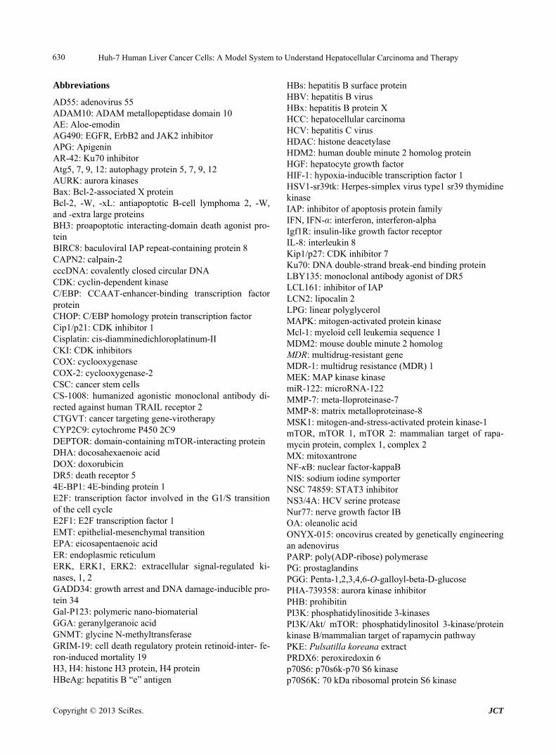
Huh-7 Human Liver Cancer Cells: A Model System to Understand Hepatocellular Carcinoma and Therapy 630
Abbreviations
AD55: adenovirus 55 ADAM10: ADAM metallopeptidase domain 10 AE: Aloe-emodin AG490: EGFR, ErbB2 and JAK2 inhibitor APG: Apigenin AR-42: Ku70 inhibitor Atg5, 7, 9, 12: autophagy protein 5, 7, 9, 12 AURK: aurora kinases Bax: Bcl-2-associated X protein Bcl-2, -W, -xL: antiapoptotic B-cell lymphoma 2, -W, and -extra large proteins BH3: proapoptotic interacting-domain death agonist pro- tein BIRC8: baculoviral IAP repeat-containing protein 8 CAPN2: calpain-2 cccDNA: covalently closed circular DNA CDK: cyclin-dependent kinase C/EBP: CCAAT-enhancer-binding transcription factor protein CHOP: C/EBP homology protein transcription factor Cip1/p21: CDK inhibitor 1 Cisplatin: cis-diamminedichloroplatinum-II CKI: CDK inhibitors COX: cyclooxygenase COX-2: cyclooxygenase-2 CSC: cancer stem cells CS-1008: humanized agonistic monoclonal antibody di- rected against human TRAIL receptor 2 CTGVT: cancer targeting gene-virotherapy CYP2C9: cytochrome P450 2C9 DEPTOR: domain-containing mTOR-interacting protein DHA: docosahexaenoic acid DOX: doxorubicin DR5: death receptor 5 4E-BP1: 4E-binding protein 1 E2F: transcription factor involved in the G1/S transition of the cell cycle E2F1: E2F transcription factor 1 EMT: epithelial-mesenchymal transition EPA: eicosapentaenoic acid ER: endoplasmic reticulum ERK, ERK1, ERK2: extracellular signal-regulated ki- nases, 1, 2 GADD34: growth arrest and DNA damage-inducible pro- tein 34 Gal-P123: polymeric nano-biomaterial GGA: geranylgeranoic acid GNMT: glycine N-methyltransferase GRIM-19: cell death regulatory protein retinoid-inter- fe- ron-induced mortality 19 H3, H4: histone H3 protein, H4 protein HBeAg: hepatitis B “e” antigen
HBs: hepatitis B surface protein HBV: hepatitis B virus HBx: hepatitis B protein X HCC: hepatocellular carcinoma HCV: hepatitis C virus HDAC: histone deacetylase HDM2: human double minute 2 homolog protein HGF: hepatocyte growth factor HIF-1: hypoxia-inducible transcription factor 1 HSV1-sr39tk: Herpes-simplex virus type1 sr39 thymidine kinase IAP: inhibitor of apoptosis protein family IFN, IFN-α: interferon, interferon-alpha Igf1R: insulin-like growth factor receptor IL-8: interleukin 8 Kip1/p27: CDK inhibitor 7 Ku70: DNA double-strand break-end binding protein LBY135: monoclonal antibody agonist of DR5 LCL161: inhibitor of IAP LCN2: lipocalin 2 LPG: linear polyglycerol MAPK: mitogen-activated protein kinase Mcl-1: myeloid cell leukemia sequence 1 MDM2: mouse double minute 2 homolog MDR: multidrug-resistant gene MDR-1: multidrug resistance (MDR) 1 MEK: MAP kinase kinase miR-122: microRNA-122 MMP-7: meta-lloproteinase-7 MMP-8: matrix metalloproteinase-8 MSK1: mitogen-and-stress-activated protein kinase-1 mTOR, mTOR 1, mTOR 2: mammalian target of rapa-mycin protein, complex 1, complex 2 MX: mitoxantrone NF-κB: nuclear factor-kappaB NIS: sodium iodine symporter NSC 74859: STAT3 inhibitor NS3/4A: HCV serine protease Nur77: nerve growth factor IB OA: oleanolic acid ONYX-015: oncovirus created by genetically engineering an adenovirus PARP: poly(ADP-ribose) polymerase PG: prostaglandins PGG: Penta-1,2,3,4,6-O-galloyl-beta-D-glucose PHA-739358: aurora kinase inhibitor PHB: prohibitin PI3K: phosphatidylinositide 3-kinases PI3K/Akt/ mTOR: phosphatidylinositol 3-kinase/protein kinase B/mammalian target of rapamycin pathway PKE: Pulsatilla koreana extract PRDX6: peroxiredoxin 6 p70S6: p70s6k-p70 S6 kinase p70S6K: 70 kDa ribosomal protein S6 kinase
Copyright © 2013 SciRes. JCT
Huh-7 Human Liver Cancer Cells: A Model System to Understand Hepatocellular Carcinoma and Therapy
Copyright © 2013 SciRes. JCT
631
p-STAT3: phospho-Stat3 PTX: paclitaxel ω-3 PUFA: omega-3 poly-unsaturated fatty acids ω-6 PUFA: omega-6 polyunsaturated fatty acids p21/WAF1: cyclin-dependent kinase inhibitor 1 RB: retinoblastoma tumour suppressor protein RB-E2F: retinoblastoma tumour suppressor transcription factor pathway rcDNA: relaxed circular deoxyribonucleic acid ROS: reactive oxygen species SB365: saponin D SC-1, -49: sorafenib derivatives SHP-1: Src homology region 2 domain-containing phos-phatase-1 SMAC: second mitochondria-derived activator caspases SNAI1: snail homolog 1
STAT3: signal transducer and activator of transcription 3 TLR, TLR2, 3, 8: toll-like receptor, toll-like receptor 2, 3, 8 TNF: tumour necrosis factor TNF-α: tumour necrosis factor-alpha TNFRSF10B: tumour necrosis factor receptor member 10b TRAIL: tumour necrosis factor-related apoptosis-inducing ligand TXNIP: thioredoxin interacting protein UA: ursolic acid UBE3A: ubiquitin-protein ligase E3A UPR: unfolded protein response U0126: MEK inhibitor VEGF: vascular endothelial growth factor XIAP: X-linked apoptotic protein


![Medley - 16 September - Scarborough Ukulele Players · [C] Well come on everybody it's six o'clock, Huh-huh, uh- [C7] huh-huh [F] Come on everybody it's time to rock, Uh- [C] huh-huh,](https://static.fdocuments.us/doc/165x107/5f097c2b7e708231d4270cd7/medley-16-september-scarborough-ukulele-players-c-well-come-on-everybody-its.jpg)