Features of Adult Derived Human Liver Stem Progenitor Cells
20
cells Article Inflammation Differentially Modulates the Biological Features of Adult Derived Human Liver Stem/Progenitor Cells Hoda El-Kehdy 1, † , Mehdi Najar 2, † , Joery De Kock 3 , Douaa Moussa Agha 4 , Vera Rogiers 3 , Makram Merimi 4 , Laurence Lagneaux 5 , Etienne M. Sokal 1 and Mustapha Najimi 1, * 1 Laboratory of Pediatric Hepatology and Cell Therapy, Institut de Recherche Expérimentale et Clinique (IREC), Université Catholique de Louvain, 1200 Brussels, Belgium; [email protected] (H.E.-K.); [email protected] (E.M.S.) 2 Osteoarthritis Research Unit, Department of Medicine, University of Montreal Hospital Research Center (CRCHUM), Montreal, QC H2X 0A9, Canada; [email protected] 3 Department of In Vitro Toxicology and Dermato-Cosmetology (IVTD), Faculty of Medicine and Pharmacy, Vrije Universiteit Brussel, 1090 Brussels, Belgium; [email protected] (J.D.K.); [email protected] (V.R.) 4 Laboratory of Experimental Hematology (HEMEXP), Institut Jules Bordet, Université Libre de Bruxelles (ULB), 1000 Brussels, Belgium; [email protected] (D.M.A.); [email protected] (M.M.) 5 Laboratory of Clinical Cell Therapy (LCCT), Institut Jules Bordet, Université Libre de Bruxelles (ULB), 1070 Brussels, Belgium; [email protected] * Correspondence: [email protected] † These authors contribute equally. Received: 8 May 2020; Accepted: 1 July 2020; Published: 8 July 2020 Abstract: The progression of mesenchymal stem cell-based therapy from concept to cure closely depends on the optimization of conditions that allow a better survival and favor the cells to achieve efficient liver regeneration. We have previously demonstrated that adult-derived human liver stem/progenitor cells (ADHLSC) display significant features that support their clinical development. The current work aims at studying the impact of a sustained pro-inflammatory environment on the principal biological features of ADHLSC in vitro. METHODS: ADHLSC from passages 4–7 were exposed to a cocktail of inflammatory cytokines for 24 h and 9 days and subsequently analyzed for their viability, expression, and secretionprofiles by using flow cytometry, RT-qPCR, and antibody array assay. The impact of inflammation on the hepatocytic differentiation potential of ADHLSC was also evaluated. RESULTS: ADHLSC treated with a pro-inflammatory cocktail displayed significant decrease of cell yield at both times of treatment while cell mortality was observed at 9 days post-priming. After 24 h, no significant changes in the immuno-phenotype of ADHLSC expression profile could be noticed while after 9 days, the expression profile of relevant markers has changed both in the basal conditions and after inflammation treatment. Inflammation cocktail enhanced the release of IL-6, IL-8, CCL5, monocyte-chemo-attractant protein-2 and 3, CXCL1/GRO, and CXCL5/ENA78. Furthermore, while IP-10 secretion was increased after 24 h priming, granulocyte macrophage colony-stimulating factor enhanced secretion was noticed after 9 days treatment. Finally, priming of ADHLSC did not affect their potential to differentiate into hepatocyte-like cells. CONCLUSION: These results indicate that ADHLSCs are highly sensitive to inflammation and respond to such signals by adjusting their gene and protein expression. Accordingly, monitoring the inflammatory status of patients at the time of cell transplantation, will certainly help in enhancing ADHLSC safety and efficiency. Keywords: liver; liver stem/progenitor cells; inflammation; immuno-biology Cells 2020, 9, 1640; doi:10.3390/cells9071640 www.mdpi.com/journal/cells
Transcript of Features of Adult Derived Human Liver Stem Progenitor Cells

cells
Article
Inflammation Differentially Modulates the BiologicalFeatures of Adult Derived Human LiverStem/Progenitor Cells
Hoda El-Kehdy 1,† , Mehdi Najar 2,†, Joery De Kock 3 , Douaa Moussa Agha 4, Vera Rogiers 3,Makram Merimi 4, Laurence Lagneaux 5, Etienne M. Sokal 1 and Mustapha Najimi 1,*
1 Laboratory of Pediatric Hepatology and Cell Therapy, Institut de Recherche Expérimentale etClinique (IREC), Université Catholique de Louvain, 1200 Brussels, Belgium;[email protected] (H.E.-K.); [email protected] (E.M.S.)
2 Osteoarthritis Research Unit, Department of Medicine, University of Montreal Hospital ResearchCenter (CRCHUM), Montreal, QC H2X 0A9, Canada; [email protected]
3 Department of In Vitro Toxicology and Dermato-Cosmetology (IVTD), Faculty of Medicine and Pharmacy,Vrije Universiteit Brussel, 1090 Brussels, Belgium; [email protected] (J.D.K.); [email protected] (V.R.)
4 Laboratory of Experimental Hematology (HEMEXP), Institut Jules Bordet, Université Libre deBruxelles (ULB), 1000 Brussels, Belgium; [email protected] (D.M.A.);[email protected] (M.M.)
5 Laboratory of Clinical Cell Therapy (LCCT), Institut Jules Bordet, Université Libre de Bruxelles (ULB),1070 Brussels, Belgium; [email protected]
* Correspondence: [email protected]† These authors contribute equally.
Received: 8 May 2020; Accepted: 1 July 2020; Published: 8 July 2020�����������������
Abstract: The progression of mesenchymal stem cell-based therapy from concept to cure closelydepends on the optimization of conditions that allow a better survival and favor the cells to achieveefficient liver regeneration. We have previously demonstrated that adult-derived human liverstem/progenitor cells (ADHLSC) display significant features that support their clinical development.The current work aims at studying the impact of a sustained pro-inflammatory environment on theprincipal biological features of ADHLSC in vitro. METHODS: ADHLSC from passages 4–7 wereexposed to a cocktail of inflammatory cytokines for 24 h and 9 days and subsequently analyzed fortheir viability, expression, and secretion profiles by using flow cytometry, RT-qPCR, and antibodyarray assay. The impact of inflammation on the hepatocytic differentiation potential of ADHLSC wasalso evaluated. RESULTS: ADHLSC treated with a pro-inflammatory cocktail displayed significantdecrease of cell yield at both times of treatment while cell mortality was observed at 9 days post-priming.After 24 h, no significant changes in the immuno-phenotype of ADHLSC expression profile could benoticed while after 9 days, the expression profile of relevant markers has changed both in the basalconditions and after inflammation treatment. Inflammation cocktail enhanced the release of IL-6, IL-8,CCL5, monocyte-chemo-attractant protein-2 and 3, CXCL1/GRO, and CXCL5/ENA78. Furthermore,while IP-10 secretion was increased after 24 h priming, granulocyte macrophage colony-stimulatingfactor enhanced secretion was noticed after 9 days treatment. Finally, priming of ADHLSC did notaffect their potential to differentiate into hepatocyte-like cells. CONCLUSION: These results indicatethat ADHLSCs are highly sensitive to inflammation and respond to such signals by adjusting theirgene and protein expression. Accordingly, monitoring the inflammatory status of patients at the timeof cell transplantation, will certainly help in enhancing ADHLSC safety and efficiency.
Keywords: liver; liver stem/progenitor cells; inflammation; immuno-biology
Cells 2020, 9, 1640; doi:10.3390/cells9071640 www.mdpi.com/journal/cells

Cells 2020, 9, 1640 2 of 20
1. Introduction
Mesenchymal stem cells (MSC) represent a fibroblast-like cell population that displays a potent abilityto differentiate and to modulate both adaptive and innate immune systems [1]. These undifferentiatedand non-hematopoietic cells have received much interest in regenerative medicine. Extensive clinicaltrials using culture expanded MSC are currently exploring their therapeutic potential in humans [2].Although a clinical improvement was noticed in most of the studies, the failure to track these cellsin situ obviously supported paracrine-mediated effects. Active rejection and/or letting host cells tosubsequently ameliorate injury and accelerate repair, could explain the documented post-transplantationeffects. Accordingly, the progression of MSC-based therapeutic approach from concept to cure closelydepends on the optimization of specific experimental conditions that allow a better survival and favorthe transplanted MSC to achieve efficient tissue regeneration.
In the liver, most of the chronic injuries progress to fibrosis and cirrhosis when regenerativepotential of the parenchymal cells is impaired. Given the major role of chronic inflammation in liverpathophysiology and the supportive function of non-parenchymal stromal/mesenchymal cells in liverhomeostasis, MSC-based therapy has also been positioned as a promising innovative strategy to contributeto the hepatic healing process. At steady state, MSC are not constitutively immunosuppressive and theirimmunomodulatory features will be influenced by the local inflammatory environment to which they areexposed upon transplantation [3]. Indeed, inflammation is emerging as an important regulator of stemcells and plays an intricate role in health and disease. There is evidence for the direct crosstalk betweenthe inflammatory response and stem cells both in cases of microbial and sterile induced inflammation [4].MSCs are environmentally responsive cells that can sense specific signals, adapt their fate and functionsin consequences, and finally respond by migrating, proliferating, and/or regenerating the tissue [5].
For liver cell therapy purposes, we have successfully and reproducibly obtained a mesenchymalstem/progenitor cell population from adult healthy human livers [6]. Pre-clinical and clinical data revealedthe ability of those hepatic MSC-like cells to display significant regenerative and immuno-modulatoryfeatures [7–9]. By enhancing our knowledge about the safety and efficiency of the transplanted cellproduct, we can ameliorate the therapeutic issue for the patient. Thus, to gain insights regarding thebehavior of adult-derived human liver stem/progenitor cells (ADHLSC) in inflammatory pathologicalconditions and the mechanisms by which such cells cooperate with inflammation, we studied in vitrothe impact of sustained pro-inflammatory environment on culture expanded ADHLSC as well as afterhepatocytic differentiation.
Our results indicate a significant impairment of ADHLSC proliferative capacity and viability aftersustained inflammation with no alteration of their hepatocytic differentiation potential. Moreover, thephenotype and secretome, both related to the immunomodulatory potential of ADHLSC, were differentiallyaltered in a time and inflammatory dependent manner. These observations could be related to the lowengraftment level and transient therapeutic activity of MSC documented upon transplantation [10]. Thus,inflammation status of the recipient should be considered when designing liver cell therapy protocols.
2. Materials and Methods
2.1. ADHLSC Culture
The protocol as well as the experiments were approved by the ethical committees of the St-LucHospital and faculty of Medicine of Université catholique de Louvain (Reference: JMM/sy/2010/12).An agreement from the Belgian Ministry of Health was obtained for the Hepatocytes and Hepatic StemCells Bank. A written and signed informed consent was obtained for each human liver used in thecurrent study.
Liver tissues were supplied via the Hepatocytes and Hepatic Stem Cells bank (Table 1).

Cells 2020, 9, 1640 3 of 20
Table 1. Characteristics of the donors used in the current study.
Cell Populations Donor Status Cause of Death Donor Age Sex
XF115 P6 No liver disease Meningitis 0.4 YO M
XF18P5 No liver disease Gas embolism 1.5 YO M
XF75 P7 Crigler Najjar type 1 NA 2 YO M
XF89 P3 No liver disease Respiratory 3 DO M
XF45 P6 and P7 No liver disease Severe asphyxia 6 DO F
XF98 P6 No liver disease Cardiorespiratory arrest 7 DO M
NA: not applicable; YO: year old; DO: day old.
Human liver cell suspensions predominantly constituted by hepatocytes were isolated fromcadaveric donated livers by using a classic two-step collagenase perfusion, filtration, and low speedcentrifugation as previously detailed [6,11]. After plating of the recovered single cell suspensionsfor 24 h, primary hepatocyte culture medium was changed in order to eliminate the non-adherentcells and thereafter renewed every 3 days. Two weeks later, primary hepatocyte culture mediumwas substituted by 4.5 g/L glucose DMEM medium (Invitrogen) supplemented with 10% FCS and1% Penicillin/Streptomycin. ADHLSCs spontaneously emerged, proliferated, and filled the emptyspace left by dead cells and became predominant at passage 2. Upon reaching confluence, cells wereenzymatically detached and re-plated at 5000 cells/cm2 on Corning CellBIND T75 flasks at 37 ◦C in afully humidified atmosphere (5% CO2). When reaching 85% confluence, ADHLSC were lifted with0.05% Trypsin-EDTA (Life Technologies). Viability of recovered cells was evaluated using trypan blueexclusion assay and often exceeded 95%. Twenty-four hours after seeding, cells were treated withan inflammatory cocktail consisting of 3000 UI/mL interferon alpha (Roferon), 103 U/mL interferonγ (R&D Systems), 12 ng/mL IL-1β (R&D Systems), and 15 ng/mL TNF-α. For 9 days treatment, mediumcontaining the inflammation cocktail was changed every 3 days. For both time points, cell pellets andrelated supernatants were recovered for further analyses.
2.2. Flow Cytometry
After enzymatic detachment, ADHLSC were suspended in D-PBS at a concentration of 105 cells/mL.Cell suspensions were washed twice with PBS. For intracellular immunostaining, cell permeabilizationwas performed with cytofix/cytoperm for 15 min at room temperature (BD Pharmingen). ADHLSC werethen washed and incubated for 30 min at room temperature with antibodies or corresponding controlisotypes (Table 2). After washing, cells were suspended in Stabilizing Fixative (BD Pharmingen) beforereading with a CANTO II flow cytometer. The analyses were performed using the BD FACSDiva Software.ADHLSC apoptosis was appreciated using DAPI Annexin V Apoptosis Detection Kit I (BD Pharmingen)following the manufacturer’s instructions. Cells treated with 1 mM Hydrogen Peroxide for 1 h were usedas positive control for apoptosis. Immunostainings were analyzed by flow cytometer (Canto II, BD).
Table 2. Antibodies used for flow cytometry analyses of adult-derived human liver stem/progenitorcells (ADHLSC).
Antibody Supplier Concentration Used Reference
CD45 BD Biosciences 1/20 557748CD73 BD Biosciences 1/20 550257CD90 BD Biosciences 1/20 559669
CD105 Ancell 1/20 326-041ASMA Abcam 1/20 Ab8211

Cells 2020, 9, 1640 4 of 20
2.3. Gene Expression Analysis
Total RNA was extracted from 1.5 million ADHLSC for each condition using Tripure isolationreagent (Roche). First strand cDNA was synthesized from 2 µg RNA using the ThermoscriptTM RTkit according to the manufacturer’s instructions (Life Technologies) and subsequently diluted withnuclease free water (Invitrogen) to 5 ng/µL cDNA. qPCR was performed using TaqMan universalMasterMix (Applied Biosystems, MA, USA) and TaqMan probes listed in Table 3 according to themanufacturer’s instructions, on a StepOnePlus real-time PCR machine (Applied Biosystems). Relativegene expression was determined using the ∆∆Ct method and PPIA as a housekeeping gene.
Table 3. Taqman probes used for RT-qPCR analyses.
Probe Reference
Albumin Hs00910225_m1CD54 Hs00164932_m1
Col1alpha1 Hs00164004_m1MRP2 Hs00166123_m1PPIA Hs99999904_m1Slug Hs00950344_m1Snail Hs0019559_m1Sox9 Hs00165814_m1
Vimentin Hs00185584_m1
2.4. qPCR Cytokine Array
To identify modulated cytokines-encoding genes, quantitative Real-Time PCR (qRT-PCR)arrays (Human cytokines and cytokines receptors 96 StellARray™ qPCR Array) from HarborBioscientific-Lonza (Verviers, Belgium) were applied. Reverse transcription of RNA to cDNA wascarried out using a M-MLV reverse transcriptase in qScriptTM cDNA SuperMix (QuantaBio- VWRInternational bvba, Leuven, Belgium) respecting the manufacturer’s recommendations. A total of500 ng of cDNA was then mixed with SYBR-green reagent (Thermo Scientific), where 20 µL of theresulting mixture was added to a 96-well microarray plate. The qPCR was carried out in a StepOnePlus™Real-Time PCR System (Applied Biosystems Inc, CA, USA) using the following program: one cycleof a holding stage at 50 ◦C for 2 min and 95 ◦C for 5 min and 40 cycles of the amplification stage at95 ◦C for 15 s and 60 ◦C for 1 min. Relative gene expression in comparative analysis between untreatedand treated conditions was determined using the ∆∆Ct method (see Supplementary file Table S1 forthe total list of genes). Normalization was performed using the “deltaCt” and “quantile” methodsavailable in the “normalizeCtData” function of the HTqPCR R package [12]. "deltaCt" normalizesthe Ct values within an array by subtracting the mean Ct value of the chosen housekeeping genesfrom the values of the other genes. Quantile normalization transforms the data to make the Ct valuesdistributions more or less identical across all arrays.
2.5. Secretome Analysis
Human cytokine (ab133998) antibody arrays (both from Abcam) were used to detect major changesin the secretion profile of ADHLSC upon pro-inflammatory stimulation. Briefly, 1 × 10⁶ ADHLSCwere stimulated or not with the pro-inflammatory cocktail, as described above, for 24 h and 9 days.Thereafter, cells were incubated with serum free medium for an additional 24 h for accumulationof secreted molecules. The medium was recovered, centrifuged to eliminate cell debris, and storedat −80 ◦C until analysis by the antibody array assay. The assay was performed according to themanufacturer’s instructions and chemiluminescent detection was done on a ChemiDocTM MP ImagingSystem (Bio-rad). Densitometry was performed to evaluate relative changes in the secretion profileof ADHLSC stimulated or not with the pro-inflammatory cocktail using Bio-rad’s Image Lab v.5.2.1software. Relative secretion levels were calculated as follows: summed signal intensities for each

Cells 2020, 9, 1640 5 of 20
marker of interest were used. Background correction was done by subtracting the average summedsignal intensities of the negative control spots. Data normalization across arrays was accomplishedby defining one array as "reference" to which the other arrays were normalized using the averagesummed signal intensities of the positive control spots. Next, for each marker of interest the averagesummed signal intensities of the respective medium controls either with or without pro-inflammatorycocktail were subtracted from the samples. Finally, the obtained secretion levels for ADHLSC withpro-inflammatory stimulation were calculated as fold change versus control ADHLSC.
2.6. Hepatocytic Differentiation
ADHLSC were seeded at a density of 1 × 10⁴ cells/cm2 on rat tail collagen type I (BD) coated flasks(Corning) using expansion medium. At 90% confluence, cells were primed or not for 24 h and 9 days,then incubated in differentiation medium which consists of Iscove’s modified Dulbecco’s medium(IMDM; Life Technologies) serum-free medium in which specific growth factors/cytokines (PerpotechEC Ltd.) were added as a sequential multi-step protocol [6]. Step 1 (20 ng/mL epidermalgrowth factor(EGF) and 10 ng/mL basic fibroblast growth factor (bFGF)) lasted for 2 days. Then, Step 2 (20 ng/mLbFGF, 10 ng/mL hepatocyte growth factor (HGF), insulin-selenium-transferrin (ITS; Life Technologies),and 0.61 g/L nicotinamide (Sigma)) lasted for 9 days. Step 3 (20 ng/mL oncostatin M (OSM), 20 ng/mLHGF, 1% ITS, 0.61 g/L nicotinamide, and 10–6 M Dexa (Sigma)) lasted for another 9 days. The cellswere microscopically followed at a regular basis and medium was replaced every 3 days throughoutthe differentiation protocol (except for Step 1). At the end of the differentiation, cells were harvestedand the quality of hepatocytic differentiation was evaluated at the morphology, gene expression profile,and functional levels.
2.7. CYP3A4 Activity
Undifferentiated and differentiated ADHLSC were detached using 0.05% trypsin and cell densitywas determined. Thereafter, cells were seeded at density of 1 × 10⁵ cells/well, on 96-well plates.CYP3A4 activity was analyzed using P450-GloTM assay according to the manufacturer’s instructions(Promega, Madison, WI, USA) and as previously adapted [13].
2.8. Statistical Analysis
Results are expressed as mean ± standard error of the mean (SEM). Statistical differences weredetermined by paired Student’s t-test for two samples analysis and one-way ANOVA followed bythe Dunnett post-hoc test for more than two samples (GraphPad Prism 8.4.2, San Diego, CA, USA).Differences were considered significant when p values * p < 0.05, ** p < 0.01, *** p < 0.001.
3. Results
3.1. Sustained Inflammation Significantly Alters the Morphology, Proliferation, and Viability of ADHLSCs
The morphology of ADHLSCs was microscopically followed at different times post-platingin presence or absence of the inflammation cocktail. Adhering untreated ADHLSCs displayedspindle-shaped morphology and proliferated starting from day 1 to reach a sub-confluence after 9 days(Figure 1). In the presence of the inflammation cocktail, ADHLSCs became less elongated, displayedmore contorted shape, and more granularity around the proximal perinuclear area. Those changeswere more pronounced at day 9 (Figure 1).
In parallel, we evaluated the impact of inflammation on the yield of ADHLSC. In control conditions,we confirmed the expansion capacity of ADHLSC as demonstrated by the increased number of cellsrecovered at day 9 (more than 10-fold) (Figure 2A). Upon treatment with inflammation cocktail,a significant massive decrease in the number of adherent ADHLSCs was observed at both day 1 andday 9. No statistically significant difference was found between the two time periods.

Cells 2020, 9, 1640 6 of 20Cells 2020, 9, x FOR 6 of 21
Figure 1. Effect of inflammation on ADHLSC culture. Morphology of ADHLSC observed microscopically after different times post-treatment with the inflammation cocktail (n = 6 samples from different donors). Magnification: 100× and 200×.
In parallel, we evaluated the impact of inflammation on the yield of ADHLSC. In control conditions, we confirmed the expansion capacity of ADHLSC as demonstrated by the increased number of cells recovered at day 9 (more than 10-fold) (Figure 2A). Upon treatment with inflammation cocktail, a significant massive decrease in the number of adherent ADHLSCs was observed at both day 1 and day 9. No statistically significant difference was found between the two time periods.
Figure 1. Effect of inflammation on ADHLSC culture. Morphology of ADHLSC observed microscopicallyafter different times post-treatment with the inflammation cocktail (n = 6 samples from different donors).Magnification: 100× and 200×.Cells 2020, 9, x FOR 7 of 21
Figure 2. Effect of inflammation on ADHLSC viability in culture. (A) Significant decrease in adherent ADHLSC number after 24 h and 9 days treatment with the inflammation cocktail (n = 4 samples from different donors for each timepoint). Results are expressed as mean ± standard error of the mean (SEM). * p value < 0.05. # p < 0.05 control-9-day inflammation vs. control-24 h inflammation, one-way ANOVA followed by Dunnett post hoc test. (B) Following Annexin V–DAPI staining, no significant difference in cell death induction was noticed after 24 h treatment with the inflammation cocktail. (C) In contrast, maintaining the treatment for 9 days significantly decreases ADHLSC viability in correlation to an increase in cell apoptosis. Results are expressed as mean ± standard error of the mean (SEM) (n = 4). ** denotes a p value < 0.01; * p < 0.05 vs. corresponding control, paired Student’s t-test.
Therefore, we evaluated the effect of the inflammation cocktail on ADHLSCs viability and death. Using Annexin V–DAPI staining, we demonstrated that following 24 h treatment with the inflammation cocktail, the majority of the analyzed cells (almost 100%) were viable with no significant difference in cell death induction (neither early nor late apoptosis) (Figure 2B). In contrast, maintaining the treatment for 9 days significantly decreased the viability of ADHLSCs by 40% (Figure 2C) while a significant increase in both early and late apoptosis was observed. To note, the % of late apoptosis was more pronounced than that of early stage with statistical significance.
3.2. Sustained Inflammation Influences the Immuno-Phenotype of ADHLSCs
We assessed the effect of inflammation on ADHLSCs mesenchymal immuno-phenotype (Figure 3) by using flow cytometry. ADHLSCs positive expression of membranous (CD73 and CD90) and intracellular (alpha-smooth muscle actin (ASMA)) mesenchymal markers, as well as the negative-expression of hematopoietic marker (CD45) were checked by using validated primary antibodies. Figure 3A indicated that no significant changes in ADHLSC expression profile could be noticed after 24 h of inflammatory cocktail treatment. Globally, ADHLSC demonstrated positivity for CD73, CD90, and ASMA markers with, however, different levels of expression and no impact of inflammation. The expression of CD45 remained negative even after inflammation treatment. Analysis of
Figure 2. Effect of inflammation on ADHLSC viability in culture. (A) Significant decrease in adherentADHLSC number after 24 h and 9 days treatment with the inflammation cocktail (n = 4 samples fromdifferent donors for each timepoint). Results are expressed as mean ± standard error of the mean (SEM).* p value < 0.05. # p < 0.05 control-9-day inflammation vs. control-24 h inflammation, one-way ANOVAfollowed by Dunnett post hoc test. (B) Following Annexin V–DAPI staining, no significant differencein cell death induction was noticed after 24 h treatment with the inflammation cocktail. (C) In contrast,maintaining the treatment for 9 days significantly decreases ADHLSC viability in correlation to an increasein cell apoptosis. Results are expressed as mean ± standard error of the mean (SEM) (n = 4). ** denotes ap value < 0.01; * p < 0.05 vs. corresponding control, paired Student’s t-test.

Cells 2020, 9, 1640 7 of 20
Therefore, we evaluated the effect of the inflammation cocktail on ADHLSCs viability and death.Using Annexin V–DAPI staining, we demonstrated that following 24 h treatment with the inflammationcocktail, the majority of the analyzed cells (almost 100%) were viable with no significant differencein cell death induction (neither early nor late apoptosis) (Figure 2B). In contrast, maintaining thetreatment for 9 days significantly decreased the viability of ADHLSCs by 40% (Figure 2C) while asignificant increase in both early and late apoptosis was observed. To note, the % of late apoptosis wasmore pronounced than that of early stage with statistical significance.
3.2. Sustained Inflammation Influences the Immuno-Phenotype of ADHLSCs
We assessed the effect of inflammation on ADHLSCs mesenchymal immuno-phenotype (Figure 3) byusing flow cytometry. ADHLSCs positive expression of membranous (CD73 and CD90) and intracellular(alpha-smooth muscle actin (ASMA)) mesenchymal markers, as well as the negative-expression ofhematopoietic marker (CD45) were checked by using validated primary antibodies. Figure 3A indicatedthat no significant changes in ADHLSC expression profile could be noticed after 24 h of inflammatorycocktail treatment. Globally, ADHLSC demonstrated positivity for CD73, CD90, and ASMA markerswith, however, different levels of expression and no impact of inflammation. The expression of CD45remained negative even after inflammation treatment. Analysis of corresponding relative MFI (meanfluorescence intensity) confirmed the absence of an inflammation effect on the expression level of themarkers studied (Table 4).
Cells 2020, 9, x FOR 8 of 21
corresponding relative MFI (mean fluorescence intensity) confirmed the absence of an inflammation effect on the expression level of the markers studied (Table 4).
After 9 days of treatment, the immuno-phenotype of ADHLSC has changed both in the basic conditions and after inflammation treatment. Thus, the constitutive expression of CD73 and ASMA markers has been substantially reduced whereas that of CD90 remained highly present (Figure 3). The inflammation cocktail induced an increase in the % of cells only positively immune-stained for CD73 (Figure 3). Analysis of corresponding relative MFI confirmed the augmentation of the % of cells immune-stained for CD73 but also clearly showed significant downregulation in the expression levels of both CD90 (by 81%) and ASMA (by 68%) markers (Table 4). No effect was observed on the expression levels of both CD45 and CD105.
Figure 3. Effect of inflammation on ADHLSC mesenchymal expression profile. Positive expression of mesenchymal cell surface (CD105, CD90, and CD73) and intracellular markers (alpha-smooth muscle actin—ASMA) was evaluated using validated corresponding primary antibodies and flow cytometry. Negative expression of CD45 was also analyzed. (A) No changes in the mesenchymal expression profile were noticed after 24 h treatment. (B) After 9 days post-treatment with the inflammation cocktail, a significant increase was only observed for CD73 expression. Results are expressed as mean ± standard error of the mean (SEM) (n = 3 samples from different donors). * denotes a p value p < 0.05 vs. corresponding control, paired Student’s t-test.
Figure 3. Effect of inflammation on ADHLSC mesenchymal expression profile. Positive expression ofmesenchymal cell surface (CD105, CD90, and CD73) and intracellular markers (alpha-smooth muscleactin—ASMA) was evaluated using validated corresponding primary antibodies and flow cytometry.Negative expression of CD45 was also analyzed. (A) No changes in the mesenchymal expression profile werenoticed after 24 h treatment. (B) After 9 days post-treatment with the inflammation cocktail, a significantincrease was only observed for CD73 expression. Results are expressed as mean ± standard error of themean (SEM) (n = 3 samples from different donors). * denotes a p value p < 0.05 vs. corresponding control,paired Student’s t-test.

Cells 2020, 9, 1640 8 of 20
Table 4. Effect of inflammation on ADHLSC expression levels of mesenchymal markers: MFI (meanfluorescence intensity) values.
CD45 CD73 CD90 CD105 ASMA
24 h inflammation
Control 0 ± 0 639.7 ± 181.0 1041.0 ± 1532.9 1621.3 ± 785.9 2101.0 ± 1034.7
Treated 0 ± 0 935.3 ± 425.3 9790.0 ± 2185.6 2408.3 ± 1222.4 2612.7 ± 1845.3
9-day inflammation
Control 0 ± 0 1026.0 ± 156.6 26,619.7 ± 7802.5 310.3 ± 310.3 1769.7 ± 222.7
Treated 0 ± 0 3674.7 ± 487.9 ** 5106.7 ± 205.6 * 386.3 ± 377.4 566.3 ± 293.9 *
Results are expressed as mean ± standard error of the mean of three independent experiments (cells populationsfrom three different donors). ** denotes a p value < 0.01; * p < 0.05 vs. corresponding control, t-test.
After 9 days of treatment, the immuno-phenotype of ADHLSC has changed both in the basicconditions and after inflammation treatment. Thus, the constitutive expression of CD73 and ASMAmarkers has been substantially reduced whereas that of CD90 remained highly present (Figure 3).The inflammation cocktail induced an increase in the % of cells only positively immune-stained forCD73 (Figure 3). Analysis of corresponding relative MFI confirmed the augmentation of the % ofcells immune-stained for CD73 but also clearly showed significant downregulation in the expressionlevels of both CD90 (by 81%) and ASMA (by 68%) markers (Table 4). No effect was observed on theexpression levels of both CD45 and CD105.
3.3. Inflammation Significantly Modulates the Mesenchymal Stem Cell Gene Expression Profile of ADHLSCs
By using RT-qPCR, we examined the effect of inflammation on ADHLSC mesenchymal stemcell gene expression pattern. Our data showed that inflammation differentially modulated severalADHLSC mRNAs. As shown in Figure 4A, we confirmed the potency of the inflammation cocktail byshowing a significant induction of CD54 mRNA expression with more than 200-fold-increase comparedto untreated ADHLSCs after 24 h of treatment.
We also noticed that while the expression of mesenchymal stem cell genes Sox9 and Snail wassignificantly increased—but to a low extent as compared to CD54—a downregulation of vimentinand COL1alpha1 expression was observed. In contrast, the expression of Slug was not impacted.When inflammation was maintained during 9 consecutive days (Figure 4B), only the upregulation ofboth CD54 and Sox9 expression was maintained. The noticed increase in CD54 mRNA expressionwas lower than after 24 h of treatment (9-fold decrease). The expression of Snail and COL1alpha1 wasdownregulated while the expression of the other analyzed genes was not modulated. Finally, AlbuminmRNA expression was significantly enhanced after 9 days of priming.
3.4. Inflammation Substantially Alters the Immunomodulatory Potential of ADHLSC
The immunomodulatory potential of ADHLSC was evaluated by determining the expression/
secretion profile of several molecules known to participate in the immune and inflammatory responses.These molecules were analyzed at both gene and protein levels by using a qPCR array and an antibodyarray, respectively.
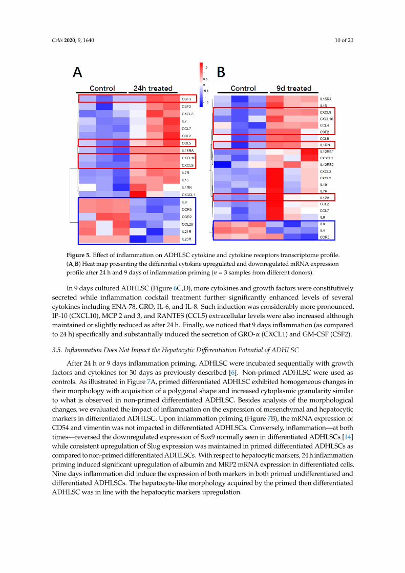
Regarding the immunomodulatory gene expression profile of ADHLSC (Figure 5A), 24 h inflammationwas found to alter the mRNA expression of 20% of analyzed genes most of them being induced (using athreshold value of 2). The plots showing the Ct values for each of these genes are provided in Supplementaryfile Figure S1. The genes that were highly induced after inflammation treatment (more than 200×)include CXCL9, CXCL10, CCL5 (RANTES), CSF3, and CXCL1 (GRO). From this list, we noticed thatIL1RN and CFS3 expression was constitutively undetectable as compared to other genes. The number ofinflammation-repressed genes (n = 6) represents 6.25% of the total number of genes analyzed. Those targetsinclude IL9, IL21R, IL23R, CCL28, CCR2, and CCR5. The plots showing the Ct values for each of these

Cells 2020, 9, 1640 9 of 20
genes are provided in Supplementary file Figure S2. When ADHLSC were primed for 9 consecutive days,≈23% of the analyzed genes were altered, most of them, as after 24 h treatment, being upregulated (using athreshold value of 2) (Figure 5B). The genes that were highly induced after 9 days inflammation (more than200×) include CXCL9, CXCL10 (IP-10) IL1RN, IL12A, CSF2 [Granulocyte-macrophage colony-stimulatingfactor (GM-CSF)), and CCL4 (Ligand of CCR5). The number of inflammation-significantly repressed genes(n = 3) represents ~3% of the total number of genes analyzed. Those targets include IL9, IL4, and CCR5(CCL4 Receptor). The plots showing corresponding Ct values are provided in Supplementary file Figure S2.
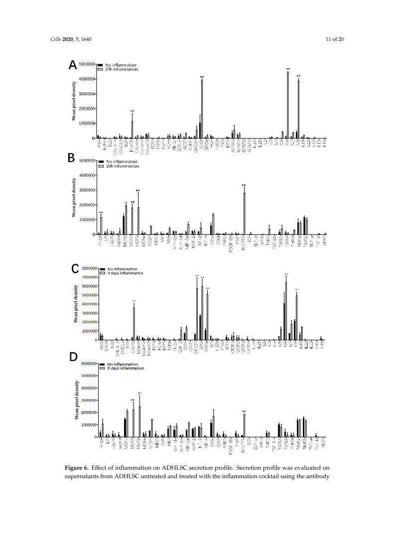
The immunomodulatory secretion profile of ADHLSC was analyzed by using a human cytokineand growth factor antibody array on supernatants recovered after 24 h and 9 days with and without theinflammation cocktail. Results from Figure 6 indicate that depending on both culture time period as well asthe presence or not of inflammation, several differences in the secretion pattern of ADHLSC were reported.After 24 h of culture in normal conditions (Figure 6A,B), few cytokines and growth factors constitutivelysecreted by ADHLSC, were detected. However, a significant increase in secreted levels of ENA-78, GRO,IL-6, IL-8, IP-10, MCP 2 and 3, and RANTES was noticed after inflammation. Of importance, the levels ofthese stimulated secretions varied and were particularly relevant for IL-6 and IL-8.
Cells 2020, 9, x FOR 9 of 21
Table 4. Effect of inflammation on ADHLSC expression levels of mesenchymal markers: MFI (mean fluorescence intensity) values.
CD45 CD73 CD90 CD105 ASMA 24 h inflammation
Control 0 ± 0 639.7 ± 181.0 1041.0 ± 1532.9 1621.3 ± 785.9 2101.0 ± 1034.7 Treated 0 ± 0 935.3 ± 425.3 9790.0 ± 2185.6 2408.3 ± 1222.4 2612.7 ± 1845.3
9-day inflammation Control 0 ± 0 1026.0 ± 156.6 26,619.7 ± 7802.5 310.3 ± 310.3 1769.7 ± 222.7 Treated 0 ± 0 3674.7 ± 487.9 ** 5106.7 ± 205.6 * 386.3 ± 377.4 566.3 ± 293.9 *
Results are expressed as mean ± standard error of the mean of three independent experiments (cells populations from three different donors). ** denotes a p value < 0.01; * p < 0.05 vs. corresponding control, t-test.
3.3. Inflammation Significantly Modulates the Mesenchymal Stem Cell Gene Expression Profile of ADHLSCs
By using RT-qPCR, we examined the effect of inflammation on ADHLSC mesenchymal stem cell gene expression pattern. Our data showed that inflammation differentially modulated several ADHLSC mRNAs. As shown in Figure 4A, we confirmed the potency of the inflammation cocktail by showing a significant induction of CD54 mRNA expression with more than 200-fold-increase compared to untreated ADHLSCs after 24 h of treatment.
Figure 4. Effect of inflammation on ADHLSC mRNA expression profile. (A) Differential modulation of ADHLSC gene expression profile by 24 h inflammation. (B) Differential modulation of ADHLSC gene expression profile by 9-days inflammation. RT-qPCR gene expression analysis demonstrated that inflammation differentially modulated the mRNA expression pattern of several ADHLSC genes. Differences in the upregulated and downregulated genes are observed between 24 h and 9 days of
Figure 4. Effect of inflammation on ADHLSC mRNA expression profile. (A) Differential modulationof ADHLSC gene expression profile by 24 h inflammation. (B) Differential modulation of ADHLSCgene expression profile by 9-days inflammation. RT-qPCR gene expression analysis demonstratedthat inflammation differentially modulated the mRNA expression pattern of several ADHLSC genes.Differences in the upregulated and downregulated genes are observed between 24 h and 9 days ofinflammatory treatment. For the ADHLSC treated group, results are expressed as mRNA relativeexpression versus untreated cells. CD54 (Intercellular Adhesion Molecule 1; ICAM-1), Sox9 (SRY-RelatedHMG-Box 9 encoding gene), Snail (SNAI1), Slug (SNAI2), COL1alpha1 (collagen type 1 alpha 1), VIM(Vimentin), and ALB (Albumin). Data shown are the mean ± SEM of three independent experiments(three samples from different donors). *** denotes a p value < 0.001; ** p < 0.01; * p < 0.05 vs. correspondingcontrol, paired Student’s t-test.
Cells 2020, 9, 1640 10 of 20Cells 2020, 9, x FOR 11 of 21
Figure 5. Effect of inflammation on ADHLSC cytokine and cytokine receptors transcriptome profile. (A,B) Heat map presenting the differential cytokine upregulated and downregulated mRNA expression profile after 24 h and 9 days of inflammation priming (n = 3 samples from different donors).
The immunomodulatory secretion profile of ADHLSC was analyzed by using a human cytokine and growth factor antibody array on supernatants recovered after 24 h and 9 days with and without the inflammation cocktail. Results from Figure 6 indicate that depending on both culture time period as well as the presence or not of inflammation, several differences in the secretion pattern of ADHLSC were reported. After 24 h of culture in normal conditions (Figure 6A,B), few cytokines and growth factors constitutively secreted by ADHLSC, were detected. However, a significant increase in secreted levels of ENA-78, GRO, IL-6, IL-8, IP-10, MCP 2 and 3, and RANTES was noticed after inflammation. Of importance, the levels of these stimulated secretions varied and were particularly relevant for IL-6 and IL-8.
In 9 days cultured ADHLSC (Figure 6C,D), more cytokines and growth factors were constitutively secreted while inflammation cocktail treatment further significantly enhanced levels of several cytokines including ENA-78, GRO, IL-6, and IL-8. Such induction was considerably more pronounced. IP-10 (CXCL10), MCP 2 and 3, and RANTES (CCL5) extracellular levels were also increased although maintained or slightly reduced as after 24 h. Finally, we noticed that 9 days inflammation (as compared to 24 h) specifically and substantially induced the secretion of GRO-α (CXCL1) and GM-CSF (CSF2).
Figure 5. Effect of inflammation on ADHLSC cytokine and cytokine receptors transcriptome profile.(A,B) Heat map presenting the differential cytokine upregulated and downregulated mRNA expressionprofile after 24 h and 9 days of inflammation priming (n = 3 samples from different donors).
In 9 days cultured ADHLSC (Figure 6C,D), more cytokines and growth factors were constitutivelysecreted while inflammation cocktail treatment further significantly enhanced levels of severalcytokines including ENA-78, GRO, IL-6, and IL-8. Such induction was considerably more pronounced.IP-10 (CXCL10), MCP 2 and 3, and RANTES (CCL5) extracellular levels were also increased althoughmaintained or slightly reduced as after 24 h. Finally, we noticed that 9 days inflammation (as comparedto 24 h) specifically and substantially induced the secretion of GRO-α (CXCL1) and GM-CSF (CSF2).
3.5. Inflammation Does Not Impact the Hepatocytic Differentiation Potential of ADHLSC
After 24 h or 9 days inflammation priming, ADHLSC were incubated sequentially with growthfactors and cytokines for 30 days as previously described [6]. Non-primed ADHLSC were used ascontrols. As illustrated in Figure 7A, primed differentiated ADHLSC exhibited homogeneous changes intheir morphology with acquisition of a polygonal shape and increased cytoplasmic granularity similarto what is observed in non-primed differentiated ADHLSC. Besides analysis of the morphologicalchanges, we evaluated the impact of inflammation on the expression of mesenchymal and hepatocyticmarkers in differentiated ADHLSC. Upon inflammation priming (Figure 7B), the mRNA expression ofCD54 and vimentin was not impacted in differentiated ADHLSCs. Conversely, inflammation—at bothtimes—reversed the downregulated expression of Sox9 normally seen in differentiated ADHLSCs [14]while consistent upregulation of Slug expression was maintained in primed differentiated ADHLSCs ascompared to non-primed differentiated ADHLSCs. With respect to hepatocytic markers, 24 h inflammationpriming induced significant upregulation of albumin and MRP2 mRNA expression in differentiated cells.Nine days inflammation did induce the expression of both markers in both primed undifferentiated anddifferentiated ADHLSCs. The hepatocyte-like morphology acquired by the primed then differentiatedADHLSC was in line with the hepatocytic markers upregulation.
Cells 2020, 9, 1640 11 of 20Cells 2020, 9, x FOR 12 of 21
Figure 6. Effect of inflammation on ADHLSC secretion profile. Secretion profile was evaluated onsupernatants from ADHLSC untreated and treated with the inflammation cocktail using the antibody

Cells 2020, 9, 1640 12 of 20
array assay. The data of each antibody array was normalized against the average of six positive controlspots which are present on each antibody array and detect all proteins present in the sample. This meansthat the expression of each protein was first normalized against the total protein present in the individualsample before comparing expression levels between samples. (A,B) After 24 h, the inflammation cocktailsignificantly increased the secretion of ENA-78, GRO, IL-6, Il-8, IP-10, MCP 2 and 3, and RANTES byADHLSC (n = 3 samples from different donors). (C,D) After 9 days of treatment, increased levels ofENA-78, GRO, IL-6, Il-8, MCP 2 and 3, and RANTES were maintained, whereas GRO-alpha and GM-CSFwere additionally augmented (n = 3 samples from different donors). Results are expressed as mean ±standard error of the mean (SEM) of calculated pixel densities in stimulated ADHLSC versus untreatedcells. ** denotes a p value < 0.01 vs. corresponding control, paired Student’s t-test.Cells 2020, 9, x FOR 14 of 21
Figure 7. Effect of inflammation on ADHLSC hepatocytic differentiation potential. (A) Primed ADHLSC acquired polygonal shape with granular cytoplasm after in vitro hepatocytic differentiation similar to the standard condition. Magnification: 200×. (B) RT-qPCR gene expression analysis demonstrated that inflammation did not alter the mRNA expression of mesenchymal markers except for slug and vimentin. Slug expression remains upregulated in differentiated 24 h-primed cells and both undifferentiated and differentiated 9-day primed cells while the expression of vimentin remains
Figure 7. Effect of inflammation on ADHLSC hepatocytic differentiation potential. (A) Primed ADHLSC

Cells 2020, 9, 1640 13 of 20
acquired polygonal shape with granular cytoplasm after in vitro hepatocytic differentiation similar tothe standard condition. Magnification: 200×. (B) RT-qPCR gene expression analysis demonstratedthat inflammation did not alter the mRNA expression of mesenchymal markers except for slugand vimentin. Slug expression remains upregulated in differentiated 24 h-primed cells and bothundifferentiated and differentiated 9-day primed cells while the expression of vimentin remainsdownregulated in non-differentiated 24 h and 9-day primed cells as well as differentiated 9-day primedcells. Furthermore, inflammation did not impact the upregulation of hepatocytic markers that normallyoccurs after hepatocytic differentiation (Albumin expression in differentiated 24 h primed cells andMRP2 expression in 9-day primed and differentiated cells) which is correlated with the morphologicalchanges described above in Figure 7A. For treated and untreated differentiated cell groups, results areexpressed as fold change in differentiated versus undifferentiated ADHLSC. *** denotes a p value < 0.001,** p < 0.01, * p < 0.05 vs. non-primed and non-differentiated control, one-way ANOVA followedby Dunnett post-hoc test. CD54 (Intercellular Adhesion Molecule 1; ICAM-1), SOX9 (SRY-RelatedHMG-Box 9 encoding gene), Snail (SNAI1), Slug (SNAI2), VIM (Vimentin), ALB (Albumin), MRP2(multi-drug resistance-associated protein-2 encoding gene). (C) Undifferentiated and differentiatedADHLSC from untreated and treated groups were incubated with IPA substrate and luciferase activitywas measured. Results are expressed as the % of relative luminescence unit detected in the differentiatedADHLSC versus undifferentiated counterparts. Data shown are the mean ± SEM of three independentexperiments (three different samples from different donors).
This was also confirmed at the functional level as the primed differentiated cells showed upregulationin Cyp3A4 activity, one of the most active phase I and II drug-metabolizing enzymes (Figure 7C).When inflammation cocktail was maintained during the whole process of hepatocytic differentiation,the hepatocyte-like morphology (Figure 8A), as well as the upregulation of the expression of hepatocyticmarkers (Figure 8B) were impaired, and CYP3A4 activity was completely inhibited (Figure 8C).
Cells 2020, 9, x FOR 15 of 21
downregulated in non-differentiated 24 h and 9-day primed cells as well as differentiated 9-day primed cells. Furthermore, inflammation did not impact the upregulation of hepatocytic markers that normally occurs after hepatocytic differentiation (Albumin expression in differentiated 24 h primed cells and MRP2 expression in 9-day primed and differentiated cells) which is correlated with the morphological changes described above in Figure 7A. For treated and untreated differentiated cell groups, results are expressed as fold change in differentiated versus undifferentiated ADHLSC. *** denotes a p value < 0.001, ** p < 0.01, * p < 0.05 vs. non-primed and non-differentiated control, one-way ANOVA followed by Dunnett post-hoc test. CD54 (Intercellular Adhesion Molecule 1; ICAM-1), SOX9 (SRY-Related HMG-Box 9 encoding gene), Snail (SNAI1), Slug (SNAI2), VIM (Vimentin), ALB (Albumin), MRP2 (multi-drug resistance-associated protein-2 encoding gene). (C) Undifferentiated and differentiated ADHLSC from untreated and treated groups were incubated with IPA substrate and luciferase activity was measured. Results are expressed as the % of relative luminescence unit detected in the differentiated ADHLSC versus undifferentiated counterparts. Data shown are the mean ± SEM of three independent experiments (three different samples from different donors).
Figure 8. Effect of 30 days-inflammation on ADHLSC hepatogenic differentiation potential. (A) alteration of the typical morphological changes noted after in vitro hepatogenic differentiation of ADHLSC when the inflammation cocktail is added. Magnification: 200×. (B) RT-qPCR gene expression analysis demonstrated the inflammation does not alter the mRNA expression of mesenchymal markers expect for slug whose expression remains upregulated. However, inflammation significantly inhibits the upregulation of hepatocytic markers that normally occurs after hepatogenic differentiation which is in correlation with the morphological changes. For treated and untreated groups, results are expressed as fold change in differentiated versus corresponding undifferentiated ADHLSC. CD54 (Intercellular Adhesion Molecule 1; ICAM-1), Sox9 (SRY-Related HMG-Box 9 encoding gene), Snail (SNAI1), Slug (SNAI2), VIM (Vimentin), ALB (Albumin), MRP2 (multi-drug resistance-associated protein-2 encoding gene). *** denotes a p value < 0.001; * p < 0.05 vs. non-primed and non-differentiated control, # denotes a p < 0.05 vs primed and differentiated cells, One-way ANOVA followed by Dunnett post-hoc test. (C) Undifferentiated and differentiated ADHLSC from untreated and treated groups were incubated with IPA substrate and luciferase activity was measured. Results are expressed as the relative luminescence unit detected in the differentiated ADHLSC versus undifferentiated counterparts. Data shown are the mean ± SEM of at least four independent experiments. ** denotes a p value < 0.01 vs. non-primed and non-differentiated control, ## denotes a p < 0.01 vs primed and differentiated cells, One-way ANOVA followed by Dunnett post-hoc test.
4. Discussion
Current cell therapy trials tend to improve the durability of the effect post-transplantation and long-term engraftment in the recipient liver. This may be achieved once the inflammatory and related immune responses are managed. The current study investigated the influence of sustained inflammation on the biology of ADHLSC in vitro. Our data demonstrate that inflammation priming
Figure 8. Effect of 30 days-inflammation on ADHLSC hepatogenic differentiation potential. (A) alterationof the typical morphological changes noted after in vitro hepatogenic differentiation of ADHLSC when theinflammation cocktail is added. Magnification: 200×. (B) RT-qPCR gene expression analysis demonstratedthe inflammation does not alter the mRNA expression of mesenchymal markers expect for slug whoseexpression remains upregulated. However, inflammation significantly inhibits the upregulation ofhepatocytic markers that normally occurs after hepatogenic differentiation which is in correlation withthe morphological changes. For treated and untreated groups, results are expressed as fold change indifferentiated versus corresponding undifferentiated ADHLSC. CD54 (Intercellular Adhesion Molecule 1;ICAM-1), Sox9 (SRY-Related HMG-Box 9 encoding gene), Snail (SNAI1), Slug (SNAI2), VIM (Vimentin), ALB(Albumin), MRP2 (multi-drug resistance-associated protein-2 encoding gene). *** denotes a p value < 0.001;

Cells 2020, 9, 1640 14 of 20
* p < 0.05 vs. non-primed and non-differentiated control, # denotes a p < 0.05 vs primed and differentiatedcells, One-way ANOVA followed by Dunnett post-hoc test. (C) Undifferentiated and differentiatedADHLSC from untreated and treated groups were incubated with IPA substrate and luciferase activitywas measured. Results are expressed as the relative luminescence unit detected in the differentiatedADHLSC versus undifferentiated counterparts. Data shown are the mean ± SEM of at least fourindependent experiments. ** denotes a p value < 0.01 vs. non-primed and non-differentiated control,## denotes a p < 0.01 vs primed and differentiated cells, One-way ANOVA followed by Dunnettpost-hoc test.
4. Discussion
Current cell therapy trials tend to improve the durability of the effect post-transplantation andlong-term engraftment in the recipient liver. This may be achieved once the inflammatory andrelated immune responses are managed. The current study investigated the influence of sustainedinflammation on the biology of ADHLSC in vitro. Our data demonstrate that inflammation primingalters ADHLSCs viability, expression, and secretion profiles while not impacting their hepatocyticdifferentiation potential.
Although with shorter half-lives, the medium containing inflammatory cytokines was renewedeach 3 days. This protocol was established based on data obtained in previous studies describingoptimal cellular effects of those cytokines when used alone or combined after 3 and 7 days [15–18].Furthermore, shorter stimulation by those cytokines (2 h) was reported to exert pronounced effects72 h later on the expression of target genes of treated MSC [19].
In our study, we used ADHLSCs from young liver donors. Indeed, MSC populations isolated/
obtained from aged donors have been abundantly reported to display decreasing proliferative potentialperformance as well as increasing death rate when compared to younger counterparts [20]. An age-dependent decrease in the expression levels of inflammatory response genes including cytokine andchemokine receptors has been demonstrated for bone marrow-MSCs. Such alterations compromise theanti-inflammatory protective role of the aged MSCs by perturbing their potential to become activated,to migrate to the site of injury and to resolve inflammation [21].
In our experimental conditions, we reported that ADHLSC expansion capacity was substantiallyreduced from 24 h to 9 days upon inflammation while apoptosis rate was considerably increasedat 9 days post-priming. These impaired features could compromise their good bio-distribution andtherapeutic effects early post-transplantation. Nevertheless, it has been shown in a mouse model ofliver cirrhosis that survival of hepatic stem/progenitor (HSP) was influenced by the inflammatoryactivity grade and fibrotic stage [22]. ADHLSCs were previously extensively studied in our laboratory.In this study, we selected a minimal list of markers that are routinely used in our laboratory to confirmtheir mesenchymal phenotype both at the cell surface (CD90, C73, and CD105) and intracellular(ASMA) levels. The analysis of CD45 expression aims at demonstrating the non-hematopoietic originof the cells we recovered. Phenotypic characterization of ADHLSCs using flow cytometry revealedthat 24 h inflammation did not alter their mesenchymal membrane expression profile whereas uponmaintained inflammation up to 9 days, significant changes occurred. Of importance, inflammationabolished the reduction of CD73 following long culture period. Conversely, although no significanteffect was noticed at the level of immune-positive cells (Figure 3B), expression level of CD90 wassignificantly decreased by inflammation. This may be crucial for an improved differentiation potentialof ADHLSC. Indeed, a lower CD90 expression, possibly occurring by using CD90-target small hairpinRNA lentiviral vectors, has been shown to be associated with a more efficient differentiation of adipose,dental, and amniotic fluid MSCs [23]. A decrease in ASMA expression levels was also noticed whichcould also be related to an improvement of ADHLSC differentiation potential. Knockdown of ASMAwas shown to be sufficient for the restoration of clonogenicity and adipogenesis in MSCs from bonemarrow, umbilical perivasculature, and adipose tissue [24].

Cells 2020, 9, 1640 15 of 20
Inflammation also altered ADHLSC mRNA level of several membranous markers includingCD54, an inducible cell adhesion glycoprotein constitutively expressed on the membrane of a widevariety of cell types including liver stem and progenitor cells [25,26]. The expression of CD54 wasanalyzed as a positive control to demonstrate the usefulness of the cell populations’ response tothe inflammatory cocktail. Indeed, the expression of CD54 has been shown to be upregulated inresponse to a variety of inflammatory mediators and in autoimmune diseases [27]. We did confirm thisdata on ADHLSC [13]. As ADHLSC have been shown to display a hepato-mesenchymal phenotype,we also analyzed the expression of intracellular (Vimentin and Col1A1) and nuclear mesenchymalmarkers that are/could be modulated after hepatocytic differentiation like Sox9 [14] and Snail and Sluginvolved in mesenchymal–epithelial transition [28]. To a lesser extent, inflammation also inducedthe expression of mesenchymal transcription factors Sox9 and Snail, markers that have been shownto be upregulated in liver inflammation; immunity and/or tissue remodeling [29–31]. Conversely,a significant downregulation of intracellular markers expression like vimentin and Col1alpha1 wasnoticed. Such transient decrease in vimentin expression and its recovery after inflammation have beensimilarly documented in previous studies using activated astrocytes [32]. The consistent decrease inCol1alpha1 mRNA expression could be linked to the anti-fibrogenic features of the cytokines used inthe inflammation cocktail [33].
We thereafter explored ADHLSC immunological profile post-inflammation by targeting an arrayof genes that, according to the literature and to our expertise, are relevant for their therapeutic value.We have previously demonstrated that the therapeutic feature of MSCs is linked to several pathwaysrather than to one specific mechanism [34]. Hence, in order to have a view on the markers linked totissue repair and inflammation, we chose to screen a panel of cytokine, chemokines, and their receptorswhich could be impacted also following hepatocytic differentiation. They can work as a networkwhere converging regulatory pathways compete to establish a tolerogenic state adequate for tissuerepair. Our data revealed that inflammation affected ADHLSC secretion profile in a time dependentmanner. Like MSCs, ADHLSC may also sense inflammatory cues and generate appropriate responseto inflammatory condition by adjusting their cytokine/chemokine profile [35].
Beyond their role in leucocyte recruitment, chemokines are key mediators of stem cells homingwhich is critical to address the current challenges of stem cell-based therapy, an optimal injection siteand engraftment level in the injured tissue. Furthermore, chemokines and their receptors are proposedto play a beneficial role in liver diseases via their ability to promote hepatic parenchymal repair. In ourstudy, a substantial upregulation of constitutively secreted chemokines CXCL1 (growth-regulatedalpha protein (GRO)), CXCL5 (ENA-78), IL-8 (CXCL8), CXCL10 (10 kDa interferon gamma-inducedprotein (IP-10), CCL2 (MCP1), CCL5 (RANTES), CCL7 (MCP-3), CCL8 (MCP-2), and IL-6, was noticedupon inflammation. A specific and substantial induction of GRO-α and GM-CSF secretion wasalso measured.
Neutrophil mobilization is a very important step for the dead cell clearance. CXCL1, CXCL2,and IL-8 are key chemokines that attract neutrophils, whose mobilization leads to a release of reactiveoxygen species and proteases [36]. Furthermore, when exogenously administered in the acetaminophentreated mouse model, IL-8 and CXCL5 agonists were reported to boost liver regeneration and diminishhepatic injury probably by damping the inflammatory response [37]. High levels of CCL2 aresecreted by Kupffer cells, injured hepatocytes, and activated HSCs which promotes the hepaticaccumulation of bone marrow-derived CCR2-expressing monocytes and expand the intrahepaticreservoir of macrophages.
CXCL10 and its receptor CXCR3 are inducible by IFN-gamma and widely expressed by a vastnumber of hepatic parenchymal and nonparenchymal cells (hepatocytes, endothelial cells, cholangiocytes,HSCs, immune cells). CXCL10 has been reported, via attraction of Th-1 cells and promotion of theinfiltration of T cells, to exert a hepatoprotective role during acute liver injury via its interaction withCXCR2 expressed on hepatocytes [38]. GM-CSF has also been reported as an exogenous hepato-trophicfactor that may play a critical function in liver regeneration like after cirrhotic liver resection [39].

Cells 2020, 9, 1640 16 of 20
These secretion profile analyses suggest that ADHLSC secreted cytokines and growth factors maybe of therapeutic importance in the context of their transplantation development. Chemokines arethought to be responsible for recruiting inflammatory and thus actively involved in inflammation, tissuerepair, and development of fibrosis [40,41]. Chemokines pattern secreted by ADHLSC was reported toregulate the infiltration of immune cells to sites of inflammatory injuries [42] while pro-inflammatorymay stimulate liver regeneration at the cellular level [43].
The process of liver repair and regeneration following hepatic injury is complex and relies on atemporally coordinated integration of several key signaling pathways including members of the CXC familyof chemokines and their respective receptors that are also expressed at the cell surface of parenchymal andnon-parenchymal liver cells [42,44]. During liver injury, a defined upregulation of inflammatory chemokinesand cytokines gene expression is observed for CXCL8/IL-8, CXCL9/MIG, CXCL10/IP-10, CXCL11/ITAC,CXCL12/SDF1, MCP1/CCL2, MIP-1α/CCL3, MIP-1β/CCL4, MIP-3α/CCL20, MIP-3β/CCL19, CXCL2/MIP-2,CXCL1/GRO, and CXCL5/ENA78 and considered to be important for inducing inflammation and acuteliver damage by recruiting specific immune and inflammatory cells [40,45].
We have previously demonstrated that ADHLSC secrete great amounts of both pro-inflammatory(IL-7, IL-8, IL-9, IL-12, interferon-γ, and TNFα) and anti-inflammatory cytokines (IL-1ra, IL-4, IL-10,and IL-13) [46]. All these secreted cytokines have to be associated with the immuno-modulatoryproperties that were attributed to ADHLSC [8]. We have previously demonstrated that the immuneinhibitory potential of ADHLSC, although lower than that of hepatocytes, increased after hepatogenicdifferentiation [47]. Similarly, to chemokines, cytokines are described to play an important role in theregulation of liver injury and repair. IL-6 and its signaling pathways through STAT3 are both significantlyimportant for liver regeneration by mediating the induction of anti-apoptotic functions and the stimulationof priming phase of hepatocytes growth and protein synthesis [48,49].
Besides modulating their expression profile, we also demonstrated that ADHLSC conserved theirhepatocytic differentiation potential at the morphology, mRNA, and functional levels up to 9 days ofinflammation. This is because (i) the key function of the liver is xenobiotic metabolism, performed byhepatic phase I and II enzymes, (ii) CYP3A4 is the predominant isoform in adult hepatocytes—the majorcell type of the liver—responsible for half of all drug metabolism [50], (iii) CYP3A4 activity is consistentlyand reproducibly measured in all our differentiation experiments and analyzed cell populations, we mostlychecked this parameter as a unique and typical indicator of the quality of the differentiated hepatocyte-likecells. This functional feature post-differentiation is also selected as one of the release criteria of thebatches produced.
In the liver, inflammation is associated with downregulation of hepatic and extrahepatic cytochromeP450s, as well as other drug metabolizing enzymes and transporters. Pro-inflammatory cytokines seemto be the principal mediators of these effects. As the liver is the most important site of metabolismclearance, changes in the activities or expression of drug metabolizing enzymes may potentially alterhepatic functions [50,51]. Extrahepatic tumors have been reported to elicit an inflammatory response thatleads to a transcriptional repression of the CYP3A4 gene as well as of other drug clearance pathways [52].The absence of noticed related effect on CYP3A4 activity could be explained by a dramatic decrease in theconcentrations of those cytokines in the extracellular medium. This is logically linked to the duration of thedifferentiation culture step (>21 days) and the number of medium renewals which will lead the cytokinesconcentrations to return to normal values. This interpretation is supported by the data of Figure 8C thatshows an alteration of CYP3A4 activity when the inflammatory cocktail is maintained during the wholedifferentiation process. With respect to IL-6, data of the literature clearly reported involvement of thiscytokine in the maintenance of MSCs stemness [53] and a logical downregulation of its expression andsecreted levels after differentiation. Such downregulation is not related to the cell type towards whichthe MSCs will be differentiated [54]. During the whole differentiation process, a potential mechanismby which IL-6 may contribute to CYP3A4 repression is the disruption of the balance of two isoforms ofCAAT/enhancer binding protein-β (C/EBP-β)—liver activating protein (LAP) and liver inhibitory protein(LIP). The relative amounts of LAP and LIP in turn have an impact on the action of C/EBP-α which

Cells 2020, 9, 1640 17 of 20
is required for basal expression of CYP3A4 [55]. It is likely that IL-6 causes a marked increase in thetranslation of C/EBP-β LIP which competes with and antagonizes constitutive C/EBP trans-activators tofinally downregulate CYP3A4 expression and activity.
Altogether, our data showed that ADHLSC, like for Adipose tissue MSC and skin progenitors [56,57],is a responsive therapeutic unit that can sense specific signals related to injury and responds accordingly.Thus, 9 day-sustained inflammatory environment negatively affected ADHLSC viability withoutimpacting the potential of surviving cells’ hepatocytic differentiation. Hence, to help optimize cell-basedtherapy, feasible approaches are needed for monitoring the inflammatory status of patients at the timetherapeutic cells are infused, while relevant biomarkers that indicate potential efficacy are mandatory.
Supplementary Materials: The following are available online at http://www.mdpi.com/2073-4409/9/7/1640/s1, TableS1: Human Cytokines and Receptors 96 StellARray, Figure S1: Effect of inflammation on cytokine and cytokinereceptors gene expression, Figure S2: Effect of 30 days-inflammation on ADHLSC hepatogenic differentiation potential.
Author Contributions: Conceptualization, M.N. (Mehdi Najar) and M.N. (Mustapha Najimi); methodology,H.E.-K., J.D.K., and D.M.A.; resources, E.M.S., V.R., L.L. and M.M.; writing—original draft preparation, H.E.-K.;writing—review and editing, M.N. (Mustapha Najimi); supervision, M.N. (Mehdi Najar) and M.N. (MustaphaNajimi); funding acquisition, E.M.S. All authors have read and agreed to the published version of the manuscript.
Funding: This research received no external funding.
Acknowledgments: We thank Nawfal Chibani and Joachim Ravau for their precious technical assistance. HodaEl-Kehdy is holder of a post-doc grant from IREC (Institut de Recherche Expérimentale & Clinique). MustaphaNajimi is Principal Investigator of IREC. This study was supported by ‘Fonds National de la Recherche Scientifique(FNRS)’, ‘Télévie’, and ‘Les Amis de l’Institut Jules Bordet’.
Conflicts of Interest: E.M.S. and M.N. (Mustapha Najimi) are founders and scientific advisors for PrometheraBiosciences and have founding shares and/or stock options. All other authors declare that they have no competingfinancial interests.
References
1. Caplan, A.I.; Sorrell, J.M. The MSC curtain that stops the immune system. Immunol. Lett. 2015, 168, 136–139.[CrossRef]
2. Squillaro, T.; Peluso, G.; Galderisi, U. Clinical Trials with Mesenchymal Stem Cells: An Update. Cell Transpl.2016, 25, 829–848. [CrossRef] [PubMed]
3. Krampera, M. Mesenchymal stromal cell ‘licensing’: A multistep process. Leukemia 2011, 25, 1408–1414.[CrossRef] [PubMed]
4. Michael, S.; Achilleos, C.; Panayiotou, T.; Strati, K. Inflammation Shapes Stem Cells and Stemness duringInfection and Beyond. Front. Cell Dev. Biol. 2016, 4, 9584. [CrossRef] [PubMed]
5. Najar, M.; Crompot, E.; Raicevic, G.; Sokal, E.M.; Najimi, M.; Lagneaux, L. Cytokinome of adult-derivedhuman liver stem/progenitor cells: Immunological and inflammatory features. HepatoBiliary Surg. Nutr.2018, 7, 331–344. [CrossRef]
6. Najimi, M.; Khuu, D.N.; Lysy, P.A.; Jazouli, N.; Abarca, J.; Sempoux, C.; Sokal, E.M. Adult-derived humanliver mesenchymal-like cells as a potential progenitor reservoir of hepatocytes? Cell Transpl. 2007, 16, 717–728.[CrossRef]
7. Khuu, D.N.; Scheers, I.; Ehnert, S.; Jazouli, N.; Nyabi, O.; Calderon, P.B.; Meulemans, A.; Nussler, A.;Sokal, E.M.; Najimi, M. In Vitro Differentiated Adult Human Liver Progenitor Cells Display Mature HepaticMetabolic Functions: A Potential Tool for in Vitro Pharmacotoxicological Testing. Cell Transpl. 2011, 20,287–302. [CrossRef] [PubMed]
8. Sana, G.; Lombard, C.; Vosters, O.; Jazouli, N.; Andre, F.; Stephenne, X.; Smets, F.; Najimi, M.; Sokal, E.M. AdultHuman Hepatocytes Promote CD4+T-Cell Hyporesponsiveness Via Interleukin-10-Producing AllogeneicDendritic Cells. Cell Transpl. 2014, 23, 1127–1142. [CrossRef] [PubMed]
9. Sokal, E.M.; Stephenne, X.; Ottolenghi, C.; Jazouli, N.; Clapuyt, P.; Lacaille, F.; Najimi, M.; De Lonlay, P.;Smets, F.; Zschocke, J.; et al. Liver Engraftment and Repopulation by In Vitro Expanded Adult DerivedHuman Liver Stem Cells in a Child with Ornithine Carbamoyltransferase Deficiency. JIMD Rep. 2013, 13,65–72.

Cells 2020, 9, 1640 18 of 20
10. Najimi, M.; Defresne, F.; Sokal, E.M. Concise Review: Updated Advances and Current Challenges in CellTherapy for Inborn Liver Metabolic Defects. STEM CELLS Transl. Med. 2016, 5, 1117–1125. [CrossRef]
11. Khuu, D.N.; Nyabi, O.; Maerckx, C.; Sokal, E.M.; Najimi, M. Adult Human Liver MesenchymalStem/Progenitor Cells Participate in Mouse Liver Regeneration after Hepatectomy. Cell Transpl. 2013,22, 1369–1380. [CrossRef]
12. Dvinge, H.; Bertone, P. HTqPCR: High-throughput analysis and visualization of quantitative real-time PCRdata in R. Bioinformatics 2009, 25, 3325–3326. [CrossRef]
13. El-Kehdy, H.; Sargiacomo, C.; Fayyad-Kazan, M.; Fayyad-Kazan, H.; Lombard, C.; Lagneaux, L.; Sokal, E.M.;Najar, M.; Najimi, M. Immunoprofiling of Adult-Derived Human Liver Stem/Progenitor Cells: Impact ofHepatogenic Differentiation and Inflammation. Stem Cells Int. 2017, 2017, 1–15. [CrossRef]
14. Paganelli, M.; Nyabi, O.; Sid, B.; Evraerts, J.; El Malmi, I.; Heremans, Y.; Dollé, L.; Benton, C.; Calderon, P.B.;Van Grunsven, L.A.; et al. Downregulation of Sox9 Expression Associates with Hepatogenic Differentiationof Human Liver Mesenchymal Stem/Progenitor Cells. Stem Cells Dev. 2014, 23, 1377–1391. [CrossRef][PubMed]
15. Prasanna, S.J.; Gopalakrishnan, D.; Shankar, S.R.; Vasandan, A.B. Pro-Inflammatory Cytokines, IFNγ andTNFα, Influence Immune Properties of Human Bone Marrow and Wharton Jelly Mesenchymal Stem CellsDifferentially. PLoS ONE 2010, 5, e9016. [CrossRef] [PubMed]
16. Bruewer, M.; Luegering, A.; Kucharzik, T.; Parkos, C.A.; Madara, J.L.; Hopkins, A.; Nusrat, A. Proinflammatorycytokines disrupt epithelial barrier function by apoptosis-independent mechanisms. J. Immunol. 2003, 171,6164–6172. [CrossRef] [PubMed]
17. Valdez, I.A.; Dirice, E.; Gupta, M.K.; Shirakawa, J.; Teo, A.K.K.; Kulkarni, R.N. Proinflammatory CytokinesInduce Endocrine Differentiation in Pancreatic Ductal Cells via STAT3-Dependent NGN3 Activation. Cell Rep.2016, 15, 460–470. [CrossRef] [PubMed]
18. Crop, M.J.; Baan, C.; Korevaar, S.S.; Ijzermans, J.N.M.; Pescatori, M.; Stubbs, A.P.; Van Ijcken, W.F.;Dahlke, M.H.; Eggenhofer, E.; Weimar, W.; et al. Inflammatory conditions affect gene expression and functionof human adipose tissue-derived mesenchymal stem cells. Clin. Exp. Immunol. 2010, 162, 474–486. [CrossRef]
19. Ralphs, J.R.; Alini, M.; Stoddart, M.J. Enhancing inflammatory and chemotactic signals to regulate boneregeneration. Eur. Cells Mater. 2014, 28, 320–334.
20. Yuan, X.; Logan, T.M.; Ma, T. Metabolism in Human Mesenchymal Stromal Cells: A Missing Link BetweenhMSC Biomanufacturing and Therapy? Front. Immunol. 2019, 10, 977. [CrossRef]
21. Bustos, M.L.; Huleihel, L.; Kapetanaki, M.G.; Lino-Cardenas, C.L.; Mroz, L.; Ellis, B.M.; McVerry, B.J.;Richards, T.J.; Kaminski, N.; Cerdenes, N.; et al. Aging Mesenchymal Stem Cells Fail to Protect Because ofImpaired Migration and Anti-inflammatory Response. Am. J. Respir. Crit. Care Med. 2014, 189, 787–798.[CrossRef]
22. Chen, Q.; You, X.; Yang, W.; Jiang, S.; Lai, J.; Zhang, H.; Bai, L. Survival of endogenous hepatic stem/progenitorcells in liver tissues during liver cirrhosis. Life Sci. 2020, 241, 117121. [CrossRef] [PubMed]
23. Moraes, D.A.; Sibov, T.T.; Pavon, L.F.; Alvim, P.Q.; Bonadio, R.S.; Da Silva, J.R.; Pic-Taylor, A.; Toledo, O.A.;Marti, L.C.; De Azevedo, R.B.; et al. A reduction in CD90 (THY-1) expression results in increased differentiationof mesenchymal stromal cells. Stem Cell Res. Ther. 2016, 7, 97. [CrossRef]
24. Talele, N.; Fradette, J.; Davies, J.E.; Kapus, A.; Hinz, B. Expression of α-Smooth Muscle Actin Determines theFate of Mesenchymal Stromal Cells. Stem Cell Rep. 2015, 4, 1016–1030. [CrossRef] [PubMed]
25. Roebuck, K.A.; Finnegan, A. Regulation of intercellular adhesion molecule-1 (CD54) gene expression.J. Leukoc. Biol. 1999, 66, 876–888. [CrossRef] [PubMed]
26. Springer, T.A. Adhesion receptors of the immune system. Nature 1990, 346, 425–434. [CrossRef] [PubMed]27. Satoh, S.; Nüssler, A.K.; Liu, Z.Z.; Thomson, A.W. Proinflammatory cytokines and endotoxin stimulate
ICAM-1 gene expression and secretion by normal human hepatocytes. Immunology 1994, 82, 571–576.28. Stemmler, M.P.; Eccles, R.L.; Brabletz, S.; Brabletz, T. Non-redundant functions of EMT transcription factors.
Nature 2019, 21, 102–112. [CrossRef]29. Cicchini, C.; Amicone, L.; Alonzi, T.; Marchetti, A.; Mancone, C.; Tripodi, M. Molecular mechanisms
controlling the phenotype and the EMT/MET dynamics of hepatocyte. Liver Int. 2014, 35, 302–310. [CrossRef]30. Rowe, R.G.; Lin, Y.; Shimizu-Hirota, R.; Hanada, S.; Neilson, E.G.; Greenson, J.K.; Weiss, S.J. Hepatocyte-Derived
Snail1 Propagates Liver Fibrosis Progression. Mol. Cell. Biol. 2011, 31, 2392–2403. [CrossRef] [PubMed]

Cells 2020, 9, 1640 19 of 20
31. Jo, A.; Denduluri, S.; Zhang, B.; Wang, Z.; Yin, L.; Yan, Z.; Kang, R.; Shi, L.L.; Mok, J.; Lee, M.J.; et al.The versatile functions of Sox9 in development, stem cells, and human diseases. Genes Dis. 2014, 1, 149–161.[CrossRef] [PubMed]
32. Schäfer, S.; Calas, A.-G.; Vergouts, M.; Hermans, E. Immunomodulatory influence of bone marrow-derivedmesenchymal stem cells on neuroinflammation in astrocyte cultures. J. Neuroimmunol. 2012, 249, 40–48.[CrossRef]
33. Robert, S.; Gicquel, T.; Bodin, A.; Lagente, V.; Boichot, E. Characterization of the MMP/TIMP Imbalance andCollagen Production Induced by IL-1β or TNF-α Release from Human Hepatic Stellate Cells. PLoS ONE2016, 11, e0153118. [CrossRef] [PubMed]
34. Najar, M.; Raicevic, G.; Crompot, E.; Kazan, H.F.; Bron, D.; Toungouz, M.; Lagneaux, L. The ImmunomodulatoryPotential of Mesenchymal Stromal Cells. J. Immunother. 2016, 39, 45–59. [CrossRef]
35. Mardpour, S.; Hamidieh, A.A. Adult-derived human liver stem/progenitor cells as sensors of inflammation:A potential therapy for liver disorders. HepatoBiliary Surg. Nutr. 2019, 8, 286–288. [CrossRef]
36. Su, L.; Li, N.; Tang, H.; Lou, Z.; Chong, X.; Zhang, C.; Su, J.; Dong, X. Kupffer cell-derived TNF-α promoteshepatocytes to produce CXCL1 and mobilize neutrophils in response to necrotic cells. Cell Death Dis. 2018, 9,323. [CrossRef] [PubMed]
37. Hogaboam, C.M.; Bone-Larson, C.L.; Steinhauser, M.L.; Lukacs, N.W.; Colletti, L.M.; Simpson, K.J.;Strieter, R.M.; Kunkel, S.L. Novel CXCR2-dependent liver regenerative qualities of ELR-containing CXCchemokines. FASEB J. 1999, 13, 1565–1574. [CrossRef] [PubMed]
38. Bone-Larson, C.L.; Hogaboam, C.M.; Evanhoff, H.; Strieter, R.M.; Kunkel, S.L. IFN-gamma-inducibleprotein-10 (CXCL10) is hepatoprotective during acute liver injury through the induction of CXCR2 onhepatocytes. J. Immunol. 2001, 167, 7077–7083. [CrossRef] [PubMed]
39. Saiman, Y.; Friedman, S.L. The Role of Chemokines in Acute Liver Injury. Front. Physiol. 2012, 3, 213.[CrossRef]
40. Amanzada, A.; Moriconi, F.; Mansuroglu, T.; Cameron, S.; Ramadori, G.; Malik, I.A. Induction of chemokinesand cytokines before neutrophils and macrophage recruitment in different regions of rat liver after TAAadministration. Lab. Investig. 2013, 94, 235–247. [CrossRef]
41. Marra, F.; Tacke, F. Roles for Chemokines in Liver Disease. Gastroenterology 2014, 147, 577–594. [CrossRef][PubMed]
42. Sahin, H.; Trautwein, C.; Wasmuth, H.E. Functional role of chemokines in liver disease models. Nat. Rev.Gastroenterol. Hepatol. 2010, 7, 682–690. [CrossRef] [PubMed]
43. Chen, X.; Xu, C.; Zhang, F.; Ma, J. Comparative analysis of expression profiles of chemokines, chemokinereceptors, and components of signaling pathways mediated by chemokines in eight cell types during ratliver regeneration. Genome 2010, 53, 608–618. [CrossRef] [PubMed]
44. Van Sweringen, H.L.; Sakai, N.; Tevar, A.D.; Burns, J.M.; Edwards, M.J.; Lentsch, A.B. CXC chemokinesignaling in the liver: Impact on repair and regeneration. Hepatology 2011, 54, 1445–1453. [CrossRef][PubMed]
45. Chen, L.-J.; Lv, J.; Wen, X.-Y.; Niu, J. CXC chemokine IP-10: A key actor in liver disease? Hepatol. Int. 2013, 7,798–804. [CrossRef] [PubMed]
46. Berardis, S.; Lombard, C.; Evraerts, J.; El Taghdouini, A.; Rosseels, V.; Sancho-Bru, P.; Lozano, J.J.;Van Grunsven, L.A.; Sokal, E.M.; Najimi, M. Gene Expression Profiling and Secretome Analysis DifferentiateAdult-Derived Human Liver Stem/Progenitor Cells and Human Hepatic Stellate Cells. PLoS ONE 2014, 9, e86137.[CrossRef]
47. Lombard, C.; Sana, G.; LeMaoult, J.; Najar, M.; Ravau, J.; André, F.; Bouhtit, F.; Daouya, M.; Loustau, M.;Najimi, M.; et al. Human Hepatocytes and Differentiated Adult-Derived Human Liver Stem/ProgenitorCells Display In Vitro Immunosuppressive Properties Mediated, at Least in Part, through the NonclassicalHLA Class I Molecule HLA-G. J. Immunol. Res. 2019, 2019, 8250584. [CrossRef]
48. Galun, E.; Axelrod, J.H. The role of cytokines in liver failure and regeneration: Potential new moleculartherapies. Biochim. Biophys. Acta Mol. Cell Res. 2002, 1592, 345–358. [CrossRef]
49. Castell, J.V.; Gómez-Lechón, M.J.; David, M.; Andus, T.; Geiger, T.; Trullenque, R.; Fabra, R.; Heinrich, P.C.Interleukin-6 is the major regulator of acute phase protein synthesis in adult human hepatocytes. FEBS Lett.1989, 242, 237–239. [CrossRef]

Cells 2020, 9, 1640 20 of 20
50. Guengerich, F.P. CYTOCHROME P-450 3A4: Regulation and Role in Drug Metabolism. Annu. Rev.Pharmacol. Toxicol. 1999, 39, 1–17. [CrossRef] [PubMed]
51. Morgan, E.T. Impact of Infectious and Inflammatory Disease on Cytochrome P450–Mediated Drug Metabolismand Pharmacokinetics. Clin. Pharmacol. Ther. 2009, 85, 434–438. [CrossRef]
52. Robertson, G.R.; Liddle, C.; Clarke, S. Inflammation and Altered Drug Clearance in Cancer: TranscriptionalRepression of a Human CYP3A4 Transgene in Tumor-bearing Mice. Clin. Pharmacol. Ther. 2008, 83, 894–897.[CrossRef] [PubMed]
53. Pricola, K.L.; Kuhn, N.Z.; Haleem-Smith, H.; Song, Y.; Tuan, R.S. Interleukin-6 Maintains Bone Marrow-DerivedMesenchymal Stem Cell Stemness by an ERK1/2-Dependent Mechanism. Cell Biochem. 2009, 108, 577–588.[CrossRef] [PubMed]
54. Li, P.; Li, S.-H.; Wu, J.; Zang, W.-F.; Dhingra, S.; Sun, L.; Weisel, R.D.; Li, R.-K. Interleukin-6 downregulationwith mesenchymal stem cell differentiation results in loss of immunoprivilege. J. Cell. Mol. Med. 2013, 17,1136–1145. [CrossRef] [PubMed]
55. Jover, R.; Bort, R.; Gómez-Lechón, M.J.; Castell, J.V. Down-regulation of human CYP3A4 by the inflammatorysignal interleukin 6: Molecular mechanism and transcription factors involved. FASEB J. 2002, 16, 1–29.[CrossRef] [PubMed]
56. Buyl, K.; Merimi, M.; Rodrigues, R.M.; Moussa Agha, D.; Melki, R.; Vanhaecke, T.; Bron, D.; Lewalle, P.;Meuleman, N.; Fahmi, H.; et al. The Impact of Cell-Expansion and Inflammation on The Immune-Biology ofHuman Adipose Tissue-Derived Mesenchymal Stromal Cells. J. Clin. Med. 2020, 9, 696. [CrossRef]
57. De Kock, J.; Rodrigues, R.M.; Branson, S.; Verhoye, L.; Colemonts-Vroninks, H.; Rombaut, M.; Boeckmans, J.;Neuckermans, J.; Lequeue, S.; Buyl, K.; et al. Inflammation Alters the Secretome and ImmunomodulatoryProperties of Human Skin-Derived Precursor Cells. Cells 2020, 9, 914. [CrossRef]
© 2020 by the authors. Licensee MDPI, Basel, Switzerland. This article is an open accessarticle distributed under the terms and conditions of the Creative Commons Attribution(CC BY) license (http://creativecommons.org/licenses/by/4.0/).