Barrett's esophagus with low grade dysplasia and incidence of adenocarcinoma

How to report Barrett’s esophagus?
Name: Raf Bisschops Institution: UZ LEUVEN Country: BELGIUM
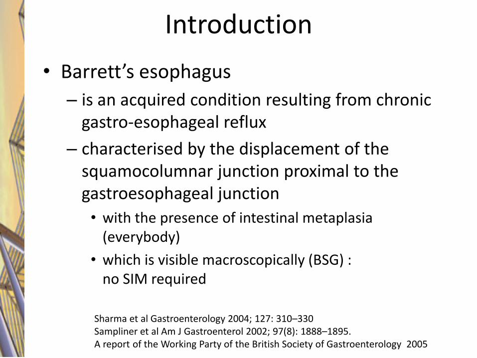
Introduction
• Barrett’s esophagus
– is an acquired condition resulting from chronic gastro-esophageal reflux
– characterised by the displacement of the squamocolumnar junction proximal to the gastroesophageal junction
• with the presence of intestinal metaplasia (everybody)
• which is visible macroscopically (BSG) : no SIM required
Sharma et al Gastroenterology 2004; 127: 310–330 Sampliner et al Am J Gastroenterol 2002; 97(8): 1888–1895. A report of the Working Party of the British Society of Gastroenterology 2005

Introduction
• Barrett’s esophagus
– is an acquired condition resulting from chronic gastro-esophageal reflux
– characterised by the displacement of the squamocolumnar junction proximal to the gastroesophageal junction
First step in diagnosis is endoscopic identification of a columnar lined esophagus to take a biopsy to confirm columnar lined esophagus (CLE) or specialized intestinal
metaplasia (SIM)

Example 1

Example 1
This is a Barrett’s Esophagus
1. Yes 2. No
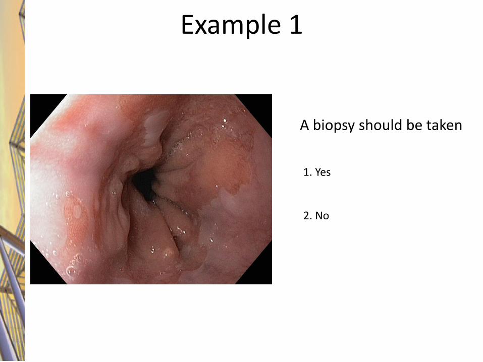
A biopsy should be taken
Example 1
1. Yes 2. No

Example 1
I believe that the biopsy will contain SIM
1. Yes 2. No

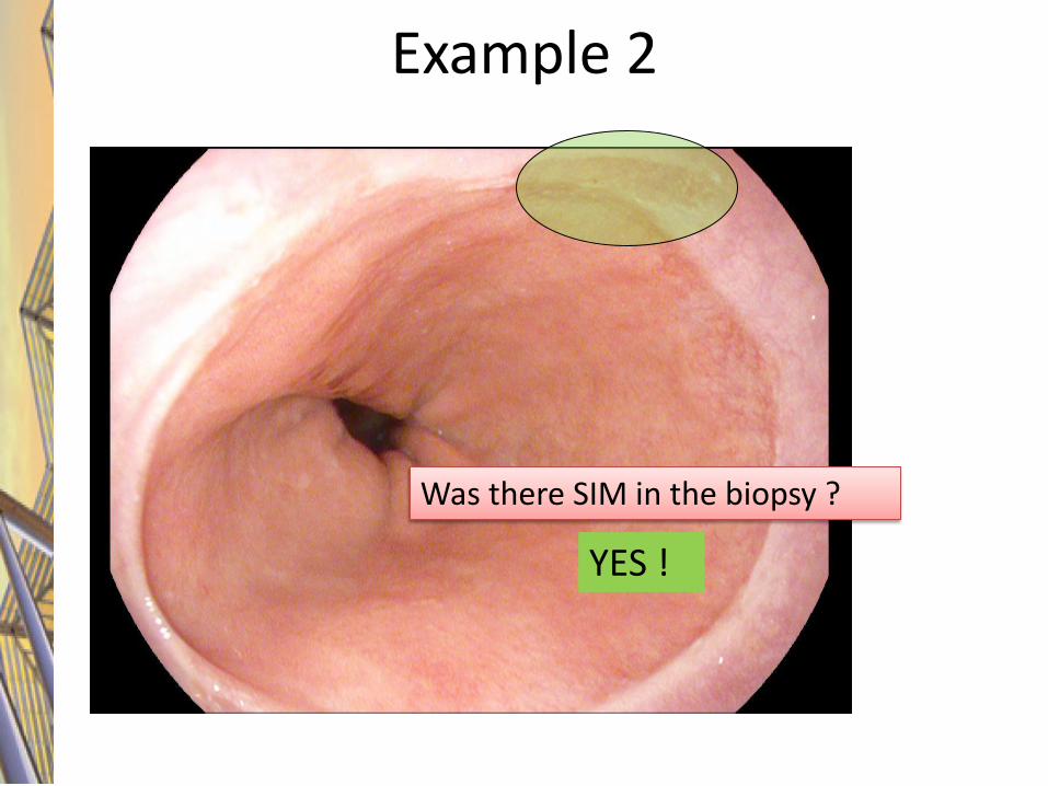
Example 2
Who thinks this is a Barrett’s Esophagus ?
1. Yes 2. No

Example 2
How long is this Barrett ? 1. >2 cm 2. <2cm

Example 2
I would take a biopsy ?
1. Yes 2. No
Example 2
Was there SIM in the biopsy ?
YES !

How to report Barrett endoscopically

Prague classification
Developed by the Barrett’s Oesophagus Subgroup of the International Working Group for the Classification of Reflux Oesophagitis (IWGCO)

Prague classification
Developed by the Barrett’s Oesophagus Subgroup of the International Working Group for the Classification of Reflux Oesophagitis (IWGCO)
Ensure Hiatus Hernia Is Recognised By
Distinguishing Diaphragmatic Hiatal Impression From
Gastroesophageal Junction

Step 1 : recognize hiatal hernia

Prague classification : Step 2
Developed by the Barrett’s Oesophagus Subgroup of the International Working Group for the Classification of Reflux Oesophagitis (IWGCO)
Locate gastroesophageal Junction by depth of endoscope
Insertion at level of: – tops of gastric mucosal folds
– sphincter “pinch”

How to determine GEJ ?
• Anatomically the GEJ is defined as the level of the Angle of His
• This corresponds best with endoscopically defined top of gastric folds
Wallner Surg Endosc (2009) 23:2155–2158
The difference between GEJ and endoscopic junction was < 5 mm

Prague classification : step 3
Developed by the Barrett’s Oesophagus Subgroup of the International Working Group for the Classification of Reflux Oesophagitis (IWGCO)
Look For Displacement Of Squamocolumnar Junction Above Gastroesophageal
Junction

Prague classification : step 3

What is a regular Z-line
• Savary and Miller: “It is serrated and shows 4 to 6 small, long or short tongues toward the esophagus.”
• DeNardi and Riddell: “The Z-line consists of small projections of red gastric epithelium, up to 5 mm long and 3 mm wide, extending upward into the pink-white squamous epithelium”
Savary M, Miller G. Handbook and atlas of endoscopy. 1978. DeNardi FG, Riddell RH. Am S Surg Pathol 1991;15:296-309.
Barrett

What is a regular Z-line
Wallner et al GIE 2002;55:65-9.)

What is a regular Z-line
Wallner et al GIE 2002;55:65-9.)

What is the prevalence of SIM at the GEJ ?
• Retrospective analyses of 2000 gastroscopies
– 166 identified with “irregular Z-line”
– No previous diagnosis of Barrett
• 43.5% of these had specialized intestinal metaplasia
– Risk factors : male, hiatal hernia
Dickman et al Eur J Gastro Hep 2010;22:135

ProGERD study
• PROGERD trial : endoscopic and symptomatic follow-up of 6215 GERD and NERD patients
• Subgroup analysis of patients without visible Barrett but SIM +.
• Biopsies were taken under Z-line and at 2 cm to distinguish also histologically if there was Barrett or not.
Leodolter et al Sc J Gastro. 2012; 47: 1429–1435

ProGERD study
Leodolter et al Sc J Gastro. 2012; 47: 1429–1435
Baseline 171 (3%) patients with SIM without BE
125 follow-up at 2 year and 68 at 5 year
0
20
40
60
80
100
120
140
160
180
baseline 2 years 5 years
0
20 33
SIM+ BE
SIM+

ProGERD study
• Risk factors for progression
– All patients had esophagitis at baseline, so none of the NERDs progressed
– Multivariate analysis :
• Smoking
• Long history of GERD (> 5 years)
• Severe esophagitis
– Male (13.9%) more than women (7.9%)
Leodolter et al Sc J Gastro. 2012; 47: 1429–1435

Prague classification : Step 4
Developed by the Barrett’s Oesophagus Subgroup of the International Working Group for the Classification of Reflux Oesophagitis (IWGCO)
Measure depth of endoscope insertion at the most proximal
Circumferential extent of suspected columnar metaplasia

Step 4 : determine C
GEJ 40 cm
C = 39 cm

Prague classification : step 5
Developed by the Barrett’s Oesophagus Subgroup of the International Working Group for the Classification of Reflux Oesophagitis (IWGCO)
Measure depth of endoscope insertion at the maximum extent
of suspected columnar metaplasia

Step 5 : determine M
GEJ 40 cm
C = 39 cm
M = 36 cm

Prague classification : step 6
Developed by the Barrett’s Oesophagus Subgroup of the International Working Group for the Classification of Reflux Oesophagitis (IWGCO)
Subtract the depth of insertion for circumferential
And maximum extents from the depth of
Endoscope insertion at the Gastroesophageal Junction

Step 6 : calculate CM
GEJ 40 cm
C = 39 cm
M = 36 cm
C1

GEJ 40 cm
C = 39 cm
M = 36 cm
C1 M4
Step 6 : calculate CM

How to take biopsies and report on them ?
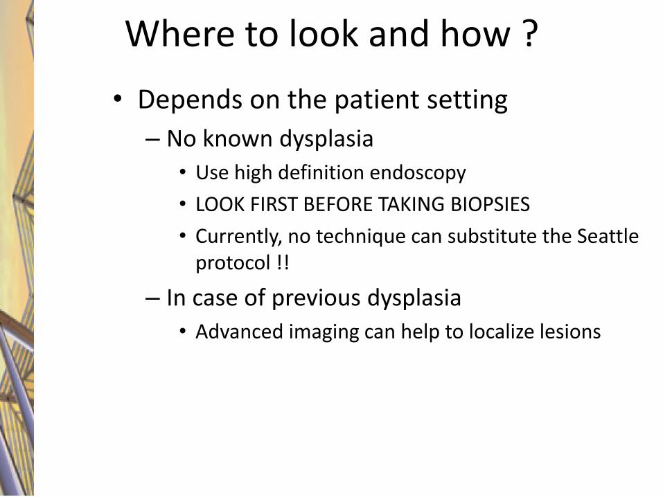
Where to look and how ?
• Depends on the patient setting
– No known dysplasia
• Use high definition endoscopy
• LOOK FIRST BEFORE TAKING BIOPSIES
• Currently, no technique can substitute the Seattle protocol !!
– In case of previous dysplasia
• Advanced imaging can help to localize lesions

USE YOUR BEST AVAILABLE ENDOSCOPE
Barrett screening
Fiberoptic
Standard high resolution endoscope
High definition endoscope
Courtesy Dr Bergman

Where is the cancer ?

INSPECT BEFORE TAKING BIOPSIES

INSPECT PRIOR TO TAKING BIOPSIES
HD-endoscopy

INSPECT PRIOR TO TAKING BIOPSIES
HD-endoscopy

Retroflex !
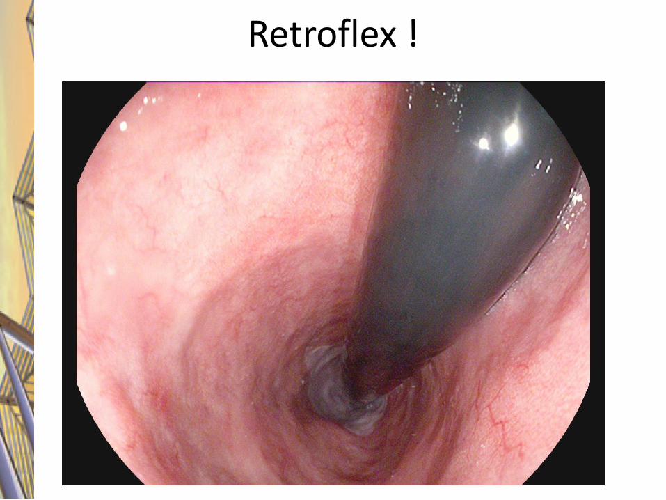
Retroflex !

Where to look for cancer ?

Pech et al Endoscopy 2007; 39: 588±593
388 neoplastic lesions
Where to look for cancer ?

How to take biopsies ?
• Seattle protocol : AFTER inspection
– First targeted biopsies of suspicious areas
• Use Paris classification to describe the lesions
– 4 quadrant biopsies each 2 cm
– Preferably in different containers

How to report biopsies ?
• Proposal for use of a standardized reporting system (xxyy)
– xx = distance from incisors
– yy = orientation circumferentially (clock system) (from 01-12)
– yy=00 for random biopsies
Lesser curvature of stomach

C8M9 Barrett
Lesion at 5 o’ clock 32 cm from incisors
Lesion 3205
IIb lesion at 3205
How to report biopsies ?

C8M9 Barrett
Lesion at 5 o’ clock 34 cm from incisors
Lesion 3205
IIb lesion at 3405
How to report biopsies ?

C8M9 Barrett
Lesion 3205
4000
3800
3600
3400
3200
IIb lesion at 3205
IIb lesion at 3405
How to report biopsies ?

Summary
Lisbon Coding for BE biopsies Random E.g. : 3600 : random at 36 cm from incisors Targeted biopsies: E.g. : 3603 : biopsy taken at 36 cm from incisors at the 3 o’ clock position

Conclusion
• Quality in reporting Barrett’s esophagus:
– Using Prague classification
– Pay attention to the identification of GEJ
– Measure hiatal hernia
– Measure C and M (islands do not count)
• Quality biopsies in Barrett’s esophagus
– FIRST look and target biopsies
– Seattle protocol
– Mark different containers according to xxyy principle : Lisbon coding ?