Infections in Immunocompromised Host A focus on Febrile Neutropenia.

Host defense against infections: Innate host defense against
infections: lessons learned from the clinic
Brahm Segal, MD [email protected]
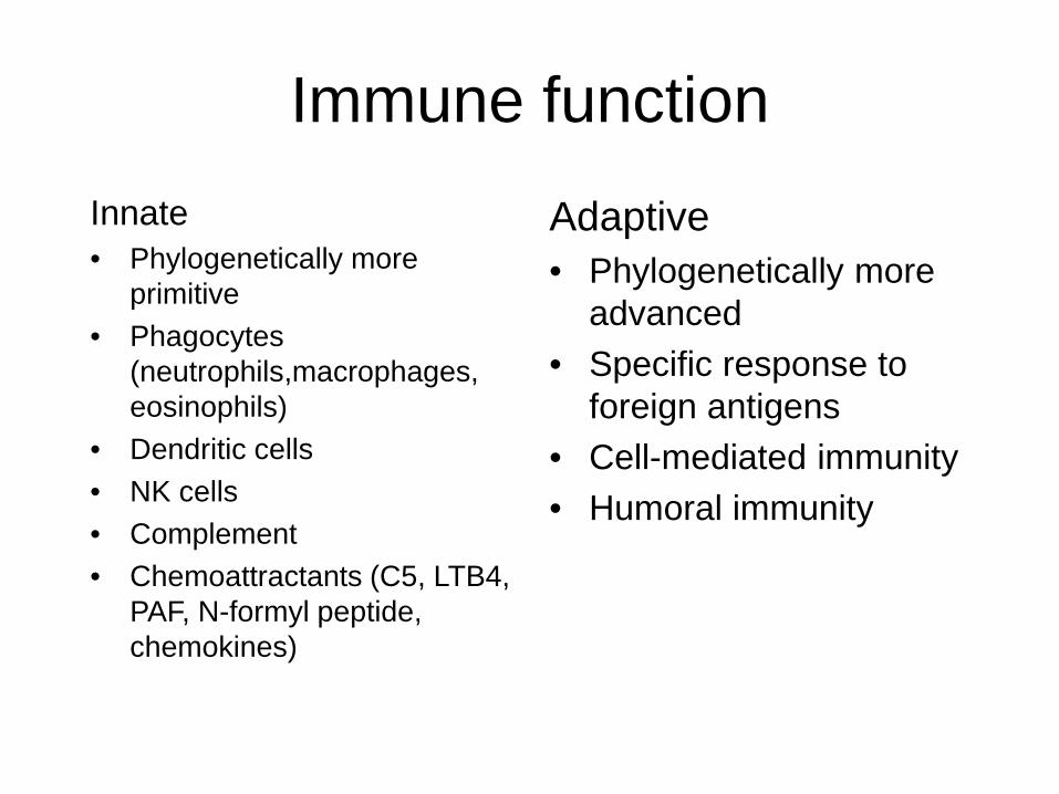
Immune function Innate • Phylogenetically more
primitive • Phagocytes
(neutrophils,macrophages, eosinophils)
• Dendritic cells • NK cells • Complement • Chemoattractants (C5, LTB4,
PAF, N-formyl peptide, chemokines)
Adaptive • Phylogenetically more
advanced • Specific response to
foreign antigens • Cell-mediated immunity • Humoral immunity
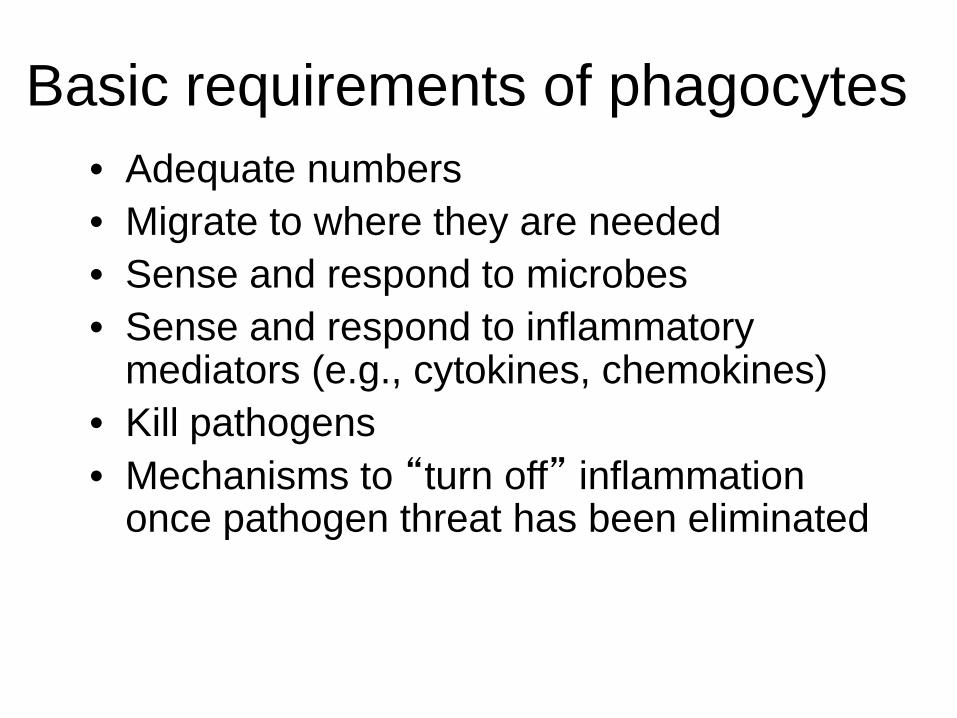
Basic requirements of phagocytes • Adequate numbers • Migrate to where they are needed • Sense and respond to microbes • Sense and respond to inflammatory
mediators (e.g., cytokines, chemokines) • Kill pathogens • Mechanisms to “turn off” inflammation
once pathogen threat has been eliminated

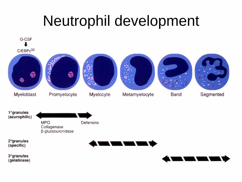
Neutrophil development

Neutrophil granules
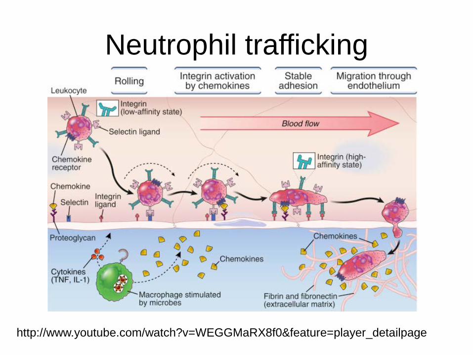
Neutrophil trafficking
http://www.youtube.com/watch?v=WEGGMaRX8f0&feature=player_detailpage

Sensing microbes and mediators of inflammation
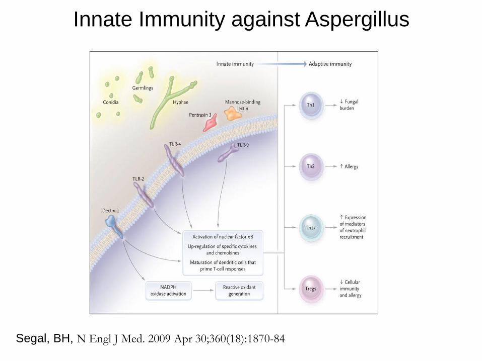
Innate Immunity against Aspergillus
Segal, BH, N Engl J Med. 2009 Apr 30;360(18):1870-84
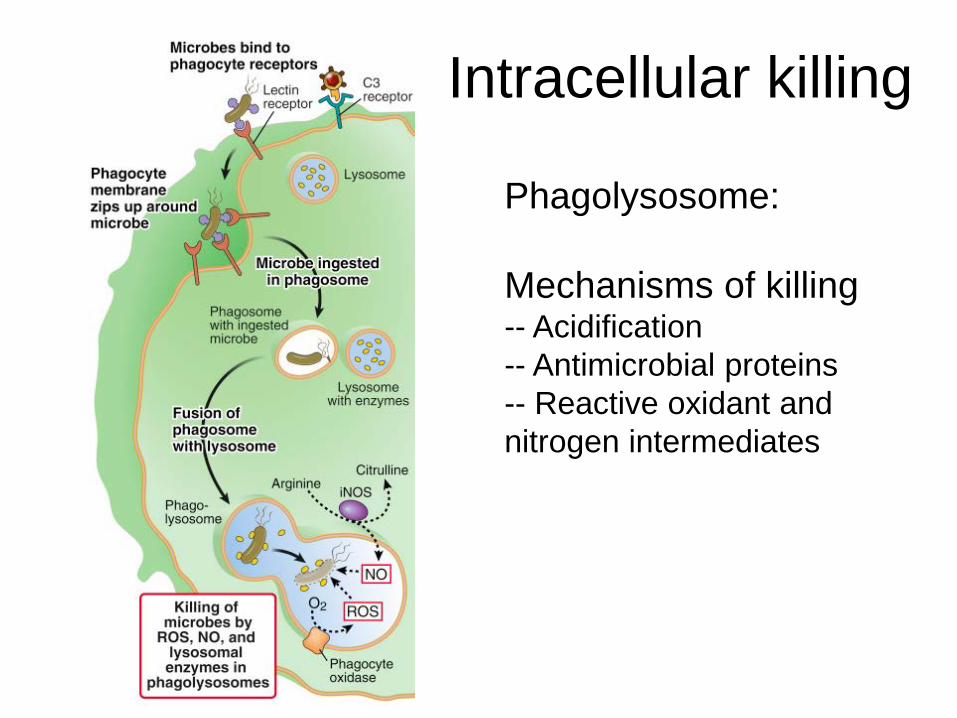
Intracellular killing
Phagolysosome: Mechanisms of killing -- Acidification -- Antimicrobial proteins -- Reactive oxidant and nitrogen intermediates

When to think of a primary phagocytic disorder?
• Usually (not always) first manifest during childhood • Unusually severe or recurrent infection by common
pathogens – S. aureus liver abscess – Multiple soft tissue infections requiring I&D – Severe dental infections --> premature tooth loss
• Opportunistic pathogens – Molds, candidiasis, nocardiosis, atypical
mycobacterial infection (disseminated)

When to think of a primary phagocytic disorder?
• Systemic features – CGD --> inflammatory complications – Chediak-Higashi --> occulocutaneous
albinism – HIESRI (Job’s) --> atopic dermatitis,
skeletal/craniofacial abnormalities • Family history

Management: basic principles
• Patients may be sicker than they look • Aggressively pursue a microbiologic dx --
“Tissue is the issue” • Prolonged systemic antimicrobial therapy • Surgical debridement of localized disease • Prophylactic ABX • Cytokine therapy, granulocyte transfusions,
BMT, gene therapy
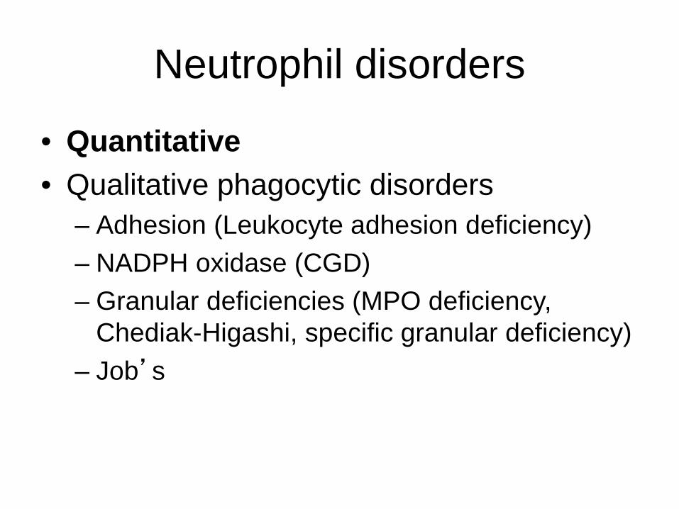
Neutrophil disorders
• Quantitative • Qualitative phagocytic disorders
– Adhesion (Leukocyte adhesion deficiency) – NADPH oxidase (CGD) – Granular deficiencies (MPO deficiency,
Chediak-Higashi, specific granular deficiency) – Job’s

Neutropenia: secondary causes
• Cytotoxic agents – Used principally as antineoplastic chemotherapy
• Other drugs with potential for marrow suppression – Bactrim, chloramphenicol, linezolid, ganciclovir,
zidovudine, clozoril • Immune-mediated • Infections
– Overwhelming sepsis, neonatal sepsis, viral

Risk of infection related to degree of neutropenia

Severe chronic neutropenias
• Heterogenous group of rare disorders – Congenital (Kostmann syndrome) – Cyclic neutropenia
• Linked to mutations in Neutrophil Elastase gene
– Adult-onset cyclic neutropenia – Primary autoimmune

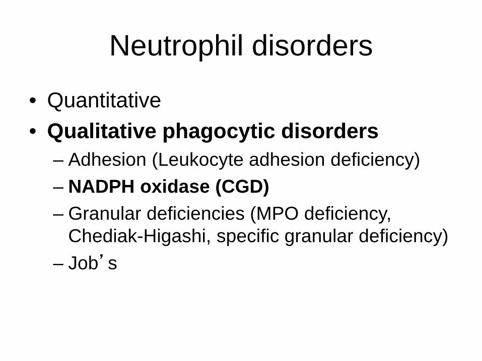
Neutrophil disorders
• Quantitative • Qualitative phagocytic disorders
– Adhesion (Leukocyte adhesion deficiency) – NADPH oxidase (CGD) – Granular deficiencies (MPO deficiency,
Chediak-Higashi, specific granular deficiency) – Job’s


How do neutrophils kill pathogens?

How do neutrophils kill pathogens?
• Neutrophils kill microorganisms through oxygen-independent and oxygen-dependent mechanisms
• Oxygen-independent mechanisms include the release of peptides and proteins from the granules, such as bactericidal permeability-increasing proteins (BPI), defensins and cathelicidins

HEME
HOCI H2O2
SOD
NADP+ + H+ NADPH
MPO
O2 O2–
p22phox gp91phox
e–
FAD
e–
rac GTP
HEME
HEME p22phox gp91phox
FAD
HEME
rac
p7phox
RhoGDI
GDP
p67phox p47phox
p40phox
p67phox
p47phox
p40phox
NADPH oxidase
Activation
Cytoplasm

Neutrophil extraceullar traps (NETs)
Shigella Candida
Neutrophil disorders
• Quantitative • Qualitative phagocytic disorders
– Adhesion (Leukocyte adhesion deficiency) – NADPH oxidase (CGD) – Granular deficiencies (MPO deficiency,
Chediak-Higashi, specific granular deficiency) – Job’s
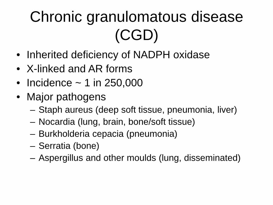
Chronic granulomatous disease (CGD)
• Inherited deficiency of NADPH oxidase • X-linked and AR forms • Incidence ~ 1 in 250,000 • Major pathogens
– Staph aureus (deep soft tissue, pneumonia, liver) – Nocardia (lung, brain, bone/soft tissue) – Burkholderia cepacia (pneumonia) – Serratia (bone) – Aspergillus and other moulds (lung, disseminated)

HEME
HOCI H2O2
SOD
NADP+ + H+ NADPH
MPO
O2 O2–
p22phox gp91phox
e–
FAD
e–
rac GTP
HEME
HEME p22phox gp91phox
FAD
HEME
rac
p7phox
RhoGDI
GDP
p67phox p47phox
p40phox
p67phox
p47phox
p40phox
NADPH oxidase
Activation
Cytoplasm
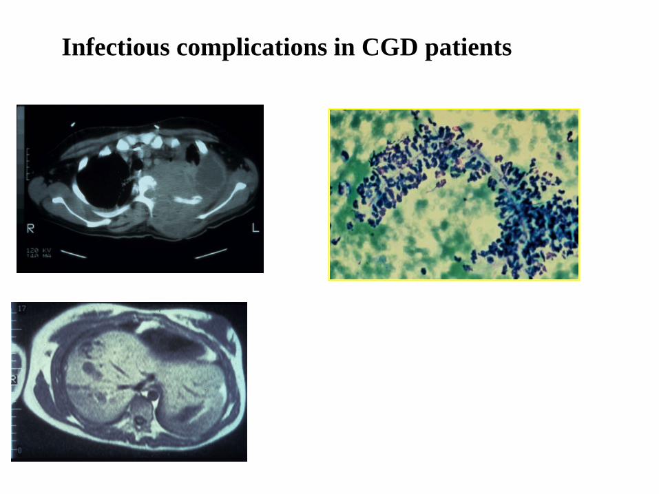
Infectious complications in CGD patients

Inflammatory complications of CGD

Prophylaxis in CGD
• Several non-randomized studies have shown benefit of antibacterial prophylaxis
• Bactrim is most commonly used • randomized placebo-controlled study
showed protective benefit of itraconazole prophylaxis (Gallin et al. NEJM, 2003)

rIFN-g prophylaxis in CGD • rIFN-g increased phagocyte superoxide production
in vitro • increased reactive oxidant intermediates (ROI)
production in human monocytes ex vivo after administration of rIFN-g to patients with advanced malignancy and lepromatous leprosy
• Early studies in patients with CGD showed that rIFN-g led to partial restoration of NADPH oxidase function, increased NADPH oxidase constituents, and enhanced bactericidal and fungicidal killing
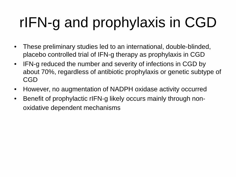
rIFN-g and prophylaxis in CGD • These preliminary studies led to an international, double-blinded,
placebo controlled trial of IFN-g therapy as prophylaxis in CGD • IFN-g reduced the number and severity of infections in CGD by
about 70%, regardless of antibiotic prophylaxis or genetic subtype of CGD
• However, no augmentation of NADPH oxidase activity occurred • Benefit of prophylactic rIFN-g likely occurs mainly through non-
oxidative dependent mechanisms

Current and future approaches • Allogeneic stem cell transplantation • Gene therapy
– Major hurdle is maintenance of a stable long-term population of gene-corrected cells

Neutrophil disorders
• Quantitative • Qualitative phagocytic disorders
– Adhesion (Leukocyte adhesion deficiency) – NADPH oxidase (CGD) – Granular deficiencies (MPO deficiency,
Chediak-Higashi, specific granular deficiency)
– Job’s

Myeloperoxidase deficiency
• Most common primary phagocytic disorder, affecting ~ 1 in 2000
• Usually asymptomatic • Occasionally invasive candidiasis
observed in diabetics with MPO • Diagnosis made by direct assay of MPO

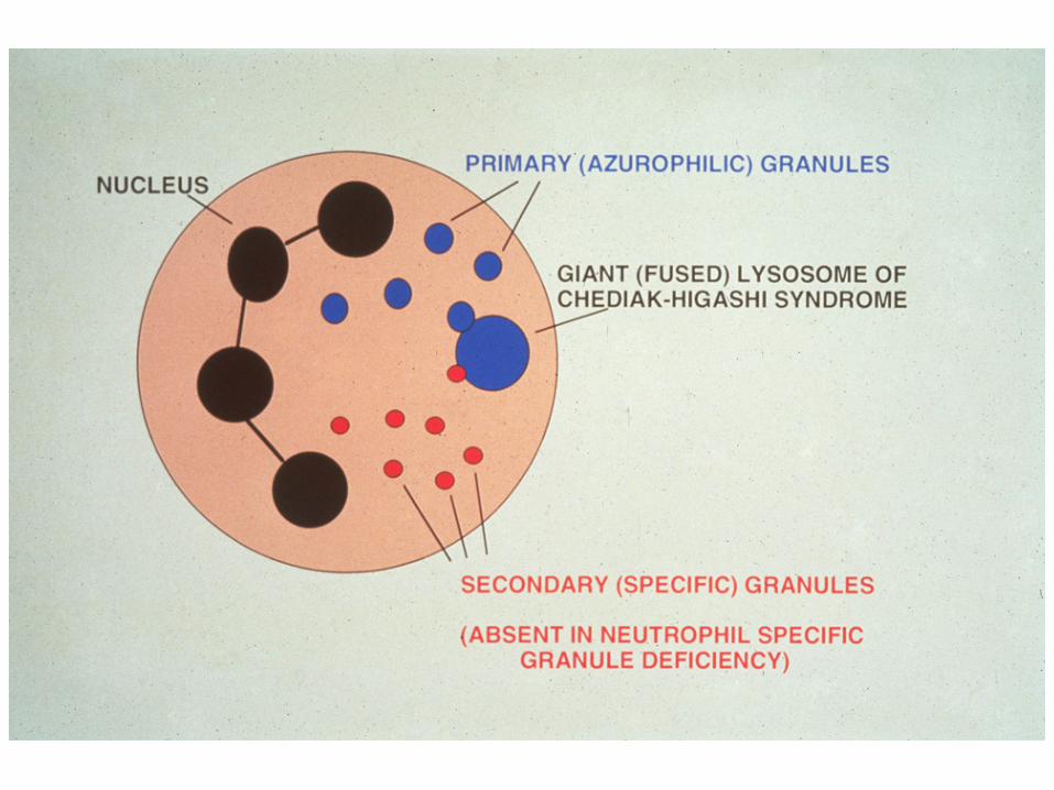
Chediak-Higashi syndrome • severe autosomal recessive condition of LYST
gene (lysosomal trafficking regulator) • partial oculocutaneous albinism with photophobia • increased susceptibility to infections (mostly
Staphylococcus and streptococci) • Neurologic manifestations • widespread visceral infiltration of
lymphohistiocytic cells gives rise to fever, jaundice, hepatosplenomegaly, lymphadenopathy, pancytopenia, bleeding
• most children dying before age of 10 from an accelerated lymphomatous phase

-- Abnormally large leukocyte granules result from fusion of lysozymes. -- may affect granulocytes and monocytes -- Chemotaxis and phagocytosis are defective -- Platelets lack dense granules and platelet function is abnormal.
CHS
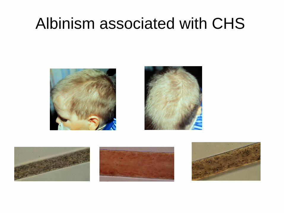
Albinism associated with CHS

Neutrophil disorders
• Quantitative • Qualitative phagocytic disorders
– Adhesion (Leukocyte adhesion deficiency) – NADPH oxidase (CGD) – Granular deficiencies (MPO deficiency,
Chediak-Higashi, specific granular deficiency) – Job’s

Job’s syndrome • Hyper-IgE syndrome with recurrent infections (HIESRI) • Severe eczema starting at infancy • Deep soft tissue infections (cold abscesses), usually
caused by Staphylococcus aureus • Pneumonias (pneumatoceles) • ↑IgE • Differentiate from atopic dermatitis by frequency and
severity of infections • Non-immunologic manifestations
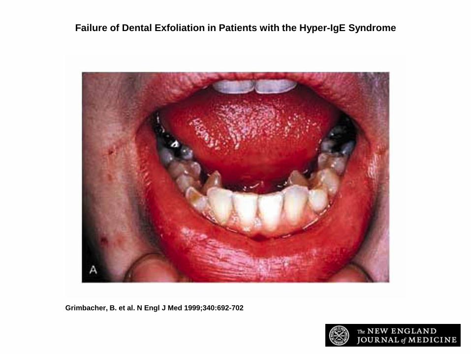
– Delayed shedding of the primary teeth owing to lack of root resorption, recurrent fractures, hyperextensible joints and scoliosis, characteristic facial appearance

Pneumonia with pneumatocele in a patient with Job’s
Grimbacher, B. et al. N Engl J Med 1999;340:692-702
Failure of Dental Exfoliation in Patients with the Hyper-IgE Syndrome

Grimbacher, B. et al. N Engl J Med 1999;340:692-702
Characteristic Facial Appearance of Men and Women of Different Races with the Hyper-IgE Syndrome

Grimbacher, B. et al. N Engl J Med 1999;340:692-702
Serum IgE Levels in Patients with the Hyper-IgE Syndrome Whose Levels Declined

Dominant-negative mutations in the DNA-binding domain of STAT3 cause hyper-IgE syndrome
Minegishi et al. Nature, 2007 Holland et al. NEJM, 2007
PBLs from Job’s patients -- Normal STAT3 levels at baseline and after IFN-α stimulation
-- Decreased DNA binding of STAT3
-- defective responses to IL-6 and IL-10
-- Reduced Th-17 development and IL-22-induced signaling

Chronic intracellular infections
• Herpes viruses • Mycobacteria • Certain fungi (e.g., Histoplasma) • Require ongoing surveillance • Don’t believe anyone that says the
immune system evolved for cancer surveillance…


M. tuberculosis
• Worldwide – > 1 Billion people infected – 8 million new cases per year – 3 million deaths per year
• Person-to-person contact through inhalation • Prolonged multi-drug regimen

Disseminated Mycobacterium avium infection in a patient with IFN-g receptor deficiency
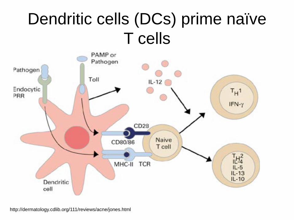
Dendritic cells (DCs) prime naïve T cells
http://dermatology.cdlib.org/111/reviews/acne/jones.html

Conclusions • Primary phagocytic defects are for the most part
rare • Many have extra-immunologic manifestations • “Experiments of nature” teach us about
neutrophil development and host defense pathways
• Management relies on targeted prophylaxis, vigilance for early signs of infection, and judicious diagnostic work-up
• Opportunities for novel therapeutics (e.g., cytokines, BMT, gene therapy)