Hormones - cnx.org · A hormone's half-life is the time required for half the concentration of the...
15
• • • • •
Transcript of Hormones - cnx.org · A hormone's half-life is the time required for half the concentration of the...
OpenStax-CNX module: m46667 1
Hormones*
OpenStax
This work is produced by OpenStax-CNX and licensed under the
Creative Commons Attribution License 3.0�
Abstract
By the end of this section, you will be able to:
• Identify the three major classes of hormones on the basis of chemical structure• Compare and contrast intracellular and cell membrane hormone receptors• Describe signaling pathways that involve cAMP and IP3• Identify several factors that in�uence a target cell's response• Discuss the role of feedback loops and humoral, hormonal, and neural stimuli in hormone control
Although a given hormone may travel throughout the body in the bloodstream, it will a�ect the activityonly of its target cells; that is, cells with receptors for that particular hormone. Once the hormone binds tothe receptor, a chain of events is initiated that leads to the target cell's response. Hormones play a criticalrole in the regulation of physiological processes because of the target cell responses they regulate. Theseresponses contribute to human reproduction, growth and development of body tissues, metabolism, �uid,and electrolyte balance, sleep, and many other body functions. The major hormones of the human bodyand their e�ects are identi�ed in Table 1.
Endocrine Glands and Their Major Hormones
Endocrine gland Associated hormones Chemical class E�ect
Pituitary (anterior) Growth hormone (GH) Protein Promotes growth ofbody tissues
Pituitary (anterior) Prolactin (PRL) Peptide Promotes milk produc-tion
Pituitary (anterior) Thyroid-stimulatinghormone (TSH)
Glycoprotein Stimulates thyroid hor-mone release
Pituitary (anterior) Adrenocorticotropichormone (ACTH)
Peptide Stimulates hormone re-lease by adrenal cortex
continued on next page
*Version 1.3: Jun 18, 2013 4:50 pm -0500�http://creativecommons.org/licenses/by/3.0/
http://cnx.org/content/m46667/1.3/

OpenStax-CNX module: m46667 2
Pituitary (anterior) Follicle-stimulating hor-mone (FSH)
Glycoprotein Stimulates gamete pro-duction
Pituitary (anterior) Luteinizing hormone(LH)
Glycoprotein Stimulates androgenproduction by gonads
Pituitary (posterior) Antidiuretic hormone(ADH)
Peptide Stimulates water reab-sorption by kidneys
Pituitary (posterior) Oxytocin Peptide Stimulates uterine con-tractions during child-birth
Thyroid Thyroxine (T4), tri-iodothyronine (T3)
Amine Stimulate basalmetabolic rate
Thyroid Calcitonin Peptide Reduces blood Ca2+
levels
Parathyroid Parathyroid hormone(PTH)
Peptide Increases bloodCa2+ levels
Adrenal (cortex) Aldosterone Steroid Increases blood Na+
levels
Adrenal (cortex) Cortisol, corticosterone,cortisone
Steroid Increase blood glucoselevels
Adrenal (medulla) Epinephrine, nore-pinephrine
Amine Stimulate �ght-or-�ightresponse
Pineal Melatonin Amine Regulates sleep cycles
Pancreas Insulin Protein Reduces blood glucoselevels
Pancreas Glucagon Protein Increases blood glucoselevels
Testes Testosterone Steroid Stimulates developmentof male secondary sexcharacteristics andsperm production
Ovaries Estrogens and proges-terone
Steroid Stimulate developmentof female secondary sexcharacteristics and pre-pare the body for child-birth
Table 1
1 Types of Hormones
The hormones of the human body can be divided into two major groups on the basis of their chemicalstructure. Hormones derived from amino acids include amines, peptides, and proteins. Those derived fromlipids include steroids (Figure 1 (Amine, Peptide, Protein, and Steroid Hormone Structure )). These chemicalgroups a�ect a hormone's distribution, the type of receptors it binds to, and other aspects of its function.
http://cnx.org/content/m46667/1.3/

OpenStax-CNX module: m46667 3
Amine, Peptide, Protein, and Steroid Hormone Structure
Figure 1
http://cnx.org/content/m46667/1.3/

OpenStax-CNX module: m46667 4
1.1 Amine Hormones
Hormones derived from the modi�cation of amino acids are referred to as amine hormones. Typically, theoriginal structure of the amino acid is modi�ed such that a �COOH, or carboxyl, group is removed, whereasthe −NH+
3 , or amine, group remains.Amine hormones are synthesized from the amino acids tryptophan or tyrosine. An example of a hor-
mone derived from tryptophan is melatonin, which is secreted by the pineal gland and helps regulate cir-cadian rhythm. Tyrosine derivatives include the metabolism-regulating thyroid hormones, as well as thecatecholamines, such as epinephrine, norepinephrine, and dopamine. Epinephrine and norepinephrine aresecreted by the adrenal medulla and play a role in the �ght-or-�ight response, whereas dopamine is secretedby the hypothalamus and inhibits the release of certain anterior pituitary hormones.
1.2 Peptide and Protein Hormones
Whereas the amine hormones are derived from a single amino acid, peptide and protein hormones consistof multiple amino acids that link to form an amino acid chain. Peptide hormones consist of short chains ofamino acids, whereas protein hormones are longer polypeptides. Both types are synthesized like other bodyproteins: DNA is transcribed into mRNA, which is translated into an amino acid chain.
Examples of peptide hormones include antidiuretic hormone (ADH), a pituitary hormone important in�uid balance, and atrial-natriuretic peptide, which is produced by the heart and helps to decrease blood pres-sure. Some examples of protein hormones include growth hormone, which is produced by the pituitary gland,and follicle-stimulating hormone (FSH), which has an attached carbohydrate group and is thus classi�ed asa glycoprotein. FSH helps stimulate the maturation of eggs in the ovaries and sperm in the testes.
1.3 Steroid Hormones
The primary hormones derived from lipids are steroids. Steroid hormones are derived from the lipid choles-terol. For example, the reproductive hormones testosterone and the estrogens�which are produced bythe gonads (testes and ovaries)�are steroid hormones. The adrenal glands produce the steroid hormonealdosterone, which is involved in osmoregulation, and cortisol, which plays a role in metabolism.
Like cholesterol, steroid hormones are not soluble in water (they are hydrophobic). Because blood iswater-based, lipid-derived hormones must travel to their target cell bound to a transport protein. This morecomplex structure extends the half-life of steroid hormones much longer than that of hormones derived fromamino acids. A hormone's half-life is the time required for half the concentration of the hormone to bedegraded. For example, the lipid-derived hormone cortisol has a half-life of approximately 60 to 90 minutes.In contrast, the amino acid�derived hormone epinephrine has a half-life of approximately one minute.
2 Pathways of Hormone Action
The message a hormone sends is received by a hormone receptor, a protein located either inside the cellor within the cell membrane. The receptor will process the message by initiating other signaling events orcellular mechanisms that result in the target cell's response. Hormone receptors recognize molecules withspeci�c shapes and side groups, and respond only to those hormones that are recognized. The same type ofreceptor may be located on cells in di�erent body tissues, and trigger somewhat di�erent responses. Thus,the response triggered by a hormone depends not only on the hormone, but also on the target cell.
Once the target cell receives the hormone signal, it can respond in a variety of ways. The responsemay include the stimulation of protein synthesis, activation or deactivation of enzymes, alteration in thepermeability of the cell membrane, altered rates of mitosis and cell growth, and stimulation of the secretionof products. Moreover, a single hormone may be capable of inducing di�erent responses in a given cell.
http://cnx.org/content/m46667/1.3/

OpenStax-CNX module: m46667 5
2.1 Pathways Involving Intracellular Hormone Receptors
Intracellular hormone receptors are located inside the cell. Hormones that bind to this type of receptor mustbe able to cross the cell membrane. Steroid hormones are derived from cholesterol and therefore can readilydi�use through the lipid bilayer of the cell membrane to reach the intracellular receptor (Figure 2 (Bindingof Lipid-Soluble Hormones )). Thyroid hormones, which contain benzene rings studded with iodine, are alsolipid-soluble and can enter the cell.
The location of steroid and thyroid hormone binding di�ers slightly: a steroid hormone may bind to itsreceptor within the cytosol or within the nucleus. In either case, this binding generates a hormone-receptorcomplex that moves toward the chromatin in the cell nucleus and binds to a particular segment of the cell'sDNA. In contrast, thyroid hormones bind to receptors already bound to DNA. For both steroid and thyroidhormones, binding of the hormone-receptor complex with DNA triggers transcription of a target gene tomRNA, which moves to the cytosol and directs protein synthesis by ribosomes.
Binding of Lipid-Soluble Hormones
Figure 2: A steroid hormone directly initiates the production of proteins within a target cell. Steroidhormones easily di�use through the cell membrane. The hormone binds to its receptor in the cytosol,forming a receptor�hormone complex. The receptor�hormone complex then enters the nucleus and bindsto the target gene on the DNA. Transcription of the gene creates a messenger RNA that is translatedinto the desired protein within the cytoplasm.
http://cnx.org/content/m46667/1.3/
OpenStax-CNX module: m46667 6
2.2 Pathways Involving Cell Membrane Hormone Receptors
Hydrophilic, or water-soluble, hormones are unable to di�use through the lipid bilayer of the cell membraneand must therefore pass on their message to a receptor located at the surface of the cell. Except for thyroidhormones, which are lipid-soluble, all amino acid�derived hormones bind to cell membrane receptors thatare located, at least in part, on the extracellular surface of the cell membrane. Therefore, they do notdirectly a�ect the transcription of target genes, but instead initiate a signaling cascade that is carried outby a molecule called a second messenger. In this case, the hormone is called a �rst messenger.
The second messenger used by most hormones is cyclic adenosine monophosphate (cAMP). Inthe cAMP second messenger system, a water-soluble hormone binds to its receptor in the cell membrane(Step 1 in Figure 3 (Binding of Water-Soluble Hormones )). This receptor is associated with an intracellularcomponent called a G protein, and binding of the hormone activates the G-protein component (Step 2).The activated G protein in turn activates an enzyme called adenylyl cyclase, also known as adenylatecyclase (Step 3), which converts adenosine triphosphate (ATP) to cAMP (Step 4). As the second messenger,cAMP activates a type of enzyme called a protein kinase that is present in the cytosol (Step 5). Activatedprotein kinases initiate a phosphorylation cascade, in which multiple protein kinases phosphorylate (adda phosphate group to) numerous and various cellular proteins, including other enzymes (Step 6).
http://cnx.org/content/m46667/1.3/

OpenStax-CNX module: m46667 7
Binding of Water-Soluble Hormones
Figure 3: Water-soluble hormones cannot di�use through the cell membrane. These hormones mustbind to a surface cell-membrane receptor. The receptor then initiates a cell-signaling pathway within thecell involving G proteins, adenylyl cyclase, the secondary messenger cyclic AMP (cAMP), and proteinkinases. In the �nal step, these protein kinases phosphorylate proteins in the cytoplasm. This activatesproteins in the cell that carry out the changes speci�ed by the hormone.
The phosphorylation of cellular proteins can trigger a wide variety of e�ects, from nutrient metabolismto the synthesis of di�erent hormones and other products. The e�ects vary according to the type of targetcell, the G proteins and kinases involved, and the phosphorylation of proteins. Examples of hormones thatuse cAMP as a second messenger include calcitonin, which is important for bone construction and regulatingblood calcium levels; glucagon, which plays a role in blood glucose levels; and thyroid-stimulating hormone,which causes the release of T3 and T4 from the thyroid gland.
Overall, the phosphorylation cascade signi�cantly increases the e�ciency, speed, and speci�city of thehormonal response, as thousands of signaling events can be initiated simultaneously in response to a verylow concentration of hormone in the bloodstream. However, the duration of the hormone signal is short, ascAMP is quickly deactivated by the enzyme phosphodiesterase (PDE), which is located in the cytosol.The action of PDE helps to ensure that a target cell's response ceases quickly unless new hormones arrive
http://cnx.org/content/m46667/1.3/

OpenStax-CNX module: m46667 8
at the cell membrane.Importantly, there are also G proteins that decrease the levels of cAMP in the cell in response to hormone
binding. For example, when growth hormone�inhibiting hormone (GHIH), also known as somatostatin, bindsto its receptors in the pituitary gland, the level of cAMP decreases, thereby inhibiting the secretion of humangrowth hormone.
Not all water-soluble hormones initiate the cAMP second messenger system. One common alternativesystem uses calcium ions as a second messenger. In this system, G proteins activate the enzyme phospholipaseC (PLC), which functions similarly to adenylyl cyclase. Once activated, PLC cleaves a membrane-boundphospholipid into two molecules: diacylglycerol (DAG) and inositol triphosphate (IP3). Like cAMP,DAG activates protein kinases that initiate a phosphorylation cascade. At the same time, IP3 causes calciumions to be released from storage sites within the cytosol, such as from within the smooth endoplasmicreticulum. The calcium ions then act as second messengers in two ways: they can in�uence enzymatic andother cellular activities directly, or they can bind to calcium-binding proteins, the most common of which iscalmodulin. Upon binding calcium, calmodulin is able to modulate protein kinase within the cell. Examplesof hormones that use calcium ions as a second messenger system include angiotensin II, which helps regulateblood pressure through vasoconstriction, and growth hormone�releasing hormone (GHRH), which causes thepituitary gland to release growth hormones.
3 Factors A�ecting Target Cell Response
You will recall that target cells must have receptors speci�c to a given hormone if that hormone is to triggera response. But several other factors in�uence the target cell response. For example, the presence of asigni�cant level of a hormone circulating in the bloodstream can cause its target cells to decrease theirnumber of receptors for that hormone. This process is called downregulation, and it allows cells to becomeless reactive to the excessive hormone levels. When the level of a hormone is chronically reduced, targetcells engage in upregulation to increase their number of receptors. This process allows cells to be moresensitive to the hormone that is present. Cells can also alter the sensitivity of the receptors themselves tovarious hormones.
Two or more hormones can interact to a�ect the response of cells in a variety of ways. The three mostcommon types of interaction are as follows:
• The permissive e�ect, in which the presence of one hormone enables another hormone to act. Forexample, thyroid hormones have complex permissive relationships with certain reproductive hormones.A dietary de�ciency of iodine, a component of thyroid hormones, can therefore a�ect reproductivesystem development and functioning.
• The synergistic e�ect, in which two hormones with similar e�ects produce an ampli�ed response. Insome cases, two hormones are required for an adequate response. For example, two di�erent repro-ductive hormones�FSH from the pituitary gland and estrogens from the ovaries�are required for thematuration of female ova (egg cells).
• The antagonistic e�ect, in which two hormones have opposing e�ects. A familiar example is the e�ectof two pancreatic hormones, insulin and glucagon. Insulin increases the liver's storage of glucose asglycogen, decreasing blood glucose, whereas glucagon stimulates the breakdown of glycogen stores,increasing blood glucose.
4 Regulation of Hormone Secretion
To prevent abnormal hormone levels and a potential disease state, hormone levels must be tightly controlled.The body maintains this control by balancing hormone production and degradation. Feedback loops governthe initiation and maintenance of most hormone secretion in response to various stimuli.
http://cnx.org/content/m46667/1.3/

OpenStax-CNX module: m46667 9
4.1 Role of Feedback Loops
The contribution of feedback loops to homeostasis will only be brie�y reviewed here. Positive feedback loopsare characterized by the release of additional hormone in response to an original hormone release. The releaseof oxytocin during childbirth is a positive feedback loop. The initial release of oxytocin begins to signal theuterine muscles to contract, which pushes the fetus toward the cervix, causing it to stretch. This, in turn,signals the pituitary gland to release more oxytocin, causing labor contractions to intensify. The release ofoxytocin decreases after the birth of the child.
The more common method of hormone regulation is the negative feedback loop. Negative feedback ischaracterized by the inhibition of further secretion of a hormone in response to adequate levels of thathormone. This allows blood levels of the hormone to be regulated within a narrow range. An example of anegative feedback loop is the release of glucocorticoid hormones from the adrenal glands, as directed by thehypothalamus and pituitary gland. As glucocorticoid concentrations in the blood rise, the hypothalamus andpituitary gland reduce their signaling to the adrenal glands to prevent additional glucocorticoid secretion(Figure 4 (Negative Feedback Loop )).
http://cnx.org/content/m46667/1.3/

OpenStax-CNX module: m46667 10
Negative Feedback Loop
Figure 4: The release of adrenal glucocorticoids is stimulated by the release of hormones from thehypothalamus and pituitary gland. This signaling is inhibited when glucocorticoid levels become elevatedby causing negative signals to the pituitary gland and hypothalamus.
4.2 Role of Endocrine Gland Stimuli
Re�exes triggered by both chemical and neural stimuli control endocrine activity. These re�exes may besimple, involving only one hormone response, or they may be more complex and involve many hormones, as
http://cnx.org/content/m46667/1.3/
OpenStax-CNX module: m46667 11
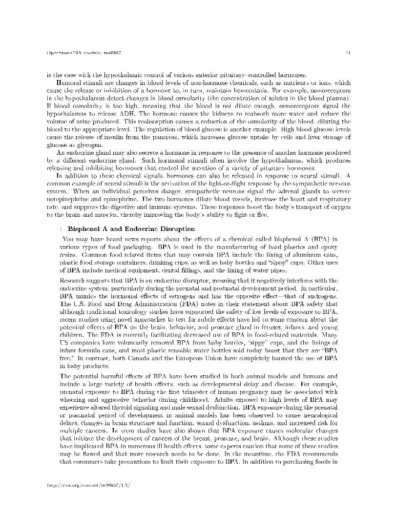
is the case with the hypothalamic control of various anterior pituitary�controlled hormones.Humoral stimuli are changes in blood levels of non-hormone chemicals, such as nutrients or ions, which
cause the release or inhibition of a hormone to, in turn, maintain homeostasis. For example, osmoreceptorsin the hypothalamus detect changes in blood osmolarity (the concentration of solutes in the blood plasma).If blood osmolarity is too high, meaning that the blood is not dilute enough, osmoreceptors signal thehypothalamus to release ADH. The hormone causes the kidneys to reabsorb more water and reduce thevolume of urine produced. This reabsorption causes a reduction of the osmolarity of the blood, diluting theblood to the appropriate level. The regulation of blood glucose is another example. High blood glucose levelscause the release of insulin from the pancreas, which increases glucose uptake by cells and liver storage ofglucose as glycogen.
An endocrine gland may also secrete a hormone in response to the presence of another hormone producedby a di�erent endocrine gland. Such hormonal stimuli often involve the hypothalamus, which producesreleasing and inhibiting hormones that control the secretion of a variety of pituitary hormones.
In addition to these chemical signals, hormones can also be released in response to neural stimuli. Acommon example of neural stimuli is the activation of the �ght-or-�ight response by the sympathetic nervoussystem. When an individual perceives danger, sympathetic neurons signal the adrenal glands to secretenorepinephrine and epinephrine. The two hormones dilate blood vessels, increase the heart and respiratoryrate, and suppress the digestive and immune systems. These responses boost the body's transport of oxygento the brain and muscles, thereby improving the body's ability to �ght or �ee.
: Bisphenol A and Endocrine Disruption
You may have heard news reports about the e�ects of a chemical called bisphenol A (BPA) invarious types of food packaging. BPA is used in the manufacturing of hard plastics and epoxyresins. Common food-related items that may contain BPA include the lining of aluminum cans,plastic food-storage containers, drinking cups, as well as baby bottles and �sippy� cups. Other usesof BPA include medical equipment, dental �llings, and the lining of water pipes.
Research suggests that BPA is an endocrine disruptor, meaning that it negatively interferes with theendocrine system, particularly during the prenatal and postnatal development period. In particular,BPA mimics the hormonal e�ects of estrogens and has the opposite e�ect�that of androgens.The U.S. Food and Drug Administration (FDA) notes in their statement about BPA safety thatalthough traditional toxicology studies have supported the safety of low levels of exposure to BPA,recent studies using novel approaches to test for subtle e�ects have led to some concern about thepotential e�ects of BPA on the brain, behavior, and prostate gland in fetuses, infants, and youngchildren. The FDA is currently facilitating decreased use of BPA in food-related materials. ManyUS companies have voluntarily removed BPA from baby bottles, �sippy� cups, and the linings ofinfant formula cans, and most plastic reusable water bottles sold today boast that they are �BPAfree.� In contrast, both Canada and the European Union have completely banned the use of BPAin baby products.
The potential harmful e�ects of BPA have been studied in both animal models and humans andinclude a large variety of health e�ects, such as developmental delay and disease. For example,prenatal exposure to BPA during the �rst trimester of human pregnancy may be associated withwheezing and aggressive behavior during childhood. Adults exposed to high levels of BPA mayexperience altered thyroid signaling and male sexual dysfunction. BPA exposure during the prenatalor postnatal period of development in animal models has been observed to cause neurologicaldelays, changes in brain structure and function, sexual dysfunction, asthma, and increased risk formultiple cancers. In vitro studies have also shown that BPA exposure causes molecular changesthat initiate the development of cancers of the breast, prostate, and brain. Although these studieshave implicated BPA in numerous ill health e�ects, some experts caution that some of these studiesmay be �awed and that more research needs to be done. In the meantime, the FDA recommendsthat consumers take precautions to limit their exposure to BPA. In addition to purchasing foods in
http://cnx.org/content/m46667/1.3/

OpenStax-CNX module: m46667 12
packaging free of BPA, consumers should avoid carrying or storing foods or liquids in bottles withthe recycling code 3 or 7. Foods and liquids should not be microwave-heated in any form of plastic:use paper, glass, or ceramics instead.
5 Chapter Review
Hormones are derived from amino acids or lipids. Amine hormones originate from the amino acids tryptophanor tyrosine. Larger amino acid hormones include peptides and protein hormones. Steroid hormones arederived from cholesterol.
Steroid hormones and thyroid hormone are lipid soluble. All other amino acid�derived hormones are watersoluble. Hydrophobic hormones are able to di�use through the membrane and interact with an intracellularreceptor. In contrast, hydrophilic hormones must interact with cell membrane receptors. These are typicallyassociated with a G protein, which becomes activated when the hormone binds the receptor. This initiatesa signaling cascade that involves a second messenger, such as cyclic adenosine monophosphate (cAMP).Second messenger systems greatly amplify the hormone signal, creating a broader, more e�cient, and fasterresponse.
Hormones are released upon stimulation that is of either chemical or neural origin. Regulation of hormonerelease is primarily achieved through negative feedback. Various stimuli may cause the release of hormones,but there are three major types. Humoral stimuli are changes in ion or nutrient levels in the blood. Hormonalstimuli are changes in hormone levels that initiate or inhibit the secretion of another hormone. Finally, aneural stimulus occurs when a nerve impulse prompts the secretion or inhibition of a hormone.
6 Review Questions
Exercise 1 (Solution on p. 14.)
A newly developed pesticide has been observed to bind to an intracellular hormone receptor. Ifingested, residue from this pesticide could disrupt levels of ________.
a. melatoninb. thyroid hormonec. growth hormoned. insulin
Exercise 2 (Solution on p. 14.)
A small molecule binds to a G protein, preventing its activation. What direct e�ect will this haveon signaling that involves cAMP?
a. The hormone will not be able to bind to the hormone receptor.b. Adenylyl cyclase will not be activated.c. Excessive quantities of cAMP will be produced.d. The phosphorylation cascade will be initiated.
Exercise 3 (Solution on p. 14.)
A student is in a car accident, and although not hurt, immediately experiences pupil dilation,increased heart rate, and rapid breathing. What type of endocrine system stimulus did the studentreceive?
a. humoralb. hormonalc. neurald. positive feedback
http://cnx.org/content/m46667/1.3/

OpenStax-CNX module: m46667 13
7 Critical Thinking Questions
Exercise 4 (Solution on p. 14.)
Compare and contrast the signaling events involved with the second messengers cAMP and IP3.
Exercise 5 (Solution on p. 14.)
Describe the mechanism of hormone response resulting from the binding of a hormone with anintracellular receptor.
http://cnx.org/content/m46667/1.3/

OpenStax-CNX module: m46667 14
Solutions to Exercises in this Module
to Exercise (p. 12)Bto Exercise (p. 12)Bto Exercise (p. 12)Cto Exercise (p. 13)In both cAMP and IP3�calcium signaling, a hormone binds to a cell membrane hormone receptor that iscoupled to a G protein. The G protein becomes activated when the hormone binds. In the case of cAMPsignaling, the activated G protein activates adenylyl cyclase, which causes ATP to be converted to cAMP.This second messenger can then initiate other signaling events, such as a phosphorylation cascade. In thecase of IP3�calcium signaling, the activated G protein activates phospholipase C, which cleaves a membranephospholipid compound into DAG and IP3. IP3 causes the release of calcium, another second messenger,from intracellular stores. This causes further signaling events.to Exercise (p. 13)An intracellular hormone receptor is located within the cell. A hydrophobic hormone di�uses through thecell membrane and binds to the intracellular hormone receptor, which may be in the cytosol or in the cellnucleus. This hormone�receptor complex binds to a segment of DNA. This initiates the transcription of atarget gene, the end result of which is protein assembly and the hormonal response.
Glossary
De�nition 4: adenylyl cyclasemembrane-bound enzyme that converts ATP to cyclic AMP, creating cAMP, as a result of G-proteinactivation
De�nition 4: cyclic adenosine monophosphate (cAMP)second messenger that, in response to adenylyl cyclase activation, triggers a phosphorylation cascade
De�nition 4: diacylglycerol (DAG)molecule that, like cAMP, activates protein kinases, thereby initiating a phosphorylation cascade
De�nition 4: downregulationdecrease in the number of hormone receptors, typically in response to chronically excessive levelsof a hormone
De�nition 4: �rst messengerhormone that binds to a cell membrane hormone receptor and triggers activation of a secondmessenger system
De�nition 4: G proteinprotein associated with a cell membrane hormone receptor that initiates the next step in a secondmessenger system upon activation by hormone�receptor binding
De�nition 4: hormone receptorprotein within a cell or on the cell membrane that binds a hormone, initiating the target cellresponse
De�nition 4: inositol triphosphate (IP3)molecule that initiates the release of calcium ions from intracellular stores
De�nition 4: phosphodiesterase (PDE)cytosolic enzyme that deactivates and degrades cAMP
http://cnx.org/content/m46667/1.3/
OpenStax-CNX module: m46667 15
De�nition 4: phosphorylation cascadesignaling event in which multiple protein kinases phosphorylate the next protein substrate by trans-ferring a phosphate group from ATP to the protein
De�nition 4: protein kinaseenzyme that initiates a phosphorylation cascade upon activation
De�nition 4: second messengermolecule that initiates a signaling cascade in response to hormone binding on a cell membranereceptor and activation of a G protein
De�nition 4: upregulationincrease in the number of hormone receptors, typically in response to chronically reduced levels ofa hormone
http://cnx.org/content/m46667/1.3/