


Expression and clinical significance of CD74 and MMP-9 in ...
HIV-1 GP41 ECTODOMAIN INDUCES ACTIVATION OF THE CD74-MEDIATED ERK/MAPK PATHWAY TO ENHANCE VIRAL INFECTION
Chang Zhou1§, Lu Lu2, 3§, Suiyi Tan3, Shibo Jiang2, 3*, Ying-Hua Chen1* 1Laboratory of Immunology, School of Life Sciences; Beijing Key Laboratory for Protein Therapeutics;
Protein Science Laboratory of MOE, Tsinghua University Beijing 100084, China; 2MOE/MOH Key Laboratory of Medical Molecular Virology, Institute of Medical Microbiology,
Shanghai Medical College, Fudan University, Shanghai 200032, China; 3Laboratory of Viral Immunology, Lindsley F. Kimball Research Institute,
New York Blood Center, New York, NY 10065, USA §Authors contributed equally to this work.
Running head: Gp41 Loop Enhances HIV-1 Infection via CD74
*Correspondence to: Prof. Ying-Hua Chen, Tel. +86-10-6277 2267, Fax. +86-10-6277 1613, E-mail: [email protected]; or Prof. Shibo Jiang, Tel. +86-21-5423 7673, Fax. +86-21-5423 7465, E-mail: [email protected].
Besides mediating the viral entry process, the human immunodeficiency virus (HIV-1) envelope protein gp41 can bind to many host cell components and regulate cell functions. Using a yeast two-hybrid system, we screened a human bone marrow cDNA library and identified a novel gp41 binding protein, CD74 (the MHC class II-associated invariant chain). Here we report possible biological effects mediated by interaction between gp41 and CD74. We found that HIV-1 gp41 could directly bind to host CD74 in HIV-1-infected cells and the peptide 6358 derived from gp41 loop region (aa597-611) could effectively block gp41-CD74 interaction. As a result of this binding, rsgp41 and gp41 peptide 6358 activated the CD74-mediated ERK/MAPK pathway and significantly enhanced HIV-1 infection in vitro. Conversely, the enhancing effect could be suppressed by recombinant CD74 extracellular domain. These results reveal a novel mechanism underlying gp41 mediation of HIV-1 infection and replication.
The human immunodeficiency virus type I (HIV-1) envelope glycoprotein (Env) transmembrane subunit gp41 has been confirmed to play a central role in syncytium formation and HIV infection (1,2). Its extracellular domain contains four major regions, including fusion peptide (FP), N-terminal heptad repeats (N-HR), loop, and C-terminal heptad repeats (C-HR). The gp41 trimer is embedded in the virus membrane
and covered by surface protein gp120 (3). In a current membrane fusion model, gp120 first binds to cell-surface CD4 and a chemokine receptor (CCR5/CXCR4), resulting in a conformational change of the complex and the exposure of gp41. Then the gp41 trimer ejects and inserts its fusion peptides into the target cell membrane. After that, the N-HR and the C-HR of gp41 are rearranged to form a stable six-helix bundle (6-HB) which brings the membranes of both virus and cells into close proximity to finally accomplish the fusion process and infection of the target cells (4,5).
It has been shown that the HIV-1 Env surface subunit gp120 can interact with cell surface receptor CD4 and a co-receptor CCR5 or CXCR4 to induce cascades of cell signals for assisting viral infection (6-11). Although we know the HIV-1 gp41 could change conformation triggered by gp120-CD4 interaction and mediate virus-cell fusion, it is unclear whether gp41 itself can interact with the cell surface proteins to trigger cell signals for regulating viral infection and host cell function.
We and others have demonstrated that besides its involvement in the viral entry process, gp41 could also regulate cell functions by interacting with a variety of cellular proteins. For instance, the recombinant soluble gp41 (rsgp41) could selectively enhance the surface expression of MHC-1 and ICAM-1 (12), as well as inhibit spontaneous proliferation of human cell lines, including H9, Raji and U937 (13). A recent study found that gp41 modulates the proliferation of T
1
http://www.jbc.org/cgi/doi/10.1074/jbc.M111.267393The latest version is at JBC Papers in Press. Published on October 28, 2011 as Manuscript M111.267393
Copyright 2011 by The American Society for Biochemistry and Molecular Biology, Inc.
by guest on April 9, 2019
http://ww
w.jbc.org/
Dow
nloaded from

lymphocytes through its transmembrane region (14). Moreover, gp41 could regulate the expression and the activity of chemokine receptors on monocytes (15). Finally, gp41 might be involved in endocytosis of HIV-1 by interacting with proteins such as cavoelin-1 and epsin (16-18). These findings suggest that gp41 is a multifunctional protein. Hence, searching for novel gp41 binding proteins and studying their involvement in regulating HIV-1 infection would provide useful information for understanding the role of gp41 in the pathogenesis of HIV-1 infection.
In this study, we carried out a yeast two-hybrid screening to discover novel gp41 binding proteins, and we discovered CD74, also known as the MHC class II-associated invariant chain. CD74 is a receptor for macrophage migration inhibitory factor (MIF) (19), and it mediates important signaling pathways for cell proliferation and differentiation (20). Here we analyzed the interaction between gp41 and CD74, as well as the functional effect of gp41-CD74 interaction on HIV-1 infection. Our data provided evidence that HIV-1 gp41 could enhance HIV-1 infection in vitro by inducing the activation of the CD74-mediated ERK/MAPK pathway.
Experimental Procedures
Cells and viruses─The following reagents were obtained through the NIH AIDS Research and Reference Reagent Program, Division of AIDS, NIAID, NIH: H9/HTLV-IIIB NIH 1983 from Dr. Robert Gallo (21-23), H9 cells, PBMCs,TZM-bl cells and HIV-1 strains IIIB, Bal, NL4-3. U937 cells, a human monocyte-cell line, Raji cells, a human B-cell line. Cells were maintained in RPMI-1640 with 10% fetal bovine serum. 293T cells were maintained in DMEM with 10% fetal bovine serum.
Yeast two-hybrid screening and assays─The Matchmaker GAL4 two-hybrid system (Clontech) was used for the screening, and all the procedures followed the manufacturer’s protocol. Briefly, the HIV-1 rsgp41 (aa539-684) gene fragment was cloned into plasmid pGBKT7 as the bait. The plasmid was then transformed into yeast strain AH109 to test for bait expression, toxicity and
autoactivation. For screening, the bait strain AH109 [pGBKT7-rsgp41] was mated with yeast strain Y187 which contained the human bone marrow cDNA library (Matchmaker 630477). The hybrid culture was plated on SD/–Trp/-Leu/-His (TDO) selective media. Clones grown on these plates were further plated onto SD/–Trp/-Leu/-His/-Ade/X-α-gal (QDO/ X-α-gal) media. Blue clones were selected, and the plasmids were recovered for sequencing.
For β-galactosidase assays, 2-Nitrophenyl β-D-galactopyranoside (ONPG, Sigma) was used as substrate. Briefly, 1.5 ml yeast culture in mid-log phase (OD600=0.5-0.8) was harvested and resuspended in 0.3 ml buffer Z (thus concentration factor = 5). The cells were broken by three freeze/thaw cycles (liquid nitrogen and 37 °C water bath). After adding ONPG, the samples were incubated at 30 °C until a yellow color developed, and the elapsed time t was recorded. The OD420 of the samples was measured using a spectrophotometer. The β-galactosidase units were calculated based on the following formula: β-Galactosidase units = 1000×OD420/ (t×V×OD600), where V=0.1×concentration factor.
Cloning and expression of recombinant CD74─The cDNA fragment coding for CD74 extracellular domain (aa76-232) was amplified from yeast vector pGADT7-CD74 and cloned into pET-28(a) vector (Novagen). The recombinant plasmid was sequenced and used to transform Escherichia coli BL21 (DE3) pLysS (Novagen). After induction with 1mM IPTG (isopropyl-β-D-thiogalactopyranoside) at 37 °C for 3 h, the E. coli cells were harvested, resuspended in MCAC-20, and broken by sonication. The sample was centrifuged at 12000 rpm at 4 °C for 20min. The protein in the supernatants was purified by Ni-NTA affinity chromatography (eluted by MCAC-250). The purified protein was analyzed by SDS-PAGE and Western blotting. The protein concentration was determined by the Bradford method.
Pull-down assay and Immunoprecipitation ─For pull-down assay, BSA and rsgp41 were respectively conjugated to Sepharose 4 Fast flow beads (GE Healthcare, Sweden). 1×10⁷ Raji cells were lysed with TGH buffer (50 mM HEPES, 10% Glycerol, 1% Triton-X100, 100 mM NaCl, 5
2
by guest on April 9, 2019
http://ww
w.jbc.org/
Dow
nloaded from

mM EDTA, 2 mM PMSF, 1 mM DTT, 1× Protein inhibitor) at 4 °C for 1 h. The lysates were centrifuged at 12000 rpm at 4 °C for 20 min. Supernatants were incubated with BSA-beads or rsgp41-beads at 4 °C for 1 h. The beads were collected by centrifuge and washed five times by washing buffer (20 mM HEPES, 10% Glycerol, 0.1% Triton-X100, 100 mM NaCl). The precipitated complexes were eluted from the beads by boiling with SDS-PAGE loading buffer. The CD74 protein was detected by Western blot, using rabbit anti-CD74 polyclonal antibodies. For immunoprecipitation assay, the full-length CD74 gene (aa17-232) was PCR-amplified and cloned into pCMV-Myc vector (24). The rsgp41 (aa539-684) gene fragment was cloned into pCMV-HA vector. 5×10⁵ 293T cells were transfected with 5 μg of plasmid DNA using Vigofect reagent (Vigorous Biotechnology, Inc., China). Cells were collected 48 h later and lysed with TGH buffer at 4 °C for 1 h. The lysates were centrifuged at 12000 rpm at 4 °C for 20 min. Supernatants were incubated with 5 μl mouse anti-HA monoclonal antibody and 20 μl Protein A/G Plus Agarose (sc-2003, Santa Cruz Biotechnology) at 4 °C for 1 h. The beads were collected by centrifuge and washed five times by washing buffer. The CD74 protein was detected by anti-CD74 polyclonal antibodies. To determine the gp41-CD74 interaction in HIV-1-infected cells, human monocytic U937 cells were infected with HIV-1 NL4-3. Three days postinfection, the cells were collected and lysed with TGH buffer at 4 °C for 1 h. The lysates were centrifuged at 12000 rpm at 4 °C for 20 min. Supernatants were incubated with 5 μl 2F5 antibody (Polymun, Austria) and 20 μl Protein A/G Plus Agarose at 4 °C for 1 h. The beads were collected and washed and CD74 was detected by as described above.
Enzyme-linked immunosorbent assay─ Proteins (5 μg/ml) and peptides (10 μg/ml) were coated on 96-well plates with 0.1 M NaHCO3 (pH 8.0), at 4 °C overnight. The plates were blocked by 0.3% gelatin at RT for 2 h. Gradient diluted (4-fold, starting from 5 μg / ml) rCD74 was added to the wells (50 μl per well) and incubated at RT for 1 h. The plates were washed five times with 0.5% PBS-Tween20. The rCD74 that bound to the proteins and peptides was detected by mouse anti-rCD74 sera, rabbit anti-mouse antibodies and
substrate o-phenylenediamine, sequentially. The absorbances at 450 nm were measured.
Isothermal Titration Calorimetry─Isothermal titration calorimetry was performed using a NANO-ITC (low volume) (TA Instruments, USA). Solutions were degassed under vacuum prior to use. Briefly, 0.05 mM rsgp41 dissolved in PBS (pH 7.2) was injected into the chamber containing 0.01 mM rCD74 (pH 7.2). The time between injections was 200 seconds, and the stirring speed was 350 rpm. The dilution temperatures were determined in control experiments by injecting rsgp41 into PBS buffer. The experiments were carried out at 37 °C. Data acquisition and analysis were performed using Launch NanoAnalyze software.
Flow cytometry─H9/HTLV-IIIB cells were incubated with or without rCD74 (50 μg / ml) at 4 °C for 1 h. Cells were washed with washing buffer (PBS supplemented with 2% FBS) three times. Then cells were incubated with anti-His monoclonal antibodies at 4 °C for 1 h. After washing three times, cells were incubated with FITC-conjugated-rabbit-anti-mouse IgG (Dako, 1:50 dilution with PBS) at 4 °C for 30 min. After three washes, the cells were analyzed on a FACSCalibur (Becton Dickinson). In the enhancement test with sCD4, the cells were first incubated with sCD4 (5 μg / ml). In the blockade test with peptide 6358, the peptide (50 μg / ml) was incubated with rCD74 and added to the cells.
Activation of the ERK/MAPK pathway─ PBMCs were maintained in RPMI-1640 with 10% fetal bovine serum. Cells (2×10⁶) were serum- starved for 24 h and stimulated by peptide 6358 or rsgp41 at indicated concentrations in serum-free medium for 2 h. After that, the cells were washed by PBS and lysed by SDS sample buffer. Equal amount of samples were fractionated by SDS-PAGE and transferred to nitrocellulose membrane. The phosphorylation of ERK-1/2 (p44/p42) was detected by Western blotting of cell lysates using specific antibodies directed against phospho-p44/p42 or total p44/p42 (Santa Cruz Biotechnology, Inc.) (25). The recombinant CD74 protein and a monoclonal antibody against CD74 (LN-2, Santa Cruz Biotechnology) were tested to inhibit the activation of the ERK signaling.
3
by guest on April 9, 2019
http://ww
w.jbc.org/
Dow
nloaded from

HIV-1 infection assay─The effects of proteins and peptides on HIV-1 infection were determined using TZM-bl reporter cells, as described previously (26), with slight modification. TZM-bl cells (1×10⁴/well) were serum-starved for 16 h and stimulated by gp41 peptides or rsgp41 at indicated concentrations for 2 h. After that, the cells were washed and incubated with virus for 3 h without serum. Next, the culture medium was replaced by fresh DMEM containing 5% FBS. The cells were harvested and lysed 3 days post-infection, and the luciferase activities were measured using a luciferase kit (Promega, Madison, WI) and a luminometer (Ultra 384, Tecan, Durham, NC). To inhibit the enhancing effect, rCD74 was incubated with peptide 6358 and rsgp41 for 1 h before adding to the cells. Alternatively, the cells were pretreated with 20 μM of PD98059 (P215, Sigma) for 1 h before adding peptide 6358 or rsgp41.
RESULTS
Identification of CD74 as a gp41-binding protein─To identify novel proteins able to bind to HIV-1 gp41, we screened a human bone marrow library, using rsgp41 as the bait. We found a candidate cDNA insert, which encoded a 157aa protein sequence similar to the C-terminal region of human CD74. CD74 is a type II integral membrane protein. It consists of a 30aa N-terminal cytoplasmic domain, a 26aa transmembrane domain and a 160aa extracellular domain (Fig.1A) (24). To verify interaction between CD74 and rsgp41, the bait plasmids, pGBKT7-rsgp41 and pGBKT7-p53 (control), and the library plasmids, pGADT7-CD74 and pGADT7-T (control), were pairwise co-transformed into yeast strain Y187. Only the yeasts containing pGADT7-CD74/pGBKT7-rsgp41 activated the reporter gene lacZ in the same manner as the positive control pGBKT7-p53/pGADT7-T (Fig. 1B), indicating that CD74 specifically binds to rsgp41. The CD74 fragment was then cloned into pET-28(a) vector and expressed in E. coli. After purification by Ni-NTA affinity chromatography, the recombinant CD74 (rCD74) showed a single band on SDS-PAGE and Western blotting (Fig. 1C).
HIV-1 gp41 bound to CD74 in HIV-1- infected cells. To test whether rsgp41 could bind endogenous CD74, BSA and rsgp41 conjugated to Sepharose 4 beads were used to pull down the CD74 in human Raji cells. As shown in Fig. 2A, the BSA-beads did not capture any proteins, whereas the rsgp41-beads precipitated CD74 in Raji cell lysates (Fig. 2A). To determine the gp41-CD74 binding in mammalian cells, plasmids encoding the full-length CD74 and HA-tagged rsgp41 were co-transfected into 293T cells. HA-rsgp41 in supernatants of the cell lysates were captured by anti-HA mAb, and the full-length CD74 was co-precipitated with rsgp41 and detected by Western blotting (Fig. 2B). To investigate the interaction of gp41 and CD74 in HIV-1-infected cells, human monocytic U937 cells that constitutively express endogenous CD74 were infected with HIV-1 NL4-3 strain. The cell lysates were immunoprecipitated with anti-gp41 antibodies (2F5) and then blotted with anti-CD74 antibodies. As shown in Fig. 2C, anti-gp41 antibodies (2F5) efficiently co-precipitated CD74 from HIV-1 NL4-3-infected cells, but could not capture CD74 in uninfected cells. These results confirmed a direct interaction between HIV-1 gp41 and host cell CD74.
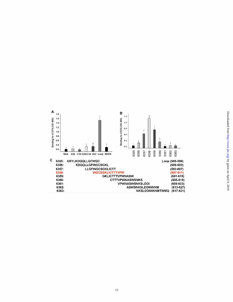
The gp41 loop region is required to interact with CD74 extracellular domain─We further characterized the interaction between rsgp41 and CD74 using several different assays. First, we confirmed the binding of rCD74 to rsgp41 by ELISA (Fig. 3A). Then, we determined the binding of rCD74 to rsgp41 in solution phase by ITC, which directly measures the heat released or absorbed during the binding process. A total of 50 µM rsgp41 dissolved in PBS was injected into the ITC cell containing 10 µM rCD74 in PBS. The binding affinity of rsgp41 to rCD74 was calculated as Ka=1.8×10⁶ M-1 (Fig. 3B). Moreover, the CD74 binding site on rspg41 was determined using different components of rsgp41, including N36, C34, N36/C34, 3NC, loop and MPER. Compared with other components, the gp41 loop strongly bound to rCD74 (Fig. 4A), indicating that gp41 interacts with CD74 mainly through its loop region. Using a series of peptides that cover the entire loop region, we further narrowed down the binding site around peptide 6358 (Fig. 4B and 4C).
4
by guest on April 9, 2019
http://ww
w.jbc.org/
Dow
nloaded from

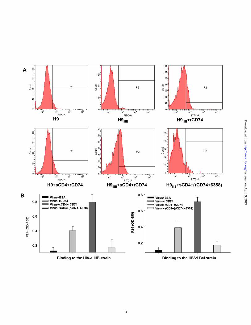
The rCD74 binds to native gp41, but the binding is blocked by the gp41 loop peptide─Since gp41 and CD74 are both membrane proteins, we reasoned that they interact on the cell surface. So we tested whether rCD74 could bind to native gp41 existing on HIV-1IIIB-infected H9 cells. As shown in Figure 5A, rCD74 could directly bind to HIV-1IIIB-infected H9 cells compared with uninfected H9 cells. Treating cells with soluble CD4 (sCD4) enhanced the binding to H9IIIB cells but not to H9 cells, since sCD4 induces the shedding of gp120, resulting in an increased exposure of gp41. Conversely, adding gp41 peptide 6358 (aa597-611) strongly inhibited the binding of rCD74 to H9IIIB cells. These results suggested that rCD74 could bind to native gp41 on cell surface. We next tested whether rCD74 could bind to HIV-1 virions. To this end, rCD74 was conjugated with Ni+-NTA and incubated with the virus on ice for 2 h. The unbound virus was washed and removed by centrifuge, and the sediment was detected using HIV-1 p24 test kit. As shown in Figure 4B, both HIV-1 strains IIIB and Bal were precipitated by rCD74. Treating virus with sCD4 significantly enhanced the binding, whereas gp41 peptide 6358 strongly inhibited it.
The rsgp41 and gp41 peptide 6358 activate the CD74-mediated ERK/MAPK pathway─CD74 mediates several important signaling pathways, including the ERK/MAPK pathway, which can be initiated by MIF. Since peptide 6358 and rsgp41 could bind CD74, we tested whether they could also activate the ERK/MAPK pathway. PBMCs were stimulated with indicated concentrations of peptide 6358 and rsgp41, respectively. Then, using specific monoclonal antibodies, total ERK and the phosphor-ERK molecules were detected by Western blotting. We found that both peptide 6358 and rsgp41 induced the phosphorylation of ERK1/2 in a dose-dependent manner (Fig. 6A), and this effect could be inhibited by an anti-CD74 mAb (LN2) and by rCD74 (Fig. 6B). These results suggest that HIV-1 gp41 is able to activate the ERK/MAPK pathway by binding to CD74.
The rsgp41 and gp41 peptide 6358 enhance HIV-1 infection, but enhancement could be suppressed by rCD74─It has been reported that activation of the ERK/MAPK pathway could enhance HIV-1 infection (27,28). Since peptide
6358 and rsgp41 activated the ERK/MAPK pathway, we tested their effect on HIV-1 infection using TZM-bl reporter cells. As shown in Fig. 7, peptide 6358 significantly enhanced HIV-1 infection in a dose-dependent manner (Fig. 7A), whereas the upstream neighboring peptide 6355 did not enhance HIV-1 infection (Fig. 7B). The peptide 6358-mediated enhancing effect was inhibited by rCD74 or PD98059 (a MEK1 inhibitor which blocks the ERK/MAPK pathway) (Fig. 7C and 7E). Similarly, rsgp41 enhanced HIV-1 infection in a dose-dependent manner, and this effect was blocked by rCD74 and PD98059 (Fig. 7D and 7E). These results suggested that gp41 could enhance HIV-1 infection by activating the ERK/MAPK pathway.
DISCUSSION
It has been reported that rsgp41 could directly bind to human lymph cell lines through interactions with several proteins on those cells, and such binding could be inhibited by the gp41-derived peptides named Env60 (aa591-605) and Env66 (aa651-665). At least five kinds of proteins were separated from those cells using rsgp41. However, their sequence remains elusive (29-32). Similarly, Henderson et al. have found that the immunosuppressive peptide (ISP) of gp41 (aa583-599) (33,34), when conjugated to BSA, could bind to CD4+ T cells and inhibit T cell proliferation; they have also separated two gp41-binding proteins with uncertain sequences (35,36). In the present study, using yeast two-hybrid screening, we identified CD74 as a gp41-binding protein. Biological analysis demonstrated that HIV-1 gp41 could bind to the extracellular domain of CD74 and that this binding is mainly mediated by the gp41 loop. Using a series of overlapping peptides covering the loop region, we further narrowed down the binding sites to the sequence around peptide 6358 (aa597-611). These studies indicate that the gp41 loop region is an important component in controlling the mediation of viral-host interactions and regulating cell functions.
CD74 is a versatile protein that performs multiple functions. It was initially recognized as the MHC II invariant chain, which binds to class II MHC molecules in endoplasmic reticulum and regulates the targeting and maturing of MHC II
5
by guest on April 9, 2019
http://ww
w.jbc.org/
Dow
nloaded from

complex (37). Subsequent studies have revealed that CD74 is also independently expressed on the cell surface (38) and serves as a receptor for MIF (19,39). Moreover, in gastric epithelial cells, CD74 is utilized by Helicobacter pylori as a receptor to facilitate virus adhesion and infection (40). Therefore, we asked what functional significance might be extrapolated from the binding of HIV-1 gp41 to CD74. Considering that surface-expressed CD74 serves as a signal receptor for MIF, we explored the effect of gp41 on CD74-mediated signal transduction. Using PBMCs as target cells, we observed that both rsgp41 and the peptide 6358 induced the phosphorylation of ERK1/2, which marked the activation of the CD74-mediated ERK/MAPK pathway.
The ERK/MAPK pathway is highly conserved and can be mediated by different kinds of stimuli and receptors (41). Besides MIF and CD74, stimuli, such as growth factors, cytokines, mitogens, as well as receptors, including receptor tyrosine kinase and G protein-coupled receptors, can also mediate the ERK/MAPK pathway (42,43). Earlier studies on RANTES (CCL5, the CCR5 ligand) have revealed that RANTES at low concentrations mainly displays HIV-1 inhibitory activities as a consequence of its strong binding to CCR5 which inhibits the membrane fusion process. However, at higher concentrations, RANTES efficiently activates the cellular ERK/MAPK pathway, resulting in an enhanced HIV-1 infection (43-45). Thus, two mechanisms have been proposed to explain the enhancement of HIV-1 infection by activation of the ERK/MAPK pathway. On the one hand, ERK/MAPK could phosphorylate various downstream substrates, including transcription factors and other protein kinases (i.e., Ets-1, c-Jun, c-Myc and NF-кB), which regulate host gene expression and lead to cell proliferation, thus priming cells for HIV-1 replication (25,46). On the other hand, the phosphor-ERK could also phosphorylate viral proteins (i.e., gag MA, Vif, Rev, Tat, Nef) and promote the nuclear translocation and integration process to enhance viral infection (28,47,48). In comparison, we observed in this study that rsgp41 and gp41 peptide 6358 activated the ERK/MAPK
pathway and enhanced HIV-1 infection in vitro. This effect was very similar to that of RANTES, except the receptor of rsgp41 and peptide 6358 was CD74, not CCR5. However, unlike RANTES, or other exogenous stimuli, rsgp41 and the peptide 6358 were both derived from gp41, indicating that HIV-1 might be able to regulate the important ERK/MAPK pathway, as well as its infectivity through gp41 or its fragments.
In an earlier report, using genetic suppressor element (GSE) screening technology, Dunn et al. identified CD74 and some other membrane proteins as essential for HIV-1 early infection (49). They found that silencing the CD74 gene with specific siRNA strongly inhibited HIV-1 infection. This study supports our results, and when taken together, we reasoned that that CD74 is involved in HIV-1 early infection through its interaction with the gp41 loop region, but that inhibition of HIV-1 infection by silencing CD74 probably results from blocking the CD74-mediated ERK/MAPK pathway.
During HIV-1 infection, there are at least three possible chances for gp41 to bind cellular CD74. (i) During the virus entry process, gp41 is sufficiently exposed to enable mediation of the viral-cell membrane fusion, thus allowing potential interaction with CD74 on the cell membrane. (ii) Under physiological conditions, most viral particles are defective with gp120 shed and gp41 exposed (50). As such, the exposed gp41 on defective virions might directly bind to cell-surface CD74. (iii) The degraded gp41 fragments containing loop sequence (like the peptide 6358) may also bind CD74 to activate the ERK/MAPK pathway and enhance HIV-1 infection. Taken together, we propose a model, as shown in Figure 8. In this model, gp41, as well as its fragments, interact with cell-surface CD74 and activate the CD74-mediated ERK/MAPK pathway to enhance HIV-1 infection.
In conclusion, we identified and confirmed the interaction of HIV-1 gp41 and CD74, and we demonstrated that gp41 could activate the CD74-mediated ERK/MAPK pathway and enhance HIV-1 infection.
6
by guest on April 9, 2019
http://ww
w.jbc.org/
Dow
nloaded from
REFERENCES
1. Wyatt, R., and Sodroski, J. (1998) Science 280(5371), 1884-1888 2. Swanson, C., and Malim, M. (2008) Cell 133(4), 742, 742 e1 3. Weissenhorn, W., Dessen, A., Harrison, S., Skehel, J., and Wiley, D. (1997) Nature 387(6631),
426-430 4. Chan, D., Fass, D., Berger, J., and Kim, P. (1997) Cell 89(2), 263-273 5. Chan, D., and Kim, P. (1998) Cell 93(5), 681-684 6. Benkirane, M., Jeang, K. T., and Devaux, C. (1994) Embo. J. 13(23), 5559-5569 7. Weissman, D., Rabin, R. L., Arthos, J., Rubbert, A., Dybul, M., Swofford, R., Venkatesan, S.,
Farber, J. M., and Fauci, A. S. (1997) Nature 389(6654), 981-985 8. Briant, L., Robert-Hebmann, V., Acquaviva, C., Pelchen-Matthews, A., Marsh, M., and Devaux,
C. (1998) J. Virol 72(7), 6207-6214 9. Cicala, C., Arthos, J., Selig, S. M., Dennis, G., Jr., Hosack, D. A., Van Ryk, D., Spangler, M. L.,
Steenbeke, T. D., Khazanie, P., Gupta, N., Yang, J., Daucher, M., Lempicki, R. A., and Fauci, A. S. (2002) Proc. Natl. Acad. Sci. U S A 99(14), 9380-9385
10. Grainger, D. J., and Lever, A. M. (2005) Retrovirology 2, 23 11. Yoder, A., Yu, D., Dong, L., Iyer, S. R., Xu, X., Kelly, J., Liu, J., Wang, W., Vorster, P. J.,
Agulto, L., Stephany, D. A., Cooper, J. N., Marsh, J. W., and Wu, Y. (2008) Cell 134(5), 782-792 12. Chen, Y. H., Bock, G., Vornhagen, R., Steindl, F., Katinger, H., and Dierich, M. P. (1994) Int.
Arch. Allergy. Immunol. 104(3), 227-231 13. Chen, Y. H., Christiansen, A., and Dierich, M. P. (1995) Immunol. Lett. 48(1), 39-44 14. Cohen, T., Cohen, S., Antonovsky, N., Cohen, I., and Shai, Y. (2010) Plos Pathog. 6(9),
e1001085 15. Ueda, H., Howard, O. M., Grimm, M. C., Su, S. B., Gong, W., Evans, G., Ruscetti, F. W.,
Oppenheim, J. J., and Wang, J. M. (1998) J. Clin. Invest. 102(4), 804-812 16. Hovanessian, A. G., Briand, J. P., Said, E. A., Svab, J., Ferris, S., Dali, H., Muller, S., Desgranges,
C., and Krust, B. (2004) Immunity 21(5), 617-627 17. Huang, J. H., Lu, L., Lu, H., Chen, X., Jiang, S., and Chen, Y. H. (2007) J. Biol. Chem. 282(9),
6143-6152 18. Huang, J. H., Qi, Z., Wu, F., Kotula, L., Jiang, S., and Chen, Y. H. (2008) J. Biol. Chem. 283(22),
14994-15002 19. Leng, L., Metz, C. N., Fang, Y., Xu, J., Donnelly, S., Baugh, J., Delohery, T., Chen, Y., Mitchell,
R. A., and Bucala, R. (2003) J. Exp. Med. 197(11), 1467-1476 20. Bernhagen, J., Calandra, T., and Bucala, R. (1998) J. Mol. Med. 76(3-4), 151-161 21. Popovic, M., Read-Connole, E., and Gallo, R. C. (1984) Lancet. 2(8417-8418), 1472-1473 22. Popovic, M., Sarngadharan, M. G., Read, E., and Gallo, R. C. (1984) Science 224(4648), 497-500 23. Ratner, L., Haseltine, W., Patarca, R., Livak, K. J., Starcich, B., Josephs, S. F., Doran, E. R.,
Rafalski, J. A., Whitehorn, E. A., Baumeister, K., and et al. (1985) Nature 313(6000), 277-284 24. Strubin, M., Long, E. O., and Mach, B. (1986) Cell 47(4), 619-625 25. Mitchell, R. A., Metz, C. N., Peng, T., and Bucala, R. (1999) J. Biol. Chem. 274(25), 18100-
18106 26. Jiang, S., Lu, H., Liu, S., Zhao, Q., He, Y., and Debnath, A. K. (2004) Antimicrob. Agents.
Chemother. 48(11), 4349-4359 27. Jacque, J. M., Mann, A., Enslen, H., Sharova, N., Brichacek, B., Davis, R. J., and Stevenson, M.
(1998) Embo. J. 17(9), 2607-2618 28. Yang, X., and Gabuzda, D. (1999) J. Virol. 73(4), 3460-3466 29. Chen, Y. H., Ebenbichler, C., Vornhagen, R., Schulz, T. F., Steindl, F., Bock, G., Katinger, H.,
and Dierich, M. P. (1992) Aids 6(6), 533-539
7
by guest on April 9, 2019
http://ww
w.jbc.org/
Dow
nloaded from

30. Chen, Y. H., Bock, G., Vornhagen, R., Steindl, F., Katinger, H., and Dierich, M. P. (1993) Mol. Immunol. 30(13), 1159-1163
31. Chen, Y. H., Bock, G., Vornhagen, R., Steindl, F., Katinger, H., and Dierich, M. P. (1993) Immunol. 188(4-5), 323-329
32. Chen, Y. H., Bock, G., Vornhagen, R., Steindl, F., Katinger, H., and Dierich, M. P. (1993) Immunol. Lett. 37(1), 41-45
33. Ruegg, C. L., Monell, C. R., and Strand, M. (1989) J. Virol. 63(8), 3257-3260 34. Denner, J., Norley, S., and Kurth, R. (1994) Aids 8(8), 1063-1072 35. Qureshi, N. M., Coy, D. H., Garry, R. F., and Henderson, L. A. (1990) Aids 4(6), 553-558 36. Henderson, L. A., and Qureshi, M. N. (1993) J. Biol. Chem. 268(20), 15291-15297 37. Cresswell, P. (1994) Annu. Rev. Immunol. 12, 259-293 38. Henne, C., Schwenk, F., Koch, N., and Moller, P. (1995) Immunology 84(2), 177-182 39. Leng, L., and Bucala, R. (2006) Cell. Res. 16(2), 162-168 40. Beswick, E. J., Bland, D. A., Suarez, G., Barrera, C. A., Fan, X., and Reyes, V. E. (2005) Infect.
Immun. 73(5), 2736-2743 41. Robinson, M. J., and Cobb, M. H. (1997) Curr. Opin. Cell. Biol. 9(2), 180-186 42. Johnson, G. L., and Lapadat, R. (2002) Science 298(5600), 1911-1912 43. Chang, T. L., Gordon, C. J., Roscic-Mrkic, B., Power, C., Proudfoot, A. E., Moore, J. P., and
Trkola, A. (2002) J. Virol. 76(5), 2245-2254 44. Roscic-Mrkic, B., Fischer, M., Leemann, C., Manrique, A., Gordon, C. J., Moore, J. P., Proudfoot,
A. E., and Trkola, A. (2003) Blood 102(4), 1169-1177 45. Trkola, A., Gordon, C., Matthews, J., Maxwell, E., Ketas, T., Czaplewski, L., Proudfoot, A. E.,
and Moore, J. P. (1999) J. Virol. 73(8), 6370-6379 46. McCubrey, J. A., Steelman, L. S., Chappell, W. H., Abrams, S. L., Wong, E. W., Chang, F.,
Lehmann, B., Terrian, D. M., Milella, M., Tafuri, A., Stivala, F., Libra, M., Basecke, J., Evangelisti, C., Martelli, A. M., and Franklin, R. A. (2007) Biochim. Biophys. Acta. 1773(8), 1263-1284
47. Yang, X., and Gabuzda, D. (1998) J. Biol. Chem. 273(45), 29879-29887 48. Marozsan, A. J., Torre, V. S., Johnson, M., Ball, S. C., Cross, J. V., Templeton, D. J., Quinones-
Mateu, M. E., Offord, R. E., and Arts, E. J. (2001) J. Virol. 75(18), 8624-8638 49. Dunn, S. J., Khan, I. H., Chan, U. A., Scearce, R. L., Melara, C. L., Paul, A. M., Sharma, V., Bih,
F. Y., Holzmayer, T. A., Luciw, P. A., and Abo, A. (2004) Virology 321(2), 260-273 50. McMichael, A. J., and Hanke, T. (2003) Nat. Med. 9(7), 874-880 51. Ramos, J. W. (2008) Int. J. Biochem. Cell. Biol. 40(12), 2707-2719
FOOTNOTES
This work was supported by grants from the National Natural Science Foundation of China #81171548 to YHC, #81173098 to SJ and #81102476 to LL. Dr. Y.H. Chen is a Janssen Investigator.
FIGURE LEGENDS
Fig. 1. Identification of CD74 as a gp41-binding protein. (A) Schematic view of gp41 and CD74 structure. Rsgp41, recombinant soluble gp41; FP, fusion peptide; N-HR, N-terminal heptad repeat; C-HR, C-terminal heptad repeat; MPER, membrane-proximal external region; TM, transmembrane domain; CP, cytoplasm domain; IC, intracellular domain; EC, extracellular domain. M1 and M17 are two alternative translation initiations (24). (B) Specific interaction of rsgp41 and CD74 determined by yeast two-hybrid assay. The samples were tested in triplicate and data are presented as means ±standard deviation (SD).
8
by guest on April 9, 2019
http://ww
w.jbc.org/
Dow
nloaded from

The experiment was repeated twice. (C) SDS-PAGE and Western blot analysis of the purified recombinant CD74 extracellular domain. Fig. 2. Binding of gp41 to CD74 in mammalian cells. (A) Binding of gp41 to endogenous CD74 in human Raji cells. Cell lysates were incubated with BSA- or rsgp41-conjugated beads. CD74 was detected by anti-CD74 antibodies. (B) Binding of gp41 with CD74 in 293T cells. 293 cells were transfected with plasmids encoding HA-rsgp41 and Myc-CD74. Cell lysates were immunoprecipitated with anti-HA mAb followed by Western blotting with anti-CD74 antibodies. (C) Binding of gp41 to CD74 in HIV-1-infected cells. Human monocytic U937 cells were infected with HIV-1 NL4-3 strain. Cell lysates were immunoprecipitated with anti-gp41 antibodies (2F5). CD74 was detected by anti-CD74 antibodies.
Fig. 3. Interaction of rsgp41 and rCD74. (A) Rsgp41 binds to rCD74, as verified by ELISA. Data are expressed as means ± SD of triplicate. The experiment was repeated once. (B) Rsgp41 binds to rCD74, as confirmed by ITC. The titration traces determined when 50 µM rsgp41 dissolved in PBS was injected into a solution containing 10 µM rCD74 (PBS) at 37ºC (panel a). The binding affinity of rsgp41 to rCD74 was calculated using Launch NanoAnalyze software. Ka=1.8×10⁶ M-1, N=0.89, and H=1365.6 kJ/mol (panel b). Fig. 4. Narrowing down the CD74-binding site on gp41. (A) rCD74 binds to the gp41 loop. (B) rCD74 binds to peptides derived from the gp41 loop. The figure is representative of two independent experiments carried out in triplicate (mean ± SD). (C) Amino acid sequences of a series of overlapping peptides which cover the gp41 loop region. Fig. 5. Binding of rCD74 to gp41 on cell surface and virions. (A) rCD74 binds to gp41 expressed on HIV-1IIIB-infected H9 cells. Cells with or without sCD4 treatment were incubated with rCD74, or rCD74+6358 mixture, and detected by anti-His antibody. The uninfected H9 cells were used as control. (B) rCD74 binds HIV-1 IIIB and Bal. Viral particles were pulled down by Ni+-NTA-conjugated rCD74 and detected by ELISA against p24. Samples were tested in triplicate (mean ± SD), and the data show one representative of three independent experiments. Fig. 6. Activation of ERK/MAPK pathway by peptide 6358 and rsgp41 binding to CD74. (A) Peptide 6358 and rsgp41 induce ERK-1/2 (p44/p42) phosphorylation. (B) Inhibition of the peptide 6358- or rsgp41-induced phosphorylation of ERK1/2 by rCD74 and anti-CD74 mAb. An irrelevant mAb (8C6) was used as control. Data shown are representative of three independent experiments. Fig. 7. Enhancement of HIV-1 infection in vitro by peptide 6358 and rsgp41. (A) Peptide 6358 enhanced HIV-1 infection. The numbers above the columns indicate the enhancing folds. (B) Peptide 6355 did not enhance HIV-1 infection. (C) rCD74 inhibited the enhancing effect of peptide 6358. (D) rsgp41 enhanced HIV-1 infection and rCD74 inhibited rsgp41-medaited enhancing effect. (E) PD98059 (20 μM) blocked enhancement of HIV-1 infection mediated by PMA (50 μM), peptide 6358 (2 μg/ml), and rsgp41 (20 μg/ml), respectively. The samples were tested in triplicate and the experiments were repeated twice. Data are presented as means ± SD of a representative experiment. Fig. 8. Model of enhancement of HIV-1 infection as a result of HIV-1 gp41 binding to CD74 and subsequent activation of the ERK/MAPK signal pathway. (a) The gp41, which is exposed during membrane fusion, binds CD74 and activates the ERK/MAPK pathway. (b) The gp41 existing on defective virions directly binds CD74 and activates the ERK/MAPK pathway. (c) The degraded gp41 fragments containing loop sequence (like peptide 6358) bind CD74 to activate the ERK/MAPK pathway and enhance HIV-1 infection. The ERK/MAPK downstream signaling pathway has been described elsewhere(46,51).
9
by guest on April 9, 2019
http://ww
w.jbc.org/
Dow
nloaded from

Chang Zhou, Lu Lu, Suiyi Tan, Shibo Jiang and Ying-Hua ChenPathway to Enhance Viral Infection
HIV-1 Gp41 Ectodomain Induces Activation of the CD74-Mediated ERK/MAPK
published online October 28, 2011J. Biol. Chem.
10.1074/jbc.M111.267393Access the most updated version of this article at doi:
Alerts:
When a correction for this article is posted•
When this article is cited•
to choose from all of JBC's e-mail alertsClick here
by guest on April 9, 2019
http://ww
w.jbc.org/
Dow
nloaded from