
A gp41 MPER-specific Llama VHH Requires a Hydrophobic CDR3 for
RESEARCH Open Access
The membrane-spanning domain of gp41 plays acritical role in intracellular trafficking of the HIVenvelope proteinKosuke Miyauchi1, A Rachael Curran2, Yufei Long3, Naoyuki Kondo3,5,6, Aikichi Iwamoto4, Donald M Engelman2,Zene Matsuda3,5*
Abstract
Background: The sequences of membrane-spanning domains (MSDs) on the gp41 subunit are highly conservedamong many isolates of HIV-1. The GXXXG motif, a potential helix-helix interaction motif, and an arginine residue(rare in hydrophobic MSDs) are especially well conserved. These two conserved elements are expected to locateon the opposite sides of the MSD, if the MSD takes a a-helical secondary structure. A scanning alanine-insertionmutagenesis was performed to elucidate the structure-function relationship of gp41 MSD.
Results: A circular dichroism analysis of a synthetic gp41 MSD peptide determined that the secondary structure ofthe gp41 MSD was a-helical. We then performed a scanning alanine-insertion mutagenesis of the entire gp41MSD, progressively shifting the relative positions of MSD segments around the helix axis. Altering the position ofGly694, the last residue of the GXXXG motif, relative to Arg696 (the number indicates the position of the aminoacid residues in HXB2 Env) around the axis resulted in defective fusion. These mutants showed impairedprocessing of the gp160 precursor into gp120 and gp41. Furthermore, these Env mutants manifested inefficientintracellular transport in the endoplasmic reticulum and Golgi regions. Indeed, a transplantation of the gp41 MSDportion into the transmembrane domain of another membrane protein, Tac, altered its intracellular distribution.Our data suggest that the intact MSD a-helix is critical in the intracellular trafficking of HIV-1 Env.
Conclusions: The relative position between the highly conserved GXXXG motif and an arginine residue around thegp41 MSD a-helix is critical for intracellular trafficking of HIV-1 Env. The gp41 MSD region not only modulatesmembrane fusion but also controls biosynthesis of HIV-1 Env.
BackgroundHIV-1, the retrovirus responsible for the current world-wide AIDS pandemic, is an enveloped virus. The envel-ope protein (Env) of HIV-1 is essential for determininghost range and for inducing the membrane fusion thatallows the virus to enter the host cell. The former andlatter functions are mediated by the SU (gp120) and theTM (gp41) subunits of the envelope protein, respectively[1-3]. The SU and TM are generated from a precursor(gp160) by cellular proteases that recognize a basicamino acid sequence between gp120 and gp41 [4-6].
This proteolytic processing is essential to generatefusion-competent HIV-1 Env and is believed to takeplace in an early Golgi region [7,8].HIV-1 Env is anchored across lipid bilayers via its
highly conserved membrane-spanning domain (MSD)[9]. Although the possibility of a transient alteration ofthe membrane topology exists [10,11], HIV-1 Env iswidely believed to be a type I membrane protein with asingle a-helical MSD in the steady state [12]. Two dif-ferent models exist within the single MSD model ofHIV-1 Env. In an initial model, the MSD is supposed tobe 23 amino acid residues long, ranging from Lys683 toVal704 in the HXB2 sequence, and has a highly con-served hydrophilic arginine residue in the midst of itshydrophobic amino acid sequence [13]. In an alternativemodel, MSD is shorter; and the arginine residue in the
* Correspondence: [email protected] Joint Laboratory of Structural Virology and Immunology,Institute of Biophysics, Chinese Academy of Sciences, 15 Datun Road, Beijing,100101 PR ChinaFull list of author information is available at the end of the article
Miyauchi et al. Retrovirology 2010, 7:95http://www.retrovirology.com/content/7/1/95
© 2010 Miyauchi et al; licensee BioMed Central Ltd. This is an Open Access article distributed under the terms of the CreativeCommons Attribution License (http://creativecommons.org/licenses/by/2.0), which permits unrestricted use, distribution, andreproduction in any medium, provided the original work is properly cited.
lipid bilayer is expected to interact with the polar headof the lipid molecule [14,15].The primary structure of the MSD of HIV-1 Env also
has a GXXXG motif, a motif often found at the helix-helix interface of transmembrane a-helices [16]; it existsupstream of the arginine residue. If an ordinary a-helixstructure is assumed for the MSD, the GXXXG motifand arginine residue are positioned on opposite sides ofthe gp41 MSD a-helix.In vitro studies of the gp41 MSD showed a high toler-
ance for mutations. For example, the above mentionedconserved arginine residue [17] and the GXXXG motifcan accommodate point mutations [18]. Even severalheterologous MSDs can replace the entire gp41 MSDwithout deteriorating effects [17,19]. These findings ledto the notion that the specific amino acid sequence inthe gp41 MSD has no significant biological role withinthe limits of the assays used. This is a curious notionsince the sequence is quite conserved in nature, despitethe virus being subject to very strong sequence diversifi-cation from errors in reverse transcription.In fact, other studies have suggested that the specific
sequence of the gp41 MSD plays a role in the functionof gp41 [20,21]. We have shown that replacing the gp41MSD with MSDs derived from glycophorin A orVSV-G, each containing the GXXXG motif, severelydecreases the fusion activity of HIV-1 Env [18,22].Furthermore, simultaneous substitution of all three gly-cine residues, within the GXXXG motif with leucineresidues, also negatively affected the function of theHIV-1 Env [23]. Shang et al. recently showed the impor-tance of the GXXXG region using a unique geneticapproach [24]. These studies clearly suggested the pre-sence of important information encoded in the sequenceof MSD. However, the nature of the code is still notevident.To further elucidate the structure-function relation-
ship of the gp41 MSD, we analyzed a circulardichroism (CD) profile of the synthetic peptide corre-sponding to the MSD and obtained the profileexpected for a-helical secondary structure. Next, weused the envelope gene of HXB2 [25] to create a seriesof alanine insertion mutants of the entire predictedMSD. We found that alteration of the relationshipbetween Gly694 and Arg696 (the number indicates theposition of the amino acid residues in HXB2 Env)around the axis of the MSD a-helix resulted in fusionincompetent Env. These mutant Envs also showeddefects in proteolytic processing and intracellulartransport in the endoplasmic reticulum (ER) and Golgiregions. We further showed that the intracellulartransport of HIV-1 Env is regulated by the MSDregion, through experiments that transplanted thegp41 MSD into another membrane protein, Tac.
This transplantation led to an alteration of the intracel-lular distribution of Tac, similar to that of HIV-1 Env.
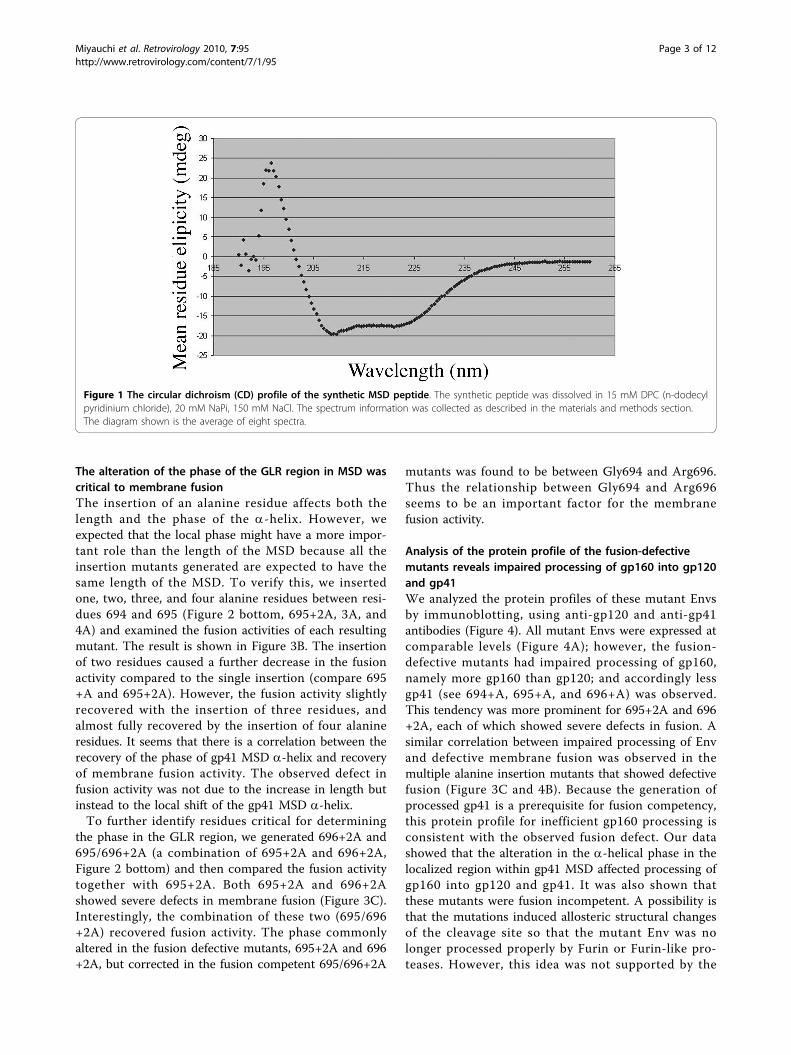
ResultsCircular dichroism analysis of the synthetic MSD peptidein lipid shows a-helical secondary structureThe primary structure of the gp41 MSD is highly con-served, and its secondary structure has been predictedto be an a-helix based on computational algorithms[26]. However, there are no physical data to support thisexpectation. We synthesized a peptide corresponding toa consensus HIV-1 clade B structure of the gp41 MSDand determined its CD spectrum in lipid bilayers. TheCD profile, shown in Figure 1, has negative maximanear 208 nm and 222 nm, indicating the presence of ana-helical structure. Although the gp41 MSD of HIV-1contains three glycine residues, thought to be helix-breaking residues in soluble proteins, the dominantstructure indicated by our CD data was an a-helix.Many glycines are found in transmembrane helices.Addition of lysine residues at both ends was necessaryto allow us to purify the extremely hydrophobic MSDpeptide. We cannot completely exclude the possibilitythat these lysine residues at the termini, especially at theC-terminus, may stabilize the a-helical structure.
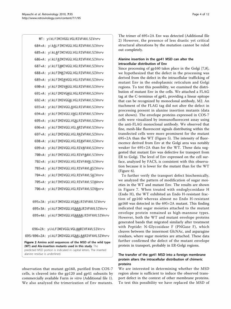
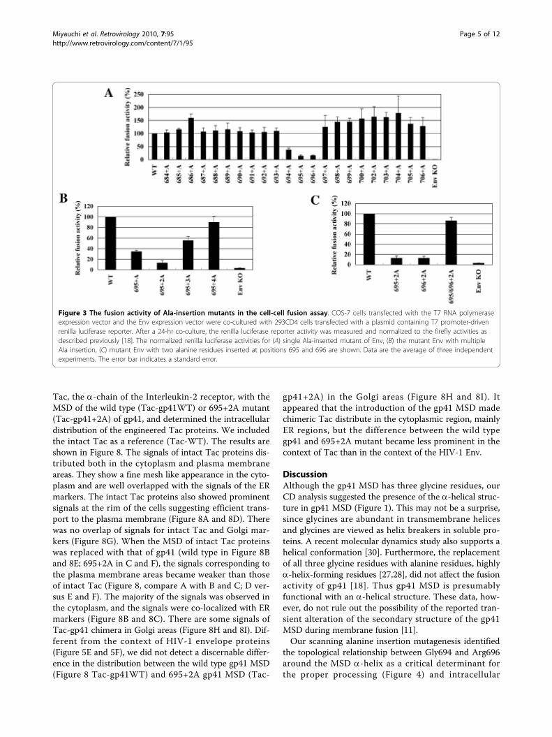
Scanning alanine-insertion mutagenesis identified theregion of gp41 MSD critical for membrane fusionTo identify the region of the gp41 MSD a-helix criticalfor its function, we generated a set of alanine-insertionmutants covering the entire predicted MSD by using theHXB2 envelope gene. The alanine residue was chosenbecause it can be well accommodated in an a-helix[27,28]. Since previous data suggest the involvement ofthe gp41 MSD in membrane fusion [18,23,24,29], mem-brane fusion activity was determined for the mutants.The primary structures of these mutants are shown inFigure 2. Nomenclature is based on the positions of theinserted alanine residues in HIV-1 Env. Therefore, 684+A mutant indicates that the inserted alanine residuecorresponds to the 684th residue of the envelope pro-tein. The mutant envelope gene was cloned into theenvelope expression vector, and the fusion activity ofeach mutant was determined by the T7 RNA polymer-ase transfer assay as described previously [18]. Theresult is shown in Figure 3A. Among the twenty-twomutants we generated, three showed a prominentdecrease in the fusion activity. These three are 694+A,695+A, and 696+A; their relative fusion activities whencompared with the wild type (WT) were 37.5%, 14.0%and 15.5%, respectively. Mutants 695+A and 696+Ashowed more severe defects than 694+A. Thus the cor-responding region from 694 to 696, the G694LR696
region, was shown to be critical for fusion activity.
Miyauchi et al. Retrovirology 2010, 7:95http://www.retrovirology.com/content/7/1/95
Page 2 of 12
The alteration of the phase of the GLR region in MSD wascritical to membrane fusionThe insertion of an alanine residue affects both thelength and the phase of the a-helix. However, weexpected that the local phase might have a more impor-tant role than the length of the MSD because all theinsertion mutants generated are expected to have thesame length of the MSD. To verify this, we insertedone, two, three, and four alanine residues between resi-dues 694 and 695 (Figure 2 bottom, 695+2A, 3A, and4A) and examined the fusion activities of each resultingmutant. The result is shown in Figure 3B. The insertionof two residues caused a further decrease in the fusionactivity compared to the single insertion (compare 695+A and 695+2A). However, the fusion activity slightlyrecovered with the insertion of three residues, andalmost fully recovered by the insertion of four alanineresidues. It seems that there is a correlation between therecovery of the phase of gp41 MSD a-helix and recoveryof membrane fusion activity. The observed defect infusion activity was not due to the increase in length butinstead to the local shift of the gp41 MSD a-helix.To further identify residues critical for determining
the phase in the GLR region, we generated 696+2A and695/696+2A (a combination of 695+2A and 696+2A,Figure 2 bottom) and then compared the fusion activitytogether with 695+2A. Both 695+2A and 696+2Ashowed severe defects in membrane fusion (Figure 3C).Interestingly, the combination of these two (695/696+2A) recovered fusion activity. The phase commonlyaltered in the fusion defective mutants, 695+2A and 696+2A, but corrected in the fusion competent 695/696+2A
mutants was found to be between Gly694 and Arg696.Thus the relationship between Gly694 and Arg696seems to be an important factor for the membranefusion activity.
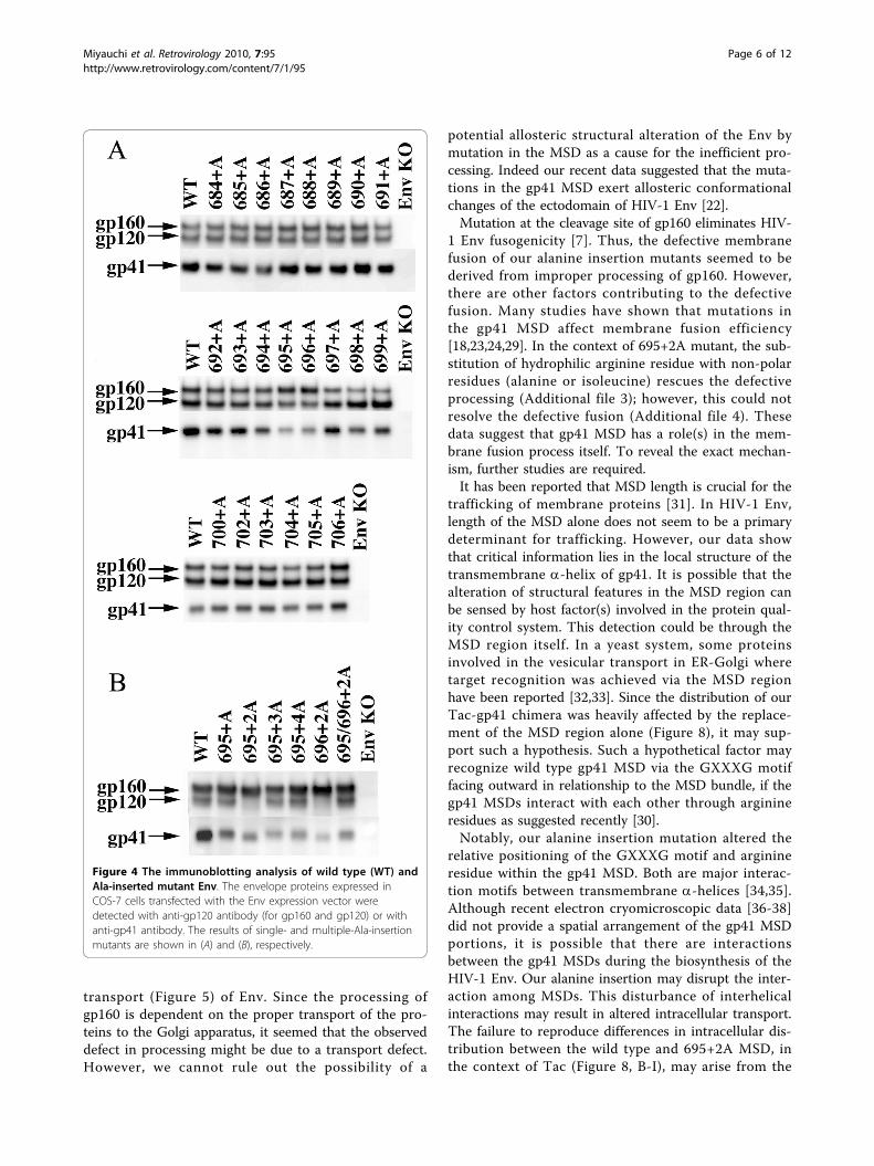
Analysis of the protein profile of the fusion-defectivemutants reveals impaired processing of gp160 into gp120and gp41We analyzed the protein profiles of these mutant Envsby immunoblotting, using anti-gp120 and anti-gp41antibodies (Figure 4). All mutant Envs were expressed atcomparable levels (Figure 4A); however, the fusion-defective mutants had impaired processing of gp160,namely more gp160 than gp120; and accordingly lessgp41 (see 694+A, 695+A, and 696+A) was observed.This tendency was more prominent for 695+2A and 696+2A, each of which showed severe defects in fusion. Asimilar correlation between impaired processing of Envand defective membrane fusion was observed in themultiple alanine insertion mutants that showed defectivefusion (Figure 3C and 4B). Because the generation ofprocessed gp41 is a prerequisite for fusion competency,this protein profile for inefficient gp160 processing isconsistent with the observed fusion defect. Our datashowed that the alteration in the a-helical phase in thelocalized region within gp41 MSD affected processing ofgp160 into gp120 and gp41. It was also shown thatthese mutants were fusion incompetent. A possibility isthat the mutations induced allosteric structural changesof the cleavage site so that the mutant Env was nolonger processed properly by Furin or Furin-like pro-teases. However, this idea was not supported by the
Figure 1 The circular dichroism (CD) profile of the synthetic MSD peptide. The synthetic peptide was dissolved in 15 mM DPC (n-dodecylpyridinium chloride), 20 mM NaPi, 150 mM NaCl. The spectrum information was collected as described in the materials and methods section.The diagram shown is the average of eight spectra.
Miyauchi et al. Retrovirology 2010, 7:95http://www.retrovirology.com/content/7/1/95
Page 3 of 12
observation that mutant gp160, purified from COS-7cells, is cleaved into the gp120 and gp41 subunits bycommercially available Furin in vitro (Additional file 1).We also analyzed the trimerization of Env mutants.
The trimer of 695+2A Env was detected (Additional file2) However, the presence of less drastic yet criticalstructural alterations by the mutation cannot be ruledout completely.
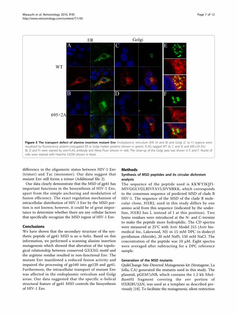
Alanine insertion in the gp41 MSD can alter theintracellular distribution of EnvSince processing of gp160 takes place in the Golgi [7,8],we hypothesized that the defect in the processing wasderived from the defect in the intracellular trafficking ofmutant Env in the endoplasmic reticulum and Golgiregions. To test this possibility, we examined the distri-bution of mutant Env in the cells. We attached a FLAGtag at the C-terminus of gp41, providing a linear epitopethat can be recognized by monoclonal antibody, M2. Anttachment of the FLAG tag did not alter the defect inprocessing present in alanine insertion mutants (datanot shown). The envelope proteins expressed in COS-7cells were visualized by immunofluorecent assay usingthe anti-FLAG monoclonal antibody. We observed thatfine, mesh-like fluorescent signals distributing within thetransfected cells were more prominent for the mutant695+2A than the WT (Figure 5). The intensity of fluor-escence derived from Env at the Golgi area was notablyweaker for 695+2A than for the WT. These data sug-gested that mutant Env was defective for transport fromER to Golgi. The level of Env expressed on the cell sur-face, analyzed by FACS, is consistent with this observa-tion because it is lower for the mutant than for the WT(Figure 6).To further verify the transport defect biochemically,
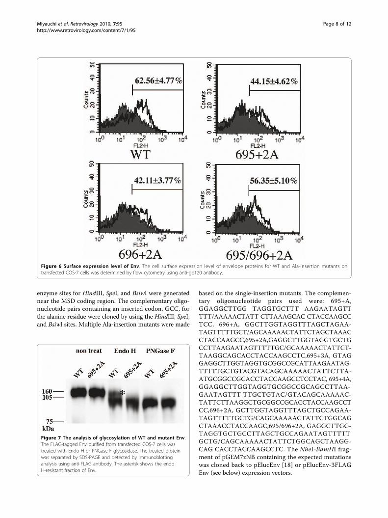
we analyzed the pattern of modification of sugar moi-eties in the WT and mutant Env. The results are shownin Figure 7. When treated with endoglycosidase H(Endo H), the WT exhibited an Endo H-resistant frac-tion of gp160 whereas almost no Endo H-resistantgp160 was detected in the 695+2A mutant. This findingindicated that sugar moieties attached to the mutantenvelope protein remained as high-mannose types.However, both the WT and mutant envelope proteinsgenerated bands that migrated similarly after treatmentwith Peptide: N-Glycosidase F (PNGase F), whichcleaves between the innermost GlcNAc, and asparagineresidues, where sugar moieties are attached. These datafurther confirmed the defect of the mutant envelopeprotein in transport, probably in ER-Golgi regions.
The transfer of the gp41 MSD into a foreign membraneprotein alters the intracellular distribution of chimericproteinsWe are interested in determining whether the MSDregion alone is sufficient to induce the observed trans-port defect in the context of other membrane proteins.To test this possibility we have replaced the MSD of
Figure 2 Amino acid sequences of the MSD of the wild type(WT) and Ala-insertion mutants used in this study. Thepredicted MSD portion is indicated in capital letters. The insertedalanine residue is underlined.
Miyauchi et al. Retrovirology 2010, 7:95http://www.retrovirology.com/content/7/1/95
Page 4 of 12
Tac, the a-chain of the Interleukin-2 receptor, with theMSD of the wild type (Tac-gp41WT) or 695+2A mutant(Tac-gp41+2A) of gp41, and determined the intracellulardistribution of the engineered Tac proteins. We includedthe intact Tac as a reference (Tac-WT). The results areshown in Figure 8. The signals of intact Tac proteins dis-tributed both in the cytoplasm and plasma membraneareas. They show a fine mesh like appearance in the cyto-plasm and are well overlapped with the signals of the ERmarkers. The intact Tac proteins also showed prominentsignals at the rim of the cells suggesting efficient trans-port to the plasma membrane (Figure 8A and 8D). Therewas no overlap of signals for intact Tac and Golgi mar-kers (Figure 8G). When the MSD of intact Tac proteinswas replaced with that of gp41 (wild type in Figure 8Band 8E; 695+2A in C and F), the signals corresponding tothe plasma membrane areas became weaker than thoseof intact Tac (Figure 8, compare A with B and C; D ver-sus E and F). The majority of the signals was observed inthe cytoplasm, and the signals were co-localized with ERmarkers (Figure 8B and 8C). There are some signals ofTac-gp41 chimera in Golgi areas (Figure 8H and 8I). Dif-ferent from the context of HIV-1 envelope proteins(Figure 5E and 5F), we did not detect a discernable differ-ence in the distribution between the wild type gp41 MSD(Figure 8 Tac-gp41WT) and 695+2A gp41 MSD (Tac-
gp41+2A) in the Golgi areas (Figure 8H and 8I). Itappeared that the introduction of the gp41 MSD madechimeric Tac distribute in the cytoplasmic region, mainlyER regions, but the difference between the wild typegp41 and 695+2A mutant became less prominent in thecontext of Tac than in the context of the HIV-1 Env.
DiscussionAlthough the gp41 MSD has three glycine residues, ourCD analysis suggested the presence of the a-helical struc-ture in gp41 MSD (Figure 1). This may not be a surprise,since glycines are abundant in transmembrane helicesand glycines are viewed as helix breakers in soluble pro-teins. A recent molecular dynamics study also supports ahelical conformation [30]. Furthermore, the replacementof all three glycine residues with alanine residues, highlya-helix-forming residues [27,28], did not affect the fusionactivity of gp41 [18]. Thus gp41 MSD is presumablyfunctional with an a-helical structure. These data, how-ever, do not rule out the possibility of the reported tran-sient alteration of the secondary structure of the gp41MSD during membrane fusion [11].Our scanning alanine insertion mutagenesis identified
the topological relationship between Gly694 and Arg696around the MSD a-helix as a critical determinant forthe proper processing (Figure 4) and intracellular
Figure 3 The fusion activity of Ala-insertion mutants in the cell-cell fusion assay. COS-7 cells transfected with the T7 RNA polymeraseexpression vector and the Env expression vector were co-cultured with 293CD4 cells transfected with a plasmid containing T7 promoter-drivenrenilla luciferase reporter. After a 24-hr co-culture, the renilla luciferase reporter activity was measured and normalized to the firefly activities asdescribed previously [18]. The normalized renilla luciferase activities for (A) single Ala-inserted mutant of Env, (B) the mutant Env with multipleAla insertion, (C) mutant Env with two alanine residues inserted at positions 695 and 696 are shown. Data are the average of three independentexperiments. The error bar indicates a standard error.
Miyauchi et al. Retrovirology 2010, 7:95http://www.retrovirology.com/content/7/1/95
Page 5 of 12
transport (Figure 5) of Env. Since the processing ofgp160 is dependent on the proper transport of the pro-teins to the Golgi apparatus, it seemed that the observeddefect in processing might be due to a transport defect.However, we cannot rule out the possibility of a
potential allosteric structural alteration of the Env bymutation in the MSD as a cause for the inefficient pro-cessing. Indeed our recent data suggested that the muta-tions in the gp41 MSD exert allosteric conformationalchanges of the ectodomain of HIV-1 Env [22].Mutation at the cleavage site of gp160 eliminates HIV-
1 Env fusogenicity [7]. Thus, the defective membranefusion of our alanine insertion mutants seemed to bederived from improper processing of gp160. However,there are other factors contributing to the defectivefusion. Many studies have shown that mutations inthe gp41 MSD affect membrane fusion efficiency[18,23,24,29]. In the context of 695+2A mutant, the sub-stitution of hydrophilic arginine residue with non-polarresidues (alanine or isoleucine) rescues the defectiveprocessing (Additional file 3); however, this could notresolve the defective fusion (Additional file 4). Thesedata suggest that gp41 MSD has a role(s) in the mem-brane fusion process itself. To reveal the exact mechan-ism, further studies are required.It has been reported that MSD length is crucial for the
trafficking of membrane proteins [31]. In HIV-1 Env,length of the MSD alone does not seem to be a primarydeterminant for trafficking. However, our data showthat critical information lies in the local structure of thetransmembrane a-helix of gp41. It is possible that thealteration of structural features in the MSD region canbe sensed by host factor(s) involved in the protein qual-ity control system. This detection could be through theMSD region itself. In a yeast system, some proteinsinvolved in the vesicular transport in ER-Golgi wheretarget recognition was achieved via the MSD regionhave been reported [32,33]. Since the distribution of ourTac-gp41 chimera was heavily affected by the replace-ment of the MSD region alone (Figure 8), it may sup-port such a hypothesis. Such a hypothetical factor mayrecognize wild type gp41 MSD via the GXXXG motiffacing outward in relationship to the MSD bundle, if thegp41 MSDs interact with each other through arginineresidues as suggested recently [30].Notably, our alanine insertion mutation altered the
relative positioning of the GXXXG motif and arginineresidue within the gp41 MSD. Both are major interac-tion motifs between transmembrane a-helices [34,35].Although recent electron cryomicroscopic data [36-38]did not provide a spatial arrangement of the gp41 MSDportions, it is possible that there are interactionsbetween the gp41 MSDs during the biosynthesis of theHIV-1 Env. Our alanine insertion may disrupt the inter-action among MSDs. This disturbance of interhelicalinteractions may result in altered intracellular transport.The failure to reproduce differences in intracellular dis-tribution between the wild type and 695+2A MSD, inthe context of Tac (Figure 8, B-I), may arise from the
Figure 4 The immunoblotting analysis of wild type (WT) andAla-inserted mutant Env. The envelope proteins expressed inCOS-7 cells transfected with the Env expression vector weredetected with anti-gp120 antibody (for gp160 and gp120) or withanti-gp41 antibody. The results of single- and multiple-Ala-insertionmutants are shown in (A) and (B), respectively.
Miyauchi et al. Retrovirology 2010, 7:95http://www.retrovirology.com/content/7/1/95
Page 6 of 12
difference in the oligomeric status between HIV-1 Env(trimer) and Tac (monomer). Our data suggest thatmutant Env still forms a trimer (Additional file 2).Our data clearly demonstrate that the MSD of gp41 has
important functions in the biosynthesis of HIV-1 Env,apart from the simple anchoring and modulation offusion efficiency. The exact regulation mechanism ofintracellular distribution of HIV-1 Env by the MSD por-tion is not known; however, it could be of great impor-tance to determine whether there are any cellular factorsthat specifically recognize the MSD region of HIV-1 Env.
ConclusionsWe have shown that the secondary structure of the syn-thetic peptide of gp41 MSD is an a-helix. Based on thisinformation, we performed a scanning alanine insertionmutagenesis which showed that alteration of the topolo-gical relationship between conserved GXXXG motif andthe arginine residue resulted in non-functional Env. Themutant Env manifested a reduced fusion activity andimpaired the processing of gp160 into gp120 and gp41.Furthermore, the intracellular transport of mutant Envwas affected in the endoplasmic reticulum and Golgiareas. Our data suggested that the specific a-helicalstructural feature of gp41 MSD controls the biosynthesisof HIV-1 Env.
MethodsSynthesis of MSD peptides and its circular dichroismanalysisThe sequence of the peptide used is KKWYIKIFI-MIVGGLVGLRIVFAVLSIVNRKK, which correspondsto the consensus sequence of predicted MSD of clade BHIV-1. The sequence of the MSD of the clade B mole-cular clone, HXB2, used in this study differs by oneamino acid from this sequence (indicated by the under-line, HXB2 has L instead of I at this position). Twolysine residues were introduced at the N- and C-terminito make the peptide more hydrophilic. The CD spectrawere measured at 25°C with Aviv Model 215 (Aviv bio-medical Inc, Lakewood, NJ) in 15 mM DPC (n-dodecylpyridinium chloride), 20 mM NaPi, 150 mM NaCl. Theconcentration of the peptide was 10 μM. Eight spectrawere averaged after subtracting for a DPC referencesample.
Generation of the MSD mutantsQuikChange Site-Directed Mutagenesis kit (Stratagene, LaJolla, CA) generated the mutants used in this study. Theplasmid, pGEM7zNB, which contains the 1.2-kb NheI-BamHI fragment covering the env portion ofHXB2RU3ΔN, was used as a template as described pre-viously [18]. To facilitate the mutagenesis, silent restriction
Figure 5 The transport defect of alanine insertion mutant Env. Endoplasmic reticulum (ER) (A and B) and Golgi (C to F) regions werevisualized by fluorescence protein-conjugated ER or Golgi marker proteins (shown in green). FLAG tagged WT (A, C and E) and 695+2A Env(B, D and F) were stained by anti-FLAG antibody and Alexa Fluor (shown in red). The close-up of the Golgi area was shown in E and F. Nuclei ofcells were stained with Hoechst 33258 (shown in blue).
Miyauchi et al. Retrovirology 2010, 7:95http://www.retrovirology.com/content/7/1/95
Page 7 of 12
enzyme sites for HindIII, SpeI, and BsiwI were generatednear the MSD coding region. The complementary oligo-nucleotide pairs containing an inserted codon, GCC, forthe alanine residue were cloned by using the HindIII, SpeI,and BsiwI sites. Multiple Ala-insertion mutants were made
based on the single-insertion mutants. The complemen-tary oligonucleotide pairs used were: 695+A,GGAGGCTTGG TAGGTGCTTT AAGAATAGTTTTT/AAAAACTATT CTTAAAGCAC CTACCAAGCCTCC, 696+A, GGCTTGGTAGGTTTAGCTAGAA-TAGTTTTTGCT/AGCAAAAACTATTCTAGCTAAACCTACCAAGCC,695+2A,GAGGCTTGGTAGGTGCTGCCTTAAGAATAGTTTTTGC/GCAAAAACTATTCT-TAAGGCAGCACCTACCAAGCCTC,695+3A, GTAGGAGGCTTGGTAGGTGCGGCCGCATTAAGAATAG-TTTTTGCTGTACGTACAGCAAAAACTATTCTTA-ATGCGGCCGCACCTACCAAGCCTCCTAC, 695+4A,GGAGGCTTGGTAGGTGCGGCCGCAGCCTTAA-GAATAGTTT TTGCTGTAC/GTACAGCAAAAAC-TATTCTTAAGGCTGCGGCCGCACCTACCAAGCCTCC,696+2A, GCTTGGTAGGTTTAGCTGCCAGAA-TAGTTTTTGCTG/CAGCAAAAACTATTCTGGCAGCTAAACCTACCAAGC,695/696+2A, GAGGCTTGG-TAGGTGCTGCCTTAGCTGCCAGAATAGTTTTTGCTG/CAGCAAAAACTATTCTGGCAGCTAAGG-CAG CACCTACCAAGCCTC. The NheI-BamHI frag-ment of pGEM7zNB containing the expected mutationswas cloned back to pElucEnv [18] or pElucEnv-3FLAGEnv (see below) expression vectors.
Figure 6 Surface expression level of Env. The cell surface expression level of envelope proteins for WT and Ala-insertion mutants ontransfected COS-7 cells was determined by flow cytometry using anti-gp120 antibody.
Figure 7 The analysis of glycosylation of WT and mutant Env.The FLAG-tagged Env purified from transfected COS-7 cells wastreated with Endo H or PNGase F glycosidase. The treated proteinwas separated by SDS-PAGE and detected by immunoblottinganalysis using anti-FLAG antibody. The asterisk shows the endoH-resistant fraction of Env.
Miyauchi et al. Retrovirology 2010, 7:95http://www.retrovirology.com/content/7/1/95
Page 8 of 12

The synthetic codon-optimized gene corresponding tothe Tac protein, a-chain of Interleukin-2 receptor, withthe gp41 MSD was custom synthesized (GenScript, Pis-cataway, NJ). The derivatives of this construct, whoseMSD portion was replaced with those of wild type ormutant gp41 or intact Tac, were generated by mutagen-esis using PCR. These genes were cloned downstream ofthe CMV promoter to generate the Tac-derivativeexpression vectors.
Addition of the 3 × FLAG tag at the C-terminus of theEnvA 3 × FLAG tag was added to the C-terminus of gp41by inserting oligonucleotides corresponding to the 3 ×FLAG tag sequence derived from the vector p3xFLAG-CMV™-7.1 (Sigma, St. Louis, MO). Following this inser-tion, the amino acid sequence after the C-terminus of
gp41 reads as RSARDYKDHDGDYKDHDIDYKDDDDK.The expression vector of FLAG-tagged Env was calledpElucEnv-3FLAG Env.
Cells and antibodiesCOS-7 cells, 293 cells, and 293-CD4 cells [18] weregrown in Dulbecco’s modified Eagle’s medium (Sigma,St. Louis, MO) supplemented with 10% fetal bovineserum (HyClone Laboratories, Logan, UT) and penicil-lin-streptomycin (Invitrogen, Carlsbad, CA). Cells werekept under 5% CO2 in a humidified incubator. Anti-gp120 polyclonal antibody was obtained from FitzgeraldIndustries International, Inc. (Concord, MA). The hybri-doma 902 and Chessie 8 were obtained from Bruce Che-sebro and George Lewis, respectively through the AIDSResearch and Reference Reagent Program, Division ofAIDS, National Institute of Allergy and Infectious
Figure 8 Intracelluar distribution of Tac-gp41MSD chimera. The influence of MSD in transport of Tac proteines. Endoplasmic reticulum (ER)(A to C) and Golgi (D to I) regions were visualized by fluorescence protein-conjugated ER or Golgi marker proteins (shown in green). Halotagged Tac-WT (A, D and G), Tac-gp41WT (B, E and H) and Tac-gp41 695+2A Env (C, F and I) were stained by anti-Halo antibody, anti-rabbit Igand Alexa Fluor (shown in red). The close-up of the Golgi area was shown in G to I. Nuclei of cells were stained with Hoechst 33258 (shown inblue).
Miyauchi et al. Retrovirology 2010, 7:95http://www.retrovirology.com/content/7/1/95
Page 9 of 12
Diseases, National Institutes of Health, USA [39-41].Anti-FLAG M2 and BioM2 were purchased from Sigma(St Louis, MO).
Cell-cell fusion assayCell-cell fusion assays, using T7 RNA polymerase (T7RNA pol) transfer, were performed as described pre-viously [18]. Briefly, 293-CD4 cells that constitutivelyexpress CD4 were transfected with pTM3hRL harboringthe T7 promoter-driven renilla luciferase gene byFuGene 6 (Roche Applied Science, Mannheim, Ger-many), and were co-cultured with COS-7 cells that hadbeen transfected with pCMMPT7iresGFP, a T7 RNApolymerase expression vector, and pElucEnv containingHIV-1 Env and firefly luciferase genes by FuGene 6.After 12 hours of co-culture, the renilla and firefly luci-ferase activities were measured using the Dual-Glo luci-ferase assay system (Promega, Madison, WI). The fusionactivity, represented by renilla luciferase activity, wasnormalized by firefly luciferase activity to obtain trans-fection efficiency [18]. The polyclonal anti-halo antibodywas obtained from Promega (Promega, Madison, WI).
Immunoblotting analysis5 × 104 COS-7 cells were transfected with pElucEnv byFuGene 6 in a 24-well culture plate. Forty-eight hoursafter transfection, the cells were lysed with radioimmu-noprecipitation assay lysis buffer (0.05 M TrisCl, 0.15M NaCl, 1% Triton X-100, 0.1% sodium dodecyl sul-fate, and 1% sodium deoxycholate). Cell lysates wereelectrophoresed (5-20% Pantera Gel, DRC, Tokyo,Japan) and transferred to a polyvinylidene fluoridemembrane (Pall, East Hills, NY). The blot was probedwith anti-gp120 antibody (Fitzgerald, Concord, MA),with the monoclonal anti-gp41 antibody (Chessie 8), orwith anti-FLAG M2 antibody. A biotinylated anti-spe-cies-specific immunoglobulin (GE Healthcare Bio-Sciences AB, Uppsala, Sweden) was used as the sec-ondary antibody. The blot was further treated with astreptavidin-horseradish peroxidase conjugate (GEHealthcare Bio-Sciences AB) and Lumi-Lightplus
(Roche, Indianapolis, IN). Images were obtained withLAS3000 (Fujifilm, Tokyo, Japan).
Immunofluorescence assayImmunofluorescence assays were used to determinethe intracellular distribution of the envelope proteins.For this purpose, we generated a modified envelopeexpression vector called pElucEnvdeltaGFP; this is thederivative of the previously described pElucEnv [18]and it has the deletion of the EGFP portion. COS-7cells transfected with pElucEnv WT or 695+2A in thedelta GFP backbone vector and ER-DsRed2 or Golgi-YPF (Clontech) or pER-mAG1 (MBL, Nagoya, Japan)
plasmid by FuGene 6 (Roche) were treated with PBSincluding 4% of PFA for 5 min at 48 hr posttransfec-tion. Cells were permeabilized by PBS including, 0.05%of saponin and 0.2% of BSA, for 30 min and thenstained with 20 μg/ml of bio-M2 (Sigma) antibody and10 μg/ml of streptavidin conjugated Alexa fluor 488 or555 (Invitrogen). In the case of Halo-tagged proteins,polyclonal anti-Halo antibodies were used as primaryantibodies. The distributions of fluorescence in cellswere visualized using a Zeiss LSM 510 meta confocalmicroscope.
Flow cytometric analysisFlow cytometric analysis was performed as describedpreviously [18]. Briefly, COS-7 cells were transfectedwith pElucEnv by FuGene 6 on a six-well plate. Forty-eight hours aftertransfection, the cells were stained withanti-gp120 monoclonal antibody 902, biotinXX anti-mouse IgG (Invitrogen) and streptavidin-Alexa 555 inPBS including 10% FBS. Cells were fixed with 1% paraf-ormaldehyde in PBS and analyzed by FACS Calibur (BDBiosciences).
Glycosidase assayCOS-7 cells transfected with pElucEnv-3FLAG byFuGene 6 on the six-well plate were lysed with radioim-munoprecipitation assay lysis buffer including Completeprotease inhibitor (Roche). Env-3FLAG was purifiedfrom cell lysates by immunoprecipitation using M2 agar-ose (Sigma) and eluted with 3XFLAG peptide (Sigma).Purified Env-3FLAG was treated with Endo H orPNGase F (Roche). For digestion by Endo H, Env-3FLAG was boiled and digested with 0.005 unit Endo Hat 37°C for 12 hr in Endo H digestion buffer [50 mMphosphate buffer (pH 5.8), 50 mM NaCl, 0.1 M 2-mer-captoethanol (2-ME), 0.01% SDS]. Env-3FLAG wasboiled in PBS including 0.1 M 2-ME and 0.1% SDS anddigested by 1 unit PNGase F at 37°C for 12 hr inPNGase F digestion buffer (74 mM TrisCl, pH 8.0;0.74% NP-40; 0.37 M 2-ME, 0.37% SDS). Env-3FLAGtreated with glycosidase was resolved by SDS-polyacryla-mide gel electrophoresis (10% polyacrylamide gel; DRC)and detected by immunoblotting analysis using anti-FLAG M2.
In vitro furin cleavage of EnvEnv-3FLAG with the 695+2A mutation was purifiedfrom COS-7 cell lysates by immunoprecipitation asdescribed above and treated with 0.7 units of furin(Alexis, Lausen, Switzerland) at 30°C for 12 hr in furin-digestion buffer (100 mM Hepes, pH 7.5; 1 mM CaCl2;0.5% Triton X-100]). Env-3FLAG, treated with furin,was detected by immunoblotting analysis using anti-FLAG M2 as described above.
Miyauchi et al. Retrovirology 2010, 7:95http://www.retrovirology.com/content/7/1/95
Page 10 of 12

The cross linking analysis of EnvAt 48 hr postransfection, 293T cells transfected withFLAG tagged WT or 695+2A Env expression vectorswere treated with 1 mM DSS for 20 min at room tem-perature in PBS (pH 8.0). Cells were incubated with 20mM Tris-Cl for 15 min at room temperature to stop thereaction and then were lysed in buffer A (10 mMHEPES, 1.5 mM MgCl2, 10 mM KCl, 0.5 mM DTT,0.05% Igepal pH 7.9). Env proteins in cellular lysatewere detected by immunoblotting analysis using anti-FLAG antibody (see above).
Additional material
Additional file 1: Supplemental Figure 1- In vitro digestion ofmutant Env with recombinant Furin. The wild type (WT) and mutant(695+2A) Env were prepared from transfected COS-7 cells and subjectedto digestion with recombinant Furin (rFurin) as described in the Methodssection. Mock indicates the result for the cell lysates prepared from mocktransfected cells.
Additional file 2: Suplemental Figure 2- Cross linking analysis of the695+2A Env. The trimerization of gp160 was examined by chemicalcross linking. The cells transfected with Env expression vectors for wildtype (WT) and mutant (695+2A) were treated with the chemical crosslinker. The cell lysates were probed with the anti-FLAG antibody. Thesingle asterisk and the double asterisk indicate the bands for trimer andmonomer of mutant gp160, respectively. Marker: HiMark Pre-StainedHigh Molecular Weight Protein Standard (Invitrogen), Mock: mocktransfection.
Additional file 3: Suplemental Figure 3A - Immunoblotting analysisof the Arg-substitution mutants in the context of 695+2A. Thedegree of processing of gp160 was examined by immunoblotting thecell lysates prepared from COS-7 cells transfected with respective Envexpression vectors. The Arg residue in the context of 695+2A wassubstituted with the indicated amino acid residue by the site directedmutagenesis (columns under 2A). One letter abbreviation for an aminoacid residue is used. Mock: mock transfection, WT: wild type MSD.
Additional file 4: Suplemental Figure 3B - Fusion activities of Arg-substitution mutants in the context of 695+2A. The fusion activitiesof the mutant shown in additional file 3A were examined by a syncytiaformation assay in 293CD4 cells. Fusion activity of the WT and MSDmutants was expressed using a fusion index (fusion index = 2x + y,where x is the number of multinucleated cells [number of nuclei ≥ 5 infive visual fields] and y is the number of multinucleated cells [number ofnuclei < 5 in five visual fields]) as described previously [18].
List of abbreviationsMSD: membrane-spanning domain; CD: circular dichroism; ER: endoplasmicreticulum; WT: wild type.
AcknowledgementsThis study was supported by a contract grant from the Ministry ofEducation, Culture, Sports, Science and Technology of Japan for the Programof Founding Research Centers for Emerging and Reemerging InfectiousDiseases and a grant from the USNIH to DME (GM073857). We thank Dr.Kunito Yoshiike for his critical reading of the manuscript. We thank A. M.Menting, an editorial consultant, for help in the preparation of themanuscript.
Author details1Laboratory of Virology and Pathogenesis, AIDS Research Center, NationalInstitute of Infectious Diseases, 1-23-1 Toyama, Shinjuku, Tokyo, Japan.2Department of Molecular Biophysics and Biochemistry, Yale University, Box
208114, New Haven, CT 06520-8114, USA. 3China-Japan Joint Laboratory ofStructural Virology and Immunology, Institute of Biophysics, ChineseAcademy of Sciences, 15 Datun Road, Beijing, 100101 PR China. 4Division ofInfectious Diseases, Advanced Clinical Research Center, University of Tokyo,4-6-1 Shirokanedai, Minato-ku, Tokyo, Japan. 5Research Center for AsianInfectious Diseases, Institute of Medical Science, University of Tokyo, 4-6-1Shirokanedai, Minato-ku, Tokyo, Japan. 6Current Address: Department ofPediatrics, Emory University School of Medicine, 2015 uppergate Dr. Atlanta,GA 30322, USA.
Authors’ contributionsKM, ARC, YL and NK performed most of the experimental work. KM, YLand NK did the cell biological analyses of mutant Envs. ARC analysed thesynthetic peptide for its biophysical properties. AI contributed to discussion.DME and ZM conceived the study and coordinated the experiments. Allauthors read and approved the final manuscript.
Competing interestsThe authors declare that they have no competing interests.
Received: 14 July 2010 Accepted: 13 November 2010Published: 13 November 2010
References1. Weiss CD: HIV-1 gp41: mediator of fusion and target for inhibition. AIDS
Rev 2003, 5:214-221.2. Eckert DM, Kim PS: Mechanisms of viral membrane fusion and its
inhibition. Annu Rev Biochem 2001, 70:777-810.3. Freed EO, Martin MA: The role of human immunodeficiency virus type 1
envelope glycoproteins in virus infection. J Biol Chem 1995,270:23883-23886.
4. Gu M, Rappaport J, Leppla SH: Furin is important but not essential for theproteolytic maturation of gp160 of HIV-1. FEBS Lett 1995, 365:95-97.
5. Moulard M, Decroly E: Maturation of HIV envelope glycoproteinprecursors by cellular endoproteases. Biochim Biophys Acta 2000,1469:121-132.
6. Ohnishi Y, Shioda T, Nakayama K, Iwata S, Gotoh B, Hamaguchi M, Nagai Y:A furin-defective cell line is able to process correctly the gp160 ofhuman immunodeficiency virus type 1. J Virol 1994, 68:4075-4079.
7. McCune JM, Rabin LB, Feinberg MB, Lieberman M, Kosek JC,Reyes GR, Weissman IL: Endoproteolytic cleavage of gp160 isrequired for the activation of human immunodeficiency virus. Cell1988, 53:55-67.
8. Kantanen ML, Leinikki P, Kuismanen E: Endoproteolytic cleavage of HIV-1gp160 envelope precursor occurs after exit from the trans-Golginetwork (TGN). Arch Virol 1995, 140:1441-1449.
9. Gabuzda D, Olshevsky U, Bertani P, Haseltine WA, Sodroski J: Identificationof membrane anchorage domains of the HIV-1 gp160 envelopeglycoprotein precursor. J Acquir Immune Defic Syndr 1991, 4:34-40.
10. Dimmock NJ: The complex antigenicity of a small external region of theC-terminal tail of the HIV-1 gp41 envelope protein: a lesson in epitopeanalysis. Rev Med Virol 2005, 15:365-381.
11. Lu L, Zhu Y, Huang J, Chen X, Yang H, Jiang S, Chen YH: Surface exposureof the HIV-1 env cytoplasmic tail LLP2 domain during the membranefusion process: interaction with gp41 fusion core. J Biol Chem 2008,283:16723-16731.
12. Haffar OK, Dowbenko DJ, Berman PW: Topogenic analysis of the humanimmunodeficiency virus type 1 envelope glycoprotein, gp160, inmicrosomal membranes. J Cell Biol 1988, 107:1677-1687.
13. Helseth E, Olshevsky U, Gabuzda D, Ardman B, Haseltine W, Sodroski J:Changes in the transmembrane region of the human immunodeficiencyvirus type 1 gp41 envelope glycoprotein affect membrane fusion. J Virol1990, 64:6314-6318.
14. Yue L, Shang L, Hunter E: Truncation of the membrane-spanning domainof human immunodeficiency virus type 1 envelope glycoprotein defineselements required for fusion, incorporation, and infectivity. J Virol 2009,83:11588-11598.
15. West JT, Johnston PB, Dubay SR, Hunter E: Mutations within the putativemembrane-spanning domain of the simian immunodeficiency virustransmembrane glycoprotein define the minimal requirements forfusion, incorporation, and infectivity. J Virol 2001, 75:9601-9612.
Miyauchi et al. Retrovirology 2010, 7:95http://www.retrovirology.com/content/7/1/95
Page 11 of 12

16. Senes A, Engel DE, DeGrado WF: Folding of helical membrane proteins:the role of polar, GxxxG-like and proline motifs. Curr Opin Struct Biol2004, 14:465-479.
17. Wilk T, Pfeiffer T, Bukovsky A, Moldenhauer G, Bosch V: Glycoproteinincorporation and HIV-1 infectivity despite exchange of the gp160membrane-spanning domain. Virology 1996, 218:269-274.
18. Miyauchi K, Komano J, Yokomaku Y, Sugiura W, Yamamoto N, Matsuda Z:Role of the specific amino acid sequence of the membrane-spanningdomain of human immunodeficiency virus type 1 in membrane fusion. JVirol 2005, 79:4720-4729.
19. Deml L, Kratochwil G, Osterrieder N, Knuchel R, Wolf H, Wagner R:Increased incorporation of chimeric human immunodeficiency virus type1 gp120 proteins into Pr55gag virus-like particles by an Epstein-Barrvirus gp220/350-derived transmembrane domain. Virology 1997,235:10-25.
20. Salzwedel K, Johnston PB, Roberts SJ, Dubay JW, Hunter E: Expression andcharacterization of glycophospholipid-anchored humanimmunodeficiency virus type 1 envelope glycoproteins. J Virol 1993,67:5279-5288.
21. Owens RJ, Burke C, Rose JK: Mutations in the membrane-spanningdomain of the human immunodeficiency virus envelope glycoproteinthat affect fusion activity. J Virol 1994, 68:570-574.
22. Kondo N, Miyauchi K, Meng F, Iwamoto A, Matsuda Z: Conformationalchanges of the HIV-1 envelope protein during membrane fusion wereinhibited by the replacement of its membrane-spanning domain. J BiolChem 2010, 285:14681-8.
23. Miyauchi K, Curran R, Matthews E, Komano J, Hoshino T, Engelman DM,Matsuda Z: Mutations of conserved glycine residues within themembrane-spanning domain of human immunodeficiency virus type 1gp41 can inhibit membrane fusion and incorporation of Env ontovirions. Jpn J Infect Dis 2006, 59:77-84.
24. Shang L, Yue L, Hunter E: Role of the membrane-spanning domain ofhuman immunodeficiency virus type 1 envelope glycoprotein in cell-cellfusion and virus infection. J Virol 2008, 82:5417-5428.
25. Ratner L, Fisher A, Jagodzinski LL, Liou RS, Mitsuya H, Gallo RC, Wong-Staal F: Complete nucleotide sequences of functional clones of the virusassociated with the acquired immunodeficiency syndrome, HTLV-III/LAV.Haematol Blood Transfus 1987, 31:404-406.
26. Andreassen H, Bohr H, Bohr J, Brunak S, Bugge T, Cotterill RM, Jacobsen C,Kusk P, Lautrup B, Petersen SB, et al: Analysis of the secondary structureof the human immunodeficiency virus (HIV) proteins p17, gp120, andgp41 by computer modeling based on neural network methods. J AcquirImmune Defic Syndr 1990, 3:615-622.
27. Pace CN, Scholtz JM: A helix propensity scale based on experimentalstudies of peptides and proteins. Biophys J 1998, 75:422-427.
28. Chakrabartty A, Schellman JA, Baldwin RL: Large differences in the helixpropensities of alanine and glycine. Nature 1991, 351:586-588.
29. Welman M, Lemay G, Cohen EA: Role of envelope processing and gp41membrane spanning domain in the formation of humanimmunodeficiency virus type 1 (HIV-1) fusion-competent envelopeglycoprotein complex. Virus Res 2007, 124:103-112.
30. Kim JH, Hartley TL, Curran AR, Engelman DM: Molecular dynamics studiesof the transmembrane domain of gp41 from HIV-1. Biochim Biophys Acta2009, 1788:1804-1812.
31. Ronchi P, Colombo S, Francolini M, Borgese N: Transmembrane domain-dependent partitioning of membrane proteins within the endoplasmicreticulum. J Cell Biol 2008, 181:105-118.
32. Sato K, Sato M, Nakano A: Rer1p, a retrieval receptor for ER membraneproteins, recognizes transmembrane domains in multiple modes. MolBiol Cell 2003, 14:3605-3616.
33. Reggiori F, Black MW, Pelham HR: Polar transmembrane domains targetproteins to the interior of the yeast vacuole. Mol Biol Cell 2000,11:3737-3749.
34. Curran AR, Engelman DM: Sequence motifs, polar interactions andconformational changes in helical membrane proteins. Curr Opin StructBiol 2003, 13:412-417.
35. Cosson P, Lankford SP, Bonifacino JS, Klausner RD: Membrane proteinassociation by potential intramembrane charge pairs. Nature 1991,351:414-416.
36. Zhu P, Liu J, Bess J, Chertova E, Lifson JD, Grise H, Ofek GA, Taylor KA,Roux KH: Distribution and three-dimensional structure of AIDS virusenvelope spikes. Nature 2006, 441:847-852.
37. Liu J, Bartesaghi A, Borgnia MJ, Sapiro G, Subramaniam S: Moleculararchitecture of native HIV-1 gp120 trimers. Nature 2008, 455:109-113.
38. Zhu P, Winkler H, Chertova E, Taylor KA, Roux KH: Cryoelectrontomography of HIV-1 envelope spikes: further evidence for tripod-likelegs. PLoS Pathog 2008, 4:e1000203.
39. Chesebro B, Wehrly K: Development of a sensitive quantitative focalassay for human immunodeficiency virus infectivity. J Virol 1988,62:3779-3788.
40. Pincus SH, Wehrly K, Chesebro B: Treatment of HIV tissue culture infectionwith monoclonal antibody-ricin A chain conjugates. J Immunol 1989,142:3070-3075.
41. Abacioglu YH, Fouts TR, Laman JD, Claassen E, Pincus SH, Moore JP,Roby CA, Kamin-Lewis R, Lewis GK: Epitope mapping and topology ofbaculovirus-expressed HIV-1 gp160 determined with a panel of murinemonoclonal antibodies. AIDS Res Hum Retroviruses 1994, 10:371-381.
doi:10.1186/1742-4690-7-95Cite this article as: Miyauchi et al.: The membrane-spanning domain ofgp41 plays a critical role in intracellular trafficking of the HIV envelopeprotein. Retrovirology 2010 7:95.
Submit your next manuscript to BioMed Centraland take full advantage of:
• Convenient online submission
• Thorough peer review
• No space constraints or color figure charges
• Immediate publication on acceptance
• Inclusion in PubMed, CAS, Scopus and Google Scholar
• Research which is freely available for redistribution
Submit your manuscript at www.biomedcentral.com/submit
Miyauchi et al. Retrovirology 2010, 7:95http://www.retrovirology.com/content/7/1/95
Page 12 of 12