Guidelines for the management of patients with NSTEMI ACS ...
22
Version 3 Date: February 2016 Author: Dr Babu Kunadian (in conjunction with CMSCN ACS Group & Pharmacist Forum) Review Date: February 2018 Guidelines for the management of patients with Non-ST Segment Elevation Myocardial Infarction (NSTEMI) Acute Coronary Syndrome including unstable angina and Non-Q wave Myocardial Infarction February 2016
Transcript of Guidelines for the management of patients with NSTEMI ACS ...

Version 3 Date: February 2016 Author: Dr Babu Kunadian (in conjunction with CMSCN ACS Group & Pharmacist Forum) Review Date: February 2018
Guidelines for the management of patients with Non-ST Segment Elevation Myocardial Infarction (NSTEMI) Acute Coronary Syndrome including unstable angina and Non-Q wave Myocardial Infarction February 2016

CNSCN Guidelines for the management of patients with NTEMI ACS including unstable angina and Non-Q wave MI – February 2016
2
GUIDELINES FOR THE MANAGEMENT OF PATIENTS WITH NON-ST SEGMENT ELEVATION ACUTE CORONARY SYNDROME
(NSTEACS) INCLUDING UNSTABLE ANGINA AND NON-Q WAVE MYOCARDIAL INFARCTION
1.0 INTRODUCTION 1.1 The leading symptom that initiates the diagnostic and therapeutic cascade in patients with
suspected acute coronary syndromes (ACS) is chest pain. Based on the electrocardiogram (ECG), two groups of patients should be differentiated: ∗ Acute ST segment elevation MI (STEMI) ∗ Non-ST segment elevation myocardial infarction (NSTEMI)
• The pathological correlate at the myocardial level is cardiomyocyte necrosis -NSTEMI • Less frequently, myocardial ischaemia without cell loss (unstable angina)
1.2 This paper is intended to provide management guidelines for NSTEACS (UA and NSTEMI)
which conform with NICE Clinical Guidelines, are consistent across the Cheshire & Merseyside Strategic Clinical Network area and which allow for equity and best practice within the context of resources currently available to the NHS locally.
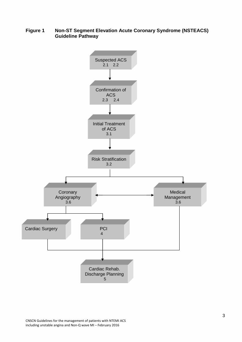
1.3 An overview of this NSTEACS guideline is shown in Fig.1 1.3.1 An integrated care pathway for NSTEACS management has been produced to complement
this guideline and to aid its implementation. It is available for downloading from the CMSCN website (www.cmscnsenate.nhs.uk)
These guidelines represent the views of the Cheshire & Merseyside Strategic Clinical Network (CMSCN), which were arrived at after consideration of the available evidence, a review of relevant NICE guidelines and the development of consensus. Health professionals are asked to take them into account when exercising their clinical judgement and are encouraged to discuss with colleagues those cases where the assessment of likely benefit from a particular intervention is equivocal. The guidelines do not override the responsibility of health professionals to make appropriate decisions in the circumstances of the individual patient in consultation with the patient and / or guardian or carer.
CNSCN Guidelines for the management of patients with NTEMI ACS including unstable angina and Non-Q wave MI – February 2016
3
Figure 1 Non-ST Segment Elevation Acute Coronary Syndrome (NSTEACS)
Guideline Pathway
Suspected ACS 2.1 2.2
Confirmation of ACS
2.3 2.4
Initial Treatment of ACS
3.1
Risk Stratification 3.2
Coronary Angiography
3.6
Medical Management
3.6
Cardiac Surgery
PCI 4
Cardiac Rehab. Discharge Planning
5

CNSCN Guidelines for the management of patients with NTEMI ACS including unstable angina and Non-Q wave MI – February 2016
4
2.0 ASSESSMENT AND DIAGNOSIS
The diagnosis is based on initial short-term ischaemic and bleeding risk stratification on a combination of clinical history, symptoms, vital signs, other physical findings, ECG and laboratory results.
2.1 Suspecting an ACS
An ACS should be suspected on clinical grounds based on the occurrence of ischaemic chest pain in a suggestive symptom pattern
2.1.1 The recognition of ischaemic chest pain depends upon a careful consideration of the
following factors (Table 1) • Chest pain features – typical pain • Patient setting – presence of known CV disease and/or risk factors • Examination findings
2.1.2 Patients with NSTEACS usually present with one or more of the following symptom patterns
o Prolonged (>20 min) anginal pain at rest; o New onset (de novo) angina (class II or III of the Canadian Cardiovascular Society
classification) o Recent destabilization of previously stable angina with at least Canadian
Cardiovascular Society Class III angina characteristics (crescendo angina); or o Post-MI angina.
2.1.3 Additional helpful diagnostic points are as follows:-
o Additional symptoms such as sweating, nausea, abdominal pain, dyspnoea and syncope may be present.
o The exacerbation of symptoms by physical exertion and their relief at rest increase the probability of myocardial ischaemia.
o The relief of symptoms after nitrates administration is not specific for anginal pain as it is reported also in other causes of acute chest pain.
o Older age, male gender, family history of CAD, diabetes, hyperlipidaemia, hypertension, renal insufficiency, previous manifestation of CAD as well as peripheral or carotid artery disease increase the likelihood of NSTE-ACS
o Conditions that may exacerbate or precipitate NSTE-ACS include anaemia, infection, inflammation, fever, and metabolic or endocrine (in particular thyroid) disorders.
o Atypical complaints are more often observed in the elderly, in women and in patients with diabetes, chronic renal disease or dementia

CNSCN Guidelines for the management of patients with NTEMI ACS including unstable angina and Non-Q wave MI – February 2016
5
Table 1 - Clinical Basis for Chest Pain Classification
Key to Abbreviations IHD - Ischaemic heart disease MI - Myocardial infarction CVA - Cerebrovascular accident (stroke) TIA - Transient cerebral ischaemic event PVD - Peripheral vascular disease M - Male F - Female IDDM - Insulin dependent diabetes mellitus (Type 1) NIDDM - Non-insulin dependent diabetes mellitus (Type 2) AF - Atrial fibrillation SVT - Supraventricular tachycardia VT - Ventricular tachycardia LVH - Left ventricular hypertrophy CCF - Congestive heart failure AR - Aortic regurgitation
BOX 1 - Chest Pain Features Typical Ischaemia All 3 of following present: • Site – Central retrosternal,
L Chest • Radiation – across chest, L
shoulder/arm, throat, jaw, L side neck
• Character – dull, tight, heavy, crushing, ache
Atypical • 1-2 of the above typical
features and • No positive features of
alternative cause Non-Cardiac • 0-1 of the above typical
features and/or • Positive features of
alternative cause e.g postural, pleuritic, post-prandial, tender
BOX 2 - Patient Setting Evidence of Cardiovascular Disease • Previous/Known IHD, Angina, MI • Previous/Known CVA, TIA • Previous/Known PVD Risk Factors • Age – M > 40 yrs; F >50 • Gender – M > F • Family IHD History – especially
premature <50 yr M, <60 F • Smoking • Dyslipidaemia • Hypertension • Diabetes Mellitus – IDDM,
NIDDM
BOX 3 - Examination Acute Coronary Syndrome • Usually normal • Arrhythmia – AF, SVT, VT,
bradycardia • LV dysfunction – S3,
pulmonary oedema Non-Ischaemic Cardiac • Pericardial rub • Valvular disease – especially
AS • Cardiomyopathy – LVH, CCF • Aortic dissection – AR,
differential arm pulses or BP (R > L), ?TIA or stroke
Non-Cardiac • Musculoskeletal - chest wall
tenderness, +ve physical manoeuvres
• Respiratory – pleural rub, pneumothorax, consolidation
• Other – pyrexia, rash, epigastric tenderness

CNSCN Guidelines for the management of patients with NTEMI ACS including unstable angina and Non-Q wave MI – February 2016
6
2.2 Initial Management of Suspected ACS
If based on the above assessment an ACS is suspected, start the following immediate management:
• Pain relief (GTN and/or an intravenous opioid – with caveat that morphine may slow
intestinal absorption of oral platelet inhibitors) • Anti-platelets – Loading dose of Aspirin to be considered • Pulse oximetry, if available. Offer oxygen:
o If oxygen saturation (SpO2) is less than 90% and in-patient in respiratory distress. • Monitor chest pain, pulse, BP, heart rhythm, pulse oximetry, 12 lead ECG (if
appropriate) • Bloods to be taken to include - Troponin, blood Glucose, HB, LFT and Creatinine (e-
GFR if possible).
Decide on Need for Admission and monitoring on the following basis:-
a) Transfer to the local acute chest pain unit (HEC, HAC, AMU depending on local designated arrangements) all patients with suspected ACS who have had chest pain within the last 72 hours.
b) Consider discharge and referral to the local Rapid Access Chest pain clinic patients
whose chest pain resolved >72 hours ago in the absence of complications.
c) Monitoring 1) Continuous rhythm monitoring is recommended until the diagnosis of NSTEMI is
established or ruled out. 2) Rhythm monitoring up to 24 h or PCI (whichever comes first) should be
considered in NSTEMI patients at low risk for cardiac arrhythmias. 3) Rhythm monitoring for >24 h should be considered in NSTEMI patients at
intermediate to high-risk for cardiac arrhythmias 4) In the absence of signs or symptoms of ongoing ischaemia, rhythm monitoring in
unstable angina may be considered in selected patients (e.g. suspicion of coronary spasm or associated symptoms suggestive of arrhythmic events)
2.3 In Hospital Assessment 2.3.1 History
• Chest Pain Features – typical, atypical, non-cardiac (Table 1) • Time Course • Patient setting – known evidence of CVD, risk factors (Table 1) • Symptoms
2.3.2 Examination
• Signs of complications e.g. pulmonary oedema, arrhythmia • Haemodynamic status • Physical examination may identify signs of non-coronary causes of chest pain e.g.
aortic dissection or non-cardiac e.g. pulmonary embolism, acute aortic syndromes, myopericarditis, aortic stenosis or extracardiac pathologies (e.g. pneumothorax, pneumonia or musculoskeletal diseases). (Table 2)
• Cardiac auscultation may reveal a systolic murmur due to ischaemic mitral regurgitation, which is associated with poor prognosis, or aortic stenosis (mimicking ACS). Rarely, a systolic murmur may indicate a mechanical complication (i.e. papillary muscle rupture or ventricular septal defect) of a subacute and possibly undetected MI.

CNSCN Guidelines for the management of patients with NTEMI ACS including unstable angina and Non-Q wave MI – February 2016
7
• According to the presentation, abdominal disorders (e.g. oesophageal spasm, oesophagitis, gastric ulcer, cholecystitis, pancreatitis) may also be considered in the differential diagnosis.
• Differences in blood pressure between the upper and lower limbs or between the arms, irregular pulse, jugular vein distension, heart murmurs, friction rub and pain reproduced by chest or abdominal palpation are findings suggestive of alternative diagnoses.
• Pallor, sweating or tremor may point towards precipitating conditions such as anaemia and thyrotoxicosis.
Table 2: Differential diagnoses of acute coronary syndromes in the setting of acute chest pain Cardiac Vascular Pulmonary Gastro-intestinal Orthopaedic Other Myo-pericarditis Cardiomyopathies
Aortic Dissection
Pulmonary embolism
Oesophagitis, reflux or spasm
Musculoskeletal disorders
Anxiety disorders
Tachy-arrhythmias Acute heart failure
Symptomatic aortic aneurysm
(Tension) -Pneumothorax
Peptic ulcer, gastritis Chest trauma Herpes
zoster
Hypertensive emergencies Stroke Bronchitis,
pneumonia Pancreatitis Cholecystitis Costochondritis Anaemia
Aortic valve stenosis Pleuritis Cervical spine
pathologies
Tako-Tsubo Cardiomyopathy
Coronary spasm Cardiac trauma 2.3.3 12 Lead ECG
• It is recommended to obtain a 12-lead ECG within 10 min after first medical contact and to have it immediately interpreted by an experienced physician.
• It is recommended to obtain an additional 12-lead ECG in case of recurrent symptoms or diagnostic uncertainty. Additional ECG leads (V3R, V4R, V7–V9) are recommended if ongoing ischaemia is suspected when standard leads are inconclusive.
The following patterns should be sought:- • Normal – this does NOT exclude ACS • ST elevation which is persistent between 2 ECGs and which does not resolve with
sublingual GTN – implement STEMI guidelines (CMCN PPCI or Thrombolysis guidelines)
• Regional ST depression, T wave inversion (especially when deep and symmetrical) or transient ST elevation (which resolves spontaneously or with GTN) suggest NSTEACS
• Pathological Q waves - consistent with an old infarction and increases suspicion of current ACS
• Left bundle branch block – if known to be old is not diagnostically useful; if new (or age unknown) implement STEMI guidelines (thrombolysis, not primary PCI)

CNSCN Guidelines for the management of patients with NTEMI ACS including unstable angina and Non-Q wave MI – February 2016
8
2.3.4 Cardiac Biomarkers
• Use Troponin T (TnT) or Troponin I (TnI) • When interpreting the TnT/I, take into account the clinical presentation, time from
onset of symptoms, the 12 lead ECG, and any cardiac imaging undertaken – See table conditions other than type 1 MI associated elevated troponin.
• Measure cardiac troponins with sensitive or high-sensitivity assays and obtain the results within 60 min.
• A rapid rule-out protocol at 0 h and 3 h is recommended if high-sensitivity cardiac troponin tests are available – See figure 2
• If the clinical presentation is compatible with myocardial ischaemia, then a dynamic elevation of cardiac troponin above the 99th percentile of healthy individuals indicates MI.
If high sensitive troponin not available then follow as below • Take an initial TnT/I on admission (providing this is at least 6 hours from the onset of
the significant chest pain); take a second TnT/I at 12 hours (or greater) if the first sample is non-diagnostic (normal, small rise, in setting of renal disease, or in suspected myocarditis) in order to detect a rise and/or fall (NB: details will vary from hospital to hospital)
• Do not routinely use cardiac enzymes such as creatine kinase or other biomarkers such as natriuretic peptides, or high sensitivity CRP
Figure 2 - 0h/3h rule-out algorithm using high-sensitive troponins
ESC (2015) Guidelines for the management of acute coronary syndromes in patients presenting without persistent ST-segment elevation (Reference 5)
CNSCN Guidelines for the management of patients with NTEMI ACS including unstable angina and Non-Q wave MI – February 2016
9
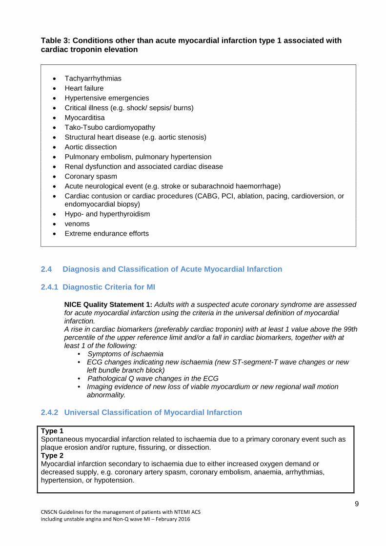
Table 3: Conditions other than acute myocardial infarction type 1 associated with cardiac troponin elevation
2.4 Diagnosis and Classification of Acute Myocardial Infarction 2.4.1 Diagnostic Criteria for MI
NICE Quality Statement 1: Adults with a suspected acute coronary syndrome are assessed for acute myocardial infarction using the criteria in the universal definition of myocardial infarction. A rise in cardiac biomarkers (preferably cardiac troponin) with at least 1 value above the 99th percentile of the upper reference limit and/or a fall in cardiac biomarkers, together with at least 1 of the following:
• Symptoms of ischaemia • ECG changes indicating new ischaemia (new ST-segment-T wave changes or new
left bundle branch block) • Pathological Q wave changes in the ECG • Imaging evidence of new loss of viable myocardium or new regional wall motion
abnormality.
2.4.2 Universal Classification of Myocardial Infarction Type 1 Spontaneous myocardial infarction related to ischaemia due to a primary coronary event such as plaque erosion and/or rupture, fissuring, or dissection. Type 2 Myocardial infarction secondary to ischaemia due to either increased oxygen demand or decreased supply, e.g. coronary artery spasm, coronary embolism, anaemia, arrhythmias, hypertension, or hypotension.
• Tachyarrhythmias • Heart failure • Hypertensive emergencies • Critical illness (e.g. shock/ sepsis/ burns) • Myocarditisa • Tako-Tsubo cardiomyopathy • Structural heart disease (e.g. aortic stenosis) • Aortic dissection • Pulmonary embolism, pulmonary hypertension • Renal dysfunction and associated cardiac disease • Coronary spasm • Acute neurological event (e.g. stroke or subarachnoid haemorrhage) • Cardiac contusion or cardiac procedures (CABG, PCI, ablation, pacing, cardioversion, or
endomyocardial biopsy) • Hypo- and hyperthyroidism • venoms • Extreme endurance efforts

CNSCN Guidelines for the management of patients with NTEMI ACS including unstable angina and Non-Q wave MI – February 2016
10
Type 3 Sudden unexpected cardiac death, including cardiac arrest, often with symptoms suggestive of myocardial ischaemia, accompanied by presumably new ST elevation, or new LBBB, or evidence of fresh thrombus in a coronary artery by angiography and/or at autopsy, but death occurring before blood samples could be obtained or at a time before the appearance of cardiac biomarkers in the blood. Type 4a Myocardial infarction associated with PCI Type 4b Myocardial infarction associated with stent thrombosis as documented by angiography or at autopsy. Type 5 Myocardial infarction associated with CABG
3.0 MANAGEMENT OF NSTEACS
3.1 Initial Treatment
3.1.1 Anti-platelet agent
Aspirin • Aspirin 300mg loading dose (if not already given in ambulance), followed by 75mg
daily indefinitely. • Consider a loading dose for all patients with cardiac sounding chest pain even if they
already take aspirin, warfarin (unless INR >3) or a new oral anticoagulant (NOAC). • If proven aspirin allergy, clopidogrel 300mg may be given as a loading dose followed
by 75mg daily maintenance. • N.B. Ticagrelor is not licensed for long term monotherapy
3.1.2 Antithrombin
Fondaparinux injection
• Consider fondaparinux 2.5mg daily subcutaneously for all patients with cardiac sounding chest pain.
• Fondaparinux does not provide therapeutic levels of anti-coagulation and will not be adequate for conditions, such as DVT/PE, mechanical prosthetic heart values or atrial fibrillation with a high risk of cardiac thromboembolism.
• Do not use in renal impairment (eGFR <20ml/min) – use enoxaparin 1mg/kg once daily as an alternative.
• Contraindicated in active clinically significant bleeding. Caution in history of known bleeding complications.
• Discontinuation of parenteral anticoagulation should be considered after PCI, unless otherwise indicated.
• In those patients not undergoing angiography, continue up to a maximum of 8 days or until hospital discharge, whichever occurs soonest.
Enoxaparin s/c
• Patients with NSTEMI requiring full anticoagulation (e.g. metallic valves, DVT/PE, high risk AF) who are not already on oral anticoagulants, should receive enoxaparin 1mg/kg BD for at least 48 hours and continue until the patient is pain free for at least 24 hours and oral anticoagulation is initiated (where appropriate).
• Patients on existing OAC (vitamin K antagonist) for metallic valves or DVT/PE should be continued on warfarin ensuring the INR remains in range. Enoxaparin is not required in these patients

CNSCN Guidelines for the management of patients with NTEMI ACS including unstable angina and Non-Q wave MI – February 2016
11
• Patients admitted on an OAC for existing AF should be switched to enoxaparin 1mg/kg BD. This should be initiated when the INR<2.0 if on a vitamin K antagonist (e.g. warfarin) or when the next dose of NOAC was due.
• Patients with severe renal impairment (eGFR or CrCl <30mL/min) and NSTEMI requiring full anticoagulation use enoxaparin 1mg/kg daily.
• Discontinuation of parenteral anticoagulation should be considered after PCI, unless otherwise indicated.
3.2. Risk Stratification
NICE Quality statement 2: Individual risk of future adverse cardiovascular events should be formally assessed using an established risk scoring system that predicts 6-month mortality (for example, Global Registry of Acute Cardiac Events [GRACE]). Using 6-month mortality, the categories for the risk of future adverse cardiovascular events are:
3.2.1 GRACE score
• There are a number of related GRACE scores – use the mini – GRACE 6 month mortality score (henceforth referred to as the GRACE score)
• It predicts the risk of death due to cardiovascular events and has been validated against the MINAP database which is most relevant to UK practice
• It can be accessed in a number of ways using: o A handheld risk calculator o Via the web (PC/MAC/PALM) using the online calculator at www.outcomes-
umassmed.org/GRACE/acs_risk/acs_risk_content.html o Use the Grace score to stratify patients into 5 risk categories as below – each of
which defines a range of 6 month mortalities. o Note that the calculator defaults to creatinine in mg/dl. There is an option to
change the units to micromol/l which should be activated because we report creatinine in the latter units.
o Table 4 GRACE Risk Categories based on MINAP Database
Range of mini- GRACE score
Defined by MINAP
Quartiles/octiles
Corresponding
Range of 6 Month
Mortality
% of ACS
Population
CMCN Guideline
Risk Category 6 Month Mortality
<70
0-1.6%
12.5%
Lowest
≤1.5%
71-87
1.6-3.1%
12.5%
Low
>1.5% to ≤3.0%
88-100
3.1-5.5%
12.5%
Intermediate
>3.0% to ≤6.0%
101-112
5.5-9.5%
12.5%
High
>6.0% to ≤9.0%
>112
>9.5%
50.0%
Highest
>9%

CNSCN Guidelines for the management of patients with NTEMI ACS including unstable angina and Non-Q wave MI – February 2016
12
3.3 Bleeding Risk Common risk factors associated with high bleeding risk in clinical trials include:-
o Advancing age o Known bleeding complications/conditions o Renal impairment o Low body weight
A more quantitative approach to estimating a patients baseline risk of in-hospital major bleeding during NSTEMI has been reported using the CRUSADE Bleeding Score. A weighted integer score is allocated to each of 8 independent predictors based on its coefficient in a regression model. The CRUSADE Bleeding Score considers baseline patient characteristics (female sex, history of diabetes, peripheral vascular disease), admission clinical variables (heart rate, systolic blood pressure, signs of CHF), and admission laboratory values (haematocrit, glomerular filtration rate, GFR, by Cockcroft-Gault formula) to estimate the patient’s likelihood of having an in-hospital major bleed event. While treatments increase the likelihood of bleeding, they were not included in the model as they are post-admission variables.
The CRUSADE Bleeding Score calculator is available on-line at:- www.crusadebleedingscore.org To use the CRUSADE Bleeding Score risk calculator input the appropriate range of each
predictor from the drop-down bar. The CRUSADE Bleeding Score and estimated risk of major bleeding will appear below the calculator. The risk of major bleeding derived from CRUSADE should be used as part of a risk/benefit analysis to guide the use of all anti-thrombotic drugs especially in combination.
NOTE: Cockcroft-Gault GFR = (140-age) x (Wt in kg) x constant Serum creatinine in micromol/l where constant = 1.23 for men and 1.04 for women 3.4 Assessment of LV function
• Echocardiography is recommended to evaluate regional and global LV function and to
rule in or rule out differential diagnoses. 3.5 Further Management Based on GRACE Score
Once a diagnosis of NSTEACS has been reached, management should be along the lines of the algorithms shown in Figs 4 (18) and 5 (20) and outlined below. Use the GRACE score to determine further treatment taking into account at each stage the bleeding risk and the patients’ co-morbidities to guide the risk-benefit balance.
Basic Medical Treatment 3.5.1 Anti-platelet agent
3.5.1.1 Aspirin
• Aspirin 300mg loading dose (if not already given in ambulance), followed by 75mg daily indefinitely.
• An additional loading dose of 600mg is required prior to transfer for PCI. This will be advised by LHCH prior to transfer.
3.5.1.2 P2Y12 inhibitor
Ticagrelor • 180mg ticagrelor loading dose (if not already given in ambulance), followed by 90mg
twice daily for 12 months.

CNSCN Guidelines for the management of patients with NTEMI ACS including unstable angina and Non-Q wave MI – February 2016
13
• An additional loading dose of 180mg is required before transfer for PCI. This will be advised by LHCH prior to transfer.
• Ticagrelor is recommended, in the absence of contraindications, for all patients at moderate-to-high risk of ischaemic events (e.g. elevated cardiac troponins), regardless of initial treatment strategy and including those pretreated with clopidogrel (which should be discontinued when ticagrelor is started).
• Should not be used as monotherapy in patients who can’t take aspirin or in combination with clopidogrel.
• Do not routinely offer warfarin in combination with prasugrel or ticagrelor. Continue 12 months(a shorter or longer duration may be specified by the cardiologist)
Other considerations
• P2Y12 inhibitor administration for a shorter duration of 3–6 months after DES implantation may be considered in patients deemed at high bleeding risk.
• P2Y12 inhibitor administration in addition to aspirin beyond 1 year may be considered after careful assessment of the ischaemic and bleeding risks of the patient. This must be discussed prior to discharge with the pharmacy department to ensure primary care medicines management teams have been informed and agreed to funding/prescribing off label use.
• In patients on P2Y12 inhibitors who need to undergo non-emergency major non-cardiac surgery, postponing surgery for at least 7 days after cessation of ticagrelor or clopidogrel, and for 7 days for prasugrel, should be considered if clinically feasible and unless the patient is at high risk of ischaemic events.
• In case of a non-cardiac surgical procedure that cannot be postponed or of a bleeding complication, discontinuation of the P2Y12 inhibitor may be considered after a minimum of 1 and 3 months from PCI with BMS and new-generation DES, respectively.
Clopidogrel
• Clopidogrel (300–600 mg loading dose, 75 mg daily dose) is recommended for patients who cannot receive ticagrelor or prasugrel or who require oral anticoagulation.
• On admission to hospital load with 300mg then continue on 75mg daily. • A 600mg loading dose is recommended before transfer to LHCH for PCI. This will be
advised directly from LHCH. • Continue for 12 months post NSTEMI/PCI unless otherwise stated by the
Cardiologist.
Addendum See NICE and ESC guidance for use – References 3-5
Prasugrel • Prasugrel (60 mg loading dose, 10 mg daily dose (5mg if less than 60Kg or over 75
years)) can be used as alternative to ticagrelor/clopidogrel in patients who are proceeding to PCI if no contraindication.
• It is not recommended to administer prasugrel in patients in whom coronary anatomy is not known.
Rivaroxaban
• Rivaroxaban could be considered as an option in ACS as per the NICE TA (LINK). • In NSTEMI patients with no prior stroke/TIA and at high ischaemic risk as well as low
bleeding risk receiving aspirin alone or the combination of aspirin and clopidogrel, low-dose rivaroxaban (2.5 mg twice daily for approximately 1 year) may be considered after discontinuation of parenteral anticoagulation. This does not provide full anticoagulation.
CNSCN Guidelines for the management of patients with NTEMI ACS including unstable angina and Non-Q wave MI – February 2016
14
3.5.2 Antithrombin
Fondaparinux injection
• Consider fondaparinux 2.5mg daily subcutaneously for all patients with cardiac sounding chest pain.
• GPIIb/IIIa inhibitors GPIIb/IIIa inhibitors during PCI should be considered for bailout situations or
thrombotic complications. It is not recommended to administer GPIIb/IIIa inhibitors in patients in whom coronary
anatomy is not known
3.5.3 Anti-ischaemia
3.5.3.1 Beta-blockers • All patients should receive a beta blocker as soon as clinically stable unless contra-
indicated. • Contra-indicated in: bradycardia <60bpm 2nd or 3rd degree AV block systolic BP <100mmHg severe heart failure, broncho-spasm or asthma significant peripheral vascular disease concomitant use of verapamil • Continue current beta blocker or start bisoprolol 2.5mg daily titrating up to a maximum
10mg daily as tolerated as per heart rate and blood pressure • For patients who have LVSD (EF<40%) or subsequently develop LVSD consider
bisoprolol, nebivolol or carvedilol. A lower starting dose of 1.25mg daily for bisoprolol/nebivolol or carvedilol 3.125mg twice daily should be initiated - see Heart Failure Guidelines
• Monitor BP, pulse rate, for conduction disturbances or worsening of heart failure. 3.5.3.2 Nitrates
• Glyceryl trinitrate (GTN) infusion (ready diluted 50mg/50ml vial) or isosorbide dinitrate 1mg/ml infusion.
• Consider for all patients with acute coronary syndrome with ongoing or persistent pain.
• Not given if patient has systolic BP < 100mmHg known nitrate sensitivity. known severe aortic stenosis. • Up-titrate as per local policy every 30 minutes until a satisfactory response. • Measure BP and HR after each dose titration and then hourly.
3.5.4 Other drugs
3.5.4.1 Statins • Start atorvastatin 80mg daily in all patients (consider 40mg daily if very elderly, frail
with multiple co-morbidities as appropriate). • Check LFT’s prior to therapy, within 1-3 months after starting therapy then 6 monthly
thereafter up to 1 year. LFT’s should then be checked yearly • Patients should be warned to report any unexplained muscle pain, tenderness or
weakness

CNSCN Guidelines for the management of patients with NTEMI ACS including unstable angina and Non-Q wave MI – February 2016
15
• It is recommended to start high-intensity statin therapy as early as possible, unless contraindicated, and maintain it long term.
• In patients with LDL cholesterol ≥70 mg/dL (≥1.8 mmol/L) despite a maximally tolerated statin dose, further reduction in LDL cholesterol with a non-statin agent should be considered.
3.5.4.2 ACE inhibitors (ACEi)
• Once clinically and haemodynamically stable, start ramipril at 2.5mg once or twice daily dependent on BP and renal function.
• Increase every 1-3 days in hospital to a maximum of 5mg BD or 10mg daily. • Patients with LVSD EF<40% and/or NYHA class IV should be started on ramipril
1.25mg daily and titrated gradually every 1-2 days to 10mg daily.
3.5.4.3 Angiotensin II Receptor Blockers (ARBs) • Should ONLY be used where patients truly intolerant of ACE inhibitors due to an ACEi
induced cough (persistent, intolerable cough - not just dry cough) • Start candesartan 8mg daily post MI and uptitrate. • If LVSD with EF<40% start candesartan 4mg daily increasing up to a maximum 32mg
daily as tolerated. • Monitor as per ACE inhibitors
3.5.4.4 Eplerenone (Aldosterone antagonist/ mineralocorticoid receptor antagonist)
• For patients with signs and/or symptoms of heart failure post myocardial infarction who are not already on an aldosterone antagonist
• Started 3-14 days post-myocardial infarction if indicated • Initially 25mg daily, increased preferably within 4 weeks to 50mg daily • Check potassium levels prior to initiation, during the 1st week and at 1 month after the
start of treatment and each dosage change. Potassium levels should be monitored periodically after this
• Patients with potassium levels >5 should not be started on eplerenone. • After initiation the dose should be adjusted based on the serum potassium • Patients admitted on spironolactone may be continued on spironolactone instead of
starting eplerenone (never use eplerenone and spironolactone together).
3.5.4.5 PPI • A proton pump inhibitor in combination with DAPT is recommended in patients at
higher than average risk of gastrointestinal bleeds (i.e. history of gastrointestinal ulcer/haemorrhage, anticoagulant therapy, chronic NSAID/corticosteroid use or two or more of the following: age ≥65 years, dyspepsia, gastro-oesophageal reflux disease, Helicobacter pylori infection, chronic alcohol use). The FDA, MHRA and EMA currently advise avoiding omeprazole and esomeprazole in patients taking clopidogrel.
3.5.5 Additional Medical Treatment
Antithrombotic strategies in NSTEMI and non- valvular atrial fibrillation • In patients with a firm indication for OAC (e.g. atrial fibrillation with a CHA2DS2-VASc
score ≥2, OAC is recommended in addition to antiplatelet therapy. • An early invasive coronary angiography (within 24 h) should be considered in
moderate- to high-risk patients, irrespective of OAC exposure, to expedite treatment allocation (medical vs. PCI vs. CABG) and to determine the optimal antithrombotic regimen.
• Initial dual antiplatelet therapy with aspirin plus a P2Y12 inhibitor in addition to OAC before coronary angiography is not recommended.

CNSCN Guidelines for the management of patients with NTEMI ACS including unstable angina and Non-Q wave MI – February 2016
16
3.5.6 Antithrombotic strategies in NSTEMI and other indications for anticoagulation
ESC (2015) Guidelines for the management of acute coronary syndromes in patients presenting without persistent ST-segment elevation
Figure 3 - Antithrombotic strategies in NSTEMI and non- valvular atrial fibrillation
• Patients with recent venous thromboembolism or LV thrombus who require an OAC to be continued should consider antiplatelet therapy as above depending on the course length of the OAC required.
• Patients with mechanical valve prosthesis will need life-long OAC, usually with a higher INR range, and this should be considered when choosing the combination and duration of antiplatelet therapy.
Anti-platelet treatment
• Cardiologists must clearly state antiplatelet/OAC strategy including duration on discharge information to primary or intermediate care
• Following coronary stenting, DAPT including new P2Y12 inhibitors should be considered as an alternative to triple therapy for patients with NSTE-ACS and atrial fibrillation with a CHA2DS2-VASc score of 1 (in males) or 2 (in females).
• If at low bleeding risk (HAS-BLED ≤2), triple therapy with OAC, aspirin (75–100 mg/day) and clopidogrel 75 mg/day should be considered for 6 months, followed by OAC and aspirin 75–100 mg/day or clopidogrel (75 mg/day) continued up to 12 months.
• If at high bleeding risk (HAS-BLED ≥3), triple therapy with OAC, aspirin (75–100 mg/day) and clopidogrel 75 mg/day should be considered for a duration of 1 month, followed by OAC and aspirin 75–100 mg/day or clopidogrel (75 mg/day) continued up to 12 months irrespective of the stent type (BMS or new-generation DES).
• Dual therapy with OAC and clopidogrel 75 mg/day may be considered as an alternative to triple antithrombotic therapy in selected patients (HAS-BLED ≥3 and low risk of stent thrombosis).
• The use of ticagrelor or prasugrel as part of triple therapy is not recommended. • Antiplatelet therapy should be stopped at one year (and OAC continued) unless
deemed to be high risk by the cardiologist.

CNSCN Guidelines for the management of patients with NTEMI ACS including unstable angina and Non-Q wave MI – February 2016
17
Medically managed patients
• One antiplatelet agent in addition to OAC should be considered for up to 1 year. 3.6 Coronary Angiography
In the setting of NSTEACS management, coronary angiography is performed partly for diagnostic reasons to confirm the presence of coronary artery disease, but more importantly as a means of identifying patients in whom coronary revascularisation by PCI or CABG would be appropriate. However, it is important to recognise that the assessed risk of death or further cardiac event on its own does not always equate with a patient’s capacity to benefit from coronary revascularisation. It is very important in making the decision as to whether to embark on the pathway of coronary angiography +/- coronary revascularisation, to take into account other factors such as clinical context, co-morbidity, contraindications and, importantly, patient preference. Patient Suitability Patients who are candidates for coronary angiography should have their suitability assessed for that investigation and for the associated coronary revascularisation. This includes a consideration of invasive strategy and, if appropriate, revascularization after careful evaluation of potential risks and benefits,
o Clinical context: pre-morbid functional status, frailty etc. o Co-morbidity: including known severe peripheral vascular disease, COPD, renal
impairment, previous strokes, co-existent malignancy etc o Contra-indications: including bleeding risk etc o Patient Preference: Where patients are in an appropriate risk category and are
suitable as defined above, the final determination in the selection of invasive management strategy should always be that of patient preference. Careful attention should be given to ensuring that patients understand the potential benefits and risks of the treatment options proposed and why other options are not proposed. It is important that realistic estimates of risk, discomfort and benefits are given in order to avoid unrealistic expectations.
Site for Coronary Angiography
o The intermediate to very high-risk patients should transferred to LHCH. Other patient group to be considered for transfer to LHCH
o Severe valvular disease associated with heart failure o Complex adult congenital heart disease o Where the DGH consultant feels that the risk/benefit ratio is in favour of transfer to
LHCH
Quality statement 3: Adults with heart conditions called NSTEMI and unstable angina who have a medium or higher risk of another heart attack are offered a test called coronary angiography, and treatment to improve blood flow to the heart if needed, within 72 hours of first being admitted to hospital. Quality statement 4: Adults with heart conditions called NSTEMI and unstable angina and whose condition is unstable are offered a test called coronary angiography and treatment to improve blood flow to the heart if needed, as soon as possible but within 24 hours of their condition becoming unstable (ongoing or recurring pain despite treatment, haemodynamic instability (low blood pressure, shock), dynamic ECG changes, left ventricular failure)

CNSCN Guidelines for the management of patients with NTEMI ACS including unstable angina and Non-Q wave MI – February 2016
18
Indications and Timing
Patients with Very high risk, high risk and Intermediate risk should be considered coronary angiography +/- coronary revascularisation for Very high risk An immediate invasive strategy (<2 h) is recommended in patients with at least one of the following very-high-risk criteria:
o Haemodynamic instability or cardiogenic shock o Recurrent or ongoing chest pain refractory to medical treatment o Life-threatening arrhythmias or cardiac arrest o Mechanical complications of MI o Acute heart failure with refractory angina or ST deviation o Recurrent dynamic ST- or T-wave changes, particularly with intermittent ST-
elevation. High Risk An early invasive strategy (<24 h) is recommended in patients with at least one of the following high-risk criteria:
o Rise or fall in cardiac troponin compatible with MI o Dynamic ST- or T-wave changes (symptomatic or silent) o GRACE score >140.
Intermediate Risk An invasive strategy (<72 h) is recommended in patients with at least one of the following intermediate-risk criteria:
o Diabetes mellitus o Renal insufficiency (eGFR <60 mL/min/1.73 m²) o LVEF <40% or congestive heart failure o Early post-infarction angina o Recent PCI o Prior CABG o GRACE risk score >109 and <140, o or recurrent symptoms or known ischaemia on non-invasive testing.
Lower risk
o In patients with none of the above mentioned risk criteria and no recurrent symptoms, non-invasive testing for ischaemia (preferably with imaging) is recommended before deciding on an invasive evaluation.
o In patients with no recurrence of chest pain, normal ECG findings and normal levels of cardiac troponin (preferably high-sensitivity), but suspected ACS, a non-invasive stress test (preferably with imaging) for inducible ischaemia is recommended before deciding on an invasive strategy.
o MDCT coronary angiography should be considered as an alternative to invasive angiography to exclude ACS when there is a low to intermediate likelihood of CAD and when cardiac troponin and/or ECG are inconclusive.

CNSCN Guidelines for the management of patients with NTEMI ACS including unstable angina and Non-Q wave MI – February 2016
19
Figure 4 - Treatment strategy and timing according to initial - risk stratification
ESC (2015) Guidelines for the management of acute coronary syndromes in patients presenting without persistent ST-segment elevation
4.0 Referral to LHCH
4.1 Referral for Coronary Angiography, Coronary angiography +/-, PCI
As soon as a patient has been deemed to need transfer to LHCH for Coronary Angiography, Coronary angiography +/-, PCI, the current appropriate arrangements need to be made (see relevant document or contact LHCH).
4.2 Referral for Cardiac Surgery
As soon as a patient has been deemed to need cardiac surgery (CABG +/- valve surgery) following DGH coronary angiography (See Fig 2), the current appropriate arrangements need to be made (see relevant document or contact LHCH).

CNSCN Guidelines for the management of patients with NTEMI ACS including unstable angina and Non-Q wave MI – February 2016
20
5.0 REHABILITATION AND DISCHARGE PLANNING
5.1 Before discharge offer patients information and advice about:
o Their diagnosis and arrangements for follow-up o Management of cardiovascular risk factors and drug therapy for secondary prevention o Lifestyle changes (including smoking cessation, regular physical activity and a healthy
diet). o Refer to Cardiac rehabilitation to modify lifestyle habits and increase adherence to
treatment should be considered. 5.2 Performance Measure to be assessed based on Use of
o Aspirin o P2Y12 inhibitors (Ticagrelor/Clopidogrel/Prasugrel) o Fondaparinux/bivalirudin/UFH/Enoxaparin o Beta-blockers in patients with LV dysfunction, o ACE-inhibitors or ARB in patients with systolic dysfunction or heart failure,
hypertension or Diabetes o Use of early invasive procedures in intermediate or high-risk patients o Smoking cessation advice / counselling o Enrolment in a secondary/ cardiac rehabilitation programme o LV function assessment
5.3 Begin discharge planning as soon as possible after admission. Produce a discharge letter at
point of discharge including diagnosis, investigations undertaken and/or planned, drug treatment given, intervention undertaken and a discharge (TTO) drug list. Follow up arrangements must be clearly stated including outpatient clinic, cardiac rehabilitation etc.
Follow UP All ACS patients who had been transferred to LHCH will be FU in ACS clinic in
1. 6/52 if LV EF <40% for with repeat ECHO and 24 hour tape a. Assess requirement for CRT-Devices
o Device therapy (CRT-D or ICD, depending on QRS duration) is recommended in symptomatic patients with severe LV dysfunction (LVEF ≤35%) despite optimal medical therapy >40 days after the acute event and without options of revascularization. Patients should be expected to survive >1 year with good functional status.
o In patients with CAD and LVEF ≤35%, testing for residual ischaemia and subsequent revascularization should be considered prior to primary prophylactic ICD/CRT-D implantation. After revascularization, assessment of reverse LV remodelling up to 6 months should be considered prior to primary prophylactic ICD/CRT-D implantation
b. Need for revascularisation in-patient with residual disease and optimise medication. 2. In 3/12 in patients with good LV function

21
Figure 5 MANAGEMENT OF NSTEACS BASED ON GRACE SCORE
Lowest risk (≤1.5%)
Low risk (>1.5-3.0%)
Highest risk* >9.0%)
• Basic Medical Treatment
• Medical Treatment
Coronary Angiography
• Refer LHCH for PCI
Intermediate risk* (>3.0-6.0%)
High risk* (>6.0-9.0%)
Basic Tx except single antiplatelet
Recurrent spontaneous ischaemia
Consider Ischaemia testing
Ischaemia Demonstrated?
• Refer LHCH for CABG +/- valve surgery
• Cardiac Rehabilitation • Secondary Prophylaxis • Discharge and Follow-up Up Planning
• Basic Medical Treatment
Offer coronary angiography +/- in <72hr or as soon as
possible
Consider referral to LHCH for coronary angiography
No
Yes
Yes No
Intermediate risk Very high risk and High risk
• * Include indications and timings criteria as per ESC. guidance

22
References
1. Acute coronary syndromes in adults: NICE quality standard [QS68] Published date:
September 2014. 2. National Institute for Health and Clinical Excellence Clinical Guidelines 95. Assessment and
diagnosis of recent onset chest pains or discomfort of suspected cardiac origin. March 2010.
3. Prasugrel with percutaneous coronary intervention for treating acute coronary syndromes: NICE technology appraisal guidance [TA317] Published date: July 2014
4. Rivaroxaban for preventing adverse outcomes after acute management of acute coronary syndrome: NICE technology appraisal guidance [TA335] Published date: March 2015
5. 2015 ESC Guidelines for the management of acute coronary syndromes in patients presenting without persistent ST-segment elevation: ESC 29/8/15Subherwal S, Bach RG et al. Baseline risk of Bleeding in Non-ST segment Elevation Myocardial Infarction. Circulation 2009; 119: 1873-82