Guidelines for Drinking-Water Quality - Second Edition ...
94
Guidelines for Drinking-Water Quality - Second Edition - Volume 2 - Health Criteria and Other Supporting Information INTERNATIONAL PROGRAMME ON CHEMICAL SAFETY World Health Organization Geneva 1996 WHO Library Cataloguing in Publication Data Guidelines for drinking-water quality. - 2nd ed. Contents: v.2. Health criteria and other supporting information 1. Drinking water - standards ISBN 92 4 154480 5 (v. 2) (NLM Classification: WA 675) The World Health Organization welcomes requests for permission to reproduce or translate its publications, in part or in full. Applications and enquiries should be addressed to the Office of Publications, World Health Organization, Geneva, Switzerland, which will be glad to provide the latest information on any changes made to the text, plans for new editions, and reprints and translations already available. © World Health Organization 1996 Publications of the World Health Organization enjoy copyright protection in accordance with the provisions of Protocol 2 of the Universal Copyright Convention. All rights reserved. The designations employed and the presentation of the material in this publication do not imply the expression of any opinion whatsoever on the part of the Secretariat of the World Health Organization concerning the legal status of any country, territory, city or area or of its authorities, or concerning the delimitation of its frontiers or boundaries. The mention of specific companies or of certain manufacturers' products does not imply that they are endorsed or recommended by the World Health Organization in preference to others of a similar nature that are not mentioned. Errors and omissions excepted, the names of proprietary products are distinguished by initial capital letters. TYPESET IN THE NETHERLANDS PRINTED IN AUSTRIA 94/9960 - Mastercom/Wiener Verlag - 8000 Ordering information Guidelines for Drinking-water Quality Volume 2: Health Criteria and Other Supporting Information Second edition 1996, xvi + 973 pages [E, F*, S*] ISBN 92 4 154480 5 Sw.fr. 260.-/US $234.00; in developing countries: Sw.fr. 182.- Order no. 1152404
Transcript of Guidelines for Drinking-Water Quality - Second Edition ...

Guidelines for Drinking-Water Quality - Second Edition - Volume 2 - HealthCriteria and Other Supporting Information
INTERNATIONAL PROGRAMME ON CHEMICAL SAFETY
World Health OrganizationGeneva
1996
WHO Library Cataloguing in Publication Data
Guidelines for drinking-water quality. - 2nd ed.
Contents: v.2. Health criteria and other supporting information1. Drinking water - standardsISBN 92 4 154480 5 (v. 2) (NLM Classification: WA 675)
The World Health Organization welcomes requests for permission to reproduce or translate itspublications, in part or in full. Applications and enquiries should be addressed to the Office ofPublications, World Health Organization, Geneva, Switzerland, which will be glad to provide thelatest information on any changes made to the text, plans for new editions, and reprints andtranslations already available.
© World Health Organization 1996
Publications of the World Health Organization enjoy copyright protection in accordance with theprovisions of Protocol 2 of the Universal Copyright Convention. All rights reserved.
The designations employed and the presentation of the material in this publication do not implythe expression of any opinion whatsoever on the part of the Secretariat of the World HealthOrganization concerning the legal status of any country, territory, city or area or of its authorities,or concerning the delimitation of its frontiers or boundaries.
The mention of specific companies or of certain manufacturers' products does not imply that theyare endorsed or recommended by the World Health Organization in preference to others of asimilar nature that are not mentioned. Errors and omissions excepted, the names of proprietaryproducts are distinguished by initial capital letters.
TYPESET IN THE NETHERLANDSPRINTED IN AUSTRIA
94/9960 - Mastercom/Wiener Verlag - 8000
Ordering information
Guidelines for Drinking-water QualityVolume 2: Health Criteria and Other Supporting InformationSecond edition1996, xvi + 973 pages [E, F*, S*]ISBN 92 4 154480 5Sw.fr. 260.-/US $234.00; in developing countries: Sw.fr. 182.-Order no. 1152404

Preface
In 1984 and 1985, the World Health Organization (WHO) published the first edition of Guidelinesfor drinking-water quality in three volumes. The development of these guidelines was organizedand carried out jointly by WHO headquarters and the WHO Regional Office for Europe (EURO).
In 1988, the decision was made within WHO to initiate the revision of the guidelines. The workwas again shared between WHO headquarters and EURO. Within headquarters, both the unit forthe Prevention of Environmental Pollution (PEP) and the ILO/UNEP/WHO InternationalProgramme on Chemical Safety (IPCS) were involved, IPCS providing a major input to the healthrisk assessments of chemicals in drinking-water.
The revised guidelines are being published in three volumes. Guideline values for variousconstituents of drinking-water are given in Volume 1, Recommendations together with essentialinformation required to understand the basis for the values. Volume 2, Health criteria and othersupporting information, contains the criteria monographs prepared for each substance orcontaminant; the guideline values are based on these. Volume 3, Surveillance and control ofcommunity supplies, is intended to serve a very different purpose; it contains recommendationsand information concerning what needs to be done in small communities, particularly indeveloping countries, to safeguard their water supplies.
The preparation of the current edition of the Guidelines for drinking-water quality covered a periodof four years and involved the participation of numerous institutions, over 200 experts from nearly40 different developing and developed countries and 18 meetings of the various coordination andreview groups. The work of these institutions and scientists, whose names appear in Annex 1,was central to the completion of the guidelines and is much appreciated.
For each contaminant or substance considered, a lead country prepared a draft documentevaluating the risks for human health from exposure to the contaminant in drinking-water. Thefollowing countries prepared such evaluation documents: Canada, Denmark, Finland, Germany,Italy, Japan, Netherlands, Norway, Poland, Sweden, United Kingdom of Great Britain andNorthern Ireland and United States of America.
Under the responsibility of a coordinator for each major aspect of the guidelines, these draftevaluation documents were reviewed by several scientific institutions and selected experts, andcomments were incorporated by the coordinator and author prior to submission for finalevaluation by a review group. The review group then took a decision as to the health riskassessment and proposed a guideline value.
During the preparation of draft evaluation documents and at the review group meetings, carefulconsideration was always given to previous risk assessments carried out by IPCS, in itsEnvironmental Health Criteria monographs, the International Agency for Research on Cancer, thejoint FAO/WHO Meetings on Pesticide Residues, and the joint FAO/WHO Expert Committee onFood Additives, which evaluates contaminants such as lead and cadmium in addition to foodadditives.
It is clear that not all the chemicals that may be found in drinking-water were evaluated indeveloping these guidelines. Chemicals of importance to Member States which have not beenevaluated should be brought to the attention of WHO for inclusion in any future revision.
It is planned to establish a continuing process of revision of the Guidelines for drinking-waterquality with a number of substances of agents subject to evaluation each year. Whereappropriate, addenda will be issued, containing evaluations of new substances or substancesalready evaluated for which new scientific information has become available. Substances for

which provisional guideline values have been established will receive high priority for re-evaluation.

Acknowledgements
The work of the following coordinators was crucial in the development of Volumes 1 and 2 of theGuidelines:
J. K. Fawell, Water Research Centre, England (inorganic constituents)
J. R. Hickman, Department of National Health and Welfare, Canada (radioactive materials)
U. Lurid, Water Quality Institute, Denmark (organic constituents and pesticides)
B. Mintz, Environmental Protection Agency, United States of America (disinfectants anddisinfectant by-products)
E. B. Pike, Water Research Centre, England (microbiology)
The coordinator for Volume 3 of the Guidelines was J. Bartram of the Robens Institute of Healthand Safety, England.
The WHO coordinators were as follows:
Headquarters:
H. Galal-Gorchev, International Programme on Chemical Safety;R. Helmer, Division of Environmental Health.
Regional Office for Europe:
X. Bonnefoy, Environment and Health;O. Espinoza, Environment and Health.
Ms Marla Sheffer of Ottawa, Canada, was responsible for the scientific editing of the guidelines.
The convening of the coordination and review group meetings was made possible by the financialsupport afforded to WHO by the Danish International Development Agency (DANIDA) and thefollowing sponsoring countries: Belgium, Canada, France, Italy, Netherlands, United Kingdom ofGreat Britain and Northern Ireland and United States of America.
In addition, financial contributions for the convening of the final task group meeting were receivedfrom the Norwegian Agency for Development Cooperation (NORAD), the United KingdomOverseas Development Administration (ODA) and the Water Services Association in the UnitedKingdom, the Swedish International Development Authority (SIDA), and the Government ofJapan.
The efforts of all who helped in the preparation and finalization of the Guidelines for Drinking-water quality are gratefully acknowledged.

Acronyms and abbreviations used in the text
AAS atomic absorption spectrometryA/C asbestos-cementADA ampicillin-dextrin agarADI acceptable daily intakea.i. active ingredientAIDS acquired immunodeficiency syndromeALAD aminolaevulinic acid dehydrataseALAT alanine aminotransferaseAOC assimilable organic carbonAPHA American Public Health Association
BOD biochemical oxygen demandBq BecquerelBSP bromosulfophthaleinBUN blood urea nitrogenbw body weight
CAS Chemical Abstracts Servicecfu colony-forming unitsCHO Chinese hamster ovaryCMC carboxymethyl cellulose
DENA diethylnitrosamineDMAA dimethylarsinic acidDNA deoxyribonucleic acidDOPA 3,4-dihydroxyphenylalanine
ECG electrocardiogramEDTA edetic acidEEG electroencephalogramEIEC enteroinvasive E. coliEP erythrocyte protoporphyrinEPA Environmental Protection Agency (USA)ETEC enterotoxigenic E. coli
FAO Food and Agriculture Organization of the United NationsFPD flame photometric detection
GC gas chromatographyGCI general cognitive indexGEMS Global Environment Monitoring SystemGOT glutamic-oxaloacetic transaminaseGPT glutamic-pyruvic transaminase
h hourHD Hodgkin diseaseHDL high-density lipoproteinHPLC high-performance liquid chromatography
IARC International Agency for Research on CancerICRP International Commission on Radiological ProtectionID infective doseIg immunoglobulinIgG immunoglobulin G

IgM immunoglobulin MILO International Labour OrganisationIPCS International Programme on Chemical SafetyIQ intelligence quotientISO International Organization for Standardization
JECFA Joint FAO/WHO Expert Committee on Food AdditivesJMPR Joint FAO/WHO Meeting on Pesticide Residues
LC50 lethal concentration, medianLD50 lethal dose, medianLH luteinizing hormoneLOAEL lowest-observed-adverse-effect levelLT heat-labile enterotoxin
MAC Mycobacterium avium complexMAIS Mycobacterium avium, M. intracellulare, M. scrofulaceum complexMDI mental development indexMFL million fibres per litreMIB 2-methylisoborneolMMAA monomethylarsonic acidMNCV motor nerve conduction velocityMS mass spectrometryMSCA McCarthy Scales of Children's AbilitiesMTD maximum tolerated dose
NADPH nicotinamide adenine dinucleotide phosphate (reduced)NAG non-agglutinableNCI National Cancer Institute (USA)NCV non-cholera vibriosNEU nitrosoethylureaNHANES US National Health and Nutrition Examination SurveyNHL non-Hodgkin lymphomaNOAEL no-observed-adverse-effect levelNTA nitrilotriacetic acidNTP National Toxicology Program (USA)NTU nephelometric turbidity unit
Pa PascalPDI psychomotor development indexpKa log acid dissociation constantPMTDI provisional maximum tolerable daily intakePTWI provisional tolerable weekly intakePVC polyvinyl chloride
RNA ribonucleic acid
SAED selected-area electron diffractionSAP serum alkaline phosphataseSGOT serum glutamic-oxaloacetic transaminaseSGPT serum glutamic-pyruvic transaminaseSMR standardized mortality ratioST heat-stable enterotoxinSTS soft tissue sarcoma
T3 triiodothyronine

T4 thyroxineTCU true colour unitTDI tolerable daily intakeTDS total dissolved solidsTEM transmission electron microscopyTOC total organic carbonTPA tetradecanoyl-phorbol-acetate
UNEP United Nations Environment ProgrammeUV ultraviolet
WHA World Health AssemblyWHO World Health Organization

1. Introduction
This volume of the Guidelines for drinking-water quality explains how guideline values fordrinking-water contaminants are to be used, defines the criteria used to select the variouschemical, physical, microbiological, and radiological contaminants included in the report,describes the approaches used in deriving guideline values, and presents, in the form of briefmonographs, critical reviews and evaluations of the effects on human health of the substances orcontaminants examined.
This edition of the Guidelines considers many drinking-water contaminants not included in thefirst edition. It also contains revised guideline values for many of the contaminants included in thefirst edition, which have been changed as a result of new scientific information. The guidelinevalues given here supersede those in the 1984 edition.
Although the number of chemical contaminants for which guideline values are recommended isgreater than in the first edition, it is unlikely that all of these chemical contaminants will occur in allwater supplies or even in all countries. Care should therefore be taken in selecting substances forwhich national standards will be developed. A number of factors should be considered, includingthe geology of the region and the types of human activities that take place there. For example, if aparticular pesticide is not used in the region, it is unlikely to occur in the drinking-water.
In other cases, such as the disinfection by-products, it may not be necessary to set standards forall of the substances for which guideline values have been proposed. If chlorination is practised,the trihalomethanes, of which chloroform is the major component, are likely to be the maindisinfection by-products, together with the chlorinated acetic acids in some instances. In manycases, control of chloroform levels and, where appropriate, trichloroacetic acid will also providean adequate measure of control over other chlorination by-products.
In developing national standards, care should also be taken to ensure that scarce resources arenot unnecessarily diverted to the development of standards and the monitoring of substances ofrelatively minor importance.
Several of the inorganic elements for which guideline values have been recommended arerecognized to be essential elements in human nutrition. No attempt has been made here to definea minimum desirable concentration of such substances in drinking-water.
1.1 General considerations
The primary aim of the Guidelines for drinking-water quality is the protection of public health. Theguidelines are intended to be used as a basis for the development of national standards that, ifproperly implemented, will ensure the safety of drinking-water supplies through the elimination, orreduction to a minimum concentration, of constituents of water that are known to be hazardous tohealth. It must be emphasized that the guideline values recommended are not mandatory limits.In order to define such limits, it is necessary to consider the guideline values in the context oflocal or national environmental, social, economic, and cultural conditions.
The main reason for not promoting the adoption of international standards for drinking-waterquality is the advantage provided by the use of a risk-benefit approach (qualitative or quantitative)to the establishment of national standards and regulations. This approach should lead tostandards and regulations that can be readily implemented and enforced. For example, theadoption of drinking-water standards that are too stringent could limit the availability of watersupplies that meet those standards - a significant consideration in regions of water shortage. Thestandards that individual countries will develop can thus be influenced by national priorities andeconomic factors. However, considerations of policy and convenience must never be allowed toendanger public health, and the implementation of standards and regulations will require suitable

facilities and expertise as well as the appropriate legislative framework.
The judgement of safety - or what is an acceptable level of risk in particular circumstances - is amatter in which society as a whole has a role to play. The final judgement as to whether thebenefit resulting from the adoption of any of the guideline values given here as standards justifiesthe cost is for each country to decide. What must be emphasized is that the guideline values havea degree of flexibility and enable a judgement to be made regarding the provision of drinking-water of acceptable quality.
Water is essential to sustain life, and a satisfactory supply must be made available to consumers.Every effort should be made to achieve a drinking-water quality as high as practicable. Protectionof water supplies from contamination is the first line of defence. Source protection is almostinvariably the best method of ensuring safe drinking-water and is to be preferred to treating acontaminated water supply to render it suitable for consumption. Once a potentially hazardoussituation has been recognized, however, the risk to health, the availability of alternative sources,and the availability of suitable remedial measures must be considered so that a decision can bemade about the acceptability of the supply.
As far as possible, water sources must be protected from contamination by human and animalwaste, which can contain a variety of bacterial, viral, and protozoan pathogens and helminthparasites. Failure to provide adequate protection and effective treatment will expose thecommunity to the risk of outbreaks of intestinal and other infectious diseases. Those at greatestrisk of waterborne disease are infants and young children, people who are debilitated or livingunder unsanitary conditions, the sick, and the elderly. For these people, infective doses aresignificantly lower than for the general adult population.
The potential consequences of microbial contamination are such that its control must always beof paramount importance and must never be compromised.
The assessment of the risks associated with variations in microbial quality is difficult andcontroversial because of insufficient epidemiological evidence, the number of factors involved,and the changing interrelationships between these factors. In general terms, the greatestmicrobial risks are associated with ingestion of water that is contaminated with human and animalexcreta. Microbial risk can never be entirely eliminated, because the diseases that arewaterborne may also be transmitted by person-to-person contact, aerosols, and food intake; thus,a reservoir of cases and carriers is maintained. Provision of a safe water supply in thesecircumstances will reduce the chances of spread by these other routes. Waterborne outbreaksare particularly to be avoided because of their capacity to result in the simultaneous infection of ahigh proportion of the community.
The health risk due to toxic chemicals in drinking-water differs from that caused bymicrobiological contaminants. There are few chemical constituents of water that can lead to acutehealth problems except through massive accidental contamination of a supply. Moreover,experience shows that, in such incidents, the water usually becomes undrinkable owing tounacceptable taste, odour, and appearance.
The fact that chemical contaminants are not normally associated with acute effects places themin a lower priority category than microbial contaminants, the effects of which are usually acuteand widespread. Indeed, it can be argued that chemical standards for drinking-water are ofsecondary consideration in a supply subject to severe bacterial contamination.
The problems associated with chemical constituents of drinking-water arise primarily from theirability to cause adverse health effects after prolonged periods of exposure; of particular concernare contaminants that have cumulative toxic properties, such as heavy metals, and substancesthat are carcinogenic.

It should be noted that the use of chemical disinfectants in water treatment usually results in theformation of chemical by-products, some of which are potentially hazardous. However, the risksto health from these by-products are extremely small in comparison with the risks associated withinadequate disinfection, and it is important that disinfection should not be compromised inattempting to control such by-products.
The radiological health risk associated with the presence of naturally occurring radionuclides indrinking-water should also be taken into consideration, although the contribution of drinking-waterto total ambient exposure to these radionuclides is very small under normal circumstances. Theguideline values recommended in this volume do not apply to water supplies contaminated duringemergencies arising from accidental releases of radioactive substances to the environment.
In assessing the quality of drinking-water, the consumer relies principally upon his or her senses.Water constituents may affect the appearance, odour, or taste of the water, and the consumer willevaluate the quality and acceptability of the water on the basis of these criteria. Water that ishighly turbid, is highly coloured, or has an objectionable taste or odour may be regarded byconsumers as unsafe and may be rejected for drinking purposes. It is therefore vital to maintain aquality of water that is acceptable to the consumer, although the absence of any adverse sensoryeffects does not guarantee the safety of the water.
Countries developing national drinking-water limits or standards should carefully evaluate thecosts and benefits associated with the control of aesthetic and organoleptic quality. Enforceablestandards are sometimes set for contaminants directly related to health, whereasrecommendations only are made for aesthetic and organoleptic characteristics. For countries withseverely limited resources, it is even more important to establish priorities, and this should bedone by considering the impact on health in each case. This approach does not underestimatethe importance of the aesthetic quality of drinking-water. Source water that is aestheticallyunsatisfactory may discourage the consumer from using an otherwise safe supply. Furthermore,taste, odour, and colour may be the first indication of potential health hazards.
Many parameters must be taken into consideration in the assessment of water quality, such assource protection, treatment efficiency and reliability, and protection of the distribution network(e.g., corrosion control). The costs associated with water quality surveillance and control mustalso be carefully evaluated before developing national standards.
1.2 The nature of the guideline values
Guideline values have been set for potentially hazardous water constituents and provide a basisfor assessing drinking-water quality.
(a) A guideline value represents the concentration of a constituent that does not result in anysignificant risk to the health of the consumer over a lifetime of consumption.
(b) The quality of water defined by the Guidelines for drinking-water quality is such that it issuitable for human consumption and for all domestic purposes, including personal hygiene.However, water of a higher quality may be required for some special purposes, such as renaldialysis.
(c) When a guideline value is exceeded, this should be a signal: (i) to investigate the causewith a view to taking remedial action; (ii) to consult with, and seek advice from, the authorityresponsible for public health.
(d) Although the guideline values describe a quality of water that is acceptable for lifelongconsumption, the establishment of these guideline values should not be regarded as implyingthat the quality of drinking-water may be degraded to the recommended level. Indeed, a

continuous effort should be made to maintain drinking-water quality at the highest possiblelevel.
(e) Short-term deviations above the guideline values do not necessarily mean that the water isunsuitable for consumption. The amount by which, and the period for which, any guidelinevalue can be exceeded without affecting public health depends upon the specific substanceinvolved. It is recommended that when a guideline value is exceeded, the surveillance agency(usually the authority responsible for public health) should be consulted for advice on suitableaction, taking into account the intake of the substance from sources other than drinking-water(for chemical constituents), the toxicity of the substance, the likelihood and nature of anyadverse effects, the practicability of remedial measures, and similar factors.
(f) In developing national drinking-water standards based on these guideline values, it will benecessary to take account of a variety of geographical, socio-economic, dietary, and otherconditions affecting potential exposure. This may lead to national standards that differappreciably from the guideline values.
(g) In the case of radioactive substances, screening values for gross alpha and gross betaactivity are given, based on a reference level of dose.
It is important that recommended guideline values are both practical and feasible to implement aswell as protective of public health. Guideline values are not set at concentrations lower than thedetection limits achievable under routine laboratory operating conditions. Moreover, guidelinevalues are recommended only when control techniques are available to remove or reduce theconcentration of the contaminant to the desired level.
In some instances, provisional guideline values have been set for constituents for which there issome evidence of a potential hazard but where the available information on health effects islimited. Provisional guideline values have also been set for substances for which the calculatedguideline value would be (i) below the practical quantification level, or (ii) below the level that canbe achieved through practical treatment methods. Finally, provisional guideline values have beenset for certain substances when it is likely that guideline values will be exceeded as a result ofdisinfection procedures.
Aesthetic and organoleptic characteristics are subject to individual preference as well as social,economic, and cultural considerations. For this reason, although guidance can be given on thelevels of substances that may be aesthetically unacceptable, no guideline values have been setfor such substances where they do not represent a potential hazard to health.
The recommended guideline values are set at a level to protect human health; they may not besuitable for the protection of aquatic life. The guidelines apply to bottled water and ice intendedfor human consumption but do not apply to natural mineral waters, which should be regarded asbeverages rather than drinking-water in the usual sense of the word. The Codex AlimentariusCommission has developed Codex standards for such mineral waters.1
1 Codex Alimentarius Commission. Codex standards for natural mineral waters. Rome, Foodand Agriculture Organization of the United Nations, 1994 (Codex Alimentarius Vol. XI, Part III).
1.3 Criteria for the selection of health-related drinking-water contaminants
The recognition that faecally polluted water can lead to the spread of microbial infections has ledto the development of sensitive methods for routine examination to ensure that water intended forhuman consumption is free from faecal contamination. Although it is now possible to detect thepresence of many pathogens in water, the methods of isolation and enumeration are oftencomplex and time-consuming. It is therefore impracticable to monitor drinking-water for every

possible microbial pathogen. A more logical approach is the detection of organisms normallypresent in the faeces of humans and other warm-blooded animals as indicators of faecalpollution, as well as of the efficacy of water treatment and disinfection. The various bacterialindicators used for this purpose are described in Chapter 9. The presence of such organismsindicates the presence of faecal material and, hence, that intestinal pathogens could be present.Conversely, their absence indicates that pathogens are probably also absent.
Thousands of organic and inorganic chemicals have been identified in drinking-water suppliesaround the world, many in extremely low concentrations. The chemicals selected for thedevelopment of guideline values include those considered potentially hazardous to human health,those detected relatively frequently in drinking-water, and those detected in relatively highconcentrations.
Some potentially hazardous chemicals in drinking-water are derived directly from treatmentchemicals or construction materials used in water supply systems. Such chemicals are bestcontrolled by appropriate specifications for the chemicals and materials used. For example, awide range of polyelectrolytes are now used as coagulant aids in water treatment, and thepresence of residues of the unreacted monomer may cause concern. Many polyelectrolytes arebased on acrylamide polymers and co-polymers, in both of which the acrylamide monomer ispresent as a trace impurity. Chlorine used for disinfection has sometimes been found to containcarbon tetrachloride. This type of drinking-water contamination is best controlled by theapplication of regulations governing the quality of the products themselves rather than the qualityof the water. Similarly, strict national regulations on the quality of pipe material should avoid thepossible contamination of drinking-water by trace constituents of plastic pipes. The control ofcontamination of water supplies by in situ polymerized coatings and coatings applied in a solventrequires the development of suitable codes of practice, in addition to controls on the quality of thematerials used.

Part 1. Microbiological aspects
2. Microbiological aspects: introduction
The most common and widespread health risk associated with drinking-water is contamination,either directly or indirectly, by human or animal excreta, particularly faeces. If such contaminationis recent, and if those responsible for it include carriers of communicable enteric diseases, someof the pathogenic microorganisms that cause these diseases may be present in the water.Drinking the water, or using it in food preparation, may then result in new cases of infection.
The pathogenic agents involved include bacteria, viruses, and protozoa, which may causediseases that vary in severity from mild gastroenteritis to severe and sometimes fatal diarrhoea,dysentery, hepatitis, or typhoid fever. Most of them are widely distributed throughout the world.Faecal contamination of drinking-water is only one of several faeco-oral mechanisms by whichthey can be transmitted from one person to another or, in some cases, from animals to people.
One ingested waterborne pathogen, namely guinea worm (Dracunculus medinensis), is not faecalin origin and deserves special mention. Although it is of limited geographical distribution, thishelminth is of major public health importance in endemic areas. It is the only human infection thatis solely transmitted by the waterborne route, and the World Health Assembly has committeditself to the eradication of dracunculiasis by the end of 1995 (resolution WHA 44.5, 1991).
Other pathogens cause infection when water containing them is used for bathing or for recreationinvolving water contact, rather than by the oral route. Some may also cause infection byinhalation when they are present in large numbers in water droplets, such as those produced byshowers and some air-conditioning systems or in the irrigation of agricultural land.
It is not only by causing infection that microorganisms in drinking-water can affect human health.In some circumstances, cyanobacteria can produce toxins that may remain in the water evenwhen the cyanobacteria themselves have been removed. Finally, there are some organismswhose presence in water is a nuisance, but which are of no significance for public health.
2.1 Agents of significance
The human pathogens potentially transmitted in drinking-water are listed in Table 2.1. Somegeneral information on those included in the table is given below.


Table 2.1 Waterborne pathogens and their significance in water supplies
Pathogen Healthsignificance
Main routeof
exposurea
Persistence inwater suppliesb
Resistanceto chlorinec
Relativeinfective dosed
Important animalreservoir
BacteriaCampylobacter jejuni, C. coli High O Moderate Low Moderate YesPathogenic Escherichia coli High O Moderate Low High YesSalmonella typhi High O Moderate Low High NoOther salmonellae High O Long Low High YesShigella spp. High O Short Low Moderate NoVibrio cholerae High O Short Low High NoYersinia enterocolitica High O Long Low High(?) YesLegionella Moderate I May multiply Moderate High NoPseudomonas aeruginosa Moderate C, IN May multiply Moderate High(?) NoAeromonas spp. Moderate O, C May multiply Low High(?) NoMycobacterium, atypical Moderate I, C May multiply High ? NoVirusesAdenoviruses High O, I, C ? Moderate Low NoEnteroviruses High O Long Moderate Low NoHepatitis A High O Long Moderate Low NoHepatitis E High O ? ? Low ProbableNorwalk virus High O ? ? Low NoRotavirus High O ? ? Moderate No(?)Small round viruses (otherthan Norwalk virus)
Moderate O ? ? Low(?) No
ProtozoaEntamoeba histolytica High O Moderate High Low NoGiardia intestinalis High O Moderate High Low YesCryptosporidium parvum High O Long High Low YesAcanthamoeba spp. Moderate C, I May multiply High 7 NoNaegleria fowleri Moderate C May multiply Moderate Low NoBalantidium coli Moderate O ? Moderate Low YesHelminthsDracunculus medinensis High O Moderate Moderate Low YesSchistosoma spp. Moderate C Short Low Low Yes
? = Not known or uncertain

a O = oral (ingestion); I = inhalation in aerosol; C = contact with skin; IN = ingestion in immunosuppressed patients.
b Detection period for infective stage in water at 20 °C: short = up to 1 week; moderate = 1 week to 1 month; long = over 1 month.
c When the infective stage is freely suspended in water treated at conventional doses and contact times: resistance moderate, agent may notbe completely destroyed; resistance low, agent completely destroyed.
d Dose required to cause infection in 50% of healthy adult volunteers.

2.1.1 Agents of high health significance
Not all potentially waterborne human pathogens are of equal public health significance (Table 2.1). Someof them, including most of the ingested pathogens, present a serious risk of disease whenever they arepresent in drinking-water, and their elimination from it should be given high priority. Examples includestrains of Escherichia coli, Salmonella, Shigella, Vibrio cholerae, Yersinia enterocolitica, andCampylobacter jejuni, the viruses described in Chapter 4, and the parasites Giardia, Cryptosporidium,Entamoeba histolytica, and Dracunculus.
While most of the pathogens of high significance in Table 2.1 are found worldwide, others are a publichealth problem only in limited regions of the world, e.g. guinea worm is found only in certain countries ofAfrica and Asia. Historically, pandemics of cholera have spread from well-defined regions where theoutbreaks first occurred. Although high priority should be given to control of these pathogens in drinking-water, this is of regional significance only.
2.1.2 Opportunistic pathogens
Some organisms, naturally present in the environment and not normally regarded as pathogens, maycause disease opportunistically. When such organisms are present in drinking-water, they cause infectionpredominantly among people whose local or general natural defence mechanisms are impaired. Thosemost likely to be at risk include the very old, the very young, and patients in hospitals, e.g. those withburns or undergoing immunosuppressive therapy, and those suffering from acquired immunodeficiencysyndrome (AIDS). Water used by such patients for drinking or bathing, if it contains excessive numbers ofthese agents, may produce a variety of infections involving the skin and mucous membranes of the eye,ear, nose, and throat. Pseudomonas, Flavobacterium, Acinetobacter, Klebsiella, and Serratia areexamples of such opportunistic pathogens, as is Legionella, which can infect the lungs if inhaled indroplets. Some of these, such as Legionella and Aeromonas, can also cause disease in otherwise healthyindividuals when the specific conditions prevailing within a water-supply system have enabled them tomultiply to unusually high concentrations. These organisms, while clearly of medical importance, onlyacquire public health significance under certain conditions. Their removal from drinking-water maytherefore be given moderate priority.
2.1.3 Nuisance organisms
Nuisance organisms, by definition, have no public health significance. However, they produce problems ofturbidity, taste and odour or appear as visible animal life in the water. As well as being aestheticallyobjectionable, they indicate that both water treatment and the maintenance and repair of the distributionsystem are defective.
2.2 Routes of exposure
For the faeco-oral pathogens, drinking-water is only one vehicle of transmission. Contamination of food,hands, utensils, and clothing can also play a role, particularly when domestic hygiene is poor. Because ofthis multiplicity of transmission routes, improvements in the quality and availability of water, in excretadisposal, and in general hygiene education are all important factors in achieving reductions in diarrhoeamorbidity and mortality rates (1).
While many faeco-oral pathogens have been shown to cause waterborne epidemics, none of them causesepidemics exclusively by this means. Neither the identification of a specific pathogen in drinking-water northe occurrence of a common-source epidemic can therefore be taken as proof of waterborne diseasetransmission. To obtain confirmatory evidence, an epidemiological investigation is required. Thoseinfections for which there is epidemiological evidence of waterborne transmission are listed in Table 2.1.
The significance of the water route varies considerably both with the disease and with local conditions.

Thus, waterborne transmission of poliomyelitis has not been conclusively demonstrated, while waterborneepidemics of giardiasis, typhoid fever, and cholera have frequently been documented. One reason for thisis that there are important differences between the pathogens in terms of their persistence in water, theirremoval by conventional water-treatment processes, and the minimum infective dose, i.e. the number oforganisms needed to cause infection when taken by mouth.
2.3 Persistence in water
The persistence of a pathogen in water is a measure of how quickly it dies after leaving the body. Inpractice, the numbers of a pathogen introduced on a given occasion will tend to decline exponentially withtime, reaching insignificant and undetectable levels after a certain period (Table 2.1).
A pathogen that persists outside the body only for a short time must rapidly find a new susceptible host. Itis therefore less likely to be transmitted through a water-supply system than within a family or some othergroup living closely together, where lax personal cleanliness will allow the infection to be transferred fromone person to another.
The persistence of most pathogens in water is affected by various factors, of which sunlight andtemperature are among the most important. Lifetimes are shorter, sometimes much shorter, at warmertemperatures. For example, whereas enteric viruses may be detected for up to 9 months at around 10 °C,their maximum period of detection at 20 °C is nearer to 2 months (2).
Some pathogens are more resistant than others to conventional water-treatment processes, particularlychlorination at the doses and contact times usually employed. This is also indicated in Table 2.1 anddiscussed in further detail in Chapter 11.
2.4 Infective dose
For several intestinal pathogens, attempts have been made to determine the number of organismsneeded to produce either a clinically apparent infection or intestinal colonization in human subjects (seeTable 2.2). The significance or the results of these studies is obscure. While they undoubtedly provide anorder of relative infectivity, it is doubtful whether the actual infective doses obtained are relevant to naturalinfections. The number of subjects exposed to infection in experimental studies is necessarily small andthe experiments are designed to ensure that many of them become infected. During an outbreak, thenumber of subjects exposed may be very large, but only a small proportion become infected. Thus theminimum infective dose in an outbreak, and the attack rate, are probably much lower than in anexperimental study. In many outbreaks of typhoid fever, the case rate can be explained only by assumingthat the infective dose was low.
The likelihood of ingesting very large numbers of a pathogen on a single occasion from drinking-water isrelatively small, both because enteric pathogens cannot normally multiply in water and because the watertends to disperse them. On the other hand, if polluted water is permitted to contaminate food, bacterialpathogens, initially present in small numbers, can multiply to produce very large doses.
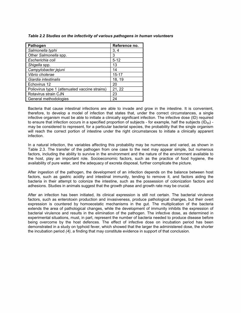
Table 2.2 Studies on the infectivity of various pathogens in human volunteers
Pathogen Reference no.Salmonella typhi 3, 4Other Salmonella spp. 3Escherichia coli 5-12Shigella spp. 13Campylobacter jejuni 14Vibrio cholerae 15-17Giardia intestinalis 18, 19Echovirus 12 20Poliovirus type 1 (attenuated vaccine strains) 21, 22Rotavirus strain CJN 23General methodologies 24
Bacteria that cause intestinal infections are able to invade and grow in the intestine. It is convenient,therefore, to develop a model of infection that states that, under the correct circumstances, a singleinfective organism must be able to initiate a clinically significant infection. The infective dose (ID) requiredto ensure that infection occurs in a specified proportion of subjects - for example, half the subjects (ID50) -may be considered to represent, for a particular bacterial species, the probability that the single organismwill reach the correct portion of intestine under the right circumstances to initiate a clinically apparentinfection.
In a natural infection, the variables affecting this probability may be numerous and varied, as shown inTable 2.3. The transfer of the pathogen from one case to the next may appear simple, but numerousfactors, including the ability to survive in the environment and the nature of the environment available tothe host, play an important role. Socioeconomic factors, such as the practice of food hygiene, theavailability of pure water, and the adequacy of excreta disposal, further complicate the picture.
After ingestion of the pathogen, the development of an infection depends on the balance between hostfactors, such as gastric acidity and intestinal immunity, tending to remove it, and factors aiding thebacteria in their attempt to colonize the intestine, such as the possession of colonization factors andadhesions. Studies in animals suggest that the growth phase and growth rate may be crucial.
After an infection has been initiated, its clinical expression is still not certain. The bacterial virulencefactors, such as enterotoxin production and invasiveness, produce pathological changes, but their overtexpression is countered by homoeostatic mechanisms in the gut. The multiplication of the bacteriaextends the area of pathological changes, while the development of immunity inhibits the expression ofbacterial virulence and results in the elimination of the pathogen. The infective dose, as determined inexperimental situations, must, in part, represent the number of bacteria needed to produce disease beforebeing overcome by the host defences. The effect of infective dose on incubation period has beendemonstrated in a study on typhoid fever, which showed that the larger the administered dose, the shorterthe incubation period (4), a finding that may constitute evidence in support of that conclusion.
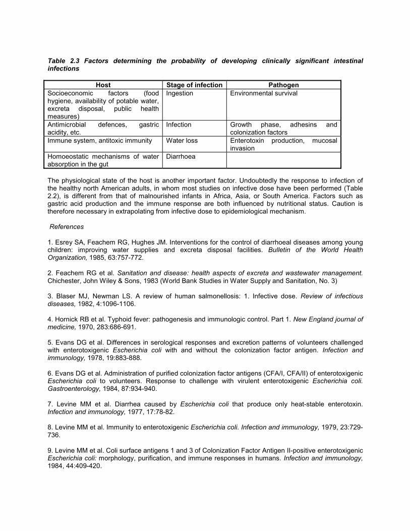
Table 2.3 Factors determining the probability of developing clinically significant intestinalinfections
Host Stage of infection PathogenSocioeconomic factors (foodhygiene, availability of potable water,excreta disposal, public healthmeasures)
Ingestion Environmental survival
Antimicrobial defences, gastricacidity, etc.
Infection Growth phase, adhesins andcolonization factors
Immune system, antitoxic immunity Water loss Enterotoxin production, mucosalinvasion
Homoeostatic mechanisms of waterabsorption in the gut
Diarrhoea
The physiological state of the host is another important factor. Undoubtedly the response to infection ofthe healthy north American adults, in whom most studies on infective dose have been performed (Table2.2), is different from that of malnourished infants in Africa, Asia, or South America. Factors such asgastric acid production and the immune response are both influenced by nutritional status. Caution istherefore necessary in extrapolating from infective dose to epidemiological mechanism.
References
1. Esrey SA, Feachem RG, Hughes JM. Interventions for the control of diarrhoeal diseases among youngchildren: improving water supplies and excreta disposal facilities. Bulletin of the World HealthOrganization, 1985, 63:757-772.
2. Feachem RG et al. Sanitation and disease: health aspects of excreta and wastewater management.Chichester, John Wiley & Sons, 1983 (World Bank Studies in Water Supply and Sanitation, No. 3)
3. Blaser MJ, Newman LS. A review of human salmonellosis: 1. Infective dose. Review of infectiousdiseases, 1982, 4:1096-1106.
4. Hornick RB et al. Typhoid fever: pathogenesis and immunologic control. Part 1. New England journal ofmedicine, 1970, 283:686-691.
5. Evans DG et al. Differences in serological responses and excretion patterns of volunteers challengedwith enterotoxigenic Escherichia coli with and without the colonization factor antigen. Infection andimmunology, 1978, 19:883-888.
6. Evans DG et al. Administration of purified colonization factor antigens (CFA/I, CFA/II) of enterotoxigenicEscherichia coli to volunteers. Response to challenge with virulent enterotoxigenic Escherichia coli.Gastroenterology, 1984, 87:934-940.
7. Levine MM et al. Diarrhea caused by Escherichia coli that produce only heat-stable enterotoxin.Infection and immunology, 1977, 17:78-82.
8. Levine MM et al. Immunity to enterotoxigenic Escherichia coli. Infection and immunology, 1979, 23:729-736.
9. Levine MM et al. Coli surface antigens 1 and 3 of Colonization Factor Antigen II-positive enterotoxigenicEscherichia coli: morphology, purification, and immune responses in humans. Infection and immunology,1984, 44:409-420.

10. Levine MM et al. The diarrheal response of humans to some classic serotypes of enteropathogenicEscherichia coli is dependent on a plasmid encoding an entero-adhesiveness factor. Journal of infectiousdiseases, 1985, 152:550-559.
11. Mathewson JJ et al. Pathogenicity of enteroadherent Escherichia coli in adult volunteers. Journal ofinfectious diseases, 1986, 154:524-527.
12. Tacket CO et al. Protection by milk immunoglobulin concentrate against oral challenge withenterotoxigenic Escherichia coli. New England journal of medicine, 1988, 318:1240-1243.
13. Dupont HL et al. Inoculum size in shigellosis and implications for expected mode of transmission.Journal of infectious diseases, 1989, 159:1126-1128.
14. Black RE et al. Experimental Campylobacter jejuni infection in humans. Journal of infectious diseases,1988, 157:472-479.
15. Cash RA et al. Response of man to infection with Vibrio cholerae. Clinical, serologic, and bacteriologicresponses to a known inoculum. Journal of infectious diseases, 1974, 129:45-52.
16. Levine MM et al. Volunteer studies of deletion mutants of Vibrio cholerae 01 prepared by recombinanttechniques. Infection and immunology, 1988, 56:161-167.
17. Levine MM et al. Safety, immunogenicity, and efficacy of recombinant live oral cholera vaccines, CVD103 and CVD 103-HgR. Lancet, 1988, ii: 467-470.
18. Nash TE et al. Experimental human infections with Giardia lamblia. Journal of infectious diseases,1987, 156:974-984.
19. Rose JB, Haas CN, Regli S. Risk assessment and control of waterborne giardiasis. American journalof public health, 1991, 81:701-713.
20. Schiff GM et al. Studies of Echovirus-12 in volunteers: determination of minimal infectious dose andthe effect of previous infection on infectious dose. Journal of infectious diseases, 1984, 150:858-866.
21. Katz M, Plotkin SA. Minimal infective dose of attenuated poliovirus for man. American journal of publichealth, 1967, 57:1838-1840.
22. Minor TE et al. Human infective dose determinations for oral poliovirus type 1 vaccine in infants.Journal of clinical microbiology, 1981, 13:388-389.
23. Ward RL et al. Human rotavirus studies in volunteers: determination of infectious dose and serologicalresponse to infection. Journal of infectious diseases, 1986, 154:871-880.
24. Haas CN. Estimation of risk due to low doses of microorganisms: a comparison of alternativemethodologies. American journal of epidemiology, 1983, 118:573-582.
3. Bacteria1
1 The valuable contribution made by Dr N.F. Pierce, Division of Diarrhoeal and Acute RespiratoryDisease Control, WHO, Geneva, in the preparation of this chapter is gratefully acknowledged.
3.1 Pathogens excreted
3.1.1 Salmonella
General description
The genus Salmonella is a member of the family Enterobacteriaceae. The genus is currently subdividedinto the subgenera I-IV, on the basis of biochemical characteristics. Most salmonella strains isolated fromhumans and warm-blooded vertebrates belong to subgenus I, while subgenera II, III (also called Arizona)and IV are more frequently associated with reptiles, in which they reside commensally. Currently morethan 2000 serotypes are named. There are considerable regional variations in the prevalence of serotypes(1).
The virulence of Salmonella spp. depends on serotype and strain specificity in host range and infectivedose and on host status. S. typhi is a specific human pathogen. In particular, S. typhi, S. paratyphi A, andS. paratyphi B are able to invade tissues and cause septicaemia with high temperature rather thandiarrhoea. This is known as enteric fever. In humans, the majority of the other serotypes give rise to atransient intestinal infection manifesting itself as acute gastroenteritis with diarrhoea. Certain serotypesare highly pathogenic for humans, while others are devoid of any pathogenic action. Many salmonellainfections are asymptomatic (2).
Routes of exposure
In the case of S. typhi and S. paratyphi A, human carriers are the source of infection, whereas milk-bornetransmission can also occur with S. paratyphi B. The incidence of enteric fever decreases as a countrybecomes more highly developed in terms of controlled sewerage systems and drinking-water supplies,and the supply of pasteurized milk and dairy products. Most salmonellae are primarily pathogens ofanimals, which constitute important reservoirs for those infections (2).
Salmonellae may be present in all kinds of food grown in faecally polluted environments, and arecommonly isolated from poultry and livestock and foods prepared from them. Furthermore, animalfeedstuffs and fertilizers prepared from animal products may be highly contaminated with salmonellae,and they are also widely distributed in the environment. The contamination of food and animal feedstuffsby water contaminated with salmonellae is considered to be an additional risk factor (3, 4). Dumping ofunprocessed slaughterhouse wastes into rivers has been found to be a cause of salmonellae infections.Contamination by salmonellae and conditions favouring their regrowth should be avoided at all stages ofthe production, transport, storage, and preparation of food, feedstuffs, and drinking-water. Sludge disposaland irrigation must always be accompanied by appropriate hygienic precautions.
The transmission routes of salmonellae are highly complex. Person-to-person transmission may occur, butthe relative importance of the human and non-human reservoirs depends on the dietary, agricultural, andsanitary situation in a particular community (2).
Significance in drinking-water
Waterborne outbreaks have been chiefly associated with S. typhi and much less frequently with S.paratyphi B or other Salmonella serotypes (2). Epidemiological studies of outbreaks suggest that theingestion of relatively few cells of S. typhi may cause typhoid fever, whereas studies in volunteers (Table2.2, p. 14) indicate that, for other Salmonella serotypes, millions of cells are usually required to cause

gastroenteritis.
Salmonellae are excreted in the faeces of infected humans or animals. Faecal contamination ofgroundwater or surface waters, as well as insufficiently treated and inadequately disinfected drinking-water, are the main causes of epidemic waterborne outbreaks caused by Salmonella spp.
Waterborne outbreaks due to heavy contamination are usually characterized by an explosive onset. Themajority of cases develop over a period of a few days, and may be followed by a secondary crop ofcontact cases (2). The geographical distribution of infections in major outbreaks is often stronglycorrelated with the pattern of a waterworks pipeline network.
Salmonellae can be found in open wells as a result of the drainage or flooding of contaminated surfacewater into unprotected well shafts. It is uncommon for salmonellae to be isolated from piped watersupplies, whether treated or untreated, and their presence suggests a serious fault in the design ormanagement of the system (2).
Penetration of pathogens into water sources must be avoided by the protection of groundwater andsurface water catchment areas. A review of the literature has shown that, in general, pathogens will nottravel further than the distance that the groundwater flows in 10 days, except in fissured rocks such aslimestone and heavily fissured soils (5).
Drinking-water must be of low turbidity after treatment if adequate chlorination is to be achieved.Furthermore, a low load of assimilable organic carbon (AOC) in the treated water is considered to be animportant factor in reducing survival time and preventing the regrowth of salmonellae within the distributionsystem. Reported survival times for salmonellae in drinking-water range from a few days to over 100 days.
Several outbreaks have been caused by the deposition of contaminated sediments in the distributionsystem for drinking-water, especially in water basins and pipes. Sediments may be shifted to newpositions in the system by water pressure oscillations or temporary scarcity of water. Regular flushing ofthe distribution system is therefore recommended.
3.1.2 Yersinia
General description
The genus Yersinia is currently placed in the family Enterobacteriaceae and comprises seven species. Y.pestis, Y. pseudotuberculosis, and certain serotypes of Y. enterocolitica are pathogens for humans.Atypical strains within Y. enterocolitica, isolated most frequently from environmental samples, are groupedtogether as Y. enterocolitica-like organisms. They are nonpathogenic for humans and can be subdividedby biochemical means into Y. intermedia, Y. frederiksenii, Y. kristensenii, and Y. aldovae.
The cells of Y. enterocolitica are Gram-negative rods, motile at 25 °C but nonmotile in cultures grown at37 °C. Certain strains of Y. enterocolitica cause acute gastroenteritis with diarrhoea, but other humandiseases caused by Y. enterocolitica are also known. Biochemical and serological typing of enteric Y.enterocolitica strains show that serotypes O:3 and O:9 are commonly found in Africa, Asia, Canada andEurope, whereas serotype O:8 is exclusively isolated in the United States (6-8).
There is some evidence that Y. enterocolitica infection may be waterborne. The following discussion isconfined to this species.
Routes of exposure
The transmission of Y. enterocolitica from the natural reservoirs to humans has been the subject of muchdebate. Many domestic and wild animals are considered to be possible reservoirs of Y. enterocolitica

because of the high isolation rates of the organism from such sources. It is likely that wild animals,particularly shrews, hares, foxes, and beavers, form a natural reservoir of Y. enterocolitica. Swine havebeen implicated as a major reservoir of Y. enterocolitica serotypes involved in human infections.
Available evidence indicates that foods, especially meat and meat products, milk and dairy products, arethe major vehicles for the transmission of Y. enterocolitica. Furthermore, Y. enterocolitica has beenisolated from a variety of environmental samples, especially from water, but the serotypes isolated differfrom those associated with human disease.
A number of different transmission routes have been suggested for Y. enterocolitica, but the ingestion ofcontaminated food and water is probably the most likely one (8). Direct transmission from person toperson and from animals to humans also occurs, but its relative importance has not been clarified. Furtherresearch is needed to define the epidemiological importance of "environmental" strains of Y.enterocolitica.
Significance in drinking-water
The apparent waterborne spread of Y. enterocolitica infection has been described in a number of reports.There is some evidence that pathogenic strains of Y. enterocolitica enter drinking-water via contaminatedsurface water or as a result of pollution with sewage (9). Recent studies have shown that humanpathogenic serotypes of Y. enterocolitica are present in sewage and polluted surface water (10, 11).
In general, pathogenic types of Y. enterocolitica are not isolated from untreated or treated drinking-waterunless faecal pollution has occurred. Occasionally, nonpathogenic serotypes of Y. enterocolitica andnonpathogenic Y. enterocolitica-like organisms (Y intermedia, Y. frederiksenii, Y. kristensenii) may also beisolated from drinking-water. However, none of these isolates exhibit the typical virulence markers. Suchisolates are probably of environmental origin without public health importance (12).
Water samples yielding Y. enterocolitica often show only slight coliform contamination. One studyindicated that 25% of Y. enterocolitica-positive samples were negative for both total and faecal coliforms(9). Intensive methods of treatment are not needed in such cases. Other studies have shown a closerelationship between faecal pollution and Y. enterocolitica isolation rates (13).
Standard chlorination procedures are sufficient to avoid the transmission of Yersinia if the treated water isof low turbidity. Free chlorine in the range required for water disinfection (0.2 - 0.5 mg/litre) for 10 minutesat pH 7.0 completely eradicates the bacteria; 0.05 mg/litre of ozone eradicates the organism after contactfor 1 minute regardless of pH (14).
A special feature of Y. enterocolitica and Y. enterocolitica-like organisms is their ability to grow at lowtemperatures, even at 4 °C. Accordingly, these organisms can survive for long periods in water habitats.For example, Y. enterocolitica was detected in previously sterilized distilled water after over 18 months at4 °C (15). Such long survival periods make it difficult to determine the origin of contamination whenYersinia are detected.
3.1.3 Campylobacter
General description
In recent years, considerable attention has been given to Campylobacter spp. as important agents ofenteritis, gastritis, and other human diseases.
Campylobacters are Gram-negative, slender, comma-shaped rods. They also appear S-shaped and gull-winged when in pairs (7, 16). The organisms show a characteristic corkscrew-like motion, which can beeasily seen by phase-contrast microscopy. Campylobacters are microaerophilic organisms, requiring a low
oxygen tension (3-6%) for growth. Under unfavourable growth conditions, cells may form coccoid bodies.
A recent review discusses 14 Campylobacter species (17). Some are pathogens for humans and animals(e.g. C. jejuni, C. coli, C. fetus), while others are considered to be nonpathogenic (e.g. C. sputorum, C.concisus) (16, 17). Most of the members of the thermophilic group (growing at 42 °C) of Campylobacterscause enteritis in humans. Worldwide, Campylobacters are much more important than salmonellae ascauses of acute gastroenteritis, but not as important as shigellas. Several major outbreaks ofcampylobacter enteritis were linked to the ingestion of contaminated food, milk, or water.
From the point of view of water hygiene, the thermophilic Campylobacters are of greatest significance; thefollowing discussion is therefore confined to these organisms.
Routes of exposure
Thermophilic campylobacters are transmitted by the oral route. The reservoirs for Campylobacters includewild birds and poultry which are the most important, and other domesticated animals, such as pigs, cattle,dogs, and cats. Meat, in particular poultry products, and unpasteurized milk are therefore importantsources of campylobacter infections (16). Milk may be contaminated with faeces or by the secretion oforganisms into milk by cows with mastitis (18). In developing countries, the faeces of infected animals areimportant reservoirs. The infective dose is low (19).
Recent studies have shown that raw sewage frequently contains 10-105 thermophilic campylobacters per100 ml (20, 21). However, Campylobacter counts in heavily contaminated sewage can be reducedconsiderably by wastewater treatment processes. Thermophilic campylobacters were found in crudesludge, but were not detectable in digested conditioned sludge of filter effluent (21). The occurrence ofcampylobacters in surface waters has proved to be strongly dependent on rainfall, water temperature, andthe presence of waterfowl.
Significance in drinking-water
Several waterborne outbreaks of campylobacteriosis have been reported in the past decade. Thenumbers of persons involved ranged from a few to several thousands. In only two of these outbreaks werecampylobacters isolated both from patients and from water samples. Unchlorinated surface water andfaecal contamination of water storage reservoirs by wild birds were found to be the main causes. Theconsumption of unchlorinated or inadequately chlorinated surface waters is associated with a considerablerisk of outbreaks of campylobacteriosis. Any contamination of drinking-water reservoirs by the excrementof waterfowl must be controlled. Consideration should be given to imposing stricter hygienic requirementsfor drinking-water, even if obtained from high-quality surface water, since it may be distributed withoutchlorination.
Campylobacters, like other bacterial pathogens, survive well at low temperatures, suggesting that coldwater may be an effective vehicle of transmission. They are able to survive for several weeks (22) in coldgroundwater or unchlorinated tapwater. The standard chlorination procedures are sufficient to prevent thespread of campylobacters along water mains if the water is of low turbidity.
3.1.4 Escherichia coli
E. coli is found in large numbers in the faeces of humans and of nearly all warmblooded animals; as suchit serves as a reliable index of recent faecal contamination of water. This topic is fully covered in Chapters9-11. Certain strains are pathogenic for humans and it is these that are described below.
General description
E. coli is a Gram-negative, non-spore-forming, rod-shaped bacterium which can be either motile or

nonmotile (motile cells are peritrichous); growth is aerobic or facultatively anaerobic. Metabolism is bothrespiratory and fermentative; acid is produced by the fermentation of glucose and lactose. Catalase isproduced but not oxidase, and nitrates are reduced to nitrites. In the microbiological examination of water,a biochemical description is used (see sections 9.2.1-9.2.3).
Serological typing is based on the somatic O antigens, the capsular K antigens, and the flagellar Hantigens. In practice, serogrouping by O antigen is often used alone and, within a particularepidemiological context, may be satisfactory. Biochemical tests do not reliably distinguish pathogenicstrains of E. coli.
Health effects
E. coli is a normal inhabitant of the intestine, and most strains are nonpathogenic. However, subtypes ableto cause gastrointestinal disease are also known. Such pathogenic E. coli strains cause intestinal diseaseby a variety of mechanisms. Infections may resemble cholera, dysentery, or gastroenteritis due tosalmonellae. Four classes of pathogenic E. coli responsible for diarrhoea are recognized:enteropathogenic, enteroinvasive, enterotoxigenic, and verocytotoxin-producing (23).
Enteropathogenic subtypes of E. coli were first recognized as a result of the serological examination ofstrains isolated from outbreaks of diarrhoea among infants. Associations of particular serotypes withdisease were observed, but the corresponding pathogenic mechanisms are not fully understood for mostof these organisms. These strains have been particularly associated with outbreaks of infantilegastroenteritis (24).
Enteroinvasive strains of E. coli (EIEC) produce dysentery by a mechanism similar to that found withShigella spp. These organisms invade the colonic mucosa and cause bloody diarrhoea. The propertyseems to be restricted to a few O groups. It must be remembered that Shigella and E. coli are closelyrelated and that genetic material is readily exchanged between them.
Although enteropathogenic or enteroinvasive strains may cause serious illness, such epidemiologicalevidence as is available suggests that enterotoxigenic strains are responsible for most episodes of E. colidiarrhoea, particularly in developing countries. Enterotoxigenic E. coli (ETEC) can cause a cholera-likesyndrome in infants, children, and adults.
ETEC produce either a heat-labile enterotoxin (LT), related to cholera enterotoxin, or a heat-stableenterotoxin (ST); some strains produce both toxins. The action of LT is the same as that of cholera toxin.The production of enterotoxin is controlled by plasmids.
The ability of ETEC to cause disease depends not only on the production of enterotoxin but also on theirability to colonize the small intestine. Various colonization factors, or adhesins, have been described,which enable the bacteria to attach themselves to the intestinal mucosa.
The fourth class, verocytotoxic E. coli, was first recognized by their production of a cytotoxin active againstVero cells in culture. The organisms commonly belong to the serogroup O157 and cause disease rangingfrom mild diarrhoea to haemorrhagic colitis characterized by blood-stained diarrhoea, usually withoutfever, but accompanied by abdominal pain. It is also a cause of the haemolytic uraemic syndrome,commonest in infants and young children, and characterized by acute renal failure and haemolyticanaemia.
Significance in drinking-water
Isolation of E. coli from a water supply indicates faecal contamination. However, E. coli is only one speciesamong many in the family Enterobacteriaceae. Members of the lactose-fermenting species of this group,which may be referred to colloquially as "coliforms", occur in a variety of ecological niches, not all of which

are intestinal. Thus, for example, some species are associated with aquatic slimes and vegetation. Thepicture is further complicated by the fact that other members of the coliform group are also found in theintestine. Thus, the definitive identification of E. coli may be needed to determine the significance of"coliforms" in a water supply.
Conventionally, thermotolerant coliforms are identified by growth at 44-45 °C, but some isolates ofKlebsiella, Citrobacter, and Enterobacter will also grow and ferment lactose under these conditions. Theterm "faecal coliforms" is often used for this group, but the term "faecal" is to be deprecated, since not allprove to be of faecal origin.
The detection of the pathogenic subtypes of E. coli in water supplies has seldom been attempted.Although this may be necessary in epidemiological research, the available methods are not suitable forthe routine examination of water samples.
3.1.5 Vibrio cholerae
General description
Vibrio species are motile, non-spore-forming, slightly curved Gram-negative rods with a single polarflagellum; they are both aerobic and facultatively anaerobic. Their metabolism is both respiratory andfermentative without the production of gas. Both catalase and oxidase are formed, and nitrates arereduced to nitrites.
Among the vibrios, special attention has focused on the identity of those causing cholera. It was for a longtime believed that Vibrio cholerae was a unique and distinct species associated with human disease, andrecognized by possession of the O1 antigens. This species was further divided into "classical" and "El Tor"biotypes, the latter distinguished by the ability to produce a dialysable, heat-labile haemolysin, activeagainst sheep and goat red cells (25).
A broader definition of V. cholerae has now been adopted. It has been known for many years that vibriosbiochemically identical to Vibrio cholerae O1, but lacking the O1 antigen, could be found in the aquaticenvironment. These were termed non-cholera vibrios (NCV) or non-agglutinable (NAG) vibrios. The term"non-O1 V. cholerae" is now preferred, since some may produce cholera toxin, while investigation ofDNA/DNA homology between Vibrio cholerae, NCVs, and NAG vibrios has conclusively demonstrated thatthey are all very closely related. Currently, all are considered to be a single species, Vibrio cholerae,divided into more than 80 serological types on the basis of the O or somatic antigens. The H or flagellaantigen is common to all the O groups of V. cholerae, and H agglutination has been used as a diagnostictest.
Only V. cholerae serogroup O1 causes cholera. The O1 group contains two serotypes based on variationsin the O antigen factors, namely Ogawa (factors A and B) and Inaba (factors A and C). These serotypesmay exist in either the classical or El Tor biotype.
Routes of exposure
Cholera has historically been one of the major pandemic diseases. The present pandemic, unlike previousones, is caused by V. cholerae O1 biotype El Tor.
Cholera is usually a waterborne disease, and numerous waterborne outbreaks have been documented.However, foodborne and nosocomial outbreaks are also important, and person-to-person transmissionmay occur under conditions of extreme crowding and poor hygiene. The problems of the transmission ofcholera have been extensively reviewed, and although waterborne transmission is undoubtedly important,many aspects of the epidemiology of cholera are a matter for debate. Evidence has accumulated tosuggest that, in some circumstances, V. cholerae, including serorype O1, may be part of the

autochthonous microbiota of natural waters (25, 26).
Health effects
Infection with V. cholerae O1 involves the small and large intestine. In the small intestine, the vibriosattach themselves to the mucous layer covering the villous epithelium, chemotactic processes apparentlyplaying a role in their migration to the epithelium. After attachment, vibrios penetrate the layer of mucusand adhere to the surface of the epithelial cells. Motility seems to be essential for mucus penetration tooccur. Adherence to the mucosal surface is specific, involving a receptor-adhesion interaction analogousto a lectin-ligand reaction. When present in large numbers, V. cholerae O1 produce an enterotoxin(cholera toxin) that causes alterations in the ionic fluxes across the mucosa with the resulting catastrophicloss of water and electrolytes in liquid stools. Cholera toxin is very similar to the heat-labile toxin producedby enterotoxigenic E. coli.
It seems likely that other accessory virulence factors, such as mucinase and protease, are also importantin the pathogenesis of cholera. Other toxins may also be involved. Enterotoxins similar to the heat-stabletoxins of E. coli are known to be formed by V. cholerae of O groups 2-84, and V. cholerae O1 may alsoproduce several toxins. Cholera toxin is not produced by all strains of V. cholerae O1, and nontoxigenicstrains are considered to be nonpathogenic.
Significance in drinking-water
The isolation of V. cholerae O1 from water used for drinking is of major public health importance and isevidence of faecal contamination. However, other serogroups of V. cholerae may be part of the normalflora of some waters.
3.1.6 Shigella
General description
Shigellae are Gram-negative, non-spore-forming, non-motile rods, capable of growth under both aerobicand anaerobic conditions. Metabolism is both respiratory and fermentative; acid, but not usually gas, isproduced from glucose. Lactose is seldom fermented. Catalase is usually produced, except by Shigelladysenteriae type 1, but oxidase is produced by one serotype only. Nitrates are reduced to nitrites.
Shigellae are serotyped on the basis of their somatic O antigens. Both group and type antigens aredistinguished, group antigenic determinants being common to a number of related types. There isevidence that type antigen specificities among Shigella flexneri are determined by the presence oflysogenic bacteriophages. It seems likely that biotypic and serological variants are determined by thepresence of phage or the carriage of plasmids. Plasmids (transferable, extrachromosomal geneticelements) were first described in this genus. Phage-typing systems exist for all groups, though they havenot been widely applied, serological typing being adequate for all species except Shigella sonnei.
Health effects and routes of infection
Though shigella infection is not often spread by waterborne transmission, major outbreaks resulting fromsuch transmission have been described. The characteristic bloody diarrhoea results from the invasion ofthe colonic mucosa by the bacteria. There is good reason to believe that the process is highly species-specific. Shigellae have no natural hosts other than the higher primates, and humans are the only effectivesource of infection. Of the enteric bacterial pathogens, shigellae seem to be the best adapted to causehuman disease. Direct transmission between susceptible individuals is the usual route of infection, and theinfective dose is lower than for other bacteria.

Significance in drinking-water
The isolation of shigellae from drinking-water indicates recent human faecal contamination and, in view ofthe extreme pathogenicity of the organisms, is of crucial public health significance. However, this is a rareevent, possibly explained in part by the absence of a useful enrichment or selective medium for theisolation of these bacteria. Those generally used have been designed for the isolation of salmonellae andare not optimal for that of shigellae. Furthermore, without confirmatory testing, some anaerogenic strainsof E. coli may be wrongly identified as shigellae.
3.2 Pathogens that grow in supply systems
All drinking-water contains assimilable organic compounds that will allow a certain degree of bacterialgrowth. The density of bacteria in drinking-water can and should be controlled for the reasons given inChapter 8.
Certain bacteria in drinking-water deserve particular attention because they are opportunistic pathogens tohumans, i.e. they are able to cause infections in susceptible persons. The most important organisms ofthis type, namely Legionella and Aeromonas, will be considered here. Other organisms, such asPseudomonas aeruginosa and opportunistically pathogenic mycobacteria, have been detected in drinking-water supplies.
3.2.1 Legionella
General description
The genus Legionella, a member of the family Legionellaceae, has 22 currently known species, of whichL. pneumophila serogroup 1 is most frequently associated with human disease. Other serogroups of L.pneumophila and occasionally other legionellae have also been reported to cause disease. Legionellaeare Gram-negative, rod-shaped, non-spore-forming bacteria that require L-cysteine for growth and primaryisolation. The cellular fatty acids in legionellae are unique among those found in Gram-negative bacilli inthat they consist essentially of branched chains. Preconcentration of legionellae from environmentalsamples may be required if low levels are to be detected. Immunofluorescence techniques may also beused to detect legionellae in the environment.
Health effects
Legionella infections can lead to two types of disease, namely Legionnaires' disease (legionellosis) andnon-pneumonic Legionnaires' disease (Pontiac fever). Legionnaires' disease is a form of pneumonia withan incubation period usually of 3 - 6 days. Males are more frequently affected than females, and mostcases occur in the 40 - 70 year age group. Risk factors include smoking, alcohol abuse, cancer, diabetes,chronic respiratory or kidney disease, and severe immunosuppression, as in transplant recipients. Thefatality rate in untreated cases may be 10% or higher, but the disease can be treated effectively withantibiotics such as erythromycin and rifampicin.
Legionnaires' disease is uncommon, but common-source outbreaks attract much attention. Between 100and 200 cases are reported each year in England and Wales, and in Germany; in France, the incidence issomewhat higher, with over 400 cases per year (27). For people living in temperate climates, travelling tosubtropical areas appears to be a significant risk factor, outbreaks being related to air-conditioning andhot-water systems in hotels. Hospital-associated Legionnaires' disease is the most serious form, becauseit usually affects debilitated persons and has a high mortality rate. The non-pneumonic form of the diseaseis milder, with a high attack rate, an acute onset (5 hours to 3 days) and symptoms similar to those ofinfluenza: fever, headache, nausea, vomiting, aching muscles, and coughing. No fatal cases have beenreported and few outbreaks have been recognized, possibly because the non-specific symptoms of thedisease hinder its diagnosis.

Routes of exposure
Legionellae are widespread in natural sources of water and may also be found in soils. They occurcommonly in man-made water systems, particularly in hot-water and cooling-water systems. Infection isthe result of the inhalation of aerosols that are small enough to penetrate the lungs and be retained by thealveoli. The degree of risk depends on four key factors: (i) the density of the bacteria in the source; (ii) theextent of aerosol generation; (iii) the number of inhaled bacteria; and (iv) the susceptibility of the exposedindividual.
Legionellae multiply in the laboratory at temperatures between 20 and 46 °C. At temperatures higher than46 °C, the bacteria will survive, but at 60 °C only for a few minutes (28). Temperatures favourable forgrowth may be found in cooling towers, spas, cold-water systems in buildings, hot-water systems operatedbelow 60 °C or "dead legs" of such systems operated at higher temperatures. Aerosols may be created bythe spraying of water in cooling towers or its agitation in spas. Hot-water systems are also likely to createaerosols in showers, through nozzle heads or by splashing in sinks, baths, etc. The number of inhaledbacteria depends on the size of the aerosol generated (<5 µm being most dangerous), the dispersal of theaerosol in the air, and the duration of the exposure. Host defence is an important factor that determineswhether exposure to legionellae will lead to clinical disease. It is primarily for this reason that high countsof L. pneumophila in water systems have been reported in the absence of disease, whereas similar orlower counts have been associated with epidemics. It is also likely, although not yet adequately proven,that differences in virulence between strains partly account for these observations.
It is now apparent that legionellae can be ingested by the trophozoites of certain amoebae(Acanthamoeba, Hartmanella, Valkampfia, and Naegleria) and even grow intracellularly and becomeincorporated in their cysts (29). This may explain the difficulty of eradicating legionellae from watersystems and may be a factor in the etiology of the non-pneumonic disease (30).
The following are generally accepted as requiring disinfection:
- sites implicated in an outbreak of Legionnaires' disease or Pontiac fever;
- hospital wards housing high-risk patients, such as an organ transplant unit;
- buildings in which the water system has not been used for some time and where large numbers oflegionellae are likely to be found.
Nevertheless, it is generally advisable to design and maintain systems in such a way that colonization byLegionella is prevented as much as possible. Detailed instructions for achieving this have been given inseveral publications (31-34) and focus on the following aspects:
- preventing the accumulation of sludge, scale, rust, algae and slime, and removing such depositsregularly;
- maintaining hot-water temperatures permanently above 60 °C or increasing them periodically toabove 70 °C, and keeping cold-water supplies below 20 °C;
- selecting materials for contact with water that do not release nutrients that support the growth ofLegionella.
The use of biocides is generally regarded as a less effective and less desirable means of controllinglegionellae in water supplies in buildings. However, their use is essential to prevent the build-up ofmicrobial slimes in air-conditioning systems in which wet, evaporative cooling towers are used. Suchsystems should be kept clean and well maintained, and should be inspected weekly for fouling,
accumulations of slime and scale, and corrosion; they should be thoroughly cleaned and disinfected twiceyearly. Biocides are best used intermittently in clean systems (33, 34).
3.2.2 Aeromonas
General description
Aeromonas spp. are Gram-negative, rod-shaped, non-spore-forming bacteria that are currently assignedto the family Vibrionaceae, although they also bear many similarities to the Enterobacteriaceae.MacDonnell et al. (35) have suggested the creation of a new family Aeromonadaceae. The genusAeromonas is divided into two groups of which the first, the group of psychrophilic non-motileaeromonads, consists of only one species, A. salmonicida, an obligate fish pathogen that will not beconsidered further here. The group of mesophilic motile aeromonads has been divided by Popoff intothree biochemically distinguishable groups (36), namely A. hydrophila, A. sobria and A. caviae. Each ofthese three species consists of at least three different DNA-hybridization groups, and later workers havedescribed new species such as A. veronii, A. media, A. schubertii, and A. eucrenophila. It may beexpected that in the near future the taxonomy of the group of mesophilic aeromonads will be changed stillfurther, but at present the classification of Popoff is that most widely accepted internationally.
Health effects
Mesophilic aeromonads have long been known to be pathogenic for coldblooded animals such as fish andamphibians, but in the last few decades greater attention has been given to their pathogenic significancefor humans. Three major types of infection are described (37): (i) systemic infections, usually in seriouslyimmunocompromised persons; (ii) wound infections (mainly after contact with surface water); and (iii)diarrhoea. In particular, the significance of Aeromonas as an enteropathogenic organism is the subject ofmuch discussion. In animal test models, such as the suckling mouse test and the rabbit ileal loop test,pure cultures of Aeromonas have been found to cause strong fluid accumulation which can partially beascribed to the production of extracelluar cytotoxins. However, there have been reports that, while theculture filtrate of some Aeromonas strains is not enteropathogenic in an animal test model, a suspensionof living cells does have this property. Cell-bound factors are apparently also of importance (38). Asao etal. (39) have purified and characterized an Aeromonas haemolysin, which was found to be a protein with arelative molecular mass of about 50 000 that was strongly enterotoxic and cytotoxic. It has not yet beenpossible to purify and characterize other toxins because the toxic activity disappears rapidly when culturefiltrates are manipulated. Despite the marked toxin production by Aeromonas strains in vitro, diarrhoeahas not yet been induced in test animals or human volunteers, and it is assumed that such strains are onlypoorly able to colonize the gastrointestinal tract (40). Little is known about the adhesion factors ofAeromonas or their interaction with receptors in the gastrointestinal tract.
Epidemiological investigations have also resulted in contradictory findings on the significance ofAeromonas as an enteropathogenic organism. In some studies, the numbers of Aeromonas in the faecesof patients with diarrhoea were greater than those in control groups, whereas in other studies there wasno difference, or the bacteria were found in even greater numbers in the latter. In general, it can be saidthat the significance of Aeromonas as an enteropathogenic organism is greater in tropical areas than inthe temperate zone. However, infections do occur in the temperate zone as well, albeit less frequently.Aeromonas-associated diarrhoea usually causes an acute but self-limiting gastroenteritis but chronicdisease with serious complications may also occur. The incidence of Aeromonas in human diarrhoealfaeces in the Netherlands was found to vary between 0.5% in winter and 3% in summer. Most isolationswere made in children under five years of age or in adults above 70 years of age. Young children yieldedmainly A. caviae, whereas A. sobria was usually isolated from the elderly (41).
Routes of exposure
Aeromonas occurs in water, soil, and food, particularly meat, fish, and milk. The occurrence of Aeromonas

in drinking-water can be studied by a variety of methods. A membrane filtration method has beendescribed in which a selective ampicillin-dextrin agar (ADA) is used (42). If water samples from houseinstallations are being examined, the addition to the sample of a complexing agent such as the disodiumsalt of edetic acid (Na2EDTA) at a concentration of 50 mg/litre is necessary. Aeromonas is extremelysensitive to the traces of copper that may be present in water in domestic installations in which copperpiping is used. Complexing of copper was also found to improve the survival of coliform bacteria andheterotrophic bacteria (43).
The number of Aeromonas in surface waters can vary between 0.01 and 1000 cfu/ml. Small numbers arefound in springs and in seawater that is not contaminated by sewage discharges. If such discharges arepresent, the number of Aeromonas can rise to 100 cfu/ml in seawater, and more than 1000 cfu/ml in freshwater, the species A. caviae then being dominant. In fresh waters not subject to sewage pollution, thenumbers of Aeromonas are usually between 10 and 100 cfu/ml. In these waters, the numbers are higherin summer than in winter, and there is a relation between the eutrophication of the water and the summerdensity of Aeromonas. In stagnant fresh water, A. sobria is usually the dominant species. When riverwater is stored in reservoirs, the number of Aeromonas decreases, but there is also a shift in speciescomposition from A. caviae to A. sobria. Aeromonas is not usually found in groundwater or is found only invery small numbers (44).
Irrespective of the contamination level of the raw water, most drinking-water treatment processes appearto be able to reduce the numbers of Aeromonas to below 1 cfu/100 ml. However, treated water cancontain larger numbers, with maxima of about 1000 cfu/100 ml as a result of regrowth in storagereservoirs with long retention times, polluted filter sand, or sudden changes in the quantities of water to beproduced. Regrowth of Aeromonas occurs in the distribution network of most drinking-water treatmentplants. The size of the Aeromonas population will depend on many factors but primarily on the organiccontent of the water and its temperature, the residence time in the distribution network, and the presenceof residual chlorine.
Little is known about the type and concentrations of nutrients for Aeromonas in drinking-water. Van derKooij (45) has suggested that Aeromonas prefers to grow on organic matter, e.g. from decaying nitrifyingand methane-oxidizing bacteria that develop in drinking-water treatment plants or from biofilm material indistribution networks.
Control of aeromonads in drinking-water requires a multiple approach, which is similar to the generalapproach to limiting the regrowth of bacteria (see Chapters 8 and 9). The treatment process shouldeffectively remove organic compounds serving as sources of carbon and energy for the growth of bacteria.Furthermore, the amount of biomass produced and subsequently released during the treatment processshould be as small as possible. The distribution system should be designed in such a way that residencetimes are short, and it should be flushed regularly to prevent the accumulation of sediments in stretcheswith low water velocities. Materials in contact with drinking-water should not be a source of biodegradablecompounds. These factors are of greatest importance in supplies that are not disinfected, or where themaintenance of a chlorine residual is not considered desirable for various reasons. Free available chlorineresiduals of 0.2 - 0.5 mg/litre will generally be sufficient to control Aeromonas densities in water in thedistribution network (46, 47). Chlorine or other disinfectants should not be used to control occasionalincreases in Aeromonas densities in supplies that are normally not chlorinated because biofilms on pipewalls will be disturbed, and this will result initially in increases rather than decreases in bacterialconcentrations. Also, in such systems the chlorine consumption will be rather high and residuals cannotbe properly maintained, thus allowing regrowth in remote stretches of the network.
The question whether the presence of Aeromonas in drinking-water is a risk to human health cannot beanswered with certainty; however, if there is a risk, it must be small because in many countries thebacterium is not important as a causative agent of diarrhoea and is not often able to colonize thegastrointestinal tract of humans. Also, the numbers present in drinking-water are small as compared withthose in other sources. In food, for instance, the numbers usually found are of the order of 103-105 cfu/g.

However, drinking-water is a product that is consumed daily by everyone, including groups with a reducedresistance to infectious diseases. Some of this water is consumed without previous heating, in contrast tomost foods contaminated with Aeromonas. A cautious approach therefore appears to be justified.Numbers of Aeromonas in drinking-water must be controlled as far as possible. Apart from the publichealth reasons for the control of Aeromonas levels in drinking-water, experience has shown that it is auseful and sensitive indicator of general hygiene within the drinking-water production and distributionprocess. Currently, no guideline value can be given because local conditions (temperature, raw watersource) may greatly influence Aeromonas counts in drinking-water.
3.2.3 Pseudomonas aeruginosa
General description
P. aeruginosa is a member of the family Pseudomonadaceae and is a monotrichate. Gram-negative rod. Itcan be recognized by its production of a blue-green fluorescent pigment (pyocyanin), which, in agarcultures, will diffuse into the medium. Pigment may not be produced by strains of P. aeruginosa recoveredfrom clinical specimens, and the ability to produce it may be lost on subculture. Like other fluorescentpseudomonads that occur in natural waters, P. aeruginosa strains produce catalase and oxidase, andammonia from arginine, grow with citrate as the sole source of carbon, and are aerobic. P. aeruginosa,however, is capable of growth at 41 - 42 °C, and the blue-green pigment that it produces differs from thefluorescent pale green pigment (fluorescein) produced by other species of fluorescent pseudomonadsfound in water. It is also capable of growing anaerobically in stab cultures of nitrate agar.
P. aeruginosa is commonly found in faeces, soil, water, and sewage but cannot be used as an indicator offaecal contamination, since it is not invariably present in faeces and sewage, and may also multiply in theenriched aquatic environment and on the surface of unsuitable organic materials in contact with water.However, its presence may be one of the factors taken into account in assessing the general cleanlinessof water distribution systems and the quality of bottled waters (see section 9.3.2).
Routes of exposure
P. aeruginosa is an opportunistic pathogen. Most of the illnesses in humans for which it is responsible arecaused, not by drinking water, but by contact with it. Water containing these bacteria may alsocontaminate food, drinks, and pharmaceutical products, causing them to deteriorate and to act assecondary vehicles for transmission. Fixtures in contact with water, such as sinks and sink drains, tapfittings and showerheads, can also be contaminated by P. aeruginosa and can serve as reservoirs ofinfection in hospitals.
Health effects and significance in drinking-water
In healthy persons, the illnesses caused by P. aeruginosa are usually mild and trivial. Waterborneinfections are usually associated with warm, moist environments; they include the skin rashes andpustules or outer ear canal infections (otitis externa) reported in users of indoor swimming-pools andwhirlpools, where bacterial counts are high and disinfection is deficient (48, 49). The presence of thisorganism in water supplied to hospitals and for the manufacture of pharmaceutical preparations anddressings is a matter of concern because P. aeruginosa is a common pathogen in infections of woundsand burns and has caused serious eye infections after the use of contaminated eye drops (50). Hospitalstrains of P. aeruginosa can first colonize and then infect patients receiving cancer chemotherapy (51).
The presence of this organism in potable water also indicates a serious deterioration in bacteriologicalquality, and is often associated with complaints about taste, odour, and turbidity linked to low rates of flowin the distribution system and a rise in water temperature (see section 9.3.2).

3.2.4 Mycobacterium
General description
Mycobacterium spp. are rod-shaped bacteria with cell walls having a high lipid content; this enables themto retain certain dyes in staining procedures that employ an acid wash, and they are therefore oftenreferred to as acid-fast bacteria. The characteristics of the cell wall structure also result in a relatively highresistance to disinfectants. All mycobacteria are characterized by slow growth (generation times underoptimal circumstances 2-20 hours), but within this range they are divided into "slow" and "rapid" growers.Most pathogenic species are found among the slow growers, which include the strictly pathogenic speciesM. tuberculosis, M. bovis, M. africanum, and M. leprae; these are not transmitted by water and have onlyhuman or animal reservoirs. Other mycobacterial species, often referred to as "atypical", haveenvironmental reservoirs. Although many are considered to be nonpathogenic, several species areopportunistic pathogens for humans, the most important being the slow growers M. kansasii, M. marinum,M. avium, M. intracellulare, M. scrofulaceum, and M. xenopi, and the rapid growers M. chelonae and M.fortuitum. Some of these species are closely related, and the literature often describes a number ofcomplexes rather than individual species. Examples are the "M. bovis complex" (which includes M.africanum), the "M. avium complex" (or MAC, which includes M. intracellulare), or the "M. avium, M.intracellulare, M. scrofulaceum complex" (or MAIS) and the "M. fortuitum-chelonae complex."
Health effects
The strictly pathogenic mycobacteria are associated with classical infectious diseases such astuberculosis and leprosy. The environmental mycobacteria may cause a range of diseases includingtuberculous lung disease and disseminated infections which may also involve the skeleton (M. kansasii,M. avium complex), infections of the lymph nodes (MAIS complex), and infections of the skin and softtissues (M. marinum, M. fortuitum-chelonae complex) (52, 53). Diseases caused by opportunisticpathogenic mycobacteria are not normally transmitted from person to person but are usually the result ofenvironmental exposure in combination with predisposing factors, such as dust retained in the lungs,surgical wounds, or immunosuppression produced by medication (transplant patients) or by underlyingdisease (AIDS, malignancies). Mycobacteria are generally resistant to many antimicrobial agents, henceeffective treatment may be difficult.
Routes of exposure
An extensive review of the occurrence of mycobacteria in environmental sources has been published (54).Tapwater has long been known to harbour saprophytic mycobacteria; in fact, one of the most commonlyoccurring species, M. gordonae, is known as the tapwater bacillus. The occurrence of opportunisticpathogenic species in tapwater has also been demonstrated by various authors (55, 56). These organismsmay accidentally contaminate clinical specimens during and after collection, or during processing in thelaboratory; this may falsely suggest that the patients concerned are suffering from a mycobacterialinfection (57, 58). A link between the occurrence of mycobacteria in drinking-water and disease hassometimes been suggested. Endemic M. kansasii infections in Czechoslovakia were studied from 1968onwards, the peak incidence being found in a small, densely populated district in which workers wereengaged in mining, heavy industry, and power generation. M. kansasii could also be isolated from showeroutlets in collieries, and it was later shown that the drinking-water system in the entire region was widelycontaminated. It was suggested that mycobacteria from drinking-water were spread via aerosols (59). Thehigh isolation frequency of M. kansasii from clinical specimens in Rotterdam, the Netherlands, led to aninvestigation of the water supply system. The organisms were frequently isolated from tapwater, and wereof the same phage type and showed the same weak nitratase activity as clinical strains (60). The increasein the isolation frequency of the M. avium complex in Massachusetts, USA, has also been attributed totheir presence in drinking-water (61). It should be noted that in all these cases there is only circumstantialevidence of a causal relationship between the occurrence of mycobacteria in drinking-water and humandisease. Certainly, the low infectivity of environmental mycobacteria does not warrant the setting of
standards or the institution of eradication programmes.
The ecology of opportunistic mycobacteria in water supplies is poorly understood. The bacteria have beenisolated infrequently from treated water or mains water (52, 57) but appear to multiply within the plumbingsystems in buildings as well as in taps. Increased isolation frequencies have been associated with highertemperatures (hot-water systems or cold-water pipes in the vicinity of central heating). Older buildingsappear to be more frequently colonized than new ones (61), and transport of drinking-water over longdistances also seems to increase the content of mycobacteria (58). Haas et al. (62) attempted to correlatetotal microscopic counts of acid-fast bacteria (hence including both pathogenic and saprophytic species)with a range of physicochemical parameters. A negative correlation with total chlorine residual and apositive correlation with turbidity and total organic carbon (TOC) was established, but these variables onlyaccounted for a small proportion of the overall variance of counts. It might also be expected that materialsused for plumbing would have an effect on mycobacterial densities, but no experimental evidence of suchan effect has yet been presented.
References
1. Leminor L. The genus Salmonella. In: Starr MP et al., eds. The prokaryotes, Vol. II. Berlin andHeidelberg, Springer-Verlag, 1981:1148-1159.
2. Lloyd B. Salmonella, enteric fevers and salmonelloses. In: Feacham RG et al., eds. Sanitation anddisease. Health aspects of excreta and wastewater management. Chichester, John Wiley & Sons,1983:251-286 (World Bank Studies in Water Supply and Sanitation, No. 3).
3. Kampelmacher EH. Spread and significance of salmonellae in surface waters in the Netherlands. In:Hoadley AW, Dutka BJ, eds. Bacterial indicators of potential health hazards associated with water.Philadelphia, American Society for Testing and Materials, 1977:148-158.
4. Mersch-Sundermann V, Wundt W. Die bakteriologische Beschaffenheit des Wassers vom Rhein undseinen Zuflussen im Rhein-Neckar-Raum. II. Mitteilung. Salmonellen - Hygienische Bedeutung undgesundheitliche Gefahren. [Bacteriological quality of water from the Rhine and its tributaries in the Rhine-Neckar area. Part II. Salmonellae - public health significance and health risks.] Zentralblatt fürBakteriologie, Mikrobiologie und Hygiene, Reihe B, 1987, 184:470-482.
5. Lewis WJ, Foster SSD, Dragar BS. The risk of groundwater pollution by on-site sanitation in developingcountries; a literature review. Dubendorf, Switzerland, International Centre for Wastes Disposal, 1982(Report No. 01/82).
6. Swaminathan B, Harmon MC, Mehlman JJ. Yersinia enterocolitica. Journal of applied bacteriology,1982, 52:151-183.
7. Bercovier H, Mollaret HH. Yersinia. In: Krieg NR, Holt JG, eds. Bergey's manual of systematicbacteriology. Vol. 1. Baltimore, Williams & Wilkins, 1984:498-506.
8. Lloyd B. Yersinia and yersiniosis. In: Feacham RG, eds. Sanitation and disease. Health aspects ofexcreta and wastewater management. Chichester, John Wiley & Sons, 1983:327-330 (World BankStudies in Water Supply and Sanitation, No. 3).
9. Schiemann DA. Isolation of Yersinia enterocolitica from surface and well waters in Ontario. Canadianjournal of microbiology, 1978, 24:1048-1052.
10. Ziegert E, Diesterweg I. The occurrence of Yersinia enterocolitica in sewage, Zentralblatt fürMikrobiologie, 1990, 145(5): 367-375.

11. Fukushima H et al. Yersinia spp. in surface waters in Matsue, Japan. Zentralblatt für Bakteriologie,Mikrobiologie und Hygiene, Reihe B, 1984, 179:235-247.
12. Aleksic S, Bockem HLJ. Serological and biochemical characteristics of 416 Yersinia strains from wellwater and drinking water plants in the Federal Republic of Germany: lack of evidence that these strainsare of public health importance. Zentralblatt für Bakteriologie, Mikrobiologie und Hygiene, Reihe B, 1988,185:527-533.
13. Weber G, Manafi M, Reisinger H. Die bedeutung von Yersinia enterocolitica und thermophilencampylobactern für die Wasserhygiene. [Significance of Yersinia enterolitica and thermophiliccampylobacter for water hygiene.] Zentralblatt für Bakteriologie, Mikrobiologie und Hygiene, Reihe B,1987, 184:504-514.
14. Krogulska B, Maleszewska J, Wichrowska B. [Sensitivity of Yersinia enterocolitica to agents used inwater disinfection.] Rocznik panstwowego zakladu higieny, 1986, 37: 434-440 (in Polish).
15. Highsmith AK et al. Isolation of Yersinia enterocolitica from well water and growth in distilled water.Applied and environmental microbiology, 1977, 34:745-750.
16. Smibert RM. The genus Campylobacter. In: Starr MP et al., eds. The prokaryotes, Vol. I. Berlin andHeidelberg, Springer-Verlag, 1981:609-617.
17. Penner JL. The genus Campylobacter: a decade of progress. Clinical reviews in microbiology, 1988,1:157-172.
18. Birkhead G et al. A multiple-strain outbreak of campylobacter enteritis due to consumption ofinadequately pasteurized milk. Journal of infectious diseases, 1988, 157:1095-1097.
19. Robinson DA. Infective dose of Campylobacter jejuni in milk. British medical journal, 1981, 282:1584.
20. Steltzer W et al. Characterisation of Campylobacter jejuni and Campylobacter coli isolated fromwastewater. Zentralblatt für Bakteriologie, Mikrobiologie und Hygiene, Reihe A, 1988, 269:188-196.
21. Holler C. Quantitative and qualitative studies of Campylobacter in the sewage of a large city.Zentralblatt für Bakteriologie, Mikrobiologie und Hygiene, Reihe B, 1988, 185:307-325.
22. Gondrosen B. Survival of thermotolerant campylobacters in water. Acta veterinaria scandinavica,1986, 27:1-10.
23. Feachem RG. Environmental aspects of cholera epidemiology. III. Transmission and control. Tropicaldisease bulletin, 1982, 79:1-47.
24. Scotland SM et al. Adhesion to cells in culture and plasmid profiles of enteropathogenic Escherichiacoli isolated from outbreaks and sporadic cases of infant diarrhoea. Journal of infection, 1989, 19(3): 237-249.
25. Attridge SR, Rowley D. Cholera. In: Smith GR, Easmon CSF, eds. Topley and Wilson's principles ofbacteriology, virology and immunity, 8th ed., Vol. 3. London, Edward Arnold, 1990:459-468.
26. Miller CJ, Drasar BS, Feachem RG. Cholera epidemiology in developed and developing countries:new thoughts on transmission, seasonality and control. Lancet, 1985:261-262.
27. Guerin JC. Epidemiology of Legionnaires' disease. From myth to reality. Revues des maladiesrespiratoires, 1992, 9 (Suppl. 1):R53-R56.

28. Brenner DJ et al. Legionella. In: Krieg NR, Holt JG, eds. Bergey's manual of systematic bacteriology.Vol. 1. Baltimore, Williams & Wilkins, 1984:279-288.
29. Lee JV, West AA. Survival and growth of Legionella species in the environment. Society of AppliedBacteriology symposium series, 1991, 20:121S-129S.
30. Rowbotham TJ. Pontiac fever explained? Lancet, 1980, ii: 969.
31. Prevention of legionellosis. The Hague, Gezondheidsraad, 1986.
32. The control of legionellae in health care premises. A code of practice. London, Her Majesty'sStationery Office, 1989.
33. Department of Health and Social Security. Health services management. Legionnaires' disease.Cooling towers and evaporative condensers. London, 1987 (Engineering Guidance Note 4).
34. Report of the Expert Advisory Committee on Biocides. London, Her Majesty's Stationery Office, 1989.
35. MacDonnell MT et al. Ribosomal RNA phylogenies for the vibrio-enteric group of eubacteria.Microbiological science, 1986, 3(6):172-175, 178.
36. Popoff M. Aeromonas. In: Krieg NR, Holt JG, eds. Bergey's manual of systematic bacteriology, Vol. 1.Baltimore, Williams & Wilkins, 1984:545-548.
37. Janda JM, Duffey PS. Mesophilic aeromonads in human disease: current taxonomy, laboratoryidentification, and infectious disease spectrum. Reviews of infectious diseases, 1988, 10:980-997.
38. Stelma GN, Johnson CH, Spaulding PL. Experimental evidence for enteropathogenicity in Aeromonasveronii. Canadian journal of microbiology, 1988, 34:877-880.
39. Asao T et al. Purification and some properties of Aeromonas hydrophila hemolysin. Infection andimmunity, 1984, 46:122-127.
40. Kuijper EJ. Aeromonas associated diarrhoea in the Netherlands. Amsterdam, University ofAmsterdam, 1989 (thesis).
41. Havelaar AH et al. Aeromonas species als verwekker van diarree en infecties buiten het maag-darmkanaal in Nederland. [Aeromonas as the cause of diarrhoea and of infections outside thegastrointestinal tract in the Netherlands.] Nederlands tijdschrift voor geneeskunde, 1990, 134:1053-1057.
42. Havelaar AH, During M, Versteegh JF. Ampicillin-dextrin agar medium for the enumeration ofAeromonas species in water by membrane filtration. Journal of applied bacteriology, 1987, 62:279-287.
43. Versteegh JF et al. Complexing of copper in drinking water samples to enhance recovery ofAeromonas and other bacteria. Journal of applied bacteriology, 1989, 67:561-566.
44. Schubert RHW. Der Nachweis von Aeromonaden der "Hydrophila-Punctata-Gruppe" im Rahmen derhygienischen Trinkwasserbeurteilung. [Detection of aero-monads of the "hydrophila punctata group" in thehygienic assessment of drinking-water.] Zentralblatt für Bakteriologie und Hygiene, I Abteilung, Originale,Reihe B, 1976, 161:482-497.
45. Van der Kooij D. Properties of aeromonads and their occurrence and hygienic significance in drinking-water. Zentralblatt für Bakteriologie, Mikrobiologie und Hygiene, Reihe B, 1988, 187:1-17.

46. Burke V et al. Isolation of Aeromonas hydrophila from a metropolitan water supply: seasonalcorrelation with clinical isolates. Applied and environmental microbiology, 1984, 48:361-366.
47. Lechevallier MW, Cawthorn CD, Lee RG. Factors promoting survival of bacteria in chlorinated watersupplies. Applied and environmental microbiology, 1988, 54:649-654.
48. Jones F, Bartlett CLR. Infections associated with whirlpools and spas. Society of Applied Bacteriologysymposium series, 1985, 14:61S-66S.
49. Calderon R, Mood EW. An epidemiological assessment of water quality and "swimmer's ear". Archivesof environmental health, 1982, 73:300-305.
50. Parker MT. Septic infections due to Gram-negative aerobic bacilli. In: Smith GR, ed. Topley andWilson's principles of bacteriology, virology and immunity, 7th ed.. Vol. 3. London, Edward Arnold,1984:280-310.
51. Wade JC et al. Potential of imipenem as single-agent empiric antibiotic therapy of febrile neutropenicpatients with cancer. American journal of medicine, 1985, 78 (6A):62-72.
52. Good RC. Opportunistic pathogens in the genus Mycobacterium. Annual reviews of microbiology,1985, 39:347-369.
53. Wallace RJ et al. Spectrum of disease due to rapidly growing mycobacteria. Reviews of infectiousdiseases, 1983, 5:657-679.
54. Collins CH, Grange JM, Yates MD. Mycobacteria in water. Journal of applied bacteriology, 1984,57:193-211.
55. McSwiggan DA, Collins CH. The isolation of M. kansasii and M. xenopii from water systems. Tubercle,1974, 55:291-297.
56. Stine TM et al. A pseudoepidemic due to atypical mycobacteria in a hospital water supply. Journal ofthe American Medical Association, 1987, 258:809-811.
57. Mankiewicz E, Majdaniw O. Atypical mycobacteria in tapwater. Canadian journal of public health,1982, 73:358-360.
58. Wright EP, Collins CH, Yates MD. Mycobacterium xenopi and Mycobacterium kansasii in a hospitalwater supply. Journal of hospital infection, 1985, 6:175-178.
59. Kaustova J et al. Endemic occurrence of Mycobacterium kansasii in water supply systems. Journal ofhygiene, epidemiology, microbiology and immunology, 1981, 25:24-30.
60. Engel HWB, Berwald LG, Havelaar AH. The occurrence of Mycobacterium kansasii in tapwater.Tubercle, 1980, 61:21-26.
61. du Moulin GC et al. Mycobacterium avium complex, an emerging pathogen in Massachusetts. Journalof clinical microbiology, 1985, 22:9-12.
62. Haas CH, Meyer MA, Paller MS. The ecology of acid-fast organisms in water supply, treatment, anddistribution systems. Journal of the American Water Works Association, 1983, 75:139-144.

4. Viruses1
1 The valuable contribution made by Dr N.F. Pierce, Division of Diarrhoeal and Acute RespiratoryDisease Control, WHO, Geneva, in the preparation of this chapter is gratefully acknowledged.
4.1 General description
The viruses of greatest significance in the waterborne transmission of infectious disease are essentiallythose that multiply in the intestine of humans and are excreted in large numbers in the faeces of infectedindividuals. Although viruses cannot multiply outside the tissues of infected hosts, some enteric virusesappear to have a considerable ability to survive in the environment and remain infective. Discharges ofsewage and human excreta constitute the main source of human enteric viruses in the aquaticenvironment. With the various analytical methods currently available, wide variations are found in thenumbers of viruses present in sewage. These belong to the families shown in Table 4.1. The numbers ofviruses and the species distribution will reflect the extent to which they are being carried by the population.Sewage treatment may reduce the numbers of viruses 10-1000-fold, depending on the nature and extentof the treatment given. However, it will not eliminate them entirely, and the sludge produced duringsewage treatment will often contain large numbers. As sewage mixes with receiving water, viruses arecarried downstream, remaining detectable for varying periods of time, depending on the temperature, thedegree to which they are adsorbed on to sediments, the depth to which sunlight penetrates into the water,and other factors. Consequently, enteric viruses can be found in sewage-polluted water at the intakes towater-treatment plants.
The relationship between the occurrence of viruses in water and risks to health is not a simple one; thefactors involved are discussed in section 4.3. Table 4.1 lists those viruses, infective for humans, whichhave been found in sewage-polluted water and the illnesses with which they have been associated.
4.1.1 The nature of viruses
Viruses are replicating infectious agents that are among the smallest of all microorganisms. In essence,they are nucleic acid molecules that can enter cells and replicate in them, and code for proteins capable offorming protective shells around them. The following characteristics are shared by all viruses:
1. The virus particle or virion consists of a genome, either RNA or DNA, that is surrounded by aprotective protein shell called the capsid. This shell is itself often enclosed within an envelope thatcontains both protein and lipid.
2. Viruses replicate only inside specific host cells. They are totally dependent on the host cell'ssynthetic apparatus and energy sources, and are thus parasites at the genetic level.

Table 4.1 Viruses pathogenic to humans which can occur in polluted water and diseases attributedto them
Virus family Members No. ofserotypes
Diseases caused
Picornaviridae Human polioviruses 3 Paralysis, meningitis, feverHuman echoviruses 32 Meningitis, respiratory disease, rash,
fever, gastroenteritisHuman coxsackie-viruses A1-22,24
23 Enteroviral vesicular pharyngitis,respiratory disease, meningitis,enteroviral vesicular stomatitis withexanthem (hand, foot and mouthdisease)
Human coxsackie-viruses B1-6 6 Myocarditis, congenital heartanomalies, rash, fever, meningitis,respiratory disease, epidemic myalgia(pleurodynia)
Human enteroviruses 68-71 4 Meningitis, encephalitis, respiratorydisease, rash, acute enteroviralhaemorrhagic conjunctivitis, fever
Hepatitis A virus 1 Hepatitis AReoviridae Human reoviruses 3 Unknown
Human rotaviruses 5 Gastroenteritis, diarrhoeaAdenoviridae Human adenoviruses 41 Respiratory disease, conjunctivitis,
gastroenteritisParvoviridae Adeno-associated viruses 4 Latent infection following integration of
DNA into the cellular genomeCaliciviridae Human caliciviruses 5 Gastroenteritis in infants and young
childrenSmall round structured viruses(including Norwalk virus)
14 Gastroenteritis, acute viralgastroenteropathy (Winter vomitingdisease)
Caliciviridae (?) Hepatitis E virus ? Hepatitis EUnknown Astroviruses 1 Gastroenteritis, neonatal necrotizing
enterocolitisPapovaviridae Papillomaviruses 2 Plantar warts
4.1.2 Classification of animal viruses
The present universal system for virus taxonomy is set arbitrarily at the hierarchical levels of family,genus, and species by the International Committee on Taxonomy of Viruses (1). The fundamental criteriaused for classification purposes are the type and strandedness of the nucleic acid of the viral genome andthe presence or absence of a lipoprotein envelope.
Virus families, designated by terms ending in -viridae, represent clusters of genera of apparently commonevolutionary origin. Virus genera are designated by terms ending in -virus and are based on commonevolutionary origin and biophysicochemical or serological properties (see Table 4.1). Virus species havenot been designated formally except for the family Adenoviridae, where the term is now defined on thebasis of immunological distinctiveness.

4.1.3 Virus families occurring in water
Picornaviruses are 27 - 28 nm particles consisting of positive-sense single-stranded RNA enclosed in aprotein coat of icosahedral symmetry, which are stable at pH 3; one member of this family, hepatitis Avirus, is particularly stable, e.g. it can survive for some hours at pH 1. They resist inactivation by variousenvironmental factors for a number of weeks, particularly when associated with sediments in naturalwaters. The genus Enterovirus, which is one of the three genera of the Picornaviridae family pathogenic tohumans, contains six major groups: human polioviruses, human echoviruses, human coxsackievirusgroups A and B, the new enterovirus serotypes 68-71, and, as mentioned above, hepatitis A virus.
The family Reoviridae contains six genera, two of which - human reoviruses (orthoreoviruses) and humanrotaviruses - have been detected in polluted water. The virus particles are approximately 70 nm indiameter and have both an inner capsid 50 - 65 nm in size, of icosahedral symmetry, enclosing a double-stranded, segmented genome, and an outer one, in which striking differences are apparent in the differentgenera. The orthoreoviruses have a well-defined outer capsid, composed of hexagonal and pentagonalsubunits. The rotavirus outer capsid lacks visible subunit structures. Both genera lose infectivity relativelyslowly even at ambient temperatures and are stable over a wide range of pH values.
The family Adenoviridae contains two genera. The mammalian adenoviruses include 41 human species,subdivided on the basis of their biophysical, biochemical, biological, and immunological characteristics intosix subgenera (A-F). The virion is a non-enveloped regular icosahedron (20 triangular surfaces and 12vertices), which is 65 - 80 nm in diameter. A fibre-like structure projects from each of the vertices. Thegenome is a single linear molecule of double-stranded DNA.
Parvoviridae are among the smallest of the DNA animal viruses. The virion is 18 - 26 nm in diameter, is oficosahedral symmetry and has a single-stranded DNA genome. The family Parvoviridae contains threegenera, for two of which, Parvovirus and Dependovirus, waterborne transmission is a possibility. Thevirion is extremely resistant to inactivation; it is stable between pH 3 and 9, and at 56 °C for 60 minutes.The genus Dependovirus (adeno-associated virus, AAV), has a relatively wide host range; infection iscommon in the general population. AAV only infects human cells cryptically; no overt disease has beenobserved.
The so-called "small round structured viruses", which include Norwalk virus, contain RNA and a singlecapsid polypeptide typical of caliciviruses; they are therefore currently included in the Caliciviridae family(see also p. 47).
Hepatitis E virus is an important cause of acute hepatitis in tropical and subtropical countries.Classification of this virus is difficult, but many have placed it among the Caliciviridae (2).
The Papovaviridae consist of several genera, among them the papilloma viruses. Papovaviridae are non-enveloped, icosahedral particles, 45 - 55 nm in diameter, which contain one molecule of double-strandedDNA. They are highly resistant to inactivating environmental factors. Natural transmission is presumed tobe through contact, and the diseases that they cause have been associated with swimming-pools.
4.2 Routes of exposure
4.2.1 General considerations
Acute gastrointestinal and diarrhoeal illnesses continue to be the major water-borne diseases throughoutthe world. Rapid methodological advances have recently been made in the study of their etiology thathave revolutionized the diagnosis of viral diarrhoeal diseases. Waterborne outbreaks due to viruses havenow been recorded from developed and developing countries all over the world (2-5). Many differentstrains of viruses have been isolated from raw and treated drinking-water (6). Isolation from water doesnot prove beyond all possible doubt that water is a vehicle for the transmission of disease, although it

does indicate that a hazard exists. Proper treatment and disinfection should result in drinking-water that isessentially virus-free. Epidemiological proof of the water-borne transmission of viral diseases is verydifficult to obtain for a variety of reasons (7), including the following:
- the symptoms may not resemble those of typical waterborne diseases;
- asymptomatic carriage and excretion occur in a large proportion of those infected;
- some infections have long incubation periods, e.g. hepatitis caused by hepatitis A virus;
- waterborne transmission may be at a low level, and secondary spread may occur by other routes;
- suitably sensitive methods for detecting the infectious agent in water may be lacking.
Waterborne transmission has been unequivocally demonstrated for hepatitis A and hepatitis E viruses,rotaviruses and Norwalk virus, and the explosive epidemics that they cause have been well documented.For the other viruses included in Table 4.1, waterborne transmission is a probability but has not beendefinitely established.
Low-level transmission may occur in which small numbers of viruses present in drinking-water, eithersporadically or continuously, produce asymptomatic infections that remain unrecognized. The person-to-person spread of such infections in the community could lead to disease outbreaks apparentlyunconnected with water. However, the existence of such a mechanism has not been confirmed.
In a prospective epidemiological study among city dwellers receiving bacteriologically satisfactorydrinking-water, it was found that the group receiving water not treated by reverse osmosis at the point ofuse had 25% more gastrointestinal symptoms than those receiving water treated by this process (8). Thesymptoms observed were compatible with infection caused by the Norwalk virus or astroviruses, whichwere probably incompletely removed from the sewage-contaminated river water used as the source.
In some areas, water sources may be heavily polluted, and the water-treatment processes used may notbe reliable. For this reason, and because of the large number of persons at risk, drinking-water must beregarded as having a very significant potential as a vehicle for the environmental transmission of entericviruses. As with other microbial infections, enteric viruses may also be transmitted by contaminated foodand aerosols, as well as by direct contact, the usual mode of transmission.
Schemes for the recycling of wastewater for domestic use are being considered in some cities, while inmany others, water for potable supplies is obtained from contaminated surface sources containing asignificant proportion of waste-water. The risk of viruses penetrating the water-treatment processes -including pretreatment storage and disinfection - must be carefully evaluated whenever wastewater is tobe reused in this way.
4.2.2 Specific families of viruses
Enteroviruses have a worldwide distribution, their prevalence increasing during the warm months of theyear in temperate climates. The epidemiology of these infections suggests that faecal-to-oral transmissionis the major means of spread and that various types of enterovirus can give rise to large outbreaks whenthey are transmitted by the water route.
Rotaviruses and orthoreoviruses have been detected in sewage, rivers, and lakes and in treated drinking-water in some countries (9-12). Transmission occurs via the faecal-to-oral route. The infection is usuallyassociated with sporadic cases, but several large waterborne outbreaks have been well documented (13,14). The rotaviruses are of considerable public health importance as a common cause of acute diarrhoea,particularly in young children. They infect and multiply in mature or differentiated enterocytes located on

the villi of the duodenum and small intestine, and are excreted in large numbers; as many as 1000 virusparticles may be present per gram of faeces for approximately 8 days after the onset of symptoms.
Adenoviruses generally infect conjunctival, respiratory, and intestinal epithelium in addition to regionallymphoid tissue. Prolonged excretion of viruses both from the pharynx and from the intestinal tract hasbeen described. Several species, particularly subgroups B, C, D and E, and serotypes 1, 2, 3, 4, 5, 6, 7and 15, have been isolated from sewage, rivers, lakes, groundwater, and water used for drinking andswimming. Waterborne transmission occurs by the faecal-to-oral route, by inhalation of adenovirusaerosols into the lower respiratory tract, and by eye contact when the conjunctival surface is mildlyirritated. Several large outbreaks of pharyngoconjunctival fever have been associated with swimming-pools (15, 16).
The use of electron microscopy for the examination of faecal specimens from persons with nonbacterialgastroenteritis resulted in many observations of small viruses ranging in size from 20 to 40 nm, the "smallround structured viruses" already mentioned on p. 45. The first of these viruses to be described was theNorwalk agent which was detected in volunteers fed filtered faecal suspension obtained from patients inan outbreak of winter vomiting disease. Morphologically similar viruses known as the Hawaii, Wollan,Ditching, Parramatta, Snow Mountain and Montgomery County agents were subsequently found. Failureto culture any of these agents satisfactorily delayed definitive classification but, as previously noted, theyare now assigned to the Caliciviridae family.
Norwalk virus infects the villi of the jejunum. Virus shedding in stools occurs during the first 72 hours afterthe onset of illness. The virus is transmitted by the faecal-to-oral route. Of all Norwalk-related outbreaks,water seems to be responsible for about 40%, the type of water involved including drinking-water supplies,recreational bathing water, and shellfish-harvesting water (17).
4.3 Health effects
Enteric viruses are capable of producing a wide variety of syndromes, including rashes, fever,gastroenteritis, myocarditis, meningitis, respiratory disease, and hepatitis (Table 4.1). In general,asymptomatic infections are common and the more serious manifestations rare. However, when drinking-water is contaminated with sewage, gastroenteritis and hepatitis may occur in epidemic proportions. Apartfrom these infections, there is little, if any, epidemiological evidence to show that adequately treateddrinking-water is involved in the transmission of virus infections.
Gastroenteritis of viral origin may be associated with a variety of agents (Table 4.1). It is usually of 24-72hours' duration with nausea, vomiting and diarrhoea; it occurs in susceptible individuals of all ages, but ismost serious in the very young and very old, where dehydration and electrolyte imbalance can occurrapidly and threaten life if not corrected without delay.
Dependoviruses (adeno-associated virus), together with adenoviruses, have been recovered from surfacewater (18); it is therefore suspected that waterborne transmission of these viruses can occur.
Hepatitis A virus (human enterovirus 72) and enterically transmitted hepatitis E virus cause infections ofthe liver typically accompanied by lassitude, anorexia, weakness, nausea, vomiting, headache, abdominaldiscomfort, fever, dark urine, and jaundice. Hepatitis, if mild, may require only rest and restricted activitiesfor a week or two, but when severe may cause death from liver failure, or may result in chronic disease ofthe liver. Severe hepatitis is tolerated less well with increasing age, and the fatality rate increases sharplybeyond middle age. The mortality rate is higher among those with pre-existing malignancy and cirrhosis(19). A fulminant form leading to death within days occurs in 0.1-0.6% of cases. Hepatitis E infection inpregnant women has a high mortality rate. Local epidemics are usually traceable to contaminated food orwater. The virus has been detected in polluted rivers (20) and in drinking-water (21). Several very largeoutbreaks of drinking-water-transmitted hepatitis have occurred in India (2), China (Mendong, personalcommunication), Algeria (22) and the former Soviet Union (23).

Adenoviruses are among the viral agents associated with acute nonbacterial infectious gastroenteritis. Ofthe various species, two (types 40 and 41) cannot routinely replicate in cell cultures and are calledfastidious variants. Such fastidious adenoviruses have been found in many parts of the world and areprobably second only to rotaviruses as a cause of gastroenteritis in young children. They tend to beendemic rather than epidemic although outbreaks have occurred. Cytopathogenic adenoviruses can easilybe detected in all kinds of water, so that waterborne transmission of the fastidious variants has also beensuspected (6).
Rotaviruses are responsible for a large proportion of severe episodes of diarrhoea in small children andinfants, and may also cause gastroenteritis in the elderly (24). They are responsible for as much as 50%of the gastroenteritis in infants and children admitted to hospital during the cooler months of the year intemperate climates. Rotaviruses have occasionally been isolated from drinking-water in some countries,but more often from sewage (9, 25). Acute infection is characterized by the abrupt onset of severe waterydiarrhoea with fever and vomiting. Dehydration and metabolic acidosis may develop, resulting in death ifuntreated. Those most severely infected and affected are between 6 and 24 months old.
The Norwalk virus usually causes self-limiting explosive epidemics of gastroenteritis that last for 24-48hours, are community-wide, and involve school-age children, family contacts, and adults. Roughly one-third of such outbreaks of gastroenteritis can be attributed to the Norwalk virus. Infections result in delayedgastric emptying, nausea, vomiting, and abdominal cramps. About 50% of infected persons haveassociated diarrhoea; some have fever and chills. A transient lymphopenia has been observed. Norwalkand Norwalk-like viruses (small round structured viruses) primarily infect and cause disease in olderchildren and adults, and have been responsible for a large number of outbreaks of acute infectiousnonbacterial gastroenteritis. Infection may be spread by municipal water systems, semi-public watersupplies, recreational swimming, and stored water (4, 26, 27) although other modes of transmission,including person-to-person spread, are usually more important.
References
1. Francki RIB et al. Classification and nomenclature of viruses. Fifth report of the International Committeeon Taxonomy of Viruses. Vienna, Springer-Verlag, 1991 (Suppl. 2 to Archives of virology).
2. Ticehurst J. Identification and characterization of hepatitis E virus. In: Hollinger FB, Lemon SM,Margolis HS, eds. Viral hepatitis and liver disease. Baltimore, Williams and Wilkins, 1991:501-513
3. Wong DC et al. Epidemic and endemic hepatitis in India: evidence for a non-A, non-B hepatitis virusaetiology. Lancet, 1980, ii: 876-879.
4. Hejkal TW et al. Viruses in a community water supply associated with an outbreak of gastroenteritis andinfectious hepatitis. Journal of the American Water Works Association, 1982, 74:318-321.
5. Murphy AM, Grohmann GS, Sexton MFH. Infectious gastroenteritis in Norfolk Island and recovery ofviruses from drinking water. Journal of hygiene (London), 1983, 91:139-146.
6. Bitton G et al. Survey of virus isolation data from environmental samples. Cincinnati, OH, USEnvironmental Protection Agency, 1986 (Contract Report 68-03-3196).
7. Rao VC, Melnick JL. Environmental virology. Wokingham, Van Nostrand Reinhold (UK), 1986:10-17(Aspects of Microbiology No. 13).
8. Payment P et al. A prospective epidemiological study of drinking water related gastrointestinal illnesses.Water science and technology, 1991, 24 (2):27-28.

9. Bates J, Goddard MR, Butler W. The detection of rotaviruses in products of waste-water treatment.Journal of hygiene (London), 1984, 93:639-643.
10. Dahling DR, Safferman RS, Wright BA. Isolation of enterovirus and reovirus from sewage and treatedeffluents in selected Puerto Rican communities. Applied and environmental microbiology, 1989, 55:503-506.
11. Gerba CP et al. Virus removal during conventional drinking water treatment. Cincinnati, OH, USEnvironmental Protection Agency, 1984 (Project Report, Grant No. CR 809331010).
12. Walter R et al. Interactions between biotic and abiotic factors and viruses in a water system. Waterscience and technology, 1985, 17:139-151.
13. Andersson Y, Stenstrom TA. Waterborne outbreaks in Sweden - causes and etiology. Water scienceand technology, 1987, 19:575-580.
14. Hopkins RS et al. A community waterborne gastroenteritis outbreak: evidence of rotavirus as theagent. American journal of public health, 1984, 74:263-265.
15. Foy HM, Cooney MK, Hatlen JB. Adenovirus type 3 epidemic associated with intermittent chlorinationof a swimming pool. Archives of environmental health, 1968, 17:795-802.
16. D'Angelo LJ et al. Pharyngoconjunctival fever caused by adenovirus type 4: report of a swimmingpool-related outbreak with recovery of virus from pool water. Journal of infectious diseases, 1979, 140:42-47.
17. Goodman RA et al. Norwalk gastroenteritis associated with a water system in a rural Georgiacommunity. Archives of environmental health, 1982, 37:358-360.
18. Towianska A, Potajallo U. Human pathogenic viruses in waters of Lake Zarnowiekie (north Poland).Bulletin of the Institute of Maritime and Tropical Medicine in Gdynia, 1990, 41:149-155.
19. National Research Council. Proceedings of a Symposium on Viral Hepatitis. Washington, DC, NationalAcademy Press, 1975.
20. Pana A et al. Isolation of Hepatitis A virus from polluted river water on FRP/3 cells. Lancet, 1987,ii(5):1328 (letter).
21. Gerba CP, Rose JB, Singh SN. Waterborne gastroenteritis and viral hepatitis. Critical reviews inenvironmental control, 1985, 15(3):213-235.
22. Belabbes EH et al. Epidemic non-A, non-B viral hepatitis in Algeria: strong evidence for its spreadingby water. Journal of medical virology, 1985, 16:257-263.
23. Khukhlovich PA et al. The epidemiological problems of viral non-A, non-B hepatitis with a fecal-oralmechanism of infection transmission. Zurnal mikrobiologii, epidemiologii i immunobiologii, 1989, 5:42-47.
24. Flewett TH, Woode GN. The rotaviruses. Archives of virology, 1978, 57:1-23.
25. Bosch A et al. Non-seasonal distribution of rotavirus in Barcelona raw sewage. Zentralblatt fürBakteriologie, Mikrobiologie und Hygiene, Reihe B, 1988, 186:273-277.
26. Baron RC et al. Norwalk gastrointestinal illness. American journal of epidemiology, 1982, 115:163-172.

27. Sekine S et al. Prevalence of small structured virus infections in acute gastroenteritis outbreaks inTokyo. Microbiology and immunology, 1989, 33:207-217.
5. Protozoa
Drinking-water plays a major role in the spread of three of the intestinal protozoa pathogenic for humans,namely Giardia intestinalis (syn. G. lamblia, the etiological agent of human giardiasis), Cryptosporidiumparvum (human cryptosporidiosis), and Entamoeba histolytica (amoebic dysentery). Balantidium coliinfection (balantidiasis) is uncommon, although the parasite has a worldwide distribution. Thesepathogenic intestinal protozoa can be transmitted to humans by any mechanism whereby materialcontaminated with faeces containing viable organisms from infected individuals can reach the mouth.However, infections with pathogenic Naegleria fowleri (naegleriasis or primary amoebicmeningoencephalitis) and Acanthamoeba spp. (meningitis, keratitis) are associated primarily withrecreation and the inhalation of warm soil-contaminated water, and are comparatively rare.
5.1 Giardia
5.1.1 General description
Life cycle
Organisms in the genus Giardia (also called Lamblia) are flagellated protozoa that parasitize the intestinesof humans and animals. These flagellates have a simple two-stage life cycle consisting of the reproductivetrophozoite stage and the environmentally resistant cyst stage. When ingested by a susceptible host, thecysts are induced to excyst by exposure to acid in the stomach and perhaps also by contact with enzymesor other as yet undefined digestants (1). After excysting, the trophozoite leaves the cyst wall behind andrapidly undergoes cytokinesis, splitting by binary fission into two daughter trophozoites (2) which arebilaterally symmetrical and vary in shape from ellipsoidal to pyriform (3). The anterior end is rounded andcontains two nuclei, while the posterior end tends to be pointed. The dorsal side is convex, and the ventralside contains an adhesive or sucking disc by which the organism attaches itself to intestinal surfaces.Each trophozoite has two slender median rods or axostyles, four pairs of flagella, and a pair of medianbodies. The trophozoites may be 9-21 µm long, 5-15 µm wide, and 2-4 µm thick.
Perhaps in response to population pressures, the trophozoites release their hold on the intestinalepithelium and enter the lumen. As they travel down the intestines, they are apparently induced to encystby exposure to bile, alkaline pH, and possibly bacterial metabolites (4). The cysts are ovoid, 8-12 µm longby 7 - 10 µm wide, and contain the same structures (nuclei, axostyles, median bodies) as thetrophozoites; however, up to four nuclei may be visible within each cyst. The cysts are discharged with thefaeces and thereby returned to the environment.
The length of time that cysts can survive depends on the temperature. G. intestinalis cysts have survivedfor at least 77 days and G. muris cysts for at least 84 days when suspended in water at less than 10 °C.Above 20 °C, cyst in-activation is relatively rapid. Sharp decreases in cyst viability have been noted after 3days' storage in water at 20 °C or after only 1 day at 37 °C. The thermal death point for G. muris cysts hasbeen reported to be 54 °C, and G. intestinalis have been inactivated by exposure to 55 °C for 5 minutes.Cysts may be inactivated in water by bringing the temperature to boiling point (5-8).
Host range
Giardia organisms are widely distributed in nature and have been reported as occurring in more than 40species of animals including amphibians, birds, and mammals (9). However, whether or not giardiasis is,or can be, a zoonosis is debatable. Some investigators have reported infecting a variety of animals -including dog, beaver, muskrat, gerbil, and rat - with cysts from human sources (10-12), but others have
been unable to infect mice, hamsters, rats, cats, and dogs with such cysts (13). However, all of them havebeen able to infect some species of animals with cysts derived from different ones. While there areanecdotal reports to suggest that humans may become infected with cysts from deer, beavers, andmuskrats (14), no controlled studies on human volunteers inoculated with organisms from animal sourceshave yet been reported. It appears that some species of Giardia may be host-specific while others maynot be. In addition, since at least some animals that inhabit watersheds can become infected with cystsfrom humans, they may act as intermediaries for human Giardia infection rather than as primaryreservoirs. Methods are needed capable of differentiating between the cysts causing human infectionsand those found in environmental samples. Until such methods are developed, it would seem prudent, ashas been suggested (15), to assume that humans may be susceptible to many of the Giardia infectinglower animals.
The North American literature strongly supports the concept that animal vectors have been the source ofthe contamination of watersheds and of waters all but inaccessible to humans.
5.1.2 Routes of exposure
As with other pathogenic intestinal protozoa, Giardia can be transmitted by any mechanism wherebymaterial contaminated with faeces containing viable organisms from infected individuals can reach themouth. Documented routes of exposure include drinking-water, recreational water, food, and person-to-person contact.
Water
Epidemic giardiasis associated with contaminated drinking-water has been reported in the United Statesof America (16), Canada (17), England (18), Scotland (19), and Sweden (20). Drinking-water has alsobeen implicated as the vehicle of transmission in outbreaks occurring among travellers in the formerSoviet Union (21). The USA has experienced a great number of reported water-borne outbreaks, over 25occurring between 1986 and 1988 (22). In some of the outbreaks, water supplies had been contaminatedwith human sewage; in others, faecal discharges from watershed animals were the suspected sources ofthe contamination. Surveys of such animals have shown very high Giardia prevalence in aquatic voles(23) and muskrats (24). Most of the outbreaks in the USA have been attributed to contaminated surfacewater treated only by disinfection (16). Giardia cysts can be inactivated by disinfection, but are among themost resistant waterborne pathogens; effective disinfection calls for consideration of the water pH,turbidity, and temperature, as well as controlling the disinfectant dose and contact time (25). The widedistribution of Giardia in humans and animals, the uncertainty concerning cross-species infectivity, theresistance of the cysts to inactivation by disinfection, and experience with the outbreaks led the USA todevelop regulations on the disinfection of all surface water supplies in the country (26). Risk analysis,using a probabilistic model, suggests that an annual risk of infection of less than one per 10 000population can be achieved for source waters with 0.7-70 cysts per 100 litres, when treatment to achieve a103-105-fold reduction is applied (27).
Endemic giardiasis has also been associated with the consumption of contaminated drinking-water in suchdiverse locations as the USA (16) and South Africa (28). In addition to endemic and epidemic giardiasisfrom drinking-water supplies, there have been reported outbreaks in the USA (29) and in Canada (30),affecting children and adults, caused by the ingestion of swimming-pool water. The source ofcontamination in these outbreaks was apparently related to defecation in the water by infected children.
Relative significance of routes of exposure
Quantifying the degree of significance of the various routes of transmission of giardiasis is difficultbecause of a lack of information on the total prevalence or incidence of infection or disease. Bennett andco-workers (31), using published material and survey data from the National Center for Health Statistics,estimated that 60% of the cases of giardiasis occurring in the USA were waterborne. Kappus & Juranek
(32) suggested that 45-50% of giardiasis cases in the USA were associated with drinking unfilteredmunicipal water. They also suggested that 40-45% of cases were associated directly or indirectly withperson-to-person transmission at day-care centres, and that the remaining cases (about 10%) involvedexposure while travelling, engaging in sexual practices that involve faecal exposure, or ingesting untreatedsurface water while hiking or camping. The contribution of waterborne as opposed to person-to-persontransmission may be expected to vary from country to country depending on a number of factors includingthe extent of water treatment, the sanitation facilities, and local customs. However, apart fromCryptosporidium (see p. 56), Giardia probably has the greatest potential for transmission through drinking-water of all the waterborne parasitic protozoa since:
- cysts from humans are infective for a wide variety of domestic and wild animals and are widelydistributed in the environment;
- some waterborne outbreaks have been attributed to the contamination of drinking-water by cysts ofnonhuman origin;
- the cysts are highly resistant to disinfection.
5.1.3 Health effects
Although the pathogenicity of the organisms was for long controversial, it is now widely accepted thatGiardia can cause disease, and Koch's postulates have been satisfied by experimental human infections(33). Much of the controversy apparently arose from the highly variable illness-to-infection ratio observed.Asymptomatic infections with Giardia have been reported to account for up to 76% of the total underepidemic conditions (34). The time between ingestion of the organism and the appearance of the parasitein the stool is about 9 - 14 days, while the incubation period may range from 1 to 75 days with a medianvalue of 8 - 15 days (35). Symptomatic infections may be acute, subacute, or chronic, and the conditionmay last for months if not diagnosed and treated. Symptoms that have been commonly reported includediarrhoea, flatulence, foul-smelling stools, cramps, distension, fatigue, anorexia, nausea, weight loss, andvomiting. Intolerance to lactose may develop during the infection and persist even after the organism hasbeen eradicated (35). Infection in children may interfere with growth and normal development (36), butmortality has rarely been reported in patients of any age.
The pathophysiological mechanisms in giardiasis remain to be clarified. As with the clinical effects,histopathological changes in the intestinal mucosa can cover a wide spectrum ranging from minimal tosignificant enteropathy with enterocyte damage, villus atrophy, and crypt hyperplasia (37).
No explanation can be given for the broad range of clinical and pathological effects observed but bothparasite and host factors are probably involved. Strain variation in pathogenicity has been demonstrated inhumans (33), while strain and host variations have been observed in animals (38). In addition to localeffects that can be produced directly by the parasites, their metabolic activity (39), and secretion products,host factors that could contribute to the degree of tissue damage include nutritional status, systemicimmune responses, and mucosal immunity (37, 40). Giardia isolated from humans and animals have beenfound to be associated with bacteria, virus-like particles and mycoplasma-like organisms (41). It has beensuggested that these apparent symbionts may be transmitted via Giardia cysts. In addition, a double-standed RNA virus has been found in Giardia (42). Some isolates of G. intestinalis are susceptible toinfection with this virus while others are not (43). What effect, if any, these associated organisms mighthave on the virulence of Giardia or on the pathogenesis of the disease is not known.
5.2 Cryptosporidium spp.
5.2.1 General description
Cryptosporidium spp. are intracellular coccidian parasites of the gastrointestinal and respiratory tracts of

numerous animals, including mammals, birds, and fish, and have a worldwide distribution. At present, sixspecies are known, namely C. parvum and C. muris, which infect mammals, C. baileyi and C. meleagridis,which infect birds, and C. serpentis and C. nasorum, which infect reptiles and fish, respectively. C. parvumis the major species responsible for clinical disease in humans and domestic animals (44). As with both E.histolytica and G. intestinalis, infection occurs by ingestion of the transmissive phase which, forCryptosporidium spp., is the oocyst. Person-to-person transmission occurs (45), and oocysts from humansare infective for numerous mammals, including cattle and sheep (46), while both domestic and feralanimals may be reservoirs of human infection (47). Infected humans can excrete 109 oocysts a day, andcalves and lambs can excrete up to 1010 oocysts daily for up to 14 days (48). The average density ofoocysts in raw sewage has been estimated at 5000 per litre (49). The broad host range together with thehigh output of oocysts ensures a high level of contamination in the environment. Cryptosporidium is anobligate parasite that develops only within a living host cell; unlike the other protozoa transmitted bydrinking-water, but in common with other coccidia, it has several characteristic developmental stages (44).Infection is initiated following ingestion of the oocyst, which contains four naked, motile sporozoites. Theseare released through the suture in the oocyst wall following exposure to trypsin and bile salts, and attachthemselves intimately to the surface of adjacent epithelial cells. They develop within a parasitophagousvacuole which is intracellular but extracytoplasmic, initially as a fixed trophozoite, then through asexualand sexual stages to finally become oocysts.
Cryptosporidium completes its life cycle within a single host; however, unlike E. histolytica and G. lamblia,endogenous reinfection (autoinfection) occurs which, together with recycling of the asexual stage, allowsparasite numbers to build up to a high level. In addition, external maturation of oocysts is not required, andthe thin-walled oocysts, which account for up to 20% of the total, excyst during passage through theintestine, releasing sporozoites which further increase the infection. The majority of the oocysts becomedetached and sporulate during passage through the gut to become thick-walled oocysts which areinfective when excreted. C. parvum oocysts are spherical; their modal size is 4.5 × 5.0 µm (range 4-6 µm).
In various surveys conducted throughout the world, Cryptosporidium infection in immunocompetentpersons has been found in 26 countries, with a reported prevalence of 0.6-20% in developed countriesand 4-20% in developing ones. The infection is more common in children than in adults (50). Among AIDSpatients, cryptosporidiosis has a prevalence of 3-4% in the USA and over 50% in some African countriesand Haiti. An asymptomatic carrier state exists, but the ratio of cases to carriers has not been determined.At present, no effective drug is available for the treatment of cryptosporidiosis.
5.2.2 Routes of exposure
As with other pathogenic intestinal protozoa, Cryptosporidium can be transmitted by any mechanismwhereby material contaminated with faeces containing viable organisms from infected humans or animalscan reach the mouth.
Drinking-water
Humans and other mammals are reservoirs for infection, and the contamination of water supplies witheither human or animal sewage can lead to the transmission of Cryptosporidium through drinking-water.Outbreaks have been traced to the contamination of drinking-water by both human and animalwastewaters (51 - 54). Oocysts can survive several months in water at 4 °C and are among the mostchlorine-resistant pathogens known (55). Waterborne outbreaks of cryptosporidiosis have been reportedfrom both the USA and the United Kingdom and, in most of the recently documented outbreaks, oocystshave been identified in drinking-water. Outbreaks have been associated with untreated drinking-water,water treated by chlorination only, and water subjected to conventional treatment (coagulation,sedimentation, sand filtration and chlorination). Because oocysts are only 4 - 6 µm in size, the extent towhich those present in raw water are removed by various water-treatment processes is still unclear. Aswith other intestinal protozoa pathogenic to humans, the infective dose is thought to be small. When twoprimates were given a dose of 10 oocysts, disease was produced in both (56). Information both on oocyst

survival in the environment and on resistance to disinfection is incomplete at present; however, oocystslose their infectivity at temperatures below 0 °C or when kept at above 45 °C for 5-20 minutes (55-57).
Apart from Giardia (see p. 52), Cryptosporidium probably has the greatest potential for transmissionthrough drinking-water of all the waterborne parasitic protozoa since:
- oocysts from humans are infective for a wide variety of domestic and wild animals, and are widelydistributed in the environment;
- some waterborne outbreaks have been attributed to the contamination of drinking-water by oocysts ofnonhuman origin;
- among the protozoa under consideration, Cryptosporidium spp. have the smallest and most chlorine-resistant oocysts.
Other routes of exposure
Swimming-pools have been incriminated in the transmission of cryptosporidiosis (54), but the evidence forthe outdoor recreational water route for the transmission of infection is circumstantial (52, 53). However,as oocysts can be detected in recreational waters, and such waters are being increasingly used forimmersion sports, it is likely that the importance of this route of infection will increase in the future.
Since both animals and humans are reservoirs of infection and both E. histolytica and G. intestinalis canbe transmitted by food, it seems likely that this may also be true for Cryptosporidium spp.
The transmission of Cryptosporidium infection between children and adults appears to be rare where goodpersonal hygiene is practised. However, the transmission of infection among preschool children in day-care centres (46, 58) and similar institutions is probably common.
5.2.3 Health effects
Immunocompetent patients
While infection may be asymptomatic, it is usually associated with diarrhoea (80 - 90% of cases).Gastrointestinal symptoms, which may be accompanied by an influenza-like illness (20 - 40% of cases),include vomiting, anorexia, and flatulence. Symptoms typically last 7 - 14 days, and prolonged excretion ofoocysts is unusual.
Immunocompromised patients
In patients with AIDS, other acquired abnormalities of T-lymphocytes, congenitalhypogammaglobulinaemia, severe combined immunodeficiency syndrome, those receivingimmunosuppressive drugs, and those with severe malnutrition, a severe cholera-like illness is produced,resulting in intractable nausea, weight loss, and severe dehydration (as much as 20 litres of liquid stoolmay be lost per day).
Except in those patients in whom the suppression of the immune system can be relieved by stoppingimmunosuppressant drugs, symptoms persist unabated until the patient dies (59).
5.3 Entamoeba histolytica
5.3.1 General description
E. histolytica is distributed worldwide and exists in trophozoite and cyst stages. Infection occurs by
ingestion of cysts; these range in size from 10 to 20 µm (average 12 µm). Since E. histolytica is primarily aparasite of primates, humans are the reservoir of infection. Dysenteric individuals pass only trophozoites,which are adversely affected by environmental factors such as drying and changes in temperature andsalt concentration, while most or all of the parasites in this active amoeboid stage are destroyed by gastricjuice (60). Consequently, chronic cases and carriers who excrete cysts are more important sources ofinfection. Various surveys throughout the world have indicated a prevalence of 10-45% for E. histolyticainfections and carriers can discharge up to 1.5 × 10⁷ cysts daily (61).
5.3.2 Routes of exposure
Since humans are the primary reservoir for infection with E. histolytica, the contamination of watersupplies with domestic sewage can lead to the transmission of this organism through drinking-water.Outbreaks have been traced to sewage contamination of drinking-water (61). The potential for waterbornetransmission may be greater in the tropics, where the carrier rate sometimes exceeds 50%, as comparedwith more temperate regions where the prevalence in the general population is generally less than 10%.The cysts can survive for several months in water at 0 °C, 3 days at 30 °C, 30 minutes at 45 °C, and 5minutes at 50 °C (55), and are extremely resistant to chlorination (62).
E. histolytica may also be transmitted by food, including raw vegetables, and food handlers may beimportant in transmission (61). Although swimming-pools have not been definitely incriminated, they are apotential source.
Of the intestinal protozoan pathogens, E. histolytica is the most prevalent worldwide. Person-to-personspread and contamination of food by infected food handlers appear to be the most significant means oftransmission, although contaminated drinking-water also plays a role.
5.3.3 Health effects
Though most infections with E. histolytica are asymptomatic or cause only minor symptoms, deaths canoccur. The usual clinical manifestations are gastroenteritis with symptoms ranging from mild diarrhoea tofulminating bloody dysentery. Liver abscess is the most common metastatic complication. Pathogenicityappears to depend both on strain virulence and on host factors, including the nutritional status of theindividual and the associated bacterial flora (61).
5.4 Balantidium coli
5.4.1 General description
Balantidium coli is a ciliated organism of worldwide distribution; both the trophozoite and cyst stages canbe infective for humans. The spherical to ovoid cysts are 40-60 µm in diameter, yellowish to greenish incolour, and have a two-membrane wall. Human infections usually occur as a result of the ingestion of foodor water contaminated with faecal material from infected swine. Other hosts include lesser primates and,rarely, dogs and rats. B. coli is very common in swine but is considerably less prevalent in humans.Asymptomatic carrier infections can occur in humans and the world incidence is estimated at less than0.7% (60).
5.4.2 Route of exposure
The only reported waterborne outbreak of balantidiasis occurred in the Truk District of Micronesia in 1971.It was concluded that the epidemic probably resulted from the contamination of water supplies by pigfaeces when a devastating typhoon destroyed pig pens and precarious water-catchment facilities (63).

5.4.3 Health effects
The incidence of balantidiasis in humans is low, and direct contact with pigs appears to be the main routeof transmission of the causative organism. The potential exists for the transmission of the organism in foodand water contaminated with pig faeces.
Balantidiasis can present as an acute bloody dysentery, but an asymptomatic carrier state also occurs inhumans (60).
5.5 Naegleria and Acanthamoeba
5.5.1 General description
Free-living amoebae cause severe human disease of waterborne origin. Naegleria fowleri is the etiologicalagent of primary amoebic meningoencephalitis (64). Although another species of Naegleria, N.australiensis, is known to produce fatal brain infection in experimental animals, no human cases due tothis species have been reported (65). Various species of the genus Acanthamoeba cause keraritis, skinand pulmonary infections, and granulomatous amoebic meningitis (66). Infections by Hartmannellareported in the older literature were all due to Acanthamoeba. Infections with N. fowleri are almost alwaysassociated with recreational contact rather than with the drinking of water. Acanthamoeba eye infectionsare mostly related to inadequate cleaning or disinfection of contact lenses.
Naegleria spp. exist in three forms, namely as a trophozoite, a flagellate, and a cyst stage (67). Thetrophozoites (10-20 µm) move by eruptive pseudopod formation. They have a single nucleus with acentral nucleolus, although binucleated and multinucleated forms do occur. A sexual stage is unknown,and reproduction is by simple binary fission. The trophozoite can transform into a flagellate stage with twoanterior flagella. The flagellate does not divide but reverts to the trophozoite stage. Under adverseconditions, the trophozoite transforms into a circular cyst, 7 - 15 µm in diameter. Although the cyst is quiteresistant to chlorination, prolonged contact does kill it.
Acanthamoeba spp. have two forms (67). The trophozoites (10-30 µm) are characterized by needle-likeprojections called filopodia or acanthopodia. Like Naegleria, they usually have a single nucleus with acentral nucleolus, and reproduce by binary fission. In most species, the cyst stage (14-25 µm) is typicallypolygonal or starlike and has two easily distinguished cell walls. In some species, including the mostvirulent ones, the cyst is more or less rounded, and the two cell walls are difficult to discern. Cysts ofAcanthamoeba are extremely resistant to chlorination.
Pathogenic species can be differentiated from nonpathogenic ones by prescreening on cell lines and thenby intranasal instillation of the cultured amoebae into mice. Different species of pathogenic Naegleria andAcanthamoeba can be identified by antigen, isoenzyme and/or DNA studies. Naegleria fowleri is typicallythermophilic, growing in water at temperatures up to 45 °C. Pathogenic Acanthamoeba rarely thrive atsuch high temperatures.
5.5.2 Routes of exposure
Because of its thermophilic nature, N. fowleri is distributed worldwide in surface waters that are naturallyheated by the sun or in industrial cooling waters and geothermal springs (68). Most infections are reportedin industrialized countries. In Australia, many fatal cases occurred through the use of unfiltered,chlorinated water for washing and bathing (69). Cases in developing countries are most probably under-reported.
Some Acanthamoeba infections are related to water, but most, except for keratitis, occur in debilitatedpersons. Keratitis can occur following a minor trauma to the eye and subsequent washing, or as a result ofwearing contact lenses. In particular, inadequate cleaning and disinfection of contact lenses favour the

occurrence of Acanthamoeba keratitis. Contact lens cases appear to be breeding places for this organism.
Acanthamoeba can be found in all environments, and particularly frequently in chlorinated swimming-pools and drinking-water. Although airborne transmission of free-living amoebae does occur, the evidencefor infection by this route is controversial.
5.5.3 Health effects
Naegleria fowleri causes fatal meningoencephalitis particularly in young and healthy individuals afterswimming or activities causing infected water to be inhaled. The amoeba enters the brain by penetratingthe olfactory mucosa and cribriform plate (70). The infection is very severe, and patients often die (5-10days after penetration) before the infectious agent can be diagnosed. In addition, treatment is difficult, asonly amphotericin B appears to be effective. Administration of other antibiotics together with amphotericinB might increase success rates. Although the infection remains rare (about 100 cases had been describedup to 1980), new cases are encountered every year.
Acanthamoeba can cause diseases ranging from meningitis to pulmonary and wound infections, but fewcases have been reported. However, the number of cases of keratitis increased considerably in the 1980s.While only 20 cases of keratitis were reported up to 1984, the number of cases in the USA had increasedto over 200 by 1989 (71). Very few treatments are effective against Acanthamoeba infections, althoughkeratitis cases can now be treated effectively; corneal transplants were usually necessary in the past.
Legionella bacteria can grow inside the cells of Naegleria, Acanthamoeba (72) and other free-livingamoebae, and are protected against disinfection when inside the cysts of these amoebae. This isdiscussed further on p. 29.
References
1. Bingham AK, Meyer EA. Giardia excystation can be induced in vitro in acidic solution. Nature (London),1979, 277:301-302.
2. Coggins JR, Schaefer FW. Giardia muris: scanning electron microscopy of in vitro excystation.Experimental parasitology, 1984, 57:62-67.
3. Levine ND. Veterinary protozoology. Ames, IA, Iowa State University Press, 1985.
4. Gillin FD et al. Giardia lamblia: the roles of bile, lactic acid, and pH in the completion of the life cycle invitro. Experimental parasitology, 1989, 69:164-174.
5. Bingham AK, Jarroll EL, Meyer EA. Giardia sp: physical factors of excystation in vitro, and excystationvs eosin exclusion as determinants of viability. Experimental parasitology, 1979, 47:284-291.
6. Deregnier DP et al. Viability of Giardia cysts suspended in lake, river and tap water. Applied andenvironmental microbiology, 1989, 55:1223-1229.
7. Wickramanayake GB, Rubin AJ, Sproul OJ. Effects of ozone and storage temperature on Giardia cysts.Journal of the American Water Works Association, 1985, 77(8):74-77.
8. Schaefer FW, Rice EW, Hoff JC. Factors promoting in vitro excystation of Giardia muris cysts.Transactions of the Royal Society of Tropical Medicine and Hygiene, 1984, 78:795-800.
9. Cox FEG. Systematics of parasitic protozoa. In: Kreier JP, Baker JR, eds. Parasitic protozoa, Vol. 1.New York, Academic Press, 1991:55-80.

10. Davies RB, Hibler CP. Animal reservoirs and cross-species transmission of Giardia. In: Jakubowski W,Hoff JC, eds. Waterborne transmission of giardiasis. Cincinnati, OH, US Environmental ProtectionAgency, 1979: 104-126 (EPA-600/9-79-001).
11. Erlandsen SL et al. Cross-species transmission of Giardia spp.: inoculation of beavers and muskratswith cysts of human, beaver, mouse and muskrat origin. Applied and environmental microbiology, 1988,54(11):2777-2785.
12. Belosevic M, Faubert GM. Animal models for infections with G. duodenalis-type organisms. In: MeyerEA, ed. Human parasitic diseases, Vol. 3. Giardiasis. Amsterdam, Elsevier, 1990:77-90.
13. Woo PTK, Paterson WB. Giardia lamblia in children in day-care centres in southern Ontario, Canada,and susceptibility of animals to G. lamblia. Transactions of the Royal Society of Tropical Medicine andHygiene, 1986, 80:56-59.
14. Jakubowski W. The control of Giardia in water supplies. In: Meyer EA, ed. Human parasitic diseases.Vol. 3. Giardiasis. Amsterdam, Elsevier, 1990:335-353.
15. Meyer EA. The epidemiology of giardiasis. Parasitology today, 1985, 1(4):101-105.
16. Craun GF. Waterborne giardiasis. In: Meyer EA, ed. Human parasitic diseases, Vol. 3, Giardiasis.Amsterdam, Elsevier, 1990:257-293.
17. Wallis PM, Zammuto RM, Buchanan-Mappin JM. Cysts of Giardia spp. in mammals and surfacewaters in southwestern Alberta. Journal of wildlife diseases, 1986, 22:115-118.
18. Jephcott AE, Begg NT, Baker IA. Outbreak of giardiasis associated with mains water in the UnitedKingdom. Lancet, 1986, i:730-732.
19. Benton C et al. The incidence of waterborne and water-associated disease in Scotland from 1945 to1987. Water science and technology, 1989, 21(3):125-129.
20. Neringer R, Andersson Y, Eitrem R. A water-borne outbreak of giardiasis in Sweden. Scandinavianjournal of infectious diseases, 1987, 19:85-90.
21. Jokipii L, Jokipii AM. Giardiasis in travelers: a prospective study. Journal of infectious diseases, 1974,130:295-299.
22. Levine WC, Stephenson WT, Craun GF. Waterborne disease outbreaks, 1986-1988. Morbidity andmortality weekly report, 1990, 39:1-13.
23. Pacha RE et al. Small rodents and other mammals associated with mountain meadows as reservoirsof Giardia spp. and Campylobacter spp. Applied and environmental microbiology, 1987, 53:1547-1579.
24. Erlandsen SL et al. Prevalence of Giardia in beaver and muskrat populations in northeastern statesand Minnesota: detection of intestinal trophozoites at necropsy provides greater sensitivity than detectionof cysts in faecal samples. Applied and environmental microbiology, 1990, 56:31-36.
25. Hoff JC. Inactivation of microbial agents by chemical disinfectants. Cincinnati, OH, US EnvironmentalProtection Agency, 1986 (EPA-600/2-86-067).
26. US Environmental Protection Agency. National Primary Drinking Water Regulations; filtration,disinfection, turbidity, Giardia lamblia, viruses, Legionella and heterotrophic bacteria; final rule. Federalregister, 1989, 54(124):27486-27541.

27. Rose JB, Haas CN, Regli S. Risk assessment and control of waterborne giardiasis. American journalof public health, 1991, 81:709-713.
28. Esrey SA et al. Drinking water source, diarrhoeal morbidity and child growth in villages with bothtraditional and improved water supplies in rural Lesotho, Southern Africa. American journal of publichealth, 1988, 78:1451-1455.
29. Porter JD et al. Giardia transmission in a swimming pool. American journal of public health, 1978:659-662.
30. Greensmith CT et al. Giardiasis associated with the use of a water slide. Pediatric infectious diseasejournal, 1988, 7:91-94.
31. Bennett JV et al. Infectious and parasitic diseases. American journal of preventative medicine, 1987, 3(Suppl. 3):102-114.
32. Kappus K, Juranek DD. Giardia in the well. Journal of the American Medical Association, 1988,259:1810 (letter).
33. Nash TE et al. Experimental human infection with Giardia lamblia. Journal of infectious diseases,1987, 156:974-984.
34. Lopez CE et al. Waterborne giardiasis; a communitywide outbreak of disease and a high rate ofasymptomatic infection. American journal of epidemiology, 1980, 112:495-507.
35. Smith JW, Wolfe MS. Giardiasis. Annual review of medicine, 1980, 31:373-383.
36. Farthing MJ et al. Natural history of Giardia infection of infants and children in rural Guatemala and itsimpact on physical growth. American journal of clinical nutrition, 1986, 43:395-405.
37. Ferguson A, Gillon J, Munro G. Pathology and pathogenesis of the intestinal mucosal damage ingiardiasis. In: Meyer EA, ed. Human parasitic diseases. Vol. 3, Giardiasis. Amsterdam, Elsevier,1990:155-173.
38. Visvesvara GS, Dickerson J, Healy GR. Variable infectivity of human-derived Giardia lamblia cysts forMongolian gerbils (Meriones unquiculatus). Journal of clinical microbiology, 1988, 26:837-841.
39. Halliday CE, Clark C, Farthin MJ. Giardia-bile salt interactions in vitro and in vivo. Transactions of theRoyal Society of Tropical Medicine and Hygiene, 1988, 82:428-432.
40. Farthing MJ. Immune responses in human giardiasis. Saudi medical journal, 1989, 10:1-8.
41. Feely DE et al. Ultra-structural evidence for the presence of bacteria, viral-like particles, andmycoplasma-like organisms associated with Giardia spp. Journal of protozoology, 1988, 35:151-158.
42. Wang AL, Wang CC. Discovery of a specific double-stranded RNA virus in Giardia lamblia. Molecularand biochemical parasitology, 1986, 21:269-276.
43. Miller RL, Wang AL, Wang CC. Identification of Giardia lamblia isolates susceptible and resistant toinfection by the double-stranded RNA virus. Experimental parasitology, 1988, 66:118-123.
44. Current WL. The biology of Cryptosporidium. American Society of Microbiology new, 1988, 54:605-611.

45. Current WL et al. Human cryptosporidiosis in immunocompetent and immunodeficient persons.Studies of an outbreak and experimental transmission. New England journal of medicine, 1983, 308:1252-1257.
46. Fayer R. Ungar BL. Cryptosporidium spp. and cryptosporidiosis. Microbiological reviews, 1986,50:458-483.
47. Centers for Disease Control. Human cryptosporidiosis - Alabama. Morbidity and mortality weeklyreport, 1982, 31:252-254.
48. Blewett DA. Quantitative techniques in Cryptosporidium research. In: Angus KW, Blewett DA, eds.Cryptosporidiosis. Proceedings of the First International Workshop, Edinburgh, The Animal DiseasesResearch Association, 1989:85-95.
49. Madore MS et al. Occurrence of Cryptosporidium oocysts in sewage effluent and selected surfacewaters. Journal of parasitology, 1987, 73:702-705.
50. Soave R, Johnson WD. Cryptosporidium and Isospora belli infections. Journal of infectious diseases,1988, 157:225-229.
51. Hayes EB et al. Large community outbreak of cryptosporidiosis due to contamination of a filteredpublic water supply. New England journal of medicine, 1989, 320:1372-1376.
52. Casemore DP. Epidemiological aspects of human cryptosporidiosis. Epidemiology and infection, 1990,104:1-28.
53. Gallaher MM et al. Cryptosporidiosis and surface water. American journal of public health, 1989,79:39-42.
54. Department of the Environment, Department of Health. Cryptosporidium in water supplies. Report ofthe Group of Experts. London, Her Majesty's Stationery Office, 1990:37-45.
55. Smith HV et al. The effect of free chlorine on the viability of Cryptosporidium spp. oocysts.Medmenham, Water Research Centre, 1988 (Report PRU 2023-M).
56. Miller RA, Bronsdon MA, Morton WR. Experimental cryptosporidiosis in a primate model. Journal ofinfectious diseases, 1990, 161(2): 312-315.
57. Sherwood D et al. Experimental cryptosporidiosis in laboratory mice. Infection and immunity, 1982,38:471-475.
58. Anderson BC. Moist heat inactivation of Cryptosporidium spp. American journal of public health, 1985,75:1433-1434.
59. Soave R, Armstrong D. Cryptosporidium and cryptosporidiosis. Reviews of infectious diseases, 1986,8:1012-1023.
60. Freeman BA. Burrows textbook of microbiology, 22nd ed. Philadelphia, Saunders Co., 1985.
61. Prevention and control of intestinal parasitic infections: report of a WHO Expert Committee. Geneva,World Health Organization, 1987 (WHO Technical Report Series, No. 749).
62. Hoff JC. Disinfection resistance of Giardia cysts: origins of current concepts and research in progress.

In: Jakubowski W, Hoff JC, eds. Waterborne transmission of giardiasis. Cincinnati, OH, US EnvironmentalProtection Agency, 1979:231-239 (EPA-600/9-79-001).
63. Walzer PD et al. Balantidiasis outbreak in Truk. American journal of tropical medicine and hygiene,1973, 22:33-41.
64. Carter R. Description of a Naegleria sp. isolated from two cases of primary amoebic meningo-encephalitis, and of the experimental pathological changes induced by it. Journal of pathology, 1970,100:217-244.
65. John DT, de Jonckheere JF. Isolation of Naegleria australiensis from an Oklahoma lake. Journal ofprotozoology, 1985, 32:571-575.
66. Hanssens M, de Jonckheere JF, de Meunynck C. Acanthamoeba keratitis. A clinico-pathological casereport. International ophthalmology, 1985, 7:203-213.
67. Page FC. A new key to freshwater and soil Gymnamoeba. Ambleside, Freshwater BiologicalAssociation, 1988.
68. de Jonckheere JF. Pathogenic and nonpathogenic Acanthamoeba spp. in thermally polluteddischarges and surface waters. Journal of protozoology, 1981, 28:56-59.
69. Dorsch MM, Cameron AS, Robinson BS. The epidemiology and control of primary amoebic meningo-encephalitis with particular reference to South Australia. Transactions of the Royal Society of TropicalMedicine and Hygiene, 1983, 77:372-377.
70. John DT. Opportunistically pathogenic free-living amoebae. In: Kreir JP, Baker JR, eds. Parasiticprotozoa. Vol. 3, New York, Academic Press, 1993:143-246.
71. Stehr-Green JK et al. The epidemiology of Acanthamoeba keratitis in the United States. Americanjournal of ophthalmology, 1989, 107:331-336.
72. Rowbotham TJ. Preliminary report on the pathogenicity of Legionella pneumophila for freshwater andsoil amoebae. Journal of clinical pathology, 1980, 33:1179-1183.
6. Helminths
The helminths or parasitic worms comprise two unrelated groups of organisms, namely flatwormsbelonging to the phylum Platyhelmintha, and roundworms belonging to the phylum Nematoda. Apart fromthe guinea worm, Dracunculus medinensis, which is transmitted solely by drinking-water, it is rare for anyof those listed in Table 6.1 to be so transmitted. On the other hand, the continued use of poor-qualityborehole or piped water is a major factor in the risk of acquiring the other helminth infections.

Table 6.1 Helminths potentially transmitted by drinking-water
Zoological classification Species Infective stage and usual mode ofinfection
Phylum Nematoda(roundworms)
Dracunculus medinensis Larvae in Cyclops ingested in water
Ascaris lumbricoides Eggs ingested from soilToxocara canis Eggs ingested from soilTrichuris trichiura Eggs ingested from soilNecator americanus Penetrative larvae in soilAncylostoma duodenale Penetrative larvae in soilStrongyloides stercoralis Penetrative larvae in water
Phylum Platyhelmintha,class Trematoda (flukes)
Schistosoma spp. Free-swimming cercarial larvaepenetrate skin
Fasciola spp. Cercarial larvae encysted and ingestedon vegetation
Class Cestoidea, subclassCestoda (tapeworms)
Taenia solium Cysticerci consumed in raw pork orwild boar
Echinococcus spp. Eggs ingested from soilSpirometra spp. Larvae in cyclops ingested in water or
from soil
6.1 Dracunculus medinensis1
1 The valuable contribution made by Dr P.J.A. Ranque, Dracunculiasis Eradication, WHO, Geneva, inthe preparation of this section is gratefully acknowledged.
6.1.1 General description
The guinea worm is the longest nematode parasite of humans, the female worm measuring up to 700 mmin length. When the female is ready to discharge its embryos, its anterior end emerges from a blister,usually on the foot or lower limb, and releases many thousands of embryos when the affected part of thebody is immersed in water. The male worms measure only 25 mm in length, remain in the tissues and soare never seen. Embryos can be released on several occasions on contact with water in ponds, or in largeopen step-wells, used as sources of drinking-water. After a few weeks the entire worm is expelled from thebody. Embryos can live in water for about 3 days but, when ingested by certain species of freshwatercyclopoid Copepoda (Crustacea), penetrate into the haemocoelom, moult twice, and are infective to a newhost in about 2 weeks. If the cyclops, which measure 0.5 - 2 mm in length, are swallowed in drinking-water, the larvae are released in the stomach, penetrate the intestinal and peritoneal walls, and inhabit thesubcutaneous tissues. Mature gravid female worms emerge about 1 year after infection (1).
Infection with guinea worm is geographically limited to rural areas of India, Pakistan, and 16 countries insub-Saharan Africa (Benin, Burkina Faso, Cameroon, Chad, Cote d'Ivoire, Ethiopia, Ghana, Kenya, Mali,Mauritania, Niger, Nigeria, Senegal, Sudan, Togo, and Uganda). The annual incidence of dracunculiasis isestimated to be less than 2 million cases; approximately 140 million people are at risk (2).
6.1.2 Routes of exposure
Drinking-water containing infected cyclops is the only source of infection with Dracunculus, which istherefore the only human parasite that can be eradicated solely by the provision of safe drinking-water.The eradication of guinea worm infection from the world by 1995 was a target of the International DrinkingWater Supply and Sanitation Decade (1981-1990), and the World Health Assembly formally committeditself to this goal in 1991 (resolution WHA 44.5).

The disease occurs in rural areas where piped water supplies are not always available. Control is basedprincipally on the provision of boreholes and safe wells, but also includes measures aimed at preventingcontamination of water sources, filtering of water by consumers, and in some situations chemicaltreatment of ponds and open wells. There are no effective antihelminthic drugs for the clinical treatment ofthe infection.
Transmission is usually highly seasonal, depending on changes in water sources. For instance,transmission is highest in the early rainy season in a dry savanna zone of Mali with under 800 mm annualrainfall, but in the dry season in the humid savanna area of southern Nigeria with over 1300 mm annualrainfall.
6.1.3 Health effects
As previously mentioned, when a female guinea worm emerges, it causes the formation of a blister, whichbursts, and a portion of the worm is extruded. In about 50% of all cases, the whole worm is extruded in afew weeks, the lesion then heals rapidly, and disability is of limited duration. However, in the remainingcases, complications ensue, and the track of the worm becomes secondarily infected, leading to morbidity,which lasts for months. Mortality is extremely rare, but permanent disability can result from contractures oftendons and chronic arthritis; in 1988, it was estimated that, in Nigeria, there were 12 000 such casesannually out of more than 600 000 infections each year.
Usually only one worm emerges, but there may be two, three, or occasionally many. Worms do notsurvive for more than one transmission season, but there does not appear to be any acquired immunity,and the same individuals can be reinfected many times. Incidence rates in infected communities can bevery high, 30% of the 14 - 45-year age group often becoming infected each year. The economic effect onagricultural productivity can be important; for instance, an 11% annual reduction in rice production hasbeen reported from an area of eastern Nigeria, at a cost of US$ 20 million (3).
6.2 Schistosoma1
1 The valuable contribution made by Dr K.E. Mott, Schistosomiasis Control, WHO, Geneva, in thepreparation of this section is gratefully acknowledged.
6.2.1 General description
Schistosoma spp. belong to the class of trematodes or flukes, whose infective larvae are able to penetratethe human skin or mucous membranes, causing schistosomiasis. They may be transmitted throughdrinking-water, but are more of a hazard when water is used for washing or bathing.
Schistosome eggs are excreted in the urine or faeces of an infected person, and break open on reachingfresh water, releasing a tiny parasite (a miracidium). This must penetrate a freshwater snail within 8-12hours if it is to develop further. Once it has penetrated the snail, the parasite divides many times until,within 4-7 weeks or longer, depending on the type of parasite, thousands of new forms (cercariae) breakout of the snail into the water. The cercariae can live for up to 48 hours, and can penetrate human skinwithin a few seconds.
After penetration, the young parasites migrate through the lymphatic system to the blood vessels of theportal system, affecting the intestine (intestinal schistosomiasis) or the blood vessels around the bladder(urinary schistosomiasis), and develop into male or female adult worms within about 4 weeks. The adultworms live less than 5 years on average, although they can live for up to 40 years. Of the eggs producedby the female worm - over 200 per day for some species - only about half leave the body in the faeces(intestinal schistosomiasis) or in the urine (urinary schistosomiasis), the rest remaining embedded in thebody, where they damage important organs. Heavy infections with schistosomes, which occur mainly in
children, cause the actual disease.
Intestinal schistosomiasis caused by Schistosoma mansoni occurs in 52 countries in Africa, the EasternMediterranean Region, the Caribbean, and South America. Oriental or Asiatic intestinal schistosomiasis,caused by the S. japonicum group of parasites (including S. mekongi in the Mekong river basin), isendemic in seven countries in the South-East Asia and Western Pacific Regions. (Another form ofintestinal schistosomiasis caused by S intercalatum has been reported from ten central African countries.)Urinary schistosomiasis, caused by S. haematobium, is endemic in 54 countries in the African andEastern Mediterranean Regions.
6.2.2 Routes of exposure
Schistosome infections are acquired when infected water is used for domestic activities, bathing orwashing, or while working in contact with water. Ingested cercariae can penetrate the buccal mucousmembranes, but this is a relatively unimportant route of entry. If safe drinking-water is readily available itwill be used for washing, thus reducing the need to use contaminated surface water.
While there is a real possibility of piped untreated surface water transmitting schistosomiasis, mosttransmission is from unpiped sources such as pools, wells, and cisterns. In regions where schistosomiasisis endemic, the construction of dams and large reservoirs often leads to an increase in the population ofthe aquatic snail host and thus favours the spread of the disease. There are also many examples ofincreased transmission of schistosomiasis as a result of irrigation, the most dramatic being found alongthe Nile valley in Egypt and Sudan (4).
Schistosome infections are a hazard of recreational and irrigational water use rather than of drinking-water. However, improvements in community water supplies will reduce the incidence of schistosomiasis,particularly in communities where incidence is high (4, 5).
6.2.3 Health effects
The human schistosomes are a cause of severe morbidity and sometimes death in the 200 million peopleinfected worldwide. In terms of socioeconomic and public health importance in tropical and subtropicalareas, the disease now ranks second to malaria (4).
In communities where it is endemic, the prevalence of infection is greatest in 10-14-year-old children; inmany African communities over 70% of village children may be infected. Pathology is due mainly to thehost's reaction to eggs that fail to escape. Primary lesions are mainly in the liver, intestine, and around thebladder, but the most severe pathological effects are the consequence of secondary damage to the upperurinary tract, bladder cancer, and liver fibrosis and its haemodynamic consequences.
Schistosomiasis has a significant effect on health (6). In infected people without clinical evidence ofdisease, it is estimated that 30 work days are lost per year as a result of S. japonicum infection and 4 workdays per year as a result of S. haematobium infection. After a latency period of 5-15 years, approximately10% of infected people will develop severe disease. An 18% reduction in the work output of persons withsevere S. mansoni infection can be expected. A reduction of 12% or more in exercise capacity was foundin children with S. haematobium infection in Zimbabwe, but this was recovered by 1 month after treatment.Similarly, a 7-10% improvement in exercise capacity was found in children with S. haematobium infection1 month after treatment in Kenya (KE Mott, personal communication).
A specific type of bladder cancer occurs in countries where urinary schistosomiasis is endemic, and is theleading cause of death due to cancer in Egypt among men aged 20-44 years. Diseases of the centralnervous system, affecting the spinal cord, are more frequent and cause more debility than is widelyrecognized, especially among migrants into endemic areas of S. mansoni transmission.

In persons with schistosomiasis and intercurrent hepatitis B or typhoid fever, the severity and duration ofboth increase markedly, with an increased risk of chronic liver disease.
Since a single cercaria is infective, there is no safe level and cercariae should be absent from drinking-water. In the absence of routine monitoring assays, reliance must be placed on preventive measures if asignificant risk from drinking-water is suspected in an area. The cercariae have a free-living life of under48 hours, and storage for this period renders water safe (7). It is likely that storage for 24 hours will greatlyreduce infectivity. Slow sand filters, provided that they are properly operated, will remove the majority ofcercariae, and disinfection at a residual level of 0.5 mg of free chlorine per litre for 1 hour will kill cercariaeof the human schistosomes (8). A sounder approach is to use a source that does not contain the hostsnails and is not subject to excretal contamination.
6.3 Other helminths
A great variety of helminth eggs and larvae have been detected in drinking-water, and it is clear that noneof those infective to humans should be present if the drinking-water is to be safe. However, the vastmajority of such helminths are not primarily waterborne, and it is neither feasible nor necessary to monitorwater for them on a routine basis (9).
Helminths that could conceivably be transmitted through drinking-water are listed in Table 6.1. Fasciolaspp., which belong to the same class as the schistosomes (the Trematoda), are principally parasites offarm and domestic animals. The cercariae which emerge from freshwater snails encyst on water plantsand infect humans if these are ingested. Some tapeworms (Cestoda) have very resistant eggs, and thoseof Taenia solium (the pork tapeworm) and Echinococcus spp. can develop in humans. Eggs are liberatedfrom the gravid proglottids (segments), and are passed out in faeces. They can then be ingested from soilor on salad vegetables, although the normal route of infection with T. solium is the ingestion of raw porkcontaining the larval cysticercus stage. Eggs of Echinococcus have been recovered from wells in an areaof East Africa and might be transmitted in drinking-water. Another tapeworm, Spirometra, has itstapeworm stage in carnivores, and two intermediate hosts, the first being a cyclopoid copepod and thesecond an amphibian, reptile, rodent or herbivore, depending on species. Humans occasionally act asintermediate hosts for the larvae (spargana) by ingesting first-stage larvae inside cyclops when drinkingwater from ponds.
The resistant eggs of various common, ubiquitous, intestinal nematode parasites of humans, such asAscaris and Trichuris (and the common dog ascarid, Toxocara, the eggs of which can hatch in humansand the larvae cause visceral damage), are passed in faeces and normally ingested in soil or on saladvegetables. The eggs occasionally enter water but have a high relative density and settle quickly; drinking-water does not play an important part in their transmission.
Other intestinal nematode parasites which infect many millions of people in the tropics and subtropics areNecator and Ancylostoma (the hookworms) and Strongyloides. Eggs (or, in the case of Strongyloides,larvae) are passed in the faeces, and the larvae develop in the soil into an infective stage which canpenetrate the skin of a new host. While the larvae of Ancylostoma are sometimes ingested on saladvegetables, there is little evidence that drinking-water is ever a source of infection for these soil-transmitted nematodes.
References
1. Muller R. Dracunculus and dracunculiasis. Advances in parasitology, 1971, 9:73-151.
2. Drancunculiasis. Global surveillance summary, 1992. Weekly epidemiological record, 1993, 68(18):125-132.
3. Edungbola LD et al. Guinea worm control as a major contributor to self-sufficiency in rice production in

Nigeria. Lagos, Nigeria, UNICEF, 1987.
4. The control of schistosomiasis: second report of the WHO Expert Committee. Geneva, World HealthOrganization, 1993 (WHO Technical Report Series, No. 830)
5. Esrey SA et al. Effects of improved water supply and sanitation on ascariasis, diarrhoea, dracunculiasis,hookworm infection, schistosomiasis and trachoma. Bulletin of the World Health Organization, 1991,69:609-621.
6. Hunter J et al. Parasitic diseases in water resources development. Geneva, World Health Organization,1993.
7. Jones MF, Brady FJ. Survival of Schistosoma japonicum cercariae at various temperatures in severaltypes of water. National Institute of Health bulletin, 1974, 189:131-136.
8. Coles GC, Mann H. Schistosomiasis and water works practice in Uganda. East African medical journal,1971, 48:40-43.
9. Feachem RG et al. Sanitation and disease. Health aspects of excreta and wastewater management.Chichester, John Wiley & Sons, 1983 (World Bank Studies in Water Supply and Sanitation No. 3).
7. Toxins from cyanobacteria
Blooms of cyanobacteria (commonly called blue-green algae) are very common in lakes and reservoirsused for potable water supply. These bacteria are capable of producing various toxins which fall into thefollowing three categories: (i) hepatotoxins produced in fresh water by Microcystis, Oscillatoria andAnabaena, and by Nodularia in brackish water; (ii) neurotoxins produced by species of Anabaena,Oscillatoria, Nostoc, Cylindrospermum and Aphanizomenon; (iii) lipopoly-saccharides from a number ofspecies (1).
The most commonly encountered are the hepatotoxins, which induce death by circulatory shock as aresult of massive liver haemorrhage within 2-24 hours of oral intake of a sufficiently large quantity (2-7). Atpresent, there are thought to be more than 13 variants of the toxin, which is a cyclic structure containingseven amino acids of relative molecular mass varying from about 800 to 1050. The best studied of thesehepatotoxins is microcystin LR:R, which has a relative molecular mass of 994.
The LD50 of microcystin LR:R has been shown to be about 30 µg/kg of body weight in mice byintraperitoneal injection (8). A lethal dose is about 1-2 µg of pure toxin per mouse; however, the toxicity bythe oral route appears to be about an order of magnitude less. There appear to be no other toxicity dataavailable on the pure toxin, although studies with diluted extracts of a toxic bloom, reported to contain 56.6µg/ml of an unknown variant of microcystin, showed that liver damage could be induced in mice given aone-quarter dilution of the extract in their drinking-water for 1 year (9). Microcystin was not mutagenic inthe Ames test (10), but purified microcystin LR inhibited protein phosphatase in vitro with the samepotency and specificity as the tumour promoter okadaic acid (11).
The hepatotoxic cyclic peptide from Nodularia, termed nodularin, has a structure similar to that ofmicrocystin, but contains only five amino acids. The oral LC50 for mice has been determined as 67 µg/mlfor females and 73 µg/ml for males receiving 4.5-7 ml of drinking-water per day containing crude extracts(7).
There are a number of unconfirmed reports of algal toxins in drinking-water supplies causing healthproblems, including an outbreak of hepatoenteritis in Palm Island, Australia (12). However, the mostconvincing evidence comes from an epidemiological study by Falconer et al. (13) of an Australian

community in which raised serum enzymes indicative of mild, reversible liver damage were observed inhospital patients who drank water from a local reservoir with a very large toxic bloom of Microcystisaeruginosa. Recent surveys of large numbers of fresh waters worldwide, which produce heavy growths ofcyanobacteria, have shown the presence of cyanobacterial toxins at over 60% of sites (14). Algal bloomsmay change from being nontoxic to toxic in a very short time, but there is at present no well establishedmethod for analysis of the toxin in drinking-water.
It has been reported that activated carbon removes microcystin to a significant extent (15-17) and thatozone at a dose of 1.0 - 1.5 mg/litre destroys toxicity (17) by converting microcystin into a less toxicsubstance (18). The use of algicides such as copper sulfate at the height of the bloom is notrecommended, since this leads to a massive release of toxin into the water, and may have beenresponsible for the unusual problems seen on Palm Island (12).
At present, it is not clear how great a hazard algal toxins pose in drinking-water, and the data areinsufficient to enable any guidelines to be drawn up. However, problems resulting from the progressiveeutrophication of inland waters appear to be increasing and with them the likelihood of cyanobacterialblooms. This emphasizes the need for the protection of sources, and particularly of lakes and reservoirs,from discharges of nutrient-rich effluent.
References
1. National Rivers Authority. Toxic blue-green algae. London, 1990 (Water Quality Series No. 2).
2. Eriksson E et al. Cell selective cytotoxicity of a peptide toxin from the cyanobacterium Microcystisaeruginosa. Biochimica et biophysica acta, 1987, 930:304-310.
3. Falconer IR, Buckley T, Runnegar MT. Biological half-life, organic distribution and excretion of 125I-labelled toxic peptide from the blue-green algae Microcystis aeruginosa. Australian journal of biologicalsciences, 1986, 39:17-21.
4. Runnegar MT et al. Lethal potency and tissue distribution of 125I-labelled toxic pep-tides from the blue-green alga Microcystis aeruginosa. Toxicon, 1986, 24:506-509.
5. Runnegar MT, Falconer IR. Effect of toxin from the cyanobacterium Microcystis aeruginosa onultrastructural morphology and actin polymerization in isolated hepatocytes. Toxicon, 1986, 24:109-115.
6. Brooks WP, Codd GA. Distribution of Microcystis aeruginosa peptide toxin and interactions with hepaticmicrosomes in mice. Pharmacology and toxicology, 1987, 60:187-191.
7. Kiviranta J et al. Mouse oral toxicity of the cyanobacterium Nodularia spumigena, and inhibition ofhepatotoxicity by hydrocortisone. Acta pharmaceutica fennica, 1990, 99:69-76.
8. Lovell RA et al. Toxicity of intraperitoneal doses of microcystin-LR in two strains of male mice. Journalof environmental pathology, toxicology and oncology, 1989, 9:221-237.
9. Falconer IR et al. Oral toxity of a bloom of the cyanobacterium Microcystis aeruginosa administered tomice over periods up to 1 year. Journal of toxicology and environmental health, 1988, 24:291-305.
10. Grabow WOK et al. Microcystis aeruginosa toxin: cell culture toxicity, haemolysis and mutagenicityassays. Applied and environmental microbiology, 1982, 43:1425-1433.
11. Mackintosh C et al. Cyanobacterial microcystin-LR is a potent and specific inhibitor of proteinphosphatases 1 and 2A from both mammals and higher plants. FEBS letters, 1990, 264:187-192.

12. Hawkins PR et al. Severe hepatotoxicity caused by the tropical cyanobacterium (blue-green alga)Cylindrospermopsis raciborskii (Woloszynska) Seenaya and Subba Raju isolated from a domestic watersupply reservoir. Applied and environmental microbiology, 1985, 50:1292-1295.
13. Falconer IR, Beresford AM, Runnegar MTC. Evidence of liver damage by toxin from a bloom of theblue-green alga, Microcystis aeruginosa. Medical journal of Australia, 1983, 1:511-514.
14. Codd GA, Bell SC, Brooks WP. Cyanobacterial toxins in water. Water science and technology, 1989,21(3):1-13.
15. Falconer IR et al. Using activated carbon to remove toxicity from drinking water containingCyanobacterial blooms. Journal of the American Water Works Association, 1989, 81:102-105.
16. Himberg K et al. The effect of water treatment processes on the removal of hepatotoxins fromMicrocystis and Oscillatoria cyanobacteria: a laboratory study. Water research, 1989, 23:979-984.
17. Lahti K, Hiisvirta L. Removal of cyanobacterial toxins in water treatment processes: review of studiesconducted in Finland. Water supply, 1989, 7:149-154.
18. Dahlem AM. Structure/toxicity relationships and fate of low molecular weight peptide toxins ofcyanobacteria. Department of Veterinary Medical Science, Graduate College, University of Illinois atUrbana-Champaign, 1989 (PhD thesis).
8. Nuisance organisms
Nuisance organisms constitute a morphologically and physiologically diverse group, including planktonicand benthic cyanobacteria (blue-green algae), actinomycetes, iron, manganese, and sulfur bacteria,Crustacea, and protozoa. These organisms cause problems when the conditions in reservoirs ordistribution systems are such as to support their growth. Thus organic matter in drinking-water supportsthe growth of bacteria and fungi, which in turn will help to maintain populations of protozoa and Crustacea.Many invertebrate animals can feed on bacteria, fungi, and protozoa. The content of organic compoundsin treated water should therefore ideally be so low as to inhibit the growth of bacteria and to prevent that ofother organisms during distribution.
8.1 Microbiological problems
Although the raw water itself does not usually contain large numbers of nuisance organisms, problemsmay develop during the water-treatment process. Nuisance organisms become concentrated on thesurfaces and inside the beds of filters, where they autolyse and release cellular compounds that causecolour, turbidity, tastes, and odours. Activated carbon filters will, after a while, contain large amounts oforganic matter, thus providing an excellent substrate for bacteria, which can create problems in the watersupply, either by causing taste, odour, and turbidity, or microbiologically by increasing the colony counts ofaerobic heterotrophic bacteria. Significant amounts of organic carbon can cause the growth of Aeromonasspp. in the distribution system during the warmer months of the year (see section 3.2.2). Large numbers ofaerobic, heterotrophic bacteria in treated water can interfere with the interpretation of the tests for thecoliform group by masking their presence or giving false positive reactions. A particular problem existswith some strains of Aeromonas spp., which produce acid and gas with coliform media, even at 44 °C.
Most of these nuisance organisms can be controlled relatively easily by care in operating water-treatmentprocesses. Nutrient-rich raw water should be avoided if proper water treatment cannot be applied.
The compounds produced by nuisance organisms have low taste and odour thresholds, e.g. the earthytaints of geosmin (trans-1,10-dimethyl-trans-9-decadole) and MIB (2-methylisoborneol) produced by

actinomycetes and cyanobacteria. These compounds cause problems in drinking-water at thresholdvalues of 10 and 25 ng/litre respectively, and are therefore often the cause of complaints by consumersbefore they are detected by analytical methods. It is therefore advisable to use panels of trained judges oftaste and odour so that the compounds can be detected and the necessary measures taken before theybecome a problem in the drinking-water supply. Another way to prevent nuisance organisms from causingtaste and odour problems is by means of regular microscopic examination of the organisms present in thewater. As soon as a group of organisms known to cause these problems becomes dominant, appropriatemeasures should be taken to deal with them.
Some of these organisms can also produce colour in drinking-water. Pigmented organisms, such ascyanobacteria and algae, can be crushed on filters, resulting in the release of pigments, while microalgaecan pass through the filters and cause both coloration and turbidity.
If water contains ferrous or manganous salts, these can be oxidized by iron or manganese bacteria,resulting in rust-coloured or black deposits in storage tanks and on the walls of pipes in parts of thedistribution system where the flow rate is low. If the flow rate is subsequently increased, however, thesedeposits can be loosened and transported to consumers. Rust-coloured deposits can stain laundry. Theslurry will also contain organic deposits which can decompose to produce tastes and odours. Manganese-oxidizing microorganisms (bacteria, fungi, and, very rarely, protozoa) produce deposits in aquifers, wells,and water conduits, the problems caused by such deposits including reduced yield, clogging of slots inwell pipes, increased turbulence in pipes resulting in reduced flow velocity, damage to equipment formeasuring water flow, black-coloured water, stains on laundry, and problems with food-handlingestablishments. The deposits can contain heavy metals such as arsenic, lead, zinc, and copper. Bacteriacan become attached to them, so that, if they are disturbed, the colony count of the water will beincreased. Prevention is based on the removal of Mn(II) from raw water, if a value of about 0.1 mg/litre isexceeded.
Iron and sulfur bacteria may contribute to the corrosion of iron and steel well pipes and drinking-watermains. Such microbially mediated corrosion can occur as a consequence of:
- the adsorption of nutrients and the depletion of dissolved oxygen by the colonies of microorganismsthat have accumulated at the metal surface;
- the liberation of corrosive metabolites, such as organic acids and other complex-forming compounds;
- the production of sulfuric acid from sulfldes or elemental sulfur; and
- the inclusion of sulfate-reducing bacteria in the cathodic process under anaerobic conditions.
The presence of certain organisms in water may be an indication either of the corrosion of cast iron or ofthe biodeterioration of construction materials to form substances that support the growth ofmicroorganisms. The latter include non-metallic materials, such as plastics, rubber-jointing compounds,and pipe-lining materials, which can provide organic nutrients and thus encourage the growth ofmicroorganisms, sometimes including coliform organisms other than Escherichia coli and Pseudomonasaeruginosa. Deterioration can occur in pipelines carrying groundwater or surface water. Unchlorinatedwaters, or water in which the chlorine residual has disappeared, appear to support higher rates of attackthan those in which a residual can be detected.
Nuisance organisms may also cause problems in groundwater sources by encrusting well screens, thusreducing yield and impairing the aesthetic quality of the supply. Their presence may also indicate organicpollution of the aquifer.
Routine monitoring of such nuisance organisms cannot be recommended because of their diverse natureand unpredictable occurrence, although bacteriologists should be aware that they can impair water quality.

It is not practicable to specify any quantitative guideline values for nuisance microorganisms.
8.2 Problems caused by invertebrate animals
Invertebrate animals often infest shallow, open wells, from which supplies are drawn by bucket, butproblems are not uncommon in large, public supplies. The animals derive their food from the bacteria,algae, and protozoa in the water or present on slimes on pipe and tank surfaces.
The types of animal concerned can be considered, for control purposes, as belonging to two groups.Firstly, there are free-swimming organisms in the water itself or on water surfaces, such as the crustaceaGammarus pulex (freshwater shrimp), Crangonyx pseudogracilis, Cyclops spp. and Chydorus sphaericus.Secondly, there are other animals that either move along surfaces or are anchored to them (such asAsellus aquaticus (water louse), snails, Dreissena polymorpha (the zebra mussel) and other bivalvemolluscs, and the bryozoan Plumatella sp.), or inhabit slimes (such as Nais spp., nematodes and thelarvae of chironomids) (1). In warm weather, slow sand filters can sometimes discharge the larvae ofgnats (Chironomus and Culex spp.) into the water, if the top layer of the bed collapses, causing draw-down of unfiltered water.
The only health hazard positively identified arises in tropical countries where water fleas (Cyclops) are theintermediate host of the guinea worm (Dracunculus medinensis) (see section 6.1).
Penetration of waterworks and mains is more likely to be a problem when low-quality raw waters areabstracted and high-rate filtration processes used. Pre-chlorination assists in destroying animal life and inits removal by filtration but, if excessive, may produce chlorinated organic compounds and convert totalorganic carbon into a biodegradable form. Maintenance of chlorine residuals in the distribution system, theproduction of high-quality water, and the regular cleaning of water mains by flushing or swabbing willusually prevent infestation.
Bryozoan infestation can be treated with a shock dose of chlorine, maintained at 10 mg/litre for about 24hours, followed by flushing. Permethrin treatment of water at an average dose of 0.01-0.02 mg/litre for 24-48 hours has been used to destroy Asellus and other Crustacea, but treated water must not be dischargedinto watercourses, as it is rapidly toxic to fish and other aquatic life at this concentration (2, 3). The mosteffective procedure is to draw treated water into the main by opening hydrants downstream of the injectionpoint. These are then closed, allowing sufficient contact time (ideally 24 hours) to paralyse the Crustacea,after which the mains are cleared by flushing and swabbing. Persons using renal dialysis should not besupplied with permethrin-treated water, and those rearing fish should be warned not to replenish theculture tanks with mains water while it is being treated. The treated water can be safely discharged intosewers for treatment at sewage works (2).
References
1. Standard methods for the examination of water and wastewater, 17th ed. Washington, DC, AmericanPublic Health Association, 1989.
2. Abram FSH et al. Permethrin for the control of animals in water mains. Medmenham, Water ResearchCentre, 1980 (TR 145).
3. Fawell JK. An assessment of the safety in use of permethrin for disinfection of water mains.Medmenham, Water Research Centre, 1987 (Report PRU 1412-M/l).

9. Microbial indicators of water quality
9.1 Rationale
The recognition that faecally polluted water is responsible for spreading enteric diseases led to thedevelopment of sensitive methods of verifying that drinking-water is free from faecal contamination. Eventhough many waterborne pathogens can now be detected, the methods are often difficult, relativelyexpensive, and time-consuming. Furthermore, pathogens are shed into water only from infected peopleand animals, and it is not possible to examine water for every possible pathogen that might be present. Itis prudent to regard as unsafe all water that contains bacteria indicating faecal pollution, because of therisk that enteric pathogens may be present. The bacteria selected as indicators of faecal pollution shouldbe universally present in the faeces of humans and warm-blooded animals in large numbers. Otherdesirable properties of faecal indicators are that they should be readily detected by simple methods andthat they do not grow in natural waters. Furthermore, it is essential that their persistence in water and theextent to which they are removed by water treatment are similar to those of waterborne pathogens.
Examination for faecal indicator bacteria in drinking-water provides a very sensitive method of qualityassessment. It is also important to determine the quality of the raw water, not only to assess the degree ofpollution but also to enable the best local source to be selected and the best form of treatment chosen.Microbiological examination for faecal indicators is the most sensitive and specific method for detectingrecent faecal pollution, i.e. pollution that is potentially dangerous, since simple chemical analysis is notadequate for this purpose. Water must be examined regularly and frequently because pollution is oftenintermittent and may not be detected if examination is limited to only one or a small number of samples.For this reason it is better to examine drinking-water frequently by means of a simple test rather than lessoften by several tests or a more complicated test. When personnel and facilities are limited, routinemicrobiological examination for evidence of faecal contamination must always be given first priority (1).
Microbiological examinations can also be carried out with other objectives than assessing the degree offaecal contamination. They may give information on the effectiveness with which specific groups ofmicroorganisms have been removed by treatment processes; thus, if bacteriophages are present this mayindicate that viruses have not been removed, and the presence of spores of sulfite-reducing clostridia alsoshows highly persistent microorganisms may have survived. Colony counts of aerobic, heterotrophicbacteria, or microscopic or indirect chemical methods (e.g. the assay of adenosine triphosphate byluminometry) may provide information on the availability of nutrients in the water that support bacterialgrowth, which may result in aesthetic problems or in the presence of opportunistic pathogens. For some ofthese latter organisms, specific culture methods are also being used, namely for Pseudomonasaeruginosa, Legionella and Aeromonas (see section 3.2); however, these should not be used routinely,but only when necessary to solve problems related to the occurrence of the organisms concerned.
9.2 Indicators of faecal contamination
The use of normal intestinal organisms as indicators of faecal pollution rather than the pathogensthemselves is universally accepted for monitoring and assessing the microbial safety of water supplies. Inpractice, the criteria to be satisfied by an ideal indicator (see section 9.1) cannot all be met by any oneorganism. However, many of them are best fulfilled by Escherichia coli, and to a lesser extent by thethermotolerant coliform bacteria; E. coli is thus the indicator of choice when resources for supplementarymicrobiological examination are limited. Other microorganisms that satisfy some of these criteria, thoughnot to the same extent as E. coli and the thermotolerant coliform organisms, can also be used assupplementary indicators of faecal pollution in certain circumstances.
Because enteroviruses and the cysts of some parasites are known to be more resistant than E. coli andcoliform organisms to disinfection, the absence of these organisms in surface water that has only beendisinfected will not necessarily indicate freedom from enteric viruses and the resting stages ofCryptosporidium, Giardia, amoebae, and other parasites.

The significance that can be attached to the presence or absence of particular faecal indicators varies witheach organism and particularly with the degree to which that organism can be specifically associated withfaeces. For example, some of the genera detected by the methods for enumeration of thermotolerant andtotal coliform bacteria have nonfaecal sources in the environment, e.g. in soil or decaying vegetation, orcan even grow in the aquatic environment, thus limiting their usefulness as indicators of faecalcontamination. Other bacterial indicators have useful properties which enable them to be used forparticular purposes. For example, although the faecal streptococci and enterococci and the spores ofsulfite-reducing clostridia, typified by Clostridium perfringens, are less numerous than coliforms in faecallypolluted water, they have greater powers of survival and so may be used to confirm the presence of faecalcontamination when E. coli is not found or to assess the efficiency of treatment processes. Anaerobicbacteria, such as bifidobacteria and the Bacteroides fragilis group, are more abundant than coliformorganisms in faeces, but decay rapidly in water, and accepted standard methods for their detection andenumeration are not yet available. Full identification of these indicator organisms would require such anextensive series of tests as to be impracticable in routine monitoring.
9.2.1 Escherichia coli
Escherichia coli is abundant in human and animal faeces, where numbers may attain 109 per gram offresh faeces. It is found in sewage, treated effluents, and all natural waters and soils subject to recentfaecal contamination, whether from humans, farm animals, or wild animals and birds. The presence of E.coli in water always indicates potentially dangerous contamination requiring immediate attention.Complete identification of E. coli is too complicated for routine use, hence certain tests have been evolvedfor identifying this organism rapidly with a high degree of certainty. Some of them are the subject ofinternational and national standards and have been accepted for routine use, whereas others are stillbeing developed or evaluated. Detection of E. coli on complex media entails incubation at the restrictivetemperature of 44-45 °C in combination with demonstration of the production of acid and gas from lactoseand of specific biochemical reactions such as indole production and �-glucuronidase activity, and theabsence of urease activity. In other tests, chemically defined media with specific substrates for the growthand detection of enzymatic activities of E. coli, such as �-galactosidase and �-glucuronidase, are used.Confirmation of the presence of E. coli, as indicated by these methods, requires extensive biochemicalidentification or the use of alternative, commercially available test systems. Such confirmation is notrecommended as a routine, but may be necessary to validate the use of routine tests under specificconditions.
9.2.2 Thermotolerant (faecal) coliform organisms
These are defined as the group of coliform organisms that are able to ferment lactose at 44-45 °C. Theycomprise the genus Escherichia and, to a lesser extent, species of Klebsiella, Enterobacter, andCitrobacter. Of these organisms, only E. coli is specifically of faecal origin, being always present in thefaeces of humans, other mammals, and birds in large numbers, and rarely found in water or soil that hasnot been subject to faecal pollution. Thermotolerant coliforms other than E. coli may also originate fromorganically enriched water such as industrial effluents or from decaying plant materials and soils. Intropical and subtropical waters, thermotolerant coliform bacteria may occur without any obvious relation tohuman pollution and have been found on vegetation in a tropical rainforest (2). This means that theoccurrence of the thermotolerant coliform group in subtropical or tropical waters or those enriched withorganic wastes does not necessarily indicate faecal contamination by humans since they can originatefrom wild animals, including birds. However, their presence in waters in warm climates should not beignored, as the basic assumption that pathogens may be present and that treatment has been inadequatestill holds good. Regrowth of thermotolerant coliform organisms in the distribution system is unlikely unlesssufficient bacterial nutrients are present (biochemical oxygen demand (BOD) greater than 10 mg/litre) orunsuitable materials are in contact with the treated water, the water temperature is above 15 °C, and thereis no free chlorine residual.

Thermotolerant coliforms are less reliable indicators of faecal contamination than E. coli, although undermost circumstances their concentrations are directly related to E. coli concentrations in water. Their usefor water-quality examination is therefore considered acceptable. Internationally standardized methodsand media for their detection have been validated, and are relatively simple and widely available. Whennecessary, thermotolerant coliform isolates can be subjected to further confirmatory tests to detect thosethat are presumptive E. coli. Normally, a test for the ability to produce indole from tryptophan at 44 ± 0.5°C is sufficient. The detection and identification of these organisms as faecal organisms or presumptive E.coli provide strong evidence of recent faecal contamination and of the need for immediate investigation.
Because thermotolerant coliform bacteria are readily detected by single-step methods, they have animportant secondary role as indicators of the efficiency of individual water-treatment processes inremoving faecal bacteria. They may therefore be used in assessing the degree of treatment necessary forwaters of different quality and for defining performance targets for bacterial removal (see section 11.3).
9.2.3 Coliform organisms (total coliforms)
Coliform organisms have long been recognized as a suitable microbial indicator of drinking-water quality,largely because they are easy to detect and enumerate in water. The term "coliform organisms (totalcoliforms)" refers to Gram-negative, rod-shaped bacteria capable of growth in the presence of bile salts orother surface-active agents with similar growth-inhibiting properties, and able to ferment lactose at 35-37°C with the production of acid, gas, and aldehyde within 24-48 hours. They are also oxidase-negative andnon-spore-forming. These definitions have recently been extended by the development of rapid and directenzymatic methods for enumerating and confirming members of the coliform group. By definition, coliformbacteria display �-galactosidase activity. Traditionally, coliform bacteria were regarded as belonging to thegenera Escherichia, Citrobacter, Enterobacter, and Klebsiella. However, the group of coliform bacteria, asdefined by modern taxonomical methods, is heterogeneous and includes lactose-fermenting bacteriawhich can be found in both faeces and the environment, namely in nutrient-rich waters, soil, decayingvegetation and drinking-water containing relatively high levels of nutrients. Examples of such species areEnterobacter cloacae and Citrobacter freundii.
The coliform group also contains species that are rarely, if ever, found in faeces and which can multiply inrelatively good-quality drinking-water, e.g. Serratia fonticola, Rahnella aquatilis, and Buttiauxella agrestis.Several lactose-fermenting species of Serratia and Yersinia can be isolated from uncontaminated water orsoil. There are also many reports of the existence of non-lactose-fermenting but otherwise characteristiccoliform bacteria. Lactose-negative strains which otherwise resemble the traditional coliform genera lackthe lactose permease enzyme. They do, however, possess the �-galactosidase enzyme and will appearas coliform bacteria if a �-galactosidase test is applied. The existence of nonfaecal bacteria that fit thedefinition of coliform bacteria and of lactose-negative coliform bacteria limits the applicability of this groupof bacteria as indicators of faecal pollution.
Coliform bacteria should not be detectable in treated water supplies and, if found, suggest inadequatetreatment, post-treatment contamination, or excessive nutrients. In this sense, the coliform test can beused to assess treatment efficiency and the integrity of the distribution system. Although coliformorganisms may not always be directly related to the presence of faecal contamination or pathogens indrinking-water, the coliform test is still useful for monitoring the microbial quality of public water supplies. Ifthere is any doubt, especially when coliform organisms are found in the absence of faecal coliforms and E.coli, secondary indicator organisms may be used to determine whether faecal contamination is present;these include the faecal streptococci and sulfite-reducing clostridia, especially Clostridium perfringens.
9.2.4 Faecal streptococci
The term "faecal streptococci" refers to those streptococci generally present in the faeces of humans and

animals. All possess the Lancefield group D antigen. Taxonomically, they belong to the generaEnterococcus and Streptococcus. The genus Enterococcus has recently been defined to include allstreptococci sharing certain biochemical properties and having a wide tolerance of adverse growthconditions. It includes the species E. avium, E. casseliflavus, E. cecorum, E. durans, E. faecalis, E.faecium, E. gallinarum, E. hirae, E. malodoratus, E. mundtii, and E. solitarius. Most of these species are offaecal origin and can generally be regarded as specific indicators of human faecal pollution under manypractical circumstances. They may, however, be isolated from the faeces of animals, whereas certainspecies and subspecies, such as E. casseliflavus, E. faecalis var. liquefaciens, E. malodoratus, and E.solitarius occur primarily on plant material. The taxonomy of enterococci has recently undergone importantchanges, and detailed knowledge of the ecology of many of the new species is lacking.
In the genus Streptococcus, only S. bovis and S. equinus possess the group D antigen and are membersof the faecal streptococcus group. They occur mainly in animal faeces. Conventional media for theisolation and identification of faecal streptococci, such as m-enterococcus agar, KF-streptococcus agar,and azide-glucose broth, generally support the growth of all faecal streptococci. However, particularly inwarm climates, other cocci may also develop on these media, so that confirmatory tests are needed. Morerestrictive media that support the growth of the enterococci in particular have also been proposed andhave been widely used in the USA (3). The applicability and specificity of these media need to be furthertested under a wide range of conditions. Faecal streptococci rarely multiply in polluted water and are morepersistent than E. coli and coliform bacteria. Their main value in assessing water quality is therefore as anadditional indicator of treatment efficiency. Furthermore, streptococci are highly resistant to drying andmay be valuable for purposes of routine control after new mains have been laid or distribution systemsrepaired, or for detecting pollution by surface run-off to groundwater or surface waters.
9.2.5 Sulflte-reducing clostridia
These are anaerobic, spore-forming organisms, of which the most characteristic, Clostridium perfringens(C. welchii), is normally present in faeces, though in much smaller numbers than E. coli. However, theyare not exclusively of faecal origin and can be derived from other environmental sources. Clostridialspores can survive in water much longer than organisms of the coliform group and will resist disinfection.Their presence in disinfected waters may thus indicate deficiencies in treatment (4). In particular, thepresence of C. perfringens in filtered supplies may be a sign of deficiencies in filtration practice, while C.perfringens spores may indicate the presence of protozoan cysts; because of their longevity, they are bestregarded as indicating intermittent or remote contamination and thus are of special value. However, theyare not recommended for the routine monitoring of distribution systems. Because they tend to survive andaccumulate, they may be detected long after pollution has occurred and far from the source, and thus giverise to false alarms.
9.2.6 Bacteriophages
Bacteriophages are viruses that infect bacterial host cells. They usually consist of a nucleic acid molecule(genome) surrounded by a protein coat (capsid). Bacteriophages may contain either deoxyribonucleic acid(DNA) or ribonucleic acid (RNA) as the genome and may have a very simple, cubic structure or a morecomplex one with heads, tails, tail fibres, or other attachments. They are in the size range 25-100 nm.Bacteriophages have been proposed as indicators of water quality, particularly with respect to humanenteric viruses, both because of their similar nature and because they are relatively easy to detect in watersamples (5). Furthermore, data are accumulating showing the similarities between certain groups ofbacteriophages and human enteric viruses in terms of survival in the aquatic environment and responsesto water- and wastewater-treatment processes. Two groups of bacteriophages have been studiedextensively in the context of viral indicators in water, namely the somatic coliphages, which infect standardE. coli host strains via cell wall (somatic) receptors, and the F-specific RNA bacteriophages which infect E.coli and related bacteria through the F- or sex-pili. Neither of these groups of organisms occurs in highconcentration in faeces, but they are invariably found in sewage. They are used, therefore, primarily as anindex of sewage contamination and, because of their high persistence as compared with bacterial

indicators, as an additional indicator of treatment efficiency or groundwater protection.
9.2.7 Miscellaneous indicators
The bifidobacteria and the Bacteroides fragilis group are anaerobes which are specific to faeces, wherethey outnumber the coliform group. They do not survive or multiply in natural waters, and have been seenas an alternative to the coliform group in tropical and semitropical regions, where the latter can multiply inwarm and organically enriched water (6). However, their numbers decline more rapidly then those ofthermotolerant coliforms and E. coli in passing from faeces through sewage and into polluted waters,indicating that their rate of decay is greater than that of other bacterial indicators (6). This is adisadvantage, since bacteria in the coliform group are themselves more sensitive to decay than viral andprotozoal pathogens. In addition, the methods of detecting them in water are not very reliable and havenot been standardized.
9.3 Indicators of water quality and treatment efficacy
9.3.1 Heterotrophic plate counts (colony counts)
Heterotrophic plate counts may be used to assess the general bacterial content of water. They do notrepresent all the bacteria present in the water but only those able to grow and produce visible colonies onthe media used and under the prescribed conditions of temperature and time of incubation. Colony countsare often determined following incubation at 22 °C and 37 °C to assess the relative proportions of naturallyoccurring water bacteria unrelated to faecal pollution and of bacteria derived from humans and warm-blooded animals, respectively. The count at 22 °C is of little sanitary value, but is useful in assessing theefficiency of water treatment, specifically the processes of coagulation, filtration, and disinfection, wherethe objective is to keep counts as low as possible. The 22 °C count may also be used to assess thecleanliness and integrity of the distribution system and the suitability of the water for use in themanufacture of food and drink, where high counts may lead to spoilage. Any increase in counts in the testat 37 °C as compared with those normally found may be an early sign of pollution.
9.3.2 Aeromonas spp. and Pseudomonas aeruginosa
Apart from heterotrophic plate counts, the use of other microorganisms, including Aeromonas andPseudomonas aeruginosa, has been advocated as a means of assessing the hygienic quality of drinking-water (7, 8). However, neither examination for these organisms nor heterotrophic plate counts areessential for the routine monitoring of hygienic quality. They are of value in certain circumstances in givingan indication of the general cleanliness of the distribution system and in assessing the quality of bottledwater. However, high heterotrophic plate counts and counts of these bacteria may interfere with thedetection of E. coli, coliforms, and other bacterial indicators of faecal pollution.
9.4 Methods
9.4.1 Standard methods
Microbiological examination provides the most sensitive, although not the most rapid, indication of thepollution of drinking-water supplies. Because the growth medium and the conditions of incubation, as wellas the nature and age of the water sample, can influence the species isolated and the count, the accuracyof microbiological examinations may vary. This means that the standardization of methods and oflaboratory procedures is of great importance if uniform criteria for the microbiological quality of water areto be used in different laboratories and countries. International standard methods should be evaluated inlocal circumstances before being adopted in national surveillance programmes. Information is given hereon established standard methods, particularly those of the International Organization for Standardization(ISO), Geneva (Table 9.1), to encourage their use. There are also other well established nationalstandards, such as those of the American Public Health Association (APHA) (3) and of the United

Kingdom (9). Established standard methods should be used for routine examinations.
Whatever method is chosen for the detection of E. coli and the coliform group, some means of"resuscitating" or recovering environmentally or disinfectant-damaged strains must be used, such aspreincubation for a short time at a lower temperature (3, 9).
9.4.2 Methods for pathogenic bacteria, protozoa, and cytopathic enteroviruses
Although the direct search for specific pathogenic bacteria has no place in the routine microbiologicalexamination of water, there are occasions when examination for intestinal pathogens may be necessaryas, for example, during an epidemic, when pollution is suspected, or in the evaluation of a new source.The chances of detecting pathogens will be greater if large samples of water are examined, and if mediaselective for certain intestinal pathogens are used. Examination for bacterial pathogens will include some,if not all, of the following steps: concentration of the organisms in the sample, inoculation into enrichmentbroth, subculture onto selective agar media, and biochemical and serological examination of suspectcolonies. Rather than rely on a single method, it is better to use as many methods as possible so that noopportunity to detect a pathogen is missed (3, 9). This is especially true for the detection of Salmonella,since no single method is suitable for all serotypes.
Table 9.1 ISO standards for water quality
Standard no. Title5667-1:1980 Sampling - Part 1: Guidance on the design of sampling programmes5667-2:1982 Sampling - Part 2: Guidance on sampling techniques5667-3:1985 Sampling - Part 3: Guidance on the preservation and handling of samples5667-4:1987 Sampling - Part 4: Guidance on sampling from lakes, natural and man-made5667-5:1991 Sampling - Part 5: Guidance on sampling of drinking-water and water used for
food and beverage processing5667-6:1990 Sampling - Part 6: Guidance on sampling of rivers and streams6222:1988 Enumeration of viable micro-organisms - colony count by inoculation in or on a
nutrient agar culture medium6461-1:1986 Detection and enumeration of the spores of sulfite-reducing anaerobes
(clostridia) - Part 1: Method by enrichment in a liquid medium6461-2:1986 Detection and enumeration of the spores of sulfite-reducing anaerobes
(clostridia) - Part 2: Method by membrane filtration7704:1985 Evaluation of membrane filters used for microbiological analyses7899-1:1984 Detection and enumeration of faecal streptococci - Part 1: Method by
enrichment in a liquid medium7899-2:1984 Detection and enumeration of faecal streptococci - Part 2: Method by membrane
filtration8199:1988 General guide to the enumeration of micro-organisms by culture8360-1:1988 Detection and enumeration of Pseudomonas aeruginosa - Part 1: Method by
enrichment in liquid medium8360-2:1988 Detection and enumeration of Pseudomonas aeruginosa - Part 2: Membrane
filtration method9308-1:1990 Detection and enumeration of coliform organisms, thermotolerant coliform
organisms and presumptive Escherichia coli - Part 1: Membrane filtrationmethod
9308-2:1990 Detection and enumeration of coliform organisms, thermotolerant coliformorganisms and presumptive Escherichia coli - Part 2: Multiple tube (mostprobable number) method
6579:1990 General guidance on methods for the detection of Salmonella

Isolation methods are available for Salmonella spp. (3, 9), Shigella spp. (10), Vibrio cholerae (11), Yersiniaenterocolitica (12), Campylobacter spp. (13-15), Legionella spp. (16, 17), P. aeruginosa (3, 9), Aeromonasspp. (18), Giardia spp. (19-22), Cryptosporidium spp. (21-23), and Naegleria spp. (24).
Standard methods are now available for concentrating and recovering cytopatic enteroviruses from largevolumes of water (i.e. in the range 10-1000 litres) (3, 25-27). A method for enumerating male-specificbacteriophages in water has been described (28), and some of the factors influencing their recovery havebeen reviewed (5).
References
1. Surveillance of drinking-water quality. Geneva, World Health Organization, 1976 (WHO MonographSeries No. 63).
2. Rivera SC et al. Isolation of faecal coliforms from pristine sites in a tropical rain forest. Applied andenvironmental microbiology, 1988, 54:513-517.
3. Standard methods for the examination of water and wastewater, 17th ed. Washington, DC, AmericanPublic Health Association, 1989.
4. Kool HJ. Treatment processes applied in public water supply for the removal of microorganisms. In:James A, Evison L, eds. Biological indicators of water quality. Chichester, John Wiley & Sons, 1979:17.1-17.31.
5. Havelaar AH et al. Bacteriophages as model viruses in water quality control. IAWPRC Study Group onHealth-related Water Microbiology. Water research, 1991, 25:529-549.
6. Allsop K, Stickler DJ. An assessment of Bacteroides fragilis group organisms as indicators of humanfaecal pollution. Journal of applied bacteriology, 1985, 58:95-99.
7. Feachem RG et al. Sanitation and disease. Health aspects of excreta and wastewater management.Baltimore, Johns Hopkins University Press, 1981 (World Bank Studies in Water Supply and Sanitation No.3).
8. Geldreich EE. Current status of microbiological water quality criteria. American Society for Microbiologynews, 1981, 47:23-27.
9. Department of Health and Social Security. The bacteriological examination of drinking-water supplies1982. London, Her Majesty's Stationery Office, 1983 (Reports on Public Health and Medical Subjects No.71)
10. Park CE et al. Improved procedure of selective enrichment of Shigella in the presence of Escherichiacoli by use of 4-chloro-2-cyclopentylphenyl beta-D-galactopyranoside. Canadian journal of microbiology,1977, 23:563-566.
11. Guidelines for cholera control. Geneva, World Health Organization, 1993.
12. Highsmith AK et al. Isolation of Yersinia enterocolitica from well water and growth in distilled water.Applied and environmental microbiology, 1977, 34:745-750.
13. Bolton FJ, Robertson L. A selective medium for isolating Campylobacter jejuni/coli. Journal of clinicalpathology, 1982, 35:462-467.

14. Bolton FJ et al. A most probable number method for estimating small numbers of campylobacter inwater. Journal of hygiene (London), 1982, 89:185-190.
15. Stelzer W. Untersuchungen zum Nachweis von Campylobacter jejuni und C. coli in Abwasser.[Detection of Campylobacter jejuni and C. coli in waste-water] Zentralblatt für Mikrobiologie, 1988, 143:47-54.
16. Colbourne JS, Trew RM. Presence of Legionella in London's water supplies. Israel journal of medicalsciences, 1986, 22:633-639.
17. Dennis PJL. Isolation of legionellae from environmental specimens. In: Harrison TG, Taylor AG, eds. Alaboratory manual for Legionella. Chichester, John Wiley & Sons, 1988:31-44.
18. Havelaar AG, During M, Versteeg JF. Ampicillin-dextrin agar medium for the enumeration ofAeromonas species in water by membrane filtration. Journal of applied bacteriology, 1987, 62:279-287.
19. Jakubowski W, Ericksen TH. Methods for detection of Giardia cysts in water supplies. In: JakubowskiW, Hoff JC, eds. Waterborne transmission of giardiasis. Cincinnati, OH, US Environmental ProtectionAgency, 1979:193-210 (EPA-600/9-79-001).
20. Sauch JF. Use of immunofluorescence and phase-contrast microscopy for detection and identificationof Giardia cysts in water samples. Applied and environmental microbiology, 1985, 50:1434-1438.
21. Department of the Environment: Standing Committee of Analysts. Isolation and identification of Giardiacysts, Cryptosporidium oocysts and free-living pathogenic amoebae in water. London, Her Majesty'sStationery Office, 1990.
22. Lechevallier MW et al. Comparison of the zinc sulfate and immunofluorescence techniques fordetecting Giardia and Cryptosporidium. Journal of the American Water Works Association, 1990, 82(9):75-82.
23. Ongerth JE, Stibbs HH. Identification of Cryptosporidium oocysts in river water. Applied andenvironmental microbiology, 1987, 53:672-676.
24. Wellings FM et al. Isolation and identification of pathogenic Naegleria from Florida lakes. Applied andenvironmental microbiology, 1977, 34:661-667.
25. Berg G et al. USEPA manual of methods for virology. Cincinnati, OH, US Environmental ProtectionAgency, 1984 (EPA-600/4-84-013).
26. Rao VC, Melnick JL. Environmental virology. Wokingham, Van Nostrand Reinhold (UK), 1986.
27. Hurst CJ et al. Detecting viruses in water. Journal of the American Water Works Association, 1989,81(9):71-80.
28. Havelaar AH, Hogeboom WM. A method for the enumeration of male-specific bacteriophages insewage. Journal of applied bacteriology, 1984, 56:439-447.
10. Microbiological criteria
10.1 Rationale
10.1.1 Overall strategy

A supply of drinking-water should be sufficient in quantity, wholesome, and not injurious to health. Theserequirements are all inter-related. The history of water-supply engineering has repeatedly shown that theprovision of safe drinking-water is the most important step which can be taken to improve the health of acommunity by preventing the spread of waterborne disease.
Microbiological monitoring provides a sensitive indication of the extent to which source protection,treatment, and distribution are effective barriers to the transmission of infectious agents of waterbornedisease at the time that the samples were taken. It is important to realize at the outset that microbiologicalintegrity is provided by source protection and treatment, and that sudden loss of this integrity or steadydeterioration may be missed if monitoring is not frequent enough. Proper design of sampling schemes isimportant.
The task of monitoring is properly that of the water-supply agency whereas surveillance - keeping publichealth and the safety and acceptability of water supplies under continuous review - is the duty of the localand national health authorities. Good communications between bodies responsible for monitoring andsurveillance are essential. The user has an important role in preserving the quality of the water deliveredto the premises through the proper design, construction, and maintenance of storage tanks, taps, andassociated plumbing so as to prevent deterioration.
10.1.2 Treatment objectives and microbiological criteria
It is difficult with the epidemiological knowledge currently available to assess the risk to health presentedby any particular level of pathogens in water, since this will depend equally on the infectivity andinvasiveness of the pathogen and on the innate and acquired immunity of the individuals consuming thewater. It is only prudent to assume, therefore, that no water in which pathogenic microorganisms can bedetected can be regarded as safe, however low the concentration.
Furthermore, only certain waterborne pathogens can be detected reliably and easily in water, and somecannot be detected at all. This has led over many years to the adoption of the concept of faecal indicatorspecies (see section 9.2) and to universal agreement that the most specific and suitable bacteriologicalindicator of faecal pollution is Escherichia coli. Any water that contains E. coli must be regarded asfaecally contaminated and unsafe, and requiring immediate remedial action.
Only strict attention to source protection and to the design and operation of efficient treatment anddistribution will guarantee the exclusion of pathogens from drinking-water delivered to the consumer. Foreach water supply, the quality of the source water must guide the selection of the treatment processes,and due attention must be given to the ability of these processes to eliminate different pathogens (seeChapter 11). The microbiological water criteria presented here provide the means for demonstrating thatthese measures have been satisfactory at the time of sampling. The selection and design of water-treatment processes capable of achieving the necessary reductions in faecal and pathogenic agents willensure that, if properly operated, these systems will always be able to produce water of the desiredquality. This strategy is the only one that can be adopted in the case of pathogens, such as Giardia,Cryptosporidium, and viruses, that are more resistant than E. coli to terminal disinfection.
10.1.3 Water supplies for small remote communities
The provision of water supplies to small remote communities encounters particular problems worldwide, inthat location, available facilities, and financial constraints often mean that only untreated water can besupplied, treatment is limited in extent because only local resources are available, or monitoring isinfrequent or impossible. In such circumstances, sanitary assessment of the supply is all-important, and itis recommended that such assessments should be carried out periodically and at least yearly. In addition,the guideline values recommended here should be regarded as a goal for the future, not an immediaterequirement. The guideline values recommended for the elimination of hazards may be very difficult to

achieve under some conditions, and must then be applied, together with adequate methods of excretadisposal, in an appropriate manner depending on those conditions and on the availability of resources.Unless other sources of risk are adequately controlled, the effect of providing pure water on thetransmission of diarrhoeal diseases may not be achieved.
The particular problems of supplies for small remote communities and their management are the subjectof Volume 3 of Guidelines for drinking-water quality.
10.2 Bacteriological quality
Guideline values for bacteriological quality are summarized in Table 10.1, but they should not be useduncritically without reference to the information given in the text. It is most important that the reasons foradopting them are properly understood.
Table 10.1 Bacteriological quality of drinking-watera
Organisms Guideline valueAll water intended for drinkingE. coli or thermotolerant coliform bacteriab,c Must not be detectable in any 100-ml sampleTreated water entering the distributionsystemE coli or thermotolerant coliform bacteriab Must not be detectable in any 100-ml sampleTotal coliform bacteria Must not be detectable in any 100-ml sampleTreated water in the distribution systemE. coli or thermotolerant coliform bacteriab Must not be detectable in any 100-ml sampleTotal coliform bacteria Must not be detectable in any 100-ml sample. In
the case of large supplies, where sufficientsamples are examined, must not be present in95% of samples taken throughout any 12-monthperiod.
a Immediate investigative action must be taken if either E. coli or total coliform bacteria are detected.The minimum action in the case of total coliform bacteria is repeat sampling; if these bacteria aredetected in the repeat sample, the cause must be determined by immediate further investigation.
b Although E. coli is the more precise indicator of faecal pollution, the count of thermotolerant coliformbacteria is an acceptable alternative. If necessary, proper confirmatory tests must be carried out. Totalcoliform bacteria are not acceptable indicators of the sanitary quality of rural water supplies, particularlyin tropical areas where many bacteria of no sanitary significance occur in almost all untreated supplies.
c It is recognized that, in the great majority of rural water supplies in developing countries, faecalcontamination is widespread. Under these conditions the national surveillance agency should setmedium-term targets for the progressive improvement of water supplies, as recommended in Volume 3of Guidelines for drinking-water quality.
It is self-evident and unquestionable that water intended for drinking must not contain agents ofwaterborne disease. However, many pathogens, including bacteria, viruses, and parasites, are difficult oreven impossible to detect. For this reason, as already explained in Chapter 9, microbial indicators of waterquality, i.e. bacteria indicating either the potential for faecal pollution or that such pollution has occurred,are used, since their presence shows that pathogens could also be present. The most numerous of thefaecal indicator bacteria is the coliform group, and the most suitable member of this group is Escherichiacoli, since it alone is derived exclusively from the faeces of humans and warm-blooded animals. Inpractice, thermotolerant coliform organisms or E. coli should not be detectable in any 100-ml sample of

any water intended for drinking.
A further reason for adopting this criterion is that it is readily achievable by water treatment. Efficienttreatment, together with terminal disinfection, should yield water free from coliform organisms, no matterhow polluted the original water may have been. Furthermore, in nearly all epidemics of waterbornedisease, it can be shown that the bacteriological quality of the water was unsatisfactory and that there wasevidence of contamination or a failure of terminal disinfection (1, 2).
In practice, the fact that E. coli can be found in wild and domestic animals and birds is not importantbecause they can also carry pathogens infectious for humans.
During the passage of water from the treatment works to the consumer, its bacteriological quality maydeteriorate. Members of the coliform group may be present in inadequately treated supplies or thosecontaminated after leaving the treatment plant, as a result either of growth on unsuitable materials incontact with the water (those used for washers, packing materials, lubricants, plastics and plasticizers, forexample) or of entry from soil or natural water through leaky valves and glands, repaired mains, or back-siphonage. This type of post-contamination will most probably be found when the water is untreated orundisinfected, or where there is limited or no residual disinfectant. The occasional occurrence of coliformorganisms in water in the distribution system or untreated supplies in up to 5% of samples taken over any12-month period, in the absence of E coli, can be regarded as acceptable. It should be stressed that theregular occurrence of these organisms, as opposed to their occasional and sporadic detection, in suchsamples must be a cause of concern.
Bottled natural mineral waters constitute a special case; their quality is the subject of Codex Alimentariusstandards. The water must be collected and bottled under conditions such that it will retain its originalquality. When such water is marketed, the Codex standard specifies that it shall not contain E. coli,coliform bacteria, group D streptococci, or Pseudomonas aeruginosa in 250-ml samples, provision beingmade for re-examination if not more than two coliforms (but not E. coli) or group D streptococci are foundin 250 ml (3).
10.3 Virological quality
10.3.1 Rationale
It is essential that drinking-water supplies should be essentially free of human enteric viruses so that therisk of transmission of waterborne viral disease is negligible. It must be assumed that any drinking-watersupply subject to faecal contamination exposes consumers to the risk of viral disease. There are thus twoapproaches to preventing viral contamination of drinking-water, namely: (i) providing drinking-water from asource that is free of faecal contamination; and (ii) producing drinking-water from a faecally contaminatedsource by treating it in a manner capable of reducing enteric viruses to a negligible level.
Although methods of concentrating and detecting low levels of viruses in water are available, they are toocomplex, expensive, and time-consuming for routine monitoring. Furthermore, not all relevant viruses canbe detected by the methods currently available. As a result, failure to detect viruses in even very largevolumes of water does not prove that the water is virus-free and that consumers are not at risk of viraldisease. In fact, a recent epidemiological - virological study indicated that drinking-water produced byconventional treatment from a faecally contaminated surface source might have been responsible for 25%of all gastrointestinal illness of probable viral etiology, even though the created water was of acceptablemicrobiological quality (4).
Progress has recently been made in modelling, assessing, and predicting the risks of waterborne diseaseassociated with drinking-water containing different concentrations of viruses and protozoan cysts (5).Although this has provided estimates of the health risks linked to the consumption of contaminateddrinking-water, the modelling and risk analyses are based on limited dose-response data and require

further refinement and verification; they are therefore not sufficiently developed to provide quantitativecriteria for virus concentrations in drinking-water. Even if such health risk assessments for waterborneviruses were possible, the inability to monitor viruses in drinking-water reliably would preclude theirpractical application.
In the light of the foregoing, the guidelines for viruses in drinking-water presented in Table 10.2 are basedon the probable virological quality of the source water and the required degree of treatment for sourcewaters containing different levels of faecal contamination and hence different levels of viruses. The aim ofthese source-protection and water-treatment guidelines is to ensure that no viruses are present even invery large volumes of drinking-water.
10.3.2 Guidelines for groundwaters
If groundwater is obtained from a protected source and found to be free of faecal contamination, it can beassumed that the water is of acceptable virological quality for drinking if other essential criteria are met.The water source and its delivery system (casing, pump, pipes, and other appurtenances) must be free offaecal contamination from either surface (e.g. waste infiltration) or subsurface (e.g. septic tanks) sources.Specifically, the water must meet the guideline criteria for turbidity and pH (Table 10.2), bacteriologicalquality (Table 10.1), and para-sitological quality (section 10.4)
Groundwater obtained from a protected source showing evidence of faecal contamination (1-20 E. coli per100 ml) or exposed indirectly to obvious surface or subsurface sources of faecal contamination (e.g.wastewater infiltration of septic tanks) must be adequately disinfected to reduce enteric viruses tonegligible levels. Adequate disinfection is defined (see Table 10.2) as the application of chlorine toachieve a free residual of at least 0.5 mg/litre after a minimum contact time of 30 minutes in water havinga median turbidity not exceeding 1 NTU and a pH of <8.0, or an equivalent disinfection process in tems ofvirus inactivation. All such disinfection processes must produce at least 99.99% reduction of entericviruses.
Groundwater sources and their delivery systems not adequately protected from either surface orsubsurface faecal contamination, such as shallow dug wells or other unsealed or uncased wells, may beused as drinking-water sources if their E. coli count does not exceed 2000 per 100 ml and the water istreated by filtration and disinfection. Unprotected groundwaters containing more than 2000 E. coli per 100ml are considered grossly faecally contaminated and are not recommended as water-supply sourcesregardless of treatment, unless no higher-quality water sources are available. In this situation, the watermust be treated by at least three unit processes each of which is individually capable of reducing viruses,as prescribed for contaminated surface water (see Table 10.2).

Table 10.2 Recommended treatments for different water sources to produce water with negligiblevirus riska
Type of source Recommended treatmentGround waterProtected, deep wells; essentially freeof faecal contamination
Disinfectionb
Unprotected, shallow wells; faecallycontaminated
Filtration and disinfection
Surface waterProtected, impounded upland water;essentially free of faecal contamination
Disinfection
Unprotected impounded water or uplandriver; faecal contamination
Filtration and disinfection
Unprotected lowland rivers; faecalcontamination
Pre-disinfection or storage, filtration, disinfection
Unprotected watershed; heavy faecalcontamination
Pre-disinfection or storage, filtration, additionaltreatment and disinfection
Unprotected watershed; gross faecalcontamination
Not recommended for drinking-water supply
a For all sources, the median value of turbidity before terminal disinfection must not exceed 1 nephelo-metric turbidity unit (NTU) and must not exceed 5 NTU in single samples.
Terminal disinfection must produce a residual concentration of free chlorine of � 0.5 mg/litre after atleast 30 minutes of contact in water at pH<8.0, or must be shown to be an equivalent disinfectionprocess in terms of the degree of enterovirus inactivation (>99.99%).
Filtration must be either slow sand filtration or rapid filtration (sand, dual, or mixed media) preceded byadequate coagulation-flocculation (with sedimentation or flotation). Diatomaceous earth filtration or afiltration process demonstrated to be equivalent for virus reduction can also be used. The degree ofvirus reduction must be >90%.
Additional treatment may consist of slow sand filtration, ozonation with granular activated carbonabsorption, or any other process demonstrated to achieve >99% enterovirus reduction.
b Disinfection should be used if monitoring has shown the presence of E. coli or thermotolerant coliformbacteria.
10.3.3 Guidelines for surface water sources
In general, surface waters are never completely pure and will always be subject to some degree of faecalcontamination, so that treatment to reduce viruses to negligible levels will always be required. The qualityof the source water in terms of the degree of faecal contamination (as defined by E. coli counts in the rawsource water) will determine the degree of treatment required (see Table 10.2).
For source waters derived from protected watersheds essentially free of human faecal contamination,possibly subject only to low levels of faecal contamination from indigenous animals, and containing fewerthan 20 E. coli per 100 ml, the required degree of treatment is adequate disinfection. However, it isessential that the source water should have a median turbidity not exceeding 1 NTU and a maximumturbidity not exceeding 5 NTU in single samples in order to ensure adequate virus inactivation bydisinfection. Surface water sources that meet these virological criteria may nevertheless be contaminatedwith unacceptable levels of Giardia cysts and Cryptosporidium oocysts, neither of which can be

adequately controlled by disinfection treatment to control viruses. If there is a risk of protozoalcontamination, the source water may have to be treated by means of strictly controlled coagulation andfiltration to ensure that these agents are removed. Where necessary, special investigations can beconducted to determine whether source water contamination by protozoan cysts or oocysts is probable ordemonstrable.
Surface waters from inadequately protected watersheds contaminated by both human and animal faecesand containing 20-2000 E. coli per 100 ml must be treated by both filtration and disinfection in order toreduce enteric viruses to negligible levels. Because such waters are likely to contain protozoal cysts oroocysts as well as enteric viruses, filtration and disinfection must be adequate to control both of theseclasses of pathogens.
Surface waters from inadequately protected watersheds heavily contaminated by human and animalfaeces and having E. coli counts of over 2000 per 100 ml but not more than 20 000 per 100 ml, will requireextensive treatment consisting of filtration, disinfection, and at least one other process (e.g. long-termstorage or an additional filtration or disinfection process) capable of producing additional reduction ofviruses of >99%. Such surface waters are clearly inferior as sources of drinking-water and should be usedonly when no other source of higher quality is available. If such a source is used, the local authorities willhave to bear the considerable burden of ensuring that the treatment is properly designed, operated, andmaintained and that there is adequate monitoring and surveillance of the water system and its waterquality to ensure a continuous supply of acceptable virological quality.
Surface waters from inadequately protected sources containing more than 20 000 E. coli per 100 ml areconsidered to be grossly faecally contaminated and hence unsuitable for drinking-water supply regardlessof the extent and type of treatment. Production of drinking-water from such a source carries a great risk ofinadequate virological quality and would be undertaken only under the most extraordinary circumstances.
10.4 Parasitological quality
It is not possible to set guideline values for pathogenic protozoa, helminths, and free-living organisms,other than that these agents should not be present in drinking-water, because only one or very feworganisms are required for humans to become infected. The analytical methods for protozoan pathogensare expensive and time-consuming and cannot be recommended for routine use. Methods forconcentrating the resting stages of Giardia and Cryptosporidium from large volumes of water are beingstandardized. When facilities are available for studying the incidence of these parasites in surface water,these methods could be used for measuring the efficiency of different water-treatment processes inremoving them and the incidence of carriage of these parasites by animal vectors in the watershed. Abetter understanding of information on the epidemiology and zoo-notic relationships of these parasitesfrom the point of view of human health will then be possible.
The control of parasitic disease and of invertebrate animal life in water mains is best accomplished bymeans of appropriate treatment.
10.5 Monitoring
10.5.1 Approaches and strategies
The monitoring of drinking-water quality ideally consists of the following components:
- the control of quality on a routine basis to verify that treatment and distribution comply with theprescribed objectives and regulations;
- the surveillance, usually at specified intervals, of the entire water-supply system from source toconsumer from the point of view of microbiological safety.

Continuous control is an integral part of the responsibilities of the water-supply agency, through which thewaterworks management ensures the satisfactory performance of the treatment processes, the quality ofthe water produced and the absence of secondary contamination within the distribution network. Inprinciple, an independent body should verify that the waterworks correctly performs its duties. Thissurveillance is usually the responsibility of the local, regional and national health authorities.
10.5.2 Sampling frequencies and procedures
The frequency of sampling will be determined by the resources available. The more frequently the water isexamined, the more likely it is that chance contamination will be detected. Two main points should benoted. Firstly, the chance of detecting pollution which occurs periodically, rather than randomly, isincreased if samples are taken at different times of the day and on different days of the week. Secondly,frequent examination by a simple method is more valuable than frequent examination by a complex test orseries of tests. Sampling frequencies for raw water sources will depend on their overall quality, their size,the likelihood of contamination, and the season of the year. They should be established by local controlagencies and are often specified in national regulations and guidelines. The results, together withinformation from the sanitary inspection of the gathering grounds, will often indicate whether increasedvigilance is needed.
Sampling frequencies for treated water leaving the waterworks depend on the quality of the water sourceand the type of treatment. Minimum frequencies are one sample every 2 weeks for waterworks with agroundwater source, and one sample every week for waterworks with a surface water source.
The frequency of sampling should be greater where a large number of people are supplied because of thelarger number of people at risk. Advice on the design of sampling programmes and on the frequency ofsampling is given in ISO standards (see Table 9.1) and in many national regulations. The minimumnumber of samples to be taken each month for water in the distribution system is given for differentpopulation sizes in Table 10.3.
Table 10.3 Minimum sampling frequencies for drinking-water in the distribution system
Population served Samples to be taken monthlyLess than 5000 1 sample5000-100 000 1 sample per 5 000 populationMore than 100 000 1 sample per 10 000 population, plus 10 additional samples
Samples should be taken at random intervals in each month from fixed points, such as pumping stationsand tanks, from random locations throughout the distribution system and from taps connected directly tothe mains in houses and large multioccupancy buildings, where there is a greater risk of contaminationthrough cross-connections and back-siphonage. The frequency of sampling should be increased at timesof epidemics, flooding, and emergency operations, or following interruptions of supply or repair work. Withsystems serving small communities, periodic sanitary surveys are likely to yield more information thaninfrequent sampling.
No general recommendation can be made for unpiped supplies and untreated water because the qualityand likelihood of contamination will vary seasonally and with local conditions. The frequency should beestablished by the local control agency and reflect local conditions, including the results of sanitarysurveys.
Detailed advice on the procedures to be used for sampling different sources of water or treatment plantsand distribution systems and at the tap are given in Volume 3 of Guidelines for drinking-water quality andin standard methods (6, 7) (see Table 9.1). However, the following general points should be noted.

Care must be taken to ensure that samples are representative of the water to be examined and that noaccidental contamination occurs during sampling. Sample collectors should therefore be trained and madeaware of the responsible nature of their work. Samples should be clearly labelled with the site, date, timeand other relevant information, and sent to the laboratory for analysis without delay.
If the water to be examined is likely to contain chlorine, chloramine, chlorine dioxide, or ozone, sodiumthiosulfate should be added to neutralize any residual disinfectant. Properly controlled, the concentrationof thiosulfate has no significant effect on the coliform organisms, including E. coli, either in chlorinated orin unchlorinated water samples during storage (6). If heavy metals, particularly copper, are present,chelating agents (e.g. edetic acid (EDTA) or nitrilotriacetic acid (NTA)) should also be added.
When samples of disinfected water are taken, the concentration of residual disinfectant at the samplingpoint and the pH should be determined at the time of collection.
When a number of samples are to be taken for various purposes from the same location, the sample forbacteriological examination should be collected first to avoid the risk of contamination of the samplingpoint.
Samples must be taken from different parts of the distribution system to ensure that all parts of it aretested. When streams, lakes, or cisterns are being sampled, the water must be taken from below thesurface, away from banks, sides of tanks, and stagnant zones, and without stirring up sediments. Taps,sampling ports, and the orifices of pumps should, if possible, be disinfected and a quantity of water run towaste to flush out the stagnant water in the pipe before the sample is taken. Sampling ports in treatmentprocesses and on water mains must be carefully sited to ensure that samples are representative. Thelength of pipework to the tap should be as short as possible.
The changes that may occur in the bacterial content of water on storage can be reduced to a minimum byensuring that samples are not exposed to light and are kept cool, preferably between 4 and 10 °C, but notfrozen. Examination should begin as soon as possible after sampling and certainly within 24 hours. Ifsamples cannot be cooled, they must be examined within 2 hours of sampling. If neither condition can bemet, the sample should not be analysed.
10.5.3 Surveillance programme requirements
Surveillance is the continuous and vigilant public health assessment and review of the safety andacceptability of drinking-water supplies. Each component of the drinking-water system - the source,treatment, storage, and distribution - must function without risk of failure. A failure in one part willjeopardize and nullify the effects of other parts that function perfectly, as well as the care that has beentaken to ensure that they do so. Water is liable to contamination at all stages in the process of supply,hence the need for constant vigilance. At the same time, careful and intelligent assessment of likelysources of risk and breakdown is necessary before a supply system is planned and installed and, indeed,continously thereafter, because of possible changes in conditions and potential sources of contamination.Contingency plans must be made to deal with any emergencies that may arise through natural or man-made disasters, such as accidents, wars, and civil commotions, or the cessation of supplies of essentialchemicals used in treatment.
An essential part of surveillance is the establishment of a proper network for regulation and command. Atgovernment level, this means the establishment and enforcement of national standards, and thepromulgation of national guidelines for achieving compliance with the laws and standards; for the water-supply agency, it means promotion of local codes of good waterworks practice together with formalinstruction and training. A national inspectorate should be established to ensure that the legalrequirements are met and standards complied with. This body should be separate from that representingthe interests of the water provider.

Both the water provider and the inspectorate should have properly equipped laboratory facilities staffed bytrained and properly qualified personnel, adequate facilities for sustaining at all times the level ofmonitoring required, and sufficient capacity to carry out additional examinations as required to meetspecial needs. Operational staff at the waterworks, should also be appropriately trained and qualified.
Lines of communication and command must be established at the outset and must be properly understoodby all staff up to the highest levels. This will ensure the effective functioning of day-to-day operations andalso that immediate remedial action is taken in emergencies and when contamination is discovered.Bacteriological failures must be acted on as soon as discovered, so that the micro-biologist must have theauthority to instruct the engineer and the operational staff to take the necessary action. The lines ofcommunication needed in an emergency will be complex, involving not only different public bodies butalso authorities responsible for different geographical regions. Appropriate instructions must be laid downand understood at each site.
The scope of surveillance, together with examples covering the points made in this section, has beenconsidered in a separate WHO publication, which should be consulted (8). The importance of surveillanceis highlighted repeatedly in official reports of serious outbreaks of waterborne disease, which usuallyreveal deficiencies in more than one area (1, 2, 9). Surveillance procedures are described further inVolume 3 of Guidelines for drinking-water quality.
The levels of surveillance of drinking-water quality differ widely in developing countries in line with thedifferences in economic development and the provision of community water supplies in those countries.Surveillance should be progressively developed and expanded by adapting the level to the local situationand economic resources, with gradual implementation, consolidation, and development to the levelultimately desired.
10.6 Action to be taken when contamination is detected
No surface water can be assumed to be free from enteric pathogens, including viruses and parasites,since they can be derived from wild or farm animals living in the catchment area as well as from humanfaecal contamination. The geographical and seasonal distribution of specific pathogens in natural waterscan provide valuable information on the epidemiology of disease in animal populations and the routes oftransmission to the human population. Such information also indicates the precautions needed tosafeguard the sources of water and the degree of treatment needed.
The occurrence of any pathogenic agent, bacterial, viral, or animal, in a drinking-water supply is always amatter for the gravest concern, demanding immediate attention to treatment and to determining the cause.The examination of drinking-water for a particular pathogen will most probably be required when the wateris suspected to be the cause of an outbreak of disease in the community supplied with the water. Thefinding of the causal agent of the disease in the water, together with the distribution of primary casesamong those using it, prove that the supply is implicated, particularly if the disease is not found amongthose not using the water and not acquiring infection secondarily.
10.6.1 Bacterial indicators of faecal contamination
The finding of Escherichia coli in any sample of water intended for drinking is a matter for concern, since itindicates that the water has been recently contaminated by faecal material from humans or animals, andthat there is a likelihood that pathogens will also be present. If the water is a treated piped supply, there isalso the strongest reason for suspecting that there has been a breakdown in disinfection, treatment beforedisinfection has failed, or contaminated water has entered the system. Immediate action must be taken todiscover the source of contamination and to increase the dosage of disinfectant so as to ensure that anadequate residual is present in the water delivered to the consumer, until the problem is overcome.Consideration should be given to telling consumers to boil water intended for drinking. If the quality of
water leaving the works is satisfactory, ingress of contaminated water will most usually arise as a resulteither of damage to the distribution system or repairs to it, or through infiltration into underground servicereservoirs or directly into the mains as a result of low pressure and back-siphonage or cross-connections.Where treatment is minimal, because source water is normally of high quality, sudden deteriorationresulting from storms, flooding, or massive pollution incidents will mean that the disinfection applied isinadequate. Prior assessment of hazards by sanitary survey or by the establishment of "early-warning"monitoring systems in the catchment area will help avoid such events.
A finding of thermotolerant or total coliform bacteria in the presumptive test demands instant attention. Thepositively reacting tubes or colonies must be examined further using confirmatory tests, and for thepresence of E. coli. The water must immediately be resampled from the same source. The waterworksengineer must be informed at once, so that investigations can be made to discover the source of thecontamination. Such action must be regarded as the minimum. Where the failure concerns water leavingthe treatment works, investigations and corrective action as outlined in the previous paragraph arenecessary immediately and before the results of the confirmatory test are known.
The finding of total coliform organisms, in the absence of thermotolerant coliforms or E. coli, in a treatedpiped supply usually indicates post-treatment contamination, or growth on pipes or fittings, when thetreated water entering the supply system is satisfactory. It suggests either that materials coming intocontact with water, such as those used for pipes, washers, pipe sealants, and packings, or rubber andplastics used for other purposes, are supporting growth or that untreated water is entering the distributionnetwork. Because total coliforms of nonfaecal origin can exist in natural waters, their presence canoccasionally be tolerated in unpiped or untreated water, if thermotolerant coliform bacteria and E. coli areabsent. If they are present repeatedly or in consecutive samples, as indicated in Table 10.1, action mustbe taken to improve the sanitary protection of the source.
In temperate or cold climates, the finding of total or thermotolerant coliform bacteria in the presumptivetest leads in a high proportion of cases to confirmation of the presence of E. coli and therefore of evidenceof faecal contamination. This may be less common in tropical and semitropical regions, particularly wherethe water is untreated. Nevertheless, the indication must not be ignored for the reasons given in section9.2. If desired, the faecal origin of such coliform organisms can be confirmed using the faecalstreptococcus and sulfite-reducing clostridia tests.
10.6.2 Miscellaneous indicators
Occasionally, and particularly where the source water is derived from lowland rivers and where the watertemperatures in the distribution system are 20 °C or higher, Aeromonas spp. can occur and will interferewith the interpretation of the total coliform tests. At these temperatures and where the free chlorineresidual is below 0.2 mg/litre, these bacteria are able to grow on assimilable organic carbon in the water.Similar significance attaches to the finding of Pseudomonas aeruginosa in supply systems. Both theseorganisms can occur in the absence of coliform bacteria and will interfere with the interpretation of thecoliform tests. Their sanitary significance is unclear, although they can be opportunistically pathogenic andare undesirable where water is used in the manufacture of food and drink, or is supplied to hospitals.Measures to eradicate them must be taken, and may include eliminating unsatisfactory materials incontact with the water, cleaning distribution systems and plumbing in the buildings affected, andmaintaining adequate residual disinfectant in the supply.
Bacteria recovered in colony counts at 22 °C are without sanitary significance. However, the occurrence ofsuch bacteria in numbers exceeding 100 per 100 ml in piped water may indicate enrichment of the waterwith assimilable organic carbon. Large numbers are undesirable in water used for preparing food anddrink or for bottling. Any increase in the numbers of colonies above normal levels when counts are madeat 37 °C should be regarded with suspicion, since it may be caused by the onset of polluting conditions,particularly if not accompanied by an increase in the count at 22 °C. An increase in the count at 37 °C isoften a valuable indication of undesirable changes and should prompt an investigation of the supply or of

the gathering grounds, if the water is untreated.
The presence of macroscopic animal life in drinking-water is aesthetically objectionable if nothing else.With piped supplies, it is an indication that flushing and cleaning of the distribution system are needed(see section 8.2). Their occurrence may sometimes reflect unusually high water temperatures as, forexample, when chironomid larvae are discharged from slow sand filters into the treated water.
References
1. Galbraith NS et al. Water and disease after Croydon: a review of water-borne and water-associateddiseases in the UK 1937-86. Journal of the Institution of Water and Environmental Management, 1987,1:7-21.
2. Craun GF. Waterborne disease outbreaks in the United States of America: causes and prevention.World health statistics quarterly, 1992, 45:192-199.
3. Codex Alimentarius Commission. Codex standards for natural mineral waters. Rome, Food andAgriculture Organization of the United Nations, 1994 (Codex Alimentarius Vol. XI, Part III).
4. Payment P et al. A randomized trial to evaluate the risk of gastrointestinal disease due to consumptionof drinking-water meeting current microbiological standards. American journal of public health, 1991,81:703-708.
5. Regli S et al. Modelling the risk from Giardia and viruses in drinking-water. Journal of the AmericanWater Works Association, 1991, 83(11):76-84.
6. Standard methods for the examination of water and wastewater, 17th ed. Washington, DC, AmericanPublic Health Association, 1989.
7. The bacteriological examination of drinking-water supplies 1982. London, Her Majesty's StationeryOffice, 1983 (Reports on Public Health and Medical Subjects No. 71).
8. Surveillance of drinking-water quality. Geneva, World Health Organization, 1976 (Monograph SeriesNo. 63).
9. Short CS. The Bramham incident, 1980 - an outbreak of water-borne infection. Journal of the Institutionof Water and Environmental Management, 1988, 2:383-390.
11. Protection and improvement of water quality
The emphasis in this section is on protecting and improving the microbiological quality of drinking-water. Itis a basic principle, long established as a result of the lessons learned from serious outbreaks ofwaterborne disease, that a single barrier to the spread of pathogenic organisms is not sufficient to ensurethe purity of drinking-water (1, 2). Purity is not the only requirement, however; the drinking-water supplymust also be capable of meeting the anticipated demand. Inadequate supply, together with geographicalfactors, often means that raw water of poor microbiological quality and possibly containing significantamounts of wastewater has to be used. A second principle is that to ensure that the drinking-waterdelivered to the consumer is free from pathogens, the level of treatment should be related to the degree ofpollution expected in the source water. In the case of contaminated water sources, several treatmentprocesses, designed primarily for such water will be necessary. Together, these processes willprogressively remove pathogens and other contaminants from raw water and consistently produce a safeand wholesome supply of drinking-water. Ideally, safety should be achieved before the final treatmentstep, so that the failure of any one process will not result in waterborne disease, i.e. the system is fail-safe.
The protection of the source from pollution and the provision of adequate and properly operated treatmentprocesses constitute the essential barriers to the transmission of disease on which the supply ofwholesome water depends (1, 2).
11.1 Water sources
11.1.1 Selection of sources
Before a new source of drinking-water supply is selected, it is important to ensure that: (1) the quality ofthe water is satisfactory or can be improved by treatment to make it suitable for drinking; (2) the sourcewill yield enough water to meet the needs of the community not only under the normal conditions of theaverage annual cycle but also under conditions which are unusual but can be expected, say, once in 10years; (3) under normal abstraction conditions, the change in local water flow patterns will not cause anyunacceptable deterioration in the quality of the water abstracted; and (4) the water to be abstracted can beprotected against pollution.
A full sanitary and microbiological survey of the catchment area should be carried out to locate all thesources of pollution. It may be that the treatment or diversion of a small polluting discharge could make alarge potential source acceptable (3). If remote upland sources of surface waters are being considered, itis important to assess whether access by people or livestock can be prevented and whether wild animalsare likely to be vectors of salmonellae, Giardia, and Cryptosporidium.
11.1.2 Source protection
In the past, isolation of the watershed from human activity was an important means of protecting awaterway or aquifer from contamination. The rising cost of land and increases in population have madethis procedure more costly and difficult, especially when new sources must be found in an area that isalready developed. It is still desirable for water suppliers to own or control the land to the extent that this isfeasible (1).
Another line of defence is to prevent polluting activities in the area which may be a source of infection.This means, for instance, defining areas where sewage sludge may not be applied, and exercising strictcontrol over discharges of sewage effluents and agricultural wastes, the location of sites for the dumpingof garbage and toxic wastes, and drilling, mining, and quarrying. Control of such activities does notnecessarily mean that they should be banned, but that, in the interests of public health, they should belicensed and open to inspection and monitoring whenever water quality could be affected. Wherepotentially harmful substances are handled or made, steps should be taken to ensure that any effluentsare either adequately treated or conveyed safely over the catchment area (3).
Sources of groundwater such as springs and wells should be sited and constructed so as to be protectedfrom surface drainage and flooding. Zones of groundwater abstraction should be fenced off to preventpublic access, kept free from rubbish, and sloped to prevent the formation of pools in wet weather. Animalhusbandry should be adequately controlled in such zones (1, 3).
Protection of open surface water is difficult. It may be possible to protect a reservoir from major humanactivity, but the protection of a river, if possible at all, may be feasible only over a limited stretch. Often it isnecessary to accept existing and historical uses of a river or lake and to design the treatment accordingly.Adequate sewage treatment is important in preserving water quality at downstream intakes (3).
11.2 Treatment processes
The fundamental purpose of water treatment is to protect the consumer from pathogens and fromimpurities in the water that may be injurious to human health or offensive. Where appropriate, treatmentshould also remove impurities which, although not harmful to human health, may make the water

unappealing to drink, damage pipes, plant, or other items with which the water may come into contact, orrender operation more difficult or costly.
These purposes are achieved, as previously mentioned, by introducing successive barriers, such ascoagulation, sedimentation (or flotation), and filtration, to remove pathogens and impurities. The finalbarrier is disinfection. The function of the entire system, and indeed of much of water treatment, may withsome justification be regarded as that of conditioning the water for effective and reliable disinfection (3).
11.2.1 Storage
The storage of water in reservoirs creates favourable conditions for the self-purification of the storedwater, but may also cause undesirable changes in water quality. The benefits of storage include theprovision of a continuous supply of water, reduction in turbidity, reduction in pathogens through the actionof sunlight and sedimentation, dilution of undesirable substances that may accidentally enter the intake,and oxidation of impurities. It also provides a buffer should pollution occur in the river. Undesirableconditions created by storage include those associated with the production of algae, pollution by birds andanimals, evaporation, and the leaching of iron and manganese from soils and rocks (2, 3).
Reservoirs should either be constructed in series or designed to prevent short-circuiting, since this willenhance removal of pathogens and self-purification. The benefits of reservoir storage are greatest in thesummer and when residence periods are about 3 - 4 weeks.
11.2.2 Presedimentation
Highly turbid surface water may require presedimentation before further treatment. Presedimentationbasins are constructed in excavated ground or of steel or concrete. Such basins may be preceded byequipment for the addition of chemicals to provide partial coagulation during periods when the water is tooturbid to clarify by sedimentation alone.
11.2.3 Prechlorination
Prechlorination to breakpoint has been widely used as an alternative to storage for water derived fromlowland rivers and is also used when stored water contains much planktonic life. Its purpose is to reducecounts of faecal bacteria and pathogens, destroy animal life and algae, and oxidize ammoniacal nitrogen,iron, and manganese, thereby assisting in their removal. The combined and free chlorine which remainseffectively discourages microbial activities, such as protozoal predation and nitrification, as well asmicrobial growth during subsequent filtration. When used to disinfect raw water, the oxidative effect ofchlorine and even more of ozone will result in the partial conversion of total organic carbon intobiodegradable organic carbon which, if not removed by biological activity during treatment (e.g. duringslow sand or granular activated carbon filtration), can result in the growth of nuisance organisms duringdistribution. Prechlorination of organically enriched surface waters has been shown to produce asubstantial increase in the content of trihalomethanes and is often a wasteful use of chlorine. It isimportant to balance the maintenance of the microbiological safety of drinking-water against possiblehazards associated with the formation of such disinfection by-products.
11.2.4 Coagulation and flocculation
To remove particulate matter, a water-treatment plant will generally include equipment for coagulation andflocculation, followed by sedimentation and filtration.
Coagulation involves the addition of chemicals (e.g. aluminium sulfate, ferric sulfate) to neutralize thecharges on particles and facilitate their agglomeration during the slow mixing provided in the flocculationstep (1). Floes thus formed co-precipitate, adsorb and entrap natural colour and mineral particles, and canbring about major reductions in counts of protozoa, faecal bacteria, pathogens, and viruses.
Coagulation and flocculation require a high level of operational skill. Chemical dosages and pH controlmust be correct, and the plant must be designed to ensure proper floe formation. Before it is decided touse coagulation as part of a treatment process, careful consideration must be given to the likelihood ofprocess stability, the availability of regular supplies of chemicals, and the need for qualified personnel (3).
11.2.5 Sedimentation or flotation
The purpose of sedimentation is to permit settleable floe to be deposited and thus reduce theconcentration of suspended solids that must be removed by filters (2). Flotation is an alternative tosedimentation, and has advantages when the amount of floe is small.
The factors that influence sedimentation include the size, shape, and weight of the floe, viscosity andhence the temperature of the water, the detention time, the number, depth, and areas of the basins, thesurface overflow rate, the velocity of flow, and the inlet and outlet design (1, 2).
Arrangements must be made for the collection and safe disposal of sludge from sedimentation tanks.
11.2.6 Rapid filtration
Typically, rapid sand filters consist of 0.4-1.2 m of sand, usually of an effective size of 0.5-1.0 mm,supported by gravel and underdrains. In recent years, single-medium filters have often been replaced bydual-medium or multimedia ones. During filtration, residual particles of floe not removed by sedimentationare trapped in the interstices of the bed, and may induce further flocculation of particles. A limited amountof biological activity may also occur, if it is not suppressed by prechlorination or by high flow rates. Bothsand and mixed-media filters are normally cleaned by reversal of the flow though the bed (backwashing).Backwash water is either discharged to the sewer or drying beds or recycled after removal of sludge.
The performance of rapid filters in removing microorganisms and turbidity varies over the duration of therun between backwashings. Immediately after backwashing, performance is poor, until the bed hascompacted. In some plants, water is filtered and diverted for recycling for 15-30 min at the start of eachfilter run. In some waterworks, a 30-min slow start for each filter run is included to prevent the initialbreakthrough. Performance will also deteriorate progressively when backwashing is needed, since floemay escape through the bed into the treated water, thereby increasing its turbidity. In view of theforegoing, proper supervision and control of filtration at the waterworks are essential.
11.2.7 Slow sand filtration
Typically, slow sand filters consist of 0.5-1.5 m of silica sand with an effective size of 0.3-0.6 mm. Theupper layer of fine sand is supported on gravel and a system of underdrains.
Slow sand filtration is simpler to operate than rapid filtration, as frequent backwashing is not required (4,5). It is therefore particularly suitable for developing countries and small rural systems, but is applicableonly if sufficient land is available. On the other hand, the filters are readily clogged by algal blooms and donot remove heavy metals and many micropollutants efficiently. They effectively remove biodegradableorganic carbon and oxidize ammonia.
When the filter is first brought into use, a microbial slime community (schmutzdecke) develops at thesurface of the bed. This consists of bacteria, free-living ciliated protozoa and amoebae, Crustacea, andinvertebrate larvae acting in food chains, resulting in the oxidation of organic substances in the water andof ammoniacal nitrogen to nitrate. Pathogenic bacteria, viruses, and resting stages of parasites areremoved, principally by adsorption on to the schmutzdecke and by subsequent predation. When correctlyloaded and operated, slow sand filtration is capable of bringing about a great improvement in water quality(6). Slow sand filters, operated at a filtration rate of 1.1-4.2 m/day, were able to remove 97-99% of

enteroviruses at water temperatures of 6-11 °C. This was somewhat greater than for E. coli, and removalwas greatest when the water was warmest (7). A slow sand filter is more efficient than any other processin removing parasites (helminths and protozoa). Nevertheless, the effluent from such a filter might wellcontain a few E. coli and viruses, especially during the early phase of a filter run. It is usual to divert orrecycle the filtered water produced immediately after commissioning or cleaning a filter bed until theschmutzdecke has been established and become effective.
11.2.8 Infiltration
Surface water can also be treated by infiltration of the raw or partly treated water into river banks or sanddunes, followed by underground passage; this is an effective means of removing undesirablemicroorganisms and viruses. Infiltration is applicable only in areas where suitable geological conditionsexist. Pretreatment is required to prevent clogging of the infiltration area. In addition, water abstractedfrom the aquifer usually needs some additional treatment, such as aeration and filtration, to remove, e.g.iron and manganese present in anaerobic groundwater. The residence time underground should be aslong as possible to obtain a water comparable in quality to groundwater.
11.2.9 Disinfection
The overall objective of disinfection is to ensure that the quality criteria specified in Table 10.1 (see p. 95)are always met.
Terminal disinfection of piped drinking-water supplies is of paramount importance and is almost universal,since it is the final barrier to the transmission of waterborne bacterial and viral diseases. Although chlorineand hypochlorite are most often used, water may also be disinfected with chloramines, chlorine dioxide,ozone, and ultraviolet irradiation (3, 8).
The efficacy of any disinfection process will depend on the degree of purity achieved by prior treatment, asdisinfectants are highly active and will be neutralized to a greater or lesser extent by organic matter andreadily oxidizable compounds in water. Microorganisms that are aggregated or adsorbed on to particulatematter will also be partly protected from disinfection. It is therefore recommended that the median turbidityof water before disinfection should not exceed 1 NTU; it should not exceed 5 NTU in any individualsample.
Practical experience has shown that the kinetics of the disinfection of drinking-water follow the first-ordermodel of Chick's law, in which the fraction of the original population surviving, xt/xo, after treatment for atime t is given by
xt/xo = e-kt,
where k is the specific death rate. This law is based on the assumption that all the agents being removedare equally sensitive to the disinfectant and that they are randomly distributed and not clumped together.
The specific death rate with disinfection, k, or the contact time, t, required to kill a given percentage of theoriginal population is usually proportional to the concentration, C, of disinfectant, as in Watson's empiricaldilution law:
Cnt = k
where k is a constant of proportionality and n is the dilution exponent. For water disinfection, the value of nis close to 1, and it is therefore convenient to express the product of the concentration and the timerequired to bring about 99% removal of a given agent as a C.t value. This must be done with caution,because it is assumed that Chick's law is followed and that the conditions of disinfection (temperature, pH,chemical composition of the water, its disinfectant demand, and the physiological state of the agents being

disinfected) are constant (9). Table 11.1 lists C.t values for different agents and disinfectants, and showsthat, of the microorganisms listed, E. coli is generally the most sensitive, that the three viruses differ insensitivity not only among themselves but also to the different disinfectants, and that the parasites Giardiaand Cryptosporidium are the most resistant (10-12). Table 11.1 also indicates that normal chlorinationconditions (i.e. free chlorine residual of 0.5 mg/litre, a contact time of 30 min, pH less than 8.0, and waterturbidity less than 1 NTU) can be expected to bring about reductions greatly in excess of 99% for E. coliand the viruses specified but not for the parasitic protozoa.
Table 11.1 C.t values (mg.min/l) for 99% inactivation of various agents by disinfectants at 5 °Ca
Agent DisinfectantFree chlorine
pH 6-7Preformed chloramine
pH 8-9Chlorine dioxide
pH 6-7OzonepH 6-7
C. coli 0.034 - 0.05 95-180 0.4 - 0.75 0.02Poliovirus type 1 1.1 - 2.5 768-3740 0.2 - 6.7 0.1 - 0.2Hepatitis A virus 1.8 ca 590 1.7 -Rotavirus 0.01 - 0.05 3810-6480 0.2 - 2.1 0.006 - 0.06Giardia lamblia cysts 47 - >150 - - 0.5-0.6Giardia muris cysts 30 - 630 - 7.2 - 18.5 1.8 - 2.0Cryptosporidium parvumoocysts
- - 6.5 - 8.9 < 3.3 - 6.4
Cryptosporidium oocystsfrom human faeces
7.7 × 10⁶ - 8.7 × 10⁶ - - -
a Calculated from data in references 10-12.
It is thus clear that great care is needed to ensure that the treatment processes preceding terminaldisinfection are operated correctly to ensure the effective removal of pathogens. There are manyinstances of disinfection failure when turbidity was 5 NTU or more. It is therefore axiomatic that, forsuccessful disinfection, turbidity should always be less than 5 NTU and preferably less than 1 NTU.
As with chemical disinfection, ultraviolet disinfection is more effective against vegetative bacteria thanagainst viruses and bacterial spores, while protozoal cysts are the most resistant. Data show that theminimum dosage recommended, 16 m W.s/cm2, is sufficient to inactivate more than 99.9% of vegetativebacteria but not the other agents (10). Disinfection by ultraviolet radiation is applicable only to clearwaters, since appreciable turbidity or dissolved organic carbon will attenuate the radiation. Although thereis no residual effect of disinfection by ultraviolet radiation, this is not a drawback when the water has beentreated to a high standard to remove biodegradable organic carbon and where the water distributionsystem is well maintained.
11.3 Choice of treatment
11.3.1 Microbiological conditions
In rural areas supplying small communities, protection of the source of water may be the only "treatment"possible. Such supplies are considered in detail in Volume 3 of Guidelines fir drinking-water quality. Inlarge communities, the demand for water is high and can often be met only by using sources of poormicrobiological quality.
Two considerations are of paramount importance: firstly, the quality of drinking-water is totally dependenton protection of the source, treatment of the water, and maintenance of the integrity of the distributionsystem; and secondly, microbiological monitoring can influence water quality only if its findings, and thoseof the agency responsible for surveillance, are made known to the water engineer and any remedial

measures necessary are implemented.
11.3.2 Treatment of groundwater
Groundwater extracted from well protected aquifers is usually free from pathogenic microorganisms, andthe distribution of such groundwater without treatment is common practice in many countries. However,the catchment area must be protected by effective regulatory measures and the distribution systemadequately protected against secondary contamination of the drinking-water. If the water, in its passagefrom source to consumer, cannot be protected at all times, disinfection and the maintenance of adequatechlorine residuals are imperative.
11.3.3 Treatment of surface water
The extent to which faecal bacteria, viruses, and parasites are removed by properly designed andoperated equipment for flocculation, coagulation, sedimentation, and rapid filtration is equivalent to thatachieved by slow sand filtration.
Additional treatment, such as ozonation, will have a considerable disinfecting action besides convertingpart of the total organic carbon into a biodegradable form. If it is followed by activated carbon treatment orother biological filtration stage, some of the biodegradable organic carbon will be removed by microbialactivity, thus reducing the potential for aftergrowth of nuisance bacteria in distribution networks.
Disinfection should be regarded as obligatory for all piped supplies of surface water, even if derived fromhigh-quality, unpolluted sources, since there should always be more than one barrier against thetransmission of infection by a water supply. In large, properly run waterworks, the criteria for the absenceof E. coli and coliform bacteria can then be met with a very high degree of probability.
Table 11.2 shows that conventional urban water treatment relies on pretreatment and terminal disinfectionto remove much of the microbial contamination. Nevertheless, conventional treatment can be effectivelyoperated as a three-stage multiple-barrier system involving: (1) coagulation and settling or flotation; (2)rapid filtration; and (3) terminal disinfection.
Table 11.2 An example to illustrate the level of performance that can be achieved in removal ofturbidity and thermotolerant coliform bacteria in conventional urban water treatment
Stage andprocess
Turbidity Thermotolerant coliform bacteria
Removala(%)
Averageloading(NTU)b
Maximumloading(NTU)b
Removala(h) %
Averageloading
(per 100 ml)
Maximumloading
(per 100 ml)Micro-straining NAc NA NA NA NA NAPretreatmentd NA NA NA >99.9 1000 10 000Coagulation/settlinge
90 50 300 NA NA NA
Rapid filtratione >80 5 30 80 1 10Terminalchlorination
NA 1 5 >99.9 <1 2
Mainsdistribution
NA <1 <5 NA <1 <1
a Required performance.b NTU, nephelometric turbidity units.c NA, not applicable, Process not designed to remove turbidity and/or bacteria. Micro-straining removes

micro-algae and zooplanktond Pretreatments that can result in significant reductions in thermotolerant coliform bacteria are storagein reservoirs for 3-4 weeks, and pre-disinfection.e Taken together, coagulation, settling, and rapid filtration should be expected to remove 99.9% ofthermotolerant coliform bacteria.
Another approach to the application of the multiple-barrier principle has been applied in urban areas forsupplies derived from rivers. It involves: (1) raw water storage (or plain sedimentation); (2) rapid sandfiltration; (3) slow sand filtration; and (4) terminal disinfection. Steps 1-3 remove turbidity, while 1, 3 and4 remove microbes. Infiltration, which is highly effective in removing bacteria, viruses, and organiccarbon, has been used, notably in the Netherlands, as an additional process following storage andrapid filtration.
11.3.4 Small-scale treatment of surface water
The multiple-barrier concept, as applied in the treatment of surface water for urban supplies, can beadapted for use in rural and remote regions. A typical series of processes would include: (1) storage,sedimentation, or screening; (2) triple-stage gravel prefiltration; (3) slow sand filtration; and (4)disinfection. Table 11.3 lists typical performance objectives for the removal of turbidity and thermotolerantcoliform bacteria in such plants.
A detailed account of water treatment and supply for small remote communities is given in Volume 3 ofGuidelines for drinking-water quality.
Table 11.3 An example of performance objectives for removal of turbidity and thermotolerantcoliform bacteria in small-scale water treatment
Stage and process Turbidity Thermotolerant coliform bacteriaRemovala
(%)Averageloading(NTU)b
Maximumloading(NTU)b
Removala(h)%
Averageloading
(per 100 ml)
Maximumloading
(per 100 ml)Screening NAc NA NA NA NA NAPlain sedimentation 50 60 600 50 1000 10 000Gravel pre-filters(3-stage)
80 30 300 90 500 5000
Slow sand filter >90 6 60 95 50 500Disinfection NA <1 <5 >99.9 <3 25Distributed water NA <1 <5 NA <1 <1
a Required performance.
b NTU, nephelometric turbidity units.
c NA, not applicable. Process not designed to remove turbidity and/or bacteria.
11.4 Distribution networks
A distribution network transports water from the place of treatment to the consumer. Its design and sizewill be governed both by the topography and the location and size of the community. The aim is always toensure that the consumer receives a sufficient and uninterrupted supply and that contamination is notintroduced in transit. The shape of the network will be influenced by the location of consumers.
Distribution systems are especially vulnerable to contamination when the pressure falls, particularly in theintermittent supplies of many cities in developing countries. Suction is often created by direct pumping

from the mains to private storage tanks, a practice which should be prohibited.
The bacteriological quality of water can deteriorate during distribution, and there are a number of placeswhere contamination can be introduced. If the water contains significant assimilable organic carbon andadequate residual levels of disinfectant are not maintained, or if water mains are not flushed and cleanedfrequently enough, growth of nuisance bacteria and other organisms can occur. Where the water containsappreciable assimilable organic carbon and where the water temperature exceeds 20 °C, a chlorineresidual of 0.25 mg/litre may be required to prevent the growth of Aeromonas and other nuisance bacteria.Attached microorganisms may grow even in the presence of residual chlorine.
Underground storage tanks and service reservoirs must be inspected for deterioration and for infiltration ofsurface water and ground-water. It is desirable for the land enclosing underground storage tanks to befenced off, both to prevent access by people and animals and to prevent damage to the structures.
Repair work on mains provides another opportunity for contamination to occur. Local loss of pressure mayresult in back-siphonage of contaminated water unless check valves are introduced into the consumers'water system at sensitive points, such as supplies to ornamental pools, garden irrigation, urinals, andwater closets. When repairs to mains have been completed or when new mains are installed, it isessential that the pipes are cleaned, disinfected, and then emptied and refilled with mains water. Thewater should then be tested bacteriologically after 24 hours, and new mains should not be brought intoservice until the water quality is bacteriologically satisfactory. If the main has been damaged and waterfrom a fractured sewer or drain may have entered, the situation is most serious and, in addition tochlorination of the water in the repaired main, the level of chlorination should be increased and the mainnot returned to service until the water quality is satisfactory. These and other actions to be taken shouldbe specified both in national codes of practice and in local instructions to waterworks staff.
As already mentioned, microbial contamination can occur as a result of the use of unsuitable materials foritems coming into contact with water; such materials include those used for washers, jointing and packingmaterials, pipe and tank lining compounds, and plastics used in pipes, tanks, and faucets, all of which candeteriorate to form substances that support the growth of microorganisms. Such materials should besubject to approval by the authority responsible for the water-supply system.
References
1. Department of National Health and Welfare, Canada Water treatment principles and applications: Amanual for the production of drinking-water. Ottawa, Canadian Government Publishing Centre, 1993.
2. Cox CR. Operation and control of water treatment processes. Geneva, World Health Organization,1969.
3. Espinoza O, Benedek P, Warden J, eds. Disinfection of rural and small-community water supplies: amanual for design and operation. Copenhagen, WHO Regional Office for Europe, 1989.
4. Visscher JT et al. Slow sand filtration for community water supply. The Hague, International ReferenceCentre for Community Water Supply, 1987 (Technical Paper Series No. 24).
5. Huisman L, Wood WE. Slow sand filtration. Geneva, World Health Organization, 1974.
6. Pescod MB, Abouzaid H, Sundaresan BB. Slow sand filtration, a low cost treatment for water suppliesin developing countries. Medmenham, Water Research Centre, 1986, (published on behalf of the WHORegional Office for Europe).
7. Slade JS. Enteroviruses in slow sand filtered water. Journal of the Institution of Water Engineers andScientists, 1978, 32:530-536.

8. White GC. Handbook of chlorination, 2nd ed. New York, Van Nostrand Reinhold, 1985.
9. Hoff JC. Inactivation of microbial agents by chemical disinfectants. Springfield, VA, National TechnicalInformation Service, 1986 (PB 86-232586/AS).
10. Sobsey MD. Inactivation of health-related microorganisms in water by disinfection processes. Waterscience and technology, 1989, 21:179-195.
11. Peeters JE et al. Effect of disinfection of drinking-water with ozone or chlorine dioxide on survival ofCryptosporidium parvum oocysts. Applied and environmental microbiology, 1989, 55:1519-1522.
12. Smith HV et al. The effect of free chlorine on the viability of Cryptosporidium spp. oocysts.Medmenham, Water Research Centre, 1989 (PRU 2023-M).