Guideline - slhd.nsw.gov.aucontent/pdf/guidelines... · pregnancies but they present with brisk...
16
Compliance with this Guideline is recommended Page 1 of 16 Guideline Blood Product Transfusion in the Newborn Document No: RPAH_GL (year number) _ Number (sequential akin to DOH) Functional Sub-Group: Clinical Governance Corporate Governance Summary: Describes the indications for and management of transfusion of blood products in the newborn. National Standard: Standard 1: Governance for Safety and Quality in Health Service Organisations Policy Author: Nick Evans. Senior Staff Specialist in Newborn Care Approved by: RPA Newborn Care Guideline Development Committee Publication (Issue) Date: October 2016 Next Review Date: October 2019 Replaces Existing Policy: Transfusion Previous Review Dates: June 2000 Note: Sydney Local Health District (LHD) and South Western Sydney LHD were established on 1 July 2011, with the dissolution of the former Sydney South West Area Health Service (SSWAHS) in January 2011. The former SSWAHS was established on 1 January 2005 with the amalgamation of the former Central Sydney Area Health Service (CSAHS) and the former South Western Sydney Area Health Service (SWSAHS). In the interim period between 1 January 2011 and the release of specific LHN policies (dated after 1 January 2011) and SLHD (dated after July 2011), the former SSWAHS, CSAHS and SWSAHS policies are applicable to the LHDs as follows: Where there is a relevant SSWAHS policy, that policy will apply Where there is no relevant SSWAHS policy, relevant CSAHS policies will apply to Sydney LHD; and relevant SWSAHS policies will apply to South Western Sydney LHD.
Transcript of Guideline - slhd.nsw.gov.aucontent/pdf/guidelines... · pregnancies but they present with brisk...
Compliance with this Guideline is recommended Page 1 of 16
Guideline
Blood Product Transfusion in the Newborn
Document No: RPAH_GL (year number) _ Number (sequential akin to DOH)
Functional Sub-Group: Clinical Governance
Corporate Governance
Summary: Describes the indications for and management of transfusion of
blood products in the newborn.
National Standard: Standard 1: Governance for Safety and Quality in
Health Service Organisations
Policy Author: Nick Evans. Senior Staff Specialist in Newborn Care
Approved by: RPA Newborn Care Guideline Development Committee
Publication (Issue) Date: October 2016
Next Review Date: October 2019
Replaces Existing Policy: Transfusion
Previous Review Dates: June 2000
Note: Sydney Local Health District (LHD) and South Western Sydney LHD were established on 1 July 2011,
with the dissolution of the former Sydney South West Area Health Service (SSWAHS) in January 2011. The
former SSWAHS was established on 1 January 2005 with the amalgamation of the former Central Sydney Area
Health Service (CSAHS) and the former South Western Sydney Area Health Service (SWSAHS).
In the interim period between 1 January 2011 and the release of specific LHN policies (dated after 1 January
2011) and SLHD (dated after July 2011), the former SSWAHS, CSAHS and SWSAHS policies are applicable to
the LHDs as follows:
Where there is a relevant SSWAHS policy, that policy will apply
Where there is no relevant SSWAHS policy, relevant CSAHS policies will apply to Sydney LHD; and relevant
SWSAHS policies will apply to South Western Sydney LHD.

Compliance with this Guideline is recommended Page 2 of 16
Contents:
Introduction Page 3
Emergency transfusion for acute blood loss Page 3
Massive transfusion protocol Page 5
Top up transfusion for anaemia, usually preterm Page 8
Platelet transfusion for thrombocytopaenia Page 12
Fresh frozen plasma transfusion for coagulopathy Page 13
Key Points Page 14
References Page 14
3
Compliance with this Guideline is recommended Page 3 of 16
1: Transfusion of blood products in the newborn.
Introduction:
The indications for the administration of blood products in newborns are:
1. Emergency blood transfusion for acute blood loss which, if severe enough, leads into the:
Massive Transfusion Protocol.
2. Top up transfusion for anaemia, usually in the very preterm baby.
3. Platelet transfusion for thrombocytopaenia.
4. Plasma and clotting factors for coagulopathy.
5. Exchange transfusion for risk of severe jaundice (see separate guideline).
2. Emergency blood transfusion for acute blood loss:
2.1 Aetiology: Hypovolaemia from acute blood loss is uncommon in the newborn but early
recognition and management can be life saving. Most of the causes, with the exception of
subgaleal haemorrhage occur during the intrapartum period so will present at birth. Causes
include:
Vasa Praevia with fetal vessel damage at rupture of membranes. Low and
velamentous cord insertion may have been identified on ultrasound in these
pregnancies but they present with brisk antepartum blood loss at rupture of
membranes.
Feto-placental haemorrhage this is seen in babies born with a tight nuchal cord
or, occasionally, after vaginal breech deliveries. In both situations, slow
compression of the cord obstructs the umbilical vein before the arteries, so blood
pools in the placenta. If the cord is then cut to facilitate delivery, this blood is
trapped in the placenta and the baby is born hypovolaemic. This is a difficult
diagnosis because the blood loss is occult.
Acute feto-fetal haemorrhage can occur in monochorionic twins during labour,
usually characterised by sudden change in the fetal trace of one twin and one very
pale twin and one very pink twin at delivery. Chronic twin anaemia polycythaemia
sequence (TAPS) is more common than acute and also results in one pale and one
pink twin.
Acute feto-maternal haemorrhage is more commonly chronic than acute but
should be considered. There are often no clinical clues to this because the blood
loss in occult across the placenta. Diagnosis is based on flow cytometry to identify
fetal blood cells in the maternal circulation.
Subgaleal Haemorrhage can be significant enough to cause hypovolaemia shortly
after birth but more usually evolves over the early postnatal hours (see guideline).
Diagnosis is clinical with the characteristic fluid-filled leather bag swelling on the
scalp which crosses suture lines.
Placental Haemorrhage can occur spontaneously but more commonly when a
caesarean section incision has to go through the placenta.
Postnatal Spontaneous Haemorrhage; This is usually due to congenital or
acquired coagulopathy such as from vitamin K deficient haemorrhagic disease of
the newborn or de novo haemophilia. These can present as acute blood loss from
the GI tract or intracranially.
2.2 Diagnosis: Pallor is the dominant clinical feature of the baby born after an acute fetal
bleed but this is non-specific because anaemic babies and asphyxiated babies are also pale at

4
Compliance with this Guideline is recommended Page 4 of 16
birth. It is important to differentiate normovolaemic chronic anaemia from hypovolaemic
acute anaemia. Both result in a clinically pale baby and both will have a low haematocrit
(PCV<40%). The following clinical features may be present after acute blood loss.
A clinical history of an antenatal bleed, particularly at rupture of membranes or if
caesarean delivery has to occur through the placenta.
Clinical signs include tachycardia and faint heart sounds.
The baby has a low haemoglobin or haematocrit. In an emergency, consider the
haemoglobin result on the blood gas machine read out.
Cardiac ultrasound, if available, provides the most accurate differentiation. The
chronically anaemic baby will have a dilated heart while the hypovolaemic baby will
have a small chambered under-filled heart.
Legend to pictures: The pictures shows the difference in ventricular volumes before and after 20mls/kg of volume
expansion in a baby hypovolaemic from a perinatal bleed.
2.3 Management is focused on urgent volume replacement with saline and uncrossmatched
O-negative blood. So if there is a high level of suspicion for acute perinatal blood loss:
Call for senior neonatal medical assistance.
Call blood bank on 57831 (Massive Transfusion Hotline) (number on resuscitaire) and
place an urgent request for a unit of uncrossmatched, non-irradiated, O-negative blood.
Send a runner to the blood bank window to collect it. (The location of the blood bank
window is Level 5 of the main building in the corridor with the outpatient
pharmacy. Go past the discharge lounge, past the outpatient pharmacy and past the
medical centre pathology blood collection to the blood bank window. There are
bright red signs in the corridor that say ‘blood bank reception’ with arrows
indicating where to go).
Establish intravenous access with an umbilical venous catheter.
If there is time, take blood for full blood count, film, coagulation studies, group and
cross match and put some blood on a newborn screening card.
Give 20 mls/kg of N-saline over 5 to 10 minutes or more rapidly if the baby is in
extremis.
When available 20 mls/kg of O-negative blood over 5 to 30 minutes depending of
estimated severity of the hypovolaemia.
Transfer to the NICU and assess volume status with vital signs (heart rate and blood
pressure) and filling status of ventricles on cardiac ultrasound.
Check and correct any coagulopathy.
Give further transfusion of cross matched blood as indicated.
If this is a postnatal bleed, consider vitamin K deficient haemorrhagic disease of the
newborn. Confirm prophylactic vitamin K was given and, if not, administer.

5
Compliance with this Guideline is recommended Page 5 of 16
2.3.1 Massive Transfusion Protocol.
2.3.1.1 Background: In situations where there is ongoing severe blood loss with need for
blood replacement, it is well recognised a coagulopathy can develop because haemostatic
factors (clotting factors and platelets) in the blood are not being replaced. The causes of this
coagulopathy are complex and poorly understood. See reference 1 for further reading. This
transfusion related coagulopathy can lead to a vicious circle of blood loss. In specialties where
such critical bleeding is common (trauma and surgery), a reactive approach to transfusion
related coagulopathy has been superseded by pro-active massive transfusion protocols. In
massive transfusion protocols, rather than wait for the coagulopathy to develop, clotting
factors and platelets are replaced pro-actively according to a defined protocol. Most massive
transfusion protocols have been developed for adults with a few for children that have been
stretched down into the neonatal period on a transfusion volume per kg body weight basis.
There is no literature on the use of massive transfusion protocols in the newborn but the
principles make sense and probably still apply.
2.3.1.2 Definition: The following criteria have been proposed as situations that should trigger
a massive transfusion protocol.1
Transfusion of more than 50% of total blood volume (40-45mls/kg) in less than 4
hours.
Transfusion of more than 100% of total blood volume (85mls/kg) in 24 hours.
Need to replace more than 10% of total blood volume per minute.
2.3.1.3 Aetiology: Ongoing critical bleeding is rare in non-surgical neonatology. Subgaleal
haemorrhage would be the main critical bleeding pathology in the newborn. This is rare and
routine scalp observations after instrumental delivery should further reduce this risk. However,
when diagnosed late, transfusion associated coagulopathy and ongoing critical bleeding can
occur.
2.3.1.4 Protocol: The flow chart below outlines a suggested guideline for managing critical
bleeding and is adapted from the review of Diab et al1 on massive transfusion in children and
neonates and the Australian Patient Blood Management Guidelines.2 The flow chart is based
on two guiding principles.
That if there has been a large bleed requiring transfusion up to 40mls/kg but there is no
ongoing haemorrhage, then the baby should be investigated for transfusion associated
coagulopathy and derangements can be treated reactively.
That if there is an ongoing blood loss requiring blood transfusion of more than 40
mls/kg then coagulopathy should be measured but treatment should not be delayed by
waiting for the results. Coagulopathy should be assumed and blood products given in
cycles of Transfusion Package A (RBCs, platelets and FFP) alternating with
Transfusion Package B (RBCs, platelets and cryoprecipitate) with volumes as detailed
in the flow chart.
Two further interventions which are not detailed in the flow chart are:
2.3.1.5 Recombinant factor VII. The evidence of use of recombinant factor VII is mainly
from adults and this is not recommended for routine treatment. The National Guidelines2
indicate that use can be considered when the following 4 criteria apply:
1. Uncontrolled bleeding in a salvageable patient and
2. Surgical and/or radiological measures to control bleeding have failed and
3. There has been adequate blood product replacement and
4. pH >7.2 and temperature > 34oC

6
Compliance with this Guideline is recommended Page 6 of 16
2.3.1.6 Tranexamic acid is an antifibrinolytic. The evidence of effect in controlling bleeding
is mainly based on surgical bleeding, particularly cardiac surgery where it has been shown to
reduce bleeding and need for blood products. There is no evidence or even case reports on its
use in non-surgical bleeding such as sub-galeal haemorrhage.
The suggested dosage is 15mg/kg loading dose over 10 minutes followed by infusion of
2mg/kg/hr until the bleeding stops.

7
Compliance with this Guideline is recommended Page 7 of 16
Massive Transfusion Protocol (MTP Hotline 57831)
Acute haemorrhage needing more than
40 mls/kg blood transfusion.
Is there hypovolaemia and evidence of
ongoing blood loss ongoing?
Check: FBC
Coagulation studies (PT, APPT, INR)
Fibrinogen, FDP/D-Dimer
Calcium, potassium, blood gas, lactate
Monitor vitals and volume status with
cardiac ultrasound and ongoing blood
loss (e.g head circumference in SGH).
Give Transfusion Package A:
15mls/kg Fresh Frozen Plasma
10mls/kg Platelets
20mls/kg RBCs
Observe and correct coagulopathy reactively:
If platelets<50x109/L give 15mls/kg platelets
If PT/APPT > 1.5x normal give 15mls/kg FFP
If Fibrinogen <2g/L give 5mls/kg cryoprecipitate
Order 15mls/kg Fresh Frozen Plasma
10mls/kg Platelets
20mls/kg RBCs
Check: FBC
Coagulation studies (PT, APPT, INR)
Fibrinogen, FDP/D-Dimer
Calcium, potassium, blood gas, lactate
Is there hypovolaemia and evidence of
ongoing blood loss ongoing?
Give Transfusion Package B:
10mls/kg Cryoprecipitate
10mls/kg Platelets
20mls/kg RBCs
Is there hypovolaemia and evidence of
ongoing blood loss ongoing?
Check: FBC
Coagulation studies (PT, APPT, INR)
Fibrinogen and FDP/D-Dimer
Calcium, potassium, blood gas, lactate
No
o
No
o
No
o
Yes
Yes
Yes

8
Compliance with this Guideline is recommended Page 8 of 16
4. Top up blood transfusions for anaemia:
This is the commonest indication for blood transfusion in neonatology,
Anaemia of Prematurity: This is common in the very preterm baby and is caused by
delayed red cell production from an immature bone marrow and iatrogenic blood loss
from repeated sampling.
Fetal anaemia: less commonly in babies born after non-haemolytic fetal anaemia from
causes such as feto-maternal haemorrhage and twin anaemia polycythaemia syndrome
or red cell production problems such as Parvovirus infection or Diamon Blackfan
Syndrome (primary red cell aplasia).
4.1 Anaemia of Prematurity.
Anaemia of prematurity is caused by delayed red cell production from an immature bone
marrow and iatrogenic blood loss from repeated sampling. The risk is dominated by lower
birth weight (hence lower blood volume and illness severity hence need for sampling). Clearly
both risk factors go together.
4.2 Red cell transfusions in preterm babies at RPAH 2011 to 2016.
The following graph is derived from RPAH data from July 2011 to June 2016 and shows, for
each year, the total number of babies transfused reds cells and the total number of red cell
transfusions. Seventy five percent of all transfusions are given to babies born before 31 weeks.
There is a trend to less transfusions over the last five years.
The major predictor for need for transfusion is gestation as shown in the following graph
where the proportion transfused for each gestation falls from 100% at 23/24 weeks to 4% at 30
weeks.
0
20
40
60
80
100
120
2012 2013 2014 2015 2016
No babies
No transfusions
0
20
40
60
80
100
23/24weeks
25weeks
26weeks
27weeks
28weeks
29weeks
30weeks
Pe
rce
nt
tran
sfu
sed
Any Transfusion by Gestation 2011-2016
9
Compliance with this Guideline is recommended Page 9 of 16
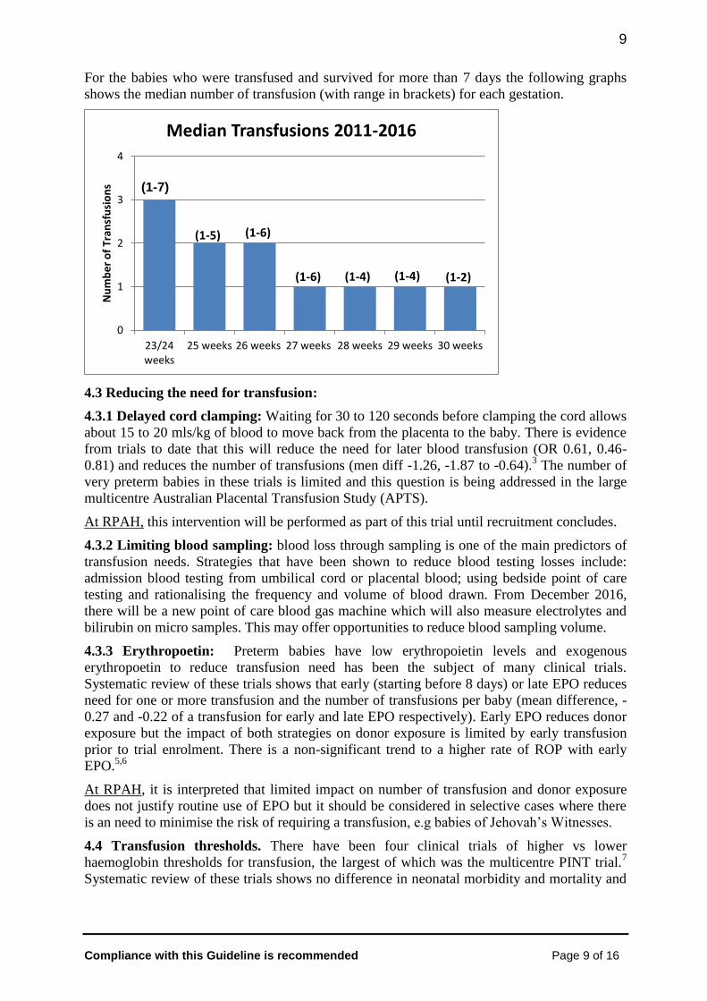
For the babies who were transfused and survived for more than 7 days the following graphs
shows the median number of transfusion (with range in brackets) for each gestation.
4.3 Reducing the need for transfusion:
4.3.1 Delayed cord clamping: Waiting for 30 to 120 seconds before clamping the cord allows
about 15 to 20 mls/kg of blood to move back from the placenta to the baby. There is evidence
from trials to date that this will reduce the need for later blood transfusion (OR 0.61, 0.46-
0.81) and reduces the number of transfusions (men diff -1.26, -1.87 to -0.64).3 The number of
very preterm babies in these trials is limited and this question is being addressed in the large
multicentre Australian Placental Transfusion Study (APTS).
At RPAH, this intervention will be performed as part of this trial until recruitment concludes.
4.3.2 Limiting blood sampling: blood loss through sampling is one of the main predictors of
transfusion needs. Strategies that have been shown to reduce blood testing losses include:
admission blood testing from umbilical cord or placental blood; using bedside point of care
testing and rationalising the frequency and volume of blood drawn. From December 2016,
there will be a new point of care blood gas machine which will also measure electrolytes and
bilirubin on micro samples. This may offer opportunities to reduce blood sampling volume.
4.3.3 Erythropoetin: Preterm babies have low erythropoietin levels and exogenous
erythropoetin to reduce transfusion need has been the subject of many clinical trials.
Systematic review of these trials shows that early (starting before 8 days) or late EPO reduces
need for one or more transfusion and the number of transfusions per baby (mean difference, -
0.27 and -0.22 of a transfusion for early and late EPO respectively). Early EPO reduces donor
exposure but the impact of both strategies on donor exposure is limited by early transfusion
prior to trial enrolment. There is a non-significant trend to a higher rate of ROP with early
EPO.5,6
At RPAH, it is interpreted that limited impact on number of transfusion and donor exposure
does not justify routine use of EPO but it should be considered in selective cases where there
is an need to minimise the risk of requiring a transfusion, e.g babies of Jehovah‟s Witnesses.
4.4 Transfusion thresholds. There have been four clinical trials of higher vs lower
haemoglobin thresholds for transfusion, the largest of which was the multicentre PINT trial.7
Systematic review of these trials shows no difference in neonatal morbidity and mortality and
0
1
2
3
4
23/24weeks
25 weeks 26 weeks 27 weeks 28 weeks 29 weeks 30 weeks
Nu
mb
er
of
Tran
sfu
sio
ns
Median Transfusions 2011-2016
(1-5) (1-6)
(1-6) (1-4) (1-4) (1-2)
(1-7)

10
Compliance with this Guideline is recommended Page 10 of 16
no difference in neurodevelopmental outcome at 18-21 months. Lower thresholds lead to a
significant reduction in the number of transfusions (mean diff -1.12 transfusions).8
At RPAH, the lack of benefit for a higher threshold indicates that we should be guided lower
haemoglobin thresholds as in the table below, which is modified from the PINT trial which
enrolled babies less than 31 weeks and less than 1000g.
Age Respiratory Support
(IPPV, CPAP, Hi Flow or Oxygen)
No Respiratory Support
<15 days <105 g/L <90 g/L
≥ 15 days <80 g/L <70 g/L
These thresholds are for guidance only and should be individualised considering the following
factors:
Severity of respiratory disease (and other condition)
Any symptoms that might relate to anaemia e.g increasing apnoeas.
The reticulocyte count.
Any evidence of on-going blood loss or haemolysis (e.g. a rhesus baby who‟d been
managed with intra-uterine transfusions).
Possible or proven sepsis
4.4 Process for Top up Blood Transfusion:
4.4.1 Consent: Informed written consent must be obtained from one of the parents in all but
the most critical situations, such as described for acute blood loss above. Use form MR 300:
Request/Consent for medical procedure/treatment – Minor.
4.4.2 Specimen collection: Initial group and cross-match will require 1.0ml of blood (into a
plain tube) from baby and 10mls of blood from mother. Because all mothers have blood taken
for cross match prior to going into labour, the blood bank may well have mother‟s blood.
Patient details must be hand written on the specimen tube. The specimens should be collected
in the presence of a witness who should confirm the identity of the patient against that on the
tube and request form and confirm that with a signature.
4.4.3 Request forms: Group and Screen/Cross-match orders are placed on a manual hand
written ‘Request for Blood Bank Services’ form. After the first transfusion, blood can be
ordered by contacting blood bank on 88033 and confirming they do not need a further
specimen for cross match. Orders are collected from blood bank with the ‘RPAH – Blood
Product Issue’ pink form.
4.4.4 Blood for top up transfusion should be:
Leucocyte depleted, CMV negative and irradiated. Irradiation for RPAH now occurs
off site so this may lead to a few hours delay in blood being available.
Infused through a leucocyte filter.
The volume should be prescribed on the fluid chart to run over four hours. There is no
indication for routine frusemide9 but consider giving 1mg/kg IV at the start of the
transfusion if there is risk of fluid overload.

11
Compliance with this Guideline is recommended Page 11 of 16
4.4.5 Volume for transfusion: There are two methods for calculating volume for transfusion:
1. A standard volume of 20mls/kg: This will suffice for most top up transfusions in
preterm babies.
2. An individualised volume: Based on current and target haemoglobin using the formula:
Transfusion Volume (mls) = Weight (kg) x Blood Volume (ml/kg)1 x (desired Hb – current
Hb) x Hb of donor blood2
Estimated blood volume will range from 100mls/kg in an extremely preterm baby to 80
mls/kg in a term baby.
Estimated haemoglobin (g/L) of leucocyte depleted donor red cells is approximately
210 g/L
Note that the “Blood and Blood Product Transfusion” sticker shown below should be
completed and stuck on the current continuation sheet in the medical record when the blood
product transfusion is set up.
5. Complications of red cell transfusion.
Complications of red cell transfusion are rare and most likely to be seen after massive or
exchange transfusion but include.
Transfusion reactions: Very unusual in the newborn due to limited ability to generate
an allergic response.
Immunological complications: e.g transfusion related lung injury, transfusion
associated graft vs host (the reason blood for neonatal transfusion is irradiated).
Metabolic complications: Hypocalcaemia, hypomagnesaemia, hypo or hyperkalaemia,
metabolic alkalosis or acidosis, impaired glucose homeostasis and hypothermia (the
reason we warm blood in exchange transfusion.
Coagulopathy (as described above) due to inadequate replacement of coagulation
factors during massive or exchange transfusion.
Infusion complications: e.g infection, air embolus.
12
Compliance with this Guideline is recommended Page 12 of 16
Transfusion associated NEC (TANEC). Defined as NEC developing within 48 hrs of a
blood transfusion. Currently uncertain whether this is association or causation. Meta-
analysis of observational studies reports moderate risk of bias and that the reports do
not address the fact that there are many cases of NEC that are not associated with
transfusion, and many transfusions happen in preterm neonates who are not linked to
NEC within 48 hours.10
Some authors have suggested that withholding feeds around a
transfusion may reduce the risk of TANEC.11
The level of evidence around this is low
therefore it is not routine practice at RPAH to withhold feeds during transfusion.
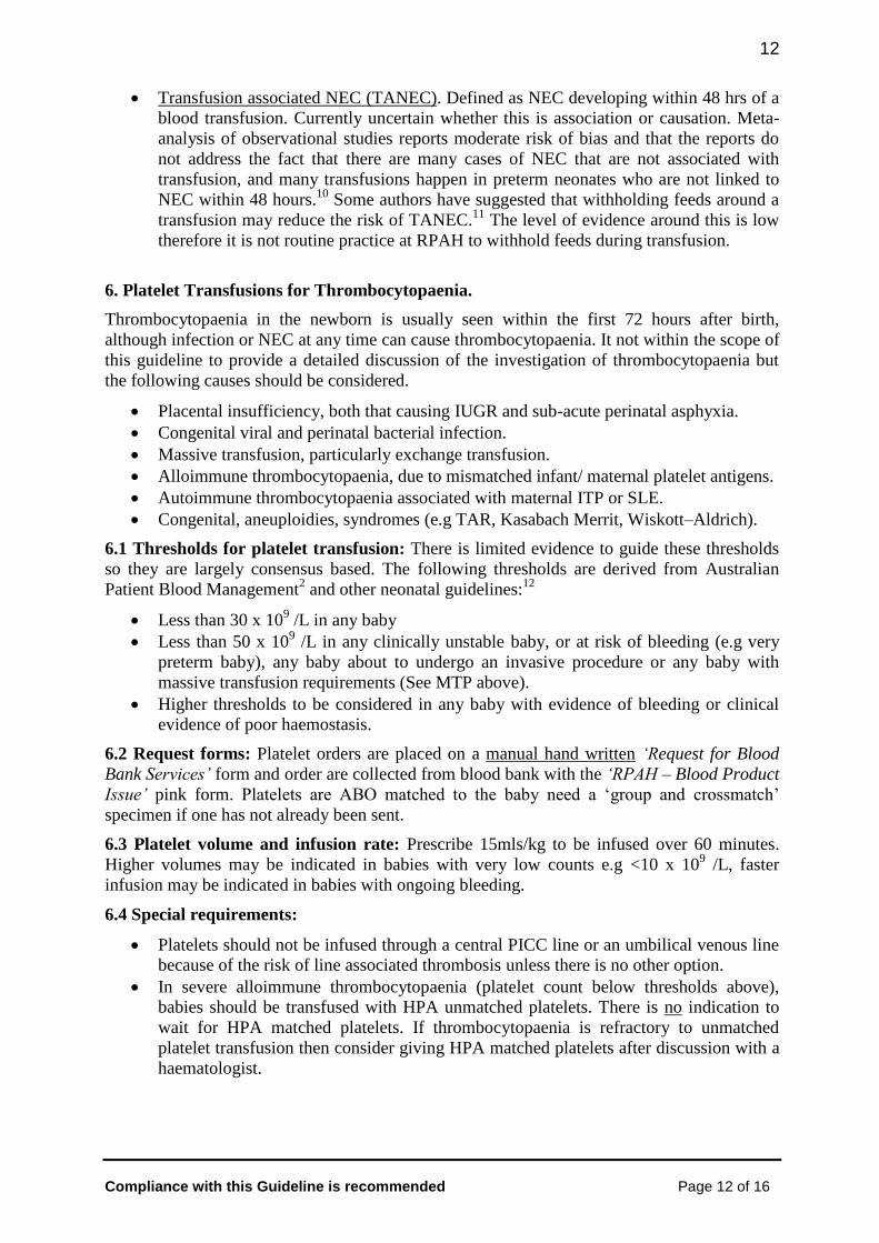
6. Platelet Transfusions for Thrombocytopaenia.
Thrombocytopaenia in the newborn is usually seen within the first 72 hours after birth,
although infection or NEC at any time can cause thrombocytopaenia. It not within the scope of
this guideline to provide a detailed discussion of the investigation of thrombocytopaenia but
the following causes should be considered.
Placental insufficiency, both that causing IUGR and sub-acute perinatal asphyxia.
Congenital viral and perinatal bacterial infection.
Massive transfusion, particularly exchange transfusion.
Alloimmune thrombocytopaenia, due to mismatched infant/ maternal platelet antigens.
Autoimmune thrombocytopaenia associated with maternal ITP or SLE.
Congenital, aneuploidies, syndromes (e.g TAR, Kasabach Merrit, Wiskott–Aldrich).
6.1 Thresholds for platelet transfusion: There is limited evidence to guide these thresholds
so they are largely consensus based. The following thresholds are derived from Australian
Patient Blood Management2 and other neonatal guidelines:
12
Less than 30 x 109 /L in any baby
Less than 50 x 109 /L in any clinically unstable baby, or at risk of bleeding (e.g very
preterm baby), any baby about to undergo an invasive procedure or any baby with
massive transfusion requirements (See MTP above).
Higher thresholds to be considered in any baby with evidence of bleeding or clinical
evidence of poor haemostasis.
6.2 Request forms: Platelet orders are placed on a manual hand written ‘Request for Blood
Bank Services’ form and order are collected from blood bank with the ‘RPAH – Blood Product
Issue’ pink form. Platelets are ABO matched to the baby need a „group and crossmatch‟
specimen if one has not already been sent.
6.3 Platelet volume and infusion rate: Prescribe 15mls/kg to be infused over 60 minutes.
Higher volumes may be indicated in babies with very low counts e.g <10 x 109 /L, faster
infusion may be indicated in babies with ongoing bleeding.
6.4 Special requirements:
Platelets should not be infused through a central PICC line or an umbilical venous line
because of the risk of line associated thrombosis unless there is no other option.
In severe alloimmune thrombocytopaenia (platelet count below thresholds above),
babies should be transfused with HPA unmatched platelets. There is no indication to
wait for HPA matched platelets. If thrombocytopaenia is refractory to unmatched
platelet transfusion then consider giving HPA matched platelets after discussion with a
haematologist.

13
Compliance with this Guideline is recommended Page 13 of 16
7. Fresh Frozen Plasma (FFP) Transfusion for Coagulopathy.
FFP is a source of all clotting factors including V, VIII and Fibrinogen. It may be indicated in
clinical situations where there is reduces production or increased losses of clotting factors.
Investigation of coagulopathy is not within the brief of this guideline but the following causes
should be considered in any baby with deranged clotting studies.
Heparin contamination of sample if taken from umbilical or peripheral arterial line.
Asphyxia, particularly if there are clinical pointers to longer standing sub-acute fetal
compromise.
Disseminated intravascular coagulation, usually together with thrombocytopaenia in a
baby with sepsis.
Transfusion associated coagulopathy, due to inadequate replacement of clotting factors
in a baby needing massive or exchange transfusion
Vitamin K deficiency, unlikely if they baby has had intramuscular Vit K prophylaxis
Inherited clotting factor deficiencies e.g haemophilia.
Inherited liver disease e.g neonatal haemochromatosis.
7.1 Threshold for FFP transfusion: An INR ≤ 2.0 should not be associated with an increased
risk of serious bleeding. FFP should be considered with an INR > 2.0 depending on the
underlying cause and the clinical condition of the baby.
7.2 Request forms:
FFP is ordered by contacting blood bank on 58033 and is collected from blood bank
with the „RPAH – Blood Product Issue‟ pink form. FFP issued for neonates is usually
group AB.
If Group AB is not available and a „group and cross match‟ specimen has not been
previously sent, then the blood bank may request a specimen depending on the urgency
of the clinical situation.
7.3 FFP volume and infusion rate: Prescribe 15mls/kg to be infused over 60 minutes. Faster
infusion may be indicated in babies with ongoing bleeding see Massive Transfusion Protocol
above.
14
Compliance with this Guideline is recommended Page 14 of 16
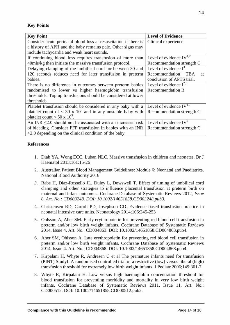
Key Points
Key Point Level of Evidence
Consider acute perinatal blood loss at resuscitation if there is
a history of APH and the baby remains pale. Other signs may
include tachycardia and weak heart sounds.
Clinical experience
If continuing blood loss requires transfusion of more than
40mls/kg then initiate the massive transfusion protocol.
Level of evidence IV1,2
Recommendation strength C
Delaying clamping of the umbilical cord for between 30 and
120 seconds reduces need for later transfusion in preterm
babies.
Level of evidence I3
Recommendation TBA at
conclusion of APTS trial.
There is no difference in outcomes between preterm babies
randomised to lower vs higher haemoglobin transfusion
thresholds. Top up transfusions should be considered at lower
thresholds.
Level of evidence I7,8
Recommendation B
Platelet transfusion should be considered in any baby with a
platelet count of < 30 x 109 and in any unstable baby with
platelet count < 50 x 109.
Level of evidence IV11
Recommendation strength C
An INR ≤2.0 should not be associated with an increased risk
of bleeding. Consider FFP transfusion in babies with an INR
>2.0 depending on the clinical condition of the baby.
Level of evidence IV2
Recommendation strength C
References
1. Diab YA, Wong ECC, Luban NLC. Massive transfusion in children and neonates. Br J
Haematol 2013;161:15-26
2. Australian Patient Blood Management Guidelines: Module 6: Neonatal and Paediatrics.
National Blood Authority 2016
3. Rabe H, Diaz-Rossello JL, Duley L, Dowswell T. Effect of timing of umbilical cord
clamping and other strategies to influence placental transfusion at preterm birth on
maternal and infant outcomes. Cochrane Database of Systematic Reviews 2012, Issue
8. Art. No.: CD003248. DOI: 10.1002/14651858.CD003248.pub3.
4. Christensen RD, Carroll PD, Josephson CD. Evidence based transfusion practice in
neonatal intensive care units. Neonatology 2014;106:245-253
5. Ohlsson A, Aher SM. Early erythropoietin for preventing red blood cell transfusion in
preterm and/or low birth weight infants. Cochrane Database of Systematic Reviews
2014, Issue 4. Art. No.: CD004863. DOI: 10.1002/14651858.CD004863.pub4.
6. Aher SM, Ohlsson A. Late erythropoietin for preventing red blood cell transfusion in
preterm and/or low birth weight infants. Cochrane Database of Systematic Reviews
2014, Issue 4. Art. No.: CD004868. DOI: 10.1002/14651858.CD004868.pub4.
7. Kirpalani H, Whyte R, Andresen C et al The premature infants need for transfusion
(PINT) StudyL A randomised controlled trial of a restrictive (low) versus liberal (high)
transfusion threshold for extremely low birth weight infants. J Pediatr 2006;149:301-7
8. Whyte R, Kirpalani H. Low versus high haemoglobin concentration threshold for
blood transfusion for preventing morbidity and mortality in very low birth weight
infants. Cochrane Database of Systematic Reviews 2011, Issue 11. Art. No.:
CD000512. DOI: 10.1002/14651858.CD000512.pub2.
15
Compliance with this Guideline is recommended Page 15 of 16
9. Balegar V, Kumar K et al. Furosemide for Packed Red Cell Transfusion in Preterm
Infants: A Randomized Controlled Trial. J Pediatr 2011;159: 913 - 918.e1
10. Mohamed A; Shah PS. Transfusion associated necrotizing enterocolitis: a meta-
analysis of observational data. Pediatrics 2012;129:529-40,
11. Keir AK; Wilkinson D. Do feeding practices during transfusion influence the risk of
developing necrotising enterocolitis in preterm infants? Archives of Disease in
Childhood 2013;98(5):386-8.
12. Sparger K, Deschmann E, Sola-Visner M. Platelet Transfusions in the Neonatal
Intensive Care Unit. Clinics in Perinatology. 2015;42(3):613-23.

16
Compliance with this Guideline is recommended Page 16 of 16
Minimum list (nothing to do with transfusions).
Legislative Compliance: Organisation, Management and Staff Obligations – Governing
Body and Management manual, Policy Number 2.7.1
Code of Conduct – Governing Body and Management Manual, Policy Number 1.1
Governing Body and Management Manual Section 4: Human Resources Management
– http://intranet.cs.nsw.gov.au/SSWPolicies/default.htm