Guide Question: Double Pump?. The main transport system in humans consists of blood, the heart and...
18
THE TRANSPORT SYSTEM Guide Question: Double Pump?
-
Upload
logan-stephens -
Category
Documents
-
view
216 -
download
1
Transcript of Guide Question: Double Pump?. The main transport system in humans consists of blood, the heart and...

THE TRANSPORT SYSTEM
Guide Question: Double Pump?
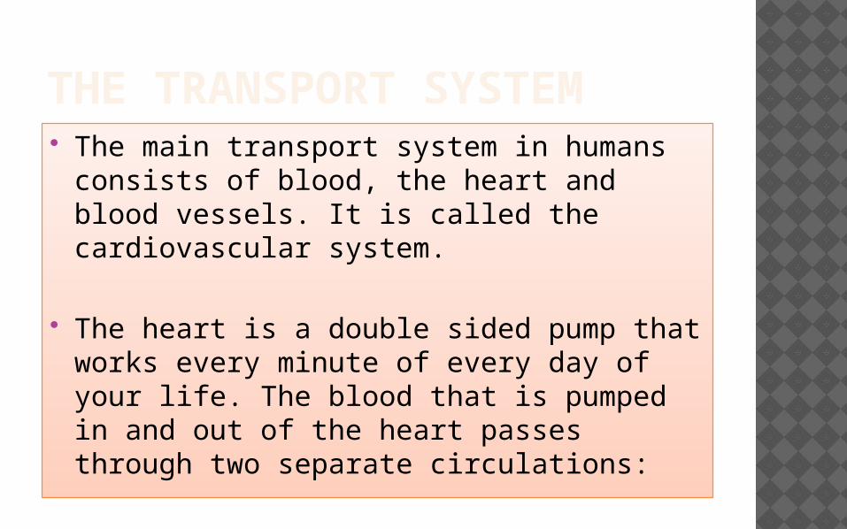
THE TRANSPORT SYSTEM The main transport system in humans
consists of blood, the heart and blood vessels. It is called the cardiovascular system.
The heart is a double sided pump that works every minute of every day of your life. The blood that is pumped in and out of the heart passes through two separate circulations:
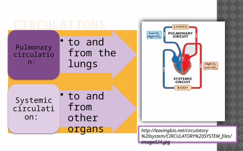
CIRCULATIONS• to and from
the lungsPulmonary circulation:
• to and from other organsSystemic
circulation:
http://leavingbio.net/circulatory%20system/CIRCULATORY%20SYSTEM_files/image034.jpg

The human heart has developed from simpler hearts in earlier organisms.
Use schemes to describe de differences observed in the hearts of other organisms including worms, amphibians, reptiles, birds and mammals (pg 171) .
Hypothesize how the human heart evolved into a double pump.

LABEL THE HUMAN HEART
http://www.abcteach.com/free/h/heartcrosssection_bw.jpg

THE RIGHT SIDE OF THE HEART SENDS BLOOD ALONG THE PULMONARY CIRCULATION.
Deoxygenated blood enters the right atrium
Right atrium contracts
Blood passes through the atrioventricular valve to the ventricle.
Right atrioventricular valve closes to prevent backflow.
Right semilunar valve opens because of pressure, due to the contraction of the right ventricle.Blood enters the pulmonary artery and is sent to the lungs, where it becomes oxygenates.From the lungs, oxygenated blood returns to the heart through the pulmonary veins right into the left atrium.
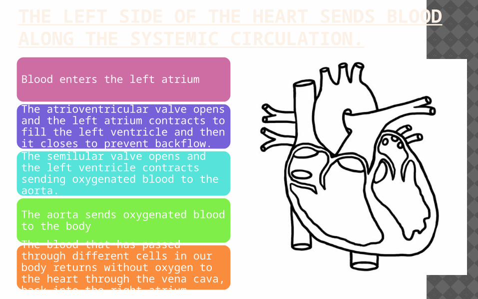
THE LEFT SIDE OF THE HEART SENDS BLOOD ALONG THE SYSTEMIC CIRCULATION.
Blood enters the left atrium
The atrioventricular valve opens and the left atrium contracts to fill the left ventricle and then it closes to prevent backflow.
The semilular valve opens and the left ventricle contracts sending oxygenated blood to the aorta.
The aorta sends oxygenated blood to the body
The blood that has passed through different cells in our body returns without oxygen to the heart through the vena cava, back into the right atrium.

Both atria and both ventricles contract simultaneously. The lup-dup sound is produced when the valves close
the lup is produced by the atrioventricular valves
the dup is produced by the semilunar valves.

CONTROL OF THE HEART BEATThe contractions of
the heart are myogenic (self estimulated)
Myogenic activity is timed
The region of the heart that sets the
overall pace of contraction is the
Sino-atrial node (SA node) or pacemaker
http://www.cvphysiology.com/Arrhythmias/A002%20SAN-AVN.gif

CONTROL OF THE HEART BEATThe pacemaker is connected to two nerves that come from the medulla of the brain:• cranial or cardiac • vagus nerves
The pacemaker also responds to the hormone adrenalin by increasing the heart rate.
http://www.cvphysiology.com/Arrhythmias/A002%20SAN-AVN.gif

CONTROL OF THE HEART BEAT
In the heart, another mass of tissue called the atrio-ventricular node (AV node) receives signals from the SA node and transmits the signal to the ventricles after 0.1 seconds approximately.
This explains why the atria contract first and then the ventricles.
http://www.cvphysiology.com/Arrhythmias/A002%20SAN-AVN.gif

BLOOD VESSELS:
There are three types of blood vessels: arteries, veins and capillaries.
Arteries are blood vessels that take blood away from the heart.
Veins are blood vessels that take blood towards the heart.
Capillaries are thin blood vessels found between an artery and a vein.
http://www.nurse-prescriber.co.uk/education/visual_lib/pp109.jpg

THE WALLS OF THESE BLOOD VESSELS CONSIST OF A SERIES OF LAYERS:
Tunica
intima:
•inner layer
•consisting of a single layer of epithelium cells, which forms a low-friction lining.
Tunica
media:
•layer of tissue containing circular muscle fibers and elastin fibers
•can stretch to accommodate blood and then contract to pump the blood onwards.
Tunica adventitia:
•outer layer of tissue
•containing longitudinal collagen and elastin fibers, which increase the overall strength of the wall and links it surrounding connective tissue.
http://www.nurse-prescriber.co.uk/education/visual_lib/pp109.jpg
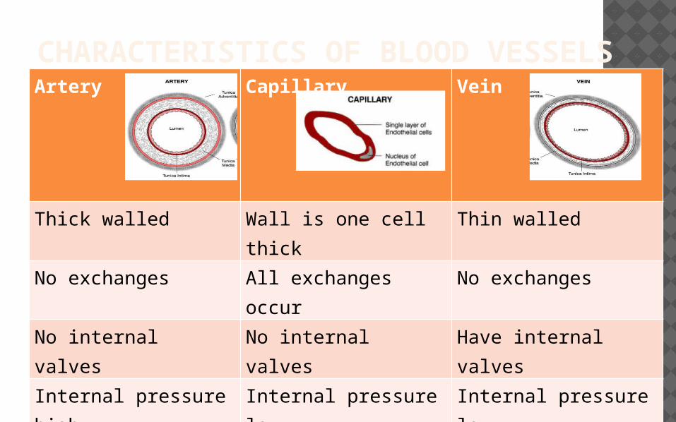
CHARACTERISTICS OF BLOOD VESSELSArtery Capillary Vein
Thick walled Wall is one cell thick Thin walled
No exchanges All exchanges occur No exchanges
No internal valves No internal valves Have internal valves
Internal pressure high
Internal pressure low Internal pressure low

THE STRUCTURE OF BLOOD:
Plasma 55%
45% Cells
99% Red blood cells – erythrocytes
0,5 % White blood cells –
leucocytes
0,5% Platelets
http://www.jonbarron.org/sites/default/files/images/blood_components.gif

PLASMA IS A FLUID THAT CONTAINS:
Water
dissolved nutrients • Glucose• Dissolved oxygen and carbon
dioxide Waste products • Urea
Hormones
Mineral salts
Plasma proteins• Antibodies
Heat
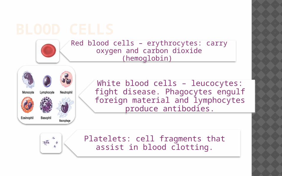
BLOOD CELLSRed blood cells – erythrocytes: carry oxygen
and carbon dioxide (hemoglobin)
White blood cells – leucocytes: fight disease. Phagocytes engulf foreign material and lymphocytes produce
antibodies.
Platelets: cell fragments that assist in blood clotting.

SOURCES: Regents Biology (2012 – 2013) Animal Nutrition,
Human Digestion. (On Line) http://www.westlake.k12.oh.us/whsteachers/ crist/2012_2013/Bio/digestion_35.pdf. PPT modified from this source.
Allot, Andrew and Mindorff, David (2010) Biology Course Companion, (2nd ed.). Oxford University Press, New York.
Damon, Alan; McGonegal, Randy; Tosto, Patricia; and Ward, William. (2007) Biology Higher Level, Pearson Baccalaureate, London.