
Guia de Pie Diabetico Infectado
69
Authors B. A. Lipsky 1 , J. Aragón-Sánchez 2 , M. Diggle 3 , J. Embil 4 , S. Kono 5 , L. Lavery 6 , É. Senneville 7 , V . Urbancic-Rovan 8 , S. Van Asten 6,9 , E. J. G. Peters 9 ; on behalf of the International Working Group on the Diabetic Foot (IWGDF) Institutions 1 Geneva University Hospitals and Faculty of Medicine, Geneva, Switzerland, and University of Oxford, Oxford, UK 2 La Paloma Hospital, Las Palmas de Gran Canaria, Spain 3 Nottingham University Hospitals Trust, Nottingham, UK 4 University of Manitoba, Winnipeg, MB, Canada 5 WHO-collaborating Centre for Diabetes, National Hospital Organization, Kyoto Medical Center, Kyoto, Japan 6 University of T exas Southwestern Medical Center and Parkland Hospital, Dallas, Texas 7 Gustave Dron Hospital, Tourcoing, France 8 University Medical Centre, Ljubljana, Slovenia 9 VU University Medical Centre, Amsterdam, The Netherlands Address of corr espondence Benjamin A. Lipsky. 79 Stone Meadow, Oxford, UK OX2 6TD [email protected]. Prepared by the IWGDF Working Group on Foot Infections IWGDF Guidance on the diagnosis and management of foot infections in persons with diabetes © 2015 International Working Group on the Diabetic Foot Recommendations Introduction Pathophysiology Diagnosis and Classifcation Soft tissue infection Osteomyelitis Assessing severity Microbiological considerations Treatment Key Controversies References IWGDF Guidance on the diagnosis and management of foot infections in persons with diabetes Systematic review
-
Upload
oscar-fabian-rojas -
Category
Documents
-
view
226 -
download
0
description
:)
Transcript of Guia de Pie Diabetico Infectado

7/21/2019 Guia de Pie Diabetico Infectado
http://slidepdf.com/reader/full/guia-de-pie-diabetico-infectado 1/69
Authors
B. A. Lipsky1, J. Aragón-Sánchez2, M. Diggle3, J. Embil4, S. Kono5, L. Lavery6, É. Senneville7,V. Urbancic-Rovan8, S. Van Asten6,9, E. J. G. Peters9; on behalf of the International Working Group on theDiabetic Foot (IWGDF)
Institutions1 Geneva University Hospitals and Faculty of Medicine, Geneva, Switzerland, and University of Oxford,
Oxford, UK2 La Paloma Hospital, Las Palmas de Gran Canaria, Spain3 Nottingham University Hospitals Trust, Nottingham, UK4 University of Manitoba, Winnipeg, MB, Canada5 WHO-collaborating Centre for Diabetes, National Hospital Organization, Kyoto Medical Center, Kyoto,
Japan6 University of Texas Southwestern Medical Center and Parkland Hospital, Dallas, Texas7 Gustave Dron Hospital, Tourcoing, France8 University Medical Centre, Ljubljana, Slovenia9 VU University Medical Centre, Amsterdam, The Netherlands
Address of correspondence
Benjamin A. Lipsky. 79 Stone Meadow, Oxford, UK OX2 [email protected].
Prepared by the IWGDF Working Group on Foot Infections
IWGDF Guidance on the diagnosis and managementof foot infections in persons with diabetes
© 2015 International Working Group on the Diabetic Foot
Recommendations
Introduction
Pathophysiology
Diagnosis and
Classifcation
Soft tissue infection
Osteomyelitis
Assessing severity
Microbiological
considerations
Treatment
Key Controversies
References
IWGDF Guidance on the diagnosis and management of foot infections in persons with diabetes
Systematic review

7/21/2019 Guia de Pie Diabetico Infectado
http://slidepdf.com/reader/full/guia-de-pie-diabetico-infectado 2/69
© 2015 International Working Group on the Diabetic Foot
Prepared by the IWGDF Working Group on Foot Infections
IWGDF Guidance on the diagnosis and managementof foot infections in persons with diabetes
Recommendations
Introduction
Pathophysiology
Diagnosis and
Classifcation
Soft tissue infection
Osteomyelitis
Assessing severity
Microbiological
considerations
Treatment
Key Controversies
References
Recommendations
Classification/Diagnosis
1. Diabetic foot infection must be diagnosed clinically, based on the presence of local or systemic signs or
symptoms of inflammation (Strong; Low).
2. Assess the severity of any diabetic foot infection using the Infectious Diseases Society of
America/International Working Group on the Diabetic Foot classification scheme (Strong; Moderate)
Osteomyelitis3. For an infected open wound, perform a probe-to-bone test; in a patient at low risk for osteomyelitis a nega-
tive test largely rules out the diagnosis, while in a high risk patient a positive test is largely diagnostic
(Strong; High)
4. Markedly elevated serum inflammatory markers, especially erythrocyte sedimentation rate, are suggestive of
osteomyelitis in suspected cases (Weak; Moderate)
5. A definite diagnosis of bone infection usually requires positive results on microbiological (and, optimally,
histological) and examinations of an aseptically obtained bone sample, but this is usually required only when
the diagnosis is in doubt or determining the causative pathogen’s antibiotic susceptibility is crucial (Strong;
Moderate)
6. A probable diagnosis of bone infection is reasonable if there are positive results on a combination of diag-
nostic tests, such as probe-to-bone, serum inflammatory markers, plain X-ray, MRI or radionuclide scanning
(Strong; Weak)
7. Avoid using results of soft tissue or sinus tract specimens for selecting antibiotic therapy for osteomyelitis as
they do not accurately reflect bone culture results (Strong; Moderate)
8. Obtain plain X-rays of the foot in all cases of non-superficial diabetic foot infection. (Strong; Low)
Systematic review

7/21/2019 Guia de Pie Diabetico Infectado
http://slidepdf.com/reader/full/guia-de-pie-diabetico-infectado 3/69
© 2015 International Working Group on the Diabetic Foot
9. Use MRI when an advanced imaging test is needed for diagnosing diabetic foot osteomyelitis
(Strong; Moderate)
10. When MRI is not available or contraindicated, consider a white blood cell-labelled radionuclide scan, or pos-
sibly SPECT/CT or 18 F- FDG PET/CT scans (Weak; Moderate)
Assessing severity
11. At initial evaluation of any infected foot, obtain vital signs and appropriate blood tests, debride the wound,
probe and assess the depth and extent of the infection to establish its severity (Strong; Moderate)
12. At initial evaluation assess arterial perfusion and decide whether and when further vascular assessment or
revascularization is needed (Strong; Low)
Microbiological considerations
13. Obtain cultures, preferably of a tissue specimen rather than a swab, of infected wounds to determine the
causative microorganisms and their antibiotic sensitivity (Strong; High)
14. Do not obtain repeat cultures unless the patient is not clinically responding to treatment, or occasionally for
infection control surveillance of resistant pathogens (Strong; Low)
15. Send collected specimens to the microbiology laboratory promptly, in sterile transport containers,
accompanied by clinical information on the type of specimen and location of the wound (Strong; Low)
Surgical treatment
16. Consult a surgical specialist in selected cases of moderate, and all cases of severe, DFI (Weak; Low)
17. Perform urgent surgical interventions in cases of deep abscesses, compartment syndrome and virtually all
necrotizing soft tissue infections (Strong; Low)
18. Consider surgical intervention in cases of osteomyelitis accompanied by: spreading soft tissue infection;
destroyed soft tissue envelope; progressive bone destruction on X-ray, or bone protruding through the ulcer
(Strong; Low)
Prepared by the IWGDF Working Group on Foot Infections
IWGDF Guidance on the diagnosis and managementof foot infections in persons with diabetes
Recommendations
Introduction
Pathophysiology
Diagnosis and
Classifcation
Soft tissue infection
Osteomyelitis
Assessing severity
Microbiological
considerations
Treatment
Key Controversies
References
Systematic review

7/21/2019 Guia de Pie Diabetico Infectado
http://slidepdf.com/reader/full/guia-de-pie-diabetico-infectado 4/69
© 2015 International Working Group on the Diabetic Foot
Antimicrobial therapy
19. While virtually all clinically infected diabetic foot wounds require antimicrobial therapy do not treat clinically
uninfected wounds with antimicrobial therapy (Strong; Low)
20. Select specific antibiotic agents for treatment based on the likely or proven causative pathogens, their
antibiotic susceptibilities, the clinical severity of the infection, evidence of efficacy of the agent for DFI and
costs (Strong; Moderate)
21. A course of antibiotic therapy of 1-2 weeks is usually adequate for most mild and moderate infections
(Strong; High)
22. Administer parenteral therapy initially for most severe infections and some moderate infections, with a switch
to oral therapy when the infection is responding (Strong; Low)
23. Do not select a specific type of dressing for a diabetic foot infection with the aim of preventing an infection or
improving its outcome (Strong; High)
24. For diabetic foot osteomyelitis we recommend 6 weeks of antibiotic therapy for patients who do not undergo
resection of infected bone and no more than a week of antibiotic treatment if all infected bone is resected.
(Strong; Moderate)
25. We suggest not using any adjunctive treatments for diabetic foot infection. (Weak; Low)
26. When treating a diabetic foot infection, assess for use of traditional remedies, previous antibiotic use, and
consider local bacterial pathogens and their susceptibility profile. (Strong; Low)
IWGDF Guidance on the diagnosis and management of foot infections in persons with diabetes
Prepared by the IWGDF Working Group on Foot Infections
IWGDF Guidance on the diagnosis and managementof foot infections in persons with diabetes
Recommendations
Introduction
Pathophysiology
Diagnosis and
Classifcation
Soft tissue infection
Osteomyelitis
Assessing severity
Microbiological
considerations
Treatment
Key Controversies
References
Systematic review

7/21/2019 Guia de Pie Diabetico Infectado
http://slidepdf.com/reader/full/guia-de-pie-diabetico-infectado 5/69
© 2015 International Working Group on the Diabetic Foot
Prepared by the IWGDF Working Group on Foot Infections
IWGDF Guidance on the diagnosis and managementof foot infections in persons with diabetes
Introduction
In the recent decades as the prevalence of diabetes has increased, so too have foot complications, including
infections. The development of a foot infection is associated with substantial morbidity, including discomfort,
reduced physical and mental quality of life (2), need for healthcare provider visits, wound care, antimicrobial
therapy, and often surgical procedures. Furthermore, foot infection remains the most frequent diabetic
complication requiring hospitalization and the most common precipitating event leading to lower extremity
amputation (3-6). Managing infection requires careful attention to properly diagnosing the condition, obtaining
appropriate specimens for culture, thoughtfully selecting empirical and then definitive antimicrobial therapy,
quickly determining when surgical interventions are needed and providing all other necessary types of woundcare. For these reasons interdisciplinary teams should, whenever possible, include an infectious diseases or
clinical microbiology specialist (7). A systematic and, to the extent possible, evidence-based approach to
diabetic foot infections (DFIs) should result in better outcomes.
Formulation of recommendations
This report from the expert panel on infectious diseases of the International Working Group on the Diabetic Foot
(IWGDF) is an update of the one published in 2012 (8). It incorporates some information from the concurrently
published ‘Systematic Review of Interventions in the Management of Infection in the Diabetic Foot’ (9) as well as
non-systematic reviews of the literature covering each of the sections in this guidance. Our intention is to present
a brief overview to assist clinicians worldwide in diagnosing and treating foot infections in persons with diabetes.
This document follows the newly adopted format of all IWGDF guidance documents, including providing
recommendations that are rated based on the GRADE system1.
Recommendations
Introduction
Pathophysiology
Diagnosis and
Classifcation
Soft tissue infection
Osteomyelitis
Assessing severity
Microbiological
considerations
Treatment
Key Controversies
References
Systematic review

7/21/2019 Guia de Pie Diabetico Infectado
http://slidepdf.com/reader/full/guia-de-pie-diabetico-infectado 6/69
© 2015 International Working Group on the Diabetic Foot
Prepared by the IWGDF Working Group on Foot Infections
IWGDF Guidance on the diagnosis and managementof foot infections in persons with diabetes
Recommendations
Introduction
Pathophysiology
Diagnosis and
Classifcation
Soft tissue infection
Osteomyelitis
Assessing severity
Microbiological
considerations
Treatment
Key Controversies
References
1 Recommendations in this guidance were formulated based on the Grading of Recommendations Assessment,
Development and Evaluation (GRADE) system for grading evidence when writing a clinical guideline (1).
For much of the older data found in the systematic review underlying this guidance we could not calculate or
assess for inconsistency, indirectness or imprecision, which are needed to fully assess the quality of evidence.
Therefore, we decided to assess the quality of evidence on: the risk of bias of included studies, effect sizes, and
expert opinion, and rate the quality of evidence as ‘high’, ‘moderate’, or ‘low’. We assessed the strength of each
recommendation as ‘strong’ or ‘weak’, based on the quality of evidence, balance between benefits and harms,
patient values and preferences, and costs (resource utilization). The rationale behind each recommendation is
described in this guidance.
Systematic review
IWGDF Guidance on the diagnosis and management of foot infections in persons with diabetes

7/21/2019 Guia de Pie Diabetico Infectado
http://slidepdf.com/reader/full/guia-de-pie-diabetico-infectado 7/69
© 2015 International Working Group on the Diabetic Foot
Prepared by the IWGDF Working Group on Foot Infections
IWGDF Guidance on the diagnosis and managementof foot infections in persons with diabetes
Recommendations
Introduction
Pathophysiology
Diagnosis and
Classifcation
Soft tissue infection
Osteomyelitis
Assessing severity
Microbiological
considerations
Treatment
Key Controversies
References
Systematic review
Pathophysiology
Diabetic foot infections
In persons with diabetes, foot infection is an increasingly common problem that is related to the duration of the
disease, and therefore the likelihood of diabetic complications. Infection is best defined as an invasion and
multiplication of microorganisms in host tissues that induces a host inflammatory response, usually followed
by tissue destruction. DFI is defined clinically as manifestations of this process in soft tissue or bone anywhere
below the malleoli in a person with diabetes. These infections usually begin with a break in the protective
cutaneous envelope, typically in a site of trauma or ulceration (10). Peripheral neuropathy (mostly sensory, but
also motor and autonomic) is the main factor leading to skin breaks; these open wounds then becomecolonised (usually with skin flora) and, in many cases, ultimately infected. Foot ischemia, related to peripheral
arterial disease, is also common in patients with a DFI. While rarely the primary cause of foot wounds, the
presence of limb ischemia increases the risk of a wound becoming infected (11,12) and adversely affects the
outcome of infection (6,13). Foot wounds in diabetic patients often become chronic, related to hyperglycemia-
induced advanced glycation end-products, persistent inflammation and apoptosis (14,15). Factors that
predispose to foot infection include having: a wound that is deep, long-standing or recurrent, or of traumatic
aetiology; ill-defined diabetes-related immunological perturbations related to neutrophil function; and, chronic
renal failure (11,16-19).
Spread of infection
While most DFIs are relatively superficial at presentation, microorganisms can spread contiguously to
subcutaneous tissues, including fascia, tendons, muscle, joints and bone. The anatomy of the foot, which is
divided into several rigid but intercommunicating compartments, fosters proximal spread of infection (20).
The inflammatory response induced by infection may cause compartmental pressure to exceed capillary
pressure, leading to ischaemic tissue necrosis (21,22). The tendons within the compartments facilitate proximal
spread of infection, which usually moves from higher to lower pressure areas. Bacterial virulence factors may
also play a role in these complex infections. Strains of Staphylococcus aureus isolated from clinically non-
infected ulcers have been shown to have a lower virulence potential than from those that are infected (23).
Similarly, a clonal complex 398 methicillin-susceptible S. aureus with a tropism for bone has emerged as the
main staphylococcal pathogen in one outbreak of diabetic foot osteomyelitis (DFO) (24).

7/21/2019 Guia de Pie Diabetico Infectado
http://slidepdf.com/reader/full/guia-de-pie-diabetico-infectado 8/69
© 2015 International Working Group on the Diabetic Foot
Systemic symptoms
Systemic symptoms (e.g., feverishness, chills), marked leukocytosis or major metabolic disturbances are uncom-
mon in patients with a DFI, but their presence denotes a more severe, potentially limb (or even life) threatening
infection (6). If not diagnosed and properly treated, DFIs tend to progress, sometimes rapidly (25). Thus, an expe-
rienced consultant (or team) should see a patient with a severe DFI within 24 hours (26).
Prepared by the IWGDF Working Group on Foot Infections
IWGDF Guidance on the diagnosis and managementof foot infections in persons with diabetes
Recommendations
Introduction
Pathophysiology
Diagnosis and
Classifcation
Soft tissue infection
Osteomyelitis
Assessing severity
Microbiological
considerations
Treatment
Key Controversies
References
Systematic review
IWGDF Guidance on the diagnosis and management of foot infections in persons with diabetes

7/21/2019 Guia de Pie Diabetico Infectado
http://slidepdf.com/reader/full/guia-de-pie-diabetico-infectado 9/69
© 2015 International Working Group on the Diabetic Foot
Prepared by the IWGDF Working Group on Foot Infections
IWGDF Guidance on the diagnosis and managementof foot infections in persons with diabetes
Recommendations
Introduction
Pathophysiology
Diagnosis and
Classifcation
Soft tissue infection
Osteomyelitis
Assessing severity
Microbiological
considerations
Treatment
Key Controversies
References
Systematic review
Diagnosis and Classification
Recommendation 1:
Diabetic foot infection must be diagnosed clinically, based on the presence of local and systemic signs and
symptoms of inflammation (Strong; moderate).
Recommendation 2:
Assess the severity of any diabetic foot infection using the Infectious Diseases Society of America/International
Working Group on the Diabetic Foot classification scheme (Strong; Moderate)
Rationale 1& 2:
The clinician seeing a patient with a diabetic foot wound should first assess for the presence of a DFI and,
if present, classify the infection’s severity. Over the past three decades experts have proposed many
classification schemes for diabetic foot wounds. Most of these take into account the size and depth of the ulcer,
and the presence or absence of gangrene, neuropathy, or arterial insufficiency. Several diabetic foot ulcer
classifications only include the presence or absence of “infection” (which is undefined). Only two, nearly
identical, schemes proposed by the Infectious Diseases Society of America and the IWGDF (the “infection” part
of the PEDIS classification) describe how to define both the presence and severity of infection (see Table 1)
(27-30). Several other guidelines, including ones produced by the Spanish, French and UK (NICE), have adopted
the IDSA/IWGDF infection classification (26,31-33).
The full PEDIS system (which includes classification of other wound descriptors, such as arterial disease,
neuropathy and wound size) of the IWGDF was originally developed for research purposes, but it can serve as
a clinical classification as well (29,34). Classification of DFIs using the full PEDIS system (35,36) or the infection
part of the IWGDF/IDSA DFI scheme (6) has been shown in several prospective studies to predict the need for
hospitalisation or lower extremity amputation. Two recently published retrospective cohort studies from one
centre addressed the issue of whether or not the presence of systemic inflammatory response syndrome (SIRS)
findings, which separate moderate from severe infections, actually predicts outcomes. They assessed the
differences in outcome between hospitalised patients without and with SIRS (i.e., PEDIS grade 3 versus grade 4)
with a DFI (37,38). In one study patients with grade 4 infections experienced a 7.1 fold higher risk of major
amputation and had a 4 day longer mean hospital stay compared to patients with grade 3 infections (37). In the
other publication, patients with grade 4 compared with grade 3 DFI had a significantly longer length of hospital

7/21/2019 Guia de Pie Diabetico Infectado
http://slidepdf.com/reader/full/guia-de-pie-diabetico-infectado 10/69
© 2015 International Working Group on the Diabetic Foot
Prepared by the IWGDF Working Group on Foot Infections
IWGDF Guidance on the diagnosis and managementof foot infections in persons with diabetes
Recommendations
Introduction
Pathophysiology
Diagnosis and
Classifcation
Soft tissue infection
Osteomyelitis
Assessing severity
Microbiological
considerations
Treatment
Key Controversies
References
stay (8 versus 5 days) and a non-significantly lower limb salvage rate (80% versus 94%) (38). Another recently
published retrospective cohort study reviewed outcomes in 57 DFI patients according to the level of adherence
of their clinicians to the IDSA practice guidelines (39). They found that rates of adherence to various
recommendations ranged from very high to very low, but in none of the patient treatment courses did clinicians
adhere to all. In this small and suboptimally designed study, adherence to the recommendations was not related
to clinical outcome, but patients with severe infections were more likely to have adverse outcomes. Surprisingly,
appropriate empiric and targeted antibiotic therapy was associated with treatment failure.
Systematic review

7/21/2019 Guia de Pie Diabetico Infectado
http://slidepdf.com/reader/full/guia-de-pie-diabetico-infectado 11/69
Clinical classification of infection, with definitions IWGDF / IDSA
classification
Uninfected: No systemic or local symptoms or signs of infection 1 (Uninfected)
Infected:
- At least 2 of the following items are present: 2 (Mild infection) • Local swelling or induration
• Erythema > 0.5 cm* around the wound • Local tenderness or pain • Local warmth • Purulent discharge
- Other causes of an inflammatory response of the skin should be excluded (e.g., trauma,
gout, acute Charcot neuro-osteoarthropathy, fracture, thrombosis, venous s tasis)
- Infection involving only the skin or subcutaneous tissue (without involvement of deeper
tissues and without systemic manifestations as described below).
- Any erythema present extends < 2 cm* around the wound - No systemic signs or symptoms of infection (see below)
- Infection involving structures deeper than skin and subcutaneous tissues (e.g., bone, 3 (Moderate infection)
joint, tendon, muscle) or erythema extending >2 cm* from the wound margin.
- No systemic signs or symptoms of infection (see below)
- Any foot infection with the systemic inflammatory response syndrome (SIRS), as 4 (Severe infection)
manifested by ≥2 of the following:
• Temperature >38˚ or <36˚ Celsius • Heart rate >90 beats/minute • Respiratory rate >20 breaths/minute or PaCO2 < 4.3 kPa (32 mmHg) • White blood cell count >12,000 or <4,000/mm3, or >10% immature (band) forms
© 2015 International Working Group on the Diabetic Foot
Note: *In any direction, from the rim of the wound; The presence of clinically significant foot ischemia makes both diagnosis and treatment of
infection considerably more difficult.
Prepared by the IWGDF Working Group on Foot Infections
IWGDF Guidance on the diagnosis and managementof foot infections in persons with diabetes
Recommendations
Introduction
Pathophysiology
Diagnosis and
Classifcation
Soft tissue infection
Osteomyelitis
Assessing severity
Microbiological
considerations
Treatment
Key Controversies
References
Systematic review
IWGDF Guidance on the diagnosis and management of foot infections in persons with diabetes
Table 1. The classification systems for defining the presence and severity of an infection of the foot in a person
with diabetes developed by the Infectious Diseases Society of America (IDSA) and the infection part of the PEDIS
classification of the International Working Group on the Diabetic Foot (IWGDF) (29,30).

7/21/2019 Guia de Pie Diabetico Infectado
http://slidepdf.com/reader/full/guia-de-pie-diabetico-infectado 12/69
© 2015 International Working Group on the Diabetic Foot
Prepared by the IWGDF Working Group on Foot Infections
IWGDF Guidance on the diagnosis and managementof foot infections in persons with diabetes
Recommendations
Introduction
Pathophysiology
Diagnosis and
Classifcation
Soft tissue infection
Osteomyelitis
Assessing severity
Microbiological
considerations
Treatment
Key Controversies
References
Systematic review
Soft tissue infection
Because all skin wounds harbour microorganisms, their mere presence (even if they are virulent species) cannot
be taken as evidence of infection. Some maintain that the presence of high numbers of bacteria (usually defined
as ≥105 colony forming units per gram per tissue) should be a basis for diagnosing infection (40), but no
convincing data support this concept for wounds, including in the diabetic foot (41). Furthermore, quantitative
microbiology is rarely available outside of research laboratories. Thus, DFI must be diagnosed clinically
(Table 1), with wound cultures serving to determine the causative organisms and their antibiotic sensitivities.
Clinicians should evaluate a diabetic patient presenting with a foot wound at three levels: the patient as a whole
(e.g., cognitive, metabolic, fluid status), the affected foot or limb (e.g., presence of neuropathy, vascularinsufficiency), and the infected wound (30). Clinical diagnosis rests on the presence of at least two local findings
of inflammation, that is, redness (erythema or rubor), warmth (calor), pain or tenderness (dolor), induration
(swelling or tumor) or purulent secretions (29,42). Other (sometimes called secondary) features suggestive of
infection include the presence of necrosis, friable or discoloured granulation tissue, non-purulent secretions,
foetid odour or the failure of a properly treated wound to heal (43). These findings may be helpful when local
and systemic inflammatory signs are diminished because of peripheral neuropathy or ischemia (44-46). Because
infection can worsen quickly, clinicians must pursue the diagnosis methodically (44) and aggressively (47).
All wounds must be carefully inspected, palpated and probed, both at initial presentation and on follow-up.
Various imaging and laboratory studies may be useful in some cases to define the extent of soft tissue infection
and any bone involvement.
IWGDF Guidance on the diagnosis and management of foot infections in persons with diabetes

7/21/2019 Guia de Pie Diabetico Infectado
http://slidepdf.com/reader/full/guia-de-pie-diabetico-infectado 13/69
© 2015 International Working Group on the Diabetic Foot
Prepared by the IWGDF Working Group on Foot Infections
IWGDF Guidance on the diagnosis and managementof foot infections in persons with diabetes
Recommendations
Introduction
Pathophysiology
Diagnosis and
Classifcation
Soft tissue infection
Osteomyelitis
Assessing severity
Microbiological
considerations
Treatment
Key Controversies
References
Systematic review
Osteomyelitis
Recommendation 3:
For an infected open wound, perform a probe-to-bone test; in a patient at low risk for osteomyelitis a negative
test largely rules out the diagnosis, while in a high risk patient a positive test is largely diagnostic (Strong; High)
Recommendation 4:
Markedly elevated serum inflammatory markers, especially erythrocyte sedimentation rate, are suggestive of
osteomyelitis in suspected cases (Weak; Moderate)
Recommendation 5:
A definite diagnosis of bone infection usually requires positive results on both histological and microbiological
examinations of an aseptically obtained bone sample, but this is usually required only when the diagnosis is in
doubt or determining the causative pathogen’s antibiotic susceptibility is crucial (Strong; Moderate)
Recommendation 6:
A probable diagnosing of bone infection is reasonable if there are positive results on a combination of
diagnostic tests, such as probe-to-bone, serum inflammatory markers, plain X-ray, MRI or radionuclide scanning
(Strong; Weak)
Recommendation 7:
Avoid using results of soft tissue or sinus tract specimens for selecting antibiotic therapy for osteomyelitis asthey do not accurately reflect bone culture results (Strong; Moderate)
Recommendation 8:
Obtain plain X-rays of the foot in all cases of non-superficial diabetic foot infection. (Strong; Low)
Recommendation 9:
Use MRI is when an advanced imaging test is needed for diagnosing diabetic foot osteomyelitis
(Strong; Moderate)

7/21/2019 Guia de Pie Diabetico Infectado
http://slidepdf.com/reader/full/guia-de-pie-diabetico-infectado 14/69
© 2015 International Working Group on the Diabetic Foot
Recommendation 10:
When MRI is not available or contraindicated, consider a white blood cell-labelled radionuclide scan, or possibly
SPECT/CT or 18 F- FDG PET scans (Weak; Moderate)
Rationale 3 - 10
Diabetic foot osteomyelitis (DFO) can present the clinician with formidable diagnostic and therapeutic
challenges (48). It is found in ~50%-60% of patients hospitalized for a DFI and ~10%-20% of apparently less
severe infections presenting in the ambulatory setting. Bone infection typically involves the forefoot (and less
often the hindfoot) and develops by contiguous spread from overlying soft tissue, penetration through the
cortical bone and into the medullary cavity. Bone destruction related to Charcot neuroostearthropathy (CN) may
be difficult to distinguish from DFO, but it is: less common; generally occurs in patients with profound peripheral
neuropathy (but usually adequate arterial perfusion); usually affects the midfoot; and, most often occurs in the
absence of a skin break (49-51). Many cases of DFO are monomicrobial, but most are polymicrobial, with
S. aureus the most commonly isolated pathogen (found in ~50% of cases), while coagulase-negative
staphylococci (~25%), aerobic streptococci (~30%) and Enterobacteriaceae (~40%) are other frequent isolates
(49).
Accurately diagnosing bone infection can be difficult, but is essential to ensure appropriate treatment. A definite
diagnosis of osteomyelitis requires both the presence of histological findings consistent with bone infection
(acute or chronic inflammatory cells, necrosis) and the isolation of bacteria from an aseptically obtained bone
sample (52). Because bone sampling and processing are not routinely available in many settings, clinicians must
often use surrogate diagnostic markers, including clinical, laboratory and imaging findings.The clinical presentation of osteomyelitis in the diabetic foot can vary with the site involved, the extent of infected
and dead bone, the presence of any associated abscess or soft tissue involvement, the causative organism(s)
and the adequacy of limb perfusion. The main problems in diagnosing osteomyelitis are that there is a delay in
the ability to detect bony changes in early infection on plain radiographs, while later when bony changes
occur it may be difficult to distinguish on imaging studies those caused by infection from those related to CN.
As discussed below, analyses from recent expert publications (52,53) and systematic reviews (52,54-56) provide
guidance on the best available diagnostic studies for DFO.
Prepared by the IWGDF Working Group on Foot Infections
IWGDF Guidance on the diagnosis and managementof foot infections in persons with diabetes
Recommendations
Introduction
Pathophysiology
Diagnosis and
Classifcation
Soft tissue infection
Osteomyelitis
Assessing severity
Microbiological
considerations
Treatment
Key Controversies
References
Systematic review

7/21/2019 Guia de Pie Diabetico Infectado
http://slidepdf.com/reader/full/guia-de-pie-diabetico-infectado 15/69
© 2015 International Working Group on the Diabetic Foot
Clinical evaluation
Clinicians should suspect osteomyelitis when an ulcer lies over a bony prominence, particularly when it fails to
heal despite adequate off-loading, or when a toe is erythematous and indurated (the so-called “sausage toe’).
The likelihood ratio (LR) of a clinician’s suspicion of osteomyelitis is surprisingly good, with a positive LR 5.5 and
negative LR 0.54 (54,55). Based on one study, the presence of exposed bone has a positive LR for osteomyelitis
of 9.2; large ulcers (area >2 cm2) are much more likely to have underlying bone infection (positive LR 7.2) than
smaller ones (negative LR 0.70) (54,55,57,58). Osteomyelitis can, however, occur in the absence of overlying
local signs of inflammation (57).
Probe-to-bone test
In the past two decades there have been at least seven published studies of the probe-to-bone test (51). When
performed correctly and interpreted appropriately, this is a useful clinical diagnostic tool for diagnosing DFO. If
a blunt sterile metal probe gently inserted through a wound strikes bone (detected by its hard, gritty feel), this
substantially increases the likelihood (positive LR 7.2, negative LR 0.48) that the patient has osteomyelitis if the
prevalence of bone infection is high (i.e., >~60%) in the population under scrutiny (59,60). Conversely, a negative
probe-to-bone test in a patient at low risk (i.e., ≤~20%) essentially rules out osteomyelitis (61-63).
The inter-observer variability of the test is relatively high for inexperienced clinicians compared to experienced
ones, but low between experienced clinicians (64). One study found a stronger correlation among clinicians’
results for ulcers located in the hallux and in the central metatarsals compared to the lesser toes (65). Combining
the results of the probe–to-bone test with those of plain radiography improves overall diagnostic accuracy of
osteomyelitis (59,64).
Blood tests
The erythrocyte sedimentation rate has proven to be useful in diagnosing DFO; a highly elevated (usually defined
as >70 mm/h) level increases the likelihood of osteomyelitis underlying a diabetic foot wound (positive LR 11),
while lower levels reduce the likelihood (negative LR of 0.34) (54,66-69). Based on fewer data, a highly elevated
C-reactive protein, procalcitonin or blood leukocyte count may be suggestive of osteomyelitis. These latter tests
tend to revert to normal levels within a week of treatment (70), while the ESR drops more slowly and can
therefore be useful for monitoring response to therapy. There is insufficient evidence to support the routine use
of any other biomarkers to document bone infection in the diabetic foot. A preliminary report suggested that
Prepared by the IWGDF Working Group on Foot Infections
IWGDF Guidance on the diagnosis and managementof foot infections in persons with diabetes
Recommendations
Introduction
Pathophysiology
Diagnosis and
Classifcation
Soft tissue infection
Osteomyelitis
Assessing severity
Microbiological
considerations
Treatment
Key Controversies
References
Systematic review

7/21/2019 Guia de Pie Diabetico Infectado
http://slidepdf.com/reader/full/guia-de-pie-diabetico-infectado 16/69
© 2015 International Working Group on the Diabetic Foot
interleukin (IL)-6, but not IL-8 may be useful in the diagnosis and follow up of diabetic foot infection (71-73).
Combining laboratory testing with clinical findings may improve the diagnostic accuracy for osteomyelitis (74).
Imaging studies
Plain radiography
Plain X-rays are often sufficient for imaging the foot in patients with suspicion of DFO. Characteristic features of
osteomyelitis on plain X-rays of the foot are summarized in Table 2. Advantages of this imaging test are that it: is
widely available (even in most centres with limited resources); has a relatively low cost; can be adequately read
by most experienced clinicians; and, is relatively easy to compare sequential radiographs over time. In addition
to bony changes, plain radiographs can demonstrate the presence of gas in the soft tissues or radiopaque f
oreign bodies. The results of two systematic reviews suggest that radiographic findings are only marginally
predictive of osteomyelitis if positive and even less predictive of the absence of osteomyelitis if negative (54,55).
While the reported sensitivity of radiography varies considerably in reported studies (57,75-82), the estimated
positive likelihood ratio (LR) is 2.3 and negative LR is 0.63 (56). The timing of the imaging greatly influences its
usefulness, as longer-standing cases are far more likely to show bony abnormalities on plain radiographs than
those present for less than two to three weeks. We know of no study that has evaluated sequential plain
radiographs of the foot over time, but changes seen over an interval of at least 2 weeks are more likely to predict
the presence of osteomyelitis than a single study. Of course, effective antibiotic therapy may prevent these bony
changes from occurring. Advanced imaging techniques are expensive, often limited in availability and difficult to
interpret by a non-expert. Thus, they are usually needed only, when there is persistent doubt about the diagnosis
of DFO or in the context of preparing a surgical intervention.
Prepared by the IWGDF Working Group on Foot Infections
IWGDF Guidance on the diagnosis and managementof foot infections in persons with diabetes
Recommendations
Introduction
Pathophysiology
Diagnosis and
Classifcation
Soft tissue infection
Osteomyelitis
Assessing severity
Microbiological
considerations
Treatment
Key Controversies
References
Systematic review

7/21/2019 Guia de Pie Diabetico Infectado
http://slidepdf.com/reader/full/guia-de-pie-diabetico-infectado 17/69
© 2015 International Working Group on the Diabetic Foot
Table 2: Typical features of diabetic foot osteomyelitis on plain X-rays* (57,75,76,103)
Periosteal reaction or elevation
Loss of bone cortex with bony erosion
Focal loss of cortical trabecular pattern or marrow radiolucency
Bone sclerosis, with or without erosion
Presence of sequestrum: devitalized bone with radiodense appearance that has become separated
from normal bone
Presence of involucrum: a layer of new bone growth outside previously existing bone resulting fromstripping off of the periosteum and new bone growing from the periosteum
Presence of cloacae: opening in the involucrum or cortex through which sequestrae or granulation tissue
may discharge
Presence of evidence of a sinus tract from the bone to the soft tissue
Note: * Some features (e.g., sequestrum, involucrum, cloacae) are seen less frequently in diabetic foot osteomyelitis than in younger patients with
osteomyelitis of larger bones.
Magnetic resonance imaging
Magnetic resonance imaging (MRI) is a valuable tool for diagnosing osteomyelitis, as well as defining the
presence and anatomy of deep soft tissue infections (30,55,83). The key features suggestive of osteomyelitison MRI are low focal signal intensity on T1-weighted images, high focal signal on T2-weighted images and high
bone marrow signal in short tau inversion recovery (STIR) sequences. Meta-analyses have found that the
sensitivity of MRI for DFO is about 90% and the specificity about 85%, diagnostic odds ratio (OR) of 24 (55,83)
and LRs estimated at positive of 3.8 and negative of 0.14. More recently performed studies reported lower
diagnostic OR compared with older ones, perhaps because they employed better study designs. The subgroups
of patients with other diagnoses (e.g., CN) were too small to analyse any differences among the studies. A recent
study found that MRI was effective in distinguishing DFO from bone marrow oedema in neuropathic ulcers but
was less accurate for the diagnosis of DFO in ischemic ulcers, presumably because of their insufficient interstitial
fluid (84).
Prepared by the IWGDF Working Group on Foot Infections
IWGDF Guidance on the diagnosis and managementof foot infections in persons with diabetes
Recommendations
Introduction
Pathophysiology
Diagnosis and
Classifcation
Soft tissue infection
Osteomyelitis
Assessing severity
Microbiological
considerations
Treatment
Key Controversies
References
Systematic review

7/21/2019 Guia de Pie Diabetico Infectado
http://slidepdf.com/reader/full/guia-de-pie-diabetico-infectado 18/69
© 2015 International Working Group on the Diabetic Foot
Nuclear medicine scans
Among the several types of nuclear imaging procedures, a bone scan, usually performed with 99mTc-methylene
diphosphonate in time-sequence phases, has been used for the longest time and is considered suggestive of
osteomyelitis when it discloses increased blood-pool activity and radionuclide intensity localized to the bone
(55). Three-phase bone scans are reasonably sensitive (~80%-90%), but not specific (~30-45%) (85); their
positive predictive value is only 65% and the pooled diagnostic OR only 2.1 with positive LR of 1.4 and negative
LR of 0.40 (56). One meta-analysis found the performance characteristics of a triple-phase bone scan markedly
inferior to MRI (83). Thus, a positive bone scan is certainly not specific for osteomyelitis (or CN), especially in the
forefoot, but a negative one largely rules it out (85).
Radiolabelled white blood cells (usually using either 99mTechnetium or 111Indium) are generally not takenup by healthy bone, making a positive leukocyte scan more specific than a bone scan for diagnosing
osteomyelitis (and excluding CN) (85). The positive predictive values for leukocytes scans for osteomyelitis are
about 70%-90% and the negative predictive values about 80% (85), the sensitivity is about 75%-80% and
specificity about 70%-85%, and the positive LR 2.3 and negative LR 0.38 (56,86). Labelling with 99mTc rather
than with 111In appears to provide superior physical characteristics, leading to better spatial resolution (86).
Most nuclear medicine authorities suggest that among radionuclide procedures, labelled leukocyte imaging is the
best choice for evaluating DFO (55,57), but MRI generally outperforms leukocyte scanning (81,83,87,88). Some
advocate combining a labelled leukocytes scan with a bone scan (dual tracer technique), but this does
not substantially improve diagnostic accuracy (89).
More recently, studies have shown that using combined 99mTc white blood cell-labelled single-photon emissioncomputed tomography and computed tomography (99mTc WBC labelled-SPECT/CT) imaging provides good
spatial resolution with the three-dimensional CT-scan images and WBC uptake intensity yielding more
information about the location and extent of infection. Although previous studies have demonstrated the value
of SPECT/CT for diagnosing inflammatory bone lesions, most focused on larger osseous structures than the foot
(86,90). In a small series of patients with suspected DFO 99mTc WBC SPECT/CT demonstrated a sensitivity of
87.5%, specificity of 71.4%, positive predictive value of 83.3% and negative predictive value of 77.8% (91).
A potential advantage of SPECT/CT is that grading the WBC uptake intensity provides a suggestion of the
physiologic response of local tissue; thus, changes in intensity might be used as a prognostic tool to predict
outcome of treatment (92,93). Thus, a recent study found that negative uptake on a WBC SPECT/CT was a
good marker for remission of DFO and was useful in guiding the optimal duration antibiotic therapy (94).
Prepared by the IWGDF Working Group on Foot Infections
IWGDF Guidance on the diagnosis and managementof foot infections in persons with diabetes
Recommendations
Introduction
Pathophysiology
Diagnosis and
Classifcation
Soft tissue infection
Osteomyelitis
Assessing severity
Microbiological
considerations
Treatment
Key Controversies
References
Systematic review

7/21/2019 Guia de Pie Diabetico Infectado
http://slidepdf.com/reader/full/guia-de-pie-diabetico-infectado 19/69
© 2015 International Working Group on the Diabetic Foot
Coupling 67Ga SPECT/CT with bedside bone puncture was found to be a simple, safe and efficient procedure
for the diagnosis of foot osteomyelitis in one study of diabetic patients (94). Other advantages are that 67Ga
SPECT/CT imaging and biopsy can both be done in an ambulatory setting and in this study the results were
used to avoid unnecessary use of antibiotics in more than half of the cases of suspected DFO (93).
Other available nuclear medicine techniques include in vivo methods of labelling leukocytes, radio-labelled
polyclonal immunoglobulin (Ig)G and radio-labelled antibiotics. Results of studies using these techniques have
varied and most of the methods are unavailable in many countries. 99mTc/111In labelled human IgG uptake is
related to vascular permeability, not inflamed tissue, and therefore not as specific as radio-labelled leukocytes
(85,95,96). Ubiquicidin 29-41 (UBI 29-41) is an antimicrobial peptide fragment reported to be highly
infection-specific that has been prospectively evaluated as a radiotracer (99mTc UBI 29-41) for the diagnosis ofDFO in a series of 55 patients (97). Among 38 patients with proven DFO and 17 patients free of bone infection
the sensitivity, specificity and accuracy of the 99mTc -UBI 29-41 scan, in combination with a three-phase bone
scan, were all 100 % (97). This technique seems worthy of further studies.
Other imaging techniques
Fluorine-18-fluorodeoxyglucose (18F-FDG) positron emission tomography (PET), which can be combined with
computed tomography (PET/CT) to improve the differentiation between osteomyelitis and soft tissue infection,
has been evaluated in the diagnosis of DFO (98-100). This technique has excellent spatial resolution and in
comparison with labelled leukocyte bone scans can be performed more quickly and does not to require blood
processing. A meta-analysis of this method reported a sensitivity of 74%, specificity of 91%, positive LR 5.6,
negative LR 0.4, and diagnostic OR of 17 (101). While the data on this new procedure are limited, there seemsto be a place for CT combined with SPECT or PET scans when MRI is unavailable or contraindicated (e.g., in a
patient with a metal implant or claustrophobia). Recently, an interdisciplinary consensus committee was tasked
with developing a suggested flow chart for imaging tests for patients with a DFI (102). They recommended that
the evaluation should begin with plain radiographs, but when advanced imaging is needed MRI is still the
modality of choice, although techniques such as molecular hybrid imaging, PET/CT and SPECT/CT using
various radiotracers are playing an increasing role.
While both PET and SPECT combined with CT have shown promise in the diagnosis of DFO, providing both
functional and anatomic information, further studies are needed to define the optimal indications and
cost-benefit of these techniques (Table 3). A recent narrative review of diagnosing DFO (56) combined a literature
review with the 2008 IWGDF proposed guidelines (52) to propose a 2-step score based diagnostic pathway for
Prepared by the IWGDF Working Group on Foot Infections
IWGDF Guidance on the diagnosis and managementof foot infections in persons with diabetes
Recommendations
Introduction
Pathophysiology
Diagnosis and
Classifcation
Soft tissue infection
Osteomyelitis
Assessing severity
Microbiological
considerations
Treatment
Key Controversies
References
Systematic review

7/21/2019 Guia de Pie Diabetico Infectado
http://slidepdf.com/reader/full/guia-de-pie-diabetico-infectado 20/69
© 2015 International Working Group on the Diabetic Foot
Table 3: Relative merits and approximated likelihood ratios of some currently available advanced imaging
techniques for diabetic foot osteomyelitis, listed in descending order of usefulness clinicians. The suggested
approach begins with a clinical assessment of 6 items (from physical examination, along with erythrocyte
sedimentation rate and plain X-rays) (56). The presence of ≥4 items suggests a high probability of DFO; if <4 are
found they recommend advanced imaging techniques to further separate patients at high versus low probability
of having DFO. While a logical approach, this scoring system has not yet been validated.
Table 3: Diabetic foot osteomyelitis
Imaging technique + LR − LR Advantages Limitations
MRI 3.8 0.14
18F-FDG PET 5.6 0.4
99mTc / 111In 4.73 / 0.12 /
labelled-leukocytes 2.31 0.38
scans
99mTc or 67mGa 3.0 0.18
SPECT/CT
99mTc-UBI 29-41 Max* Min*
scan
99mT bone scan 1.11 0.71
Note: From references (55,56,83,85,86,97); + LR = positive likelihood radio); − LR = negative likelihood ratio); *: specificity=100%, specificity=100%
Good spatial resolution,
high accuracy, can assess
both soft tissues and
bone
Good spatial resolution
High sensitivity; moderate
specificity
Good spatial resolution
Very high predictive
values
Widely available
Reduced performance with
severe ischemia
Limited availability; high cost
Requires blood handling;
time consuming
Limited availability
Limited clinical data
Low specificity
Prepared by the IWGDF Working Group on Foot Infections
IWGDF Guidance on the diagnosis and managementof foot infections in persons with diabetes
Recommendations
Introduction
Pathophysiology
Diagnosis and
Classifcation
Soft tissue infection
Osteomyelitis
Assessing severity
Microbiological
considerations
Treatment
Key Controversies
References
Systematic review

7/21/2019 Guia de Pie Diabetico Infectado
http://slidepdf.com/reader/full/guia-de-pie-diabetico-infectado 21/69
© 2015 International Working Group on the Diabetic Foot
Bone biopsy
Available evidence supports evaluating a bone specimen as the best available diagnostic technique for both
diagnosing bone infection and providing reliable data on the responsible organisms and their antibiotic suscepti-
bility profiles (9). Several studies have found that soft tissue or sinus tract cultures are not sufficiently accurate in
predicting bone pathogens (104-106). A retrospective review suggested that cultures from wound swabs
correlate with bone biopsy culture results in only 23% (107). Although a recent study suggested that cultures of
deep wound swabs correlated well enough with osseous cultures to make them useful for assessing and
targeting likely pathogens in patients with suspected DFO (108), among the 34 patients who had both types of
cultures results were completely the same in only 16 (47%).
Bone samples can be obtained either during a surgical intervention or by percutaneous biopsy. Obtain aspecimen by going through intact, uninfected skin; going through a wound risks of contamination of the
specimen by soft tissue organisms. Using an 11-gauge (or smaller for phalanges) bone-cutting needle, such as
Jamshidi (Perfectum Corporation, distributed by Propper and Sons, or CareFusion), Ostycut (Bard Products,
distributed by Angiomed), or T-lok (Angiotech) it is possible to obtain a sample of bone large enough to send one
part for microbiological culture and another part for histopathological examination (Figure 1). Histological
examination of bone specimens may be helpful in interpreting the results of culture, especially in case of a
negative culture or one growing only commensal skin flora (e.g., coagulase-negative staphylococci,
Propionibacterium spp, corynebacteria). Any properly trained physician can perform a percutaneous bone bio-
psy; it can usually be done at the bedside (for simple cases with a relatively large area of bone infection) or in the
radiology suite (when imaging is need to localize the involved bone). Anaesthesia is often not required because
most affected patients have sensory neuropathy. Complications, such as minimal bleeding (≤3%), introducingbacteria into bone or inducing a fracture or acute Charcot arthropathy, are extremely rare (94,104,109-111).
Prepared by the IWGDF Working Group on Foot Infections
IWGDF Guidance on the diagnosis and managementof foot infections in persons with diabetes
Recommendations
Introduction
Pathophysiology
Diagnosis and
Classifcation
Soft tissue infection
Osteomyelitis
Assessing severity
Microbiological
considerations
Treatment
Key Controversies
References
Systematic review
G G

7/21/2019 Guia de Pie Diabetico Infectado
http://slidepdf.com/reader/full/guia-de-pie-diabetico-infectado 22/69
© 2015 International Working Group on the Diabetic Foot
Ideally, the bone specimen should be processed for both culture and histopathology. Infected bone usually has
inflammatory cells (granulocytes early and mononuclear cells later), while the histomorphology of uninfected
bone is normal in diabetic patients, including those with neuropathy or peripheral arterial disease (112,113).
Work by one group has suggested that histopathology examination may help to define three types of DFO: (1)
acute, defined by necrosis and infiltration of polymorphonuclear granulocytes in cortical and medullary sites,
usually associated with congestion or thrombosis of small vessels; (2) chronic, characterized by destroyed bone
and infiltration of lymphocytes, histiocytes or plasma cells; and, (3) acute exacerbation of chronic osteomyelitis,
with a background of chronic osteomyelitis with infiltration of polymorphonuclear granulocytes (114). However,
we need further evaluation of these findings from other groups. The concordance among several pathologists in
diagnosing DFO in bone samples was found to be low in one study, but this may have been related to a lack of
Prepared by the IWGDF Working Group on Foot Infections
IWGDF Guidance on the diagnosis and managementof foot infections in persons with diabetes
Recommendations
Introduction
Pathophysiology
Diagnosis and
Classifcation
Soft tissue infection
Osteomyelitis
Assessing severity
Microbiological
considerations
Treatment
Key Controversies
References
Systematic review
Divide specimen for:
- Microbiology
- Histopathology
Figure 1: Technique of percutaneous bone biopsy of the foot
Note: May be done at bedside, in a radiology suite or in the operating theatre. If needed, can use fluoroscopic or
computed tomographic guidance. If bone core obtained, send to microbiology for aseptic division with one piece
for culture and the other sent to histopathology.
(Photographs courtesy of Dr E. Beltrand, Orthopedic Surgery Department, Dron Hospital, Tourcoing France)
IWGDF G id th di i d t

7/21/2019 Guia de Pie Diabetico Infectado
http://slidepdf.com/reader/full/guia-de-pie-diabetico-infectado 23/69
© 2015 International Working Group on the Diabetic Foot
Prepared by the IWGDF Working Group on Foot Infections
IWGDF Guidance on the diagnosis and managementof foot infections in persons with diabetes
Recommendations
Introduction
Pathophysiology
Diagnosis and
Classifcation
Soft tissue infection
Osteomyelitis
Assessing severity
Microbiological
considerations
Treatment
Key Controversies
References
Systematic review
IWGDF Guidance on the diagnosis and management of foot infections in persons with diabetes
an agreed definition of histopathological criteria (115). A more recent study using an agreed DFO classification
scheme that included the additional histopathological type “fibrosis”, reported a high correlation in the reading
by two independent pathologists (116). A review comparing the microbiological versus histopathological aspects
of 44 bone specimens of patients with DFI concluded that the two methods performed similarly in identifying the
presence of pedal osteomyelitis (117).
Unfortunately, both histology and culture results of bone specimens may be misleading. False-positive results
caused by skin contamination can be reduced by using a dorsal route in case of a plantar ulcer and by keeping
a minimal distance of 20 mm from the ulcer periphery when introducing the biopsy needle. Culture of a bone
specimen may be falsely negative because of sampling errors, prior antibiotic therapy or a failure to isolate
fastidious organisms. Similarly, bone histopathology may be falsely negative due to sampling error or falselypositive in patients with some non-infectious inflammatory disorders. To reduce the likelihood of false-negatives
it is likely best to perform bone biopsy using fluoroscopic or CT guidance and to impose an antibiotic-free period
(ideally 2 weeks, but even a couple of days may be helpful) in clinically stable patients (118). Because DFO in
the absence of substantial soft tissue infection is typically a slowly progressive disease, such an antibiotic-free
interval is usually safe.
In one retrospective multicentre study, using bone culture guided antibiotic treatment was associated with a
significantly better clinical outcome than using soft tissue culture results (119); this finding requires confirmation
by a prospective study. A reassuring finding from a retrospective study of 41 patients with suspected DFO is that
among those with a negative bone culture only ~25% developed bone infection during a 2 year follow-up (120).
While success rates of 75% or higher have been reported with empiric treatment of DFO it is difficult to compare
the results of available published studies because of differences in the populations enrolled, in the criteria usedfor both diagnosis and remission of infection and in the durations of follow-up (48). Bone culture is not always
needed when DFO is suspected, but clinicians should consider this procedure when the diagnosis of osteomye-
litis remains uncertain despite clinical and imaging evaluations, in cases where data from soft tissue cultures are
non-informative, when the infection has failed to respond to initial empiric antibiotic therapy or when considering
an antibiotic regimen with a higher potential for selecting resistant organisms (e.g., rifamp(ic)in, fluoroquinolones,
fusidic acid or clindamycin) (52).
IWGDF G id th di i d t

7/21/2019 Guia de Pie Diabetico Infectado
http://slidepdf.com/reader/full/guia-de-pie-diabetico-infectado 24/69
© 2015 International Working Group on the Diabetic Foot
Prepared by the IWGDF Working Group on Foot Infections
IWGDF Guidance on the diagnosis and managementof foot infections in persons with diabetes
Recommendations
Introduction
Pathophysiology
Diagnosis and
Classifcation
Soft tissue infection
Osteomyelitis
Assessing severity
Microbiological
considerations
Treatment
Key Controversies
References
Systematic review
Assessing severity
Recommendation 11:
At initial evaluation of any infected foot, obtain vital signs and appropriate blood tests, debride the wound, probe
and assess the depth and extent of the infection to establish its severity (Strong; Low)
Recommendation 12:
At initial evaluation assess arterial perfusion and decide whether and when further vascular assessment or
revascularization is needed (Strong; Low)
Rationale 11 & 12:
Accurately assessing a diabetic foot wound usually requires first debriding any callus and necrotic tissue to fully
visualize the wound. Keys to classifying a foot infection are defining at initial evaluation the depth and extent of
the tissues involved, determining the adequacy of arterial perfusion and possible need for revascularization, and
assessing for systemic toxicity (6,30,121). While mild infections are relatively easily treated, moderate infections
may be limb-threatening and severe infections may be life-threatening (Table 4A). Infection severity largely guides
the choice of the empiric antibiotic regimen and its route of administration and helps to determine the need for
hospitalisation (Table 4B), the potential necessity and timing of foot surgery and the likelihood of amputation
(6,121-123).
Severity of infection is first determined by the clinical classification scheme described above. Other clinical
features of sepsis include acute oliguria or ileus. Laboratory findings suggesting a serious infection include aplasma C-reactive protein or procalcitonin level >2 standard deviations above the upper limit of normal,
uncontrolled hyperglycaemia, hyperlactaemia (>1 mmol/L), serum creatinine increase >0.5 mg/dL (44 µmol/L),
coagulation abnormalities, or arterial hypoxemia (124)
IWGDF Guidance on the diagnosis and management

7/21/2019 Guia de Pie Diabetico Infectado
http://slidepdf.com/reader/full/guia-de-pie-diabetico-infectado 25/69
© 2015 International Working Group on the Diabetic Foot
Table 4. Characteristics suggesting a more serious diabetic foot infection and potential indications for
hospitalization
A - Findings suggesting a more serious diabetic foot infection
Wound specific
Wound Penetrates to subcutaneous tissues, (e.g., fascia, tendon, muscle, joint, bone)
Cellulitis Extensive (>2 cm), distant from ulceration or rapidly progressive
Local signs Severe inflammation or induration, crepitus, bullae, discoloration, necrosis or
gangrene, ecchymoses or petechiae, new anaesthesia
GeneralPresentation Acute onset/worsening or rapidly progressive
Systemic signs Fever, chills, hypotension, confusion, volume depletion
Laboratory tests Leukocytosis, very high C-reactive protein or erythrocyte sedimentation rate,
severe/worsening hyperglycaemia, acidosis, new/worsening azotaemia, electrolyte
abnormalities
Complicating features Presence of a foreign body (accidental or surgically implanted), puncture wound, deep
abscess, arterial or venous insufficiency , lymphedema, immunosuppressive illness or
treatment
Current treatment Progression while on apparently appropriate antibiotic and supportive therapy
Prepared by the IWGDF Working Group on Foot Infections
IWGDF Guidance on the diagnosis and managementof foot infections in persons with diabetes
Recommendations
Introduction
Pathophysiology
Diagnosis and
Classifcation
Soft tissue infection
Osteomyelitis
Assessing severity
Microbiological
considerations
Treatment
Key Controversies
References
Systematic review
>>
IWGDF Guidance on the diagnosis and management

7/21/2019 Guia de Pie Diabetico Infectado
http://slidepdf.com/reader/full/guia-de-pie-diabetico-infectado 26/69
© 2015 International Working Group on the Diabetic Foot
Prepared by the IWGDF Working Group on Foot Infections
IWGDF Guidance on the diagnosis and managementof foot infections in persons with diabetes
Recommendations
Introduction
Pathophysiology
Diagnosis and
Classifcation
Soft tissue infection
Osteomyelitis
Assessing severity
Microbiological
considerations
Treatment
Key Controversies
References
Systematic review
B - Factors suggesting hospitalization may be necessary
• Severe infection (See Table 4A)
• Metabolic or hemodynamic instability
• Intravenous therapy needed (and not available/appropriate as outpatient)
• Diagnostic tests needed that are not available as outpatient
• Critical foot ischemia present
• Surgical procedures (more than minor) required
• Failure of outpatient management
• Patient unable or unwilling to comply with outpatient-based treatment
• Need for more complex dressing changes than patient/caregivers can provide• Need for careful, continuous observation
Note: A deep space infection may have deceptively few superficial signs, but clinicians should consider this possibility in a patient with evidence
of systemic toxicity, inflammation distant from the skin wound, persistent infection or elevated inflammatory markers despite apparently
appropriate therapy, deterioration of previously controlled glycaemia or pain in a previously insensate foot (21,47,125). The presence of foot
ischemia is of particular concern, as it can both diminish clinical findings and worsen prognosis. If in doubt, consider seeking consultation from an
experienced surgeon and evaluating with ultrasound, MRI or potentially other imaging techniques.
Some “real-world” data on the presentation and outcome is available from a prospective, multicentre observati-
onal study from France of patients hospitalized for DFI (126). Among 291 included patients most infections were
graded as moderate, but 42% met criteria for sepsis; of note was that in 8 patients the investigators found the
infection was clearly of a higher severity than graded by the treating clinicians. Half the patients were suspected
of having accompanying osteomyelitis and more than half had peripheral arterial disease. Despite absent foot
pulses in about half the patients, the ankle-brachial index was measured in only a third of all patients. Even
though the included centres had a particular interest and expertise in diabetic foot problems, the outcome was
considered unfavourable in 48% of the patients. Specifically, lower extremity amputation was performed during
hospitalization in 35%, and in another 19% of the 150 non-amputated patients in the year after discharge; risk
factors for amputation included severity of the infection and the presence of osteomyelitis. As in other studies
(127), the presence of multidrug resistant pathogens (especially methicillin-resistant Staphylococcus aureus
[MRSA]) was not associated with more severe infection or worse outcome. These findings emphasize the severity
of DFI in hospitalized patients and how often this is under-appreciated and inadequately assessed.
IWGDF Guidance on the diagnosis and management of foot infections in persons with diabetes
IWGDF Guidance on the diagnosis and management

7/21/2019 Guia de Pie Diabetico Infectado
http://slidepdf.com/reader/full/guia-de-pie-diabetico-infectado 27/69
© 2015 International Working Group on the Diabetic Foot
Prepared by the IWGDF Working Group on Foot Infections
IWGDF Guidance on the diagnosis and managementof foot infections in persons with diabetes
Recommendations
Introduction
Pathophysiology
Diagnosis and
Classifcation
Soft tissue infection
Osteomyelitis
Assessing severity
Microbiological
considerations
Treatment
Key Controversies
References
Systematic review
Microbiological considerations
Recommendation 13:
Obtain cultures, preferably of a tissue specimen rather than a swab, of infected wounds to determine the identify
of causative microorganisms and their antibiotic sensitivity (Strong; High)
Recommendation 14:
Do not obtain repeat cultures unless the patient is not clinically responding to treatment, or occasionally for
infection control surveillance of resistant pathogens (Strong; Low)
Rationale 13 & 14 - When to send specimens for testing:Since infection is diagnosed clinically, the purpose of microbiological sampling is to identify the likely pathogens
and their antibiotic susceptibilities to enable the clinician to select the most appropriate antimicrobial therapy.
Acute infection in a previously untreated patient is usually caused by aerobic gram-positive cocci (often as a
monomicrobial infection), but deep or chronic wounds often harbour polymicrobial flora, including aerobic
gram-negative and obligate anaerobic bacteria (128,129). Skin disorders, environmental exposures, and
especially recent antibiotic therapy can predispose to unusual or antibiotic-resistant pathogens. Wound cultures
are helpful for most DFIs, but are difficult to obtain in cases of cellulitis without ulceration (where skin aspiration
has limited sensitivity) and unnecessary for clinically uninfected wounds. One exception is culturing uninfected
wounds when seeking evidence of colonisation with highly resistant organisms to determine if isolation of an
institutionalised patient is needed. Clinicians should try to stay updated on antibiotic-resistance patterns of
common pathogens in their area of practice. Blood cultures are only indicated for severe infections, where thereare signs of systemic manifestations of sepsis (30). When osteomyelitis is suspected a key consideration
(discussed in the osteomyelitis section) is when to obtain a specimen of bone for culture (and histopathology).
It is usually best to obtain specimens for culture as soon after the patient presents as possible, but for patients
already receiving antibiotic therapy it is sometimes useful to discontinue that treatment (if the patient is stable)
and wait a few days before sampling to avoid false-negative cultures. Repeat cultures are usually unnecessary
unless the patient is not clinically responding to treatment, or if the initial specimen was likely to be contamina-
ted.
IWGDF Guidance on the diagnosis and management

7/21/2019 Guia de Pie Diabetico Infectado
http://slidepdf.com/reader/full/guia-de-pie-diabetico-infectado 28/69
© 2015 International Working Group on the Diabetic Foot
Prepared by the IWGDF Working Group on Foot Infections
IWGDF Guidance on the diagnosis and managementof foot infections in persons with diabetes
Recommendations
Introduction
Pathophysiology
Diagnosis and
Classifcation
Soft tissue infection
Osteomyelitis
Assessing severity
Microbiological
considerations
Treatment
Key Controversies
References
Systematic review
Recommendation 15:
Send collected specimens to the microbiology laboratory promptly, in sterile transport containers, accompanied
by clinical information on the type of specimen and location of the wound (Strong; Low)
Rationale15 - Obtaining specimens from wounds:
The results of a wound culture are useful only if the specimen is appropriately collected and processed.
Although swabs of open wounds are easy to collect, several studies have clearly shown that culture results with
these specimens are both less sensitive and specific than tissue specimens. Aseptically obtained deep tissue
specimens usually contain only true pathogens, while cultures of superficial lesions often yield a mixture of
pathogens, colonising organisms and contaminants, and miss facultative and anaerobic organisms (128,130).
Curettage (tissue scraping) with a dermal curette or scalpel from the base of a debrided ulcer, punch bio-psy or needle aspirate of purulent secretions, generally provide more accurate results than wound swabbing
(128,131,132). If swabs are the only available method, they should be taken only after debriding and cleaning the
wound. Specimens of soft tissue or bone should be sent to the laboratory promptly, in suitable sterile transport
containers, and all organisms isolated should be identified.
Laboratory testing of wound specimens
Clinicians must provide the microbiology laboratory with key clinical details associated with the sample (e.g., site
and type of infection, type of specimen obtained, whether or not the patient is taking antibiotics), as these will
influence the specimen processing and reporting. Unfortunately, there are no internationally agreed guidelines for
laboratory processing or reporting for either tissue specimens or superficial swabs from an infected foot ulcer.
Such a tissue sample or swab would generally be evaluated by one of two distinct routes: phenotypic or
genotypic testing.
Phenotypic analysis
Phenotypic testing uses observational physical or biochemical characteristics to determine the identity of a
microorganism. This can be accomplished by culture of a specimen using standard or selective growth media,
along with antimicrobial sensitivity testing informed by local, national or international prescribing policies.
Traditional microscopy and staining techniques, such as the Gram-stained smear (133), can provide additional
organism characterisation. In principle, these processes are relatively cost-effective and low in complexity to
perform and interpret. The organisms most often reported as causing infections include most aerobic
gram-positive cocci (e.g., staphylococci, streptococci) and gram-negative rods (e.g., Enterobacteriaceae,
IWGDF Guidance on the diagnosis and management

7/21/2019 Guia de Pie Diabetico Infectado
http://slidepdf.com/reader/full/guia-de-pie-diabetico-infectado 29/69
© 2015 International Working Group on the Diabetic Foot
Prepared by the IWGDF Working Group on Foot Infections
IWGDF Guidance on the diagnosis and managementof foot infections in persons with diabetes
Recommendations
Introduction
Pathophysiology
Diagnosis and
Classifcation
Soft tissue infection
Osteomyelitis
Assessing severity
Microbiological
considerations
Treatment
Key Controversies
References
Systematic review
Pseudomonas aeruginosa) and common obligate anaerobes (e.g., peptostreptococci, Bacteroides).
Disadvantages of these techniques include the fact that they take at least a couple of days to process, miss
some facultative organisms, and are less useful in patients taking antibiotic therapy.
Genotypic analysis
Genotypic (molecular) analysis is a more sophisticated approach to identify pathogens, where various techniques
help to define the genetic makeup of an organism or group of organisms with reference to a single, or set of,
trait(s). The most commonly used methods in clinical laboratories include polymerase chain reaction (PCR) (134),
real-time (RT) PCR and sequencing technologies (Sanger or next generation) (135). These techniques are
currently relatively more complex than phenotypic testing, but their sensitivity and specificity is considerably
higher and they can produce results within hours. Thus, they offer the opportunity to rapidly and reliably detectthe presence of genetic material encoding for various features used for identification, characterisation,
determination of virulence and potentially antibiotic resistance of pathogens (136). While these methods detect
many more organisms than phenotypic analysis, especially obligately anaerobic and fastidious species, the
clinical significance of these additional isolates is not yet clear (137).
Interpreting wound culture results
Sole or predominant bacterial species identified on culture of a good quality specimen (and seen, where
available, on Gram-stained smear) are likely true pathogens. If multiple organisms are isolated, especially from
superficial ulcers, it can be difficult to determine which are pathogens. Clinical microbiology services must
work closely with clinicians and report results in a manner that is easily understood by the recipients. Targeting
antibiotic treatment against likely colonisers (e.g., coagulase-negative staphylococci, corynebacteria) may be
unnecessary. These species can, however, sometimes be true pathogens, especially if they grow repeatedly or
from reliable specimens. In most centres, S. aureus is the most frequently isolated, and perhaps most virulent,
pathogen, whether alone or in combination. Streptococci (various groups of -haemolytic and others) are also
important pathogens. Enterococci are relatively frequent isolates but usually of secondary clinical importance.
Infections requiring hospitalisation are often polymicrobial and may include various types of aerobes and
anaerobes (30,138). Gram-negative bacilli (mainly Enterobacteriaceae, sometimes P. aeruginosa, or other gram
negative species) are usually isolated in conjunction with gram-positive cocci from patients with chronic or
previously treated infections; they are often, but not always, true pathogens. Many recent studies have
reported that gram-negative organisms (especially P. aeruginosa) are the most frequent isolates in DFIs occurring
IWGDF Guidance on the diagnosis and management

7/21/2019 Guia de Pie Diabetico Infectado
http://slidepdf.com/reader/full/guia-de-pie-diabetico-infectado 30/69
© 2015 International Working Group on the Diabetic Foot
Prepared by the IWGDF Working Group on Foot Infections
IWGDF Guidance on the diagnosis and managementof foot infections in persons with diabetes
Recommendations
Introduction
Pathophysiology
Diagnosis and
Classifcation
Soft tissue infection
Osteomyelitis
Assessing severity
Microbiological
considerations
Treatment
Key Controversies
References
Systematic review
in patients in warm climates, especially in Asia and Africa (139-142). It is unclear if this is related to environmental
factors, footwear, personal hygiene, antimicrobial pre-treatment or other factors. Obligate anaerobic species are
most frequently isolated from ischaemic or necrotic wounds or those that involve deep tissues; they are rarely
the sole pathogen and most often are part of a mixed infection with aerobes (143).
Multidrug-resistant organisms, especially MRSA, are more often isolated from patients who have recently
received antibiotic therapy, have been previously hospitalized or reside in a chronic care facility or who have had
a previous amputation (144,145). After the prevalence of MRSA dramatically increased in many countries starting
in the late 1990s, it has recently begun to decline in most countries, concomitant with improved hospital infection
control measures (146-148). DFIs caused by MRSA have been thought to be associated with more severe
infections, but a recent review found that they had a similar clinical presentation and outcomes to otherpathogens (127). The previously useful distinction of community-acquired (less likely to be resistant to other
antibiotics and often more virulent) versus healthcare-associated strains has become less reliable in recent years.
In the past decade, other multidrug-resistant organisms, especially gram-negatives with extended-spectrum
ß-lactamases (ESBL) (149,150), and even carbapenamases (151,152) have been reported to cause DFIs.
Vancomycin-resistant enterococci are occasionally recovered from infections of the foot in persons with
diabetes, but are rarely a clinically significant pathogen. Most cases of infection with the very rare, but truly
dreaded superbug, vancomycin-resistant S. aureus, have been from patients with diabetic foot infections
(153,154).
IWGDF Guidance on the diagnosis and management

7/21/2019 Guia de Pie Diabetico Infectado
http://slidepdf.com/reader/full/guia-de-pie-diabetico-infectado 31/69
© 2015 International Working Group on the Diabetic Foot
Prepared by the IWGDF Working Group on Foot Infections
IWGDF Guidance on the diagnosis and managementof foot infections in persons with diabetes
Recommendations
Introduction
Pathophysiology
Diagnosis and
Classifcation
Soft tissue infection
Osteomyelitis
Assessing severity
Microbiological
considerations
Treatment
Key Controversies
References
Systematic review
Treatment
Surgical
Recommendation 16:
Consult a surgical specialist in selected cases of diabetic foot infections that are moderate, and in all cases that
are severe, (Weak; Low)
Recommendation 17:
Perform urgent surgical intervention is necessary in most cases of deep abscesses, compartment syndrome and
virtually all necrotizing soft tissue infections (Strong; Low)
Recommendation 18:
Consider surgical intervention is usually advisable in cases of osteomyelitis accompanied by: spreading soft
tissue infection; destroyed soft tissue envelope; progressive bone destruction on X-ray; or, bone protruding
through the ulcer (Strong; Low)
Rationale 16 – 18:
Surgery is the cornerstone of t reating many deep soft tissue infections (125) and early intervention may be
associated with better outcomes (47,155-157). Emergent surgery, however, is only needed in specific
circumstances, such as gas gangrene or necrotizing fasciitis, compartment syndrome or systemic sepsis. The
treating clinician should consider the need for surgery in every infection, which may range from minor
debridement or drainage to extensive resections, revascularisation or major amputation. When the wound has a
dry eschar, especially in an ischemic foot, it is often best to avoid debriding the necrotic tissue; often these will
resolve with autoamputation. Major amputation should, and usually can, be avoided unless the limb is:
non-viable; affected by a potentially life-threatening infection (e.g., gas gangrene or necrotizing fasciitis); or, is
functionally useless. Revascularisation (either endovascular or open bypass) may be needed for a severely
ischaemic infected limb. In many non-urgent infections the initial surgical intervention should be limited to
incision and drainage, with further resection needed only if the patient is not responding.
IWGDF Guidance on the diagnosis and management

7/21/2019 Guia de Pie Diabetico Infectado
http://slidepdf.com/reader/full/guia-de-pie-diabetico-infectado 32/69
© 2015 International Working Group on the Diabetic Foot
Prepared by the IWGDF Working Group on Foot Infections
g gof foot infections in persons with diabetes
Recommendations
Introduction
Pathophysiology
Diagnosis and
Classifcation
Soft tissue infection
Osteomyelitis
Assessing severity
Microbiological
considerations
Treatment
Key Controversies
References
Systematic review
Figure 2 shows an algorithmic overview of the approach to treating a patient with diabetes and a foot infection.
Operative treatment of a DFI should be carried out by surgeon with thorough knowledge of the anatomy of the
foot and the ways in which infection spreads through its fascial planes (see Figures 3, 4)(47,158). The aim of
surgical treatment is to drain any deep pus and to minimise tissue necrosis by decompressing foot
compartments and to removing devitalized and infected tissue. There is a relationship between the point of entry
of an infection and the compartment in which the infection spreads: those arising from the great toe and first
metatarsal head usually spread through the medial compartment; those arising in the second, third and fourth
toes and metatarsal heads spread through the central compartment; and, those arising from the fifth toe and fifth
metatarsal head spread through the lateral compartment (47,159). The dorsal compartment may be involved in
infections arising in web spaces or in advanced infections of a plantar ulcer, either by involving a metatarsal head
or via an interosseus compartment. Acute infections often spread along the tendons, as they are the path of leastresistance and run within the compartments, and infected tendons must be widely removed.
Bone resection and amputation is often necessary when there is extensive soft tissue necrosis or to provide a
more functional foot. A specimen of bone should be obtained at the time of surgery for analysis by culture and
histopathology. Some data suggest that if there is a “clear margin,” i.e., uninfected bone by culture at the site of
resection, antibiotic therapy can be safely reduced from several weeks to just days, and the rate of clinical cure
is significantly higher than when the margin is culture-positive (160). Surgical procedures in the infected diabetic
foot should be conducted as part of an interdisciplinary approach, as it must be accompanied by proper wound
care, treatment of any co-morbid medical conditions, and appropriate revascularization (when needed).
Once any necessary surgical drainage and debridement have been performed and infection is under control, the
long-term function of the foot is a key issue. Patients who have undergone previous surgeries or amputationsmay have biomechanical consequences that can potentially result in an unstable foot, or lead to a foot prone to
re-ulceration. The surgeon should consider these concerns when contemplating any ablative forefoot operation
and balance preservation of tissue with a transmetatarsal amputation (161).
IWGDF Guidance on the diagnosis and management

7/21/2019 Guia de Pie Diabetico Infectado
http://slidepdf.com/reader/full/guia-de-pie-diabetico-infectado 33/69
© 2015 International Working Group on the Diabetic Foot
Prepared by the IWGDF Working Group on Foot Infections
g gof foot Infections in persons with diabetes
Recommendations
Introduction
Pathophysiology
Diagnosis and
Classifcation
Soft tissue infection
Osteomyelitis
Assessing severity
Microbiological
considerations
Treatment
Key Controversies
References
Systematic review
Figure 2. Algorithm overview of the approach of the patient with diabetes and a foot infection
Person with diabetes with suspected foot infection
• Assess patient characteristics
- Neurological and vascular status
- Comorbidity and psycho-social status
• Assess wound characteristics
- Cleanse, debride and probe the wound
- Assess for purulence or signs of inammation
- Consider plain radiographs
• Optimise glucose and metabolic status
• Obtain specimen(s) for microbiology
• Obtain other laboratory tests
• Determine if surgical consulation is needed
• Assess patient’s psycho-social situation
• Hospitalize the patient
• Attend to uid, electrolyte, metabolic needs
• Consider obtaining blood cultures
• Arrange for surgery, if needed
• Select empiric, broad-spectrum parenteral antibiotic regimen
• Select appropriate wound care (debridement, dressings, off-loading)
If clinically infected,
classify infection severity
Mild/moderate infection
• Assess the need for inpatient treatment
• Review any available microbiological data
• Arrange for surgery, if needed
• Select initial antibiotic regimen (considering wound and patient characteristics)
• Select appropriate wound care (dressings, off-loading)
• If treated as outpatient, set up return visit, consultations
• Reassess need for surgery
• Consider deep abscess, osteomyelitis
• Assess patient’s adherence to therapy
• Reassess wound care
• Reassess need for hospitalization
• Consider consultation of infectious
diseases specialist or microbiologist
• Review microbiology results and
change antibiotics accordingly
• Consider repeating obtaining wound
specimens
Reassess in 2-4 days, or earlier if situation worsens
Infection cured?
Improving Not improving/worsening
Hospitalized
Improvement
Worsening
Consider de-escalating
antibiotic regimen
(narrower spectrum,
less toxic, less expensive)
Reassess, weekly until
infection resolves
• Schedule rst
follow-up within 30 days
• Further patient
education
• Regular follow-up
Severe infection
No
No
Yes
Yes
Reassess clinically at least once daily;- Check inammatory markers if needed
- Review culture and sensitivity results
Not improving/worseningImproving
• Switch to appropriate oral
antimicrobial regimen
• Consider follow-up as an
outpatient
• Reassess need for surgery, including abscess
drainage, revascularization, amputation
• Dene extent of tissue involved (advanced imaging,
surgical exploration)
• Consider consultation of infectious diseases
specialist or microbiologist
• Ensure all identied isolates are optimally covered
• Consider broadening antibiotic spectrum
IWGDF Guidance on the diagnosis and management
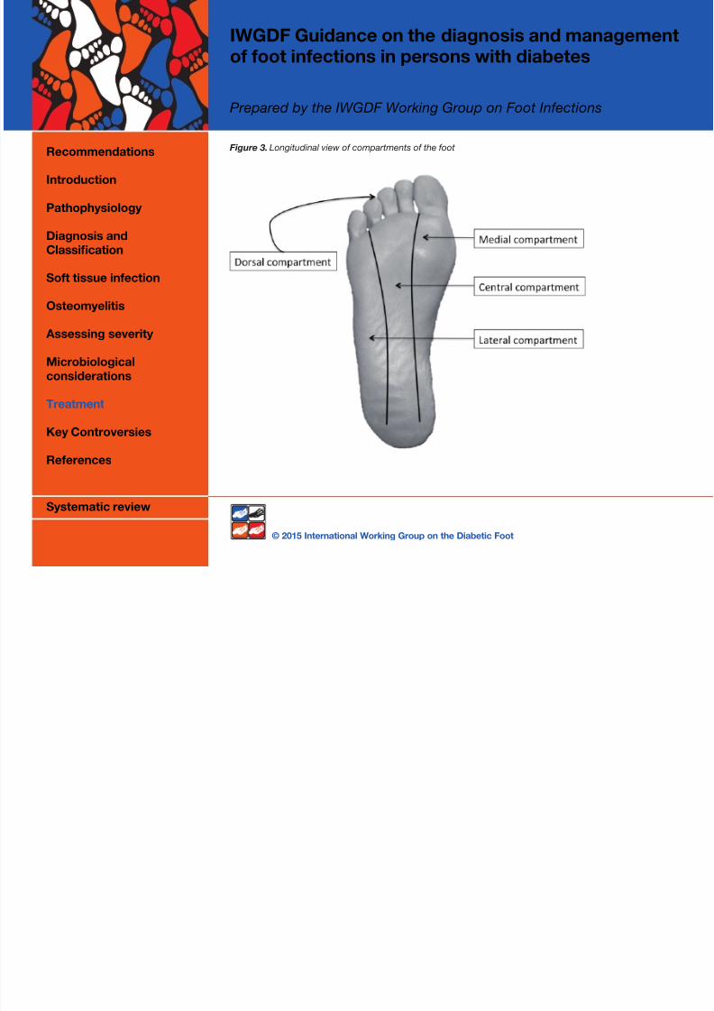
7/21/2019 Guia de Pie Diabetico Infectado
http://slidepdf.com/reader/full/guia-de-pie-diabetico-infectado 34/69
© 2015 International Working Group on the Diabetic Foot
Prepared by the IWGDF Working Group on Foot Infections
of foot infections in persons with diabetes
Recommendations
Introduction
Pathophysiology
Diagnosis and
Classifcation
Soft tissue infection
Osteomyelitis
Assessing severity
Microbiological
considerations
Treatment
Key Controversies
References
Systematic review
Figure 3. Longitudinal view of compartments of the foot
IWGDF Guidance on the diagnosis and management

7/21/2019 Guia de Pie Diabetico Infectado
http://slidepdf.com/reader/full/guia-de-pie-diabetico-infectado 35/69
© 2015 International Working Group on the Diabetic Foot
Prepared by the IWGDF Working Group on Foot Infections
of foot infections in persons with diabetes
Recommendations
Introduction
Pathophysiology
Diagnosis and
Classifcation
Soft tissue infection
Osteomyelitis
Assessing severity
Microbiological
considerations
Treatment
Key Controversies
References
Systematic review
Figure 4. Transversal view of compartments of the foot
IWGDF Guidance on the diagnosis and management

7/21/2019 Guia de Pie Diabetico Infectado
http://slidepdf.com/reader/full/guia-de-pie-diabetico-infectado 36/69
© 2015 International Working Group on the Diabetic Foot
Prepared by the IWGDF Working Group on Foot Infections
of foot infections in persons with diabetes
Recommendations
Introduction
Pathophysiology
Diagnosis and
Classifcation
Soft tissue infection
Osteomyelitis
Assessing severity
Microbiological
considerations
Treatment
Key Controversies
References
Systematic review
Antimicrobial therapy
Recommendation 19:
While virtually all clinically infected diabetic foot wounds require antimicrobial therapy, do not treat clinically
uninfected diabetic foot wounds with antimicrobial therapy (Strong; Low)
Recommendation 20:
Select specific antibiotic agents for treatment based on the likely or proven causative pathogens, their antibiotic
susceptibilities, the clinical severity of the infection, and evidence of efficacy for DFI and costs (Strong;
Moderate)
Recommendation 21:
A course of antibiotic therapy of 1-2 weeks is usually adequate for most soft tissue diabetic foot infections
(Strong; High)
Rationale 19 - 21 - Indications for therapy:
Failure to treat an infected diabetic foot wound with antimicrobial therapy is usually associated with progressive
tissue destruction and poor wound healing. However, antibiotic therapy is also associated with frequent adverse
effects, financial costs, and increasing the risk of antibiotic resistance (144), so it should be reserved for
treating wounds that are infected. Treatment with antimicrobials has not been proven to be beneficial for
managing clinically uninfected skin wounds, irrespective of theoretical considerations of the bacterial
“bioburden” (a poorly defined concept) of chronic wounds (162-166). There is no published evidence that
antimicrobial therapy either accelerates wound healing or reduces the likelihood of clinical infection developing.Where the clinical assessment for the presence of infection is equivocal, the clinician must make a decision to
treat the wound as either uninfected or as infected (using an infection grading system) and then carefully monitor
progress.
Recommendation 22:
Administer parenteral therapy initially for most severe infections and some moderate infections, with a switch to
oral therapy when the infection is responding (Strong; Low)
IWGDF Guidance on the diagnosis and managementf f i f i i i h di b

7/21/2019 Guia de Pie Diabetico Infectado
http://slidepdf.com/reader/full/guia-de-pie-diabetico-infectado 37/69
© 2015 International Working Group on the Diabetic Foot
Prepared by the IWGDF Working Group on Foot Infections
of foot infections in persons with diabetes
Recommendations
Introduction
Pathophysiology
Diagnosis and
Classifcation
Soft tissue infection
Osteomyelitis
Assessing severity
Microbiological
considerations
Treatment
Key Controversies
References
Systematic review
Rationale 22 - Route of therapy:
For an antibiotic to reach a therapeutic concentration at the site of infection it must first achieve an adequate
serum level (167). Because parenteral antibiotics achieve therapeutic serum levels faster and more reliably, they
are recommended for patients who are systemically ill or have a severe infection. They may also be required for
those unable to tolerate oral agents or who are infected with pathogens insensitive to available oral agents. After
the patient’s clinical condition has stabilized and the infection is responding, most can switch to oral therapy.
Where available, consider outpatient intravenous antibiotic therapy for those requiring prolonged parenteral
treatment, for example, for some cases of osteomyelitis or infections with organisms found resistant to available
oral agents.
Compared with parenteral therapy, treatment with oral antibiotic agents is more convenient, not associatedwith infusion-related complications, and generally less expensive. Gastrointestinal absorption of oral antibiotics
(bioavailability), while variable, is excellent for several agents, such as fluoroquinolones, clindamycin, rifamp(ic)
in, trimethoprim/sulfamethoxazole, linezolid, and doxycycline (168). Fluoroquinolones in particular achieve high
tissue concentrations in diabetic foot infections (167,169,170), even in patients with gastroparesis (171), but most
other currently used oral antibiotics also achieve adequate serum and tissue levels (168). Unfortunately,
fluoroquinolones are also associated with an increased risk of adverse effects, including Clostridium difficile
disease, and failure with one of these agents may cause resistance to others (172). No data are currently
unavailable to determine if adequate tissue levels predict successful clinical outcome (173). Newly marketed
agents generally have an expanded spectrum of activity, greater activity against antibiotic-resistant gram-positive
cocci, a longer half-life (allowing for less frequent dosing) or good oral bioavailability. However, they are generally
considerably more expensive and have a shorter track record for safety evaluations. Virtually all comparisons of
different antibiotic regimens for DFI have reported no clinically significant differences between them, and nospecific agents have emerged as being preferred. One new agent, tigecycline (which has broad-spectrum
activity, including against MRSA) when compared to ertapenem (with or without vancomycin) was found in a
recent large, multicentre, randomised controlled trial to be significantly inferior in clinical outcomes and to have
a significantly higher rate of adverse effects (174).
Peripheral vascular disease, but not diabetes per se, may limit the delivery, and therefore penetration, of
antibiotics to infected foot tissues (171,175). Optimally, patients with severe arterial insufficiency should
undergo revascularization, but even in an ischemic limb, however, antibiotics play an important role in treating
and preventing further spread of infection. Problems with limb arterial insufficiency have led some to experiment
IWGDF Guidance on the diagnosis and managementf f t i f ti i ith di b t

7/21/2019 Guia de Pie Diabetico Infectado
http://slidepdf.com/reader/full/guia-de-pie-diabetico-infectado 38/69
© 2015 International Working Group on the Diabetic Foot
Prepared by the IWGDF Working Group on Foot Infections
of foot infections in persons with diabetes
Recommendations
Introduction
Pathophysiology
Diagnosis and
Classifcation
Soft tissue infection
Osteomyelitis
Assessing severity
Microbiological
considerations
Treatment
Key Controversies
References
Systematic review
with novel methods of antibiotic delivery to the lower limb, for example, retrograde intravenous perfusion under
pressure (176,177), intra-arterial (e.g., femoral) administration (178), primary closure of debrided wounds with
catheter instillation of antibiotics (179), or negative pressure wound therapy with installation of saline, antiseptics
or antibiotics (180-184). At this time there is insufficient evidence upon which to recommend any of these
approaches.
Using topical antibiotic therapy for a foot wound is appealing, as it allows high concentrations at the site of
infection without potentially toxic systemic levels (185,186). It would also allow treatment with agents not
available for systemic therapy. There are, however, some theoretical and practical caveats to its use, such as a
potentially higher susceptibility to the occurrence of hypersensitivity and limited effectiveness for infection in
surrounding intact tissue and possibly a lower threshold for development of antimicrobial resistance (186). A large randomised trial of 835 patients treated for an infected DFU (most of which would meet the current PE-
DIS criteria for grade 2, and some grade 3) found that an investigational topical antimicrobial peptide
(pexiganan) was as effective as oral therapy with a fluoroquinolone, with clinical improvement rates of 85%-90%
(187). Topical antimicrobial therapy may also be used in combination with systemic antibiotic therapy. One trial
compared outcomes in patients with a moderately infected DFU who were treated with standard therapy
(including levofloxacin) with or without the addition of the daily application of a topical gentamicin-collagen
sponge (188). Among 56 randomised patients the clinical cure rate for the sponge group was significantly lower
at day 7 (the primary outcome), but was significantly higher at the test of cure visit (2 weeks after discontinuation
of therapy, which was up to 28 days).
A limited number of marketed topical antimicrobial agents, as well as antimicrobial impregnated wound
dressings [e.g., those containing various forms of silver and iodine]) might be useful for preventing, or possibly
even treating, mild infections (186). Currently supporting data are too limited to recommend topical antimicrobialtherapy, but further research is warranted (186,189-192). For deep surgical wounds, antibiotic impregnated
beads, cement, or biodegradable bovine collagen sponges can supply high local antibiotic concentrations
(for a few days), and in some instances fill dead space (192,193). A systematic review and an expert opinion
paper concluded that the data supporting the use of gentamicin-impregnated beads is too limited to make any
recommendations (186,194).
Choice of antibiotics
Selection of an initial antibiotic regimen is usually empirical, i.e., a best guess at what agent(s) will cover the likely
pathogen(s). These should be selected to cover the most common infecting organisms, but be modified
IWGDF Guidance on the diagnosis and managementof foot infections in persons ith diabetes

7/21/2019 Guia de Pie Diabetico Infectado
http://slidepdf.com/reader/full/guia-de-pie-diabetico-infectado 39/69
© 2015 International Working Group on the Diabetic Foot
Prepared by the IWGDF Working Group on Foot Infections
of foot infections in persons with diabetes
Recommendations
Introduction
Pathophysiology
Diagnosis and
Classifcation
Soft tissue infection
Osteomyelitis
Assessing severity
Microbiological
considerations
Treatment
Key Controversies
References
Systematic review
according to infection severity and available clinical or microbiological information. We prefer relatively narrow-
spectrum agents for mild infections, with adjustments if clinical response is inadequate, especially if cultures
disclose pathogens resistant to the selected agent(s). Initial regimens for many moderate and all severe infec-
tions should be broader spectrum and treatment must be delivered promptly. An empirical regimen must also
consider factors related to the current infection, the likely pathogen(s), the patient co-morbidities, and potential
drug-related issues (see Table 5).
IWGDF Guidance on the diagnosis and managementof foot infections in persons with diabetes
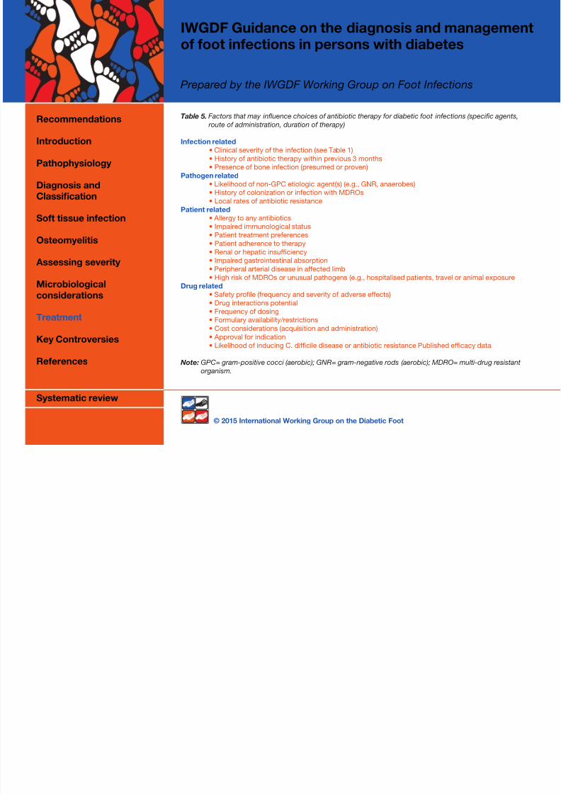
7/21/2019 Guia de Pie Diabetico Infectado
http://slidepdf.com/reader/full/guia-de-pie-diabetico-infectado 40/69
© 2015 International Working Group on the Diabetic Foot
Prepared by the IWGDF Working Group on Foot Infections
of foot infections in persons with diabetes
Recommendations
Introduction
Pathophysiology
Diagnosis and
Classifcation
Soft tissue infection
Osteomyelitis
Assessing severity
Microbiological
considerations
Treatment
Key Controversies
References
Systematic review
Table 5. Factors that may influence choices of antibiotic therapy for diabetic foot infections (specific agents,
route of administration, duration of therapy)
Infection related
• Clinical severity of the infection (see Table 1)
• History of antibiotic therapy within previous 3 months
• Presence of bone infection (presumed or proven)
Pathogen related
• Likelihood of non-GPC etiologic agent(s) (e.g., GNR, anaerobes)
• History of colonization or infection with MDROs
• Local rates of antibiotic resistance
Patient related
• Allergy to any antibiotics
• Impaired immunological status
• Patient treatment preferences
• Patient adherence to therapy
• Renal or hepatic insufciency
• Impaired gastrointestinal absorption
• Peripheral arterial disease in affected limb
• High risk of MDROs or unusual pathogens (e.g., hospitalised patients, travel or animal exposure
Drug related
• Safety prole (frequency and severity of adverse effects)
• Drug interactions potential • Frequency of dosing
• Formulary availability/restrictions
• Cost considerations (acquisition and administration)
• Approval for indication
• Likelihood of inducing C. difcile disease or antibiotic resistance Published efcacy data
Note: GPC= gram-positive cocci (aerobic); GNR= gram-negative rods (aerobic); MDRO= multi-drug resistant
organism.
IWGDF Guidance on the diagnosis and managementof foot infections in persons with diabetes

7/21/2019 Guia de Pie Diabetico Infectado
http://slidepdf.com/reader/full/guia-de-pie-diabetico-infectado 41/69
© 2015 International Working Group on the Diabetic Foot
Prepared by the IWGDF Working Group on Foot Infections
of foot infections in persons with diabetes
Recommendations
Introduction
Pathophysiology
Diagnosis and
Classifcation
Soft tissue infection
Osteomyelitis
Assessing severity
Microbiological
considerations
Treatment
Key Controversies
References
Systematic review
A Gram-stained smear of a wound specimen may help direct empiric antibiotic therapy by informing the clinician
of the number and gram-types of pathogens present (195). This simple and inexpensive procedure is particularly
useful in regions of limited resources. A recent study from Tanzania found that among 128 diabetic patients with
a limb ulcer the positive predictive value of a Gram-stain for bacterial growth was 93% and the predictive value
was 75% (15/20) for gram-positive organisms and 82% (31/38) for gram-negative organisms (133).
An empiric regimen should virtually always include an antibiotic usually active against standard strains of
staphylococci and streptococci. Consider adding an agent active against MRSA if there is substantial risk of
infection with this organism (e.g., a high local prevalence of MRSA, a patient with a recent stay in a health-care
institution, recent antibiotic therapy or known MRSA colonization). Patients who have been previously treated
with an antibiotic (for whatever reason), or who have a more severe infection, may need extended coverage for
common gram-negative bacilli, and perhaps in rare cases for Enterococcus species. Empiric anti-pseudomonal
therapy is usually not required unless risk factors for Pseudomonas infection are present, e.g., high local
prevalence of Pseudomonas infections, warm climate, or frequent exposure of the foot to water. Empiric
anti-anaerobic therapy is appropriate for necrotic, gangrenous, or foul-smelling wounds, which also require
debridement. Combination therapy may be appropriate for infections presumed (or proven) to be caused by
more than one organism, when the pathogen has a high potential for developing resistance (e.g., Pseudomonas)
or when selecting an agent (e.g., rifampi(ci)n when treating osteomyelitis) to which resistance may quickly
develop when used alone. Some DFI pathogens are highly resistant to antibiotics, such as those reported from
Italy caused by extensively-resistant P. aeruginosa that required treatment with colistin combined with rifamp(ci)
in and imipenem (196).
When culture and sensitivity results are available, consider changing to a more specific regimen that targets justthe isolated pathogens. To reduce the likelihood of antibiotic resistance, narrower spectrum agents are
preferable, but it is important to assess how the infection has responded to the empirical regimen. If the infection
is improving and the patient is tolerating therapy, there may be no reason to change, even if some or all of the
isolated organisms are resistant to the agents prescribed (197,198). If the infection is not responding, however,
modify treatment to cover all isolated organisms. When the infection is worsening despite the isolated bacteria
being susceptible to the selected regimen consider if: surgical intervention is needed; fastidious infecting
organisms were not recovered on culture; patient adherence to the treatment regimen has been suboptimal;
serum levels of the prescribed antibiotic are inadequate because of decreased intestinal absorption or drug
interactions causing more rapid metabolism of the antibiotic.
IWGDF Guidance on the diagnosis and managementof foot infections in persons with diabetes

7/21/2019 Guia de Pie Diabetico Infectado
http://slidepdf.com/reader/full/guia-de-pie-diabetico-infectado 42/69
© 2015 International Working Group on the Diabetic Foot
Prepared by the IWGDF Working Group on Foot Infections
of foot infections in persons with diabetes
Recommendations
Introduction
Pathophysiology
Diagnosis and
Classifcation
Soft tissue infection
Osteomyelitis
Assessing severity
Microbiological
considerations
Treatment
Key Controversies
References
Systematic review
Several antibiotic agents have been used successfully (including some for decades) to treat DFIs despite not
having been evaluated in prospective comparative studies; these include semisynthetic penicillinase-resistant
penicillins (e.g., dicloxacillin, nafcillin, flucloxacillin), cephalosporins (e.g., cefazolin, ceftriaxone, ceftazidime),
glycopeptides (teicoplanin, oritavancin, telavancin, dalbavancin), rifampi(ci)n, fusidic acid, trimethoprim/
sulfamethoxazole, and doxycycline. The following agents that have demonstrated clinical effectiveness, alone or
in combination, in published prospective studies that include patients with diabetic foot infections (see Table 6)
(8,9):
• Cephalosporins (cephalexin orally; cefoxitin, ceftizoxime, ceftibiprole, ceftaroline (199) parenterally)
• Penicillin/ß-lactamase inhibitor combinations (amoxicillin/clavulanate orally; ampicillin/sulbactam,
piperacillin/tazobactam, and ticarcillin/clavulanate parenterally)
• Carbapenems (imipenem/cilastatin and ertapenem, parenterally)
• Fluoroquinolones (ciprooxacin, levooxacin, and moxioxacin, all of which can be administered orally or
parenterally)
• Other agents: clindamycin (orally and parenterally); linezolid (orally and parenterally); daptomycin
(parenterally); tigecycline (parenterally); and vancomycin (parenterally)
Other agents in the same antibiotic classes as those listed in Table 6 are also likely to be effective. Overall, the
clinical and microbiological response rates have been similar in published trials with various antibiotics, and
there is no one preferred agent or combination (8,30,52,173,201-203). Understanding the principles of antibiotic
therapy is more important than knowing the specific agents currently in favour, especially as new antibiotics are
introduced and some older ones are made obsolete by emergence of resistance or newly appreciated toxicities
or adverse interactions (195,202,204,205). In the absence of a compelling reason to choose a specific antibiotic,the one with the lowest acquisition cost is preferred, even though antibiotics account for only a small portion of
the treatment costs for a foot infection (206). There is an urgent need for comparative trials and economic analy-
ses of various anti-infective regimens for DFIs (8,30,207,208). Suggested empirical antibiotic regimens, by type
of infection, are given in Table 5. Fungi are occasional pathogens in DFI, most often as part of a mixed infection
(209)
IWGDF Guidance on the diagnosis and managementof foot infections in persons with diabetes

7/21/2019 Guia de Pie Diabetico Infectado
http://slidepdf.com/reader/full/guia-de-pie-diabetico-infectado 43/69
© 2015 International Working Group on the Diabetic Foot
Prepared by the IWGDF Working Group on Foot Infections
of foot infections in persons with diabetes
Recommendations
Introduction
Pathophysiology
Diagnosis and
Classifcation
Soft tissue infection
Osteomyelitis
Assessing severity
Microbiological
considerations
Treatment
Key Controversies
References
Systematic review
Table 6. Selecting an empiric antibiotic regimen for diabetic foot infections
Infection Additional Usual Potential Empirical
Severity Factors Pathogen(s) Regimens a
Mild No complicating GPC S-S pen; 1st gen ceph
features
ß -lactam allergy GPC Clindamycin; FQ; T/S;
or intolerance macrolide; doxy
Recent antibiotic GPC + GNR ß -L-ase-1; T/S; FQ
exposure
High risk for MRSA MRSA Linezolid; T/S ; doxy;
macrolide; FQ
Moderate and Severe b No complicating features GPC ± GNR ß -L-ase 1; 2nd /3rd gen ceph
Recent antibiotics GPC ± GNR ß -L-ase 2; 3rd gen ceph,
group 1 carbapenem (depends
on prior therapy; seek advice)
Macerated ulcer, GNR, including ß -Lase-2; S-S pen +
warm climate Pseudomonas ceftazidime, S-S pen + cipro,
group 2 carbapenem
Ischemic limb/ necrosis/ GPC ± GNR ± ß -L-ase 1 or 2; group 1 or 2
gas forming anaerobes carbapenem; 2nd /3rd gen
ceph + clindamycin or
metronidazole
>>
IWGDF Guidance on the diagnosis and managementof foot infections in persons with diabetes

7/21/2019 Guia de Pie Diabetico Infectado
http://slidepdf.com/reader/full/guia-de-pie-diabetico-infectado 44/69
© 2015 International Working Group on the Diabetic Foot
Prepared by the IWGDF Working Group on Foot Infections
of foot infections in persons with diabetes
Recommendations
Introduction
Pathophysiology
Diagnosis and
Classifcation
Soft tissue infection
Osteomyelitis
Assessing severity
Microbiological
considerations
Treatment
Key Controversies
References
Systematic review
Table 6. Selecting an empiric antibiotic regimen for diabetic foot infections
Infection Additional Usual Potential Empirical
Severity Factors Pathogen(s) Regimens a
MRSA risk factors MRSA Consider addition of, or
substituting with,
glycopeptides; linezolid;
daptomycin; fusidic
acid; T/S (±rif)*; doxycycline;
FQ Risk factors for ESBL carbapenems, FQ,
resistant GNR aminoglycoside, colistin
Note: GPC = gram positive cocci (staphylococci and streptococci); GNR = gram negative rod; MRSA = methicil-
lin-resistant Staphylococcus aureus; ESBL = extended spectrum beta lactamase producing organism; S-S pen =
semisynthetic penicillinase-resistant penicillin; ß -L-ase= ß-lactam, ß-lactamase inhibitor; ß -L-ase 1= amoxicillin/
clavulanate, ampicillin/sulbactam; ß -L-ase 2= ticarcillin/clavulanate, piperacillin/tazobactam;
doxy = doxycycline; Group 1 carbapenem= ertapenem; Group 2 carbapenem= imipenem, meropenem,
doripenem; Ceph= cephalosporin; gen= generation; Pip/tazo=piperacillin/tazobactam; FQ=fluoroquinolone with
good activity against aerobic gram-positive cocci (e.g., levofloxacin or moxifloxacin); Cipro = antipseudomonal
fluoroquinolone e.g. ciprofloxacin; T/S=trimethoprim/sulfamethoxazole; T/S (±rif) = trimethoprim/sulfamethoxazole
with or without *rifamp(ic)in (200) (for now we think rifamp(ic)in should only be used for osteomyelitis).
a Given at usual recommended doses for serious infections. Modify doses or agents selected for azotaemia, liver
dysfunction, etc. Recommendations based upon theoretical considerations and available clinical trials. b Oral antibiotic agents should generally not be used for severe infections, except as follow-on (switch) after initial
parenteral therapy.
IWGDF Guidance on the diagnosis and managementof foot infections in persons with diabetes

7/21/2019 Guia de Pie Diabetico Infectado
http://slidepdf.com/reader/full/guia-de-pie-diabetico-infectado 45/69
© 2015 International Working Group on the Diabetic Foot
Prepared by the IWGDF Working Group on Foot Infections
of foot infections in persons with diabetes
Recommendations
Introduction
Pathophysiology
Diagnosis and
Classifcation
Soft tissue infection
Osteomyelitis
Assessing severity
Microbiological
considerations
Treatment
Key Controversies
References
Systematic review
Another factor that appears to impair response to antibiotic therapy in DFI is the presence of biofilm. These
slime-enclosed aggregates of sessile bacteria adhering to surfaces are present in many chronic infections, and
the majority of DFIs, and demonstrate great resistance to most antibacterial agents as well as to host defences
(210,211). Eradicating bacteria in a biofilm usually requires physical removal, often combined with high doses
of an antimicrobial agent found to be more active against these organisms. These include topical agents such
as hypochlorous acid (212), cadexomer iodine (213) and systemic agents such as fluoroquinolones, rifamp(ci)in,
daptomycin, or fosfomycin (214,215).
Duration of therapy.
The optimal durations of antibiotic therapy for diabetic foot infections involving skin and soft tissue or bone are
unknown. Based on data from available studies, for mild to moderate skin and soft tissue infections 1 to 2 weeks
is usually effective (9,131,173), while for more serious skin and soft tissue infections 3 weeks is usually sufficient
(9,173,197,198,216,217). Antibiotic therapy can generally be discontinued when signs and symptoms of infection
have resolved, even if the wound has not healed—as the antibiotics are employed to treat infection, not to heal
wounds. More extended treatment may be needed for immunocompromised patients, for wounds that are poorly
perfused, deep, large or necrotic, or for osteomyelitis (vide infra), but this decision should be accompanied by
clinical re-evaluations to support the treatment strategy. In the occasional instances in which prolonged paren-
teral therapy is needed, outpatient therapy should be considered (218). The necessary duration of therapy may
be shortened by adequate debridement, resection or amputation of infected tissue. Some patients who cannot
(or refuse to) undergo surgical resection, or who have an implanted foreign body at the infection site, may require
prolonged or intermittent suppressive antibiotic therapy.
Wound care
Recommendation 23:
Do not select a specific type of dressing for a diabetic foot infection with the aim of preventing an infection, or
improving its outcome (Strong; High)
Rationale 23:
For treating DFIs, antibiotics (and often surgery) are necessary, but not sufficient to overcome inadequate vascu-
lar supply, poor glycaemic control, persistent wound trauma or improper wound care (219,220). Most DFUs need
IWGDF Guidance on the diagnosis and managementof foot infections in persons with diabetes

7/21/2019 Guia de Pie Diabetico Infectado
http://slidepdf.com/reader/full/guia-de-pie-diabetico-infectado 46/69
© 2015 International Working Group on the Diabetic Foot
Prepared by the IWGDF Working Group on Foot Infections
p
Recommendations
Introduction
Pathophysiology
Diagnosis and
Classifcation
Soft tissue infection
Osteomyelitis
Assessing severity
Microbiological
considerations
Treatment
Key Controversies
References
Systematic review
to be carefully cleaned and debrided to remove devitalized tissue that may impede wound healing and foster
infection. No prospective studies have evaluated the optimal frequency or type of debridement for diabetic foot
ulcers, but post hoc evaluations of clinical studies in non-infected DFUs suggest that more frequent
debridement is associated with a higher healing rate (221,222). Systematic reviews of various wound
dressings and topical antimicrobials have found no evidence that any specific type of therapy is better than
others (223,224). For example, simple gauze dressings have performed as well for healing DFUs as silver
dressings, hydrogels, alginates, and foam dressings. In general, DFUs with heavy exudate need a dressing that
absorbs moisture, while dry wounds need topical treatments that add moisture. Dressings should optimally be
changed at least daily, both to apply a clean wound covering and to allow careful examination of the wound for
infection. Applying a total contact cast makes it difficult for the clinician and patient to visualise the wound for
evaluation of response to treatment between changes, and is generally not appropriate for infected wounds.
For further discussion of wound care the reader is referred to the IWGDF guidance document on wound care.
Treating osteomyelitis
Recommendation 24:
For diabetic foot osteomyelitis we recommend 6 weeks of antibiotic therapy for patients who do not undergo
resection of infected bone and no more than a week of antibiotic therapy if all infected bone is resected. (Strong;
Moderate)
Rationale 24:
While many cases of DFO require, or benefit from, surgical debridement or resection of bone, some can be
treated successfully by medical therapy alone. Several published retrospective series have shown that DFO canbe arrested (or even apparently cured) with antibiotic therapy in the absence of surgical intervention in about
two-thirds of cases (111,119,225-229). In these reports, clinicians have generally employed the higher recom-
mended daily doses of antibiotics given for at least two (and usually 3–6) months. Unfortunately, available studies
do not provide information to inform which types of DFO cases may be successfully treated without surgery
(111,119,225-229). In some cases, limited surgery (resection of infected and necrotic bone without amputation)
combined with antibiotic therapy may be most appropriate (157,230-233). A retrospective study from four
centres in France and Spain compared outcomes of patients with bone culture-proven S. aureus DFO who were
treated by either “medical” (just antibiotic therapy, other than soft tissue debridement at the bedside) or
“surgical” (operative treatment combined with prolonged antibiotic therapy) (234). Outcomes were similar for the
IWGDF Guidance on the diagnosis and managementof foot infections in persons with diabetes

7/21/2019 Guia de Pie Diabetico Infectado
http://slidepdf.com/reader/full/guia-de-pie-diabetico-infectado 47/69
© 2015 International Working Group on the Diabetic Foot
Prepared by the IWGDF Working Group on Foot Infections
p
Recommendations
Introduction
Pathophysiology
Diagnosis and
Classifcation
Soft tissue infection
Osteomyelitis
Assessing severity
Microbiological
considerations
Treatment
Key Controversies
References
Systematic review
two groups (favourable in 80% in the surgical group and 87% in the medical group), but significant differences
between patients in the medical group compared to the surgical group were that they were less frequently
hospitalized (49% versus 94%), had a shorter length of hospital stay (17 versus 12 days), had a slightly longer
course of antibiotic therapy (11 versus 10 weeks) and more treatment related side effects (33% versus 9%).
Recently the first published prospective, randomized trial was published that compared the outcomes of the
treatment of DFO in patients who received exclusively antibiotic therapy (for up to 90 days) versus those who
underwent limited resection of the osteomyelitic bone (accompanied by ~10 days of antibiotic therapy) (235).
The primary end point was foot wound healing, which occurred in 18 antibiotic-treated patients compared with
19 predominantly surgically treated patients (75% versus 86.3% healing rates, respectively, p=0.33). There was
no significant difference in median time to healing (6-7 weeks), the need for surgery (first or repeat procedure,
including minor amputations), reulceration (up to 12 weeks after healing) or treatment related complications.
This study suggests that the short-term results of therapy with either antibiotics alone or predominantly surgical
treatment (with some antibiotic therapy) are similar in patients who have neuropathic forefoot ulcers complicated
by osteomyelitis, but without ischemia or necrotizing soft tissue infections. Noteworthy aspects of this trial were
that the number of enrolled patients was relatively small, only about a third of the patients they evaluated for the
study were eligible for inclusion and the duration of follow-up was rather short (236). Table 7 summarises factors
potentially favouring selecting either primarily antibiotic or surgical treatment for diabetic foot osteomyelitis.
The IWGDF produced a full systematic review of, and guidelines for, the treatment of DFO in 2008 (52), and
updated the review for all types of DFI in 2012 and 2015 (9,173). Recently a non-systematic review provided
guidance on selecting systemic antibiotic therapy for chronic osteomyelitis (237). Among the important factors to
consider when treating osteomyelitis are the following: the anatomic site of infection, the local vascular supply,the extent of both soft tissue and bone destruction, the presence of any systemic signs of infection and the
patient’s preferences for treatment. The choice of an antimicrobial agent for treating osteomyelitis should
optimally be based on the results of a bone culture, especially because of the need for long-duration therapy
(49,119). If empiric therapy is necessary, the regimen should usually cover S. aureus as it is the most common
pathogen, but the patient’s history or culture results may suggest a need for broader coverage. Some antibiotics
may not penetrate well into infected bone, but the unreliability of measuring bone levels limits the value of
published data on this issue. Furthermore, the association between high bone levels of an antibiotic and
improved outcome has not yet been studied. Although treatment of osteomyelitis has traditionally been
IWGDF Guidance on the diagnosis and managementof foot infections in persons with diabetes

7/21/2019 Guia de Pie Diabetico Infectado
http://slidepdf.com/reader/full/guia-de-pie-diabetico-infectado 48/69
© 2015 International Working Group on the Diabetic Foot
Prepared by the IWGDF Working Group on Foot Infections
Recommendations
Introduction
Pathophysiology
Diagnosis and
Classifcation
Soft tissue infection
Osteomyelitis
Assessing severity
Microbiological
considerations
Treatment
Key Controversies
References
Systematic review
parenteral (at least initially) and prolonged (at least 4 weeks), these recommendations are not based on strong
data. Many patients can probably be switched to oral therapy after about a week of parenteral treatment to
complete their treatment course. Any oral antibiotics selected should have good bioavailability [e.g.
fluoroquinolones, rifampi(ci)n (always combined with another agent), clindamycin, linezolid, fusidic acid or
trimethoprim–sulfamethoxazole]. If all of the infected bone is surgically removed a shorter course of antibiotic
therapy (i.e. 2–14 days) may be sufficient, depending on the status of the soft tissues (9).
Extending post-debridement antibiotic therapy beyond six weeks, or giving IV treatment longer than one week,
does not appear to increase the remission rate. A recent randomised controlled trial that compared 6 versus 12
weeks of antibiotic therapy for non-surgically treated DFO in 40 patients found no significant difference in the
remission rate (60% versus 70%), but significantly fewer adverse effects with the shorter treatment (238,239).
For some patients with apparently incurable infection, long-term suppressive therapy, or intermittent short
courses of treatment for recrudescent symptoms, may be the most appropriate approach. When there are
clinical signs of persistent or recurrent infection the clinician should strongly consider a percutaneous bone
biopsy for culture to determine if there is persistent infection or any changes in the pathogens or their antibiotic
susceptibilities. Antibiotic impregnated beads (192), sponges (188), cement or orthopaedic implants have been
used successfully to treat DFO in a few small studies (193).
IWGDF Guidance on the diagnosis and managementof foot infections in persons with diabetes

7/21/2019 Guia de Pie Diabetico Infectado
http://slidepdf.com/reader/full/guia-de-pie-diabetico-infectado 49/69
© 2015 International Working Group on the Diabetic Foot
Prepared by the IWGDF Working Group on Foot Infections
Recommendations
Introduction
Pathophysiology
Diagnosis and
Classifcation
Soft tissue infection
Osteomyelitis
Assessing severity
Microbiological
considerations
Treatment
Key Controversies
References
Systematic review
Table 7. Factors potentially favouring selecting either primarily antibiotic or surgical resection for diabetic foot
osteomyelitis
Medical
• Patient is too medically unstable for surgery
• Poor postoperative mechanics of foot is likely (e.g., with mid- or hind-foot infection)
• No other surgical procedures on foot are needed
• Infection is conned to small, forefoot lesion
• No adequately skilled surgeon is available
• Surgery costs are prohibitive for the patient
• Patient has a strong preference to avoid surgery
Surgical
• Foot infection is associated with substantial bone necrosis or exposed joint
• Foot appears to be functionally nonsalvageable
• Patient is already nonambulatory
• Patient is at particularly high risk for antibiotic-related problems
• nfecting pathogen is resistant to available antibiotics
• Limb has uncorrectable ischemia (precluding systemic antibiotic delivery)
• Patient has a strong preference for surgical treatment
Note: Modified from Lipsky, 2014, Diabetes Care (236).
IWGDF Guidance on the diagnosis and managementof foot infections in persons with diabetes

7/21/2019 Guia de Pie Diabetico Infectado
http://slidepdf.com/reader/full/guia-de-pie-diabetico-infectado 50/69
© 2015 International Working Group on the Diabetic Foot
Prepared by the IWGDF Working Group on Foot Infections
Recommendations
Introduction
Pathophysiology
Diagnosis and
Classifcation
Soft tissue infection
Osteomyelitis
Assessing severity
Microbiological
considerations
Treatment
Key Controversies
References
Systematic review
Adjunctive therapies
Recommendation 25:
We suggest not using any adjunctive treatments for diabetic foot infection [Weak; Low]
Rationale 25:
Several studies have reported the results of additional approaches (beyond antibiotics and surgery) to help re-
solve infection, accelerate wound healing or improve host response. These include negative pressure wound
therapy (NPWT), systemic hyperbaric oxygen therapy (HBOT), granulocyte colony stimulating factors (G-CSF)
and larval (maggot) therapy (9,240). While NPWT is often used for infected cardiothoracic, traumatic and
orthopaedic wounds, we know of no studies that have specifically investigated the role of NPWT to treat infected
diabetic foot wounds. A randomized controlled study of patients with chronic diabetic foot wounds after partialamputation reported a nonsignificantly higher rate of infection in those treated with NPWT than in the controls
(16.8% versus 9.4%) (241). One retrospective cohort study reported a higher proportion of healed or surgically
closed wounds and shorter periods of hospitalisation in infected diabetic patients treated with NPWT with
simultaneous irrigation with an antiseptic solution (182). One controlled trial of treatment of diabetic foot wounds
included a group of 130 patients randomized after surgical debridement of an infected open minor amputation
to either NPWT or a semi-occlusive silver dressing (242). The authors reported that the NPWT group had a
significantly “more rapid development of granulation tissue covering exposed bone” and “better and more rapid
control of infections” and reduced time to complete close of the wound. We find it difficult to interpret these
endpoints and await further prospective trials of this therapy for infected wounds.
Several randomized clinical trials evaluated HBOT for treating DFUs and some have shown an increasedlikelihood or faster rates of wound healing and fewer major amputations (243-246). Most of these studies
included Wagner 3 ulcers, which can include patients with osteomyelitis, but none presented any subanalyses of
patients with infected DFUs or specifically reported on infection related outcome measures. To date, there are no
data to support using HBOT to treat either soft tissue infection or osteomyelitis.
A meta-analysis of five studies with a total of 167 patients with DFIs found that therapy with various types of
investigational G-CSF treatments was associated with significantly fewer surgeries and amputations and shorter
hospital stays, but did not increase the likelihood of resolving infection, healing wounds or shortening the
duration of systemic antibiotic therapy (9,247,248). Maggot debridement, or larval biotherapy, has been shown
IWGDF Guidance on the diagnosis and managementof foot infections in persons with diabetes

7/21/2019 Guia de Pie Diabetico Infectado
http://slidepdf.com/reader/full/guia-de-pie-diabetico-infectado 51/69
© 2015 International Working Group on the Diabetic Foot
Prepared by the IWGDF Working Group on Foot Infections
Recommendations
Introduction
Pathophysiology
Diagnosis and
Classifcation
Soft tissue infection
Osteomyelitis
Assessing severity
Microbiological
considerations
Treatment
Key Controversies
References
Systematic review
to have antibacterial effects (249). A recent systematic review of its value for chronic wounds, including DFUs,
reported that in one study there was a significantly longer antibiotic-free time period in patients who received
maggot therapy than in those who did not, but in two studies the proportion of antibiotic use was similar for
those who did and did not receive maggot therapy (250,251).
Outcome of treatment
With appropriate treatment the signs and symptoms of mild DFIs almost always resolve without need for ampu-
tation. When infection involves deep soft tissue structures or bone, the outcome is often less favourable; many
require surgical debridement, bone resection or partial amputations. With extensive infection, or in medical
centres with limited expertise or resources, lower extremity amputation rates may reach 50–60% (9,252). For
hospitalized patients, poor outcomes (mostly amputations) occur in almost half, even in expert centres (126).
A recent study from the US found that of 57 hospitalized DFI patients who were discharged to an outpatientparenteral antibiotic therapy program, 93% were considered a treatment success on discharge, but only 64%
had resolution of the DFI at 6 months follow-up (39). Not surprisingly, treatment success was significantly higher
with moderate compared to severe infections (79% vs 21%, p=0.04). Regrettably, in this small, retrospective
study, adherence to the IDSA diabetic foot infection guidelines was suboptimal, and did not correlate with clinical
outcome. Another recent US study found that of 234 patients with a DFI hospitalised in three different types of
university-affiliated centres, only 17% of wounds healed and the amputation rate was 42% (253). Independent
risk factors for amputation were the presence of gangrene or osteomyelitis and a wound area of >5 cm2.
In the hands of an experienced surgeon, most amputations can be foot sparing (i.e., below the malleoli), and
long-term control of infection is achieved in over 80% of cases (114). The presence of limb or foot ischemia has
an important adverse effect on the outcome, synergising with infection to worsen the prognosis (254).Unfortunately, having had one foot infection is associated with an increased likelihood of another; foot infection
recurs in 20% to 30% of diabetic patients, especially those with underlying osteomyelitis (255).
It is difficult to know when osteomyelitis is cured, but clinical experience suggests that evidence of remission
includes a drop in the erythrocyte sedimentation rate (and to a lesser extent, the C-reactive protein level),
reconstitution of destroyed bone on plain radiograph and healing of any overlying soft tissue wound. While not
recommended for this purpose, a negative nuclear medicine scan makes active ongoing infection unlikely. A
negative culture of the bone margin left after operative resection of infected bone is associated with a lower
incidence of recrudescence of infection than if the bone margin is culture-positive.(256) Because DFO
recurrences are common, it is best to consider apparent treatment success a “remission” for at least a year,
IWGDF Guidance on the diagnosis and managementof foot infections in persons with diabetes

7/21/2019 Guia de Pie Diabetico Infectado
http://slidepdf.com/reader/full/guia-de-pie-diabetico-infectado 52/69
© 2015 International Working Group on the Diabetic Foot
Prepared by the IWGDF Working Group on Foot Infections
Recommendations
Introduction
Pathophysiology
Diagnosis and
Classifcation
Soft tissue infection
Osteomyelitis
Assessing severity
Microbiological
considerations
Treatment
Key Controversies
References
Systematic review
before calling it a cure. Factors that predict healing include the absence of any exposed bone, palpable pedalpulses, blood pressure in the toe of >45 mmHg or in the ankle of >80 mmHg, peripheral white blood cell count
of <12 000/mm3 and a lower extremity transcutaneous oxygen tension of >40 mmHg (13,257). There is no
convincing evidence that clinical outcome is related to the specific infecting organism, even multidrug resistant(e.g., MRSA) strains (127), including in cases involving bone (258). Because of the risk of reinfection, it is crucialto educate patients who have a DFI about prevention techniques and the need for prompt consultation for anyfuture foot problems.
Issues of particular importance in developing (low-income) countries
Recommendation 26:
When treating a diabetic foot infection, assess for use of traditional remedies, previous antibiotic use andconsider local bacterial pathogens and their susceptibility profile. [Strong; Low]
Rationale 26:
These guidelines must, of course, be adapted to the local circumstances in which a healthcare provider seespatients. Many aspects of the management of DFIs may differ in developing (or low income), compared withmore developed (higher income), countries. In resource constrained regions infections are often a consequenceof wounds caused by the diabetic person wearing footwear that is not sufficiently protective (e.g., sandals) orpoorly fitting, or wearing none at all. Poor hygiene may be associated with risk of rat bites (259) and increasesthe risk of ulcer infection and may enable larval infestation (myiasis) (260). Persons with foot wounds may delayseeing a healthcare provider because they lack health-related education, nearby healthcare services or financial
resources (261). During this period of delay the person may attempt to treat the infection with various traditionalremedies, including plants or other locally accepted treatments (262-264), seek treatment from a faith or herbalhealer, or have to be referred from primary to district to regional health centres (265). In a recent questionnairestudy of patients with a DFI in the West Indies, 382 who had sought medical attention soon after detecting theinfection were compared to 313 who voluntarily chose to delay medical therapy in favour of home remedies(266). The home remedy group had significantly worse outcomes for duration of hospitalization (16.3 versus8.5 days) and number of (and need for operative) debridements. They also had a non-significant trend towardmore major amputations (9.3% versus 5.2%) and an estimated increase cost for their treatment of $10,821 (US).Furthermore, in developing countries people can often buy antibiotics without a prescription; thus, they may havetreated themselves, occasionally with the advice of a local pharmacist or other trusted but non-licensed persons,
IWGDF Guidance on the diagnosis and managementof foot infections in persons with diabetes

7/21/2019 Guia de Pie Diabetico Infectado
http://slidepdf.com/reader/full/guia-de-pie-diabetico-infectado 53/69
© 2015 International Working Group on the Diabetic Foot
Prepared by the IWGDF Working Group on Foot Infections
Recommendations
Introduction
Pathophysiology
Diagnosis and
Classifcation
Soft tissue infection
Osteomyelitis
Assessing severity
Microbiological
considerations
Treatment
Key Controversies
References
Systematic review
before presenting to a physician. This unsupervised treatment, sometimes with generic drugs of questionablequality, expired medications or at inadequate doses, is likely to result in infections caused by more antibiotic-resistant organisms (262,267).
Healthcare providers in developing or low-income countries may also face a lack of access to a microbiologylaboratory, so cannot determine the identity and antibiotic susceptibility of foot pathogens infecting anindividual patient, or of current isolates and susceptibilities in the community. Recent studies have demonstratedsubstantial variations in the causative pathogens of DFIs in different regions of the world (268). In contrast toWestern countries, studies from Asia and Africa have reported that aerobic gram-negative organisms (especiallyP. aeruginosa) are more common. Similarly, many clinicians will not have access to even basic (not to mentionmore sophisticated) imaging equipment or to specialist consultants with adequate knowledge of foot anatomy
and the available conservative management methods for treating DFIs. Even when a patient sees a physician andreceives an antibiotic prescription, indigent patients may be unable to afford the full course of therapy, or may beprescribed inexpensive but potentially more toxic or less effective agents.
Adverse social situations for many patients in these regions may also impair proper treatment. Home or workcircumstances may make it difficult for them to stay off the affected foot, or to afford or be able to purchase oruse an off-loading device. Furthermore, they may have travelled a long distance to see a physician and cannoteasily return for follow-up visits. Understandably, patients and providers in low-income countries do not want“second-class,” or “best we can afford,” medical care. Improving management of DFIs in developing countrieswill likely require a combination of education (for patients, pharmacists and healthcare providers) and funding(for diagnostic, therapeutic and preventative services) (263,269,270).
IWGDF Guidance on the diagnosis and management of foot infections in persons with diabetes
IWGDF Guidance on the diagnosis and managementof foot infections in persons with diabetes

7/21/2019 Guia de Pie Diabetico Infectado
http://slidepdf.com/reader/full/guia-de-pie-diabetico-infectado 54/69
© 2015 International Working Group on the Diabetic Foot
Prepared by the IWGDF Working Group on Foot Infections
Recommendations
Introduction
Pathophysiology
Diagnosis and
Classifcation
Soft tissue infection
Osteomyelitis
Assessing severity
Microbiological
considerations
Treatment
Key Controversies
References
Systematic review
Key Controversies
1. How should we monitor treatment and determine when infection has resolved?
This is an important unmet need as it serves as one means to limit unnecessarily prolonged antibiotic therapy.
2. What is the optimal duration of antimicrobial treatment for osteomyelitis?
Since bone infection is more difficult to cure than just soft tissue involvement, and the duration of antibiotic
therapy is more prolonged for osteomyelitis than soft tissue infection, this is a key issue.
3. How should we adapt approaches to DFI management in low-income countries?
The rise in incidence of DFIs in some of these countries is steep and with their constrained resources, finding
optimal approaches, without recommending “second-class” care, is crucial to improve outcomes.
4. When, and which, imaging studies should we order for a patient with a DFI?
Imaging studies can be expensive and time-consuming and awaiting their results may delay appropriate
therapy. Especially with the advent of new technologies, evaluating their cost-effectiveness to optimise use
would improve management.
5. When should we select primarily medical versus surgical treatment for osteomyelitis?
This has been a controversial and simmering issue for some time, addressed by several retrospective studies
but to date only one prospective one. An additional large,well-designed prospective study could largely answer
the question.
6. Is there a definition and practical clinical use for the concept of wound “bacterial bioburden”?
This term is widely used in the wound healing community (and by industry) but has no agreed upon definition.
Deciding if it has value and standardising the definition could help industry develop useful products and
clinicians know which to employ.
7. What is the value and proper interpretation of molecular (genotypic) microbiological testing for DFI?
The era of molecular microbiology is inexorably approaching, but it is crucial for clinicians to understand when
to order and how to interpret the results of these tests in deciding on antibiotic therapy.
IWGDF Guidance on the diagnosis and managementof foot infections in persons with diabetes

7/21/2019 Guia de Pie Diabetico Infectado
http://slidepdf.com/reader/full/guia-de-pie-diabetico-infectado 55/69
© 2015 International Working Group on the Diabetic Foot
Prepared by the IWGDF Working Group on Foot Infections
Recommendations
Introduction
Pathophysiology
Diagnosis and
Classifcation
Soft tissue infection
Osteomyelitis
Assessing severity
Microbiological
considerations
Treatment
Key Controversies
References
Systematic review
AcknowledgementsWe thank the following “corresponding members” for their helpful comments on the manuscript: Zulfiqarali G.
Abbas (United Republic of Tanzania); M. Bulent Ertugrul (Republic of Turkey); Alexandra Jirkovska (Czech Repu-
blic); José Luis Lázaro Martinez (Kingdom of Spain); Aziz Nather (Republic of Singapore); Nina Rojas (Republic of
Chile); Carlo Tascini (Italian Republic); Oleg Udovichenko (Russian Federation); Zhangrong Xu (People’s Republic
of China).
Conflicts of InterestBAL: Research funding from Innocoll; consulting for Innocoll, Merck, Pfizer, Dipexium, Cubist, Cerexa,
KCI/Acelity.
LL: is on the speaker’s bureau for Osiris, Integra, PamLabs, Smit&Nephew; consultant for KCI, PamLabs,Innovacyn; Stock ownership in Prizm Medical; received research grants from Osiris, MacroCure, ThermoTrek,
Integra, GlaxoSmithKline, KCI, Cardinal, Dipexium.
ES: speaker and received congress support from Sanofi-Aventis and Novartis; consulting and received congress
support from Pfizer; consultant for Cubist.
JAS, MD, JE, SK, VUR, SVA, and EJP: none declared.
IWGDF Guidance on the diagnosis and management of foot infections in persons with diabetes
IWGDF Guidance on the diagnosis and managementof foot infections in persons with diabetes

7/21/2019 Guia de Pie Diabetico Infectado
http://slidepdf.com/reader/full/guia-de-pie-diabetico-infectado 56/69
© 2015 International Working Group on the Diabetic Foot
Prepared by the IWGDF Working Group on Foot Infections
Recommendations
Introduction
Pathophysiology
Diagnosis and
Classifcation
Soft tissue infection
Osteomyelitis
Assessing severity
Microbiological
considerations
Treatment
Key Controversies
References
Systematic review
References
1. Guyatt GH, Oxman AD, Vist GE, Kunz R, Falck-Ytter Y, Alonso-Coello P, Schunemann HJ: GRADE: an emerging consensus on rating quality of
evidence and strength of recommendations. BMJ 336:924-926, 2008
2. Raspovic KM, Wukich DK: Self-reported quality of life and diabetic foot infections. J Foot Ankle Surg 53:716-719, 2014
3. International Working Group on the Diabetic Foot. International Consensus on the Diabetic Foot and Supplements, DVD. Apelqvist, J., Bakker,
K., Van Houtum, W. H., Nabuurs-Fransen, M. H., and Schaper, N. C. Complete IWGDF data DVD Guidelines 2011 at http://shop.idf.org. 2011.
4. Pecoraro RE: Chronology and determinants of Tissue Repair in Diabetic Lower Extremity Ulcers. Diabetes 40:1305-1313, 1991
5. Reiber GE, Pecoraro RE, Koepsell TD: Risk factors for amputation in patients with diabetes mellitus. A case-control study. Ann Intern Med
117:97-105, 1992
6. Lavery LA, Armstrong DG, Murdoch DP, Peters EJ, Lipsky BA: Validation of the Infectious Diseases Society of America’s diabetic foot infection
classification system. Clin Infect Dis 44:562-565, 2007
7. Paisley AN, Kalavalapalli S, Subudhi CP, Chadwick PR, Chadwick PJ, Young B: Real time presence of a microbiologist in a multidisciplinary
diabetes foot clinic. Diabetes Res Clin Prac 96:e1-e3, 2012
8. Lipsky BA, Peters EJ, Senneville E, Berendt AR, Embil JM, Lavery LA, Urbancic-Rovan V, Jeffcoate WJ: Expert opinion on the management ofinfections in the diabetic foot. Diabetes Metab Res Rev 28 Suppl 1:163-178, 2012
9. Peters EJ, Lipsky BA, Aragón-Sánchez J, Bakker K, Boyko EJ, Diggle M, Embil JM, Kono S, Lavery LA, Senneville E, Urbancic-Rovan V,
Van Asten SA, Jeffcoate WJ: A systematic review of interventions in the management of infection in the diabetic foot. Diabetes Metab Res Rev
In Press: 2015
10. Peters EJ, Lipsky BA: Diagnosis and management of infection in the diabetic foot. Med Clin North Am 97:911-946, 2013
11. Lavery LA, Armstrong DG, Wunderlich RP, Mohler MJ, Wendel CS, Lipsky BA: Risk factors for foot infections in individuals with diabetes.
Diabetes Care 29:1288-1293, 2006
12. Hao D, Hu C, Zhang T, Feng G, Chai J, Li T: Contribution of infection and peripheral artery disease to severity of diabetic foot ulcers in
Chinese patients. Int J Clin Pract 68:1161-1164, 2014
13. Prompers L, Schaper N, Apelqvist J, Edmonds M, Jude E, Mauricio D, Uccioli L, Urbancic V, Bakker K, Holstein P, Jirkovska A, Piaggesi A,
Ragnarson-Tennvall G, Reike H, Spraul M, Van Acker K, Van Baal J, Van Merode F, Ferreira I, Huijberts M: Prediction of outcome in individuals
with diabetic foot ulcers: focus on the differences between individuals with and without peripheral arterial disease. The EURODIALE Study.
Diabetologia 51:747-755, 2008
14. Acosta JB, del Barco DG, Vera DC, Savigne W, Lopez-Saura P, Guillen NG, Schultz GS: The pro-inflammatory environment in recalcitrant
diabetic foot wounds. Int Wound J 5:530-539, 2008
15. Berlanga-Acosta J: Diabetic lower extremity wounds: the rationale for growth factors-based infiltration treatment. Int Wound J 8:612-620,
2011
16. Lavery LA, Peters EJ, Armstrong DG, Wendel CS, Murdoch DP, Lipsky BA: Risk factors for developing osteomyelitis in patients with diabetic
foot wounds. Diabetes Res Clin Prac 83:347-352, 2009
17. McMahon MM, Bistrian BR: Host defenses and susceptibility to infection in patients with diabetes mellitus. Infect Dis Clin North Am 9:1-9,
1995
18. Perner A, Nielsen SE, Rask-Madsen J: High glucose impairs superoxide production from isolated blood neutrophils. Intensive Care Med
29:642-645, 2003
19. Delamaire M, Maugendre D, Moreno M, Le Goff MC, Allannic H, Genetet B: Impaired leucocyte functions in diabetic patients. Diabet Med
14:29-34, 1997
IWGDF Guidance on the diagnosis and managementof foot infections in persons with diabetes

7/21/2019 Guia de Pie Diabetico Infectado
http://slidepdf.com/reader/full/guia-de-pie-diabetico-infectado 57/69
© 2015 International Working Group on the Diabetic Foot
Prepared by the IWGDF Working Group on Foot Infections
Recommendations
Introduction
Pathophysiology
Diagnosis and
Classifcation
Soft tissue infection
Osteomyelitis
Assessing severity
Microbiological
considerations
Treatment
Key Controversies
References
Systematic review
20. Aragón-Sánchez FJ, Lázaro-Martínez JL, Pulido-Duque J, Maynar M: From the diabetic foot ulcer and beyond: how do foot infections spread
in patients with diabetes? Diabet Foot Ankle 3: 2012
21. Bridges RM, Jr., Deitch EA: Diabetic foot infections. Pathophysiology and treatment. Surg Clin North Am 74:537-555, 1994
22. Maharaj D, Bahadursingh S, Shah D, Chang BB, Darling RC, III: Sepsis and the scalpel: anatomic compartments and the diabetic foot.
Vasc Endovascular Surg 39:421-423, 2005
23. Sotto A, Lina G, Richard JL, Combescure C, Bourg G, Vidal L, Jourdan N, Etienne J, Lavigne JP: Virulence potential of Staphylococcus aureus
strains isolated from diabetic foot ulcers: a new paradigm. Diabetes Care 31:2318-2324, 2008
24. Senneville E, Briere M, Neut C, Messad N, Lina G, Richard JL, Sotto A, Lavigne JP: First report of the predominance of clonal complex 398
Staphylococcus aureus strains in osteomyelitis complicating diabetic foot ulcers: a national French study. Clin Microbiol Infect 20:O274-O277,
2014
25. Tobalem M, Uckay I: Images in clinical medicine. Evolution of a diabetic foot infection. N Engl J Med 369:2252, 2013
26. National Institute for Health and Clinical Excellence. Diabetic foot - inpatient management of people with diabetic foot ulcers and infection.
http://guidance.nice.org.uk/CG119. 2011.
27. Lipsky BA, Peters EJ, Berendt AR, Senneville E, Bakker K, Embil JM, Lavery LA, Urbancic-Rovan V, Jeffcoate WJ, International Working
Group on Diabetic Foot: Specific guidelines for the treatment of diabetic foot infections 2011. Diabetes Metab Res Rev 28 Suppl 1:234-235,2012
28. Lipsky BA, Berendt AR, Deery HG, Embil JM, Joseph WS, Karchmer AW, LeFrock JL, Lew DP, Mader JT, Norden C, Tan JS: Diagnosis and
treatment of diabetic foot infections. Clin Infect Dis 39:885-910, 2004
29. Schaper NC: Diabetic foot ulcer classification system for research purposes: a progress report on criteria for including patients in research
studies. Diabetes Metab Res Rev 20 Suppl 1:90-95, 2004
30. Lipsky BA, Berendt AR, Cornia PB, Pile JC, Peters EJ, Armstrong DG, Deery HG, Embil JM, Joseph WS, Karchmer AW, Pinzur MS, Senneville
E: 2012 Infectious Diseases Society of America Clinical Practice Guideline for the Diagnosis and Treatment of Diabetic Foot Infections.
Clin Infect Dis 54:e132-e173, 2012
31. Blanes JI: Consensus document on treatment of infections in diabetic foot. Rev Esp Quimioter 24:233-262, 2011
32. Société de Pathologie Infectieuse de Langue Française: [Management of diabetic foot infections. Long text. Société de Pathologie Infectieuse
de Langue Française]. Med Mal Infect 37:26-50, 2007
33. Tan T, Shaw EJ, Siddiqui F, Kandaswamy P, Barry PW, Baker M: Inpatient management of diabetic foot problems: summary of NICE guidance.
BMJ 342:d1280, 2011
34. Widatalla AH, Mahadi SE, Shawer MA, Elsayem HA, Ahmed ME: Implementation of diabetic foot ulcer classification system for research
purposes to predict lower extremity amputation. Int J Diabetes Dev Ctries 29:1-5, 200935. Prompers L, Huijberts M, Apelqvist J, Jude E, Piaggesi A, Bakker K, Edmonds M, Holstein P, Jirkovska A, Mauricio D, Ragnarson TG, Reike
H, Spraul M, Uccioli L, Urbancic V, Van AK, Van BJ, Van MF, Schaper N: High prevalence of ischaemia, infection and serious comorbidity in
patients with diabetic foot disease in Europe. Baseline results from the Eurodiale study. Diabetologia 50:18-25, 2007
36. Jeandrot A, Richard JL, Combescure C, Jourdan N, Finge S, Rodier M, Corbeau P, Sotto A, Lavigne JP: Serum procalcitonin and C-reactive
protein concentrations to distinguish mildly infected from non-infected diabetic foot ulcers: a p ilot study. Diabetologia 51:347-352, 2008
37. Wukich DK, Hobizal KB, Raspovic KM, Rosario BL: SIRS is valid in discriminating between severe and moderate diabetic foot infections.
Diabetes Care 36:3706-3711, 2013
38. Wukich DK, Hobizal KB, Brooks MM: Severity of diabetic foot infection and rate of limb salvage. Foot Ankle Int 34:351-358, 2013
39. Pence LM, Mock CM, Kays MB, Damer KM, Muloma EW, Erdman SM: Correlation of adherence to the 2012 Infectious Diseases Society of
America practice guidelines with patient outcomes in the treatment of diabetic foot infections in an outpatient parenteral antimicrobial
programme. Diabet Med 31:1114-1120, 2014
IWGDF Guidance on the diagnosis and managementof foot infections in persons with diabetes

7/21/2019 Guia de Pie Diabetico Infectado
http://slidepdf.com/reader/full/guia-de-pie-diabetico-infectado 58/69
© 2015 International Working Group on the Diabetic Foot
Prepared by the IWGDF Working Group on Foot Infections
Recommendations
Introduction
Pathophysiology
Diagnosis and
Classifcation
Soft tissue infection
Osteomyelitis
Assessing severity
Microbiological
considerations
Treatment
Key Controversies
References
Systematic review
40. Gardner SE, Hillis SL, Frantz RA: Clinical signs of infection in diabetic foot ulcers with high microbial load. Biol Res Nurs 11:119-128, 2009
41. Kallstrom G: Are quantitative bacterial wound cultures useful? J Clin Microbiol 52:2753-2756, 2014
42. Lipsky BA, Berendt AR, Cornia PB, Pile JC, Peters EJ, Armstrong DG, Deery HG, Embil JM, Joseph WS, Karchmer AW, Pinzur MS, Senneville
E: 2012 Infectious Diseases Society of America Clinical Practice Guideline for the Diagnosis and Treatment of Diabetic Foot Infections.
J Am Podiatr Med Assoc 103:2-7, 2013
43. Cutting KF, White R: Defined and refined: criteria for identifying wound infection revisited. Br J Community Nurs 9:S6-S15, 2004
44. Edelson GW, Armstrong DG, Lavery LA, Caicco G: The acutely infected diabetic foot is not adequately evaluated in an inpatient setting.
Arch Intern Med 156:2373-2376, 1996
45. Eneroth M, Apelqvist J, Stenstrom A: Clinical characteristics and outcome in 223 diabetic patients with deep foot infections. Foot Ankle Int
18:716-722, 1997
46. Armstrong DG, Perales TA, Murff RT, Edelson GW, Welchon JG: Value of white blood cell count with differential in the acute diabetic foot
infection. J Am Podiatr Med Assoc 86:224-227, 1996
47. Aragón-Sánchez J: Seminar review: a review of the basis of surgical treatment of diabetic foot infections. Int J Low Extrem Wounds 10:33-65,
2011
48. Lipsky BA: Bone of contention: diagnosing diabetic foot osteomyelitis. Clin Infect Dis 47:528-530, 200849. Lipsky BA: Osteomyelitis of the foot in diabetic patients. Clin Infect Dis 25:1318-1326, 1997
50. Berendt AR, Lipsky B: Is this bone infected or not? Differentiating neuro-osteoarthropathy from osteomyelitis in the diabetic foot. Curr Diab
Rep 4:424-429, 2004
51. Ertugrul BM, Lipsky BA, Savk O: Osteomyelitis or Charcot neuro-osteoarthropathy? Differentiating these disorders in diabetic patients with a
foot problem. Diabet Foot Ankle 4: 2013
52. Berendt AR, Peters EJ, Bakker K, Embil JM, Eneroth M, Hinchliffe RJ, Jeffcoate WJ, Lipsky BA, Senneville E, Teh J, Valk GD: Diabetic foot
osteomyelitis: a progress report on diagnosis and a systematic review of treatment. Diabetes Metab Res Rev 24 Suppl 1:S145-S161, 2008
53. Teh J, Berendt T, Lipsky BA: Rational Imaging. Investigating suspected bone infection in the diabetic foot. BMJ 339:b4690, 2009
54. Butalia S, Palda VA, Sargeant RJ, Detsky AS, Mourad O: Does this patient with diabetes have osteomyelitis of the lower extremity? JAMA
299:806-813, 2008
55. Dinh MT, Abad CL, Safdar N: Diagnostic accuracy of the physical examination and imaging tests for osteomyelitis underlying d iabetic foot
ulcers: meta-analysis. Clin Infect Dis 47:519-527, 2008
56. Markanday A: Diagnosing Diabetic Foot Osteomyelitis: Narrative Review and a Suggested 2-Step Score-Based Diagnostic Pathway for
Clinicians. Open Forum Infect Dis 1:1-6, 2014
57. Newman LG, Waller J, Palestro CJ, Schwartz M, Klein MJ, Hermann G, Harrington E, Harrington M, Roman SH, Stagnaro-Green A:Unsuspected osteomyelitis in diabetic foot ulcers. Diagnosis and monitoring by leukocyte scanning with indium in 111 oxyquinoline.
JAMA 266:1246-1251, 1991
58. Ertugrul MB, Baktiroglu S, Salman S, Unal S, Aksoy M, Berberoglu K, Calangu S: The diagnosis of osteomyelitis of the foot in diabetes:
microbiological examination vs. magnetic resonance imaging and labelled leucocyte scanning. Diabet Med 23:649-653, 2006
59. Aragón-Sánchez J, Lipsky BA, Lázaro-Martínez JL: Diagnosing diabetic foot osteomyelitis: is the combination of probe-to-bone test and plain
radiography sufficient for high-risk inpatients? Diabet Med 28:191-194, 2011
60. Morales Lozano R, González Fernández ML, Martinez Hernández D, Beneit Montesinos JV, Guisado Jiménez S, Gonzalez Jurado MA:
Validating the probe-to-bone test and other tests for diagnosing chronic osteomyelitis in the diabetic foot. Diabetes Care 33:2140-2145, 2010
61. Grayson ML, Gibbons GW, Balogh K, Levin E, Karchmer AW: Probing to bone in infected pedal ulcers. A clinical sign of underlying
osteomyelitis in diabetic patients. JAMA 273:721-723, 1995
IWGDF Guidance on the diagnosis and managementof foot infections in persons with diabetes

7/21/2019 Guia de Pie Diabetico Infectado
http://slidepdf.com/reader/full/guia-de-pie-diabetico-infectado 59/69
© 2015 International Working Group on the Diabetic Foot
Prepared by the IWGDF Working Group on Foot Infections
Recommendations
Introduction
Pathophysiology
Diagnosis and
Classifcation
Soft tissue infection
Osteomyelitis
Assessing severity
Microbiological
considerations
Treatment
Key Controversies
References
Systematic review
62. Shone A, Burnside J, Chipchase S, Game F, Jeffcoate W: Probing the validity of the probe-to-bone tes t in the diagnosis of osteomyelitis of the
foot in diabetes. Diabetes Care 29:945, 2006
63. Lavery LA, Armstrong DG, Peters EJ, Lipsky BA: Probe-to-bone test for diagnosing diabetic foot osteomyelitis: reliable or relic? Diabetes Care
30:270-274, 2007
64. Alvaro-Afonso FJ, Lazaro-Martinez JL, Aragón-Sánchez J, Garcia-Morales E, Garcia-Alvarez Y, Molines-Barroso RJ: Inter-observer
reproducibility of diagnosis of diabetic foot osteomyelitis based on a combination of probe-to-bone test and simple radiography. Diabetes Res
Clin Prac 105:e3-e5, 2014
65. Alvaro-Afonso FJ, Lazaro-Martinez JL, Aragón-Sánchez FJ, Garcia-Morales E, Carabantes-Alarcon D, Molines-Barroso RJ: Does the location
of the ulcer affect the interpretation of the probe-to-bone test in the diagnosis of osteomyelitis in diabetic foot ulcers? Diabet Med 31:
112-113, 2014
66. Kaleta JL, Fleischli JW, Reilly CH: The diagnosis of osteomyelitis in diabetes using erythrocyte sedimentation rate: a pilot s tudy. J Am Podiatr
Med Assoc 91:445-450, 2001
67. Rabjohn L, Roberts K, Troiano M, Schoenhaus H: Diagnostic and prognostic value of erythrocyte sedimentation rate in contiguous
osteomyelitis of the foot and ankle. J Foot Ankle Surg 46:230-237, 2007
68. Michail M, Jude E, Liaskos C, Karamagiolis S, Makrilakis K, Dimitroulis D, Michail O, Tentolouris N: The performance of serum inflammatorymarkers for the diagnosis and follow-up of patients with osteomyelitis. Int J Low Extrem Wounds 12:94-99, 2013
69. Ertugrul BM, Savk O, Ozturk B, Cobanoglu M, Oncu S, Sakarya S: The diagnosis of diabetic foot osteomyelitis: examination findings and
laboratory values. Med Sci Monit 15:CR307-CR312, 2009
70. Fleischer AE, Wrobel JS, Leonards A, Berg S, Evans DP, Baron RL, Armstrong DG: Post-treatment leukocytosis predicts an unfavorable
clinical response in patients with moderate to severe diabetic foot infections. J Foot Ankle Surg 50:541-546, 2011
71. Saeed K, Ahmad N, Dryden M: The value of procalcitonin measurement in localized skin and skin structure infection, diabetic foot infections,
septic arthritis and osteomyelitis. Expert Rev Mol Diagn 14:47-54, 2014
72. Altay FA, Sencan I, Senturk GC, Altay M, Guvenman S, Unverdi S, Acikgoz ZC: Does treatment affect the levels of serum interleukin-6,
interleukin-8 and procalcitonin in diabetic foot infection? A pilot study. J Diabetes Complications 26:214-218, 2012
73. Dinh T, Snyder G, Veves A: Current techniques to detect foot infection in the diabetic patient. Int J Low Extrem Wounds 9:24-30, 2010
74. Fleischer AE, Didyk AA, Woods JB, Burns SE, Wrobel JS, Armstrong DG: Combined clinical and laboratory testing improves diagnostic
accuracy for osteomyelitis in the d iabetic foot. J Foot Ankle Surg 48:39-46, 2009
75. Yuh WT, Corson JD, Baraniewski HM, Rezai K, Shamma AR, Kathol MH, Sato Y, el-Khoury GY, Hawes DR, Platz CE: Osteomyelitis of the foot
in diabetic patients: evaluation with plain film, 99mTc-MDP bone scintigraphy, and MR imaging. AJR Am J Roentgenol 152:795-800, 1989
76. Weinstein D, Wang A, Chambers R, Stewart CA, Motz HA: Evaluation of magnetic resonance imaging in the diagnosis of osteomyelitis indiabetic foot infections. Foot Ankle 14:18-22, 1993
77. Wang A, Weinstein D, Greenfield L, Chiu L, Chambers R, Stewart C, Hung G, Diaz F, Ellis T: MRI and diabetic foot infections. Magn Reson
Imaging 8:805-809, 1990
78. Johnson JE, Kennedy EJ, Shereff MJ, Patel NC, Collier BD: Prospective study of bone, indium-111-labeled white blood cell, and gallium-67
scanning for the evaluation of osteomyelitis in the diabetic foot. Foot Ankle Int 17:10-16, 1996
79. Enderle MD, Coerper S, Schweizer HP, Kopp AE, Thelen MH, Meisner C, Pressler H, Becker HD, Claussen C, Haring HU, Luft D: Correlation of
imaging techniques to histopathology in patients with diabetic foot syndrome and clinical susp icion of chronic osteomyelitis. The role of
high-resolution ultrasound. Diabetes Care 22:294-299, 1999
80. Shults DW, Hunter GC, McIntyre KE, Parent FN, Piotrowski JJ, Bernhard VM: Value of radiographs and bone scans in determining the need for
therapy in diabetic patients with foot ulcers. Am J Surg 158:525-529, 1989
IWGDF Guidance on the diagnosis and managementof foot infections in persons with diabetes

7/21/2019 Guia de Pie Diabetico Infectado
http://slidepdf.com/reader/full/guia-de-pie-diabetico-infectado 60/69
© 2015 International Working Group on the Diabetic Foot
Prepared by the IWGDF Working Group on Foot Infections
Recommendations
Introduction
Pathophysiology
Diagnosis and
Classifcation
Soft tissue infection
Osteomyelitis
Assessing severity
Microbiological
considerations
Treatment
Key Controversies
References
Systematic review
81. Croll SD, Nicholas GG, Osborne MA, Wasser TE, Jones S: Role of magnetic resonance imaging in the diagnosis of osteomyelitis in diabetic
foot infections. J Vasc Surg 24:266-270, 1996
82. Harwood SJ, Valdivia S, Hung GL, Quenzer RW: Use of Sulesomab, a radiolabeled antibody fragment, to detect osteomyelitis in diabetic
patients with foot ulcers by leukoscintigraphy. Clin Infect Dis 28:1200-1205, 1999
83. Kapoor A, Page S, Lavalley M, Gale DR, Felson DT: Magnetic resonance imaging for diagnosing foot osteomyelitis: a meta-analysis. Arch
Intern Med 167:125-132, 2007
84. Fujii M, Armsrong DG, Terashi H: Efficacy of magnetic resonance imaging in diagnosing diabetic foot osteomyelitis in the presence of
ischemia. J Foot Ankle Surg 52:717-723, 2013
85. Capriotti G, Chianelli M, Signore A: Nuclear medicine imaging of diabetic foot infection: results of meta-analysis. Nucl Med Commun 27:
757-764, 2006
86. Palestro CJ, Love C: Nuclear medicine and diabetic foot infections. Semin Nucl Med 39:52-65, 2009
87. Remedios D, Valabhji J, Oelbaum R, Sharp P, Mitchell R: 99mTc-nanocolloid scintigraphy for assessing osteomyelitis in diabetic neuropathic
feet. Clin Radiol 53:120-125, 1998
88. Levine SE, Neagle CE, Esterhai JL, Wright DG, Dalinka MK: Magnetic resonance imaging for the diagnosis of osteomyelitis in the diabetic
patient with a foot ulcer. Foot Ankle Int 15:151-156, 199489. Keenan AM, Tindel NL, Alavi A: Diagnosis of pedal osteomyelitis in diabetic patients using current scintigraphic techniques. Arch Intern Med
149:2262-2266, 1989
90. Horger M, Eschmann SM, Pfannenberg C, Storek D, Dammann F, Vonthein R, Claussen CD, Bares R: The value of SPET/CT in chronic
osteomyelitis. Eur J Nucl Med Mol Imaging 30:1665-1673, 2003
91. Przybylski MM, Holloway S, Vyce SD, Obando A: Diagnosing osteomyelitis in the diabetic foot: a pilot study to examine the sensitivity and
specificity of Tc white blood cell-labelled single photon emission computed tomography/computed tomography. Int Wound J 2014
92. Erdman WA, Buethe J, Bhore R, Ghayee HK, Thompson C, Maewal P, Anderson J, Klemow S, Oz OK: Indexing severity of diabetic foot
infection with 99mTc-WBC SPECT/CT hybrid imaging. Diabetes Care 35:1826-1831, 2012
93. Vouillarmet J, Morelec I, Thivolet C: Assessing diabetic foot osteomyelitis remission with white blood cell SPECT/CT imaging. Diabet Med
31:1093-1099, 2014
94. Aslangul E, M’bemba J, Caillat-Vigneron N, Coignard S, Larger E, Boitard C, Lipsky BA: Diagnosing diabetic foot osteomyelitis in patients
without signs of soft tissue infection by coupling hybrid 67Ga SPECT/CT with bedside percutaneous bone puncture. Diabetes Care 36:
2203-2210, 2013
95. Oyen WJ, Netten PM, Lemmens JA, Claessens RA, Lutterman JA, van der Vliet JA, Goris RJ, van der Meer JW, Corstens FH: Evaluation of
infectious diabetic foot complications with indium-111-labeled human nonspecific immunoglobulin G. J Nucl Med 33:1330-1336, 199296. Unal SN, Birinci H, Baktiroglu S, Cantez S: Comparison of Tc-99m methylene diphosphonate, Tc-99m human immune globulin, and
Tc-99m-labeled white blood cell scintigraphy in the diabetic foot. Clin Nucl Med 26:1016-1021, 2001
97. Saeed S, Zafar J, Khan B, Akhtar A, Qurieshi S, Fatima S, Ahmad N, Irfanullah J: Utility of 99mTc-labelled antimicrobial peptide ubiquicidin
(29-41) in the diagnosis of diabetic foot infection. Eur J Nucl Med Mol Imaging 40:737-743, 2013
98. Palestro CJ: 18F-FDG and diabetic foot infections: the verdict is.. J Nucl Med 52:1009-1011, 2011
99. Gnanasegaran G, Vijayanathan S, Fogelman I: Diagnosis of infection in the diabetic foot using (18)F-FDG PET/CT: a sweet alternative?
Eur J Nucl Med Mol Imaging 39:1525-1527, 2012
100. Liodaki E, Liodakis E, Papadopoulos O, Machens HG, Papadopulos NA: PET scanning in plastic and reconstructive surgery. Ann Nucl Med
26:115-122, 2012
IWGDF Guidance on the diagnosis and managementof foot infections in persons with diabetes

7/21/2019 Guia de Pie Diabetico Infectado
http://slidepdf.com/reader/full/guia-de-pie-diabetico-infectado 61/69
© 2015 International Working Group on the Diabetic Foot
Prepared by the IWGDF Working Group on Foot Infections
Recommendations
Introduction
Pathophysiology
Diagnosis and
Classifcation
Soft tissue infection
Osteomyelitis
Assessing severity
Microbiological
considerations
Treatment
Key Controversies
References
Systematic review
101. Treglia G, Sadeghi R, Annunziata S, Zakavi SR, Caldarella C, Muoio B, Bertagna F, Ceriani L, Giovanella L: Diagnostic performance of
Fluorine-18-Fluorodeoxyglucose positron emission tomography for the diagnosis of osteomyelitis related to diabetic foot: a systematic
review and a meta-analysis. Foot (Edinb) 23:140-148, 2013
102. Israel O, Sconfienza LM, Lipsky BA: Diagnosing diabetic foot infection: the role of imaging and a proposed flow chart for assessment.
Q J Nucl Med Mol Imaging 58:33-45, 2014
103. Mettler MA: Essentials of Radiology. Philadephia, PA, Elsevier Saunders, 2005
104. Elamurugan TP, Jagdish S, Kate V, Chandra Parija S: Role of bone biopsy specimen culture in the management of diabetic foot osteomyelitis.
Int J Surg 9:214-216, 2011
105. Ertugrul MB, Baktiroglu S, Salman S, Unal S, Aksoy M, Berberoglu K, Calangu S: Pathogens isolated from deep soft tissue and bone in
patients with diabetic foot infections. J Am Podiatr Med Assoc 98:290-295, 2008
106. Mutluoglu M, Sivrioglu AK, Eroglu M, Uzun G, Turhan V, Ay H, Lipsky BA: The implications of the presence of osteomyelitis on outcomes of
infected diabetic foot wounds. Scand J Infect Dis 45:497-503, 2013
107. Senneville E, Melliez H, Beltrand E, Legout L, Valette M, Cazaubiel M, Cordonnier M, Caillaux M, Yazdanpanah Y, Mouton Y: Culture of
percutaneous bone biopsy specimens for diagnosis of diabetic foot osteomyelitis: concordance with ulcer swab cultures. Clin Infect Dis
42:57-62, 2006108. Malone M, Bowling FL, Gannass A, Jude EB, Boulton AJ: Deep wound cultures correlate well with bone biopsy culture in diabetic foot
osteomyelitis. Diabetes Metab Res Rev 29:546-550, 2013
109. Duda SH, Johst U, Krahmer K, Pereira P, Konig C, Schafer J, Huppert P, Schott U, Bohm P, Claussen CD: [Technique and results of CT-
guided percutaneous bone biopsy]. Orthopade 30:545-550, 2001
110. Pressney I, Saifuddin A: Percutaneous image-guided needle biopsy of clavicle lesions: a retrospective study of diagnostic yield with
description of safe biopsy routes in 55 cases. Skeletal Radiol 44:497-503, 2015
111. Senneville E, Yazdanpanah Y, Cazaubiel M, Cordonnier M, Valette M, Beltrand E, Khazarjian A, Maulin L, Alfandari S, Caillaux M, Dubreuil
L, Mouton Y: Rifampicin-ofloxacin oral regimen for the treatment of mild to moderate diabetic foot osteomyelitis. J Antimicrob Chemother
48:927-930, 2001
112. Chantelau E, Wolf A, Ozdemir S, Hachmöller A, Ramp U: Bone histomorphology may be unremarkable in diabetes mellitus. Med Klin
(Munich) 102:429-433, 2007
113. Aragón-Sánchez J, Lázaro-Martínez JL, Cabrera-Galvan JJ: Additional information on the role of histopathology in diagnosing diabetic foot
osteomyelitis. Diabet Med 31:113-116, 2014
114. Aragón-Sánchez FJ, Cabrera-Galván JJ, Quintana-Marrero Y, Hernandez-Herrero MJ, Lazaro-Martinez JL, Garcia-Morales E, Beneit-
Montesinos JV, Armstrong DG: Outcomes of surgical treatment of diabetic foot osteomyelitis: a series of 185 patients with histopathologicalconfirmation of bone involvement. Diabetologia 51:1962-1970, 2008
115. Meyr AJ, Singh S, Zhang X, Khilko N, Mukherjee A, Sheridan MJ, Khurana JS: Statistical reliability of bone biopsy for the diagnosis of
diabetic foot osteomyelitis. J Foot Ankle Surg 50:663-667, 2011
116. Cecilia-Matilla A, Lazaro-Martinez JL, Aragón-Sánchez J: Statistical reliability of bone biopsy for the diagnosis of diabetic foot osteomyelitis.
J Foot Ankle Surg 52:692, 2013
117. Weiner RD, Viselli SJ, Fulkert KA, Accetta P: Histology versus Microbiology for Accuracy in Identification of Osteomyelitis in the Diabetic
Foot. J Foot Ankle Surg 50:197-200, 2011
118. Lesens O, Desbiez F, Vidal M, Robin F, Descamps S, Beytout J, Laurichesse H, Tauveron I: Culture of per-wound bone specimens: a
simplified approach for the medical management of diabetic foot osteomyelitis. C lin Microbiol Infect 17:285-291, 2011
119. Senneville E, Lombart A, Beltrand E, Valette M, Legout L, Cazaubiel M, Yazdanpanah Y, Fontaine P: Outcome of d iabetic foot osteomyelitis
treated nonsurgically: a retrospective cohort study. Diabetes Care 31:637-642, 2008
IWGDF Guidance on the diagnosis and managementof foot infections in persons with diabetes

7/21/2019 Guia de Pie Diabetico Infectado
http://slidepdf.com/reader/full/guia-de-pie-diabetico-infectado 62/69
© 2015 International Working Group on the Diabetic Foot
Prepared by the IWGDF Working Group on Foot Infections
Recommendations
Introduction
Pathophysiology
Diagnosis and
Classifcation
Soft tissue infection
Osteomyelitis
Assessing severity
Microbiological
considerations
Treatment
Key Controversies
References
Systematic review
120. Senneville E, Gaworowska D, Topolinski H, Devemy F, Nguyen S, Singer B, Beltrand E, Legout L, Caillaux M, Descamps D, Canonne JP,
Yazdanpanah Y: Outcome of patients with diabetes with negative percutaneous bone biopsy performed for suspicion of osteomyelitis of the
foot. Diabet Med 29:56-61, 2012
121. Armstrong DG, Lavery LA, Harkless LB: Validation of a diabetic wound classification system. The contribution of depth, infection, and ische-
mia to risk of amputation. Diabetes Care 21:855-859, 1998
122. Lipsky BA, Polis AB, Lantz KC, Norquist JM, Abramson MA: The value of a wound score for diabetic foot infections in predicting treatment
outcome: a prospective analysis from the SIDESTEP trial. Wound Repair Regen 17:671-677, 2009
123. Lipsky BA, Tabak YP, Johannes RS, Vo L, Hyde L, Weigelt JA: Skin and soft tissue infections in hospitalised patients with diabetes: culture
isolates and risk factors associated with mortality, length of stay and cost. Diabetologia 53:914-923, 2010
124. Dellinger RP, Levy MM, Rhodes A, Annane D, Gerlach H, Opal SM, Sevransky JE, Sprung CL, Douglas IS, Jaeschke R, Osborn TM, Nun-
nally ME, Townsend SR, Reinhart K, Kleinpell RM, Angus DC, Deutschman CS, Machado FR, Rubenfeld GD, Webb SA, Beale RJ, Vincent
JL, Moreno R: Surviving sepsis campaign: international guidelines for management of severe sepsis and septic shock: 2012. Crit Care Med
41:580-637, 2013
125. Ger R: Newer concepts in the surgical management of lesions of the foot in the patient with diabetes . Surg Gynecol Obstet 158:213-215,
1984126. Richard JL, Lavigne JP, Got I, Hartemann A, Malgrange D, Tsirtsikolou D, Baleydier A, Senneville E: Management of patients hospitalized for
diabetic foot infection: results of the French OPIDIA study. Diabetes Metab 37:208-215, 2011
127. Zenelaj B, Bouvet C, Lipsky BA, Uckay I: Do diabetic foot infections with methicillin-resistant Staphylococcus aureus differ from those with
other pathogens? Int J Low Extrem Wounds 13:263-272, 2014
128. Wheat LJ, Allen SD, Henry M, Kernek CB, Siders JA, Kuebler T, Fineberg N, Norton J: Diabetic foot infections. Bacteriologic analysis. Arch
Intern Med 146:1935-1940, 1986
129. Lipsky BA, Pecoraro RE, Wheat LJ: The diabetic foot. Soft tissue and bone infection. Infect Dis Clin North Am 4:409-432, 1990
130. Pellizzer G, Strazzabosco M, Presi S, Furlan F, Lora L, Benedetti P, Bonato M, Erle G, de Lalla F: Deep tissue biopsy vs. superficial swab
culture monitoring in the microbiological assessment of limb-threatening diabetic foot infection. Diabet Med 18:822-827, 2001
131. Lipsky BA, Pecoraro RE, Larson SA, Hanley ME, Ahroni JH: Outpatient management of uncomplicated lower-extremity infections in diabetic
patients. Arch Intern Med 150:790-797, 1990
132. Nelson EA, Backhouse MR, Bhogal MS, Wright-Hughes A, Lipsky BA, Nixon J, Brown S, Gray J: Concordance in diabetic foot ulcer infec-
tion. BMJ Open 3: 2013
133. Abbas ZG, Lutale JK, Ilondo MM, Archibald LK: The utility of Gram stains and culture in the management of limb ulcers in persons with
diabetes. Int Wound J 9:677-682, 2012134. Singh SK, Gupta K, Tiwari S, Shahi SK, Kumar S, Kumar A, Gupta SK: Detecting aerobic bacterial diversity in patients with diabetic foot
wounds using ERIC-PCR: a preliminary communication. Int J Low Extrem Wounds 8:203-208, 2009
135. Dowd SE, Wolcott RD, Sun Y, McKeehan T, Smith E, Rhoads D: Polymicrobial nature of chronic diabetic foot ulcer biofilm infections deter-
mined using bacterial tag encoded FLX amplicon pyrosequencing (bTEFAP). PloS one 3:e3326, 2008
136. Lavigne JP, Sotto A, Dunyach-Remy C, Lipsky BA: New molecular techniques to study the skin microbiota of diabetic foot ulcers. Adv
Wound Care (New Rochelle) 4:38-49, 2015
137. Lipsky BA, Richard JL, Lavigne JP: Diabetic foot ulcer microbiome: one small step for molecular microbiology . . . One giant leap for under-
standing diabetic foot ulcers? Diabetes 62:679-681, 2013
138. Citron DM, Goldstein EJ, Merriam CV, Lipsky BA, Abramson MA: Bacteriology of moderate-to-severe diabetic foot infections and in vitro
activity of antimicrobial agents. J Clin Microbiol 45:2819-2828, 2007
IWGDF Guidance on the diagnosis and managementof foot infections in persons with diabetes

7/21/2019 Guia de Pie Diabetico Infectado
http://slidepdf.com/reader/full/guia-de-pie-diabetico-infectado 63/69
© 2015 International Working Group on the Diabetic Foot
Prepared by the IWGDF Working Group on Foot Infections
Recommendations
Introduction
Pathophysiology
Diagnosis and
Classifcation
Soft tissue infection
Osteomyelitis
Assessing severity
Microbiological
considerations
Treatment
Key Controversies
References
Systematic review
139. Martínez-Gómez DA, Ramírez-Almagro C, Campillo-Soto A, Morales-Cuenca G, Pagán-Ortiz J, Aguayo-Albasini JL: [Diabetic foot infections.
Prevalence and antibiotic sensitivity of the causative microorganisms] (Abstract). Enferm Infecc Microbiol Clin 27:317-321, 2009
140. Bansal E, Garg A, Bhatia S, Attri AK, Chander J: Spectrum of microbial flora in diabetic foot ulcers. Indian J Pathol Microbiol 51:204-208,
2008
141. Yoga R, Khairul A, Sunita K, Suresh C: Bacteriology of diabetic foot lesions. Med J Malaysia 61 Suppl A:14-16, 2006
142. Shakil S, Khan AU: Infected foot ulcers in male and female diabetic patients: a clinico-bioinformative study. Ann Clin Microbiol Antimicrob
9:2, 2010
143. Gerding DN: Foot infections in diabetic patients: the role of anaerobes. Clin Infect Dis 20 Suppl 2:S283-S288, 1995
144. Tentolouris N, Jude EB, Smirnof I, Knowles EA, Boulton AJ: Methicillin-resistant Staphylococcus aureus: an increasing problem in a diabetic
foot clinic. Diabet Med 16:767-771, 1999
145. Ertugrul BM, Oncul O, Tulek N, Willke A, Sacar S, Tunccan OG, Yilmaz E, Kaya O, Ozturk B, Turhan O, Yapar N, Ture M, Akin F:
A prospective, multi-center study: factors related to the management of diabetic foot infections. Eur J Clin Microbiol Infect Dis 31:
2345-2352, 2012
146. Dang CN, Prasad YD, Boulton AJ, Jude EB: Methicillin-resistant Staphylococcus aureus in the diabetic foot clinic: a worsening problem.
Diabet Med 20:159-161, 2003147. Eleftheriadou I, Tentolouris N, Argiana V, Jude E, Boulton AJ: Methicillin-resistant Staphylococcus aureus in diabetic foot infections. Drugs
70:1785-1797, 2010
148. Lagace-Wiens PR, Ormiston D, Nicolle LE, Hilderman T, Embil J: The diabetic foot clinic: not a significant source for acquisition of
methicillin-resistant Staphylococcus aureus. Am J Infect Control 37:587-589, 2009
149. Turhan V, Mutluoglu M, Acar A, Hatipoglu M, Onem Y, Uzun G, Ay H, Oncul O, Gorenek L: Increasing incidence of Gram-negative organisms
in bacterial agents isolated from diabetic foot ulcers. J Infect Dev Ctries 7:707-712, 2013
150. Islam S, Cawich SO, Budhooram S, Harnarayan P, Mahabir V, Ramsewak S, Naraynsingh V: Microbial profile of diabetic foot infections in
Trinidad and Tobago. Prim Care Diabetes 7:303-308, 2013
151. Boyanova L, Mitov I: Antibiotic resistance rates in causative agents of infections in diabetic patients: rising concerns. Expert Rev Anti Infect
Ther 11:411-420, 2013
152. Tascini C, Lipsky B, Iacopi E, Ripoli A, Sbrana F, Coppelli A, Goretti C, Piaggesi A, Menichetti F: KPC-producing Klebsiella pneumoniae rectal
colonization is a risk factor for mortality in patients with diabetic foot infections. Clin Microbiol Inf In press: 2015
153. Chang S, Sievert DM, Hageman JC, Boulton ML, Tenover FC, Downes FP, Shah S, Rudrik JT, Pupp GR, Brown WJ, Cardo D, Fridkin SK:
Infection with vancomycin-resistant Staphylococcus aureus containing the vanA resistance gene. N Engl J Med 348:1342-1347, 2003
154. Dezfulian A, Aslani MM, Oskoui M, Farrokh P, Azimirad M, Dabiri H, Salehian MT, Zali MR: Identification and Characterization of a High Vancomycin-Resistant Staphylococcus aureus Harboring VanA Gene Cluster Isolated from Diabetic Foot Ulcer. Iran J Basic Med Sci 15:803-
806, 2012
155. Tan JS, Friedman NM, Hazelton-Miller C, Flanagan JP, File TMJ: Can aggressive treatment of diabetic foot infections reduce the need for
above-ankle amputation? Clin Infect Dis 23:286-291, 1996
156. Faglia E, Clerici G, Caminiti M, Quarantiello A, Gino M, Morabito A: The role of early surgical debridement and revascularization in patients
with diabetes and deep foot space abscess: retrospective review of 106 patients with diabetes. J Foot Ankle Surg 45:220-226, 2006
157. Aragón-Sánchez J: Treatment of diabetic foot osteomyelitis: A surgical critique. Int J Low Extrem Wounds 9:37-59, 2010
158. Armstrong DG, Lipsky BA: Diabetic foot infections: stepwise medical and surgical management. Int Wound J 1:123-132, 2004
159. La Fontaine J, Bhavan K, Talal TK, Lavery LA: Current concepts in the surgical management of acute diabetic foot infections. Foot (Edinb)
24:123-127, 2014
IWGDF Guidance on the diagnosis and managementof foot infections in persons with diabetes

7/21/2019 Guia de Pie Diabetico Infectado
http://slidepdf.com/reader/full/guia-de-pie-diabetico-infectado 64/69
© 2015 International Working Group on the Diabetic Foot
Prepared by the IWGDF Working Group on Foot Infections
Recommendations
Introduction
Pathophysiology
Diagnosis and
Classifcation
Soft tissue infection
Osteomyelitis
Assessing severity
Microbiological
considerations
Treatment
Key Controversies
References
Systematic review
160. Kowalski TJ, Matsuda M, Sorenson MD, Gundrum JD, Agger WA: The effect of residual osteomyelitis at the resection margin in patients with
surgically treated diabetic foot infection. J Foot Ankle Surg 50:171-175, 2011
161. Miller JD, Zhubrak M, Giovinco NA, Mills JL, Armstrong DG: The Too Few Toes principle: A formula for limb-sparing low-level amputation
planning. Wound Medicine 4:37-41, 2014
162. Robson MC, Mannari RJ, Smith PD, Payne WG: Maintenance of wound bacterial balance. Am J Surg 178:399-402, 1999
163. O’Meara SM, Cullum NA, Majid M, Sheldon TA: Systematic review of antimicrobial agents used for chronic wounds. Brit J Surg 88:4-21,
2001
164. Chantelau E, Tanudjaja T, Altenhofer F, Ersanli Z , Lacigova S, Metzger C: Antibiotic treatment for uncomplicated neuropathic forefoot ulcers
in diabetes: a controlled trial. Diabet Med 13:156-159, 1996
165. Hirschl M, Hirschl AM: Bacterial flora in mal perforant and antimicrobial treatment with ceftriaxone. Chemotherapy 38:275-280, 1992
166. Foster AVM, Bates M, Doxford M, Edmonds ME: Should oral antibiotics be given to “clean” foot ulcers with no cellulitis? Abstract
International Working Group on the Diabetic Foot, Noordwijkerhout, Netherlands 1999
167. Majcher-Peszynska J, Sass M, Schipper S, Czaika V, Gussmann A, Lobmann R, Mundkowski RG, Luebbert C, Kujath P, Ruf BR, Koch H,
Schareck W, Klar E, Drewelow B, Moxifloxacin-DFI Study Group: Pharmacokinetics and penetration of moxifloxacin into infected diabetic
foot tissue in a large diabetic patient cohort. Eur J Clin Pharmacol 67:135-142, 2011168. Grayson.L.M., Crowe, S. M., McCarthy, J. S., Mills, J., Mouton, J. W., Norrby, S. R., Paterson, D. L., and Pfaller, M. A. Kucers’ The Use of
Antibiotics Sixth Edition: A Clinical Review of Antibacterial, Antifungal and Antiviral Drugs. 6th. 2010. Boca Raton, FL, USA, CRC Press.
169. Kuck EM, Bouter KP, Hoekstra JB, Conemans JM, Diepersloot RJ: Tissue concentrations after a single-dose, orally administered ofloxacin in
patients with diabetic foot infections. Foot Ankle Int 19:38-40, 1998
170. Muller M, Brunner M, Hollenstein U, Joukhadar C, Schmid R, Minar E, Ehringer H, Eichler HG: Penetration of ciprofloxacin into the interstitial
space of inflamed foot lesions in non-insulin-dependent diabetes mellitus patients. Antimicrob Agents Chemother 43:2056-2058, 1999
171. Marangos MN, Skoutelis AT, Nightingale CH, Zhu Z, Psyrogiannis AG, Nicolau DP, Bassaris HP, Quintiliani R: Absorption of ciprofloxacin in
patients with diabetic gastroparesis. Antimicrob Agents Chemother 39:2161-2163, 1995
172. Tascini C, Piaggesi A, Tagliaferri E, Iacopi E, Fondelli S, Tedeschi A, Rizzo L, Leonildi A, Menichetti F: Microbiology at first visit of moderate-
to-severe diabetic foot infection with antimicrobial activity and a survey of quinolone monotherapy. Diabetes Res Clin Prac 94:133-139, 2011
173. Peters EJ, Lipsky BA, Berendt AR, Embil JM, Lavery LA, Senneville E, Urbancic-Rovan V, Bakker K, Jeffcoate WJ: A systematic review of the
effectiveness of interventions in the management of infection in the diabetic foot. Diabetes Metab Res Rev 28 Suppl 1:142-162, 2012
174. Lauf L, Ozsvar Z, Mitha I, Regoly-Merei J, Embil JM, Cooper A, Sabol MB, Castaing N, Dartois N, Yan J, Dukart G, Maroko R: Phase 3 study
comparing tigecycline and ertapenem in patients with diabetic foot infections with and without osteomyelitis. Diagn Microbiol Infect Dis
78:469-480, 2014175. Raymakers JT, Houben AJ, van dH, Tordoir JH, Kitslaar PJ, Schaper NC: The effect of diabetes and severe ischaemia on the penetration of
ceftazidime into tissues of the limb. Diabet Med 18:229-234, 2001
176. el Sherif el Sarky M: Local intravenous therapy in chronic inflammatory and vascular disorders of the foot. Int Surg 82:175-181, 1997
177. de Lalla F, Novelli A, Pellizzer G, Milocchi F, Viola R, Rigon A, Stecca C, Dal Pizzol V, Fallani S, Periti P: Regional and systemic prophylaxis
with teicoplanin in monolateral and bilateral total knee replacement procedures: study of pharmacokinetics and tissue penetration.
Antimicrob Agents Chemother 37:2693-2698, 1993
178. Dorigo B, Cameli AM, Trapani M, Raspanti D, Torri M, Mosconi G: Efficacy of femoral intra-arterial administration of teicoplanin in
gram-positive diabetic foot infections. Angiology 46:1115-1122, 1995
179. Connolly JE, Wrobel JS, Anderson RF: Primary closure of infected diabetic foot wounds. A report of closed instillation in 30 cases.
J Am Podiatr Med Assoc 90:175-182, 2000
IWGDF Guidance on the diagnosis and managementof foot infections in persons with diabetes

7/21/2019 Guia de Pie Diabetico Infectado
http://slidepdf.com/reader/full/guia-de-pie-diabetico-infectado 65/69
© 2015 International Working Group on the Diabetic Foot
Prepared by the IWGDF Working Group on Foot Infections
Recommendations
Introduction
Pathophysiology
Diagnosis and
Classifcation
Soft tissue infection
Osteomyelitis
Assessing severity
Microbiological
considerations
Treatment
Key Controversies
References
Systematic review
180. Gabriel A, Shores J, Heinrich C, Baqai W, Kalina S, Sogioka N, Gupta S: Negative pressure wound therapy with instillation: a pilot study
describing a new method for treating infected wounds. Int Wound J 5:399-413, 2008
181. Bernstein BH, Tam H: Combination of Subatmospheric Pressure Dressing and Gravity Feed Antibiotic Instillation in the Treatment of
Post-Surgical Diabetic Foot Wounds: A Case Series. Wounds 17:37-48, 2005
182. Kim PJ, Attinger CE, Steinberg JS, Evans KK, Powers KA, Hung RW, Smith JR, Rocha ZM, Lavery L: The impact of negative-pressure wound
therapy with instillation compared with standard negative-pressure wound therapy: a retrospective, historical, cohort, controlled study. Plast
Reconstr Surg 133:709-716, 2014
183. Kim PJ, Attinger CE, Steinberg JS, Evans KK, Lehner B, Willy C, Lavery L, Wolvos T, Orgill D, Ennis W, Lantis J, Gabriel A, Schultz G:
Negative-pressure wound therapy with instillation: international consensus guidelines. Plast Reconstr Surg 132:1569-1579, 2013
184. Brinkert D, Ali M, Naud M, Maire N, Trial C, Teot L: Negative pressure wound therapy with saline instillation: 131 patient case series. Int
Wound J 10 Suppl 1:56-60, 2013
185. Lipsky BA, Hoey C: Topical antimicrobial therapy for treating chronic wounds. Clin Infect Dis 49:1541-1549, 2009
186. Gottrup F, Apelqvist J, Bjansholt T, Cooper R, Moore Z, Peters EJ, Probst S: EWMA document: Antimicrobials and non-healing wounds.
Evidence, controversies and suggestions. J Wound Care 22:S1-89, 2013
187.Lipsky BA, Holroyd KJ, Zasloff M: Topical versus systemic antimicrobial therapy for treating mildly infected diabetic foot ulcers: arandomized, controlled, double-blinded, multicenter trial of pexiganan cream. Clin Infect Dis 47:1537-1545, 2008
188. Lipsky BA, Kuss M, Edmonds M, Reyzelman A, Sigal F: Topical application of a gentamicin-collagen sponge combined with systemic
antibiotic therapy for the treatment of diabetic foot infections of moderate severity: a randomized, controlled, multicenter clinical trial.
J Am Podiatr Med Assoc 102:223-232, 2012
189. Vermeulen H, van Hattem JM, Storm-Versloot MN, Ubbink DT: Topical silver for treating infected wounds. Cochrane Database Syst Rev Jan
24:CD005486, 2007
190. Storm-Versloot MN, Vos CG, Ubbink DT, Vermeulen H: Topical silver for preventing wound infection. Cochrane Database Syst Rev Mar
17:CD006478, 2010
191. Silver dressings--do they work? Drug Ther Bull 48:38-42, 2010
192. Roeder B, Van Gils CC, Maling S: Antibiotic beads in the treatment of diabetic pedal osteomyelitis. J Foot Ankle Surg 39:124-130, 2000
193. Yamashita Y, Uchida A, Yamakawa T, Shinto Y, Araki N, Kato K: Treatment of chronic osteomyelitis using calcium hydroxyapatite ceramic
implants impregnated with antibiotic. Int Orthop 22:247-251, 1998
194. Barth RE, Vogely HC, Hoepelman AI, Peters EJ: To bead or not to bead? Treatment of osteomyelitis and prosthetic joint associated infections
with gentamicin bead chains. Int J Antimicrob AgentsIn, 2011
195. Lipsky BA: Evidence-based antibiotic therapy of diabetic foot infections. FEMS Immunol Med Microbiol 26:267-276, 1999196. Tascini, C., Gemignani, G., Palumbo, F., Leonildi, A., Tedeschi, A., Lambelet, P., Lucarini, A., Piaggesi, A., and Menichetti, F. Clinical and
microbiological efficacy of colistin therapy alone or in combination as treatment for multidrug resistant Pseudomonas aeruginosa diabetic
foot infections with or without osteomyelitis. Journal of chemotherapy (Florence, Italy) 18(1120-009; 6), 648-651. 2006.
197. Lipsky BA, Armstrong DG, Citron DM, Tice AD, Morgenstern DE, Abramson MA: Ertapenem versus piperacillin/tazobactam for diabetic foot
infections (SIDESTEP): prospective, randomised, controlled, double-blinded, multicentre trial. Lancet 366:1695-1703, 2005
198. Lipsky BA, Itani K, Norden C: Treating foot infections in diabetic patients: a randomized, multicenter, open-label trial of linezolid versus
ampicillin-sulbactam/amoxicillin-clavulanate. Clin Infect Dis 38:17-24, 2004
199. Lipsky BA, Cannon CM, Ramani A, Jandourek A, Calmaggi A, Friedland HD, Goldstein EJ: Ceftaroline fosamil for treatment of diabetic foot
infections: the CAPTURE study experience. Diabetes Metab Res Rev Epub ahead of print: 2014
IWGDF Guidance on the diagnosis and managementof foot infections in persons with diabetes

7/21/2019 Guia de Pie Diabetico Infectado
http://slidepdf.com/reader/full/guia-de-pie-diabetico-infectado 66/69
© 2015 International Working Group on the Diabetic Foot
Prepared by the IWGDF Working Group on Foot Infections
Recommendations
Introduction
Pathophysiology
Diagnosis and
Classifcation
Soft tissue infection
Osteomyelitis
Assessing severity
Microbiological
considerations
Treatment
Key Controversies
References
Systematic review
200. Harbarth S, von DE, Pagani L, Macedo-Vinas M, Huttner B, Olearo F, Emonet S, Uckay I: Randomized non-inferiority trial to compare
trimethoprim/sulfamethoxazole plus rifampicin versus linezolid for the treatment of MRSA infection. J Antimicrob Chemother 70:264-272,
2015
201. Vardakas KZ, Horianopoulou M, Falagas ME: Factors associated with treatment failure in patients with diabetic foot infections: An analysis of
data from randomized controlled trials. Diab Res Clin Pract 80:344-351, 2008
202. Cunha BA: Antibiotic selection for diabetic foot infections: a review. J Foot Ankle Surg 39:253-257, 2000
203. Byren I, Peters EJ, Hoey C, Berendt A, Lipsky BA: Pharmacotherapy of diabetic foot osteomyelitis. Expert Opin Pharmacother 10:3033-3047,
2009
204. Chou HW, Wang JL, Chang CH, Lee JJ, Shau WY, Lai MS: Risk of severe dysglycemia among d iabetic patients receiving levofloxacin,
ciprofloxacin, or moxifloxacin in Taiwan. Clin Infect Dis 57:971-980, 2013
205. Parekh TM, Raji M, Lin YL, Tan A, Kuo YF, Goodwin JS: Hypoglycemia after antimicrobial drug prescription for older patients using sulfonylu-
reas. JAMA Intern Med 174:1605-1612, 2014
206. Ragnarson Tennvall G, Apelqvist J, Eneroth M: Costs of deep foot infections in patients with diabetes mellitus. PharmacoEconomics 18:225-
238, 2000
207. McKinnon PS, Paladino JA, Grayson ML, Gibbons GW, Karchmer AW: Cost-effectiveness of ampicillin/sulbactam versus imipenem/cilastatin
in the treatment of limb-threatening foot infections in diabetic patients. Clin Infect Dis 24:57-63, 1997
208. Jeffcoate WJ, Lipsky BA, Berendt AR, Cavanagh PR, Bus SA, Peters EJ, Van Houtum WH, Valk GD, Bakker K, International Working Group
on the Diabetic Foot: Unresolved issues in the management of ulcers of the foot in diabetes. Diabet Med 25:1380-1389, 2008
209. Papini M, Cicoletti M, Fabrizi V, Landucci P: Skin and nail mycoses in patients with diabetic foot. G Ital Dermatol Venereol 148:603-608, 2013
210. Malik A, Mohammad Z, Ahmad J: The diabetic foot infections: biofilms and antimicrobial resistance. Diabetes Metab Syndr 7:101-107, 2013
211. Percival SL, McCarty SM, Lipsky BA: Biofilms and wounds: an overview of the evidence. Adv Wound Care ePub, Sept: 2014
212. Sakarya S, Gunay N, Karakulak M, Ozturk B, Ertugrul B: Hypochlorous Acid: an ideal wound care agent with powerful microbicidal, antibio-
film, and wound healing potency. Wounds 26:342-350, 2014
213. Percival SL, Finnegan S, Donelli G, Vuotto C, Rimmer S, Lipsky BA: Antiseptics for treating infected wounds: Efficacy on biofilms and effect
of pH. Crit Rev Microbiol1-17, 2014
214. Luther MK, Arvanitis M, Mylonakis E, LaPlante KL: Activity of daptomycin or linezolid in combination with rifampin or gentamicin against
biofilm-forming Enterococcus faecalis or E. faecium in an in vitro pharmacodynamic model using s imulated endocardial vegetations and an
in vivo survival assay using Galleria mellonella larvae. Antimicrob Agents Chemother 58:4612-4620, 2014
215. Mihailescu R, Furustrand TU, Corvec S, Oliva A, Betrisey B, Borens O, Trampuz A: High activity of Fosfomycin and Rifampin against methi-
cillin-resistant staphylococcus aureus biofilm in vitro and in an experimental foreign-body infection model. Antimicrob Agents Chemother58:2547-2553, 2014
216. Lipsky BA, Baker PD, Landon GC, Fernau R: Antibiotic therapy for diabetic foot infections: comparison of two parenteral-to-oral regimens.
Clin Infect Dis 24:643-648, 1997
217. Grayson ML, Gibbons GW, Habershaw GM, Freeman DV, Pomposelli FB, Rosenblum BI, Levin E , Karchmer AW: Use of ampicillin/sulbactam
versus imipenem/cilastatin in the treatment of limb-threatening foot infections in diabetic patients. Clin Infect Dis 18:683-693, 1994
218. Mackintosh CL, White HA, Seaton RA: Outpatient parenteral antibiotic therapy (OPAT) for bone and joint infections: experience from a UK
teaching hospital-based service. J Antimicrob Chemother 66:408-415, 2011
219. Jones V: Debridement of diabetic foot lesions (Abstract). The Diabetic Foot 1:88-94, 1998
220. Gershater MA, Londahl M, Nyberg P, Larsson J, Thorne J, Eneroth M, Apelqvist J: Complexity of factors related to outcome of neuropathic
and neuroischaemic/ischaemic diabetic foot ulcers: a cohort s tudy. Diabetologia 52:398-407, 2009
IWGDF Guidance on the diagnosis and managementof foot infections in persons with diabetes

7/21/2019 Guia de Pie Diabetico Infectado
http://slidepdf.com/reader/full/guia-de-pie-diabetico-infectado 67/69
© 2015 International Working Group on the Diabetic Foot
Prepared by the IWGDF Working Group on Foot Infections
Recommendations
Introduction
Pathophysiology
Diagnosis and
Classifcation
Soft tissue infection
Osteomyelitis
Assessing severity
Microbiological
considerations
Treatment
Key Controversies
References
Systematic review
221. Saap LJ, Falanga V: Debridement performance index and its correlation with complete closure of diabetic foot ulcers. Wound Repair Regen
10:354-359, 2002
222. Steed DL, Donohoe D, Webster MW, Lindsley L: Effect of extensive debridement and treatment on the healing of diabetic foot ulcers.
Diabetic Ulcer Study Group. J Am Coll Surg 183:61-64, 1996
223. Gottrup F, Apelqvist J: Present and new techniques and dev ices in the treatment of DFU: a critical review of evidence. D iabetes Metab Res
Rev 28 Suppl 1:64-71, 2012
224. Gottrup F, Apelqvist J, Bjarnsholt T, Cooper R, Moore Z, Peters EJ, Probst S: Antimicrobials and Non-Healing Wounds. Evidence,
controversies and suggestions-key messages. J Wound Care 23:477-8, 480, 482, 2014
225. Venkatesan P, Lawn S, Macfarlane RM, Fletcher EM, Finch RG, Jeffcoate WJ: Conservative management of osteomyelitis in the feet of
diabetic patients. Diabet Med 14:487-490, 1997
226. Pittet D, Wyssa B, Herter-Clavel C, Kursteiner K, Vaucher J, Lew PD: Outcome of diabetic foot infections treated conservatively:
a retrospective cohort study with long-term follow-up. Arch Intern Med 159:851-856, 1999
227. Ulcay A, Karakas A, Mutluoglu M, Uzun G, Turhan V, Ay H: Antibiotherapy with and without bone debridement in diabetic foot osteomyelitis:
A retrospective cohort study. Pak J Med Sci 30:28-31, 2014
228. Acharya S, Soliman M, Egun A, Rajbhandari SM: Conservative management of diabetic foot osteomyelitis. Diabetes Res Clin Prac 101:e18-
e20, 2013
229. Embil JM, Rose G, Trepman E, Math MC, Duerksen F, Simonsen JN, Nicolle LE: Oral antimicrobial therapy for diabetic foot osteomyelitis.
Foot Ankle Int 27:771-779, 2006
230. Shaikh N, Vaughan P, Varty K, Coll AP, Robinson AH: Outcome of limited forefoot amputation with primary closure in patients with diabetes.
Bone Joint J 95-B:1083-1087, 2013
231. Aragón-Sánchez J, Lazaro-Martinez JL, Hernandez-Herrero C, Campillo-Vilorio N, Quintana-Marrero Y, Garcia-Morales E, Hernandez-Herrero
MJ: Does osteomyelitis in the feet of patients with diabetes really recur after surgical treatment? Natural history of a surgical series. Diabet
Med 29:813-818, 2012
232. Widatalla AH, Mahadi SE, Shawer MA, Mahmoud SM, Abdelmageed AE, Ahmed ME: Diabetic foot infections with osteomyelitis: efficacy of
combined surgical and medical treatment. Diabet Foot Ankle 3: 2012
233. Beieler AM, Jenkins TC, Price CS, Saveli CC, Bruntz M, Belknap RW: Successful limb-sparing treatment strategy for diabetic foot
osteomyelitis. J Am Podiatr Med Assoc 102:273-277, 2012
234. Lesens O, Desbiez F, Theis C, Ferry T, Bensalem M, Laurichesse H, Tauveron I, Beytout J, Aragon SJ: Staphylococcus aureus-Related
Diabetic Osteomyelitis: Medical or Surgical Management? A French and Spanish Retrospective Cohort. Int J Low Ext rem Wounds 2014
235. Lázaro-Martínez JL, Aragón-Sánchez J, Garcia-Morales E: Antibiotics versus conservative surgery for treating diabetic foot osteomyelitis: arandomized comparative trial. Diabetes Care 37:789-795, 2014
236. Lipsky BA: Treating diabetic foot osteomyelitis primarily with surgery or antibiotics: have we answered the question? Diabetes Care 37:593-
595, 2014
237. Spellberg B, Lipsky BA: Systemic antibiotic therapy for chronic osteomyelitis in adults. Clin Infect Dis 54:393-407, 2012
238. Rod-Fleury T, Dunkel N, Assal M, Rohner P, Tahintzi P, Bernard L, Hoffmeyer P, Lew D, Uckay I: Duration of post-surgical antibiotic therapy
for adult chronic osteomyelitis: a single-centre experience. Int Orthop 35:1725-1731, 2011
239. Tone A, Nguyen S, Devemy F, Topolinski H, Valette M, Cazaubiel M, Fayard A, Beltrand E, Lemaire C, Senneville E: Six- Versus Twelve-Week
Antibiotic Therapy for Nonsurgically Treated Diabetic Foot Osteomyelitis: A Multicenter Open-Label Controlled Randomized Study. Diabetes
Care 2014
240. Dumville JC, Hinchliffe RJ, Cullum N, Game F, Stubbs N, Sweeting M, Peinemann F: Negative pressure wound therapy for treating foot
wounds in people with diabetes mellitus. Cochrane Database Syst Rev 10:CD010318, 2013
IWGDF Guidance on the diagnosis and managementof foot infections in persons with diabetes

7/21/2019 Guia de Pie Diabetico Infectado
http://slidepdf.com/reader/full/guia-de-pie-diabetico-infectado 68/69
© 2015 International Working Group on the Diabetic Foot
Prepared by the IWGDF Working Group on Foot Infections
Recommendations
Introduction
Pathophysiology
Diagnosis and
Classifcation
Soft tissue infection
Osteomyelitis
Assessing severity
Microbiological
considerations
Treatment
Key Controversies
References
Systematic review
241. Armstrong DG, Lavery LA: Negative pressure wound therapy after partial diabetic foot amputation: a multicentre, randomised controlled trial.Lancet 366:1704-1710, 2005
242. Dalla Paola L, Carone A, Ricci S, Russo A, Ceccacci T, Ninkovic S: Use of vacuum assisted closure therapy in the treatment of diabetic foot
wounds. J Diabetic Foot Complications 2:33-44, 2010
243. Löndahl M, Katzman P, Nilsson A, Hammarlund C: Hyperbaric oxygen therapy facilitates healing of chronic foot ulcers in patients with
diabetes. Diabetes Care 33:998-1003, 2010
244. Kessler L, Bilbault P, Ortega F, Grasso C, Passemard R, Stephan D, Pinget M, Schneider F: Hyperbaric oxygenation accelerates the healing
rate of nonischemic chronic diabetic foot ulcers: a prospective randomized study. Diabetes Care 26:2378-2382, 2003
245. Faglia E, Favales F, Aldeghi A, Calia P, Quarantiello A, Oriani G, Michael M, Campagnoli P, Morabito A: Adjunctive systemic hyperbaric
oxygen therapy in treatment of severe p revalently ischemic diabetic foot ulcer. A randomized study. Diabetes Care 19:1338-1343, 1996
246. Abidia A, Laden G, Kuhan G, Johnson BF, Wilkinson AR, Renwick PM, Masson EA, McCollum PT: The role of hyperbaric oxygen therapy in
ischaemic diabetic lower extremity ulcers: a double-blind randomised-controlled trial. Eur J Vasc Endovasc Surg 25:513-518, 2003
247. Cruciani M, Lipsky BA, Mengoli C, de LF: Granulocyte-colony stimulating factors as adjunctive therapy for diabetic foot infections. Cochrane
Database Syst Rev 8:CD006810, 2013
248. Cruciani M, Lipsky BA, Mengoli C, de Lalla F: Granulocyte-colony stimulating factors as adjunctive therapy for diabetic foot infections.
Cochrane Database Syst Rev Jul 8:CD006810, 2009
249. Margolin L, Gialanella P: Assessment of the antimicrobial properties of maggots. Int Wound J 7:202-204, 2010
250. Sun X, Jiang K, Chen J, Wu L, Lu H, Wang A, Wang J: A systematic review of maggot debridement therapy for chronically infected wounds
and ulcers. Int J Infect Dis 25:32-37, 2014
251. Edwards J, Stapley S: Debridement of diabetic foot ulcers. Cochrane Database Syst Rev Jan 20:CD003556, 2010
252. Aragón-Sánchez J, Quintana-Marrero Y, Lazaro-Martinez JL, Hernandez-Herrero MJ, Garcia-Morales E, Beneit-Montesinos JV,
Cabrera-Galvan JJ: Necrotizing soft-tissue infections in the feet of patients with diabetes: outcome of surgical treatment and factors
associated with limb loss and mortality. Int J Low Extrem Wounds 8:141-146, 2009
253. Blumberg SN, Warren SM: Disparities in initial presentation and treatment outcomes of diabetic foot ulcers in a public, private, and Veterans
Administration hospital. J Diabetes 6:68-75, 2014
254. Edmonds M: Double trouble: infection and ischemia in the diabetic foot. Int J Low Extrem Wounds 8:62-63, 2009
255. Gottrup F: Management of the diabetic foot: surgical and organisational aspects. Horm Metab Res 37 Suppl 1:69-75, 2005
256. Atway S, Nerone VS, Springer KD, Woodruff DM: Rate of residual osteomyelitis after partial foot amputation in diabetic patients: a
standardized method for evaluating bone margins with intraoperative culture. J Foot Ankle Surg 51:749-752, 2012
257.Hauser CJ: Tissue salvage by mapping of skin surface transcutaneous oxygen tension index. Arch Surg 122:1128-1130, 1987
258. Aragón-Sánchez J, Lázaro-Martínez JL, Quintana-Marrero Y, Hernández-Herrero MJ, García-Morales E, Cabrera-Galván JJ,
Beneit-Montesinos JV: Are diabetic foot ulcers complicated by MRSA osteomyelitis associated with worse prognosis? Outcomes of a
surgical series. Diabet Med 26:552-555, 2009
259. Abbas ZG, Lutale J, Archibald LK: Rodent bites on the feet of diabetes patients in Tanzania. Diabet Med 22:631-633, 2005
260. Olea MS, Centeno N, Aybar CA, Ortega ES, Galante GB, Olea L, Juri MJ: First report of myiasis caused by Cochliomyia hominivorax (Diptera:
Calliphoridae) in a diabetic foot ulcer patient in Argentina. Korean J Parasitol 52:89-92, 2014
261. Lamchahab FZ, El KN, Khoudri I, Chraibi A, Hassam B, Ait OM: Factors influencing the awareness of diabetic foot risks. Ann Phys Rehabil
Med 54:359-365, 2011
262. Biswas M, Roy MN, Manik MI, Hossain MS, Tapu SM, Moniruzzaman M, Sultana S: Self medicated antibiotics in Bangladesh:
a cross-sectional health survey conducted in the Rajshahi City. BMC Public Health 14:847, 2014
IWGDF Guidance on the diagnosis and managementof foot infections in persons with diabetes

7/21/2019 Guia de Pie Diabetico Infectado
http://slidepdf.com/reader/full/guia-de-pie-diabetico-infectado 69/69
© 2015 International Working Group on the Diabetic Foot
Prepared by the IWGDF Working Group on Foot Infections
Recommendations
Introduction
Pathophysiology
Diagnosis and
Classifcation
Soft tissue infection
Osteomyelitis
Assessing severity
Microbiological
considerations
Treatment
Key Controversies
References
Systematic review
263. Shankhdhar K, Shankhdhar LK, Shankhdhar U, Shankhdhar S: Diabetic foot problems in India: an overview and potential simple approachesin a developing country. Curr Diab Rep 8:452-457, 2008
264. Thng P, Lim RM, Low BY: Thermal burns in d iabetic feet. Singapore Med J 40:362-364, 1999
265. Abbas ZG, Lutale JK, Bakker K, Baker N, Archibald LK: The ‘Step by Step’ Diabetic Foot Project in Tanzania: a model for improving patient
outcomes in less-developed countries. Int Wound J 8:169-175, 2011
266. Cawich SO, Harnarayan P, Islam S, Budhooram S, Ramsewak S, Naraynsingh V: Adverse events in diabetic foot infections: a case control
study comparing early versus delayed medical treatment after home remedies. Risk Manag Healthc Policy 7:239-243, 2014
267. Reardon S: Antibiotic resistance sweeping developing world. Nature 509:141-142, 2014
268. Hatipoglu M, Mutluoglu M, Uzun G, Karabacak E, Turhan V, Lipsky BA: The microbiologic profile of diabetic foot infections in Turkey: a 20-
year systematic review: diabetic foot infections in Turkey. Eur J Clin Microbiol Infect Dis 33:871-878, 2014
269. Abbas ZG, Archibald LK: Challenges for management of the d iabetic foot in Africa: doing more with less. Int Wound J 4:305-313, 2007
270. Schaper NC, Apelqvist J, Bakker K: Reducing lower leg amputations in diabetes: a challenge for patients, healthcare providers and the heal-
thcare system. Diabetologia 55:1869-1872, 2012
IWGDF Guidance on the diagnosis and management of foot infections in persons with diabetes


![Pie Diabetico IDSA 2012[1]](https://static.fdocuments.us/doc/165x107/577cdb0b1a28ab9e78a739aa/pie-diabetico-idsa-20121.jpg)
