Growth and the Cell Cycle in Cancer
26
Growth and the Cell Cycle in Cancer Michael Lea 2012 2012 Michael Lea
Transcript of Growth and the Cell Cycle in Cancer
Growth and the Cell Cycle in yCancer
Michael Lea
20122012
Michael Lea
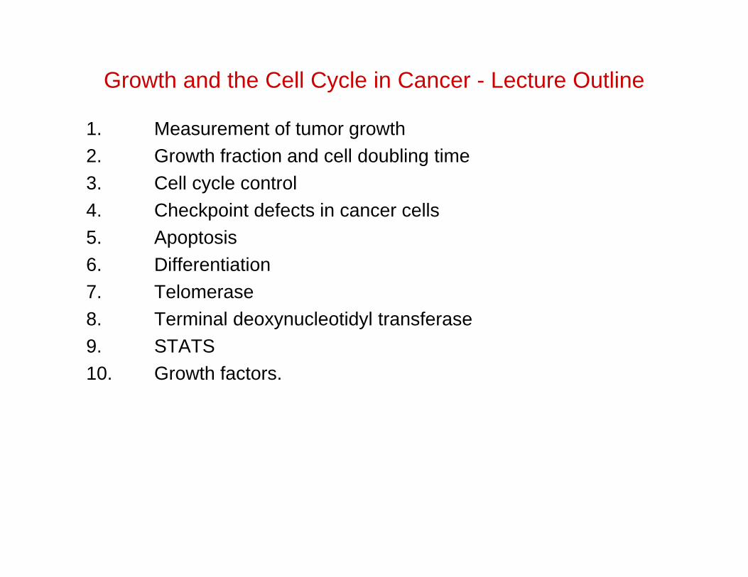
Growth and the Cell Cycle in Cancer - Lecture Outline
1. Measurement of tumor growth2. Growth fraction and cell doubling time3 Cell c cle control3. Cell cycle control4. Checkpoint defects in cancer cells5. Apoptosis6. Differentiation7. Telomerase8. Terminal deoxynucleotidyl transferase8. Terminal deoxynucleotidyl transferase9. STATS 10. Growth factors.
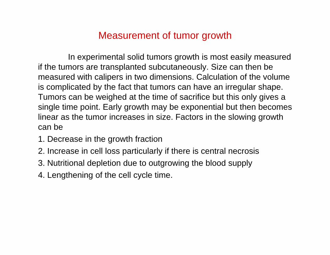
Measurement of tumor growth
In experimental solid tumors growth is most easily measured if the tumors are transplanted subcutaneously. Size can then be measured with calipers in two dimensions. Calculation of the volumemeasured with calipers in two dimensions. Calculation of the volume is complicated by the fact that tumors can have an irregular shape. Tumors can be weighed at the time of sacrifice but this only gives a single time point. Early growth may be exponential but then becomes linear as the tumor increases in size. Factors in the slowing growth can be1. Decrease in the growth fraction2. Increase in cell loss particularly if there is central necrosis3. Nutritional depletion due to outgrowing the blood supply4 Lengthening of the cell cycle time4. Lengthening of the cell cycle time.
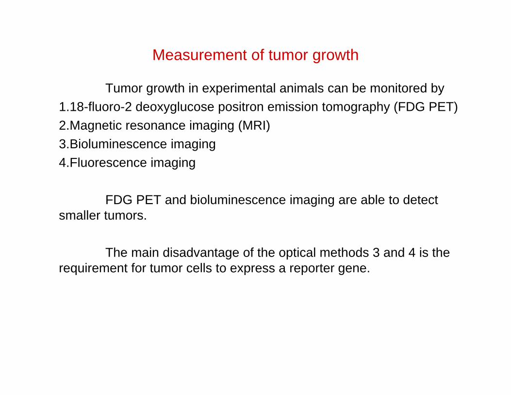
Measurement of tumor growth
Tumor growth in experimental animals can be monitored by1.18-fluoro-2 deoxyglucose positron emission tomography (FDG PET)2 Magnetic resonance imaging (MRI)2.Magnetic resonance imaging (MRI)3.Bioluminescence imaging4.Fluorescence imaging
FDG PET and bioluminescence imaging are able to detect smaller tumors.
The main disadvantage of the optical methods 3 and 4 is the requirement for tumor cells to express a reporter gene.equ e e t o tu o ce s to e p ess a epo te ge e
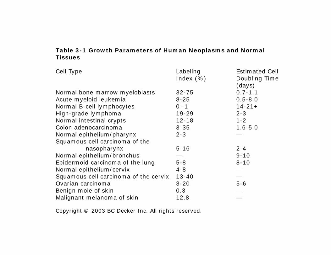
Table 3-1 Growth Parameters of Human Neoplasms and Normal Tissues
Cell Type Labeling Estimated CellIndex (%) Doubling Time Index (%) Doubling Time
(days)Normal bone marrow myeloblasts 32-75 0.7-1.1Acute myeloid leukemia 8-25 0.5-8.0Normal B-cell lymphocytes 0 -1 14-21+Hi h d l h 19 29 2 3High-grade lymphoma 19-29 2-3Normal intestinal crypts 12-18 1-2Colon adenocarcinoma 3-35 1.6-5.0Normal epithelium/pharynx 2-3 —Squamous cell carcinoma of the q
nasopharynx 5-16 2-4Normal epithelium/bronchus — 9-10Epidermoid carcinoma of the lung 5-8 8-10Normal epithelium/cervix 4-8 —Squamous cell carcinoma of the cervix 13 40Squamous cell carcinoma of the cervix 13-40 —Ovarian carcinoma 3-20 5-6Benign mole of skin 0.3 —Malignant melanoma of skin 12.8 —
Copyright © 2003 BC Decker Inc. All rights reserved.
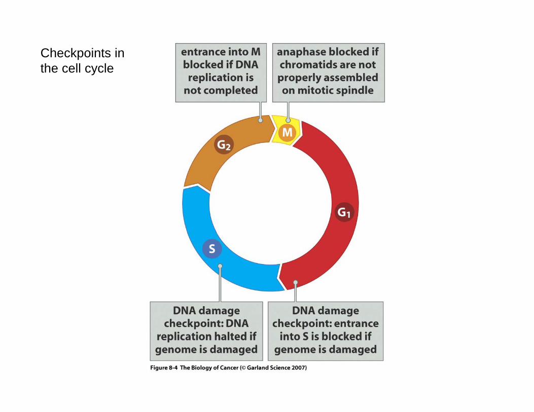
Checkpoints in the cell cycle
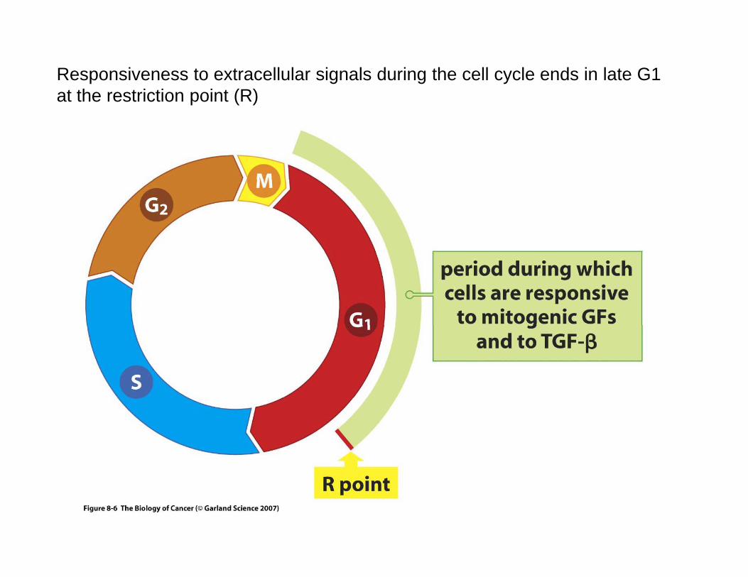
Responsiveness to extracellular signals during the cell cycle ends in late G1 at the restriction point (R)
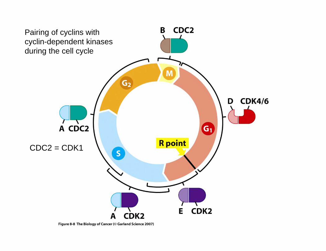
Pairing of cyclins with cyclin-dependent kinases during the cell cycle
CDC2 = CDK1
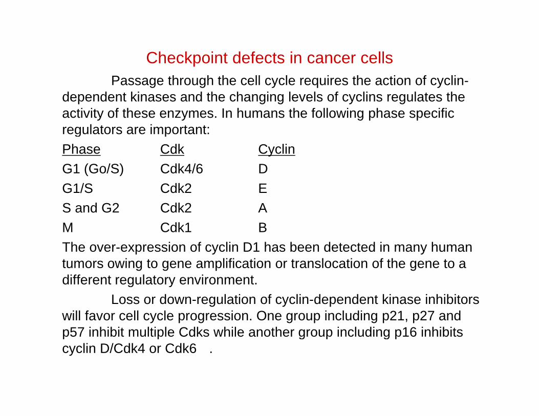
Checkpoint defects in cancer cellsP th h th ll l i th ti f liPassage through the cell cycle requires the action of cyclin-
dependent kinases and the changing levels of cyclins regulates the activity of these enzymes. In humans the following phase specific regulators are important:regulators are important:Phase Cdk CyclinG1 (Go/S) Cdk4/6 DG1/S Cdk2 ES and G2 Cdk2 AM Cdk1 BThe over-expression of cyclin D1 has been detected in many human tumors owing to gene amplification or translocation of the gene to a different regulatory environment. g y
Loss or down-regulation of cyclin-dependent kinase inhibitors will favor cell cycle progression. One group including p21, p27 and p57 inhibit multiple Cdks while another group including p16 inhibits p p g p g pcyclin D/Cdk4 or Cdk6 .
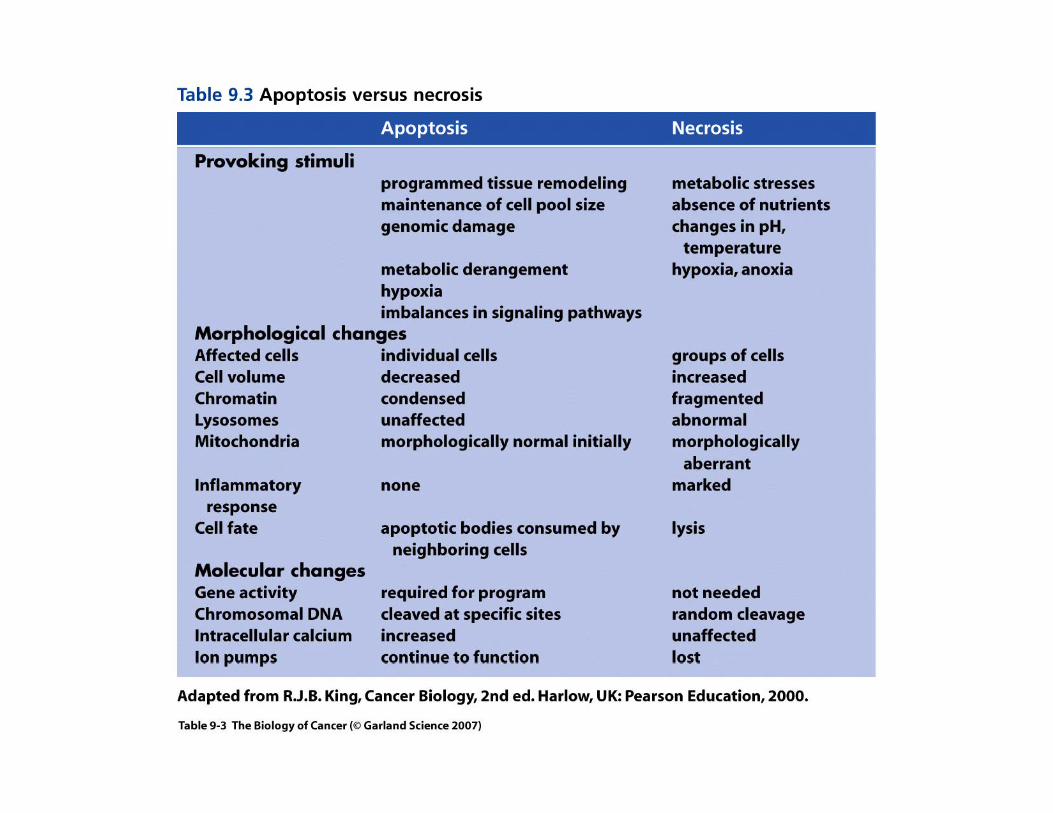
Apoptosis
A decrease in programmed cell death (apoptosis) will favor the expansion of a cancer cell population. This can be achieved by a decrease in pro-apoptotic factors and/ or an increase in anti-apoptotic f t M l l lif ti li i h h ifactors. Many slowly proliferating malignancies such as chronic lymphocytic leukemia, multiple myelomas, and colon and breast cancers over-express the antiapoptotic proteins Bcl-2 and Bcl-XL.
Survivin
Survivin is a bifunctional protein that has a critical role in the regulation of both cell division and survival. Survivin is a member of th i hibit f t i f il f t i Th l l tthe inhibitor of apoptosis family of proteins. These molecules act as suppressors of caspases, the effector enzymes of apoptosis. Survivin affects multiple signaling networks implicated in the regulation of apoptosis including the mitochondrial pathway of cell deathapoptosis including the mitochondrial pathway of cell death, modulation of p53 checkpoints, and control of spindle formation and proper kinetochore attachment during cell division.
Several clinical trials targeting survivin are underway includingSeveral clinical trials targeting survivin are underway including immunotherapy or small-molecule antagonists.
References: D.C. Altieri. Targeted therapy by disabling crossroad signaling networks: the survivin paradigm. Mol. Cancer Ther. 5: 478-482, 2006.
Ambion TechNotes Newsletter 13, 7-11, 2007.
DNA methylation
DNA meth lation in e kar otes in ol es addition of a meth lDNA methylation in eukaryotes involves addition of a methyl group to the 5 position of a cytosine ring. DNA methylation is often associated with the silencing of gene transcription. Genes that may be silenced in cancer cells by methylation include tumor suppressorsilenced in cancer cells by methylation include tumor suppressor genes, genes that suppress tumor invasion and metastasis and DNA repair genes. 5-azacytidine, deoxyazacytidine and zebularine are compounds that block DNA methylation and they have shown promisecompounds that block DNA methylation and they have shown promise in vitro and in clinical trials in leukemias.
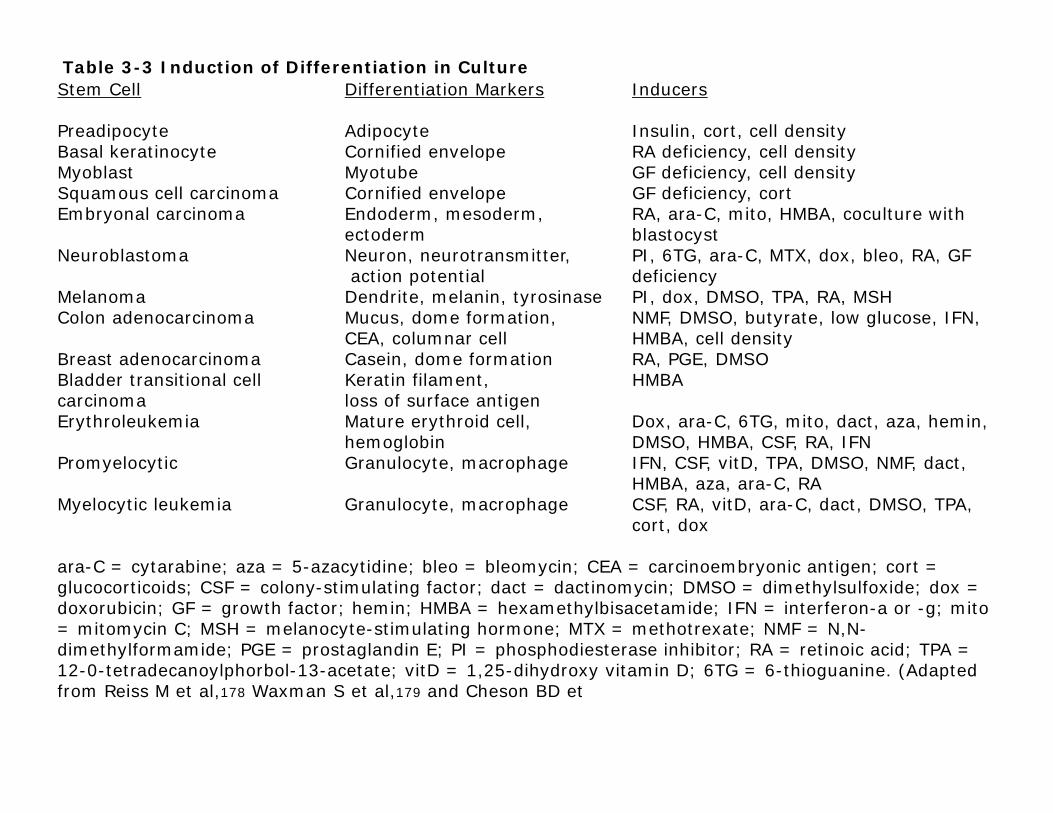
Table 3-3 Induction of Differentiation in CultureStem Cell Differentiation Markers Inducers
Preadipocyte Adipocyte Insulin, cort, cell densityBasal keratinocyte Cornified envelope RA deficiency cell densityBasal keratinocyte Cornified envelope RA deficiency, cell densityMyoblast Myotube GF deficiency, cell densitySquamous cell carcinoma Cornified envelope GF deficiency, cortEmbryonal carcinoma Endoderm, mesoderm, RA, ara-C, mito, HMBA, coculture with
ectoderm blastocystNeuroblastoma Neuron neurotransmitter PI 6TG ara C MTX dox bleo RA GF Neuroblastoma Neuron, neurotransmitter, PI, 6TG, ara-C, MTX, dox, bleo, RA, GF
action potential deficiencyMelanoma Dendrite, melanin, tyrosinase PI, dox, DMSO, TPA, RA, MSHColon adenocarcinoma Mucus, dome formation, NMF, DMSO, butyrate, low glucose, IFN,
CEA, columnar cell HMBA, cell density B east adenoca cinoma Casein dome fo mation RA PGE DMSOBreast adenocarcinoma Casein, dome formation RA, PGE, DMSOBladder transitional cell Keratin filament, HMBAcarcinoma loss of surface antigen Erythroleukemia Mature erythroid cell, Dox, ara-C, 6TG, mito, dact, aza, hemin,
hemoglobin DMSO, HMBA, CSF, RA, IFNP l ti G l t h IFN CSF itD TPA DMSO NMF d t Promyelocytic Granulocyte, macrophage IFN, CSF, vitD, TPA, DMSO, NMF, dact,
HMBA, aza, ara-C, RAMyelocytic leukemia Granulocyte, macrophage CSF, RA, vitD, ara-C, dact, DMSO, TPA,
cort, dox
C t bi 5 tidi bl bl i CEA i b i ti t ara-C = cytarabine; aza = 5-azacytidine; bleo = bleomycin; CEA = carcinoembryonic antigen; cort = glucocorticoids; CSF = colony-stimulating factor; dact = dactinomycin; DMSO = dimethylsulfoxide; dox = doxorubicin; GF = growth factor; hemin; HMBA = hexamethylbisacetamide; IFN = interferon-a or -g; mito = mitomycin C; MSH = melanocyte-stimulating hormone; MTX = methotrexate; NMF = N,N-dimethylformamide; PGE = prostaglandin E; PI = phosphodiesterase inhibitor; RA = retinoic acid; TPA = 12-0-tetradecanoylphorbol-13-acetate; vitD = 1,25-dihydroxy vitamin D; 6TG = 6-thioguanine. (Adapted from Reiss M et al,178 Waxman S et al,179 and Cheson BD et
TELOMERASE
The ends of linear chromosomes in eukaryotes are known as telomeres. They contain tandem repeated sequences which in humans may be TTAGGGmay be TTAGGG
In the replication of DNA, after removal of the RNA primer at the 5’ end of a strand by RNAseH activity conventional DNAthe 5 end of a strand by RNAseH activity, conventional DNA polymerases can not fill in the gap. This problem can be solved by the telomerase enzyme. Telomerase consists of RNA and protein. The RNA hybridizes with the 3’ end of the DNA duplex and serves as aRNA hybridizes with the 3 end of the DNA duplex and serves as a template for extension of the 3’ end. It does this in a repetitive manner to provide a sufficient extension for an RNA primer to be added that is complementary to the 3’ end. DNA polymerase can then fill the gap from the 5’ end. The eventual loss of the RNA primer is compensated by the telomerase catalyzed extension.
Without telomerase activity there will be a progressive loss of DNA at the end of the chromosome.
TELOMERASE
Telomerase activity is found in embryonic tissues and in germ cells and some adult tissues that have high rates of division including thymus and intestine. It is not found in most adult tissues but has beenthymus and intestine. It is not found in most adult tissues but has been detected in many types of tumor and a variety of human cancer cell lines.
It has been suggested that telomerase activity may be an important factor in the immortalization of cancer cell lines.
The reproductive cell death induced by ionizing radiation in cancer cells has been shown to be accompanied by a decrease in telomerase activity Detection of telomerase activity has beentelomerase activity. Detection of telomerase activity has been proposed as a diagnostic procedure for cancer tissue including pancreatic cancer.
Reference: Robert Weinberg, The Biology of Cancer, 2007, pages 368-398.
TERMINAL DEOXYNUCLEOTIDYL TRANSFERASE
Unlike other DNA polymerases, terminal deoxynucleotidyl t f d t i t l t t d f DNA th i Ittransferase does not require a template strand for DNA synthesis. It adds a single strand DNA sequence. Terminal deoxynucleotidyl transferase activity is normally found only in the precursor cells for lymphocytes in bone marrow The enzyme is believed to have a rolelymphocytes in bone marrow. The enzyme is believed to have a role in immune function. Terminal deoxynucleotidyl transferase can serve as a diagnostic marker for circulating leukemic cells.
DNA REPAIR1. nucleotide excision repairXeroderma pigmentosum can be caused by a defect in the excinuclease that cleaves the DNA near a pyrimidine dimer. There is a very high risk of skin py y gcancer with this hereditary condition.2. base excision repairIn this mechanism repair is initiated by a purine or pyrimidine glycosylase.3. mismatch repairHereditary nonpolyposis colon cancer (HNCC) may affect 1 in 200 people). It results from defects in mismatch repair. In most cases there are mutations in
f hMSH2 d hMLH1) Th h h i l fone of two genes, hMSH2 and hMLH1). These are the human equivalents of MutS and MutL of E. coli. hMSH2 binds at a mismatched DNA base pair and hMLH1 participates in cleaving DNA near the mismatch to initiate the repair process.process.4. O6 alkylguanine alkyl transferaseThis protein removes small alkyl groups such as methyl groups from the O6 position of guanine residues.p g
ATM
Ataxia telangiectasia is caused by mutations in the ATM gene encoding a protein kinase that is activated by d bl t d DNA b k ATM ki ti it i iti tdouble stand DNA breaks. ATM kinase activity initiates a phosphorylation cascade that modifies substrates controlling cell cycle arrest and DNA repair.
Ataxia telangiectasia has an autosomal recessive i h it d i h t i d b iinheritance and is characterized by progressive neurodegeneration, immunodeficiency and a high predisposition to the development of lymphoid p p p y pmalignancies.
STATS and oncogenic signaling pathways
STAT (Signal transducer and activator of transcription) family proteins are latent transcription factors that convey signals from cytokine and growth-factor receptors to the nucleus.cytokine and growth factor receptors to the nucleus.
STAT proteins, particularly STAT3 and STAT5 proteins are frequently over-activated in a variety of human solid tumors and blood malignancies.malignancies.
Persistent STAT3 signaling promotes the growth and survival of cancer cells and induces angiogenesis.
Tumor cells that become dependent on persistent STAT3Tumor cells that become dependent on persistent STAT3 signaling are more sensitive to STAT3 inhibitors than normal cells. Small molecule inhibitors of STAT3 and STAT 5 are being investigated e.g. WP-1034.est gated e g 03
Reference:H. Yu and R. Jove. The STATS of cancer - new molecular targets come of age. Nature Reviews Cancer 4: 97-105, 2004
GROWTH FACTORS
Insulin-like growth factor-I (IGF-I) levels are known to be decreased by caloric restriction. Supplementation with IGF-I was shown to prevent the protective effect of caloric restriction on tumor progression in p53 deficient mice.(Dunn et al Cancer Res 57:4667 1997)(Dunn et al., Cancer Res., 57:4667, 1997).
The loss of TGF-beta receptor gene in fibroblasts of a knockout mouse resulted in neoplasia in the prostate and forestomach associatedmouse resulted in neoplasia in the prostate and forestomach associated with an abundance of stromal cells. The occurrence of transformation in the adjacent epithelial cells was accompanied by activation of hepatocyte growth factor signaling.�(Bhowmick et al., Science 303: 848-851, 2004)
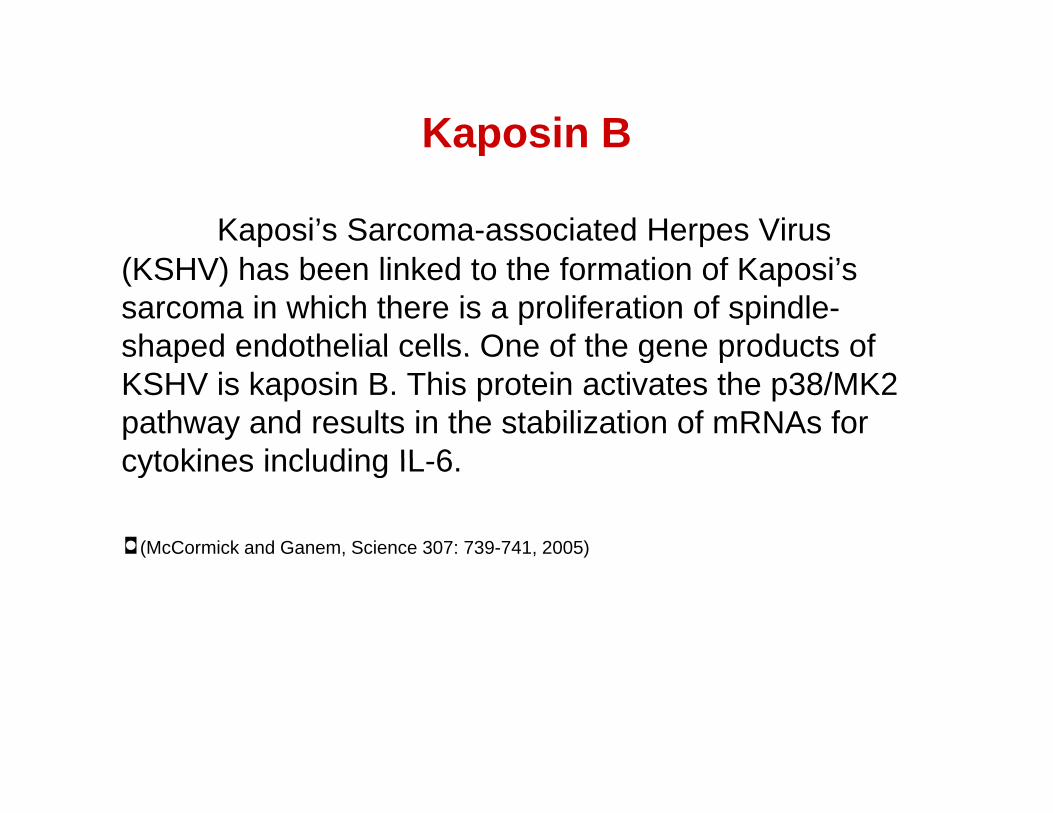
Kaposin Bp
Kaposi’s Sarcoma-associated Herpes Virus (KSHV) has been linked to the formation of Kaposi’s sarcoma in which there is a proliferation of spindle-shaped endothelial cells. One of the gene products ofshaped endothelial cells. One of the gene products of KSHV is kaposin B. This protein activates the p38/MK2 pathway and results in the stabilization of mRNAs for cytokines including IL 6cytokines including IL-6.
� (McCormick and Ganem, Science 307: 739-741, 2005)
Growth and the Cell Cycle in Cancer -Suggested Reading
M. Andreeff, D.W. Goodrich and H.P. Koeffler, In Holland-Frei Cancer Medicine 8th ed Part II Section I 3 Cell Proliferation andMedicine - 8th ed., Part II, Section I, 3. Cell Proliferation and Differentiation (2010).
J C Reed In Holland-Frei Cancer Medicine - 8th ed Part II Section I 4J.C. Reed, In Holland-Frei Cancer Medicine - 8th ed., Part II, Section I, 4. Apoptosis and Cancer (2010).
S.A. Aaronson, In Holland-Frei Cancer Medicine - 8th ed., Part II, SectionS.A. Aaronson, In Holland Frei Cancer Medicine 8th ed., Part II, Section I, 5. Growth Factors and Signal Transduction in Cancer (2010).
R. Weinberg, The Biology of Cancer, Garland Press, Chapter 8, 9 and 10, g, gy , , p , ,(2007).