Granular cell tumor in two oral anatomic sites
3
CASE REPORT Granular cell tumor in two oral anatomic sites Marianna Sampaio Serpa 1 • Hugo Costa-Neto 1 • Patrı ´cia Teixeira de Oliveira 1 • E ´ ricka Janine Dantas da Silveira 1 • Ana Miryam Costa de Medeiros 1 Received: 30 November 2015 / Accepted: 21 March 2016 / Published online: 23 March 2016 Ó Springer-Verlag Berlin Heidelberg 2016 Abstract Granular cell tumor (GCT) is an uncommon benign soft tissue neoplasm, which usually presents as a solitary nodule, appearing especially in the tongue. There are few cases of multiple oral GCT in the English-language literature, with only three cases reported in the last 20 years. This report describes a case of two oral GCT in a young patient and provides a literature review from 1995 to 2015. Clinical characteristics of the reviewed cases were retrieved and compared with the present case. Exception- ally, the current case was the only one that occurred in an adolescent and solely affected the oral cavity. Besides the oral cavity, the other cases also observed GCT lesions in the skin. Although rare, it is important to know multiple oral GCT clinical and histopathological characteristics so they can be correctly diagnosed, treated and followed up. Keywords Oral granular cell tumor Á Clinical characteristics Á Diagnosis Á Treatment Introduction Granular cell tumor (GCT) is an uncommon benign neo- plasm that has been thought to be of neural origin due to its strong and consistent stain for S-100 protein [1]. Clinically, GCT is typically an asymptomatic sessile nodule of pinkish color, occurring most frequently between the fourth and sixth decades of life [2]. Although usually solitary, multi- ple GCT lesions can occur in up to 25 % of the cases, being most often located in the intradermal or subcutaneous tis- sue [3]. Few cases of multiple oral GCT have been reported in the English-language literature. We report herein a case of GCT occurring in the dorsum and lateral tongue of a young patient and provide a literature review from 1995 to 2015. Case presentation A 12-year-old female patient was referred to the Depart- ment of Stomatology of the Federal University of Rio Grande do Norte, complaining of asymptomatic swellings in the tongue of unknown evolution time. Medical history was not helpful and extraoral physical exam showed no alterations. Intraoral examination revealed two lesions with nodular aspect, sessile implantation, normal mucosa color and firm consistence (Fig. 1). The lesions were located in the dorsum and lateral border of the tongue, measuring, respectively, 0.8 and 0.5 cm in diameter. According to the report, no trauma occurred in these areas. Clinical diag- nosis hypotheses were GCT, neurilemmoma and fibroma. Excisional biopsy of the lesions was performed and microscopic examination revealed both specimens were nonencapsulated neoplasms composed of polygonal cells with large cytoplasm, remarkably granular appearance and indistinct margins, presenting ovoid and pycnotic nuclei. Close relationship between the skeletal muscle and the neoplastic cells was also observed (Fig. 2a). Immunohis- tochemical analysis of both lesions revealed strong posi- tivity of the tumor cells for S-100 protein (rabbit polyclonal; dilution 1:2000; Dako, Glostrup, Denmark) (Fig. 2b). Periodic acid-Schiff (PAS) was performed to & Marianna Sampaio Serpa [email protected] 1 Postgraduate Program in Oral Pathology, Department of Dentistry, Federal University of Rio Grande do Norte, Avenida Senador Salgado Filho, 1787, Lagoa Nova, Natal, Rio Grande do Norte CEP 59.056-000, Brazil 123 Eur Arch Otorhinolaryngol (2016) 273:3439–3441 DOI 10.1007/s00405-016-4006-5
Transcript of Granular cell tumor in two oral anatomic sites

CASE REPORT
Granular cell tumor in two oral anatomic sites
Marianna Sampaio Serpa1 • Hugo Costa-Neto1 • Patrıcia Teixeira de Oliveira1 •
Ericka Janine Dantas da Silveira1 • Ana Miryam Costa de Medeiros1
Received: 30 November 2015 / Accepted: 21 March 2016 / Published online: 23 March 2016
� Springer-Verlag Berlin Heidelberg 2016
Abstract Granular cell tumor (GCT) is an uncommon
benign soft tissue neoplasm, which usually presents as a
solitary nodule, appearing especially in the tongue. There
are few cases of multiple oral GCT in the English-language
literature, with only three cases reported in the last
20 years. This report describes a case of two oral GCT in a
young patient and provides a literature review from 1995 to
2015. Clinical characteristics of the reviewed cases were
retrieved and compared with the present case. Exception-
ally, the current case was the only one that occurred in an
adolescent and solely affected the oral cavity. Besides the
oral cavity, the other cases also observed GCT lesions in
the skin. Although rare, it is important to know multiple
oral GCT clinical and histopathological characteristics so
they can be correctly diagnosed, treated and followed up.
Keywords Oral granular cell tumor � Clinicalcharacteristics � Diagnosis � Treatment
Introduction
Granular cell tumor (GCT) is an uncommon benign neo-
plasm that has been thought to be of neural origin due to its
strong and consistent stain for S-100 protein [1]. Clinically,
GCT is typically an asymptomatic sessile nodule of pinkish
color, occurring most frequently between the fourth and
sixth decades of life [2]. Although usually solitary, multi-
ple GCT lesions can occur in up to 25 % of the cases, being
most often located in the intradermal or subcutaneous tis-
sue [3]. Few cases of multiple oral GCT have been reported
in the English-language literature. We report herein a case
of GCT occurring in the dorsum and lateral tongue of a
young patient and provide a literature review from 1995 to
2015.
Case presentation
A 12-year-old female patient was referred to the Depart-
ment of Stomatology of the Federal University of Rio
Grande do Norte, complaining of asymptomatic swellings
in the tongue of unknown evolution time. Medical history
was not helpful and extraoral physical exam showed no
alterations. Intraoral examination revealed two lesions with
nodular aspect, sessile implantation, normal mucosa color
and firm consistence (Fig. 1). The lesions were located in
the dorsum and lateral border of the tongue, measuring,
respectively, 0.8 and 0.5 cm in diameter. According to the
report, no trauma occurred in these areas. Clinical diag-
nosis hypotheses were GCT, neurilemmoma and fibroma.
Excisional biopsy of the lesions was performed and
microscopic examination revealed both specimens were
nonencapsulated neoplasms composed of polygonal cells
with large cytoplasm, remarkably granular appearance and
indistinct margins, presenting ovoid and pycnotic nuclei.
Close relationship between the skeletal muscle and the
neoplastic cells was also observed (Fig. 2a). Immunohis-
tochemical analysis of both lesions revealed strong posi-
tivity of the tumor cells for S-100 protein (rabbit
polyclonal; dilution 1:2000; Dako, Glostrup, Denmark)
(Fig. 2b). Periodic acid-Schiff (PAS) was performed to
& Marianna Sampaio Serpa
1 Postgraduate Program in Oral Pathology, Department of
Dentistry, Federal University of Rio Grande do Norte,
Avenida Senador Salgado Filho, 1787, Lagoa Nova, Natal,
Rio Grande do Norte CEP 59.056-000, Brazil
123
Eur Arch Otorhinolaryngol (2016) 273:3439–3441
DOI 10.1007/s00405-016-4006-5

further characterize this cell population (Fig. 2c). In view
of these findings, the histopathological diagnosis was GCT.
The patient has been under follow-up for 1 year without
any signal of recurrence.
Discussion
Three cases of multiple oral GCT were identified in the
English-language literature from a search on PubMed and
Scopus from 1995 to 2015. These cases, along with the
current case are listed in Table 1. Additionally, all clinical
features of the cases are summarized in Table 2.
Adding up the four cases, a total of 28 GCT were found;
the range was 2–12 lesions per patients, and the mean
number of lesions was seven. Of the 28 lesions, 16 lesions
were located in the oral cavity and the tongue was the most
common site (56.25 %). The mean age was 29.5 and all
cases occurred in melanodermic women. Furthermore, the
present case was the only one to solely affect the oral
cavity. In the other cases, gastrointestinal tract and, par-
ticulary, subcutaneous involvement was common, involv-
ing hands, feet, elbow, face, groin, perineum and vulva. In
cases 1 and 2, after surgical excision and histopathological
diagnosis, more GCT lesions appeared in other locations,
including oral and extraoral sites.
Multiple GCT are uncommon lesions and most com-
monly affect melanodermic women between 30 and
50 years old [2], although the present case affected an
adolescent patient. As for the location, solitary oral GCT
has been described to mainly affect the dorsal surface of
Fig. 1 Intraoral clinical exam. Nodular lesions in dorsum (a) and
lateral border (b) of the tongue
Fig. 2 Photomicrographs of GCT. a Polygonal cells with large
granular cytoplasm and small nuclei. Close relationship between
neoplastic cells and muscle tissue is also observed (hematoxylin–
eosin stain; 9200 lm). b Strong and uniform positivity for S-100
protein (immunohistochemical stain, 920 lm). c Cytoplasmic gran-
ules highlighted (PAS stain, 920 lm)
3440 Eur Arch Otorhinolaryngol (2016) 273:3439–3441
123
the tongue [7]. Based on this review, it was observed
multiple oral GCT appear to involve most frequently the
lateral border of the tongue, following the dorsum.
Except for the present case, besides the oral region, the
other cases also involved lesions in the skin and gastroin-
testinal tract. Moreover, because of the patient’s young age,
and previous reports of new GCT lesions appearing after
histopathological diagnosis, it is possible that as the patient
gets older, other GCT lesions show up in other locations
besides the oral cavity.
Multiple GCT have previously been associated with
systemic disorders, such as growth retardation, lentiginosis,
cafe-au-lait spots and neurofibromatosis type I, occurring
mainly in children or adolescents [8]. Due to that, it is
important to consider other abnormalities in a young
patient with multiple GCT. In the current case, the patient
reported no relevant medical history and no other physical
alterations were observed in clinical examination, dis-
carding any possible systemic disorder.
Surgical excision with margin of safety is the treatment
of choice and recurrences are rare [1]. In this review, there
was no case of recurrence, however, in two cases more
GCT lesions showed up after histopathological diagnosis,
which suggests the need for a long-term follow-up.
Compliance with ethical standards
Funding None.
Conflict of interest All authors declare that they have no conflict of
interest.
Ethical approval All procedures performed in this study involving
the patient were in accordance with the ethical standards of the
institutional and/or national research committee and with the 1964
Helsinki declaration and its later amendments or comparable ethical
standards.
Informed consent Informed consent was obtained from the patient
in this case report.
References
1. Costa NCS, Bertini F, Carvalho YR, Almeida JD, Rodrigues
Cavalcante AS (2012) Granular cell tumor presenting as a tongue
nodule: two case reports. J Med Case Rep 6:56
2. Cole E, Rahman N, Webb R (2012) Case series: two cases of an
atypical presentation of oral granular cell tumour. Case Rep Med
2012:159803
3. Bomfin LE, Alves FA, Almeida OP, Kowalski LP, Perez DE
(2009) Multiple granular cell tumors of the tongue and parotid
gland. Oral Surg Oral Med Oral Pathol Oral Radiol Endod
107:e10–e13
4. Collins BM, Jones AC (1995) Multiple granular cell tumors of the
oral cavity. J Oral Maxillofac Surg 53:707–711
5. Sargenti-Neto S, Brazao-Silva MT, Souza KCN, Faria PR,
Durighetto-Junior AF, Loyola AM, Cardoso SV (2009) Multicen-
tric granular cell tumor: report of a patient with oral and cutaneous
lesions. Br J Oral Maxillofac Surg 47:62–64
6. Varghese J, Hardcastle N, Slater D, Cockayne SE (2010)
Cutaneous and oesophageal granular cell tumours. Clin Exp
Dermatol 35:551–552
7. Daniels JSM (2009) Granular cell tumour of tongue: a case report.
Saudi Dent J 21:75–78
8. Tomson N, Abdullah Tan CY (2006) Multiple granular cell tumors
in a child with growth retardation: report of a case and review of
the literature. Int J Dermatol 45:1358–1361
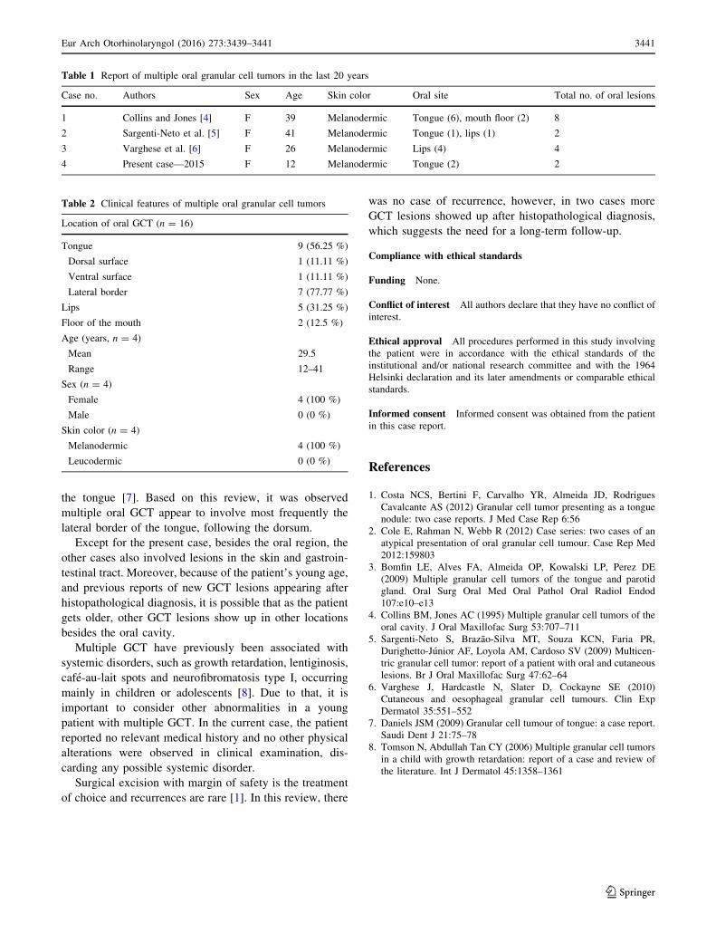
Table 1 Report of multiple oral granular cell tumors in the last 20 years
Case no. Authors Sex Age Skin color Oral site Total no. of oral lesions
1 Collins and Jones [4] F 39 Melanodermic Tongue (6), mouth floor (2) 8
2 Sargenti-Neto et al. [5] F 41 Melanodermic Tongue (1), lips (1) 2
3 Varghese et al. [6] F 26 Melanodermic Lips (4) 4
4 Present case—2015 F 12 Melanodermic Tongue (2) 2
Table 2 Clinical features of multiple oral granular cell tumors
Location of oral GCT (n = 16)
Tongue 9 (56.25 %)
Dorsal surface 1 (11.11 %)
Ventral surface 1 (11.11 %)
Lateral border 7 (77.77 %)
Lips 5 (31.25 %)
Floor of the mouth 2 (12.5 %)
Age (years, n = 4)
Mean 29.5
Range 12–41
Sex (n = 4)
Female 4 (100 %)
Male 0 (0 %)
Skin color (n = 4)
Melanodermic 4 (100 %)
Leucodermic 0 (0 %)
Eur Arch Otorhinolaryngol (2016) 273:3439–3441 3441
123