Goals*of*the*lecture Evaluation*and* Managementof* Strabismus
18
2/5/17 1 Evaluation and Management of Strabismus Don W. Lyon, OD, MS Chief of Pediatrics/Binocular Vision Services Goals of the lecture • Briefly, ever so briefly, review strabismus • Primary goal: Discuss clinical exam elements in regards to diagnosing strabismus • Discuss treatment options • Monitoring • Lenses • Vision Therapy • Surgery History • Patient here for routine examination, previously diagnosed with esotropia, would like prism to correct the eye turn • ALT ET since infancy, has not changed according to parents • No medical issues no family history Exam data • Dist. and Near VA cc 20/25 OD, OS • CT Dist. 16 Alt ET Near 16 Alt ET • Stereo 200” (animals) • Worth dot 4 all distances • Versions Full and Smooth • Pupils PERRLA OD, OS • VF Full OD, OS • MEM +0.50 OD, OS • Color Normal OD, OS • Ret +0.501.25X180 OD, OS • Wet Ret +0.751.50X180 OD, OS • Ocular Health Normal OD, OS
Transcript of Goals*of*the*lecture Evaluation*and* Managementof* Strabismus

2/5/17
1
Evaluation and Management of Strabismus
Don W. Lyon, OD, MSChief of Pediatrics/Binocular Vision Services
Goals of the lecture
• Briefly, ever so briefly, review strabismus• Primary goal: Discuss clinical exam elements in regards to diagnosing strabismus• Discuss treatment options• Monitoring• Lenses• Vision Therapy• Surgery
History
• Patient here for routine examination, previously diagnosed with esotropia, would like prism to correct the eye turn
• ALT ET since infancy, has not changed according to parents
• No medical issues no family history
Exam data
• Dist. and Near VA cc 20/25 OD, OS• CT Dist. 16 Alt ET Near 16 Alt ET• Stereo 200” (animals)• Worth dot 4 all distances• Versions Full and Smooth• Pupils PERRLA OD, OS• VF Full OD, OS• MEM +0.50 OD, OS• Color Normal OD, OS• Ret +0.50-‐1.25X180 OD, OS• Wet Ret +0.75-‐1.50X180 OD, OS• Ocular Health Normal OD, OS

2/5/17
2
Exam Data
• Visuoscopy Central Steady OD, OS
• Synoptophore <S 0rtho <D 18 BO
• Patient has Harmonious Anomalous Correspondence along with her Alt ET
• What is the treatment?
Characteristics of strabismus
• Direction of turn (Eso, Exo, Vertical)• Amount of deviation (in prism diopters) at both distance
and near• Patient control• Frequency (constant, intermittent)• Unilateral or alternating• Comitancy• Impact on Sensory abilities
Esotropia
• Majority of ET develops by age 6 years• Congenital (before 6 months of age)• Infantile (before one year of age)• Acquired (after one year of age)
Esotropia
• Majority of ET develops by age 6 years• Congenital• Large angle, can be >50pd• Low hyperopic Rx-‐ (so partial or non-‐accommodative)• Can have more sensory abnormalities

2/5/17
3
Esotropia
• Majority of ET develops by age 6 years• Infantile• A large “range” of esotropia types• Various prognosis based upon size, consistency, sensory abnormalities
• DDX:• Pseudostrabismus• Accommodative ET• Duane’s• Moebius syndrome
Esotropia
• Majority of ET develops by age 6 years• Acquired-‐ can occur in childhood or adults [adults decomp]• Group of different ET types• DDX:• Pseudostrabismus• Concomitant deviations• Nonconcomitant deviations
Classification of Esotropia
• Accommodative• Acquired• Basic Esotropia• Congenital• Convergence Excess• Divergence insufficiency• Infantile• Non-‐accommodative• Partially Accommodative• Pattern Esotropia
When Esotropia is not Esotropia
• Differential diagnoses• Vaccinations• Viral illness• Trauma• Neoplasm• Duane’s syndrome• NBS (Nystagmus Blocking Syndrome)• Other pathologies

2/5/17
4
Classification of Exotropia
• Basic Exotropia• Convergence Insufficiency• Divergence Excess• Pattern Exotropia• Consecutive Exotropia
Exotropia
• Prevalence is lower than esotropia• May be artificial, many patients with exotropia may not
be seen in eye care professional offices
• Most common is intermittent exotropia
• Clinical picture not widely known
• Onset ~age 5 years
Exotropia: To worry or not
• Acquired XT• Tumor (supra nuclear)• Duane’s II• Medial Rectus Palsy• Vascular• Trauma• Tumor
• Myasthenia gravis• Internuclear Ophthalmoplegia
Exotropia
• Patients present with• Eye strain or headaches after prolonged near tasks• Lack of concentration during reading• Tiredness when reading• These symptoms increase as the amount of time the eye
posture is tropic increases

2/5/17
5
Specific Tests to Diagnose Amblyopia and Strabismus
Evaluation of Strabismus
• Case History• Diagnostic Test Sequence• Refractive Status• Visual Acuity• Monocular Fixation• Ocular Alignment• Correspondence• Sensorimotor Fusion• Ocular Health if not done previously
Development of Amblyopia
• Amblyopia more likely to occur if strabismus is present during critical period• Critical period is birth to 3 years of age• Sensitive period is 3 years to 10 years of age
• Amblyopia more likely to occur if strabismus is constant, unilateral
• If amblyopia is present initial treatment designed to improve visual acuity
Monocular Fixation: Evaluating for EF
• Eccentric Fixation: Using a non-‐foveal point in monocular conditions
• Test for in a monocular condition• Cover the eye not being tested
• Visuoscopy is an easy clinical testing method

2/5/17
6
Visuoscopy with Central, Steady Fixation OS
Visuoscopy with 3 pdNasal, Steady Fixation OS
Eccentric Fixation
• Impact on measured VA• MAR=E(pd)+1• Patient with 4 Nasal EF• MAR=5• 20/x=1/5MAR• Expected acuity=20/100
• Impact on CT (<D)• +=Nasal EF -‐ =Temporal EF• Ortho UCT with 5 Nasal EF= 5pd Eso• <Dt= <Dm + EF
Measurement of Deviation
• A measure of Concomitancy• Is the strabismus concomitant or non-‐concomitant
• Non-‐concomitancy can indicate• Trauma• Pathology• Congenital
• What you find will impact your treatment decisions

2/5/17
7
Deviation Measurement
• Objective and Subjective measurements• Observation• Version testing• Duction testing• Park’s 3-‐step• Cover Test• Red lens with or without Maddox Rod
Direct Observation
• Look at head posture when patient is sitting in the chair• Look for facial asymmetry• Facial hypoplasia• Eg. SO palsy, head tilt to opposite side of palsy, facial hypoplasia on opposite side as well
Facial Hypoplasia
Hypoplasia on patient’s left side
Diagnostic Action Field
OD Gaze OS Gaze
RLR R LLR L
RMR L LMR R
RSR R & UP LSR L & UP
RIR R & DOWN LIR L & DOWN
RSO L & DOWN LSO R & DOWN
RIO L& UP LIO R & UP

2/5/17
8
Cover Test
• Mohney BG, Holmes JM. An office-‐based scale for assessing control in intermittent exotropia. Strabismus 2006;14:147-‐50.
• Intermittent Exotropia Control Scale• 5 = Constant Exotropia• 4 = Exotropia > 50% of the 30-‐second period before dissociation
• 3 = Exotropia < 50% of the 30-‐second period before dissociation
• 2 = No exotropia unless dissociated, recovers in > 5 seconds• 1 = No exotropia unless dissociated, recovers in 1-‐5 seconds• 0 = No exotropia unless dissociated, recovers in < 1 second (phoria)
• Needs to be done before any dissociation including monocular visual acuity
Park’s 3-‐‑step
• Limited clinical value• If the test shows a SO palsy it is believable• Any other muscle shown as deficient is suspect
40 year old male with 20 R hyper, Vertical worse in Left Gaze, Vertical worse in Right Head Tilt
RSR RIO LIO LSR
RIR RSO LSO LIR
Cover test in 9 directions of gaze
• Distance only• Accommodative and vergence issues when performed at near
• Have the patient move their chin to get all nine fields
• Primary deviation when non-‐paretic eye is fixating
• Secondary deviation when paretic eye is fixating, also large magnitude

2/5/17
9
ExampleRSO Palsy
RE Fixating LE Fixating
02BI
02BI
4BU2BI
4BU4BI
04BI
08BI
8BU8BI
8BU6BI
12BU10BI
02BI
04BI
08BI
02BI
02BI
04BI
4BD6BI
6BD8BI
8BD8BI
Red Lens Test
• Useful to find which field of action(s) the patient perceives greater amount of diplopia• Tools needed• Penlight• Red filter (possible Maddox Rod)
• It is better for recent onset and constant strabismus• Red lens goes over eye with better acuity
Red Lens over Patient’s Left Eye
RSO Palsy
Clinical Example
Right Left
ç
çç
çç
ç
ç
çç
ç ç
ç
Doctor’s View of Patient
Patient’s Motor and Sensory Status
ç
ç
ç
2/5/17
10
THE WORLD WOULD BE A HAPPIER PLACE IF NO ONE HAD THOUGHT ABOUT ANOMALOUS
RETINAL CORRESPONDENCE
-‐‑ARTHUR JAMPOLSKY1973
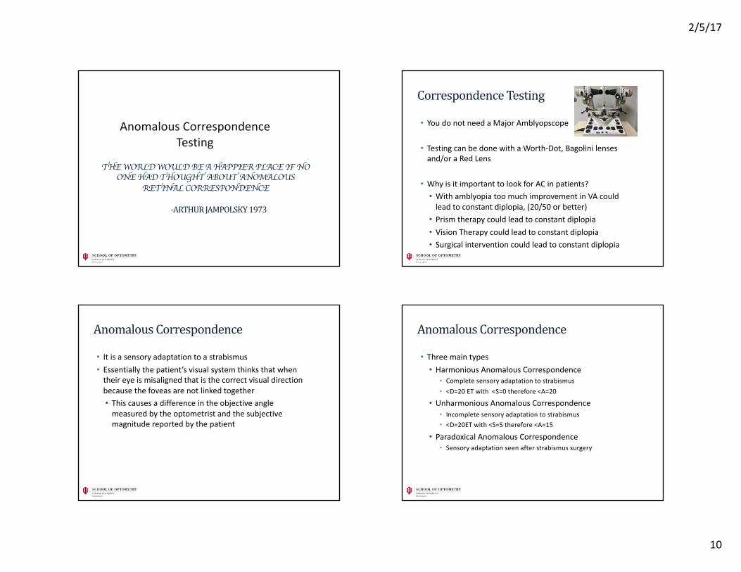
Anomalous Correspondence Testing
Correspondence Testing
• You do not need a Major Amblyopscope
• Testing can be done with a Worth-‐Dot, Bagolini lenses and/or a Red Lens
• Why is it important to look for AC in patients?• With amblyopia too much improvement in VA could lead to constant diplopia, (20/50 or better)• Prism therapy could lead to constant diplopia• Vision Therapy could lead to constant diplopia• Surgical intervention could lead to constant diplopia
Anomalous Correspondence
• It is a sensory adaptation to a strabismus• Essentially the patient’s visual system thinks that when their eye is misaligned that is the correct visual direction because the foveas are not linked together• This causes a difference in the objective angle measured by the optometrist and the subjective magnitude reported by the patient
Anomalous Correspondence
• Three main types• Harmonious Anomalous Correspondence• Complete sensory adaptation to strabismus• <D=20 ET with <S=0 therefore <A=20
• Unharmonious Anomalous Correspondence• Incomplete sensory adaptation to strabismus• <D=20ET with <S=5 therefore <A=15
• Paradoxical Anomalous Correspondence• Sensory adaptation seen after strabismus surgery

2/5/17
11
Unharmonious AC with ET and no EF
<D
<A<S
Unharmonious <S less than <D but greater than 0
D > S> 0 D>A
Clinically what does this mean:
We measure with cover test 20 BO [Objective measurement <D]
On synoptophore the patient places the targets so they align at 5BO [Subjective measurement <S]
<A=15pd Unharmonious AC
Impact of EF on measurement of Strabismus
Need to take into account the amount and direction of EF in measuring <D and <A
<Dt =<Dm+<E<At=<Am+<E
+<D=Eso or BO-‐<D=Exo or BI
+<E=nasal EF-‐<E=temporal EF
CLINICAL MEASUREMENT OF AC
Worth Dot Test• On cover test you measure a 20 Right Constant ET • What should the patient report when you perform Worth Dot
Uncrossed Diplopia Suppression of Right Eye

2/5/17
12
Worth 4-‐‑dot• Under what circumstances would your patient with a constant right 20pd ET report fusion?
• The patient actually has intermittent ETOR
• The patient has Harmonious Anomalous Correspondence
• How do you tell the difference? Repeat the unilateral cover test with the red/green glasses on while viewing the target. If you see movement, it strongly suggest HAC
• No movement strongly suggest IET
Bagolini Lenses
• Free space testing• Can be worn over glasses• Each eye has a lens with striations• RE orientated 135• LE orientated 45
• Testing is done in normal room illumination
Bagolini Lenses-‐‑Subjective responses
NC or HACLE RE Crossed Diplopia
RE Suppression Uncrossed Diplopia
Bagolini Lenses
• If patient subjectively reports crossed or uncrossed diplopia you should place corrective prism over the deviated eye until the patient reports a single light with the X
• Next the perform UCT and if there is no movement NC is suggested, if there is movement AC is suggested
• This is the most natural test for AC and more likely to show AC in patients, (when they have AC)

2/5/17
13
Red Lens test for Correspondence
• Not the same test as for concomitancy • Procedure• Place a red lens and vertical prism (~6 BU or BD) over the “normal” non-‐strabismic eye• Have the patient fixate at a distant white muscle light• Add horizontal prism over the deviating eye until patient reports the two dots are aligned to find subjective angle
• Remove lens and vertical prism and perform ACT to find objective angle
• If they are equal it suggest Normal correspondence
Testing of Strabismus
• Do you have to perform every test listed?
• Find out which tests you are most comfortable with and use those tests in assessing a patient with strabismus
• The results of the tests will help you decide your course of treatment
• Treatment options include• Monitoring• Vision therapy• Patching• Surgery
TREATMENT OPTIONS FOR STRABISMUS
When is Strabismus fixed?
• Loaded question and can depend on who you ask
Functional cure• Maintaining bifoveal fixation in ordinary life situations, (loss of bifoveal fixation should not occur more than 1% of the time)
• Vision should be clear and generally comfortable• The range of bifoveal fixation should be a range that extends from a few centimeters from the patient to a great distance
• Corrective lenses and reasonable amounts of prism may be worn, if necessary

2/5/17
14
Treating Strabismus in Presence of Amblyopia
• The presence of amblyopia will have an impact on your treatment decisions
• For surgical intervention the surgeon will want resolution or close to resolution before performing corrective surgery
• For Vision Therapy it will add another layer to the therapy activities that you could perform
Therapy Goals
• Elimination of motor angle• Decrease of frequency• Elimination of Diplopia• Elimination of Suppression• Elimination of Pulling, Loss of Concentration• Improved Binocularity• Improved Comfort
Prismatic Correction: For patients with intermittent sensory fusion
• This means those patients with intermittent strabismus or those who have normal sensory fusion with added prism• Sheard’s criterion
• Associated phoria (Saladin Card)
• Residual Vergence Demand Criterion • (Caloroso EE, RouseMW: Clinical Management of Strabismus, Boston, 1993,
Butterworth-Heinemann, p. 104)
• This can be used when the patient cannot achieve sensory fusion with clinical testing
• Percentage of dissociated prism amount
Residual Vergence Demand Criterion
Direction Magnitude (r) RVD
Esodeviation 6-20 4-6
Exodeviation 20-30 10-15
Hyperdeviation 3-10 2-4
2/5/17
15
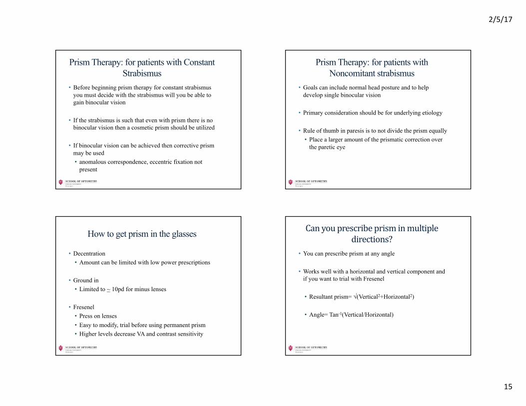
Prism Therapy: for patients with Constant Strabismus
• Before beginning prism therapy for constant strabismus you must decide with the strabismus will you be able to gain binocular vision
• If the strabismus is such that even with prism there is no binocular vision then a cosmetic prism should be utilized
• If binocular vision can be achieved then corrective prism may be used• anomalous correspondence, eccentric fixation not
present
Prism Therapy: for patients with Noncomitant strabismus
• Goals can include normal head posture and to help develop single binocular vision
• Primary consideration should be for underlying etiology
• Rule of thumb in paresis is to not divide the prism equally• Place a larger amount of the prismatic correction over
the paretic eye
How to get prism in the glasses
• Decentration• Amount can be limited with low power prescriptions
• Ground in• Limited to ~ 10pd for minus lenses
• Fresenel• Press on lenses• Easy to modify, trial before using permanent prism• Higher levels decrease VA and contrast sensitivity
Can you prescribe prism in multiple directions?
• You can prescribe prism at any angle
• Works well with a horizontal and vertical component and if you want to trial with Fresenel
• Resultant prism= √(Vertical2+Horizontal2)
• Angle= Tan-1(Vertical/Horizontal)

2/5/17
16
VISION THERAPY
General Strategy
1. Stimulate convergence and accommodative responses2. Teach eye movement awareness3. Reduce suppression4. Normalize gross (voluntary) convergence5. Gain sensory fusion at ortho6. Teach diplopia awareness when strabismic7. Teach voluntary recovery when intermittent8. Improve accommodative accuracy9. Improve convergence accuracy
Flow of Therapy Without AmblyopiaEXODEVIATION
Initial Rx
Wear 2-‐4 weeks
Reassess Exo
XT IXTXP
Correspondence
Normal Abnormal VT ACVT NC
Phases of VT
1. Find initial glasses Rx2. Improve monocular visual function• Accommodation• Visual acuity with amblyopia• Motility
3. Eliminate Suppression (if needed)4. Improve Peripheral Fusion• Ortho position vs. Strabismic position
5. Improve Central/Foveal Fusion6. Clear, Comfortable Binocular Vision in Free Space

2/5/17
17
Active Vision TherapyEXODEVIATION
Develop Gross Convergence
Improve Monocular Efficiency
Stick-‐n-‐StrawPencil Push-‐UpBarrell Card
Hart chartAccom Push-‐UpLens Sorting
Sensory-‐Motor Fusion
Active Vision Therapy
Strabismic AngleOrtho Position
Sensory-‐Motor Fusion
Suppression?
Bar ReadersBrock String
Bi-‐Ocular AccomSherman Cards
Active Vision Therapy
Strabismic AngleOrtho Position
Sensory-‐Motor Fusion
Apeture RuleVTS-‐3
TranaglyphsVectogramsChiroscope
Start with large peripheral targets and move into smaller (central/foveal) targets
General Strategy
1. Stimulate convergence and accommodative responses2. Teach eye movement awareness3. Reduce suppression4. Normalize gross (voluntary) convergence5. Gain sensory fusion at ortho6. Teach diplopia awareness when strabismic7. Teach voluntary recovery when intermittent8. Improve accommodative accuracy9. Improve convergence accuracy

2/5/17
18
Home Therapy
Home Therapy is designed to augment In-‐Office therapyTypical schedule is 4-‐5 sessions per week.Also after In-‐office therapy is completed use home therapy as maintenance
Typical time-‐‑frame
IXT typically takes 3-‐4 monthsConstant XT can take up to 6 months
This does not include possible surgical correction and post-‐op care.
Surgical Consideration
• To cut or not to cut?• Varied opinions on the matter• The Lyon principle • Surgery is a useful tool in the arsenal to treat exotropia• It is not a cure-‐all and not for everyone• Can be performed at any age• Ideally we will handle sensory motor issues before and after surgery
• I have seen great outcomes and bad outcomes• With surgical and non-‐surgical treatment
V758
Spring Lyon
Final Thoughts
• Evaluation of strabismus can go well beyond a simple cover test
• Need to look at sensory components as well as motor components
• Sometimes strabismus is simply strabismus and sometimes it is pathology
• Treatment options can go beyond lenses and can be performed in a primary care setting