Global Foot-and-Mouth Disease Research Alliance Scientific ... Scientific Meeting 2017/GFRA... ·...
89
in Incheon, Korea 2017. 10. 25 (WED) - 27 (FRI) DATE Scientific Meeting Global Foot-and-Mouth Disease Research Alliance 2017 GFRA VENUE Nest Hotel Incheon Research Alliance Global Foot-and-Mouth Disease
Transcript of Global Foot-and-Mouth Disease Research Alliance Scientific ... Scientific Meeting 2017/GFRA... ·...

in Incheon, Korea2017. 10. 25 (WED) - 27 (FRI)D A T E
Scientific MeetingGlobal Foot-and-Mouth Disease Research Alliance
2017 GFRA
VENUE Nest Hotel Incheon
Research AllianceGlobal Foot-and-Mouth Disease
i
Welcome from the GFRA Executive Committee
Welcome to the GFRA 2017 scientific meeting! Our aim is to bring research scientists from all over the world together to discuss progress regarding vaccines, diagnostic assays, role of carriers and the socio-economic impact of FMD. In addition, we want to engage endemic countries to determine their research needs and invite the global community to help address those issues. We are meeting in South Korea, one of the countries in South East Asia (SEA) where efforts are being made to control the disease and its impacts. The SEA region has been collaborating under the leadership of the World Organisation for Animal Health (OIE) South East Asia and China (SEACFMD) Campaign since 1997 with the aim of controlling and finally eradicating FMD from the region. However, there are numerous aspects where research is needed to ensure success. We trust that with this meeting we will have the opportunity to engage with each other and discuss the research needs that could assist in global disease control. Our aim is to identify the gaps to help endemic countries, and also free countries should they have an outbreak, control FMD more effectively and return to trade in the shortest time possible. We encourage you to interact, debate and enjoy the experience! Wilna Vosloo (CEO, GFRA)
xi
2017 GFRA Scientific Meeting Sponsors
Research AllianceGlobal Foot-and-Mouth Disease
xi
2017 GFRA Scientific Meeting Sponsors

ii
iii
Dedication
Ngo Thanh Long
6 November 1960 – 14 June 2017
This book is dedicated to Long Ngo, president of the GFRA 2014-2015 in remembrance of his contribution to the GFRA and FMD control in Vietnam
ii
Dedication
Ngo Thanh Long
6 November 1960 – 14 June 2017
This book is dedicated to Long Ngo, president of the GFRA 2014-2015 in remembrance of his contribution to the GFRA and FMD control in Vietnam
iv
v
Table of Contents
Welcome from the GFRA Executive Committee ........................................................ i
Dedication ........................................................................................................................... iii
Organising Committee .................................................................................................. xiii
Gap Analysis and Top Hat Instructions ....................................................................xiv
GFRA 2017 Scientific Meeting Program ...................................................................xvi
ABSTRACTS ........................................................................................................................ 24
Oral Presentations .......................................................................................................... 24
UPDATE ON THE CURRENT GLOBAL SITUATION FOR FMD: WORK OF THE OIE/FAO NETWORK TO DETECT NEW OUTBREAKS AND MONITOR THREATS 24
CURRENT FMD STATUS IN SOUTH-EAST ASIA ........................................................... 25
FOOT-AND-MOUTH DISEASE CONTROL: REVIEW OF THE SOUTH AMERICAN EXPERIENCE ....................................................................................................................... 26
PROGRESS AND PERSPECTIVE: THE INTERNATIONAL APPLICATION OF THE PCP-FMD, FIVE YEARS AFTER THE LAUNCH OF THE GLOBAL STRATEGY ......... 27
EPIDEMIOLOGICAL INVESTIGATION ON FMD OUTBREAKS IN REPUBLIC OF KOREA .................................................................................................................................. 28
FOOT-AND-MOUTH DISEASE SURVEILLANCE PROGRAM IN REPUBLIC OF KOREA .................................................................................................................................. 29
FOOT-AND-MOUTH DISEASE CONTROL MEASURES THROUGH NATIONWIDE VACCINATION IN REPUBLIC OF KOREA ....................................................................... 30
CHARACTERISTICS OF A RECENT FOOT-AND-MOUTH DISEASE OUTBREAK IN KOREA .................................................................................................................................. 31
EVALUATING CROSS-SPECIES TRANSMISSION OF FOOT-AND-MOUTH DISEASE IN RANGELANDS SHARED BY AFRICAN BUFFALO AND CATTLE IN KENYA ....... 33
HOW ECOLOGY AND EPIDEMIOLOGY OF FMDV IN SUB-SAHARAN AFRICA GOVERNS CONTROL STRATEGIES ................................................................................. 34
ISOLATION OF A NEW TOPOTYPE OF FOOT-AND-MOUTH DISEASE VIRUS SEROTYPE SAT1 IN CATTLE IN NIGERIA ..................................................................... 35
GENETIC CHARACTERIZATION OF FOOT-AND-MOUTH DISEASE VIRUSES ISOLATED DURING CROSS SECTIONAL SURVEILLANCE STUDY IN CATTLE FROM UGANDA DURING 2014-2016 ......................................................................................... 36
LIVESTOCK MOVEMENTS AS DETERMINANTS OF FOOT-AND-MOUTH DISEASE VIRUS CIRCULATION IN NORTHERN TANZANIA ....................................................... 37
RISK FACTORS FOR ENDEMIC AND EMERGING FOOT-AND-MOUTH DISEASE VIRUSES ON SMALLHOLDER FARMS IN LAO PDR ..................................................... 38
vi
INVESTIGATION OF SMALLHOLDER FARMER BIOSECURITY AND IMPLICATIONS FOR SUSTAINABLE FOOT-AND-MOUTH DISEASE CONTROL IN CAMBODIA ...... 39
ECONOMIC IMPACT OF FOOT-AND-MOTH DISEASE IN INDIA: AN EVIDENCE FROM ANDHRA PRADESH ............................................................................................... 40
THE SOCIOECONOMIC IMPACT OF THE FOOT-AND-MOUTH DISEASE VACCINATION PROJECT IMPLEMENTED IN NORTHERN AND CENTRAL LAO PDR ................................................................................................................................................ 41
WAS BIOSECURITY AWARENESS MORE EFFECTIVE THAN VACCINATION OF PIGS FOR FMD IN THE PHILIPPINES? ........................................................................... 42
FULL PROTECTION OF SWINE AGAINST FOOT-AND-MOUTH DISEASE BY A BIVALENT B-CELL EPITOPE DENDRIMER PEPTIDE ................................................. 43
FOOT-AND-MOUTH DISEASE VIRUS EXPRESSING CHIMERIC CAPSID PROTEIN: A TOOL FOR DELINEATION OF NEW ANTIGENIC SITES AND VACCINE STRAIN SELECTION .......................................................................................................................... 44
GOOD QUALITY A MALAYSIA 97 PROTECTS AGAINST A/ASIA/G-VII (A/IRN/22/2015) .............................................................................................................. 45
THE DEVELOPMENT OF NEW MASTER VACCINE SEED STOCKS FOR FMD CONTROL IN EAST AFRICA .............................................................................................. 46
RAPID ENGINEERING OF FOOT-AND-MOUTH DISEASE VACCINE AND CHALLENGE VIRUSES ....................................................................................................... 47
PATHOGENESIS AND TRANSMISSION OF FMDV IN PIGS ........................................ 48
FOOT-AND-MOUTH DISEASE VIRUS MODULATION OF EARLY INNATE IMMUNE RESPONSE IN SWINE ........................................................................................................ 49
LOCAL AND SYSTEMIC IMMUNE RESPONSES IN PIGS ............................................. 50
EXPERIMENTAL INFACTIONS OF COWS AND GOATS WITH A FOOT-AND-MOUTH DISEASE VIRUS ISOLATED FROM THE 2017 EPIDEMIC IN MONGOLIA 51
A MALAYSIA-12 PROTECTION AGAINST VIRULENT CHALLENGE: AN EXAMPLE OF CLINICAL PROTECTION OF A VACCINE DESPITE LOW R1 VALUES ................ 53
ASSESSING PROTECTIVE ANTIBODY LEVELS IN BUFFALOES USING NOVEL AND TRADITIONAL TESTS IN THE PRESENCE OF MATERNAL ANTIBODIES .............. 54
VACCINE MATCHING STUDIES OF RECENT FMDV SEROTYPE A AND O ISOLATES FROM SOUTHEAST ASIA (2015–2017) ......................................................................... 55
IMMUNOGENIC SPECTRUM OF FOOT-AND-MOUTH DISEASE O1/CAMPOS SOUTH AMERICAN STRAIN AGAINST CURRENTLY CIRCULATING ASIAN TOPOTYPES. EFFICACY AGAINST CHALLENGE WITH RECENT O/SKR/JINCHEON FIELD ISOLATE ................................................................................................................... 56
IN VITRO POTENCY TESTING OF FMD VACCINES ..................................................... 57
IMPROVING FMD VACCINE POTENCY BY MODIFICATION OF VACCINATION PROTOCOLS ......................................................................................................................... 58
INTRADERMAL APPLICATION OF FMD VACCINES.................................................... 59
vii
A HIGHLY SENSITIVE, SPECIFIC AND RAPID cELISA USING A NOVEL CONSERVED 3B EPITOPE FOR THE SEROLOGICAL DIAGNOSIS OF FOOT-AND-MOUTH DISEASE ................................................................................................................ 60
USE OF LATERAL FLOW DEVICE FOR SAFE AND COST-EFFECTIVE SHIPMENT OF FMDV SUSPECTED SAMPLES .................................................................................... 62
DEVELOPMENT OF RAPID DETECTION LATERAL FLOW STRIP KIT FOR FOOT-AND-MOUTH DISEASE VIRUS SEROTYPES O, A AND ASIA1 IN CLINICAL SAMPLES .............................................................................................................................. 63
PREPAREDNESS AND CONTROL CHALLENGES FOR AN OUTBREAK OF FMD IN THE UNITED STATES ........................................................................................................ 64
EVALUATION OF MASS SYSTEMIC VACCINATION AGAINST FOOT-AND-MOUTH DISEASE IN ARGENTINA. ................................................................................................. 65
EVALUATING VACCINATION STRATEGIES TO CONTROL FOOT-AND-MOUTH DISEASE: A COUNTRY COMPARISON STUDY .............................................................. 66
RECENT PROGRESS IN PREVENTION AND CONTROL OF FMD IN LARGE SCALE LIVESTOCK FARMS IN CHINA ......................................................................................... 67
SYSTEMIC ANTIBODIES ADMINISTERED BY PASSIVE IMMUNISATION PREVENT GENERALISATION OF THE INFECTION BY FOOT-AND-MOUTH DISEASE VIRUS IN CATTLE AFTER THE ORONASAL CHALLENGE ...................................................... 68
GET PREPARED: DEVELOPMENTS IN FMD TRAINING AND EMERGENCY RESPONSE ............................................................................................................................ 69
INDICATORS OF INFECTIOUSNESS AND THE EFFECTS OF INCUBATION PHASE TRANSMISSION FOR MODELING OF FMDV IN PIGS .................................................. 70
EVALUATION OF FOOT-AND-MOUTH DISEASE OUTBREAK TRANSMISSION MODELS ................................................................................................................................ 71
USING DIVERSITY-BASED METHODS TO ESTIMATE TRUE EPIDEMIC SIZES FROM SAMPLED OUTBREAKS ........................................................................................ 72
A SIMULATION MODEL OF FOOT-AND-MOUTH DISEASE IN BANGLADESH TO SUPPORT RESPONSE AND CONTROL ACTIONS ......................................................... 73
TRANSMISSION OF FOOT-AND-MOUTH DISEASE VIA OROPHARYNGEAL FLUID FROM PERSISTENTLY INFECTED FMDV CARRIER CATTLE .................................... 74
CLEARANCE OF PERSISTENT FMDV INFECTION REQUIRES ENHANCED PRO-APOPTOTIC AND CELLULAR IMMUNE RESPONSES .................................................. 75
INFECTION DYNAMICS OF SAT FMDV SEROTYPES IN AFRICAN BUFFALO ........ 76
EVOLUTIONARY DYNAMICS OF FMDV IN BUFFALO: A TALE OF QUASI-SPECIES, SELECTION, RECOMBINATION AND PERSISTENCE .................................................. 77
FOOT-AND-MOUTH DISEASE VIRUS PERSISTENCE IN HEALTHY ASIAN BUFFALOES ......................................................................................................................... 78
viii
Poster Presentations ...................................................................................................... 79
FOOT-AND-MOUTH DISEASE RISK-BASED STRATEGY PLAN IN GEORGIA ......... 79
2015-2016 FOOT-AND-MOUTH DISEASE MONITORING IN GEORGIA .................. 80
COMBINED USE OF IMMUNOSTIMULATOR PREPARATION WITH FMD VACCINE ................................................................................................................................................ 81
ANALYSIS OF SEROLOGICAL-MONITORING ACTIVITIES RELATED TO FOOT-AND-MOUTH DISEASE IN BUFFER MARZES OF THE REPUBLIC OF ARMENIA IN 2016 ...................................................................................................................................... 82
FMDV- HOST INTERACTION IN A MODEL OF PERSISTENTLY INFECTED BOVINE CELL ...................................................................................................................................... 83
PERSISTENT OUTBREAK OF FOOT-AND-MOUTH DISEASE IN ISINGIRO DISTRICT, UGANDA. .......................................................................................................... 84
STUDY ON THE VICISSITUDE OF ANTIBODIES INDUCED BY VACCINATING A SINGLE DOSE HIGH POTENCY FMD VACCINE IN SPF AND COMMERCIAL PIGS . 85
EVALUATION OF RT-PCR USING A PRIMER SET TARGETING 3D REGION FOR DIAGNOSIS OF FOOT-AND-MOUTH DISEASE .............................................................. 86
DETECTION OF ANTIBODIES AGANIST CONSERVED CAPSID EPITOPES PROVIDES A UNIVERSAL SEROLOGY ASSAY FOR DIAGNOSIS OF FMDV ............. 87
A MODEL OF FOOT-AND-MOUTH DISEASE TRANSMISSION, DETECTION, AND INTERVENTION STRATEGIES FOR MODERN CONCENTRATED CATTLE FEEDING FARMS .................................................................................................................................. 88
AN EXPERT OPINION SURVEY OF FOOT-AND-MOUTH DISEASE NATURAL HISTORY AND CLINICAL MANIFESTATIONS IN BEEF CATTLE .............................. 89
A NOVEL OIL ADJUVANT ENHANCES THE PROTECTION CONFERRED BY SWINE FOOT-AND-MOUTH DISEASE VACCINES ...................................................................... 90
ADAPTATION OF FMDV ASIA 1 TO SUSPENSION CULTURE: CELL RESISTANCE IS OVERCOME BY VIRUS CAPSID ALTERATIONS ............................................................ 91
VARIABLE INACTIVATION OF FOOT-AND-MOUTH DISEASE VIRUS BY COMMERCIAL LYSIS BUFFERS ....................................................................................... 92
COMPARATIVE SUSCEPTIBILITY OF FOOT-AND-MOUTH-DISEASE VIRUS (FMDV) AND TWO VIRAL SURROGATES TO USDA-RECCOMMENDED DISINFECTANTS FOR USE IN FMDV DECONTAMINATION AND CONTROL ........ 93
FIELD APPLICATION OF ORAL FLUID COLLECTION USING COTTON ROPE FOR FOOT-AND-MOUTH DISEASE .......................................................................................... 94
INTRADERMAL DELIVERY OF OIL-ADJUVANTED COMMERCIAL FMD VACCINES WITH NEEDLESS DEVICE FOR PIG ................................................................................ 95
COMPARATIVE GENOME ANALYSIS OF FOOT-AND-MOUTH DISEASE VIRUS OF SEROTYPE O ISOLATED FROM VIETNAM IN 2014 .................................................... 96
ISOLATION AND MOLECULAR CHARACTERISTIZATION OF FMDV SEROTYPE O IN OUTBREAKS OF 2013-2014 IN VIETNAM .............................................................. 97
ix
DETECTION OF FOOT-AND-MOUTH-DISEASE VIRUS SEROTYPE A BY REAL TIME RT-PCR USING PEPTIDE NUCLEIC ACID ....................................................................... 98
MODELLING THE ROLE OF CARRIERS IN ENDEMIC FOOT-AND-MOUTH DISEASE ................................................................................................................................................ 99
COMPARATIVE GENETIC ANALYSIS OF FOOT-AND-MOUTH DISEASE VIRUS OF SEROTYPE A OF KOREA IN 2017.................................................................................. 100
STRUCTURE ANALYSIS OF VP1 AND 3D PROTEINS IN FOOT-AND-MOUTH DISEASE VIRUS IN SOUTH KOREA, 2017 ................................................................... 101
PROTECTION IN PIGS AGAINST HETEROLOGOUS CHALLENGE WITH SEROTYPE A FOOT-AND-MOUTH DISEASE VIRUS USING HIGH POTENCY SINGLE STRAIN OR COMBINATION VACCINES ............................................................................................. 102
ANTIVIRAL ACTIVITY OF INTERFERON TAU 4 AGAINST FOOT-AND-MOUTH DISEASE VIRUS ................................................................................................................. 103
DETECTION OF FMDV SEROTYPE O BY REAL TIME RT-PCR ASSAY USING PEPTIDE NUCLEIC ACID PROBE. .................................................................................. 104
EVALUATION OF IMMUNE RESPONSE OF INDUCED ANTIBODIES AGAINST RECOMBINANT 3AB PROTEIN OF FMDV IN PIGS .................................................... 105
STRATEGIC PRODUCTION, MANAGEMENT AND UTILIZATION OF SEROLOGICAL SAMPLES TO BOOST FOOT-AND-MOUTH DISEASE RESEARCH ........................... 106
GENETIC CHARACTERIZATION OF THE FOOT-AND-MOUTH DISEASE VIRUS SEROTYPE O IN REPUBLIC OF KOREA, 2017 ............................................................ 107
EARLY DECISION INDICATORS TO PREDICT THE SEVERITY OF AN FMD OUTBREAK ........................................................................................................................ 108
HOW CAN THE INSIDE OF A VIRUS CAPSID CONTAIN TARGETS FOR NEUTRALISING ANTIBODIES? ...................................................................................... 109
INVESTIGATING PERSONNEL RESOURCE REQUIREMENTS FOR RESPONDING TO POTENTIAL FMD OUTBREAKS IN NEW ZEALAND USING STAMPING-OUT WITH OR WITHOUT EMERGENCY VACCINATION: A SIMULATION STUDY .................. 110
EFFECTS OF TWO AMINO ACID SUBSTITUTIONS IN THE CAPSID PROTEIN ON THE VIRULENCE AND FITNESS OF A RECOMBINANT SAT3 FOOT-AND-MOUTH DISEASE VIRUS ................................................................................................................. 111
A REPLICATION-COMPETENT FOOT-AND-MOUTH DISEASE VIRUS EXPRESSING A LUCIFERASE REPORTER ............................................................................................ 112
RESULTS OF FMD SEROSURVEY IN AZERBAIJAN (2016) ...................................... 113
SPATIAL AND TEMPORAL DISTRIBUTION OF FOOT-AND-MOUTH DISEASE IN DISTRICTS SITUATED AT UGANDA-TANZANIAN BORDER: IMPLICATIONS FOR CROSS-BORDER EFFORTS IN DISEASE CONTROL. .................................................. 114
ECONOMIC EFFECTS OF FOOT-AND-MOUTH DISEASE OUTBREAKS ALONG THE CATTLE MARKETING CHAIN IN UGANDA.................................................................. 115
MOLECULAR SURVEILLANCE AS A MONITORING TOOL TO DETECT FMDV .... 116
AMONG ASYMPTOMATIC CATTLE POPULATIONS IN UGANDA. .......................... 116
x
FOOT-AND-MOUTH DISEASE IN UGANDA: SEROPREVALENCE AND ASSOCIATED RISK FACTORS AMONG CATTLE .................................................................................. 117
UPDATE ON FOOT-AND-MOUTH DISEASE IN ETHIOPIA: PREVALENCE AND CIRCULATING SEROTYPES ............................................................................................ 118
SPATIOTEMPORAL EPIDEMIOLOGY OF FOOT-AND-MOUTH DISEASE (FMD) IN FOUR REGIONS OF UGANDA ......................................................................................... 119
INCURSION OF FOOT-AND-MOUTH DISEASE VIRUS O/IND-2001d IN VIETNAM AND SUBSEQUENT ACTIVE SURVEILLANCE ............................................................. 120
STRATEGIES TOWARDS UNIVERSAL VACCINES. ..................................................... 121
MATHEMATIC MODELS TO UNDERSTAND CURRENT ISSUES ON THE FOOT-AND-MOUTH DISEASES IN KOREA .............................................................................. 122
LPB-CHROMATOGRAPHIC STRIP TEST: USEFUL ANTIBODY RAPID TEST TOOL FOR USE IN THE FIELD ................................................................................................... 123
POSITIVITY OF SP ELISA WOULD NOT ALWAYS BE CORRELATED WITH PROTECTIVE EFFICACY BY FMD VACCINATION. ..................................................... 124
SPECIFIC RT-qPCR PROTOCOL USING PIN PARTICLE TO MINIMIZE FALSE-POSITIVE SIGNALS FOR PRECISE SEROTYPING OF FMDV .................................... 125
COMPARISON OF SENSITIVITY AND SPECIFICITY IN THREE COMMERCIAL FOOT-AND-MOUTH DISEASE VIRUS NSP ELISA KITS WITH SWINE SERA IN SOUTH KOREA .................................................................................................................. 126
PRODUCTION OF MONOCLONAL ANTIBODY AGAINST FOOT-AND-MOUTH DISEASE (FMD) TYPE O VIRUS ..................................................................................... 127
PRELIMARY STUDY OF PIGS’ IMMUNE RESPONSE TO FOOT-AND-MOUTH DISEASE VIRUS VACCINE ............................................................................................... 128
FMD SEROSURVEILLANCE TESTING IN LAOS AND MYANMAR – PART OF THE NEW ZEALAND OIE FMD CONTROL PROGRAMME IN SOUTH EAST ASIA ......... 129
ATTENUATED FOOT-AND-MOUTH DISEASE VACCINE CANDIDATES BY ENGINEERING VIRAL POLYMERASE FIDELITY ........................................................ 131
MARKER FOOT-AND-MOUTH DISEASE VACCINE PLATFORM: A SAFE AND POTENTIALLY LOW-COST OPTION FOR GLOBAL PRODUCTION OF INACTIVATED FMDV VACCINES ................................................................................... 132
THERMOSABILIZING EFFECT OF ADJUVANTS ON AN ADENOVIRUS-VECTORED FMD VACCINE ................................................................................................................... 133
COINJECTION OF A VACCINE AND ANTIVIRA-AGENT CAN PROVIDE FAST-ACTING PROTECTION FROM FOOT-AND-MOUTH DISEASE ................................. 134
STUDIES ON THE CORRELATION OF GENOTYPING AND CHALLENGE TEST FOR FMDV TYPE O, A AND SAT2 OF FIELD ISOLATES AND VACCINAL STRAINS IN EGYPT. ................................................................................................................................ 135
STUDY ON REAL CORRELATION BETWEEN FMDV SEROTYPE O NEUTRALIZING ANTIBODY AND PROTECTIVE PERCENTAGE IN VACCINATED CALVES ............ 136
xi
DURATION OF FOOT-AND-MOUTH DISEASE VIRUS PERSISTENT INFECTION IN CATTLE AND ABSENCE OF TRANSMISSION TO NAÏVE CATTLE UNDER FIELD CONDITIONS IN VIETNAM ............................................................................................. 137
LONGITUDINAL STUDY OF FMDV SUBCLINICAL INFECTION IN VACCINATED CATTLE IN THE NORTH REGION OF CAMEROON .................................................... 138
MOLECULAR DIFFERNTIATION AND PHYLOGENETIC ANALYSIS OF THE EGYPTIAN FOOT-AND-MOUTH DISEASE VIRUS COLLECTED FROM ISMAILIA GOVERNORATE, 2016 ..................................................................................................... 139
DISEASE IN EASTERN AFRICA: A REVIEW OF THE EPIDEMIOLOGY, PERSISTENCE AND EFFORTS TOWARDS CONTROL ............................................... 140
GENETIC VARIANTS OF FOOT-AND-MOUTH DISEASE VIRUS SEROTYPE A IN NIGERIA 2009-2015. ....................................................................................................... 141
EPIDEMIOLOGICAL FEATURES AND FINANCIAL IMPACT OF FOOT-AND-MOUTH DISEASE IN BOVINES IN SOUTHERN INDIA .............................................................. 142
GENETIC CHARACTERISATION OF FMD VIRUSES RECENTLY DETECTED IN TANZANIA: INSIGHTS FOR VIRUS DIVERSITY AND EVOLUTION IN AFRICA .... 143
ADAPTATION OF REVERSE-TRANSCRIPTION LOOP-MEDIATED ISOTHERMAL AMPLIFICATION FOR DETECTION OF FOOT-AND-MOUTH DISEASE VIRUS IN ENDEMIC SETTINGS IN TANZANIA ............................................................................. 144
INTRA-FARM AND INTER-FARMS FOOT-AND-MOUTH DISEASE (FMD) OUTBREAK(S) INVESTIGATION: GENETIC DIVERSITY OF FMD VIRUS STRAINS RECOVERED IN MOROGORO, TANZANIA ................................................................... 145
PROVIDING EFFICACIOUS, SAFE-TO-PRODUCE COUNTERMEASURES FOR FOOT-AND-MOUTH DISEASE EMERGENCY PREPAREDNESS IN THE UNITED STATES .............................................................................................................................................. 146
SEROLOGICAL SURVEILLANCE FOR FOOT-AND-MOUTH DISEASE VIRUS (FMDV) INFECTION IN TANZANIA: INSIGHTS FOR MULTI-HOST INVOLVEMENT OF FMDV IN ENDEMIC SETTINGS IN AFRICA ................................................................. 147
POTENTIAL USE OF NOVEL SINGLE-CHAIN ANTIBODY FRAGMENTS AGAINST FOOT-AND-MOUTH DISEASE SAT SEROTYPE VIRUSES IN VACCINE DEVELOPMENT AND DIAGNOSTICS............................................................................ 148
RISK FACTORS FOR THE CONTAMINATION OF STOMOXYS NIGER NIGER MACQUART 1851 (DIPTERA: MUSCIDAE) WITH THE FOOT-AND-MOUTH DISEASE VIRUS ................................................................................................................. 149
A FIELD STUDY OF RISK FACTORS FOR FOOT-AND-MOUTH DISEASE IN BANGLADESH ................................................................................................................... 150
FOOT-AND-MOUTH DISEASE VIRUS PERSISTENCE IN EQUATORIAL AFRICA: DOES THE VIRUS LIVE TO MID-20th CENTURY STANDARDS? .............................. 151
LESSONS FROM FOOT-AND-MOUTH DISEASE OUTBREAKS IN ZIMBABWE AND PROSPECTIVE SOLUTIONS TOWARDS ITS ERADICATION .................................... 152
ANTIGENIC AND GENOMIC CHARACTERIZATION OF FMDV TYPE O IN SOUTH EAST ASIA IN 2015-2016 ............................................................................................... 153
xii
FMD VIRUS ECOLOGY AND ITS LANDSCAPE EPIDEMIOLOGY IN CATTEL AND BUFFALO UNDERNATURAL CONDITION IN INDIA ................................................. 154
DEVELOPMENT OF DIVA STRATEGY FOR FMD VIRUS BY USING LATERAL FLOW IMMUNOCHROMATOGRAPHIC STRIP IN EGYPT...................................................... 155
PRESERVED IMMUNOGENICITY OF AN INACTIVATED VACCINE BASED ON FOOT-AND-MOUTH DISEASE VIRUS PARTICLES WITH IMPROVED STABILITY .............................................................................................................................................. 156
FMD OUTBREAK IN LUKULU DISTRICT OF WESTERN ZAMBIA, EVIDENCE OF VIRAL SPREAD OUTSIDE THE KNOWN ENDEMIC AREAS .................................... 157
EVALUATION OF CIRCULATING FOOT-AND-MOUTH DISEASE VIRUS (FMDV) SEROTYPES IN NIGERIA AS CANDIDATES FOR INDIGENOUS VACCINE DEVELOPMENT ................................................................................................................ 158
DETECTION OF FMDV THROUGH RT-LAMP ASSAY IN PAKISTAN ...................... 159
BUILDING CAPACITY FOR FMD LABORATORY DIAGNOSIS IN THE FRAMEWORK OF THE PCP-FMD IN NIGERIA (2011-2016) .............................................................. 160
AN EVOLUTIONARY HISTORY OF FOOT-AND-MOUTH DISEASE VIRUSES IN SOUTHEAST ASIA BASED UPON FULL OPEN READING FRAME SEQUENCE ANALYSES .......................................................................................................................... 161
ALTERNATIVE METHOD FOR EVALUATION OF FOOT-AND-MOUTH VACCINE .............................................................................................................................................. 162
GENETIC DIVERSITY OF FOOT-AND-MOUTH DISEASE IN NORTHWESTERN PAKISTAN .......................................................................................................................... 163
THE IMPACT OF BINARY ETHYLENIMINE (BEI) ON THE STABILITY, ANTIGENICITY, AND GENOMIC INTEGRITY OF FOOT-AND-MOUTH DISEASE VIRUS .................................................................................................................................. 164
xiii
Organising Committee Wilna Vosloo – CSIRO-Australian Animal Health Laboratory, Australia
Luis Rodriquez – USDA/ARS Plum Island Animal Disease Center, USA
Bryan Charleston – The Pirbright Institute, UK
Toby Tuthill – The Pirbright Institute, UK
Cyril Gay – Agricultural Research Service, USA
Dung Do - Ministry of Agriculture and Rural Development, Vietnam
Dylan Helgeson – CRDF Global, USA
Young Lyoo – College of Veterinary Medicine, Konkuk University, Korea
Jong Hyeon Park – Animal and Plant Quarantine Agency, Korea
Bok Kyung Ku - Animal and Plant Quarantine Agency, Korea
Scientific Committee
Luis Rodriquez, Wilna Vosloo, Young Lyoo, Bryan Charleston
Jonathan Arzt - USDA/ARS Plum Island Animal Disease Center, USA
Elzabeth Rieder - USDA/ARS Plum Island Animal Disease Center, USA
Nagendra Singanallur – CSIRO-Australian Animal Health Laboratory, Australia
Jacquelyn Horsington - CSIRO-Australian Animal Health Laboratory, Australia
Rebecca Garabed - Ohio State University, Department of Veterinary Preventive Medicine, USA
Charles Nfon – National Centre for Foreign Animal Diseases, Canada
Francois Maree – ARC Onderstepoort Veterinary Institute, South Africa
David Lefebvre – CODA-CERVA, Belgium
Consuela Carrillo – USDA/APHIS, USA
Amy Delgado – USDA/APHIS, USA
Workshop
Wilna Vosloo, Cyril Gay, Rebecca Garabed, Nagendra Singanallur, Jacquelyn Horsington
Kathy Gibson – Animal Health Australia
Corissa Miller – Department of Agriculture and Water Resources, Australia

xiv
Gap Analysis and Top Hat Instructions
We Need Your Help!
One of our aims during this meeting is to capture the research priorities and problems to be solved for each session. The purpose is to inform an in-depth research gap analysis that the GFRA will organize in 2018. Your opinion matters and we therefore ask that delegates provide their input after each session via electronic format using Top Hat (see intructions on the next page), or if you prefer, using the paper forms provided. You can remain anonymous, or provide your name, but we would like to know which country you represent, so please always start by mentioning your country/region. The epidemiology and control needs for FMD are so variable it would help us to place your comments into regional context. Please assist us in this task of identifying the most pressing research gaps. On Friday we will provide feedback on the major themes from your contributions and a more detailed analysis will be released after the workshop in 2018.
xv

xvi
GFRA 2017 Scientific Meeting Program Science and Innovation for FMD Control and Response. Day 1: Theme: FMD in Global Perspective
8:00-16:00 REGISTRATION DESK OPEN
8:30- 9:30 OPENING CEREMONY OFFICIAL OPENING OF THE MEETING SESSION I Global and Regional Disease Status Reports 09:30-10:35 Session Chair: Keith Sumption 9:30-9:35 Session Chair Opening Remarks 9:35-9:50 World Reference Laboratory Global Update King, Don 9:50-10:05 Current FMD Status in South-East Asia Qiu, Yu
10:05-10:20 Foot-and-Mouth Disease Control: Review of the South American Experience
D'Alessio, Francisco
10:20-10:35
Progress and Perspective: The International Application of the PCP-FMD, Five Years After the Launch of the Global Strategy
Sumption, Keith 10:35-11:00 TEA TIME SESSION II FMD in Korea, Remarks from QIA 11:00-12:15 Session Chair: Young Lyoo
11:00-11:05 Session Chair Opening Remarks, Introduction from QIA
11:05-11:20 Epidemiological Investigation on FMD Outbreaks in Republic of Korea
Jeong, Woo Seog
11:20- 11:35 Foot-and-Mouth Disease Surveillance Program in Republic of Korea
Byun, Jaewon
11:35 - 11:50 Foot-and-Mouth Disease Control Measures through Nationwide Vaccination in Republic of Korea
Kim, Jae Jo
11:50-12:05 Characteristics of a Recent Foot-and-Mouth Disease Outbreaks in Korea
Kim, Hyun Il
xvii
12:05-12:15 POSTER PROMOTIONAL TALKS (3 MIN PITCHES) 12:15 - 13:45 LUNCH AND POSTER VIEWING
SESSION III FMD Ecology and Epidemiology: Differences in Africa and Asia
13:45-15:15 Session Chair: Francois Maree 13:45- 13:50 Session Chair Opening Remarks
13:50-14:00
Evaluating Cross-Species Transmission of Foot-and-Mouth Disease in Rangelands Shared by African Buffalo and Cattle in Kenya
VanderWaal, Kim
14:00-14:10 How Ecology and Epidemiology of FMDV in Southern Africa Governs Control Strategies
Scott, Katherine
14:10- 14:20 Isolation of a New Totoype of Foot-and-Mouth Disease Virus Serotype SAT1 in Cattle in Nigeria
Lefebvre, David
14:20-14:30
Genetic Characterization of Foot-and-Moth Disease Viruses Isolated During Cross Sectional Surveillance Study in Cattle from Uganda During 2014-2016
Mwiine, Frank
14:30-14:40 Livestock Movements as Determinants of Foot-and-Mouth Disease Virus Circulation in Northern Tanzania
Ekwem, Divine
14:40-14:50 Risk Factors for Endemic and Emerging Foot-and-Mouth Disease Viruses on Smallholder Farms in Lao PDR
Miller, Corissa
14:50-15:00
Investigation of Small-holder Farmer Biosecurity and Implications for Sustainable Foot-and-Moth Disease Control in Cambodia
Young, James R. 15:00- 15:15 POSTER PROMOTIONAL TALKS (3 MIN PITCHES) 15:15 - 15:45 TEA TIME
SESSION IV Socio-Economics of FMD: Endemic and Non-endemic Settings
15:45- 17:15 Session Chair: Ganesh Kumar 15:45-15:50 Session Chair Opening Remarks
15:50-16:05 Economic Impact of Foot-and-Moth Disease In India: An Evidence from Andhra Pradesh
Balasubramanian, Ganesh Kumar
08:00-16:00 REGISTRATION DESK OPEN
08:30-09:30 OPENING CEREMONY OFFICIAL OPENING OF THE MEETING
SESSION I Global and Regional Disease Status Reports
09:30-10:35 Session Chair: Keith Sumption
09:30-09:35 Session Chair Opening Remarks
09:35-09:50 World Reference Laboratory Global Update King, Don
09:50-10:05 Current FMD Status in South-East Asia Qiu, Yu
10:05-10:20 Foot-and-Mouth Disease Control: Review of the South American Experience D'Alessio, Francisco
10:20-10:35 Progress and Perspective: The International Application of the PCP-FMD, Five Years After the Launch of the Global Strategy
Sumption, Keith
10:35-11:00 TEA TIME
SESSION II FMD in Korea, Remarks from APQA
11:00-12:15 Session Chair: Young Lyoo
11:00-11:05 Session Chair Opening Remarks, Introduction from APQA
11:05-11:20 Epidemiological Investigation on FMD Outbreaks in Republic of Korea Jeong, Woo Seog
11:20- 11:35 Foot-and-Mouth Disease Surveillance Program in Republic of Korea Byun, Jae Won
11:35-11:50 Foot-and-Mouth Disease Control Measures through Nationwide Vaccination in Republic of Korea Kim, Jae Jo
11:50-12:05 Characteristics of a Recent Foot-and-Mouth Disease Outbreaks in Korea Kim, Hyun Il
12:05-12:15 POSTER PROMOTIONAL TALKS (3 MIN PITCHES)
12:15-13:45 LUNCH AND POSTER VIEWING
DAY 1 [ Theme: FMD in Global Perspective ]
GFRA 2017 Scientific Meeting ProgramScience and Innovation for FMD Control and Response.
10. 25(WED)
SESSION III FMD Ecology and Epidemiology: Differences in Africa and Asia
13:45-15:15 Session Chair: Francois Maree
13:45- 13:50 Session Chair Opening Remarks
13:50-14:00 Evaluating Cross-Species Transmission of Foot-and-Mouth Disease in Rangelands Shared by African Buffalo and Cattle in Kenya
VanderWaal, Kim
14:00-14:10 How Ecology and Epidemiology of FMDV in Southern Africa Governs Control Strategies Scott, Katherine
14:10- 14:20 Isolation of a New Topotype of Foot-and-Mouth Disease Virus Serotype SAT1 in Cattle in Nigeria Lefebvre, David
14:20-14:30 Genetic Characterization of Foot-and-Mouth Disease Viruses Isolated During Cross Sectional Surveillance Study in Cattle from Uganda During 2014-2016
Mwiine, Frank
14:30-14:40 Livestock Movements as Determinants of Foot-and-Mouth Disease Virus Circulation inNorthern Tanzania
Ekwem, Divine
14:40-14:50 Risk Factors for Endemic and Emerging Foot-and-Mouth Disease Viruses on Smallholder Farmsin Lao PDR
Miller, Corissa
14:50-15:00 Investigation of Small-holder Farmer Biosecurity and Implications for Sustainable Foot-and-Mouth Disease Control in Cambodia
Young, James R.
15:00-15:15 POSTER PROMOTIONAL TALKS (3 MIN PITCHES)
15:15-15:45 TEA TIME
SESSION IV Socio-Economics of FMD: Endemic and Non-Endemic Settings
15:45- 17:00 Session Chair: Ganesh Kumar
15:45-15:50 Session Chair Opening Remarks
15:50-16:05 Economic Impact of Foot-and-Mouth Disease in India: An Evidence from Andhra Pradesh Balasubramanian,
Ganesh Kumar
16:05-16:20 The Socioeconomic Impact of the Foot-and-Mouth Disease Vaccination Project Implemented in Northern and Central Lao PDR
Nampanya, Sonevilay
16:20-16:35 Was Biosecurity Awareness more Effective than Vaccination of Pigs for FMD in the Philippines? Windsor, Peter A
16:35-17:00 POSTER PROMOTIONAL TALKS (3 MIN PITCHES)
17:00-18:30 Poster Session - I
18:30-20:00 DINNER: BBQ - MIXER
DAY 1 [ Theme: FMD in Global Perspective ] 10. 25(WED)

xviii
16:05-16:20
The Socioeconomic Impact of the Foot-and-Mouth Disease Vaccination Project Implemented in Northern and Central Lao PDR
Nampaya, Sonevilay
16:20-16:35 Was Biosecurity Awareness More Effective Than Vaccination of Pigs for FMD in the Philippines?
Windsor, Peter A 16:35-17:00 POSTER PROMOTIONAL TALKS (3 MIN PITCHES) 17:00-18:30 Poster Session - I 18:30 - 20:00 Dinner: BBQ - Mixer
xix
Day 2: Theme: Understanding and Combatting FMD 8:00-16:00 REGISTRATION DESK OPEN SESSION V FMD Vaccines in the 21st Century 8:30-10:00 Session Chair: Elizabeth Rieder and Cyril Gay 8:30-8:35 Session Chair Opening Remarks
08:35-08:50 Full Protection of Swine Against Foot-and-Mouth Disease by a Bivalent B-Cell Epitope Dendrimer Peptide
Sobrino, Francisco
08:50-09:05
Foot-and-Mouth Disease Virus Expressing Chimeric Capsid Protein: A Tool For Delineation of New Antigenic Sites And Vaccine Strain Selection
Biswal, Jitendra
09:05-09:20 Good Quality A Malaysia 97 Protects Against A/Asia/G-VII (A/IRN/22/2015)
Vosloo, Wilna
09:20-09:30 The Development Of New Master Vaccine Seed Stocks for FMD Control in East Africa
Seago, Julian
09:30-09:40 Rapid Engineering Of Foot-and-Mouth Disease Vaccine and Challenge Viruses
Park, Jong-Hyeon 09:40-10:00 POSTER PROMOTIONAL TALKS (3 MIN PITCHES) 10:00- 10:30 TEA TIME SESSION VI FMD in Swine: Pathogenesis and Immunology 10:30-11:35 Session Chair: Jonathan Arzt 10:30-10:35 Session Chair Opening Remarks 10:35- 10:50 Pathogenesis And Transmission of FMDV in Pigs Stenfeldt, Carolina
10:50-11:05 Foot-and-Mouth Disease Virus Modulation of Early Innate Immune Response in Swine
Diaz-San Segundo, Fayna 11:05-11:20 Local And Systemic Immune Responses In Pigs Tchilian, Elma
11:20-11:35 Experimental Infections of Pigs With a Foot-and-Mouth Disease Virus Isolated From the 2017 Epidemic In Mongolia
Fukai, Katsuhiko SESSION VII Vaccine Efficacy/Potency Testing 11:35-12:45 Session Chair: Wilna Vosloo and Aldo Dekker
DAY 2 [ Theme : Understanding and Combatting FMD ] 10. 26(THU)
08:00-16:00 REGISTRATION DESK OPEN
SESSION V FMD Vaccines in the 21st Century
08:30-10:00 Session Chair: Elizabeth Rieder and Cyril Gay
08:30-08:35 Session Chair Opening Remarks
08:35-08:50 Full Protection of Swine Against Foot-and-Mouth Disease bya Bivalent B-Cell Epitope Dendrimer Peptide
Sobrino, Francisco
08:50-09:05 Foot-and-Mouth Disease Virus Expressing Chimeric Capsid Protein:A Tool For Delineation of New Antigenic Sites And Vaccine Strain Selection
Biswal, Jitendra
09:05-09:20 Good Quality A Malaysia 97 Protects Against A/Asia/G-VII (A/IRN/22/2015) Vosloo, Wilna
09:20-09:30 The Development of New Master Vaccine Seed Stocks for FMD Control in East Africa Seago, Julian
09:30-09:40 Rapid Engineering of Foot-and-Mouth Disease Vaccine and Challenge Viruses Park, Jong-Hyeon
09:40-10:00 POSTER PROMOTIONAL TALKS (3 MIN PITCHES)
10:00-10:30 TEA TIME
SESSION VI FMD in Swine: Pathogenesis and Immunology
10:30-11:35 Session Chair: Jonathan Arzt
10:30-10:35 Session Chair Opening Remarks
10:35-10:50 Pathogenesis and Transmission of FMDV in Pigs Stenfeldt, Carolina
10:50-11:05 Foot-and-Mouth Disease Virus Modulation of Early Innate Immune Response in Swine Diaz-San Segundo, Fayna
11:05-11:20 Local and Systemic Immune Responses In Pigs Tchilian, Elma
11:20-11:35 Experimental Infections of Pigs With a foot-and-Mouth Disease Virus Isolated fromthe 2017 Epidemic In Mongolia
Fukai, Katsuhiko
DAY 2 [ Theme : Understanding and Combatting FMD ] 10. 26(THU)
SESSION VII Vaccine Efficacy/Potency Testing
11:35-12:45 Session Chair: Wilna Vosloo and Aldo Dekker
11:35-11:40 Session Chair Opening Remarks
11:40-11:50 Validation of Serological Potency Estimation Dekker, Aldo
11:50-12:00 A Malasya-12 Protection Against Virulent Challenge: An Example of Clinical Protection ofa Vaccine Despite Low R1 Values
Hamers, Claude
12:00-12:10 Assessing Protective Antibody Levels in Buffaloes Using Novel and Traditional Tests in thePresence of Maternal Antibodies
Capozzo, Alejandra
12:10-12:20 Vaccine Matching Studies of Recent FMDV Serotype A and O Isolates FromSoutheast Asia (2015–2017)
Singallur, Nagendrakumar
12:20-12:30 Immunogenic Spectrum of Foot-and-Mouth Disease 01/Campos South AmericanStrain Against Currently Circulating Asian Topotypes. Efficacy Against Challenge with Recent O/SKR/Jincheon Field Isolate
Galdo Novo, Sabrina
12:30-12:45 POSTER PROMOTIONAL TALKS (3 MIN PITCHES)
12:45-14:00 LUNCH AND POSTER VIEWING
SESSION VIII Title: Vaccine Delivery Routes and Adjuvants
14:00-14:45 Session Chair: Jacquelyn Horsington and Erwin van den Born
14:00-14:05 Session Chair Opening Remarks
14:05-14:15 Improving FMD Vaccine Potency by Modification Of Vaccination Protocols Diaz-San Segundo, Fayna
14:15-14:25 Intradermal Application of FMD Vaccines Van Den Born, Erwin
14:25-14:45 POSTER PROMOTIONAL TALKS (3 MIN PITCHES)

xx
11:35-11:40 Session Chair Opening Remarks 11:40-11:50 Validation of Serological Potency Estimation Dekker, Aldo
11:50-12:00
A Malasya-12 Protection Against Virulent Challenge: an Example of Clinical Protection of a Vaccine Despite Low R1 Values
Hamers, Claude
12:00-12:10
Assessing Protective Antibody Levels in Buffaloes Using Novel and Traditional Tests in the Presence of Maternal Antibodies
Capozzo, Alejandra
12:10-12:20 Vaccine Matching Studies of Recent FMDV Serotype A and O Isolates From Southeast Asia (2015–2017)
Singallur, Nagendrakumar
12:20-12:30
Immunogenic Spectrum of Foot-and-Mouth Disease 01/Campos South American Strain Against Currently Circulating Asian Topotypes. Efficacy Against Challenge with Recent O/SKR/Jincheon Field Isolate
Galdo Novo, Sabrina
12:30-12:40 In-vitro Potency Testing of FMD Vaccines Regulier, Emmanuel
12:40-12:55 POSTER PROMOTIONAL TALKS (3 MIN PITCHES) 12:55 - 14:00 LUNCH AND POSTER VIEWING SESSION VIII Title: Vaccine Delivery Routes and Adjuvants
14:00-14:45 Session Chair: Jacquelyn Horsington and Erwin van den Born
14:00-14:05 Session Chair Opening Remarks
14:05-14:15 Improving FMD Vaccine Potency By Modification Of Vaccination Protocols
Diaz-San Segundo, Fayna 14:15-14:25 Intradermal Application Of FMD Vaccines Van Den Born, Erwin 14:25-14:45 POSTER PROMOTIONAL TALKS (3 MIN PITCHES)
SESSION IX Research on Diagnostics, including Sample Collection and Management and Disinfection
14:45 - 15:45 Session Chair: David Lefebvre 14:45 - 14:50 Session Chair Opening Remarks
xxi
14:50- 15:00
A Highly Sensitive, Specific and Rapid Celisa Using a Novel Conserved 3B Epitope for the Serological Diagnosis Of Foot-and-Mouth Disease
Chungwon, Chung
15:00-15:10 Use of Pooled Milk for Foot-and-Mouth Disease Surveillance: Field Validation in Endemic Settings
Armson, Bryony
15:10-15:20 Use of Lateral Flow Device for Safe and Cost-Effective Shipment of FMDV Suspected Samples
Zientara, Stephan
15:20-15:30
Development of Rapid Detection Lateral Flow Strip Kit for Foot-and-Mouth Disease Virus Serotypes O, A And Asia1 in Clinical Samples
Ku, Bok Kyung 15:30-15:45 POSTER PROMOTIONAL TALKS (3 MIN PITCHES) 15:45 - 16:15 TEA TIME
SESSION X Intervention Strategies and Control Methods. 16:15-17:45 Session Chair: James Roth and Luis Rodriguez 16:15-16:20 Session Chair Opening Remarks
16:20-16:35 Preparedness and Control Challenges for an Outbreak of FMD in the United States
Roth, James
16:35-16:45 Evaluation of Mass Systemic Vaccination Against Foot-and-Mouth Disease in Argentina
D'Alessio, Francisco
16:45 - 16:55 Evaluating Vaccination Strategies to Control Foot-and-Mouth Disease: A Country Comparison Study
Rawdon, Thomas
16:55-17:05 Recent Progress in Prevention and Control of FMD in Large Scale Livestock Farms in China
Wei, Xuefeng
17:05-17:15
Systemic Antibodies Administered by Passive Immunisation Prevent Generalisation of The Infection by Foot-and-Mouth Disease Virus in Cattle After the Oronasal Challenge
Perez-Filgueria, Mariano
17:15-17:30 Get Prepared: Developments in FMD Training and Emergency Response
Sumption, Keith 17:30- 17:45 POSTER PROMOTIONAL TALKS (3 MIN PITCHES) 17:45-18:30 POSTER SESSION - II
DAY 2 [ Theme : Understanding and Combatting FMD ] 10. 26(THU)
SESSION IX Research on Diagnostics, including Sample Collection and Management and Disinfection
14:45-15:45 Session Chair: David Lefebvre
14:45-14:50 Session Chair Opening Remarks
14:50-15:00 A Highly Sensitive, Specific and Rapid Celisa Using a Novel Conserved 3B Epitope for the Serological Diagnosis of Foot-and-Mouth Disease
Chung, Chung Won
15:00-15:10 Use of Pooled Milk for Foot-and-Mouth Disease Surveillance: Field Validation inEndemic Settings
Armson, Bryony
15:10-15:20 Use of Lateral Flow Device for Safe and Cost-Effective Shipment of FMDV Suspected Samples Zientara, Stephan
15:20-15:30 Development of Rapid Detection Lateral Flow Strip Kit for Foot-and-Mouth Disease Virus Serotypes O, A And Asia1 in Clinical Samples
Ku, Bok Kyung
15:30-15:45 POSTER PROMOTIONAL TALKS (3 MIN PITCHES)
15:45-16:15 TEA TIME
SESSION X Intervention Strategies and Control Methods.
16:15-17:45 Session Chair: James Roth and Luis Rodriguez
16:15-16:20 Session Chair Opening Remarks
16:20-16:35 Preparedness and Control Challenges for an Outbreak of FMD in the United States Roth, James
16:35-16:45 Evaluation of Mass Systemic Vaccination Against Foot-and-Mouth Disease in Argentina D'Alessio, Francisco
16:45-16:55 Evaluating Vaccination Strategies to Control Foot-and-Mouth Disease:A Country Comparison Study
Rawdon, Thomas
16:55-17:05 Recent Progress in Prevention and Control of FMD in Large Scale Livestock Farms in China Wei, Xuefeng
17:05-17:15 Systemic Antibodies Administered by Passive Immunisation Prevent Generalisation ofthe Infection by Foot-and-Mouth Disease Virus in Cattle After the Oronasal Challenge
Perez-Filgueria, Mariano
17:15-17:30 Get Prepared: Developments in FMD Training and Emergency Response Sumption, Keith
17:30-17:45 POSTER PROMOTIONAL TALKS (3 MIN PITCHES)
17:45-18:30 POSTER SESSION - II
18:30-21:00 GALA DINNER
DAY 3 [ Theme: Research Gaps for the Control Of FMD ] 10. 27(FRI)
SESSION XI FMD Modeling: Connecting Theory, Experiments, and Reality
08:30-09:40 Session Chair: Rebecca Garabed and Amy Delgado
08:30-08:35 Session Chair Opening Remarks
08:35-08:50 Indicators of Infectiousness and the Effects of Incubation Phase Transmission for Modeling ofFMDV in Pigs
Delgado, Amy
08:50-09:05 Evaluation of Foot-and-Mouth Disease Outbreak Transmission Models Firestone, Simon
09:05-09:15 Using Diversity-Based Methods to Estimate True Epidemic Sizes from Sampled Outbreaks Mitchell, Sonia
09:15-09:25 A Simulation Model of Foot-and-Mouth Disease in Bangladesh to Support Response andControl Actions
Osmani, Mozaffar
09:25-09:40 POSTER PROMOTIONAL TALKS (3 MIN PITCHES)
Session XII Persistent FMD: Old Problem - New Knowledge
09:40-10:40 Session Chair: Stephan Zientara
09:40-09:45 Session Chair Opening Remarks
09:45-10:00 Transmission of Foot-and-Mouth Disease via Oropharyngeal Fluid from Persistently InfectedFMDV Carrier Cattle
Arzt, Jonathan
10:00-10:10 Clearance of Persistent FMDV Infection Requires Enhanced Pro-Apoptotic andCellular Immune Responses
Stenfeldt, Carolina
10:10-10:20 Infection Dynamics of Sat FMDV Serotypes in African Buffalo Perez, Eva
10:20-10:30
Evolutionary Dynamics of FMDV in Buffalo: A Tale of Quasi-Species, Selection,Recombination and Persistence
Feretti, Luca
10:30-10:40 Foot-and-Mouth Disease Virus Persistence in Healthy Asian Buffaloes Farooq, Umer
10:40-11:10 TEA TIME

xxii
18:30-21:00 GALA DINNER Day 3 Theme: Research Gaps for the Control Of FMD
Session XI FMD Modeling: Connecting Theory, Experiments, and Reality
8:30-9:40 Session Chair: Rebecca Garabed and Amy Delgado 8:30-8:35 Session Chair Opening Remarks
8:35-8:50 Indicators of Infectiousness and the Effects of Incubation Phase Transmission for Modeling of FMDV in Pigs
Delgado, Amy
8:50-9:05 Evaluation of Foot-and-Mouth Disease Outbreak Transmission Models
Firestone, Simon
9:05-9:15 Using Diversity-Based Methods to Estimate True Epidemic Sizes from Sampled Outbreaks
Mitchell, Sonia
9:15-9:25 A Simulation Model of Foot-and-Mouth Disease in Bangladesh to Support Response and Control Actions
Osmani, Mozaffar 09:25-09:40 POSTER PROMOTIONAL TALKS (3 MIN PITCHES) Session XII Persistent FMD: Old Problem - New Knowledge 09:40-10:40 Session Chair: Stephan Zientara 09:40 - 09:45 Session Chair Opening Remarks
09:45-10:00
Transmission of Foot-and-Mouth Disease via Oropharyngeal Fluid from Persistently Infected FMDV Carrier Cattle
Arzt, Jonathan
10:00-10:10 Clearance of Persistent FMDV Infection Requires Enhanced Pro-Apoptotic and Cellular Immune Responses
Stenfeldt, Carolina
10:10-10:20 Infection Dynamics of Sat FMDV Serotypes in African Buffalo
Perez, Eva
10:20-10:30 Evolutionary Dynamics of FMDV in Buffalo: a Tale of Quasi-Species, Selection, Recombination and Persistence
Feretti, Luca
10:30-10:40 Foot-and-Mouth Disease Virus Persistence in Healthy Asian Buffaloes
Farooq, Umer
xxiii
10:40-11:10 TEA TIME Session XIII Research Gaps Identified - Session Chair Reports 11:10-12:20 Session Chairs: Cyril Gay and Wilna Vosloo 11:10-11:15 Opening Remarks 11:15-12:20 Workshop on research gaps Session XIV CONCLUSIONS AND AWARDS 12:20-13:00 12:20-12:45 Presentation of Awards 12:45-13:00 Closing Remarks and Next GFRA Announcement 13:00-14:00 CLOSING LUNCH
DAY 3 [ Theme: Research Gaps for the Control Of FMD ] 10. 27(FRI)
Session XIII Research Gaps Identified - Session Chair Reports
11:10-12:20 Session Chairs: Cyril Gay and Wilna Vosloo
11:10-11:15 Opening Remarks
11:15-12:20 Workshop on Research gaps
Session XIV Conclusions and GFRA Awards
12:20-12:45 Presentation of Awards
12:45-13:00 Closing Remarks and Next GFRA Announcement
13:00-14:00 CLOSING LUNCH
24
ABSTRACTS
Oral Presentations
UPDATE ON THE CURRENT GLOBAL SITUATION FOR FMD: WORK OF THE OIE/FAO NETWORK TO DETECT NEW OUTBREAKS AND MONITOR THREATS King, D.; Mioulet, V.1; Ludi, A. & Knowles, N.1. and colleagues from WRLFMD, on behalf of the OIE/FAO FMD Laboratory Network 1WRLFMD, The Pirbright Institute, Ash Road, Pirbright, GU24 0NF, UK The OIE/FAO FMD Laboratory Network has recently detected and monitored the spread of a number of viral lineages that have emerged from their established endemic pools to cause outbreaks in geographically distant locations. Particular attention has focused on two virus lineages that normally circulate only within the Indian subcontinent (O/ME-SA/Ind-2001d and A/ASIA/G-VII). FMD outbreaks due to the O/ME-SA/Ind-2001d lineage have been detected in the Middle East (UAE, Saudi Arabia, Bahrain and most recently in Jordan), and have spread in a westerly direction across North Africa from Libya into Tunisia, Algeria and Morocco and on the islands of Mauritius in the Indian Ocean (in 2016). During 2015-17, this viral lineage also spread east into Southeast Asia (Laos, Vietnam, Thailand and Myanmar), and has recently been identified as causing FMD outbreaks in the eastern part of Russia, a Province in western China and in the Republic of Korea. During 2015, another FMD viral lineage (A/ASIA/G-VII) also emerged from the Indian subcontinent to rapidly spread in some countries of the Middle East (Saudi Arabia, Iran, Armenia, Turkey, and in northern Israel during 2017). The epidemiological situation in Israel and Palestine is further complicated by the emergence of the O/EA-3 lineage that has caused new outbreaks during 2017. Importantly, in vitro vaccine-matching data indicates that established international and local vaccines that are used in the West Eurasia region might not be adequately matched against the A/ASIA/G-VII viral lineage; findings that have motivated the development of new tailored vaccines that can be used to control these outbreaks. Most recently, new FMD outbreaks detected in Algeria and Tunisia have been shown to be due to the A/AFRICA/G-IV lineage. These are first FMD cases in any Maghreb country due to serotype A in >30 years and represent yet another FMD virus lineage that has entered the European neighbourhood. The upsurge of these FMD cases inevitably raises the threats to FMD-free countries. Together, these unexpected events highlight the ease by which FMDV can cross international boundaries and emphasize the importance of the work undertaken by field teams to collect representative good-quality samples, and the OIE/FAO FMD Laboratory Network to continuously monitor the global epidemiology of FMD.
25
CURRENT FMD STATUS IN SOUTH-EAST ASIA Qiu, Y. 1 & Abila, R.
1OIE Sub-Regional Representation for South-East Asia, c/o DLD, 69/1 Phaya Thai Road, Ratchathewi 10400, Bangkok, Thailand
FMD is endemic in mainland South-East Asia (SEA) comprising of Cambodia, Lao PDR, Peninsular Malaysia, Myanmar, Thailand and Vietnam, with outbreaks reported throughout the year. The island states comprising of Brunei Darussalam, Indonesia, the Philippines, and Singapore, as well as East Malaysia, are recognised by OIE as FMD free countries/zones without vaccination.
Serotypes O, A and Asia 1 are the only three serotypes reported in SEA. Serotype O is the most dominant serotype with three distinct topotypes cocirculating, namely the SEA topotype, the ME-SA topotype and the Cathay (pig-adapted) topotype. First reported in Myanmar in 1998, the Mya-98 strain of the SEA topotype is the most common FMDV lineage found in SEA. The PanAsia strain of the ME-SA topotype was introduced into SEA from the Indian subcontinent in late 1990s and has established itself since then. The Ind2001d strain of the ME-SA topotype, originated from the Indian subcontinent, was isolated from field outbreaks in Lao PDR, Vietnam and Myanmar in 2015, representing the first detections of this strain in SEA. This strain further spread to Thailand in 2016 and has caused widespread outbreaks in this country. Genetic analysis showed that viruses recovered from Lao PDR and Vietnam were closely related to each other, but they were divergent from viruses of the same lineage recovered from Myanmar and Thailand, indicating that at least two independent incursions of this strain into SEA have occurred. The Cathay topotype is reported on a very sporadic basis in SEA in recent years, with the latest outbreaks in Thailand in 2012 and in Vietnam in 2015.
In contrast to the various lineages within the serotype O, the SEA-97 strain of the Asia topotype is the only identified serotype A virus strain in SEA. The serotype A virus is widespread in this region, but it had never been isolated in Myanmar since 2010 until its recent detections in August/September 2015. Genetic analysis of these Myanmar isolates showed that they were most related to the isolates recovered in China in 2013 and in Thailand during 2014-2015, suggesting that these viruses share a common recent ancestor and that the Myanmar serotype A isolates unlikely represent a new viral incursion from the western border.
Phylogenetic analysis of serotype O and A FMDVs collected in SEA from 2012 to 2017 demonstrated the viral genetic diversity within both serotypes. Based on the VP1 gene, the serotype O viruses were grouped into eight clusters, and the serotype A viruses were grouped into six clusters. Viruses of the same cluster were often found in various neighboring countries, suggesting significant epidemiological links between them.
Serotype Asia 1 was last detected in Lao PDR in 1999, Malaysia in 2005, Thailand in 1998, Cambodia in 1997, and Vietnam in 2008. In 2017, viruses belonging to serotype Asia 1 were detected from field outbreaks in Myanmar. Genetic analysis revealed that these viruses were more similar to the isolates found in India and Bangladesh during 2012-2013, than to the historical serotype Asia 1 viruses circulating in SEA. This implies a new incursion of this serotype into Myanmar from the Indian subcontinent. So far, no further outbreaks due to Asia 1 have been reported in SEA countries.
The free grazing system and irregular animal movement pattern are considered as significant risk factors in the spread of FMD. Myanmar is generally considered as a key country in terms of regional FMD epidemiology, due to its significant cattle population, its role as a “gateway” for exotic FMDV incursion from South Asia, and its significant livestock export flow into other SEA countries and China. Thailand, Cambodia and Lao PDR are transiting countries, where cattle of Myanmar, India or Bangladesh origin pass through before entering Malaysia, Vietnam and China. Indeed, matching the pathways of FMD virus dissemination and animal movements in SEA reveals a good agreement: the clustering of outbreaks in southern Thailand and northern Malaysia fits the flow of cattle movement, which is most obvious prior to Malaysia’s religious festivals; the clustering of outbreaks southern of Cambodia and Vietnam also follows the flow of cattle movement. All these reinforce the need for strong official controls on cross-border animal movements, as well as for enhancing multinational cooperative measures on FMD surveillance and control.
26
FOOT-AND-MOUTH DISEASE CONTROL: REVIEW OF THE SOUTH AMERICAN EXPERIENCE Perez, A.1, D'Alessio, F.2, Rivera, A.3 1University of Minnesota, 1988 Fitch Ave, MN 55108, St. Paul, U.S. 2Servicio Nacional de Sanidad y Calidad Agroalimentaria, Av. Paseo Colón 367, Buenos Aires, Argentina 3Panaftosa-PAHO-WHO, Av.Gov. Leonel Brizola 7778, 25045-002, Duque de Caxias, Rio de Janeiro, Brazil South American economy heavily depends on producing and exporting livestock products. There is a population of >500 million head of foot-and-mouth disease (FMD) susceptible animals in South America, which literally exceeds the size of the human population in the region. For those reasons, prevention and control of FMD has been strategically important for the development of the region. The FMD virus was likely introduced into South America in the 19th century and informal control actions were subsequently implemented by producers to mitigate the impact of the disease. Those actions evolved into structured control programs at the national level and in the 1950s, the first attempts to coordinate actions between and among countries started, which ultimately resulted in the creation of the South American Commission for Fight Against FMD in the 1970s. This regional commission has kept the vision of providing a framework for the prevention, control, and eradication of the disease in South America. Since 1988, the commission approved two Plans of Actions fpr the FMD Hemispheric Eradication Program aimed to eradicate the disease by 2020 and has acted as a steering committee for the region. In the 1980s, a combination of mass vaccination campaigns, regionalization, active surveillance, and movement control was implemented by a number of countries, with variable degree of success. In the 1990s, the quality of the vaccines and diagnostic tests improved substantially, and vaccination campaigns gained efficiency through the creation of local committees in which government and producers coordinated the implementation of required activities. Since then, an increasing number of countries and zones in the region gained the FMD-free status, with or without vaccination. As of today, 95% of the cattle population is located in FMD-free zones and no FMD outbreak has been detected in the last four years. As the region approximates the last stages of the eradication program, current challenges relate to the design of surveillance and risk assessment strategies to support the transition to a free status without vaccination. The presentation here will review more than 50 years of FMD control history in the region, summarizing lessons learned through the process, and emphasizing the new actions that the region is developing to support and sustain disease eradication at the regional level.
27
PROGRESS AND PERSPECTIVE: THE INTERNATIONAL APPLICATION OF THE PCP-FMD, FIVE YEARS AFTER THE LAUNCH OF THE GLOBAL STRATEGY Sumption, K.1; Metwally, S.2; Torres, G.3; Kreindel, S.2; Montabord, D.3; Raizman, E.2, Bartels, C.1; Ferrari, G.1,2 & Weber-Vintzel, L. 3
1EuFMD Commission, FAO; 2Food and Agriculture Organization (FAO) of the United Nations, Rome, Italy, 3World Organisation for Animal Health (OIE), Paris, France The OIE-FAO Global Strategy for the Control of FMD was launched in June 2012, as an integrated effort to improve animal health systems in which the progressive control of FMD would be achieved through a three-component approach, with FMD risk management going hand-in-hand with strengthening of veterinary services at national level, and with the progressive management of other transboundary diseases where synergies in strategy, surveillance or control were identified. The risk management framework to be applied at national level was the Progressive Control Pathway (PCP-FMD), with the vision of a 3 Phase, 15-year programme in which countries (87 in 2012) would advance 2 stages in this period. After the first 5 years, it was envisaged that no country would remain in Stage 0, and only 10% of those initially PCP-Stage 1 would not have progressed to Stage 2. A Working Group (FMD-WG) under GF-TADS was established by FAO and OIE Headquarters to manage the range of supportive activities, such as Regional Roadmap meetings, needed to support and monitor progress. This WG reports to GF-TADS Management Committee and a recent review of the first 5 years of progress has been made, and a vision and work plan for the next period agreed between FAO and OIE, to which the EuFMD, and others, have been requested to support. In the first 5 years, some 16 Regional Roadmap meetings have been organised, and the PCP Stage assessed of 59 countries (of the 87 identified as potential beneficiaries in 2012). Support to countries to apply the PCP and achieve stage progression has yielded positive results where funds allowed a significant national effort over several years; notably several of these were among the least developed, and faced severe security and political issues. Several countries have achieved OIE endorsement of their national Official Control Programmes or recognised zonal freedom. In 2017, only 4 countries (from 15, among the 59 assessed) were recognised as remaining in Stage 0, and these have been priorities for support in 2017-18; the progress to exit Stage 1 has been less than envisaged, as a result of shortage of funds for the depth of national work required; nevertheless at least 36 countries were actively developing strategic plans under guidance from FAO and OIE, in 2017. This presentation will cover:
• the progress made and lesson learnt in the application of the PCP-FMD, and its relevance and synergy with other progressive management pathways;
• how the Global Strategy is implemented, at regional and national scales; • the importance of the OIE/FAO FMD Reference Laboratory network to provide support
to regional co-ordinated programmes and national control programmes, and how this may be further strengthened;
• the support documents and new tool available in 2017 to assist national and regional experts to apply the PCP, including training on the Post-Vaccination Monitoring Guidelines;
• the training support available, under FAO, EuFMD and OIE, to those wishing to develop or use their expertise to support regional and national FMD control programmes.
28
EPIDEMIOLOGICAL INVESTIGATION ON FMD OUTBREAKS IN REPUBLIC OF KOREA
Jeong, W.1 Yoon, H. Kang, Y-M. Cho, K-H. Park, H-S. Jung, S.
1Animal and Plant Quarantine Agency, Gimcheon, 39660, Gyeongbuk, Republic of Korea Foot-and-mouth disease (FMD) is a viral disease that affects cloven-hoofed animals including cattle, pig, sheep, goat, deer, boar, and wild animals. FMD is regarded as a national disaster disease in Republic of Korea (ROK) and copes with it at the national level. ROK has had nine outbreaks of FMD since the first outbreak in 2000 until 2017. It occurred in 2000, 2002, January, April and November 2010, July, December 2014, January 2016 and February 2017. In January 2010 and 2017, type A FMD occurred and in the others type O FMD occurred. Particularly, in 2017, O type and A type FMD occurred at the same time. In the first case of 2000, ring vaccination was conducted centering on the area of origin, but the stamping out policy was implemented until April 2010, the fourth occurrence. However in November 2010, outbreaks of FMD occurred nationwide except for some parts of the country, and the nationwide vaccine policy was implemented thereafter. The largest outbreak occurred in December 2014, with 185 cases for 147 days, followed by 153 cases for 145 days in November 2010. The most recent outbreak of 2017 occurred in the shortest period of nine days with nine outbreaks. Until November 2010, before the nationwide vaccine, the first infected animals from FMD occurred three times in cows and two times in pigs, and after the nationwide vaccine was administered, the first infection occurred in pigs except the last one (cow, 2017). Except for FMD outbreaks in January 2016, it is estimated that all of the FMD outbreaks in ROK are newly imported from overseas. In January 2016, the virus was found to be the same as last year’s virus, and it is believed that the infection was caused by the virus that remained in 2015. It is estimated that farmers’ travel to FMD areas and neglect of foreign worker management are major sources of introduction, and imports of hay and other feeds are also estimated to be possible. The epidemiological characteristics of the largest outbreaks of FMD from November 2014 to 2015 are as follows. 1) It has been observed that 180 farms out of 185 outbreak farms had access to livestock transportation vehicles and feed vehicles, and 2) the rate of antibody positivity was low due to avoidance of vaccination. 3) Stamping out farms out of a total of 185 farms were just 54 (29.2%) farms. So the virus was continuously released from the remaining pigs in the partially-disposed farms. The route of the virus to the farms was estimated to in the order of vehicle (146 cases, 78.9%), people (20 cases, 10.8%), nearby transmission (16 cases, 8.6%) and animal movements (3 cases, 1.6%). The major infection sources were slaughter house (74 cases, 40%), outbreak farms (73 cases, 39.5%), animal feed factories (17 cases, 9.7%) and outbreak areas (19 cases, 10.3%).
29
FOOT-AND-MOUTH DISEASE SURVEILLANCE PROGRAM IN REPUBLIC OF KOREA Byun, J-W.1, Park, M.1, Pyo, H.1, Ryoo, S.1, Nah, J.1, Ku, B.1 Wee, S.1 1 Animal and Plant Quarantine Agency, Gimcheon, 39660, Gyeongbuk, Republic of Korea Foot-and-mouth disease (FMD) has sporadically occurred in Republic of Korea since 2001. The outbreaks contributed devastating effects to producers and citizens given adverse effects on economic cost of society including economic losses of livestock and mental damage to death of livestock. Vaccination is considered one of the effective tools proven to manage or eliminate the disease when properly applied and with good quality and composition. Vaccination policy has been implemented since 2011 in Korea. However, nonstructural protein (NSP) positive farm is ongoing obstacles to declare FMD free country where vaccination is practiced and the farm showing low positive rate of structural protein(SP) antibody (positive rate : pig < 30%, cattle < 80%) might contribute to vulnerable condition to field infection. Here, we focused on the problems presented from FMD antigen and antibody (NSP, SP) surveillance program following national vaccination in Korea. It would be helpful to set up effective control measures based on the surveillance in case vaccination policy is implemented. Antigen detection was applied for NSP positive farms identified from NSP antibody surveillance program. No antigen was confirmed in any farms. NSP serosurveillance has been applied to the farm or slaughterhouse since 2001. Recently, NSP seropositivity was detected in 169 pig and 11 cattle farm in 2016. Among NSP positive farms, 107 farms (63.3%) were detected in March and April, 2016. This was consistent with the periods of time of FMD outbreak in HongSung and Gimje area. After culling and vaccination of pigs, the number of NSP positive farms was significantly decreased 2 cases in May, 2016. However, NSP was continuously detected in the pig and cattle farm even though the number of NSP positive farm was under 10 cases all over the country. NSP positive farm should be monitored for the presence of antigen and circulation. After all, it is important that the application of diagnostic method minimizing the non-specific reaction be applied. SP serosurveilance has been done in the cattle and pig farm since 2011, to which national vaccine policy was applied. Annually, over 200,000 heads of cattle and pigs have been applied to SP ELISA in 2016. Although three types of O FMD vaccine (Merial, Campos and Primoskyi) were used, approximately, 5% of farm tested showed low seropositivity (pig farm: < 30%, cattle farm: < 80%). In current review, our data would give us clues to improve the surveillance program for the country using FMD vaccination.
30
FOOT-AND-MOUTH DISEASE CONTROL MEASURES THROUGH NATIONWIDE VACCINATION IN REPUBLIC OF KOREA Kim, J. 1; Jo, H.; Choi, J.; Lee, T Y.; Ko, Y-J.; Lee, K-N. ; Park, J-H. ; Kim, Y-J.; Kim B. 1Animal and Plant Quarantine Agency, Gimcheon, 39660, Gyeongbuk, Republic of Korea In South Korea, nationwide mandatory vaccinations have been performed to control Foot-and-Mouth Disease (FMD), since Nov 2010 in which massive outbreaks were spread out from Andong city. After eradication of FMD outbreaks from Nov 2010 to Apr 2011, there were 5 major outbreaks until Jul 2017. According to serological consideration, such as vaccine matching, related with FMD outbreaks and epidemiological changes in South Korea and neighboring countries, the antigen formulations of commercial FMD vaccines used in South Korea changed. In vaccine formulation, selection of O type strains was a main concern because 8 outbreaks among 10 major outbreaks in South Korea were due to the O type field viruses since 2000. Therefore, main antigens in vaccine formulation have been O type strains such as O1 Manisa, O 3039, O SKR 7/10 (originated from Andong isolates, 2010), O Primorsky14, and O1 Campos. Formulated with or without other serotypes such as A22 Iraq, A Malaysia97, A Iran05 and Asia1 Shamir, monovalent to quadrivalent vaccines have been used to date. However, though vaccination policy has been strictly maintained since 2010, relatively low seropositive rates in pig sero-surveillance reports and several FMD outbreaks in pig farms have often made relative authorities and pig farmers reconsider the effectiveness of FMD vaccines applied in field. Especially after massive outbreaks originated from Jincheon County at Nov 2014, the efficacy of several imported FMD vaccines in SPF pigs was evaluated to demonstrate the protection against a heterogeneous strain, O Jincheon strain (O/SEA/Mya-98). The efficacy of FMD vaccines was estimated with scoring the FMD clinical signs generated by challenging a heterogeneous strain, Jincheon strain after vaccination. O SKR7/10 vaccine (Netherlands) induced full protection against heterogeneous strain challenge 14 days after vaccination. And bivalent vaccine O1 Manisa/O 3039 (France) conferred better protection immunity on pigs against the same challenge than monovalent vaccine O 3039 (France) did. According to challenge results, O1 Campos vaccine (Argentina), O Primorsky14 vaccine and O Taiwan97 vaccine (Russia) were considered to provide proper protection immunity against Jincheon strain in pigs. And according to serological results from vaccinated SPF pigs, virus neutralization test (VNT) titers against Jincheon strain were good estimates to expect protection against challenge. To prove that the vaccine can overcome maternally derived antibody (MDA) against FMD in field, field trials were carried out with several vaccines. O SKR7/10 vaccine induced poor serological reactions which showed failure to overcome MDA, even though good results were shown in challenge test in SPF pigs. According to results of field trials, O1 Manisa/O3039 vaccine, O1 Campos vaccine, and O Primorsky14 vaccine were considered to overcome MDA. In many countries experiencing FMD outbreaks, FMD vaccine strains could be selected by vaccine matching test. On the other hand, it is generally accepted that high potency serotype O FMD vaccine give good protection against heterogeneous FMD virus. In these studies, it could be concluded that cross protection test and field trials should be essentially considered to select better vaccines among commercial vaccines to control FMD outbreaks in field.
31
CHARACTERISTICS OF A RECENT FOOT-AND-MOUTH DISEASE OUTBREAK IN KOREA
Hyunil K.; Ph.D. DVM1
1CTO, Optipharm The first foot-and-mouth disease outbreak in Boeun On February 5, 2017, a suspected case of foot-and-mouth disease (FMD) was reported at a cattle farm in Gwangi-ri, in Chungbuk province. Fifteen cattle showed salivation and five showed vesicles around the nipples. The clinical signs were very similar to FMD. Results from the precision test were expected at approximately 8:00 p.m. the same day, but the municipal authority began preparations to cull all cattle from that farm. There was no swine farm within 500 meters, but there were 12 cattle farms with 655 cattle. Within 3 km, there were 83 cattle farms with 4,191 cattle and four swine farms with 5,141 pigs. Failure to control the situation early could lead the FMD virus to spread to neighboring areas. Different antibody positive-rate from previous monitoring results The Korean government confirmed an FMD diagnosis for the first suspected case in Chungbuk Province, furthermore, releasing a report that the nation's average antibody-positive rate in cattle was 97.5%. This meant that we did not need to worry about further FMD cases. However, the actual antibody-positive rate for serotype O was 20% (4 of 20 sera were positive), and for serotype A was 15%. At that time, the antibody-positive rates were monitored at slaughter. Many veterinarians and experts could not understand the data or the reason for the animals at that farm to have such a low antibody-positive rate. The Republic of Korea tested 827,544 serum samples for FMD and 392,503 serum samples for the FMD vaccine antibody, in 2016. In the Bueun area, where the FMD occurred, the antibody positive-rate was 30% according to a report released on February 6, 2017. However, the antibody positive-rate increased to 63.5% on Feb 8 and 92.8% on Feb 11. If the farm animals had been completely inoculated with the FMD vaccine, it would have been impossible to see a 30% FMD antibody-positive rate on Feb. 6. One day later, a second FMD outbreak occurred 130 km away in Jeongeup The second FMD case occurred in the Jeongeup area, 130 km away from the first FMD outbreak. Due to the distance between the two farms, it was unclear whether they could be epidemiologically connected. Later, we learned that the new FMD cases were caused by serotype O ME-SA Ind 2001, which is different from the FMD virus that was detected in Korea, from 2014 to 2016 (O SEA Mya-98). This meant that the Boeun and Jeongeup viruses were new FMD viruses, and not residual FMD viruses. Genetic analysis revealed that the Boeun and Jeongeup FMD viruses presented four gene differences in the 639-bp VP1 gene. We have not yet determined how similar but non-identical viruses were found in two places simultaneously. Data suggested that there was a problem with FMD vaccine inoculation As the second case of the outbreak was the first case in Jeonbuk province, all the cattle on that farm were culled. Only one cow tested seropositve for FMD (5%). As the Korean government thought that the total seropositive rate in cattle was approximately 97.5%, the true seropositive rates in the Boeun and Jeoneup areas were shocking. Some questions have been raised concerning problems with the efficacy of the vaccines. In cattle, it is natural to have a seropositive rate of almost 100% after FMD vaccine inoculation. A 5% positive rate

32
indicated a possibility that the farmer did not inoculate his cattle, or that vaccine storage conditions were not adequate. The fact that no action had been taken on farms with low antibody rates is more important than the low antibody rate. Supplied vaccine could not be inoculated at 100%. Antibody levels after FMD vaccination need to be checked. Vaccination could be unsuccessful because of problems with inoculation or storage. The FMD outbreak could have been prevented through accurate FMD antibody monitoring. Serotypes O and A were the two types of FMD in the outbreak On Feb 8, 2017, three days after the first outbreak of FMD, another FMD-suspected case was reported in the Yeoncheon area of Kyounggi province. The farm had 114 cattle, and 10 showed salivation and vesicles. At first, it was regarded as simply a third outbreak, but that farm was 280 km from the first FMD outbreak. Soon, that case was determined to be FMD virus serotype A. The quarantine authorities were not the only ones surprised to see the sudden outbreak of FMD; many farmers and veterinarians were also surprised. Since 2016, we had used serotype O vaccine to inoculate pigs. If serotype A virus spread throughout the country, it could be a revival of the FMD disaster of 2010. The Korean government surveyed the total number of O- + A-type vaccines; 1.41 million doses were available. It was impossible to inoculate 11 million pigs and 3 million cattle. Fortunately, serotype A no longer occurs. In an additional investigation, it was discovered that the owner of that farm had traveled to Vietnam in September 2016. Was international travel a cause of the FMD outbreak? After the FMD outbreak, the Korean government tried to determine the origin of the outbreak. The major news media announced that the owner of that farm had visited Russia in October 2016 and China in December 2016. Additionally, the son of the owner visited Vietnam in November 2016. Of course, the news reports did not say that those international trips were the direct cause of the FMD outbreak. However, people who read the articles could misunderstand that the recent FMD originated due to international travel. There were 37 FMD outbreaks in 14 European countries between 1985 and 2006. Authorities announced that they failed to determine the origin of 22 (59.5%) of the 37 outbreaks (Valarcher, Leforban et al. 2008). However, the Korean government pointed to international travel or international parcel services as a primary origin of five FMD outbreaks from 2000 to 2011 (Park, Lee et al. 2014).
33
EVALUATING CROSS-SPECIES TRANSMISSION OF FOOT-AND-MOUTH DISEASE IN RANGELANDS SHARED BY AFRICAN BUFFALO AND CATTLE IN KENYA VanderWaal, K. 1, Obanda, V.2, Sangula, A.3, Gakuya, F.2, Hartwig, E.4, Pauszek, S.4, Smoliga, G.4, Brito, B., Alkhamis, M.1, Perez, A.1, Arzt, J.4, Omondi, G.1 1University of Minnesota College of Veterinary Medicine, 1365 Gortner Ave, 55108, St. Paul, MN USA 2 Kenya Wildlife Service, 40241-00100, Off Langata Road, Nairobi, Kenya 3 Foot-and-Mouth Disease Laboratory, P.O. Box 18021-00500 Enterprise Road, Embakasi, Kenya 4 Foreign Animal Disease Research Unit, ARS/USDA-Plum Island Animal Disease Center, 40550 Route 25, 11957, Orient Point, NY USA
One of the greatest challenges for the control of foot-and-mouth disease (FMD) in eastern Africa is transmission of the virus between cattle and the primary wildlife reservoir, African buffalo (Syncerus caffer). Although FMD transmission between buffalo and cattle has been widely demonstrated, the relative importance of buffalo-cattle transmission compared to the introduction of FMD to cattle from other livestock sources is uncertain. The majority of research has been performed in southern Africa, where unlike eastern Africa, land-use strategies are characterized by spatial separation of cattle and buffalo. In Kenya, cattle and wildlife share rangelands and FMD outbreaks in cattle are frequent. To characterize the frequency and directionality of FMD transmission between buffalo and wildlife, we collected blood and probang samples from 92 buffalo and 98 cattle in January 2016 in Laikipia County, Kenya, an area of extensive commercial and pastoral cattle production and abundant wildlife. Seroreactivity was measured by NSP-ELISA. The proportion of seropositive samples was 77% (95% CI: 66-85%) in buffalo and 93% (95% CI: 87-99%) in cattle. All cattle owners reported a history of FMD clinical outbreaks. Thus far, 59VP1 sequences have been recovered from buffalo, all of which were either SAT1 topotype I(NWZ) or SAT2 topotype IV. Using a combination of virus isolation and next generation sequencing techniques, we also characterized sequences of infectious viruses from four buffalo with concurrent infections of SAT1 and SAT2. Analysis of cattle samples is ongoing. Preliminarily, phylogenetic analyses demonstrated that SAT2 sequences recovered from buffalo mostly formed a distinct clade from previous east African SAT2 viruses found in Genbank. However, several buffalo viral sequences clustered with cattle-derived sequences, suggesting that there has been viral exchange between host species to a limited extent. In contrast, SAT1 sequences from buffalo were more closely intermixed with other east African SAT1 reference viruses from cattle. This provides some of the strongest evidence to date that SAT1 viruses in buffalo and cattle hosts do not represent distinctive viral populations in East Africa, and there is at least some natural transmission between host species. Results of our study significantly advance current knowledge on the epidemiology of FMD within buffalo in east Africa, thus filling a significant knowledge gap for control measures in areas where buffalo and cattle populations share rangelands extensively.
34
HOW ECOLOGY AND EPIDEMIOLOGY OF FMDV IN SUB-SAHARAN AFRICA GOVERNS CONTROL STRATEGIES Scott, K. 1; Opperman, P.; Chitray, M.,2; Ramulongo, T.,2; van Schalkwyk3, L.; de Klerk-Lorist L-M; Maree, F.,2 1Vaccine Diagnostic Development programme, Transboundary Animal Diseases, Agricultural Research Council, 100 Old Soutpan rd, Onderstepoort (ARC-OVI), 0110, Pretoria, South Africa. 2Department of Microbiology and Plant Pathology, University of Pretoria, Pretoria 0002, South Africa. 3 Office of the state Veterinarian, Department of Agriculture, Forestry and Fisheries, P.O.Box 12 Skukuza, 1350, South Africa. The epidemiology of foot-and-mouth disease (FMD) in Africa is unique in that six of the seven serotypes of FMD viruses (Southern African Territories [SAT] 1, SAT2, SAT3, A, O, and C), with the exception of Asia-1, have occurred in the last decade. Due to underreporting of FMD, the current strains circulating throughout sub-Saharan Africa are in many cases unknown. For SAT1, SAT2, and serotype A viruses, the genetic diversity is reflected in antigenic variation, and indications are that vaccine strains may be needed for each topotype. This has serious implications for control using vaccines and for choice of strains to include in regional antigen banks. The epidemiology is further complicated by the fact that SAT1, SAT2, and SAT3 viruses are maintained and spread by wildlife, persistently infecting African buffalo in particular. Although the precise mechanism of transmission of FMD from buffalo to cattle is not well understood, it is facilitated by direct contact between these two species. Once cattle are infected they may maintain SAT infections without the further involvement of buffalo. No single strategy for control of FMD in Africa is applicable. Decision on the most effective regional control strategy should focus on an ecosystem approach, identification of primary endemic areas, animal husbandry practices, climate, and animal movement. Within each ecosystem, human behavior could be integrated in disease control planning. Different regions in sub-Saharan Africa are at different developmental stages and are thus facing unique challenges and priorities in terms of veterinary disease control. Therefore this aims to emphasize, on one hand, the progress that has been achieved in the development of new technologies, including research towards improved tailored vaccines, appropriate vaccine strain selection, vaccine potency, and diagnostics, and how it relates to the conditions in Africa. On the other hand, we focus on the unique epidemiological, ecological, livestock farming models to predict risk, socioeconomic, and governance issues that constrain effective FMD control. Any such new technologies should have the availability of safe livestock products for trade as the ultimate goal.
35
ISOLATION OF A NEW TOPOTYPE OF FOOT-AND-MOUTH DISEASE VIRUS SEROTYPE SAT1 IN CATTLE IN NIGERIA Ehizibolo D.1,2,*, Haegeman A.3,*, De Vleeschauwer A.3, Ularamu H., Lazarus D.1, Yiltawe W.1, Umoh J.2, Kazeem H.2, Okolocha E.2, Van Borm S.3, Lefebvre D.3, De Clercq K.3 1 National Veterinary Research Institute (NVRI), Vom, Nigeria 2 Faculty of Veterinary Medicine, Ahmadu Bello University, Zaria, Nigeria 3 Veterinary and Agrochemical Research Center (CODA-CERVA), Brussels, Belgium * Both authors contributed to the same extent. Foot-and-mouth disease (FMD) is widespread in Nigeria. The disease is under-reported, data are scarce and targeted control measures are not implemented. Recent investigations indicate that FMD virus (FMDV) serotypes O, A and SAT2 are the predominant serotypes responsible for periodic outbreaks. The last report on isolation of FMDV SAT1 dates from 1981. In October 2015, a suspicion of FMD clinical signs was reported in a cattle herd of 40 animals in Jos South local government area of Plateau State, Nigeria. Upon examination by field veterinarians, classical FMD lesions of about 3–4 days old were observed in the mouth and on the feet of two cows that were newly introduced into the herd from a nearby local cattle market. Epithelial tissue samples from these two cows were collected and transported to NVRI, processed and shipped to CODA-CERVA in the framework of an OIE Twinning laboratory project with the objective of capacity building for the diagnosis of FMD at NVRI. FMD virus of serotype SAT1 was isolated, identified and characterized by NVRI staff at CODA-CERVA, 35 years after the last report of FMDV SAT1 in West Africa. VP1 sequences of both samples were identical and showed the highest homologies (65%–71%) with FMDV SAT1. Fifty-seven FMDV SAT1 sequences belonging to topotypes I to IX were obtained from GenBank and corresponding maximum-likelihood and neighbor-joining trees were constructed. In both trees, SAT1/NIG/1/15 and SAT1/NIG/2/15 branched out as a separate topotype whereas the previous SAT1 isolates from Nigeria were mapped to topotype V (isolates of 1975–1976) and topotype VI (isolates of 1979–1981). The classification as a novel topotype is substantiated by a nucleotide (nt) divergence of ≥29% between the 2015 SAT1 isolates from Nigeria and isolates from other topotypes. The homology on the nt level within each of the topotypes was between 80 and 100%, whereas it varied from 65 to 79% between the topotypes. Similar observations were made at the amino acid level. The reporting of a novel FMDV SAT1 strain in the virus pool 5 (West and Central Africa) highlights the dynamic and complex nature of FMDV occurring in this region. Whether the present SAT1 isolates represent the re-emergence of undetected circulating SAT1 or a new introduction in Nigeria is not known. Sustained surveillance is needed to understand its origin and the extent of its distribution as well as to detect and monitor co-occurring serotypes and strains. The knowledge of FMDV dynamics and epidemiology is important to support future control plans in West Africa and to support risk assessment and legal international trade. Vaccine matching studies should help to determine the best suited vaccines. The countries in West Africa have porous borders and it would be highly interesting to define legal and illegal livestock trade patterns and nomad routes and to combine it with phylogenetics to help understand the spread of FMD.
36
GENETIC CHARACTERIZATION OF FOOT-AND-MOUTH DISEASE VIRUSES ISOLATED DURING CROSS SECTIONAL SURVEILLANCE STUDY IN CATTLE FROM UGANDA DURING 2014-2016 Mwiine, F.1; Ahmed, Z.6; Velazquez-Salinas, L.5,6; Ochwo, S.1; Lutwama, J.2; Maree, F.3; Lobel, L.4; Perez, A.5; VanderWaal, K.5; Munsey, A.5; Rodriguez, L.6; Rieder, E.6 1College of Veterinary Medicine, Animal Resources and Biosecurity (COVAB), Makerere University 2Department of Emerging and Re-emerging Diseases, Uganda Virus Research Institute, Entebbe, Uganda 3Onderstepoort Veterinary Institute, Agricultural Research Council (ARC-OVI). South Africa 4Department of Virology & Developmental Genetics, Ben Gurion. University, Israel 5University of Minnesota, USA 6Foreign Animal Disease Research Unit- ARS-USDA at the Plum Island Animal Disease Center, USA Foot-And-Mouth Disease (FMD) is the most contagious animal disease affecting domestic cloven hoofed species around the world. This disease is caused by a virus of the Aphthovirus genus in the Picornaviridae family. A cross sectional study was designed in Uganda to monitor and isolate FMDV serotype(s) circulating in the country divided into 4-regions namely Northern, Western, Central and Eastern. A total of 32 representative districts from all the regions of Uganda were selected for surveillance of FMDV including those that are bordering neighbouring countries like Tanzania and Rwanda to the South/Central, South Sudan to the North, Democratic Republic of Congo to the West and Kenya to the East. Samples including serum and oral-pharyngeal fluids were collected from cattle and were shipped to Plum Island Animal Disease Center. A total of 1730 oral-pharyngeal fluid samples were tested by rRTPCR and virus isolation (VI) in cell culture followed by P1 sequencing to determine the FMDV serotypes. FMDV serotype O was isolated from Northern and Eastern regions while serotype SAT 2 was isolated from Western region of Uganda in 2014. FMDV serotype SAT 1 was isolated from Western region and serotype O were isolated in oral-pharyngeal fluid samples collected from all four regions of Uganda in 2015. Furthermore, in 2016 FMDV SAT1 was isolated from western and Central regions of Uganda and serotype O from Northern region. The phylogenetic analysis of the P1 sequences for the viruses isolated in relation to geographical distribution of FMDV serotypes isolated during 2014-2016 in Uganda will be discussed. This three-year study period provides knowledge about the geographical distribution of FMDV serotypes isolated in Uganda. These field circulating FMDV serotype O, SAT 1 and SAT 2 viruses will assist in antigenic matching studies to devise improved FMDV control strategies with vaccination and for vaccine strain selection for Uganda.
37
LIVESTOCK MOVEMENTS AS DETERMINANTS OF FOOT-AND-MOUTH DISEASE VIRUS CIRCULATION IN NORTHERN TANZANIA Ekwem, D.1,2; Reeve, R.1; Morrison, T.1; Buza, J.2; Shirima, G.2; Hopcraft, G.1; Lembo, T.1
1Boyd Orr Centre for Population and Ecosystem Health, Institute of Biodiversity, Animal Health and Comparative Medicine, College of Medical, Veterinary and Life Science, University of Glasgow, UK
2Nelson Mandela African Institution of Science and Technology, School of Life Sciences and Bioengineering, Arusha, Tanzania Foot-and-mouth disease (FMD) is endemic in Tanzania and livestock-associated factors are the main drivers of infection and outbreaks. Vaccination interventions have the potential to mitigate the burden of FMD in these areas. However, the development of vaccines suitable for these settings is constrained by poor information on the multiple serotypes and virus variants responsible for outbreaks. FMD control is further complicated by unrestricted animal movements. In traditional livestock management systems in East Africa where livestock are kept in communal herds, restricting or controlling movement is currently not possible. The use of vaccination and other FMD control measures, will only be effective in Tanzania if the wide range of circulating virus variants is systematically and continuously monitored to identify the most appropriate strains for vaccine selection, together with some understanding of livestock mobility patterns, especially herd connectivity at key resource areas and market networks. In this study, we investigated how herds move around the landscape during wet and dry seasons in northern Tanzania, especially for grazing and water, in order to better understand the distribution and spread of FMD virus. We have gathered information on livestock movements across two districts in the Serengeti ecosystem, comprising pastoral and agro-pastoral systems, using mixed methods such as household-level surveys, community-level participatory mapping and tracking of selected cattle herds using Global Positioning System (GPS) collars. Through these studies we have generated insight into: the detailed position of key livestock resources (e.g. areas allocated to grazing, crop cultivation areas, watering points, dipping locations, salt points, livestock movement channels and community settlements) for each village in the study area, frequency of livestock interactions at specific points, the volume of livestock traffic at each location, and land use plans in communities where they exist. In addition, we now have some understanding of how seasonality affects livestock mobility (especially in extreme weather conditions such droughts), livestock population numbers and location, and market networks. Analyses of the movement data have allowed us to described contact patterns and identify herds (nodes) that are critical in forming linkages within livestock networks. We are now building on the network analyses to parameterise FMD virus transmission models that will allow us to assess the risk of transmission at specified shared resource areas and how (potentially targeted) vaccination would affect such transmission events.
38
RISK FACTORS FOR ENDEMIC AND EMERGING FOOT-AND-MOUTH DISEASE VIRUSES ON SMALLHOLDER FARMS IN LAO PDR Miller, C. 1; Young, J. 1; Nampanya, S.; Khounsy, S.2; Singanallur, N.3; Vosloo, W.3; Abila, R.4; Bush, R.; Windsor, P. 1The University of Sydney, Sydney School of Veterinary Science, Camden, NSW 2570, Australia 2Department of Livestock and Fisheries, Ministry of Agriculture and Forestry, Vientiane Capital, Lao PDR 3Australian Animal Health Laboratory, Private Bag 24, Geelong, Victoria 3220, Australia 4OIE Sub-Regional Representation for South-East Asia, Bangkok, Thailand
Foot-and-mouth disease (FMD) is a significant endemic transboundary animal disease in Lao People’s Democratic Republic (Lao PDR). FMD has been shown to perpetuate the cycle of smallholder poverty through reduced animal production, plus limitations on market access for trading in livestock and their products. Despite significant national and multilateral efforts to control FMD over the past two decades, endemic FMD viruses (FMDV) continue to circulate in Lao PDR. Further, the threat from new and emerging FMDVs is increasing as transboundary movements in the region intensify in response to increasing regional demand for meat. Although the economic impacts of FMD on smallholder farmers in Lao PDR are significant, studies investigating household-level risk factors for FMD are lacking. Following an outbreak of a novel FMDV (O/ME-SA/Ind2001d) in Lao PDR in 2015, a case-control questionnaire and serological study was conducted in Naxaythong District to identify both the risk factors associated with this emerging virus at the household level and the endemic circulating viruses in the outbreak area. Data were analysed using a multivariable generalized estimating equation (GEE) model with a logit link function. After adjusting for other variables, the practice of quarantining new livestock for a minimum of two weeks prior to introduction to a herd was found to be a significant protective factor during the 2015 outbreak (odds ratio (OR) 0.225, CI95% [0.06, 0.88], p-value 0.003). Results also indicated that households owning one or more animals with a laboratory-confirmed positive titre (PI ≥ 50%) for FMDV, as determined by the non-specific protein antibody (NSP-Ab) ELISA test, had 5.5 times the odds (CI95% [6.16, 49.11], p-value <0.001) of sharing communal grazing land with neighbouring villages. These findings indicate that implementing basic biosecurity and improved husbandry measures to minimise FMDV circulation at the household level are important, and reinforce the need to enhance the education of smallholder farmers in infectious disease control.
39
INVESTIGATION OF SMALLHOLDER FARMER BIOSECURITY AND IMPLICATIONS FOR SUSTAINABLE FOOT-AND-MOUTH DISEASE CONTROL IN CAMBODIA Young, J.1, Suon, S.2, Olmo, L.1, Bun, C.2, Hok, C.2, Ashley, K.1, Bush, R.1, Windsor, P.1
1Faculty of Veterinary Science, The University of Sydney, Camden, NSW, Australia 2Department of Animal Health and Production, Ministry of Agriculture, Forestry and Fisheries, Phnom Penh, Cambodia In Cambodia, the majority of the population is rural and reliant on subsistence agriculture, with cattle raised by smallholder farmers using traditional practices, resulting in low productivity and vulnerability to foot-and-mouth disease (FMD). As FMD causes deleterious impacts on rural livelihoods, known FMD risk factors were reviewed, using knowledge, attitudes and practice (KAP) surveys of smallholders (n = 240) from four regions. The study aimed to understand current biosecurity threats to smallholder livelihoods and investigate the hypothesis that smallholder farmers practising FMD risk management should be associated with higher incomes from cattle. Descriptive data were examined to demonstrate trends in KAP and a multivariable linear regression model developed to identify cattle income predictors. Results showed that baseline mean knowledge scores were low at 28.4% across all regions and basic biosecurity practices, including quarantine of new cattle, isolation of sick cattle and FMD vaccination, were lacking. As farmers purchase and sell cattle from and to various administration levels (including export), there is high risk of FMD transmission into and from smallholder communities. The final multivariable linear regression model identified significant explanatory parameters for annual cattle income, including region, number of calves born, forage plot size (ha), vaccination of cattle and the number of cattle purchased (F pr. < 0.001, R2 = 29.9). Individual biosecurity practices including FMD vaccination were not significant predictors of income. With the current focus of farmers on treatment of FMD with inappropriate antibiotics leading to potential anti-microbial residue issues, yet receptivity to payment for vaccine in most regions, there is an urgent need for a coordinated national biosecurity and FMD management public awareness campaign. Further, to enhance the association between improved cattle health and rural livelihoods, it is recommended that livestock development programmes implement a systems approach to enhance farmer KAP in biosecurity, nutrition, reproduction and marketing of cattle.
40
ECONOMIC IMPACT OF FOOT-AND-MOTH DISEASE IN INDIA: AN EVIDENCE FROM ANDHRA PRADESH Balasubramanian, G.1
1ICAR - National Academy of Agricultural Research Management, Hyderabad – 500 030, India Effective animal disease control is crucial to the optimal contribution of livestock to the economy of our country. One of the most contagious and the most devastating diseases of farm animals is foot-and-mouth disease (FMD). The direct economic losses caused by the disease are mainly due to loss in milk production and reduction in the working ability of work animals. The indirect losses are due to the non-acceptance of milk and milk products, meat and hide by countries free from the disease-causing reduction in the export potential of livestock industry. Control of FMD is mainly through prophylactic immunization of susceptible animal population. This pilot study was conducted mainly with the objectives of (i) estimating the costs and losses associated with FMD; and (ii) characterizing and quantifying the costs and benefits associated with FMD-Control Programme (FMD-CP) in two groups of FMD-CP and FMD non-CP districts in Andhra Pradesh during 2009-10. Results revealed that FMD outbreaks persisted more number of days in the areas where there is no vaccination programme against FMD. Despite the FMD-CP, farmers reported that FMD outbreaks still persisted. It was also found that the morbidity was higher than mortality and they were more in the areas where there is no vaccination coverage. The total economic loss per farm was found to be Rs. 41,482 and Rs. 63,768 due to FMD in an average CP and non-CP districts, indicating the impact of CP in the state of Andhra Pradesh to the tune of Rs. 22,286 per farm in an outbreak. Among the components of economic losses considered in this study, the loss due to the value of milk lost was the major factor, followed by value of draught power lost, loss due to treatment and the loss due to mortality of livestock. Factors such as education of the farmers, their experience in dairy farming and their total income positively influenced their urge to go for vaccination of their animals against FMD. Farmers from lower social strata, were found not getting covered completely by the vaccination programme. The major reason for not vaccinating their livestock, especially milch animals, against FMD was due to the notion that ‘milk production might fall’. The total economic loss estimated could have been to the tune of Rs.1147.31 crores in a year in the state of Andhra Pradesh due to FMD outbreaks. Expansion of FMD-CP to the whole of the state / region, ring vaccination, incentive system for the farmers to comply for vaccinating their animals, complete coverage of the susceptible animal population, quick response of the veterinary health care system in the event of outbreaks, regulation on movement of the animals across regions, etc. are suggested for effective control and ultimate stamping out of the disease from the country.
41
THE SOCIOECONOMIC IMPACT OF THE FOOT-AND-MOUTH DISEASE VACCINATION PROJECT IMPLEMENTED IN NORTHERN AND CENTRAL LAO PDR Nampanya, S.1, Khounsy, S.2, Abila, R. 3, Bush, R. 1 & Windsor, P.1
1 The University of Sydney, Sydney School of Veterinary Science, Camden, NSW 2570, Australia. 2 Department of Livestock and Fisheries, Vientiane Capital, Lao PDR 3 OIE Sub-Regional Representation for South-East Asia (SRR-SEA), Bangkok, Thailand This study assessed the impact of the Foot Mouth Disease (FMD) vaccination program implemented in northern & central Lao PDR in recent years. Surveys of large ruminant smallholder farmers to assess changes in livelihoods through improved health and production outcomes, were conducted in early 2017 in the three provinces of Xayyabouli(XYL), Xiengkhoung (XK) and Huaphan (HP). In each province, 4 villages (n=12) were randomly selected, with a total of 168 farmers interviewed. Each interview took approximately one hour to complete, with data managed in an Excel spreadsheet and comparisons between surveyed location and gender respondent categories, as determined in the Genstat statistical program. The predicted mean total income per household ranged from USD1691 (±676) and USD5,060 (±650) in HP and XK, respectively (p=0.001). Of the interviewed farmers in XYL, XK and HP, 83%, 93 and 70 (p=0.009) respectively, said their annual income increased compared to 2012. Similarly, 47%, 64% and 41% (p=0.005) respectively, claimed that their annual income increased from the sale of more large ruminant livestock. Over 86% of farmers ranked FMD vaccination activities as good or very good (p=0.4), with almost all farmers indicating they were still interested in participating in the vaccination program (p=0.3). Further, over 91% of the farmers advised that their livestock were vaccinated for FMD in the last six months (p=0.2), with over 75% indicating that their stock were regularly vaccinated for FMD in the last 4 years (p=0.03). Of the interviewed farmers, 14-29% claimed that their stock were managed by women, with 59-85% of the farmers indicating that women have a significant role in managing household finances. At the village level, 60% of the total households with cattle and buffalo participated in the FMD vaccination, with 62% of their adult livestock vaccinated and no FMD cases reported since May 2013. As women were identified as important in caring for large ruminant livestock, promotion of their role in FMD prevention should be enhanced in future programmes. This study provides evidence that a large targeted FMD vaccination program was generally well regarded by the participating farmers and may have provided satisfactory protection against the disease in northern and central Lao PDR, despite not achieving the most desired levels of vaccination coverage.
42
WAS BIOSECURITY AWARENESS MORE EFFECTIVE THAN VACCINATION OF PIGS FOR FMD IN THE PHILIPPINES? Windsor, P.1 and Ronel, A.2
1 The University of Sydney, Sydney School of Veterinary Science, Camden, NSW 2570, Australia 2 OIE Sub-Regional Representation for South-East Asia (SRR-SEA), Bangkok, Thailand An epidemic of foot and mouth disease (FMD) serotype O (Cathay topotype) commencing in 1994 and affecting mainly pigs in the Philippines, spread southwards from the vicinity of the airport in Manila, prompting creation of a disease surveillance buffer zone in the southern Luzon region of Bicol to protect the Visayas and Mindanao from infection. This strategy proved successful and the epidemic was eventually eliminated in 2005 from Luzon, with declaration of OIE-certified FMD freedom with vaccination achieved in 2011. The country has since remained FMD-free. Retrospective examination was conducted of the four components of the Bicol buffer-zone strategy, including: (1) quarantine and animal movement controls; (2) strategic vaccination; (3) surveillance and enhanced disease investigation with ‘FMD negative reporting’; and (4) public awareness of biosecurity with ‘school on the air radio programs’ and numerous other initiatives for farmers and traders. The initial spread of the epidemic to Bicol was likely due to clandestine movements of infected pigs, with the illegal slaughter of infected animals in the urban environment and the feeding of uncooked swill to pigs identified as the main source of persistence of outbreaks. This prompted the intensive public awareness campaign that promoted the cooking of swill for pigs. A vaccination program between November 1997 and March 1998 administered 230,000 doses of trivalent FMD vaccine to Bicol livestock. However, evaluation of the vaccination program found ~65% of the estimated susceptible livestock population was presented for initial vaccination with <50% re-presented for the second vaccination. Measurement of LPB-ELISA antibodies between 4-8 weeks post-vaccination in pigs known to have been vaccinated twice, revealed <50% of pigs had detectable antibodies to FMD and the herd immunity was estimated to be less than 20%. As numbers of outbreaks declined during the Bicol control program, the low level immune protection achieved by vaccination suggested that diminution then cessation of outbreaks was more likely due to animal movement controls, improved surveillance, and reduction in FMD-risk behaviours by livestock owners and traders, particularly through public awareness of simple biosecurity measures enabling implementation of timely disease emergency response interventions. These findings suggest that promotion of biosecurity measures may have had more impact than vaccination in managing outbreaks of porcine FMD in smallholder communities.
43
FULL PROTECTION OF SWINE AGAINST FOOT-AND-MOUTH DISEASE BY A BIVALENT B-CELL EPITOPE DENDRIMER PEPTIDE Blanco, E.1; Guerra, B.1; de la Torre, B.2; Defaus, S. 2, Cañas, R.4, Forner, M.2, de León, P.4, Rodríguez-Pulido, M.4, Dekker, A.3, Andreu, D.2 and Sobrino, F.4
1Centro de Investigación en Sanidad Animal (CISA-INIA), Valdeolmos, 28130 Madrid, Spain. 2Departament de Ciències Experimentals i de la Salut, Universitat Pompeu-Fabra, 08003 Barcelona, Spain. 3Central Veterinary Institute (CVI), Wageningen UR, 8200 AB Lelystad, The Netherlands. 4Centro de Biología Molecular "Severo Ochoa" (CSIC-UAM), 28049 Madrid, Spain. Development of new, safe and cost-effective foot-and-mouth disease virus (FMDV) vaccines remains a challenge. We have reported that two doses of a synthetic dendrimeric peptide consisting of four copies of a B-cell epitope (VP1 residues 136-154) linked through thioether bonds to a T-cell epitope (3A residues (21-35) of FMDV [B4T(thi)] elicits potent B- and T-cell specific responses and confers solid protection in pigs to type C FMDV challenge. Herein we show that downsized versions of this peptide bearing two copies of a B-cell epitope from a type O isolate (O-UKG 11/01) and using thioether [B2T(thi)] or maleimide [B2T(mal)] conjugation chemistries for their synthesis elicited in swine similar or higher B and T-cell specific responses than tetravalent B4T(thi). Moreover, while partial protection was observed in animals immunized with B4T(thi) (60%) and B2T(thi) (80%), B2T(mal) conferred full (100%) protection against FMDV challenge, associated to high levels of circulating IgG2 and mucosal IgGA, and entirely prevented virus shedding. Interestingly, B2T(mal) is also the most advantageous option in terms of synthetic practicality. Taken together, the results reported here point out to B2T(mal) as a highly valuable, cost-effective FMDV candidate vaccine. Further characteristics of the protective responses elicited by these dendrimeric peptides will be discussed.
44
FOOT-AND-MOUTH DISEASE VIRUS EXPRESSING CHIMERIC CAPSID PROTEIN: A TOOL FOR DELINEATION OF NEW ANTIGENIC SITES AND VACCINE STRAIN SELECTION Biswal, J. 1, Rajeev R.1, Subramaniam, S. 1, Das B. 1, Rodriguez, L. 1, & Pattnaik, B. 1 1ICAR-Directorate of Foot and Mouth Disease, Mukteshwar, Nainital, Uttarakhand, PIN: 263138, India
Among the three serotypes of FMD virus (O, A and Asia1) prevalent in India, serotype A virus population is genetically and antigenically most heterogeneous in nature. VP1 coding region based molecular phylogeny has established that four genotypes (2, 10, 16 and 18) of serotype A so-far circulating in India. Since 2001, genotype-18 has been exclusively responsible for all the field outbreaks and has been outcompeted all other genotypes. Within the gentotype-18, a divergent and unique lineage emerged in late part of 2002 (VP359-deletion group) and recently, it has been observed that the deletion group is on the verge of overthrowing the non-deletion variants and has been established itself as the only prevalent genetic cluster. Recently, majority of the field isolates belonging to this VP359-deletion group were found antigenically unrelated to the in-use vaccine strain. Analysis of P1-capsid coding region in the countrywide longitudinal data-set could not determine any specific fixation of amino acid substitution at the known antigenically critical positions. Therefore, it is essential to determine the hitherto unknown antigenic sites on the new gentotype-18 (VP359-deletion group) of FMDV serotype A. In this study, through the reverse genetics approach chimeric viruses were created from the backbone of infectious cDNA clone of A IND 40/2000 (in-use vaccine strain) by substitution of the individual viral protein (VP4, VP2, VP3 and VP1) with the matching viral protein from an antigenically unrelated virus belonging to the gentotype-18 (VP359-deletion group). After recovery of infectious FMDV serotype A from the chimeric cDNA clone, the chimeric viruses were tested against post-vaccination antisera raised against parental A IND 40/2000 by 2-dimensional virus neutralization assay (2D-VNT). From the analysis it was evident that the VP2 capsid protein has been responsible for the antigenic un-relatedness of the recent genotype-18 (VP359-deletion group) viruses. Further, a heuristic approach was adopted to develop a scoring matrix for the identification of putative amino acid residue of antigenic-site significance in the VP2 protein. Different parameters like surface accessibility, proximity to identified antigenic sites, secondary structural elements, unique nature of substitution and physico-chemical properties of the amino acids were considered for the development of scoring matrix. Based on the final score, the putative amino acid residues were substituted by site-directed mutagenesis experiments conducted on the VP2-coding sequence of the infectious cDNA copy of A IND 40/2000 to determine the impact of the putative amino acid residue substitutions on the antigenicity of rescued virus. Recovered virus containing mutation in VP2-74 position exhibited complete resistance to neutralization with post-vaccination bovine antisera against the parental A IND 40/2000. Therefore, the results from our study have identified new antigenic epitope (VP2-74) on the capsid surface of FMDV serotype A and will improve our knowledge for the selection of vaccine strain, and development of new-generation customised FMD vaccine.
45
GOOD QUALITY A MALAYSIA 97 PROTECTS AGAINST A/ASIA/G-VII (A/IRN/22/2015)
Vosloo, W. 1; Dekker, A.2; Sanz-Bernardo, B.3; Eblé, P.2; Horsington, J.; Ludi, A.3; King, D.3 & Singanallur, N.1 1CSIRO Australian Animal Health Laboratory, Private Bag 24, Geelong, Victoria 3220, Australia 2Wageningen Bioveterinary Research (WBVR), Department of Virology, Laboratory Vesicular Diseases, Houtribweg 39, 8221 RA Lelystad, The Netherlands 3The Pirbright Institute, Ash Road, Pirbright, Woking, GU24 0NF, United Kingdom The A/ASIA/G-VII lineage has recently caused widespread outbreaks in the Middle East and Caucasus; with Iran, Saudi Arabia, Turkey and Armenia reporting major outbreaks and apparent vaccine failures. Vaccine matching studies at The Pirbright Institute (TPI) showed that isolates belonging to this lineage were antigenically heterologous to the commercial vaccine strains, A22 IRQ, A IRN 96, A IRN 05, A SAU 95 and A MAY 97. A study performed by the TPI using the regular polyvalent vaccine, incorporating 7 different strains of serotypes O, Asia-1, SAT-2 and serotype A (A IRN 05 and A SAU 95), offered only partial protection in a modified PPG test (56% protection i.e. <1 PD50/dose) against this lineage of virus. A pilot study was subsequently performed at Wageningen Bioveterinary Research (WBVR) where 7 cattle were each vaccinated with monovalent A22 IRQ or A Malaysia 97 emergency vaccine (according to the producer >6 PD50/dose) and challenged using an A/ASIA/G-VII lineage virus, A/IRN/22/2015, by intra-dermo-lingual route. Only 2 out of 7 cattle vaccinated with A22 IRQ were protected whereas 5 out of 7 cattle vaccinated with A Malaysia 97 were protected. Based on these results, a full potency test, according to the European Pharmacopoeia, was done at WBVR using monovalent A Malaysia 97 emergency vaccine (>6PD50/dose) where 5 cattle in three groups were vaccinated with neat, 1/3 or 1/9 dilutions of vaccine. The vaccinated animals were challenged at 21 days post vaccination with A/IRN/22/2015 along with three unvaccinated control cattle. The estimated PD50/dose for the monovalent A Malaysia 97 vaccine was 6.47 (Spearman-Karber method). These studies showed that good quality A Malaysia 97 vaccines protect cattle against the A/ASIA/G-VII lineage of viruses, despite the poor r1-values, and could be used in routine vaccination campaigns.
46
THE DEVELOPMENT OF NEW MASTER VACCINE SEED STOCKS FOR FMD CONTROL IN EAST AFRICA Jackson, B.1; Pérez-Martín, E.1, Harvey Y.1, Carr, V.1, King, D.1, Knowles, N.1, Mioulet, V.1, Harmsen, M.2, Moore, B.1, Charleston, B.1 and Seago, J.1 1 The Pirbright Institute, Ash Road, Pirbright, Woking, Surrey GU24 0NF, UK 1 Wageningen Bioveterinary Research, Division Virology, P.O. Box 65, 8200 AB Lelystad, The Netherlands. FMDV serotypes O, A, SAT1 and SAT2 are endemic in eastern Africa, damaging livestock industries and increasing poverty. Current vaccines are made from chemically inactivated FMD virus (FMDV) and need to contain intact viral capsids (146S) for maximum efficacy. Frequent development of new vaccine strains is required due to the lack of immunological cross-reactivity between and often within serotypes. Additional obstacles for vaccine manufacturers are the instability of viral particles, in particular those of the O and SAT serotypes, and cell adaptation of different virus strains. Our current objective is to produce new improved FMD vaccine seed stocks for eastern Africa. We have used a comprehensive work flow to select new seed stock candidates based on geographical region, strain classification and comparative capsid stability. Alongside these selection criteria we have investigated the use of reverse genetics to cell adapt potential vaccine candidates and have performed cross protection analyses using serum generated from vaccinated cattle. Work to date will be presented. Project funding: The Bill and Melinda Gates Foundation (OPP1131495); BBSRC Institute Strategic Programme on Livestock Viral Diseases at The Pirbright Institute.
47
RAPID ENGINEERING OF FOOT-AND-MOUTH DISEASE VACCINE AND CHALLENGE VIRUSES Park, J-H. 1, Lee, S-Y. 1,2, Lee, Y-J., Kim, R-H., Park, J-N., Park, M-E.,2, Ko, M-K., Choi, J-H.,2, Chu, J-Q. 3, Lee K-N., Kim, S-M., Tark, D.4, Lee, H-S., Ko, Y-J., Seo, M-G., Park, J-W.1, Lee, M-H.1, Lee , J-S.2, Kim, B.1 Animal and Plant Quarantine Agency, 177 Hyeoksin 8-ro, Gimcheon City, Gyeongsangbuk-do, 39660, Republic of Korea 2 College of Veterinary Medicine (BK21 Plus Program), Chungnam National University, 220, Gungdong, Daejon City, 305-764, Republic of Korea, 3 Clinical Research Center of the Affiliated Hospital of Guangdong Medical College, No. 57 South Renmin Avenue, Zhanjiang 524001, China, 4 Korea Zoonosis Research Institute, Chonbuk National University, 820-120 Hana-ro, Iksan Si, Jeollabuk-do, Republic of Korea There are seven antigenically distinct serotypes of foot-and-mouth disease virus (FMDV), each of which has intra-typic variants. In the present study, we have developed methods to efficiently generate promising vaccines against seven serotypes or subtypes. The capsid-coding gene (P1) of the vaccine strain O1/Manisa/Turkey/69 was replaced with the amplified or synthetic genes from the O, A, Asia1, C, SAT 1, SAT 2, and SAT 3 serotypes. The seven serotype viruses were rescued successfully. Each chimeric FMDV with replacing P1 showed its serotype-specific antigenicity and varied in terms of pathogenesis in pigs and mice. Pigs vaccinated with an experimental trivalent vaccine containing the inactivated recombinants based on the main serotypes O, A, and Asia1 effectively protected them from virus challenge. This technology could be a potential strategy for customized vaccine with challenge tool to protect against epizootic disease from specific serotypes or subtypes of FMDV.
48
PATHOGENESIS AND TRANSMISSION OF FMDV IN PIGS Stenfeldt, C.1, 2, Rodriguez, L.1, Arzt, J.1
Foreign Animal Disease Research Unit, USDA-ARS, Plum Island Animal Disease Center, Greenport, NY, USA 2 Department of Veterinary population medicine, University of Minnesota, St. Paul, MN, USA Accumulated evidence from foot-and-mouth disease (FMD) outbreaks and experimental investigations suggest that critical components of FMD pathogenesis, immunology, and vaccinology cannot be extrapolated from investigations performed in cattle to explain or predict outcomes of infection or vaccination in pigs. Despite this disparity, the greatest proportion of FMD clinical research has been dedicated to elucidating pathogenesis and enhancing vaccine protection in cattle, with less effort invested in studies which are specific to pigs. Through recent years, a series of experimental investigations aimed at elucidating specific aspects of FMDV pathogenesis and transmission in pigs, have been conducted at the Plum Island Animal Disease Center. Milestone achievements include demonstration of a substantially higher susceptibility of pigs to FMDV infection via exposure of the upper gastrointestinal tract (oropharynx) compared to the upper respiratory tract (nasopharynx). Additionally, it has been demonstrated that segments of specialized epithelium within porcine oropharyngeal tonsils support both primary and sustained FMDV replication through pre-viremic and viremic phases of infection. Immuno-microscopic investigations identified foci of FMDV replication localized to intra-epithelial micro-vesicles within tonsillar crypt epithelium during the clinical phase of disease. Contrastingly, there was no concurrent evidence of FMDV replication in pulmonary tissues. After resolution of the clinical phase of disease, FMDV genome and structural antigen could be detected in lymph nodes draining previous lesion sites up to 60 days post infection (dpi). However, there was no recovery of infectious virus from any tissues harvested beyond 28 dpi. These findings all contrast knowledge obtained from cattle which have primary infection in the nasopharynx, no substantial sustained pharyngeal replication, and are capable of prolonged carrier state with recovery of infectious FMDV. FMDV-infected pigs shed considerable amounts of infectious virus into the environment, and the infection is capable of rapid spread within groups of pigs that are housed together. A recent serial-transmission investigation demonstrated that efficient within-pen transmission of FMD occurred during the incubation period, approximately 24 hours prior to the first appearance of clinical signs. However, the same investigation also concluded that transmission did not occur until oropharyngeal shedding of FMDV increased above a distinct threshold. These concepts further emphasize the importance of development of diagnostic tools for early detection of FMDV on infected premises. Additionally, the potential for very early, and potentially undetected dissemination of infection from piggeries should be considered when modelling FMD outbreak scenarios
49
FOOT-AND-MOUTH DISEASE VIRUS MODULATION OF EARLY INNATE IMMUNE RESPONSE IN SWINE Diaz-San Segundo, F. 1 & de los Santos, T. 1 1Plum Island Animal Disease Center, Agricultural Research Service, US Department of Agriculture, Orient, NY, 11957, USA. As a successful pathogen, foot-and-mouth disease virus (FMDV) has developed several mechanisms to counteract or evade the host immune response. After the virus gets in contact with susceptible animals, the viremic phase generally occurs and peaks during the first 24 to 48 hours coinciding with the clinical onset. FMDV accomplishes such a rapid colonization of the host by manipulating the early innate immune response ensuring a window of opportunity to replicate and spread before the onset of effective adaptive immunity. Understanding of host/pathogen interaction and viral escape mechanisms of immunity is particularly important in swine since acutely infected pigs massively shed virus into the environment. Early after infection, swine showed impaired natural killer (NK) cell activity parallel to abortive infection of dendritic cells (DCs) precursors that interfere with maturation and antigen presentation capacity. Furthermore, FMDV infection suppresses interferon (IFN)-α production by skin, myeloid, and plasmacytoid DCs. In vivo cytokine profile analysis during the first week of infection demonstrated a systemic decrease in the protein levels of pro-inflammatory cytokines (interleukin [IL]-1β, IL-6 and tumor necrosis factor [TNF]-α), while an increase of the anti-inflammatory cytokine IL-10. Furthermore, transcriptome analysis of skin, peripheral blood mononuclear cells (PBMCs) and lymph nodes revealed no upregulation of IFN and IFN stimulated genes (ISGs) in the pre-viremic phase, while IFN-β and some ISGs were upregulated at 6 days post-infection. These results, together with in vitro evidence of FMDV blockade of IFN expression, demonstrate that FMDV is highly susceptible to IFN. In fact, pigs pretreated with human adenovirus vectors expressing either porcine type I II or III IFNs can be efficiently protected against challenge with different FMDV serotypes at 1 day after IFN delivery. Despite the success of this experimental therapy, implementation in the fields faces several challenges. Only a clear understanding of the intricate relationships between FMDV and the host innate immune response will warrant the design and improvement of control strategies for such an important livestock disease.
50
LOCAL AND SYSTEMIC IMMUNE RESPONSES IN PIGS Tchilian, E.1
1The Pirbright Institute A major advance in understanding of disease pathogenesis has been the recognition that local immune responses play a much more important role in protective immunity than previously thought. It has become clear that many memory lymphocytes reside and proliferate outside the lymphoid system and often these are the first line of defence against infection. These tissue resident memory cells (TRM) have been well described in mice and humans, but we have now developed methods to characterise them in pigs. We have also identified mucosally associated invariant T (MAIT) cells. Development of MHC class I tetramers has shown that TRM contain large cells clones against immunodominant epitopes and MR1 tetramers allow tracking of MAIT cells. These new tools will contribute to understanding better the phenotype, distribution and function of these subsets in infection. Novel methods for analysing the Ab repertoire in pigs, immortalising B cells and strategies for inducing broadly neutralising antibodies, will also lead to more effective vaccines as well as opening up possibilities for therapy. Better methods for inducing and harnessing local immune cell populations will play a role in the development of next generation vaccines.
51
EXPERIMENTAL INFACTIONS OF COWS AND GOATS WITH A FOOT-AND-MOUTH DISEASE VIRUS ISOLATED FROM THE 2017 EPIDEMIC IN MONGOLIA Ulziibat, G.1, Tsogzol, E.2, Yamada, M.3, Kato, T.3, Furukawa, M.3, Watanabe, T.3, Yamaguchi, A.3, Tatsuya, N.3, Kameyama, K.3, Masujin K.3, Shimada, N.3, Morioka, K.3, Yamazoe, R.3, Shimazaki, T.3, Yamakawa, M.3 & Fukai, K.3 1State Central Veterinary Laboratory, Ulaanbaatar-17024, Khan-Uul district, Post Box 53/03, Mongolia 2Veterinary Laboratory, Sukhbaatar province, Baruun-Urt sum 5-bag, Mongolia 3Exotic Disease Research Station, National Institute of Animal Health, National Agriculture and Food Research Organization, 6-20-1 Josui-honcho, Kodaira, Tokyo 187-0022, Japan Foot-and-mouth disease (FMD) has occurred intermittently in Mongolia since 2010. FMD also occurred from January this year and the outbreak continues now. The aim of this study was to investigate pathogenicity, such as clinical manifestations, virus-shedding patterns and antibody responses, for ruminants of an FMD virus (FMDV) isolated from the 2017 epidemic in Mongolia. In experimental infections of cows, a 6-month-old Holsten cow was inoculated intradermally with 107.5 TCID50 (measured in the LFBK-αvβ6 cells) of the FMDV O/MOG/4/Ca/SB/2017 and the other cow was contact with the inoculated cow. The same infections were performed in two animal rooms. Observation of clinical signs and collection of clinical samples were performed to total of four cows for approximately 2 weeks. In experimental infections of goats, two 4-month-old Japanese Saanen goats were inoculated intradermally with the same strain and the other goats were contact with the inoculated goats in one animal room. Observation of clinical signs and collection of clinical samples were performed to total of four goats for approximately 2 weeks. Viruses were isolated and titrated using the LFBK-αvβ6 cells. Viral genes were detected by an RT-PCR assay. Antibody responses were determined by a neutralization test and ELISA for detection of antibodies to a nonstructural protein of FMDV. In the inoculated cows, clinical signs, such as fever, nasal discharge, salivation, anorexia, lameness and vesicular development, were observed from 1 day post-inoculation (dpi). The vesicular development was found on the tongue, inside lips and feet. In addition, viruses and viral genes were obtained from sera, saliva and nasal swabs from 1 to 4, 1 to 10 and 1 to 10 dpi, respectively. Viral titers in sera, saliva and nasal swabs were from 101.8 to 103.8, 102.6 to 105.3 and 102.9 to 107.6 TCID50/mL, respectively. Antibody responses were observed from 5 dpi. In the contact cows, the same clinical signs were observed from 4 days post-contact (dpc). Viruses and viral genes were obtained from the clinical samples from 2 to 6, 3 to 8 and 2 to 10 dpc, respectively. Viral titers in sera, saliva and nasal swabs were from 102.3 to 105.6, 102.6 to 106.1 and 102.6 to 106.3 TCID50/mL, respectively. Antibody responses were observed from 8 dpc. In the experimental infection of goats, clinical signs, such as fever, nasal discharge and vesicular development, were observed from 2 dpi. The vesicular development was found inside lips and feet. In addition, viruses and viral genes were obtained from sera, saliva and nasal swabs from 1 dpi. In the contact goats, the same clinical signs were observed from 6 dpc. Viruses and viral genes were obtained from the clinical samples from 2 dpc. These results suggest that the O/MOG/4/Ca/SB/2017 is virulent in cows and goats, produced clinical signs, and is spread efficiently by direct contact within the same species. A time course data of titers, excretion and transmission of the FMDV O/MOG/4/Ca/SB/2017 in this study are of importance in providing quantitative data for epidemiological investigation and risk analyses in relation to disease controls.
52
VALIDATION OF SEROLOGICAL POTENCY ESTIMATION Dekker, A.1
1WBVR Lelystad, P.O. box 62, 8200AB Lelystad, the Netherlands. Vaccine efficacy depends on vaccine quality and vaccine matching. Although vaccine quality is probably more important, vaccine matching has received more attention in research. Vaccine matching is mostly studied by comparing homologous and heterologous antibody titres in sera from vaccinated animals. Recently several cross-protection studies have been performed that show that good quality FMD vaccines can provide good cross-protection, even in absence of high levels of heterologous antibodies (low r1-values). However, in these cross-protection studies the homologous potency is often not (precisely) known. As protection against FMD challenge is strongly correlated with the antibody response it is possible to use the antibody response as proxy for protection. In the current study, we validate this methodology using data from homologous challenge studies with data from 61 FMD vaccine batches, using 9 different strains belonging to FMDV serotype A, O and Asia1. Serological data as well as protection results from 61 challenge experiments were available; which included 16 European Pharmacopeia potency tests using A22 Iraq (n=3), A24 Cruzeiro (n=1), A TUR/14/97 (n=3), Asia1 Shamir (n=3), O BFS (n=1) and O Manisa (n=5) vaccines. Using the neutralising titres observed at the time of challenge of 447 cattle, the relation between antibody response and protection was estimated by logistic regression. Using protection as results variable, two logistic regression models were used, the first using the neutralising antibody response and serotype as explanatory variables (AIC = 395), the second using neutralising antibody response, serotype, antigen content of the vaccine and the amount of vaccine injected as explanatory variables (AIC = 375). The relation between antibody response and protection was used to estimate the potency using either the results from the 2 models on a continuous scale, or the predicted protection from the 2 models on a binary scale (predicted protection <0.5 than unprotected). Using the observed protection, potency of the 16 vaccines was calculated either using the Spearmann Kaerber method, or by using logistic regression. In all logistic regression models the logarithm of the dose and batch were the explanatory variables. The correlation between the calculated potency was high between models using the same result variable, e.g. 0.96 between the Spearmann Kaerber estimate and the estimate from logistic regression model using the challenge result as result variable. The correlation between the potency estimate of the logistic regression model using the challenge outcome and the models using estimated protection on a continuous scale was higher (0.49 – 0.51) than the correlation with the models using dichotomised outcomes (0.3). Less extreme potency estimates were observed when using the output on a continuous scale compared to the dichotomised output. The potency estimates using dose and antigen content were closer to the observed potency. The current validation shows that models that predict protection based on antibody response can be used to estimate the potency of a vaccine. The correlation with the observed potency is higher when using estimates on a continuous scale (between 0 and 1) than dichotomised estimates (0 or 1) and fewer extreme estimates are observed. The relative low correlation with the observed protection is partly due to inherent statistical variability in challenge tests. There is a need for international standards for the relation between antibody response and protection.
53
A MALAYSIA-12 PROTECTION AGAINST VIRULENT CHALLENGE: AN EXAMPLE OF CLINICAL PROTECTION OF A VACCINE DESPITE LOW R1 VALUES Mouton, L1. & Dekker, A.2; Waters, R.3; Zhang, G.4; Hamers, C. 1; Goutebroze, S. 1; Ludi, A.3; Gaude, H.; Sanz-Bernardo, B.3; King, D.3; Hudelet, P. 1Boehringer Ingelheim AH (BI-AH), 29 Av Tony Garnier, 69007, Lyon, France 2 Wageningen Bioveterinary Research (WBR), Houtribweg 39, 8200 AB Lelystad, NL 3 The Pirbright Institute (TPI), Ash Road, Pirbright, Woking, Surrey GU24 0NF, UK 4 Boehringer Ingelheim AH Pirbright, Ash Road, Pirbright, Woking, Surrey GU24 0NF, UK Because of the high degree of antigenic variability of field isolates of FMDV, it is important that vaccine strains exhibit a broad cross-protection spectrum. Vaccine-matching r1 values are used by reference laboratories, governments and vaccine manufacturers to predict how a vaccine strain might perform against different field isolates. Using the virus neutralization test, an r1 value greater than 0.3 is indicative of a good antigenic match while an r1 value below 0.3 suggests apoor match. However, the r1 method can intrinsically give highly variable results and r1 values below 0.3 may not necessarily be associated with a poor vaccine performance. To illustrate this, we relate hereafter our experience during the development and evaluation of a new BI-AH A Malaysia -12 vaccine strain (from the A/ASIA/Sea-97 lineage): In the final stages of development of this vaccine strain, an in vivo potency test (PD50) was performed in cattle at the WBR by challenge against a closely related isolate (A/VIT/17/2010). This study demonstrated the high potency of the vaccine, with a result exceeding 18 PD50. Pre-challenge sera (collected at 21 days post-vaccination) were used to measure vaccine-matching to assess the breadth of protection of this new vaccine strain. r1 values were unexpectedly low (< 0.2) for several isolates from the A/ASIA/Sea-97 lineage. Sera were retested, including in other laboratories and results confirmed both the variability of and the lower than expected r1 values for several isolates of the same lineage. To further assess the true protection spectrum of this new strain, a second PD50 study was performed against more a genetically distant isolate (A/TAI/17/2016) from the A/ASIA/Sea-97 lineage. This study, conducted at TPI also resulted in a potency exceeding 18 PD50. Sera from this second study were used for vaccine-matching and low r1 values were also obtained. In particular, the r1 value against the challenge isolate A/TAI/17/2016 was found to be 0.15, while a strong protection against challenge was demonstrated. Globally these results, together with those of other authors, show that a strict interpretation of r1 values, with 0.3 as a threshold for vaccine matching can provide a misleading interpretation of vaccine performance. However, it is not economically or ethically justifiable to continuously assess vaccine performance through in-vivo studies, including challenges, in target host species. Therefore, alternative laboratory tools are required to provide a more robust indication of heterologous vaccine performance and while this current method is employed as the gold standard, education of all stakeholders is required to help interpret the strengths and weaknesses of this method for vaccine selection.
54
ASSESSING PROTECTIVE ANTIBODY LEVELS IN BUFFALOES USING NOVEL AND TRADITIONAL TESTS IN THE PRESENCE OF MATERNAL ANTIBODIES Sala, J.2; Mansilla, F.’ Celeste, T1., Myrian, V.1; Di Giácomo, S.1; Barrionuevo, F.1; Pérez-Filgueira, M.1 and Capozzo, A.1 1 Institute of Virology, CICVyA, INTA, Buenos Aires, Argentina. 2 EEA Mercedes, INTA. Corrientes, Argentina. Serological assays currently used to evaluate protective humoral responses elicited by vaccination are based on Liquid Phase Blocking ELISA (LPBE) and the virus neutralization test (VNT). However these assays give different information: while LPBE titrate total serum antibodies, VNT measures a biological activity. We have recently shown LPBE over-estimates antibody levels in buffalo serum samples, presumable due to the presence of natural, non-specific and non-neutralizing antibodies that might cross-react with FMDV antigen. To overcome these constrain, we have developed two high-throughput single-dilution indirect ELISAs that use sucrose-gradient purified inactivated 140S viral particles as capture antigen and a PBS or urea washing step for the bound antibodies, assessing total and high avidity anti-FMDV-antibodies, respectively. These are high-throughput tests, easily adaptable to any new strain, and have been already set up for bovine, buffalo and swine serum samples. In this study we applied both indirect ELISAs; LPBE and VNT to assess the kinetics of humoral responses in a group of 42 water buffaloes (Bubalus bubalis) that were immunized as part of the national FMD vaccination program using a tetravalent oil-adjuvanted commercial vaccine. This event was actually a re-vaccination for 25 of the 42 buffaloes, as they were over 2 years-old and had undergone at least 2 official campaigns. The other 17 animals were primo-vaccinated calves, they were less than 7 months old and had different levels of maternal antibodies (MatAbs) against FMDV. Kinetics of humoral responses against two of the vaccine strains were assessed with individual serum samples taken at 0, 7, 14, 21, 30, 60, 90 and 120 days post-vaccination (dpv). Shortly after vaccination, kinetics of mean total antibodies titers measured by LPBE and indirect ELISA, VNT and also avidity values, seemed to be around that corresponding to the 75% percentage expected protection, estimated for each assay. However, when animals were categorized according to their vaccination status (primo vs. re-vaccinated), titers were similar for LPBE and indirect ELISA; although VNT titers and avidity values were lower in primo-vaccinated animals. Thus, primo-vaccinated buffaloes were further grouped according to their initial maternal VNT titers. It became evident that the higher the VNT-Mat Abs titer at 0 dpv, the lower the neutralizing response elicited by the vaccine. Although the tendency was similar for the other assessments, it was statistically significant only for VNT. We also observed that regardless the initial levels of Mat Abs, avidity of antibodies in primo-vaccinated animals were lower than those of the re-vaccinated ones. Analyzing these data we confirm our previous observation, that LPBE assessment overestimates anti-FMDV of buffalo´s antibody levels and confirm the lack of correlation between ELISA and VNT. VNT titers provided accurate information regarding antibody responses to vaccination, particularly in the presence of MatAbs, while avidity ELISA arises as a useful tool for discriminating between primo and re-vaccinated animals. Our results show that considering the initial status of the animal in terms of number of vaccinations and the presence of MatAbs is paramount when selecting the serological assay that will be applied to measure levels of protective antibodies against FMDV.
55
VACCINE MATCHING STUDIES OF RECENT FMDV SEROTYPE A AND O ISOLATES FROM SOUTHEAST ASIA (2015–2017) Singanallur, N.1; Seeyo, K.2; Hung, V V.; Horsington, J.1; Udon, R.2; Nguyen, P T.3; Huy, H.3; Ngo, L.3*; Rodtian, P.2 & Vosloo, W.1 1CSIRO Australian Animal Health Laboratory, Private Bag 24, Geelong, Victoria 3220, Australia 2OIE-Regional Reference Laboratory for FMD in SEA, Pakchong, Nakhonratchasima 30130, Thailand** 3Centre for Veterinary Diagnostics, Regional Animal Health Office No6, Ho Chi Minh city, Vietnam *In memory of our friend and colleague **We acknowledge all the National FMD Laboratories of Southeast Asia that have sent samples for virus identification and characterisation to the OIE-RRL Pakchong Three serotypes of foot-and-mouth disease (FMD) virus, viz. O, A and Asia1 are endemic across Asia. Two main pools of viruses are present in the region: Pool 1 mostly in South-East Asia (SEA) and Pool 2 in South Asia. Despite significant investment, FMD virus continues to cause outbreaks in most regions of SEA. Recently new viruses have emerged within Pool 1 and ‘trans-pool’ migration of serotype O viruses from Pool 2 has been detected, resulting in the O/ME-SA/Ind2001d lineage now being a major virus lineage in SEA. In addition, the emergence of A/Asia/SEA-97 variants in 2011–2012 was of major concern to the region and vaccine failures against some of these variant viruses were reported. Vaccine matching data are limited due to lack of reagents and low numbers of samples submitted to specialised laboratories. Antigen matching studies, using the O1 Manisa, O-3039 and A Malaysia 97 post-vaccinal sera, were carried out against serotype O (n=58) and A isolates (n=75) collected from Lao PDR, Cambodia, Myanmar, Thailand and Vietnam, and relative homology (r1) values against each of the vaccine strains determined. Most of the serotype A viruses (~93%) were homologous, 4% were intermediary while ~3% of viruses were heterologous to the A Malaysia 97 vaccine strain. For serotype O, ~43% of the isolates matched with O1 Manisa and ~7% had an intermediary relationship. A large number of isolates (~40%) showed poor binding and ~10% were heterologous to O1 Manisa. Only one isolate did not match with the vaccine strain O-3039, while ~90% showed a homologous and 9% an intermediary relationship. The results indicated that the current A Malaysia 97 and O-3039 vaccine strains are suitable against viruses emerging from SEA. Appearance of new lineages of viruses and ‘trans-pool’ migration is of major concern affecting the FMD control programs and preparedness. Continued, real time monitoring for the emergence of variant strains in SEA by both vaccine matching studies and phylogenetic analysis is required for effective control of the disease in this region. Australia is committed to working closely with the countries in the region for management and control of the disease.
56
IMMUNOGENIC SPECTRUM OF FOOT-AND-MOUTH DISEASE O1/CAMPOS SOUTH AMERICAN STRAIN AGAINST CURRENTLY CIRCULATING ASIAN TOPOTYPES. EFFICACY AGAINST CHALLENGE WITH RECENT O/SKR/JINCHEON FIELD ISOLATE
Galdo Novo, S.1; Malirat, V.2; Maradei, E.1; Espinoza, A. M.; Smitsaart, E.; Pedemonte, A.1; Ham, A.3; Jaejo, K.4; Jida, C.4; Taffarel A.1; Nora, M.2; Bergmann, I.2.
1Servicio Nacional de Sanidad y Calidad Agroalimentaria (SENASA), Talcahuano 1660, CP 1640, Martínez, Argentina 2 Centro de Virología Animal, Instituto de Ciencia y Tecnología Dr. César Milstein, Consejo Nacional de Investigaciones Científicas y Técnicas (CONICET), Saladillo 2468, CP 1440, Buenos Aires, Argentina 3 Biogénesis Bagó S.A., Ruta Panamericana Km 38.2, CP 1619, Garín, Argentina 4 Center for Foot and Mouth Disease Vaccine Research, Animal and Plant Quarantine Agency, 177 Hyeoksin 8-ro, Gimcheon City, Gyeongsangbuk-do, 39660, Republic of Korea
Identifying vaccine strains to control outbreaks of foot-and-mouth disease (FMD) virus that could spread to new regions is essential for contingency plans. FMD is endemic in much of Asia and spread of viruses from these regions caused devastating epidemics during the past decade, even reaching very distant regions and impacting extra-continental countries which had been free of the disease for decades. In this study the antigenic/immunogenic relationships of the South American O1/Campos vaccine strain with representative isolates of the three currently active Asian O topotypes: South East Asia (SEA), Middle East South Asia (ME-SA) and Cathay are reported. Virus neutralization tests using O1/Campos post-vaccination sera derived from cattle and pigs predicted for both species acceptable cross-protection, even after single vaccination, established by r1 values and by expectancy of protection using monovalent or polyvalent vaccines. In addition, genetic, antigenic and immunogenic characterization of viruses collected during the 2014-2015 epidemic in the Republic of South Korea (Jincheon Province), indicated close similarities with the O/SK 2010 virus (SEA topotype). Further in vivo challenge studies in the PD 50% trial clearly indicated, in agreement with the in vitro trials, that O1/Campos primo vaccinated pigs were properly protected against one of the 2014-2015 field isolates. The results indicate high potency oil vaccines containing the O1/Campos strain can successfully be used against Asian isolates, expanding the scope of O1/Campos strain included in vaccine banks to control emergencies caused by Asian viruses, even on single-dose vaccination, and to cover the need of effective vaccines in Asia during systematic vaccination.
57
IN VITRO POTENCY TESTING OF FMD VACCINES Régulier, E1.; Magi, N. 1; Pairetti, A.1; Smitsaart, E. 1; Bellinzoni, R. 1 and Spitteler, M. 1 1Biogénesis Bagó S.A., Ruta Panamericana Km 38.5, CP 1619, Garín, Argentina An estimated 2.5 to 3 billion doses of FMD vaccine are produced and administered annually worldwide. The efficacy of vaccination campaigns is highly dependent on the quality of the vaccine used. In vivo potency clinical trials in target animals are commonly used nowadays for the registration of vaccines and even in some countries for the control of every batch of FMD vaccine produced. In vivo potency assays raise several key issues: the intrinsic variability of this bioassay and the influence of environmental factors, the manipulation of live virus for challenge studies, the complex standardization of in vivo potency assays worldwide (ELISA tests, VNT tests, PD50 and PGP challenge studies, etc…), the need of naïve animals, the high costs associated with in vivo clinical trials and last, but not least, animal welfare. FMD vaccine represents a great opportunity to apply the 3R concepts for animal welfare as the key parameters that correlate with potency and protection in target animals have already been established. Indeed, it is known that the structural integrity of inactivated FMD viral particles is essential to confer protection. Also the antigenic payload per dose of vaccine has been shown to correlate with protection in dose-response studies. Another critical parameter for oil vaccines is the stability of the vaccine emulsion as the adjuvant effect is highly dependent on the integrity of oil emulsions. In this study, the results of the development of a set of analytical methods that enable the in vitro potency testing of FMD vaccine are shown. First a new method to accurately quantify FMD virus particles based on separation of components by size exclusion chromatography (SEC) and high performance liquid chromatography (HPLC) was successfully developed and validated. The SEC/HPLC system has several advantages when compared with the 140S test since it is much more precise, easily reproducible in different laboratories and easy to automate. Furthermore, the tandem coupling of an in line dynamic light scattering (DLS) detector to the HPLC equipment affords a simultaneous, direct assessment of viral particle size and integrity of the FMD virus antigen. This combined quantitative and qualitative approach, which can be used in a high throughput setting, provides a robust control for the quality parameters of the antigen in both intermediate manufacturing materials and final vaccine products. Finally, Mie scattering laser analysis is used to characterize the distribution of the sizes of the dispersed phase particles for oil emulsions, thus providing direct information on the stability of the vaccine emulsion. These well-defined, precise and reproducible methods can also be applied to monitor stability of stockpiles in antigen and vaccine banks. The qualitative and quantitative determination of the antigen content in the final vaccines along with a detailed characterization of the formulations resulting from a defined and validated manufacturing process provides a much higher degree of confidence in the product quality than bioassays. Therefore, regulatory agencies and vaccine manufacturers, with the support of the scientific community, should encourage the replacement of the use of test animals for FMD vaccines potency testing.

58
IMPROVING FMD VACCINE POTENCY BY MODIFICATION OF VACCINATION PROTOCOLS de los Santos, T.1, Ramirez-Medina, E.1, Medina, G.1, Rodriguez, L.1, Diaz-San Segundo, F.1. 1Plum Island Animal Disease Center, Agricultural Research Service, North East Area, U.S. Department of Agriculture, Orient, NY, 11957 The route of vaccination can be critical for success of immunization. In addition, use of adjuvants may be limited to possible routes of inoculation while offering selective vaccine performance. Current inactivated whole antigen FMD vaccines are usually formulated as w:o:w emulsions or with alum/saponin adjuvants, and they are administered intramuscularly (IM). Slight modifications in vaccination strategies have shown improvements in vaccine potency. Eble et al (2009) have demonstrated that the vaccine dose could be spared in pigs when applied by the intradermal (ID) in comparison to intramuscular (IM) route. Similarly, Pandya et al (2012) demonstrated that the commercial FMD vaccine formulation is 16 fold more effective in cattle when administered ID vs IM. Over the last 15 years ARS has developed a replication defective human adenovirus-vectored vaccine platform that delivers FMDV empty capsids, has DIVA properties and can be produced in biosafety laboratories class 2 (Ad5-FMD). A single dose of this vaccine applied IM can fully protect swine and cattle against homologous challenge as early as 4 days post vaccination. We have recently evaluated the effect of changing the route of Ad5-FMD vaccine delivery in swine. In potency studies, a side by side comparison demonstrated that subcutaneous (SC) inoculation of pigs with Ad5-FMD at two sites in the neck allows for 25 fold vaccine dose sparing when compared to one site IM. On the same theme, a renewed interest in using needle-free devices for vaccine administration, mostly aimed at improving the immune response while warranting quality assurance standards of livestock products, led us to evaluate the use of automated devices for vaccination with Ad5-FMD in swine. Interestingly, transdermal vaccination with a commercial needle free system resulted in comparable efficacy to SC needle inoculation. These results highlight the potential practical use of this method in the field thus minimizing needle site lesions and possible bacterial contamination for live Ad5-vectored FMD vaccines.
59
INTRADERMAL APPLICATION OF FMD VACCINES Van den Born, E.1; Horsington, J.2; Honemann, H.1; Nfon, C.3; Vosloo, W.2
1 R&D Swine Biologicals, MSD Animal Health, Boxmeer, Netherlands 2 Australian Animal Health Laboratory, Private Bag 24, Geelong, Victoria, 3220, Australia 3 National Centre for Foreign Animal Disease, 820 Berry Street, Winnipeg, R3H 1H2, Canada Vaccines are traditionally administered intramuscularly, but intradermal (ID) administration is an alternative that is advancing how animals and humans are being vaccinated. An important reason is that the skin contains many cells involved in the immune response, like powerful antigen presenting cells. Also, pre-existing immunity can potentially be overcome, because (maternal) antibodies are not located in the skin. Therefore, the skin is an interesting target for antigen delivery. The presentation will give a general overview of ID vaccination and the development of ID vaccines and their adjuvants. The benefits of ID administration of FMD vaccines will also be discussed and recent data will be presented.
60
A HIGHLY SENSITIVE, SPECIFIC AND RAPID cELISA USING A NOVEL CONSERVED 3B EPITOPE FOR THE SEROLOGICAL DIAGNOSIS OF FOOT-AND-MOUTH DISEASE Chung, C. 1, Clavijo, A.,2 Bounpheng, M.,2 Rieder, E.,3 Blakeney, S.,3 Burrage, T.,1 Sayed, A.4 Dancho, B.,4 Olesen, I.,4 Pacheco, J.,5 Kamicker, B.,6 Brake, D.7; Bandaranayaka-Mudiyanselage, C.,8 Adams, S.8
1U.S. Department of Homeland Security Science & Technology Directorate, Plum Island Animal Disease Center, P.O. Box 948, 11944, Greenport, NY, USA 2 Institute for Infectious Animal Diseases, 2501 Earl Rudder Fwy S, Suite 701, Mail Stop 2129, 77845, College Station, Texas, USA 3 U.S. Department of Agriculture (USDA) Agricultural Research Service, Foreign Animal Disease Research Unit, Plum Island Animal Disease Center, Greenport, NY, 11944, USA 4 U.S. Department of Agriculture, Animal and Plant Health Inspection Service, Foreign Animal Disease Diagnostic Laboratory, Plum Island Animal Disease Center, Greenport, NY, 11944, USA 5 Oak Ridge Institute for Science and Education, Plum Island Animal Disease Center Research Participation Program, Oak Ridge, TN, USA. 6 Leidos, Plum Island Animal Disease Center, Greenport, NY, 11944, USA 7 BioQuest Associates, LLC, Plum Island Animal Disease Center, Greenport, NY, 11944, USA 8 VMRD, Inc., P.O. Box 502, Pullman, WA, 99163, USA Seven FMDV serotypes and multiple subtypes cause challenges in diagnosis and control by vaccination. An effective assay must detect all 7 serotypes in host species and differentiate infected from vaccinated animals (DIVA). Therefore, a U.S. FMDV consortium developed and validated a competitive ELISA (cELISA) that detected antibodies against the FMDV 3ABC non-structural polyprotein (NSP) by inhibiting binding of a monoclonal antibody specific to a highly immunogenic, conserved epitope in the 3B protein. The cELISA using a 3B epitope specific monoclonal blocking antibody and a mutant 3ABC coating protein was optimized at format variables including a 90 min serum incubation. The cELISA was validated in 5 labs regarding: 1) optimal cutoff for positive and negative results using receiver operating characteristic (ROC) curve analysis, 2) analytical sensitivity and specificity using previously characterized sera, 3) diagnostic sensitivity and specificity of sera from natural and experimental infections of 7 FMDV serotypes and naive U.S. animals, 4) DIVA capability using sera from vaccinated and challenged animals. Three pre-licensing serials were assessed for reproducibility and quality. Tested sera were mainly from cattle, pigs, and sheep from different geographical regions. The comparator assay was a commercial NSP cELISA used in global FMDV diagnostic labs. The optimal cutoff of 40% inhibition was determined based on ROC curve analysis using the cELISA % inhibition data against independently characterized positive and negative samples. Sera from non-FMD vesicular diseased animals (n=99 from 8 species) were negative in this assay, demonstrating analytical specificity. The 3B cELISA was more sensitive than the comparator and a reference assay, and detected FMDV antibodies in sera from animals infected with one of four tested serotypes by 7-11 d post-infection, demonstrating analytical sensitivity. The diagnostic sensitivity was 100% when determined with sera from 128 animals infected with one of 7 FMDV serotypes, and 98.4% with the comparator assay. The diagnostic specificities determined with 486 U.S. cattle sera were 99.8% and 96.0% by the 3B cELISA and the comparator cELISA. The coefficient of variation was <10% when determined by intra- and inter-run variability by several operators. Results were identical between bench and biosafety cabinet tests, meeting different needs in diagnostic labs. DIVA capability of the 3B cELISA was confirmed by negative results in sera from vaccinated, unchallenged cattle. The performance data demonstrated that this rapid (3 h), select agent-free assay can be a pivotal tool for FMD surveillance and emergency response. USDA review of pre-licensing serials and validation data resulted in a U.S. Veterinary Biological Product License
61
USE OF POOLED MILK FOR FOOT-AND-MOUTH DISEASE SURVEILLANCE: FIELD VALIDATION IN ENDEMIC SETTINGS. Armson, B.1, 2, Fowler, V.1, Bachanek-Bankowska, K.1, Lembo, T.2, Reeve, R.2, King, D., Lyons N., 3. 1The Pirbright Institute, Ash Road, Pirbright, Woking, GU24 0NF, UK. 2Institute of Biodiversity, Animal Health and Comparative Medicine, College of Medical, Veterinary & Life Sciences, Graham Kerr Building, University of Glasgow, G12 8QQ, UK. 3European Commission for the Control of Foot-and-Mouth Disease (EuFMD), Animal Production and Health Division, FAO, Viale delle Terme di Caracalla, Rome, Italy. Foot-and-mouth disease (FMD) is a highly contagious transboundary disease of cloven-hooved mammals caused by FMD virus (FMDV). Rapid and accurate detection of FMDV is central to facilitate control. Currently, the most common sample types submitted to laboratories for FMD diagnosis include epithelial tissue and vesicular fluid from acutely diseased animals. Consequently, reporting of infection and the typing information generated is biased towards clinical cases and reliant on clinical examinations and outbreak investigations. This is likely to underestimate the true burden of disease and will not detect subclinical infections that may occur particularly in endemic areas with pre-existing immunity. FMDV can be detected in milk from experimentally infected animals, before, during and after the appearance of clinical signs. As milk is a non-invasive sample type that is routinely collected from dairy farms, it is possible that this type of sample may be used for FMDV surveillance in disease-free and endemic settings. The aim of this study was to compare different laboratory methods for FMDV detection in milk and evaluate the potential of pooled milk for FMDV surveillance using 1206 samples collected over a six month period from two vaccinated farms in Saudi Arabia. During the sampling period, Farm 1 experienced two separate FMD outbreaks caused by A/ASIA/G-VII and O/ME-SA/Ind-2001d, while Farm 2 experienced an outbreak due to A/ASIA/G-VII. Both viral lineages are not normally present in this region. Two RNA extraction (MagMAX™-96 Viral RNA Isolation Kit [A] and MagMAX™ Pathogen RNA/DNA Kit [B]) and rRT-PCR (SuperScript® III Platinum® One-Step qRT-PCR [A] and TaqMan® Fast Virus 1-Step kit [B]) methods were evaluated to ensure the greatest analytical sensitivity of the FMDV detection system. A combination of extraction method B with rRT-PCR method A was found to be the most sensitive, which detected FMDV RNA in 4.23% of pooled milk samples. The FMDV positive milk samples were temporally clustered around clinical disease reports, but there was also some evidence of FMDV within milk samples in advance of clinical disease. Both farms from Saudi Arabia were vaccinated with a hexavalent FMDV vaccine containing strains of serotypes O, A, Asia 1 and Southern African Territories (SAT) 2, and it is possible that FMDV detection in milk in the absence of clinical signs may result from subclinical infection. The results from this study indicate that FMDV can be detected in pooled milk from large scale dairy farms before and during outbreaks, and that pooled milk has the potential to be utilised as a FMD surveillance tool. These real-time RT-PCR results also indicate that pooled milk may be a useful sample type to detect subclinical FMD virus circulation that may occur in vaccinated herds.
62
USE OF LATERAL FLOW DEVICE FOR SAFE AND COST-EFFECTIVE SHIPMENT OF FMDV SUSPECTED SAMPLES Zientara, S.1; Romey, A.; Relmy, A. 1; Gorna, K1.; Laloy, E1.; Blaise-Boisseau, S.1; Bakkali-Kassimi, L.1. 1Laboratoire de Santé Animale de Maisons-Alfort, Laboratoire de référence Nationale et OIE pour la Fièvre Aphteuse, UMR Virologie 1161, Université Paris-Est, Anses, Maisons-Alfort, France. Identification of circulating FMDV strains is an essential step towards the global eradication of FMD. However, the cost of sending FMD samples due to shipping conditions is a major obstacle to submission of samples to reference laboratories. In this study, we developed a low cost and safe method for shipment of samples from FMD suspected cases, based on the use of FMDV lateral flow device (LFD, penside test routinely used in the field for rapid immunodetection of FMDV). This study matches with the topic “Research on diagnostics, including sample collection”. Preliminary results were presented at the GFRA meeting 2015. FMDV strains were deposited onto LFDs (FMDV-Ag Svanodip®). After 30 min, LFDs were soaked in a 0.2% citric acid bath for 15 minutes. Strips were then completely disassembled and grounded. Sensitive cells were incubated with the grinding suspension. Appearance of CPE (cytopathic effect) was monitored during 48 hours. If no CPE was observed, a second passage was realized. In parallel, viral RNA was extracted from the grinding suspension. Real-time RT-PCR targeting FMDV genome (IRES and 3D coding region) were performed. VP1 coding region was amplified by conventional RT-PCR and the resulting amplicons were sequenced. Viral RNA extracted were then chemically transfected into cells for live virus rescue. Cells were monitored for appearance of CPE and the rescued virus was characterized by antigen capture ELISA. This protocol was applied on seven FMDV strains (representative of the 7 serotypes). After treatment of positive LFDs in a 0.2% citric acid bath, FMDV was efficiently inactivated. Viral RNA was however detected by 3D and IRES rtRT-PCR. VP1 coding region was sequenced, showing 100% identity with the homologous virus strain used in each experiment. Live virus was efficiently rescued after transfection of RNA extracted from LFD. The serotype involved was confirmed by ELISA. This protocol was then evaluated on three positive field samples available in the laboratory. FMDV was completely neutralized. Viral genome was detected, serotype was characterized and VP1 was sequenced showing 100% of homology with the original sample. However, FMD live virus was rescued only from two out of three RNA extracted from field samples. Overall these results show that after live FMDV collection onto LFD strip and citric acid treatment, the virus is hence totally inactivated. Viral RNA is however still detectable by rtRT-PCR and the virus strain can be characterized by sequencing of the VP1 coding region. In addition, live virus can be rescued by transfecting RNA extracted from treated LFD into cells. Evaluation and validation of this process on field samples will be continued, particularly by improving RNA transfection method in the framework of the project “LFD_Field_Eval_Inact” supported by the EuFMD (2018-2019). This protocol should help promoting submission of FMD suspected samples to reference laboratories by reducing the cost of sample shipment and thus characterization of FMDV strains circulating in endemic regions
63
DEVELOPMENT OF RAPID DETECTION LATERAL FLOW STRIP KIT FOR FOOT-AND-MOUTH DISEASE VIRUS SEROTYPES O, A AND ASIA1 IN CLINICAL SAMPLES Ku, B. 1; Nah, J.1 & Ryoo, S.1 Sagong, M.1 & Kim, T.1 & Park, S-H.1 & Lee, J-W.1 & Lee, H J.1 & Wee, S-H.1 Animal and Plant Quarantine Agency, 177 Hyeoksin 8-ro, 39660, Gimcheon, Republic of Korea In Korea, there have been a total nine outbreaks of foot-and-mouth disease. Despite the national efforts of control Foot-and-mouth disease, FMD has been occurred every year since 2014. For the first time, two serotypes (A and O) of FMD occurred simultaneously in 2017. In case of two different types of FMDV occurred simultaneously during the same period, rapid differentiate serotypes of FMDV at the site is very important for early prevention. Thus, the development of a rapid and simple method for the serological (O, A and Asia1) detection of FMDV in field is very effective. In this study, a lateral flow strip kit was developed and validated to differentiate serotypes (O, A and Asia1) of foot-and-mouth disease infected cattle and swine. The strip for the detection of major 3 serotypes of FMDV was developed using a monoclonal antibody shown to be O, A, Asia1 reactive to FMDV of each serotypes. Cattle and swine saliva samples were collected to evaluate the characteristics of the strip in comparison with existing commercial Antigen ELISA kit and lateral flow strip kit. The strip was shown to be a high specificity and sensitivity using animals negative saliva samples and infected samples including vesicle fluids, saliva. This suggests that the strip kit is a very useful for rapidly differentiate FMDV serotypes in field diagnosis
64
PREPAREDNESS AND CONTROL CHALLENGES FOR AN OUTBREAK OF FMD IN THE UNITED STATES Roth, J. 1
1Center for Food Security and Public Health, College of Veterinary Medicine, Iowa State University, Ames, Iowa 50011, USA The size, structure, efficiency, and extensive movement inherent in the United States livestock industry will present unprecedented challenges in the event of an FMD outbreak. No FMD-free country with a livestock industry comparable to that of the U.S. has had to deal with an outbreak of FMD. The U.S. has many extremely large herds that are often co-located in livestock dense areas with poor biosecurity between herds. These premises are too large to rapidly depopulate to stamp out the disease, and carcass disposal would present enormous environmental problems. Livestock production in the U.S. depends on extensive movement of animals. Approximately 400,000 cattle and one million swine are estimated to be on the road in trucks each week day. Many of these animals are crossing multiple state lines. If swine movement is stopped for more than a few days, it may be necessary to euthanize animals for welfare reasons. If FMD infection is not detected quickly, it will likely spread rapidly across the U.S. due to extensive animal and related movements and a complete lack of immunity in livestock. The USDA APHIS Foot and Mouth Disease Response Plan is described in the “Foreign Animal Disease Preparedness and Response Plan (FAD PReP) Foot-And-Mouth Disease Response Plan – The Red Book (http://www.aphis.usda.gov/animal_health/emergency_management/downloads/fmd_responseplan.pdf). It will be nearly impossible to control an FMD outbreak in livestock dense areas without the rapid use of tens of millions of doses of FMD vaccine. The North American FMD Vaccine Bank does not have enough vaccine for anything more than a small outbreak. Currently, it would take many months to obtain the volume of vaccine needed. A long term, very expensive and extensive control program would be needed to return the U.S. to FMD-free status (potentially FMD-free with vaccination initially and eventually FMD-free without vaccination). In recognition of these problems, the planned response to an outbreak of FMD has evolved from reliance on stop movement and stamping out only, to approaches focused on preserving animal agriculture. Federal and State Officials are working with the livestock industry and academia to develop “Secure Food Supply Plans” (milk, beef, pork, eggs and turkeys). These Secure Food Supply Plans are designed to provide business continuity in the face of a foreign animal disease outbreak: (http://www.cfsph.iastate.edu/Secure-Food-Supply/index.php). The planned response tailored to the magnitude and extent of an FMD outbreak is described in a document entitled “FAD PReP Strategy Document: Classification of Phases and Types of a Foot-And-Mouth Disease Outbreak and Response” (http://www.cfsph.iastate.edu/pdf/fmd-vaccine-surge-capacity-for-emergency-use-in-the-US). There is a recognition that it may be necessary to allow herds to recover from the disease and return to productivity. A plan has been drafted with a potential approach to develop FMD vaccine surge capacity to adequately respond to a U.S. outbreak (http://www.cfsph.iastate.edu/pdf/fmd-vaccine-surge-capacity-for-emergency-use-in-the-US). Livestock commodity groups are working together to seek funding from the federal government to enable development of an FMD vaccine stockpile to meet the potential surge capacity needs for vaccine in the U.S.
65
EVALUATION OF MASS SYSTEMIC VACCINATION AGAINST FOOT-AND-MOUTH DISEASE IN ARGENTINA. Donato, E.1; Marcos, A.1; D’Alessio, F.1, Maradei, E. 1, Pedemonte, A.1, Bottini, R.1 and León, E.2
1Servicio Nacional de Sanidad y Calidad Agroalimentaria, Av. Paseo Colón 367, Buenos Aires, Argentina. 2Instituto de Patobiología, CICVyA-Instituto Nacional de Tecnología Agropecuaria (INTA), CC25, 1712 – Castelar, Argentina Foot-and-mouth disease (FMD) control programs exist all over the world, attempting to control and eradicate the disease in endemic countries and to prevent its entry to disease-free areas. Vaccination is a key tool to fight the disease, especially since efficient methods to differentiate vaccinated from naturally infected animals are available. Cattle, swine, sheep and goats can be vaccinated, although cattle and pigs are the most important hosts for the dissemination and maintenance of the virus. The role of the different species varies according to the characteristics of the production system and the type of FMD virus involved, defining specific ecosystems in each country or zone. Vaccination can be used occasionally, with low long-term impact in endemic areas, or systematically, aiming to achieve sustained high levels of herd immunity, decreasing the number of susceptible animals and thus reducing the capacity of transmission and circulation of the FMD Virus (FMDV) in the population, progressively leading to eradication. In Argentina, the disease has been mainly associated to bovines. Currently, the whole country is recognized as free from FMD by the World Organization for Animal Health, with zones where vaccination is practiced and zones where it is not. The current national FMD program was set up in 2001 following a major outbreak of the disease after having reached a disease-free status and stopped vaccination some years before. Vaccination was highly effective in controlling the disease and later in avoiding its reintroduction when it was still endemic in neighboring countries. Systematic and mandatory mass vaccination of cattle is one of the pillars of the national Program and covers the principal productive areas including the main borders. Around 85,000,000 doses of tetravalent oil-adjuvant vaccine (types A, O, C) are applied annually in two vaccination campaigns. The national vaccination plan is defined by the National Veterinary Service and executed under official supervision by 300 nonprofit organizations with the participation of producers that organize the territory in 410 jurisdictions, called Local Vaccination Plans. This study represents an in-depth evaluation of the national vaccination program. The analysis is based on an estimation of the proportion of protected animals and establishments through the determination of antibody titers against FMDV capsid proteins by a liquid phase ELSA for type A24 and O1 viruses. This presentation analyses the results of samples from 215,500 animals, which are part of a larger study analyzing all Local Vaccination Plans. According to its serological titers, the population is classified in 2 categories: inadequately protected or adequately protected. Then, proportions of livestock and establishments are analyzed and compared according to their immune level, and these results are also compared between the different Local Vaccination Plans, including the evaluation of statistical association between these results and certain factors potentially affecting the vaccination. The findings of this study will be of great value to define future steps in the national and region programs against FMD. It will also provide valuable information for other veterinary services planning to design, implement and monitor FMD control and eradication strategies using vaccination
66
EVALUATING VACCINATION STRATEGIES TO CONTROL FOOT-AND-MOUTH DISEASE: A COUNTRY COMPARISON STUDY Rawdon, T.1 & Garner, G.2 & Sanson, R. & Stevenson, M.4 & Cook, C.5 & Birch, C.5 & Roche, S.2 & Patyk, K.6 & Forde Folle, K.6 & Dubé, C.7 & Smylie, T.8 & Yu, Z.9
1 Diagnostics and Surveillance Services Directorate, Ministry for Primary Industries, Upper Hutt 5140, New Zealand 2 Epidemiology and One Health Program, Department of Agriculture and Water Resources, Canberra City ACT 2016, Australia 3 AsureQuality Limited, Palmerston North 4440, New Zealand 4 Faculty of Veterinary and Agricultural Sciences, The University of Melbourne, Parkville Victoria 3010, Australia 5 Animal and Plant Health Agency (APHA), Weybridge, United Kingdom 6 Science Technology and Analysis Services, Veterinary Services, Animal and Plant Health Inspection Service, United States Department of Agriculture, Colorado, United States 7 Animal Health Risk Assessment Unit, Canadian Food Inspection Agency, Ontario, Canada 8 Foreign Animal Disease Section, Canadian Food Inspection Agency, Ontario, Canada 9 Readiness and Response Services Directorate, Ministry for Primary Industries, Wellington 6140, New Zealand Vaccination is increasingly being recognized as a potential tool to supplement ‘stamping out’ for controlling foot-and-mouth disease (FMD) outbreaks in non-endemic countries. Infectious disease simulation models provide the opportunity to determine how vaccination might be used in the face of an FMD outbreak. Previously, consistent relative benefits of specific vaccination strategies across different FMD simulation modelling platforms have been demonstrated, using a United Kingdom FMD outbreak scenario. We extended this work to assess the relative effectiveness of selected vaccination strategies in five countries: Australia, New Zealand, the United States, the United Kingdom and Canada. A comparable, but not identical, FMD outbreak scenario was developed for each country with initial seeding of Pan Asia type O FMD virus into an area with a relatively high density of livestock farms. A series of vaccination strategies (in addition to stamping out) were selected to evaluate key areas of interest from a disease response perspective, including: timing of vaccination, species considerations (e.g. vaccination of only those farms with cattle), risk area vaccination, and resources available for vaccination. The study found that vaccination used with stamping out was effective in reducing epidemic size and duration in a severe outbreak situation. Early vaccination and unconstrained resources for vaccination consistently outperformed other strategies. Vaccination of only those farms with cattle produced comparable results, with some countries demonstrating that this could be as effective as all species vaccination. Restriction of vaccination to higher risk areas was less effective than other strategies. This study demonstrated consistency in the relative effectiveness of selected vaccination strategies under different start up conditions. We conclude that the preferred approach to FMD control depends on clearly defining outbreak management objectives, while having a good understanding of logistic requirements, and the socio-economic implications of different measures.
67
RECENT PROGRESS IN PREVENTION AND CONTROL OF FMD IN LARGE SCALE LIVESTOCK FARMS IN CHINA Wei, X.1, Chen, J.1, Liu Y.1
1Jinyu Baoling Biopharmaceutical Co., Ltd, Hohhot, Inner Mongolia, China
China is the world’s largest livestock producer. According to national statistical data, Chinese pig production in 2016 was 685 million head and the sow inventory was 40 million head. There were 261 farms in which the annual pig production was more than 50,000 head and a further 4388 farms with an annual pig production between 10000 and 49999 head. Among the 261 farms, the annual pig production of the largest farm exceeded more than a half million head. In 2016, the national statistical data indicated 8 million cows, with 986 individual farms having more 2000 head. The largest 20 dairy enterprises have more than 1.5million cows, accounting for about 20% of the total national cow population.
FMD is endemic in China and there have been 12 outbreaks of disease reported by the Chinese Authorities in 2016 and 2017. Because of the intensification of pig production and scale up of breeding programmes in recent years, along with the introduction of new topotypes from regions neighbouring China, the current situation of FMD prevention and control in China is both severe and highly complicated, presenting a massive challenge for the farms and the local and national veterinary authorities. To address the problem, many of the large scale farms have begun to use JinYu FMD vaccine formulated with a high payload of purified antigens of the relevant strains with the result that FMD has been efficiently controlled in these farms.
Over the last 6 years, JinYu high antigen payload vaccine has been used to vaccinate annually a total sow inventory of 11.4 million head, 225 million fattening pigs and 12 million cows across 5162 pig farms and 1265 cattle farms. The vaccination efficacy was monitored by measuring the antibody by LPB-ELISA and the antibody positive cutoff was set at 1:128. The percentage of antibody positive sows, fattening pigs and cows after vaccination was more than 90%, 80% and 95% respectively. The results show that FMD can be controlled using high quality vaccine, combined with the appropriate vaccination procedure and high density vaccination “and that FMD freedom- with-vaccination can be achieved” in large scale farms.
68
SYSTEMIC ANTIBODIES ADMINISTERED BY PASSIVE IMMUNISATION PREVENT GENERALISATION OF THE INFECTION BY FOOT-AND-MOUTH DISEASE VIRUS IN CATTLE AFTER THE ORONASAL CHALLENGE Barrionuevo, F.1,2; Bucafusco, D.1,2; Di Giacomo, S.1; Ayude, A.1; Schammas, J.1; Capozzo, A.1,2; Borca, M.3 and Pérez-Filgueira, M.1,2. 1. Instituto de Virología, CICVyA, INTA. N. Repetto y De Los Reseros s/n, Hurlingham, Buenos Aires, Argentina 2. CONICET. Godoy Cruz 2370, Buenos Aires, Argentina 3. Plum Island Animal Disease Center, ARS, USDA. This work describes the ability of systemic antibodies (Ab) against foot-and-mouth disease virus (FMDV), administered by passive immunisation to seronegative calves, in preventing generalisation of the infection after oronasal challenge. Two groups of calves were transferred with sera from cattle immunised with a high payload O1 Campos monovalent FMD vaccine using serum obtained 7 or 26 days post-vaccination (dpv) (n=3 each). Both groups were infected by aerogenous challenge 24 hours after passive immunisation. Similarly, a group of vaccinated calves (one of 7 dpv and one of 26 dpv) and a group of naïve calves (n = 2) were infected by the oronasal route using the same dose of infective FMDV O1 Campos strain. Seven days post-infection (dpi) animals could be classified into those with generalised infection, showing lesions in the extremities (naïve control groups and calves transferred with serum of 7 dpv), and those without generalisation (both vaccinated calves and those transferred with serum from 26 dpv). Interestingly, systemic and antibody secreting cells (ASC) responses, detected in respiratory lymph nodes (LN), showed particular patterns after infection, according to the previous immune status. Naïve calves and those transferred with 7 dpv serum showed primary responses (starting at 4 dpi) at both systemic and local levels, with IgM titers always above IgG1 levels. The calf vaccinated and challenged at 7 dpv, however, showed no signs of FMD despite having neutralising Ab titres similar to those of the group passively immunised with 7 dpv serum. In this animal, the predominant isotype of the ASC from the tracheobronchial LN was IgG1, followed by IgM and IgA with similar values. Also distinctly, IgM and IgG1 titres increased at the systemic level, starting as soon as 24 h post infection up to 7 dpi. In a yet different pattern, the infection did not modify the titres of the circulating isotypes after infection: high levels of IgG1 and low levels of IgM were observed at all time-points assayed. At the local level, isotype profiles of ASC at 7 dpi correspond to an already switched antibody response with similar levels of IgG1, IgM and IgG2. The post-infection responses in the 3 calves transferred with serum of 26 dpv showed more variations than the vaccinated animal. Even so, they showed constant and elevated levels of IgG1 during all the week after infection. The average levels of IgM, already detectable before infection, dropped to 5 dpi and then increased towards 7 dpi. At the local level, the amount of IgM and IgG1 ASC was similar in all 3 animals. Taken together, our results demonstrate that systemic Ac in sufficient quantity can prevent generalisation of the FMDV aerosol infection in cattle. However, the results of vaccinated and infected cattle at 7 dpv also suggest that vaccination may promote, after infection, the early switch to the IgG immunoglobulins at both the systemic and local levels, which may potentially collaborate preventing the dissemination of the virus within the individual.
69
GET PREPARED: DEVELOPMENTS IN FMD TRAINING AND EMERGENCY RESPONSE Sumption, K.1; Rumich, N.; Hovari, M.; Bartels, C.; & Maud, J. 1European Commission for the Control of Foot-and-Mouth Disease (EuFMD), Rome, Italy The EuFMD is an inter-governmental body that operates under the legal framework of the FAO. It currently has 38 member states (MS), not all officially free of FMD. Its programme of work is agreed biennially with the MS, and over half of the programme is devoted to prevention of incursions and improving the preparedness of the MS for an FMD emergency. In addition to an innovative training programme, offering a menu of options for MS, there is attention to how critical emergency resources, such as vaccine and diagnostic stocks, could be made available in a crisis, augmenting the “”vaccine banks”” held by the EU and by several individual countries. Three new developments may be of interest beyond the boundaries of Europe, and the EuFMD is interested to discuss with other regions the relevance of the approach for wider uptake. Relating to vaccine availability in a crisis situation, the feasibility of an “”assured right to buy”” scheme is being investigated, to ensure that antigens are held available for “’assured countries”” that can be formulated and supplied in an emergency mode (within a week of order). The costs in this system would relate to 1) storage of the rolling reserve by suppliers and 2) cost of the formulated vaccines. The potential exists for “”donors”” to assure the existence of vaccine stocks and the final users to pay the cost of the formulated vaccines. In order to assist countries to test their level of preparedness, Guidelines for Exercises and Training (GET) for Preparedness have been developed and EuFMD is assisting countries to develop a long term vision for improved emergency preparedness (GET Prepared Pathway). This pathway recognises that countries are at different risks, start at different preparedness levels, and have different resources for emergency management, and aims to fill a gap by assisting national planning based on identified risks and available resources. Globally, EuFMD assists OIE and FAO to roll-out the Global Strategy for FMD Control, through provision of training courses, resources and peer-to-peer learning, to build local, national, regional and global capacity to prevent and control FMD. Over 5000 veterinarians from across the world are using EuFMD training materials and courses, and the current programme has a range of tutored courses in English, Spanish, French, Russian, Arabic, Turkish and other languages, led by expert mother-tongue trainers. The tutored e-learning courses are in high demand and to assist roll-out to wider regions and future course development, and we encourage FMD scientists and those involved in control to register on www.eufmdlearning.works, join the “”PCP Practitioners Network”” and enter into dialog with the training team. We hope in this way you may assist us to share knowledge and experience, as an expert or as a practitioner, and build the international expertise network to manage FMD.
70
INDICATORS OF INFECTIOUSNESS AND THE EFFECTS OF INCUBATION PHASE TRANSMISSION FOR MODELING OF FMDV IN PIGS Stenfeldt, C.2, Delgado, A.3, Branan, M.3, Yadav, S.3,4, Moreno-Torres, K., Brito, B., Rodriguez, L.1, Arzt, J.1 1Foreign Animal Disease Research Unit, Plum Island Animal Disease Center, USDA-ARS, Greenport, NY, USA 2 Department of Veterinary population medicine, University of Minnesota, St. Paul, MN, USA 3Monitoring and Modeling, Center for Epidemiology and Animal Health, USDA-APHIS, Fort Collins, CO, USA 4 Oak Ridge Institute for Science and Education, PIADC research participation program, Oak Ridge, TN, USA Successful control of outbreaks of infectious diseases, such as foot-and-mouth disease (FMD), requires thorough understanding of the quantitative characteristics of the pathogen’s capability to transmit during distinct phases of infection. Additionally, it is critical that mathematical models that are designed to predict disease spread and impact are built upon parameters that are representative for the modeled host-pathogen combination. Infectious diseases typically have an incubation (pre-clinical) as well as a latent (pre-infectious) period. Outbreak control becomes increasingly challenging when the latent period is shorter than the incubation period as this facilitates disease transmission prior to identification of clinical cases. Therefore, variations in the relationship between the latent- and incubation periods may have substantial impact on detection, spread, and control of disease outbreaks. The intention of the current investigation was to evaluate (model) the interplay of incubation and latency in the transmission of FMD-virus (FMDV) amongst group-housed domestic pigs using primary data from experimental transmission studies. Additionally, the accuracy of different proxy measures for the onset of infectiousness were evaluated for modeling FMD transmission, compared to the true onset of infectiousness as determined by confirmed transmission to contact-exposed pigs. A Bayesian model was constructed to evaluate the influence of using FMDV detection in oropharyngeal fluid (OPF) or serum, compared to detection of clinical FMD as proxies for infectiousness. The estimated duration of the latent period, and thereby the onset of incubation phase infectiousness, varied significantly depending on the transmission proxy measure that was used. Estimating the onset of infectiousness based upon threshold-defined detection of FMDV in OPF provided a close semblance of true onset of contagiousness. The difference between the durations of the incubation and latent periods, corresponding to the duration of incubation phase infectiousness (defined herein as ω), was estimated at 22 hours (95% CI: 1.1-45). Thus, the use of clinical signs as a proxy measure of contagiousness overestimated the latent period by approximately 1 day compared to detection of transmission to sentinel pigs. The effect of variation of durations of latency and incubation was further modeled through simulations of outbreak scenarios of FMD in domestic pigs in the US. Animal-level clinical and virological data was used to estimate herd-level parameters in a within-herd model. Herd-level parameters were subsequently incorporated into a regional simulation model (Interspread Plus) to simulate an FMD outbreak in a US pig-production sector. The input parameters for latent- and incubation periods were defined to represent the baseline estimated values, or values that were incrementally increased or decreased (by 1 day) compared to the baseline. Simulation outputs suggested that the estimated epidemic duration increased significantly when incubation-phase infectiousness (ω) was increased by successive 1 day increments. The baseline scenario of ω=1 led to an estimated 57 (1st, 3rd quartiles: 13, 124) median infected farms whereas ω=5 resulted in 120 infected farms (1st, 3rd quartiles: 43, 230). The combined output of this investigation emphasizes the critical importance of carefully defined input parameters for modelling of FMD outbreaks. The modeled scenarios demonstrate that relatively small changes in the duration of infectiousness during the incubation phase may substantially affect the epidemic outcome. Furthermore, using the appearance of clinical signs as indicator of infectiousness will significantly underestimate FMD transmission in pigs.
71
EVALUATION OF FOOT-AND-MOUTH DISEASE OUTBREAK TRANSMISSION MODELS Firestone, S. 1,2 & Hayama, Y.3 & Yamamoto, T.3 & Bradhurst, R.4 & Stevenson, M.1,2 & Tsutsui, T.3 1Asia-Pacific Centre for Animal Health, Faculty of Veterinary and Agricultural Sciences, The University of Melbourne, Parkville, VIC 3010, Australia 2 World Organisation for Animal Health (OIE) Collaborating Centre for Diagnostic Test Validation Science in the Asia-Pacific Region 3 Viral Disease and Epidemiology Research Division, National Institute of Animal Health, National Agriculture and Food Research Organization, Tsukuba, Ibaraki 305-0856, Japan 4 Centre of Excellence for Biosecurity Risk Assessment, The University of Melbourne, Parkville, VIC 3010, Australia Modelling the transmission network of outbreaks is a very active research area. A number of dynamic models have recently been published that combine genomic and epidemiological data to reconstruct the network of who infected whom in outbreaks. For such models to reliably inform decision-making in future foot and mouth disease (FMD) outbreaks they must be transparently validated, and be capable of producing accurate robust predictions within short timeframes based on the sparse data available early in FMD outbreaks. Several such models have recently been assessed based on (a) FMD outbreak datasets simulated in the same framework that the models themselves were developed, and/or (b) model cross-comparisons of predicted transmission networks for the ‘Darlington cluster’ of 14 infected premises in the 2001 FMD outbreak in the United Kingdom. These two approaches, however, only provide a low level of ‘pseudo-validation’. The first approach is not a robust estimate of predictive performance for outbreaks arising from stochastic processes that differ from those underpinning the models themselves. The second approach does not provide a 'gold standard' as the true transmission network of the ‘Darlington cluster’ remains unknown, with conflicting genomic and epidemiological data. The predictive accuracy of the models thus remains unclear, especially as each one predicts starkly different transmission networks for the same outbreak. We undertook a formal comparison of the performance of several published transmission network models based on a set of FMD outbreaks simulated using the baseline settings in the Australian Animal Disease (AADIS) hybrid model. The two-parameter Kimura model, parameterised from estimates of virus mutation in the UK 2001 outbreak, was used to simulate genomic data for animals in each infected herd at every important timepoint in the outbreak. Of the transmission network models tested, Lau’s Systematic Bayesian Integration Framework was found to be the most accurate, correctly identifying the source of 88% of the infected premises in the simulated outbreaks. A modification of Cottam’s frequentist approach to include a spatial kernel exhibited 77% accuracy. Two other dynamic Bayesian models (‘Phybreak’ and ‘Outbreaker’) had lower accuracy overall, nonetheless they provided other useful outputs. The findings of this formal validation study point to which models might reliably reconstruct future FMD outbreaks and how to interpret the outputs to inform control. Further research will involve extending the models to explicitly represent within-host diversity so they can handle next generation sequencing data and validation for endemic FMD scenarios.
72
USING DIVERSITY-BASED METHODS TO ESTIMATE TRUE EPIDEMIC SIZES FROM SAMPLED OUTBREAKS Mitchell, S.,2; Di Nardo, A.3; Cobbold, C.4,2; Matthews, L.1,2; and Reeve, R.1,2
1Institute of Biodiversity, Animal Health and Comparative Medicine, College of Medical, Veterinary and Life Sciences, University of Glasgow, Glasgow, G12 8QQ, UK
2Boyd Orr Centre for Population and Ecosystem Health, University of Glasgow, Glasgow, UK
3The Pirbright Institute, Ash Road, Pirbright, Woking, Surrey, GU24 0NF, UK
4School of Mathematics and Statistics, College of Science and Engineering, University of Glasgow, Glasgow, G12 8SQ, UK
Phylodynamic studies of viral evolution and population genetics models more generally provide valuable information reflecting viral transmission dynamics. These methods typically use coalescent approaches to provide insight into epidemiological processes such as changes in effective population size (Ne), which can be used as an estimate of the true size of an outbreak. However, incomplete surveillance data may cause a mismatch between estimates of incidence reconstructed from phylodynamic models and those observed empirically, with the effective population size often much smaller than the actual number of infected hosts. This means that as sampling resolution worsens, estimates of the true scale of an epidemic worsen just as such estimators become more important.
In response to this, previous work has sought to define the relationship between the effective population size and the true number of infected hosts. With data from the exhaustively-sampled UK 2001 FMDV epidemic (whole genome sequences and simulated sequences generated from transmission tree mutations), Bayesian skyline methods were used to reconstruct population dynamics and the effects of sample size observed through different stages in the transmission process. It was found that the effective population size based estimators break down when sample size is low.
We propose new methods based on recently developed measures of similarity-sensitive diversity to examine viral genetic and phylogenetic diversity during the UK 2001 FMDV outbreak, and we examine whether these methods allow outbreak size to be inferred from subsampled data more accurately than existing techniques, thus providing a robust and general approach to viral demographic inference.
73
A SIMULATION MODEL OF FOOT-AND-MOUTH DISEASE IN BANGLADESH TO SUPPORT RESPONSE AND CONTROL ACTIONS Osmani M G1, Probert, WJ M2, Ward, MP3, Tildesley MJ2, Giasuddin M4, Chawdhury E H1 and Islam M R1 1 Department of Pathology, Bangladesh Agricultural University, Mymensingh 2 University of Warwick, Coventry, CV4 7AL, UK 3 Sydney School of Veterinary Science, C01A - Jl Shute - Camden , The University of Sydney, Australia 4 Bangladesh Livestock Research Institute, Savar, Dhaka Simulation modelling of foot and mouth disease (FMD) outbreaks and control measures was carried out within three endemically-infected districts of Bangladesh. The basic reproduction number (R0) was calculated from real-time outbreak data gathered by active surveillance between 2013 and 2015 in three selected regions having different husbandry practices. Outbreak investigations were carried out and samples for laboratory confirmation were collected. The index case and secondary cases were identified. These outbreak data were used to estimate R0 and to describe the dispersion pattern of FMD. To estimate the transmission rate parameter β in a susceptible population caused by an infectious individual per unit of time, a Generalized Linear Model (GLM) based on a stochastic SusceptibleInfectedResistant (SIR) process was used.
During the period of active surveillance, a total of 21 outbreaks affecting 64 farms were recorded. There were 5 outbreaks in region-1 (Shahjadpur), 4 outbreaks in region-2 (Gazipur, Savar and Keraniganj) and 12 outbreaks in region-3 (Ghatail and Madhupur).
Following determination of R0 and using published FMD modelling parameters, an individual-based herd-level stochastic simulation model was used to simulate FMD outbreaks and control actions in three regions of Bangladesh. Using historical data from 458 herds and 103 outbreaks in these three regions, parameters for an exponential spatial transmission kernel were estimated using an Approximate Bayesian Computation (ABC) rejection algorithm. Three herds were used to seed outbreaks in each of the three regions. For each seeding condition, 100 outbreaks were simulated using one control strategy from the beginning until the end of the outbreak. Culling and vaccination were simulated using a range of radii (at 0, 1, 3, 5, and 10 km) around infected premises.
The R0 was estimated to be 8.2; all the susceptible animals within an epidemiological unit were affected within 1020 days from the detection of the index case. The average duration of infection within individual herds was 5 days.
Based on this simulation model we concluded that vaccination is less effective in areas of densely populated livestock (region-1); in contrast, in region-2 ring vaccination at a 3 km radius had similar effectiveness to ring culling at a 1 km radius; and in region-3 ring culling was predicted to be impractical, because the farms are unstructured in this region. Moreover, outbreak duration in region-1 and region-2 using 1 kilometer radius ring culling (38 & 33 days) was similar to the use of 5 kilometer radius ring vaccination (38 & 30 days).
74
TRANSMISSION OF FOOT-AND-MOUTH DISEASE VIA OROPHARYNGEAL FLUID FROM PERSISTENTLY INFECTED FMDV CARRIER CATTLE Arzt, J.1, Belsham, G.2, Lohse, L.2, Bøtner, A.2, Rodriguez, L.1, Stenfeldt, C.1, 3
1Foreign Animal Disease Research Unit, USDA-ARS, Plum Island Animal Disease Center, Greenport, NY, USA 2 National Veterinary Institute, Technical University of Denmark, Lindholm, Kalvehave, Denmark 3 Department of Veterinary population medicine, University of Minnesota, St. Paul, MN, USA The existence of a prolonged, asymptomatic carrier state is a critical political impediment for control and potential eradication of foot-and-mouth disease (FMD). When FMD outbreaks occur, they are often extinguished by massive depopulation of livestock due to the perceived risk of undiagnosed subclinically infected animals which may become persistently infected carriers. This continues despite uncertainty of the biological relevance of FMD virus (FMDV) transmission during the carrier state. Multiple experimental investigations have failed to demonstrate transmission of FMDV from persistently infected carrier cattle to naïve sentinels via direct contact exposure. However, there is some conflicting evidence from field investigations in which epidemiological and phylogenetic analyses have suggested that FMDV from persistently infected animals may have been the source of novel outbreaks. This current investigation was designed to investigate the infectivity of oropharyngeal fluid (OPF) harvested from persistently infected cattle in a controlled experimental environment. Probang-collected OPF samples (at 28 days post-inoculation (dpi)) from seven cattle that had become persistently infected with FMDV strain A24 Cruzeiro were pooled and used to challenge eight naïve cattle through simulated-natural intra-nasopharyngeal (INP) inoculation. The INP inoculation system is a recently validated, standardized, needle-free system for FMDV studies in cattle which deposits the virus at the natural primary site of infection in cattle. The pooled inoculum was homogenized through cannulation but was not further processed or purified prior to inoculation. All eight recipient cattle developed characteristic vesicular lesions within 4 to 7 days post inoculation (dpi). The infection dynamics in cattle inoculated with OPF from carriers were slightly delayed (1-2 days) compared to cattle that had been infected using a high-titer inoculum of the same virus strain via the same route. However, the levels of viremia and FMDV shedding as well as the characteristics of the clinical disease were identical in the two groups of cattle. This experiment demonstrated that OPF from persistently infected FMDV carrier cattle can cause infection in susceptible cattle despite containing low quantities of FMDV and potential interference of secreted neutralizing antibodies. This study substantiates the potential infectivity of carrier cattle under simulated natural experimental conditions. Continued accrual of experimental and field data will ultimately further elucidate the role of persistently infected FMDV carriers in the epidemiology of FMD.
75
CLEARANCE OF PERSISTENT FMDV INFECTION REQUIRES ENHANCED PRO-APOPTOTIC AND CELLULAR IMMUNE RESPONSES Stenfeldt, C. 1,2, Zhu, J.1, Smoliga, G.1, Eschbaumer, M.3, Rodriguez, L.1, Arzt, J.1 1Foreign Animal Disease Research Unit, USDA-ARS, Plum Island Animal Disease Center, Greenport, NY, USA 2 Department of Veterinary population medicine, University of Minnesota, St. Paul, MN, USA 3 Institute of Diagnostic Virology, Friedrich-Loeffler-Institut, Greifswald- Insel Riems, Germany Persistent foot-and-mouth disease virus (FMDV) has been localized to epithelium and associated lymphoid tissue of the bovine nasopharynx. Infectious FMDV is capable of long-term persistence at these micro-anatomic locations despite the presence of a strong adaptive immune response. In this investigation, a bi-modal approach of gene expression analysis (qRT-PCR and microarray) was used to further investigate regulation of the local immune response in association with clearance versus persistence of FMDV. The analyses were elaborated by considering and comparing four categories of animals: infected and non-infected in two distinct time periods defined as persistent phase or transitional phase. The transitional phase was defined as the temporal window bridging acute and persistent phases of infection, during which clearance of infection was ongoing. Nasopharyngeal tissue samples were processed using laser-capture micro-dissection to obtain distinct samples of lymphoid (follicle)-associated epithelium (FAE) and mucosa-associated lymphoid tissue follicles (MALT). Transcriptome profiles were subsequently obtained by quantitative RT-PCR and bovine whole transcriptome microarray. The overarching trends of altered gene regulation amongst animal cohorts suggested that activation of a cell-mediated, cytotoxic, response was associated with clearance of FMDV during the transitional phase of infection. Additionally, multiple genes associated with pathways of apoptosis or inhibition of cellular proliferation were upregulated in animals that had cleared infection (non-carriers). Contrastingly, in animals that harbored persistent FMDV infection, there was activation of pathways associated with promotion of an antibody-mediated immune response as well as inhibition of apoptosis-associated pathways. The combined findings of this investigation suggest that an activated cytotoxic cellular response is a key function that is critical for clearance of FMDV-infected cells from the bovine nasopharynx. Contrastingly, inhibition of Th1 associated immunity by polarization towards a strong antibody-mediated (Th2) response may promote FMDV persistence. This finding is of specific interest as an efficient (Th2-driven) humoral immune response is essential for clearance of viremia and is thus critical for protection and recovery from acute FMDV infection. However, as FMDV persistence is still a substantial concern with regards to trade regulations and classification of FMD-free countries, efforts invested in development of improved vaccines and antiviral products should consider the potential importance of the cellular immune response in preventing the FMDV carrier state.
76
INFECTION DYNAMICS OF SAT FMDV SEROTYPES IN AFRICAN BUFFALO Pérez-Martín, E. 1, Zhang, F.1, Maree, F.2, Scott, K.2, Gubbins, S.1, Seago, J.1, Ribeca, P.1, Ferretti, L.1 , de Klerk-Lorist, L-M.3, van Schalkwyk, L.3, Jolles, A.4, Beechler, B.5, Juleff, N.1 and Charleston, B.1
1The Pirbright Institute, Ash Road, Woking, Surrey, GU24 0NF, United Kingdom; 2Transboundary Animal Disease Programme, ARC-Onderstepoort Veterinary Institute, Private Bag X05, Onderstepoort 0110, South Africa; 3State Veterinary Services, P.O. Box 12, Skukuza, 1350, South Africa; 4Department of Integrative Biology, Oregon State University, Corvallis, Oregon Severity of clinical FMD, pathogenesis, and virus persistence varies depending on host species, the FMDV isolate, and route of infection. Thus, goats seem to be more resistant to FMDV than sheep; and pigs and cattle have been described as the most susceptible animals to virus infection. A peculiar scenario is found in African buffalo (Syncerus caffer) infected with the South African Territories (SAT) serotypes of FMD virus (FMDV). We have previously described that African buffalo developed a sub-clinical infection after being experimentally infected with high doses of the 3 SATs, while the same viruses in Ngu cattle caused acute FMD (Maree et al., 2016). African buffalo, not only do not show apparent clinical signs but they are the wildlife reservoir of the SATs. They are also the only ruminant described so far that can transmit the virus to naïve animals during the carrier state and therefore, the only wildlife species that plays a significant role in the epidemiology of FMD.In order to investigate the dynamics of FMDV infection in subclinically infected animals and compare it to that in cattle; we experimentally infected African buffalo with different SAT serotypes and different routes of virus exposure (needle and contact) and sampled periodically for the detection of persistentlyinfected animals. Results from these analyses indicated that irrespective of the differences in the outcome of the disease, cattle and buffalo show comparable levels and duration of viraemia for about 4-6 days, peaking at days 2 and 4, depending on the route of virus exposure (needle inoculation or contact challenge, respectively). The clearance of viraemia correlated with the appearance of neutralizing antibodies against the homologous virus although some cross-reaction is observed between the SAT1 and SAT3 FMDV serotypes. The levels of neutralizing antibodies, as well as antibodies against the non-structural proteins, remained elevated for the duration of the experiment regardless of detectable persistent infection. This indicates that immune evasion by loss of antigenicity is not the main mechanism of persistence for FMDV. These results also showed that the virus is present in the oropharynx as soon as day 2 postinfection and can persist in the tissues for at least for 400 days. The dorsal soft palate and the palatine and pharyngeal tonsils seem to be the main virus reservoirs. Sequencing of the viruses persisting at the different sites revealed only a limited increase in nucleotide diversity and recombination rates with time, indicating that although replication occurs during the carrier state, viral evolution occurs predominantly during acute infection.
77
EVOLUTIONARY DYNAMICS OF FMDV IN BUFFALO: A TALE OF QUASI-SPECIES, SELECTION, RECOMBINATION AND PERSISTENCE Ferretti, L. 1 & Pérez-Martín, E.1 &, Zhang, F.1 & Maree, F.2 & Ribeca, P.1 & Charleston, B.1 1The Pirbright Institute, Ash Road, GU24 0NF Woking, United Kingdom 2Onderstepoort Veterinary Institute-Transboundary Animal Diseases Programme (OVI-TADP), Onderstepoort, Gauteng, South Africa It is well-known that FMDV has a rich evolutionary dynamics with quasi-species structure, high rates of mutation and recombination. However, direct observations of such dynamics on the timescale of a single infection are quite rare. We present a study of the evolution of viral populations during the course of an experiment on buffalos infected with FMDV SAT serotypes. Viral SAT1 sequences from samples collected during both acute infection and persistent phase reveal an interesting quasi-species population dynamics, whereby a heterogeneous inoculum undergoes strong and deterministic selection during entry and early infection, together with extensive recombination. We observe strong changes in the composition of the quasi-species before and after the acute phase of the infection, along with a few non-synonymous nucleotide substitutions. Similar quasi-species are found in germinal centres and epithelial cells from tonsils and dorsal soft palate of different individuals at different times post-inoculation. Hence, selection acts in a similar way across different individuals and tissues. The selective pressure on the quasi-species is stronger during the acute infection phase. Viral replication, mutation and recombination occur both during the acute and the persistent phase. The rate of viral replication during the persistent phase is 10-50 times slower than during the acute phase of the infection. Our results illustrate the complexity of within-host FMDV evolution and pose new questions on quasi-species structure, selection after cross-species infections, replication of the virus during the persistent phase in buffalos and within-host recombination. Furthermore, thanks to the peculiar quasi-species structure, we are able to build for the first time a high-resolution map of recombination rates within capsid genes and uncover a mosaic structure in 1D
78
FOOT-AND-MOUTH DISEASE VIRUS PERSISTENCE IN HEALTHY ASIAN BUFFALOES Farooq, U.1, Naeem, K. 1, Afzal, M. 2; Ahmed, Z.3 and Rodriguez, L.3
1Animal Health Laboratories, Animal Sciences Institute, National Agricultural Research Center, Park Road, Islamabad 45500; 2Progresive Control of Foot-and-mouth disease in Pakistan, FAO, Pakistan; 3Plum Island Animal Disease Centre, Foreign Animal Disease Research Unit, P.O. Box 848 Greenport, New York 11944, USA Foot-and-mouth disease (FMD) is a highly contagious viral disease of both domesticated and wild cloven footed animals. This disease is a constant and a continuous threat to global livestock industries and small-holder farming. In countries like Pakistan where the nature of the disease is endemic the disease causes huge production losses and compromises food security due to decreased milk yields and growth rates besides the direct costs and logistical requisites of vaccination campaigns that are necessary for control and prospective eradication in agricultural systems of many developing countries. One of the major problems in controlling FMDV is its carrier status in animals. After the disease outbreak a variable quantity of animals in a herd might be infected sub-clinically without showing any clinical signs. FMDV persistence occurs in both vaccinated and un-vaccinated animals, irrespective of clinical manifestations of the disease for several years. In some clinically and sub-clinically infected ruminants, FMDV can be isolated from oropharyngeal fluids and/or tissues >28 days after infection. This condition is referred to as persistent FMDV infection, and such animals are referred to as “carriers”. The presence of persistently infected animals in a herd can have profound implications for international and domestic trade and has a substantial impact on the establishment of control measures in response to outbreaks. FMDV persistence has been reported in cattle, sheep, goats, various wildlife species and notably in African buffalo. The duration of persistence of virus is different in different animals. For example, virus persists for 3.5 years in cattle, 5 to 12 months in sheep and goats, 14 days in camels and Lamas and up to 5 years in individual African buffalo and 24 years within a single herd in Africa. Pigs not maintain persistent FMDV infection. The information regarding the persistence of FMDV in Asian buffaloes is lacking and in this regard, a longitudinal study was conducted to understand the FMDV persistence pattern in Asian buffaloes. Thirty peri-urban dairy farms with >20 buffaloes and previous history of FMD during last six month were included in the study at Islamabad, Pakistan during the year 2010-12. Initially, blood and OP fluids were collected from these buffaloes and later OP fluids were then collected regularly at quarterly interval for a year. Sera were analyzed for the presence of non-structure protein (NSP) of FMD viruses and OP fluids were analyzed for the presence of FMDV using real time PCR. Attempts were made to isolate FMDV onto porcine kidney cell line (LFBK). The overall FMDV NSP sero-conversion in selected buffaloes was 77.67% and 55.67% buffaloes were found infected/carriers. Ancestral relationship described a close relationship between outbreak viruses and viruses recovered from persistently infected Asian buffalo.
79
Poster Presentations
FOOT-AND-MOUTH DISEASE RISK-BASED STRATEGY PLAN IN GEORGIA Rukhadze, Z. 1; Avaliani, L. 1; and Chaligava, T. 1 1National Food Agency (NFA) of the Ministry of Agriculture (MoA), 6, Marshal Gelovani Ave, 0159, Tbilisi, Georgia
Foot-and-mouth disease (FMD) is endemic in the South Caucasus region. The last outbreak of FMD in Georgia occurred in 2002. An annual sero-survey in Georgia shows that the FMD virus (FMDV) is still circulating in the country. FMD has been successfully controlled by using vaccination campaigns for large and small ruminants (LR, and SR), and annual risk assessment analyses. The National Animal Health Program (NAHP), Animal Health Action Plans, and FMD Risk-Based Strategy Plan (RBSP) are responsible for outlining goals and FMD control strategies. In 2016, Georgia conducted a bi-annual blanket FMD vaccination campaign for LR and SR. The project goal was to immunize all domestic and wild ruminants. All FMD confirmation testing was performed by the Laboratory of Ministry of Agriculture in Tbilisi, Georgia. For FMD vaccinations, the NFA used a trivalent vaccine protective dose (min 6PD50), which included four strains recommended by the European Commission for the control of Foot-and-Mouth Disease (EuFMD): A-Iran05, A G-VII, O-PanAsia2, and Asia1-Shamir. In collaboration with the EuFMD, we created an FMD RBSP. According to the RBSP, Georgia was divided and ranked into seven risk zones: (1) Samtskhe-Javakheti, Kvemo Kartli, and Kakheti Regions (high risk area); (2) villages with live animal markets (in high risk areas); (3) villages with live animal markets (outside high risk areas); (4) villages located through or on pastures; (5) villages at the border with uncontrolled territories (within 5 km); (6) bordering villages in Turkey, Armenia, and Azerbaijan (within 5 km); and (7) other areas not included in Risk Zones 1-6. From our preliminary laboratory data, the Department of Statistics of Georgia determined that the livestock population size susceptible to FMD was estimated to be 950,000 LR and 870,000 SR. From our campaign, we successfully vaccinated a total of 3,000,646 animals (1,465,966 LR, and 1,534,680 SR). A significant portion of the Georgian GDP is based on livestock trade. As Georgia is a bridge between Asia and Europe, FMD outbreaks will have an adverse impact on transit potential of the country and will pose a disease threat to EU countries. Our study will help develop next year’s risk based vaccination strategy.
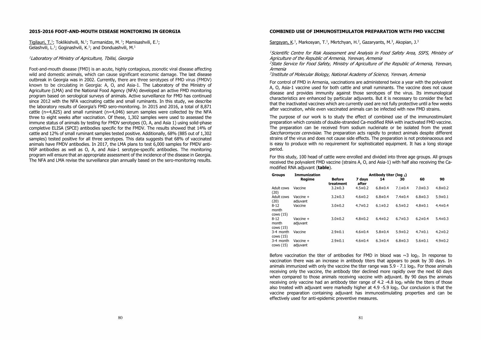
80
2015-2016 FOOT-AND-MOUTH DISEASE MONITORING IN GEORGIA Tigilauri, T.1; Toklikishvili, N.1; Turmanidze, M. 1; Mamisashvili, E.1; Gelashvili, L.1; Goginashvili, K.1; and Donduashvili, M.1
1Laboratory of Ministry of Agriculture, Tbilisi, Georgia Foot-and-mouth disease (FMD) is an acute, highly contagious, zoonotic viral disease affecting wild and domestic animals, which can cause significant economic damage. The last disease outbreak in Georgia was in 2002. Currently, there are three serotypes of FMD virus (FMDV) known to be circulating in Georgia: A, O, and Asia-1. The Laboratory of the Ministry of Agriculture (LMA) and the National Food Agency (NFA) developed an active FMD monitoring program based on serological surveys of animals. Active surveillance for FMD has continued since 2012 with the NFA vaccinating cattle and small ruminants. In this study, we describe the laboratory results of Georgia’s FMD sero-monitoring. In 2015 and 2016, a total of 8,871 cattle (n=4,825) and small ruminant (n=4,046) serum samples were collected by the NFA three to eight weeks after vaccination. Of these, 1,302 samples were used to assessed the immune status of animals by testing for FMDV serotypes (O, A, and Asia 1) using solid-phase completive ELISA (SPCE) antibodies specific for the FMDV. The results showed that 14% of cattle and 12% of small ruminant samples tested positive. Additionally, 68% (885 out of 1,302 samples) tested positive for all three serotypes. This data suggests that 68% of vaccinated animals have FMDV antibodies. In 2017, the LMA plans to test 6,000 samples for FMDV anti-NSP antibodies as well as O, A, and Asia-1 serotype-specific antibodies. The monitoring program will ensure that an appropriate assessment of the incidence of the disease in Georgia. The NFA and LMA revise the surveillance plan annually based on the sero-monitoring results.
81
COMBINED USE OF IMMUNOSTIMULATOR PREPARATION WITH FMD VACCINE Sargsyan, K.1, Markosyan, T.1, Mkrtchyan, H.2, Gazaryants, M.3, Akopian, J.3 1Scientific Centre for Risk Assessment and Analysis in Food Safety Area, SSFS, Ministry of Agriculture of the Republic of Armenia, Yerevan, Armenia 2State Service for Food Safety, Ministry of Agriculture of the Republic of Armenia, Yerevan, Armenia 3Institute of Molecular Biology, National Academy of Science, Yerevan, Armenia
For control of FMD in Armenia, vaccinations are administered twice a year with the polyvalent A, O, Asia-1 vaccine used for both cattle and small ruminants. The vaccine does not cause disease and provides immunity against those serotypes of the virus. Its immunological characteristics are enhanced by particular adjuvants. But it is necessary to consider the fact that the inactivated vaccines which are currently used are not fully protective until a few weeks after vaccination, while even vaccinated animals can be infected with new FMD strains.
The purpose of our work is to study the effect of combined use of the immunostimulant preparation which consists of double-stranded Ca-modified RNA with inactivated FMD vaccine. The preparation can be received from sodium nucleinate or be isolated from the yeast Saccharomyces cerevisiae. The preparation acts rapidly to protect animals despite different strains of the virus and does not cause side effects. The preparation is not proteinaceous and is easy to produce with no requirement for sophisticated equipment. It has a long storage period.
For this study, 100 head of cattle were enrolled and divided into three age groups. All groups received the polyvalent FMD vaccine (strains A, O, and Asia-1) with half also receiving the Ca-modified RNA adjuvant (table).
Groups Immunization Regime
Antibody titer (log 2) Before
treatment 7 days after
14 30 60 90
Adult cows (20)
Vaccine 3.2±0.3 4.5±0.2 6.8±0.4 7.1±0.4 7.0±0.3 4.8±0.2
Adult cows (20)
Vaccine + adjuvant
3.2±0.3 4.6±0.2 6.8±0.4 7.4±0.4 6.8±0.3 5.9±0.1
8-12 month cows (15)
Vaccine 3.0±0.2 4.7±0.2 6.1±0.2 6.5±0.2 4.8±0.1 4.4±0.4
8-12 month cows (15)
Vaccine + adjuvant
3.0±0.2 4.8±0.2 6.4±0.2 6.7±0.3 6.2±0.4 5.4±0.3
3-4 month cows (15)
Vaccine 2.9±0.1 4.6±0.4 5.8±0.4 5.9±0.2 4.7±0.1 4.2±0.2
3-4 month cows (15)
Vaccine + adjuvant
2.9±0.1 4.6±0.4 6.3±0.4 6.8±0.3 5.6±0.1 4.9±0.2
Before vaccination the titer of antibodies for FMD in blood was ~3 log2. In response to vaccination there was an increase in antibody titers that appears to peak by 30 days. In animals immunized with only the vaccine the titer range was 5.9 - 7.1 log2. For those animals receiving only the vaccine, the antibody titer declined more rapidly over the next 60 days when compared to those animals receiving vaccine with adjuvant. By 90 days the animals receiving only vaccine had an antibody titer range of 4.2 -4.8 log2 while the titers of those also treated with adjuvant were markedly higher at 4.9 -5.9 log2. Our conclusion is that the vaccine preparation containing adjuvant has immunostimulating properties and can be effectively used for anti-epidemic preventive measures.
82
ANALYSIS OF SEROLOGICAL-MONITORING ACTIVITIES RELATED TO FOOT-AND-MOUTH DISEASE IN BUFFER MARZES OF THE REPUBLIC OF ARMENIA IN 2016 Tumanyan, P.1, Kharatyan, S.2 and Danelyan, H.1
1Yerevan, RA MOA SFSS RVSPSCLS SNCO, Reference Laboratory for Especially Dangerous Pathogens 2 Yerevan, RA MOA SFSS, “Food Safety Risk Analysis and Assessment Research Center” SNCO
Introduction: Foot and mouth disease is considered one of the most important diseases of cloven-hoofed animals in view of its fulminating progression and the significant economic damage it causes. Prevention of the disease requires permanent control, risk assessment and appropriate risk management. Armenia is considered a high-risk zone amongst countries where foot and mouth disease is endemic. To minimize potential risks, vaccinations are administered twice a year for all cattle, sheep and goats using the Russian trivalent vaccine encompassing the A, O, and Asia-1 types, which are the main strains circulating in our country.
Goal: “Buffer zone” marzes that border neighboring countries are considered especially high-risk so monitoring activities were undertaken in three of them in 2016: Ararat, Syunik and Shirak. The aim of the survey was to assess post-vaccination immunity and to study FMD virus circulation, which would allow implementation of a corresponding analysis of outcomes, detection of potential risks and allow the implementation of measures to reduce future risk.
Methods: The size of the sample was determined using WinEpi software and based on the following indicators: prevalence of indicators being studied - 80%, level of reliability -95%, absolute error-10%. Randomized sample collection occurred in Ararat (750 cattle, 550 sheep and goats), Syunik (1200 cattle, 700 sheep and goats) and in Shirak (750 cattle, 600 sheep and goats). ELISA was used to analyze Non Specific Protein and Specific Protein antibodies. SP analyses were conducted against A, O, Asia-1 types.
Outcomes: Marz/ Analysis (% positive, number)
NSP SP (A type) SP (O type) SP (Asia-1 type)
Ararat, cattle 20% (150) 91% (682) 94% (705) 95% (712) Ararat, sheep/goats 31% (170) 94% (517) 91% (500) 96% (528) Syunik, cattle 22% (264) 98% (1172) 96% (1151) 96% (1149) Syunik, sheep/goats 26% (182) 90% (633) 90% (629) 90% (631) Shirak, cattle 28% (210) 91% (679) 96% (727) 89% (669) Shirak, sheep/goats 23% (138) 93% (557) 90% (543) 92% (551)
Conclusion: Analyzing the outcomes of the serological-monitoring, it may be assumed that the disease poses a risk at some border zones of the country. However, annual vaccinations against FMD are highly effective.
83
FMDV- HOST INTERACTION IN A MODEL OF PERSISTENTLY INFECTED BOVINE CELL Zientara, S.1 & Blaise-Boisseau, S.1 & Laloy, E.1 & Hägglund, S.2 & Näslund, K. 2 & Caignard, G.1 & Romey, A.1 & Relmy, A.1 & Gorna, K.1 & Kundlacz C.1 & Vitour, D.1 & Zientara, S.1 & Valarcher J-F. & Bakkali-Kassimi, L.1 1Laboratoire de Santé Animale de Maisons-Alfort, Laboratoire de référence Nationale et OIE pour la Fièvre Aphteuse, UMR Virologie 1161, Université Paris-Est, Anses, Maisons-Alfort, France 2 Department of Clinical Science at the Swedish University of Agricultural Sciences (SLU), Ulls väg 26, 75007 Uppsala, Sweden In a variable proportion of infected ruminants, regardless of their immune (vaccinated) status, FMDV persists in certain tissues after FMDV clearance from lesion sites. This state is termed persistence and such animals named “asymptomatic carriers”. These “carriers” are still perceived as a threat of FMDV transmission and their existence therefore complicates control and eradication of FMD. The underlying mechanisms of FMDV persistence remain however almost unknown but several studies have shown evidence for host/virus co-adaptation during persistence as well as modulation of innate immune response against FMDV. In the framework of the “Transcriptovac” ANIHWA project, we aim to study host-FMDV interactions and their modulation in the context of persistent infection by using bovine epithelial cell models to identify virus and cell gene signatures associated with persistence. For this purpose we have established FMDV type O persistent infection in epithelial bovine cells MDBK. Persistent viruses (FMDVOp) have been collected for both phenotypic and genotypic characterization. In parallel, FMDV-host protein interaction map has been realized using the yeast two-hybrid (Y2H) system to screen a cDNA prey library derived from MDBK cells with 13 FMDV “bait” proteins. This Y2H screening identified 313 interactions corresponding to 18 candidate interacting bovine proteins. These candidate interactions are currently confirmed. Furthermore, since persistence takes place in vivo namely in epithelium of dorsal soft palate (DSP), similar studies have been carried out, using primary epithelial bovine cells derived from dorsal soft palate cells (DSP) in comparison with alveolar pneumocytes (AP), derived from lung (site of FMDV replication, no persistence described in this tissue in vivo). Two models of persistent infection in the bovine host (MDBK or DSP) have been thus developed so far. Mutations affecting viral proteins known to modulate the antiviral response during acute infection and appearing during persistence are under study (luciferase reporter test). The candidate interactions are also undergoing biochemical validation. The impact of the mutations identified in the FMDVOp on these interactions is also investigated. Transcriptomic analyzes are also planned in order to identify differential cellular genetic expressions, potential "signatures" of persistence. The identification of modulated cellular signaling pathways during infection (acute or persistent) may indeed contribute to the development of better control strategies for foot-and-mouth disease. This study matches with the topic “persistent FMD: new knowledge, old problem”.
84
PERSISTENT OUTBREAK OF FOOT-AND-MOUTH DISEASE IN ISINGIRO DISTRICT, UGANDA. Atim, S.1, Ntaate, A1, Nantima, N.1 and Okurut, A R.1 1Animal Disease Diagnosis and Epidemiology Center (NADDEC) Ministry of Agriculture, Animal Industry and Fisheries, Entebbe, Uganda The objective of this study was to investigate persistent outbreak of Foot and Mouth disease (FMD) in Isingiro district neighboring northern part of Tanzania. Key informant interviews and focus group discussions were conducted drawing participants from both Ugandan and Tanzanian side. Blood and oropharyngeal samples were also collected from cattle showing clinical signs of FMD. The qualitative information collected showed that FMD infection is maintained by seasonal cross border movement of livestock from Uganda to northern Tanzania in search for pasture and water for livestock during drought in Uganda coupled with livestock markets both at Ugandan and Tanzanian borders that draw livestock from both side. Also interaction of livestock with buffalos in Rumanyika game reserve, Tanzania and Lake Mburo national park, Uganda. While Uganda implements a vaccination policy against FMD, the Republic of Tanzania does not vaccinate. The vaccination coverage in Isingiro were also below 50% attributed to mainly inadequate vaccine and seasonal movement of livestock. The SAT 1 & 2 was isolated in outbreak of FMD in Isingiro. The results have important implications for control strategy of FMD in Uganda. In planning for FMD control, there need for cross border agreement and collaboration between the Tanzania and Uganda. Closure of border livestock markets, Animal movement control and programmed vaccination to increase herd immunity. The experience of conducting this investigation highlighted critical factors that need to be considered in control FMD spread, including community engagement, public trust and benefit of the intervention to farmers.
85
STUDY ON THE VICISSITUDE OF ANTIBODIES INDUCED BY VACCINATING A SINGLE DOSE HIGH POTENCY FMD VACCINE IN SPF AND COMMERCIAL PIGS Lin, Y-L. 1 & Chen, R-S.1
1Animal Health Research Institute, Council of Agriculture, Executive Yuan, 376 Jhung-Jheng Rd, Tamsui District, 25158, New Taipei City, Taiwan, R.O.C. Commercial foot-and-mouth disease vaccines were inspected on a batch by batch basis by using SPF pigs to ensure their safety and efficacy before being put on the market in Taiwan. However, these vaccines were used to immunize the pigs at farms. Therefore, the aim of this study was to understand the situation of antibody inducing and eliminating after vaccinated with a high potency FMD vaccine in SPF and commercial pigs, respectively. And to clarify the consideration of farmers whom worried about the immune response of pigs was not strong enough by only one shot, even though the vaccines contained more than 6PD50 antigen per dose. In this study, five 8-week-old SPF piglets without antibodies against FMD virus and totally 4 pig farms, each farm was randomly selected 5 12-week-old pigs to be vaccinated with one dose (2 ml) 6PD50 O / TW / 98 commercial FMD vaccine. Each vaccinated pig was tagged with ear tag for further identification during the test. The bloods of these pigs were collected before vaccination and collected at an interval of 4 weeks after vaccination. Sera were obtained from the collected bloods and were implemented the neutralization antibody test. The results showed that the induced antibody titers of SPF pigs were higher than those of commercial pigs. The induced antibody titers of vaccinated pigs were not consistent in the 4 farms. However, the induced antibody titer of each vaccinated pigs was more than 1:16 and could persistent to 20 weeks post vaccination. The results demonstrate that the strategy for FMD prevention and control, which performed the one dose vaccination with high potency FMD vaccine in cloven-hoofed animals, is feasible. The evidence is that Taipei China regained the recognition from OIE as FMD free zones where vaccination is practiced in Taiwan, Penghu and Matsu Islands in May 2017.
86
EVALUATION OF RT-PCR USING A PRIMER SET TARGETING 3D REGION FOR DIAGNOSIS OF FOOT-AND-MOUTH DISEASE Tatsuya, N.1, Katsuhiko, F.1, Nobuaki, S.1, Kazuki, M.1, Manabu, Y.1, Makoto, Y.1, Toru, K.1
1Exotic Disease Research Station, National Institute of Animal Health, National Agriculture and Food Research Organization, 6-20-1 Josui-honcho, Kodaira, Tokyo 187-0022, Japan Rapid and accurate methods are essential for diagnosis of Foot-and-mouth disease (FMD) because of its potential for the extensive spread, therefore, reverse transcription-PCR (RT-PCR) plays one part of important role for the diagnosis. Though FMD virus (FMDV) genome has a high mutation rate as with other RNA viruses, a highly conserved region should be selected as a target of RT-PCR for a broad range detection of seven serotypes. In this study, a primer set (FM8/9) targeting a conserved region of 3D was designed and its performance was compared with that of a primer set (1F/1R) targeting 5’UTR described in OIE manual. The primer set FM8/9 was designed to amplify 644 bases of the conserved 3D region of all seven serotypes based on FMDV sequences available in GenBank. To assess the specificity of RT-PCR using FM8/9, RNAs of 14 and 33 strains of types O and A from different geographical origins and isolation years, respectively, and one strain each of types Asia1, C, SAT1, 2, and 3 were tested. In addition, RNA samples extracted from one strain of swine vesicular disease virus, two strains of vesicular stomatitis virus, and four strains of bovine rhinovirus were also tested as negative control for specificity validation. The RT-PCR using FM8/9 showed positive for all 52 FMDV strains, but no false-positive reactions for other viruses. The detection limits of the RT-PCR using each primer set were determined for seven strains isolated from East, Southeast, and West Asia, and Africa. RNAs were extracted from each serial 10-fold dilution ranged from 10-3 to 103 TCID50/0.1ml in IBRS-2 cell and subjected to the RT-PCR. As a result, detection limits of the RT-PCR using FM8/9 primer set were 100 TCID50/0.1ml against strains of types O, Asia1, SAT1, and SAT3, 101 TCID50/0.1ml against type A, and 102 TCID50/0.1ml against type SAT2. On the other hand, those of 1F/1R were 101 TCID50/0.1ml against types O, A, Asia1, and SAT2, and negative for all dilutions of types SAT1 and SAT3. Therefore, compared to sensitivity of the RT-PCR using 1F/1R, that of FM8/9 was increased more than 10-fold against five strains of types O, Asia1, SAT1, and SAT3. To assess validity of the methods in clinical samples, samples from two 2-month-old pigs inoculated intradermally with O/JPN/2010 and four pigs cohabited with the inoculated pigs at 1 day post inoculation (dpi). Sera and saliva samples were collected from them daily until 11 dpi, and were analyzed by the RT-PCR using the two primer sets. As a result, in case of FM8/9, positive results were detected from 1 to 10 dpi and 1 to 10 days post cohabitation (dpc) in the inoculated pigs and cohabitants, respectively. On the other hand, positive results were detected from 1 to 8 dpi and 2 to 9 dpc by using 1F/1R. Therefore, the RT-PCR using FM8/9 revealed higher sensitivity for the clinical samples from infected pigs. Our results suggest that this primer set enables specific and highly sensitive RT-PCR for diagnosis of FMD.
87
DETECTION OF ANTIBODIES AGANIST CONSERVED CAPSID EPITOPES PROVIDES A UNIVERSAL SEROLOGY ASSAY FOR DIAGNOSIS OF FMDV Asfor, A.1 Grazioli, S.2; Brocchi, E.2; King, D.1 Parida, S.1 and Tuthill, T.1
1The Pirbright Institute, Ash Road, Woking, GU24 0NF, UK 2Istituto Zooprofilattico Sperimentale della Lombardia e dell'Emilia Romagna, Brescia, Italy Foot-and-mouth disease virus (FMDV) causes one of the most economically important livestock diseases worldwide. FMDV exists as 7 serotypes and evolves rapidly so that circulating viruses display high levels of antigenic variation. Diagnosis of FMD can use various approaches including serological tests to detect FMDV specific antibodies. Conventional serology based diagnostics are reliable and rapid but require tests specific to each virus serotype. A number of monoclonal antibodies (mAbs) have previously been reported with cross-reactivity against multiple FMDV serotypes. Some of these mAbs have been mapped to the highly conserved N terminus of FMDV capsid protein VP2, suggesting that such conserved sequences might be useful diagnostic reagents. The aim of this study was to assess the potential of conserved N-terminal sequences of capsid proteins VP2 and VP4 as universal epitopes for the detection of FMDV specific antibodies against multiple FMDV serotypes. Synthetic peptides of various lengths were used to represent the conserved target epitopes at the N terminus of VP2 (VP2N) and N terminus of VP4 (VP4N). A panel of mAbs with existing evidence for cross-serotype activity and sera from cattle infected with each of the 7 serotypes of FMDV were tested for reactivity against the peptides by indirect peptide ELISA. Three mAbs showed strong reactivity to VP2N peptides, including to the shortest peptide tested (the first N-terminal 15 amino acids) suggesting this contained the epitope for these antibodies. Cattle sera against all 7 serotypes of FMDV reacted strongly with VP2N peptides and also with VP4N peptides demonstrating the peptides are indeed able to function as universal detection reagents for FMDV specific antibodies. This study demonstrates that conserved peptide epitopes in the FMDV capsid can be used as serotype-independent antibody capture reagents. This may have utility in the development of new universal and rapid laboratory or field-based diagnostic tests.
88
A MODEL OF FOOT-AND-MOUTH DISEASE TRANSMISSION, DETECTION, AND INTERVENTION STRATEGIES FOR MODERN CONCENTRATED CATTLE FEEDING FARMS Cabezas, A.1; Sanderson, M.1; Volkova, V.1
1College of Veterinary Medicine, Kansas State University, 1710 Denison Avenue, 66506, Manhattan, USA Mathematical modeling is an important tool to represent a potential epidemic of infectious diseases such as FMD in disease-free countries. Most published models focus on modeling between-farm FMD transmission and represent the farm as a homogenous unit in respect to FMD transmission. Our objective is to realistically project FMDv transmission dynamics, clinical manifestation, and control within modern concentrated livestock farms with large and compartmentalized animal populations. A cattle feedlot is used as an example. Our model incorporates a typical feedlot farm layout, production management, and animal demographics. We use the model to assess FMD spread within the farm, estimate the impact of surveillance sensitivity on time-to-detection, estimate the impacts of various interventions post-detection, and study logistics of control. The stochastic SLIR (susceptible-latent-infectious-recovered) models in individual animal pens were nested in the meta-population of the home and hospital pens on the farm. Transmission within home-pens occurred via direct contact, based on homogeneous animal mixing within the pen. The FMDv transmission between home-pens occurred via direct contact of cattle in the hospital pen, fence-line direct contact, consumption of contaminated water by home pens that share water troughs, environmental (dirt/feces) transfer by caretakers moving between home-pens, and by airborne transmission. Morbidity rates for endemic diseases were used to model movements of cattle to the hospital pen. Model parameterization was based on literature review and an on-line world-wide FMD expert survey. We modeled a farm of 12,000 cattle distributed in 60 pens with 200 head per pen, and 1 hospital pen. Ten FMD-latent cattle were introduced into an index pen. For surveillance, the detection threshold was 3% prevalence of clinical FMD cattle in the index pen. Stopping animal movements to the hospital pen on the day of detection and three targeted culling interventions were modeled: cull index pen, cull pens located in the same row of the index pen, and cull a region around the index pen (20 pens, 4,000 animals). With detection at 3% clinical FMD in the index pen, the median time-to-detection was 8 days. Simulations showed that without interventions the outbreak took 62-84 days to fade-out and all pens were infected by a median of 21 days. Stopping mixing of cattle in the hospital pen decreased the speed of transmission, but prolonged the duration of the outbreak on the farm. Culling interventions resulted in similar outbreak speed and total duration compared to no intervention. Moreover, combining culling strategies with stopped mixing of cattle in the hospital pen did not result in significant differences when compared to stopped mixing of cattle in the hospital pen alone. Our results suggested that stopping hospital-pen mixing upon detection played a key role in slowing transmission across the farm, though it did not prevent eventual infection of all pens. Culling interventions and stopping hospital-pen mixing might be challenging to implement due to associated human labor requirements and animal welfare complications; however, slowing outbreak speed within the farm might allow time for vaccination to limit outbreak size. Future work will assess the impacts of vaccination strategies.
89
AN EXPERT OPINION SURVEY OF FOOT-AND-MOUTH DISEASE NATURAL HISTORY AND CLINICAL MANIFESTATIONS IN BEEF CATTLE Cabezas, A.1; Sanderson, M.1; Volkova, V.1
1College of Veterinary Medicine, Kansas State University, 1710 Denison Avenue, 66506, Manhattan, USA A Foot and Mouth Disease outbreak in an FMD-free country will have severe consequences for the livestock industry. Implementation of effective and efficient intervention strategies is necessary to minimize epidemic size. Simulation models are powerful tools to represent and analyze potential interventions in countries where FMD in non-endemic. While extensive research has been conducted in controlled in-vivo FMD experiments, there are gaps and deficiencies in data generated by those studies for application in field settings. This complicates the parameterization of simulation models to produce robust predictions of potential epidemics. We developed a questionnaire to collect expert opinion about key parameters of the potential natural history, transmissibility, and clinical manifestation of FMD in infected naïve cattle of the age and health status typical for modern beef production. The questionnaire was administered on-line, allowing 4 weeks for completion. Targeted experts (virologists, epidemiologists, modelers, practicing veterinarians, and governmental veterinary officers) with experience working on FMD in endemic settings or outbreaks in non-endemic settings, including those in the OIE FMD reference laboratories, were invited. In the questionnaire, the most likely health status of the cattle and most common management practices were described. Likely scenarios of FMDv exposure (route and dose of infection) were provided. For each exposure scenario, quantitative estimates were requested for the duration of stages of infectiousness and clinical disease, proportion of animals exhibiting specific clinical signs, reduction in feed consumption, and a qualitative description of FMDv transmissibility. Twenty-seven participants agreed to participate and sixteen completed the questionnaire (56% completion rate). Of the latter, six reported having experience in FMD in-vivo experiments and the other ten in FMD epidemiology and outbreak investigation; nine had primary experience in endemic and the other seven in outbreak settings. The survey results indicated the median estimated duration of latent and infectious periods in cattle infected by high virulent strains ranged from 1 to 7 and 3 to 10 days, respectively. For subclinical, incubation, and clinical period the median estimated duration ranged from 0.5 to 4, 2 to 7, and 3.5 to 10 days, respectively. Expert estimates of the probability of exposed cattle developing clinical disease varied for high virulent strains (40-100%) and for low virulent strains (10-90%). The median duration of reduction in feed consumption in clinical cattle infected by high virulent strains ranged from 2 to 8.5 days. The survey results indicated that the FMDv exposure dose influences the incubation period duration but possibly not the disease severity. Experts indicated factors such as virus strain and intrinsic characteristics of the host might substantially affect the disease progression and animal infectiousness. Data generated in this survey will improve the robustness of FMD epidemic predictions for cattle industries in non-endemic settings, when combined with experimental and outbreak investigation data. Moreover, the data will be useful in modeling within-farm FMD dynamics for realistic analyses of early detection and rapid response in the face of an outbreak.
90
A NOVEL OIL ADJUVANT ENHANCES THE PROTECTION CONFERRED BY SWINE FOOT-AND-MOUTH DISEASE VACCINES Deville, S.1, Li, D.2, Ben Arous, J.3, Dupuis, L.3
1 SEPPIC China, 819 West Nanjing Road, Shanghai 200041, China 2 Lanzhou Veterinary Research Institute, OIE/National FMD reference lab, Chinese Academy of Agricultural Sciences, Lanzhou 730046, China 3 SEPPIC, 22 Terrasse Bellini, Paris La Défense, 92806 Puteaux Cedex, France Introduction Water in oil in water adjuvants such as MontanideTM ISA 206 VG (ISA 206) are widely used for FMD swine vaccination. These oil emulsion technologies have replaced aluminium hydroxide adjuvants. They induce a strong short-term and long-term immune response which is mainly mediated by neutralizing antibodies (1), with low viscosity and low side reactions. To improve cross-protective properties of FMD vaccines, one option is to develop new adjuvants that enhance cell mediated immune response. We have therefore selected the new adjuvant MontanideTM ISA 201 VG (ISA 201) which stimulates both humoral and cell mediated immune responses. Here we show that a swine FMD vaccine based on ISA 201 is safe, and induces higher immune response and protection against FMD than a reference vaccine based on ISA 206. Materials and Methods 9 groups of 5 pigs received respectively inactivated FMDV type O vaccine based on ISA 206, ISA 201 or non-adjuvanted at full dose (2ml), 1/3 dose or 1/9 dose. 5 control pigs were not vaccinated. Anti-FMDV antibody titers, IFN titers, and CD4+ and CD8+ T cells concentrations were measured at 0, 3, 7, 14, 21 and 28 dpv. All 50 pigs were challenged intramuscularly with 1000 PID50 of FMDV type O at 28 dpv, and the PD50 of each vaccine was calculated using Karber’s method. Results All vaccines were safe in swine. Antibody titers against FMDV were signicantly higher in the ISA 201 group compared to the reference ISA 206 group for full dose, 1/3 dose and 1/9 dose vaccinated animals at 14dpv, 21dpv and 28dpv. After full dose vaccination with ISA 201 vaccine, % of circulating CD4+ and CD8+ lymphocytes were enhanced compared to the ISA 206 group and the non adjuvanted vaccine group at 7dpv and 28dpv. Finally, full doses or 1/3 doses of both ISA 206 and ISA 201 vaccines were fully protective against FMDV type O challenge at 28dpv (5/5 pigs without any clinical signs), whereas non adjuvanted vaccine failed to protect the animals (protection rate 2/5). All non vaccinated control animals showed clinical signs. However, when the vaccines were used at 1/9 dose, only the vaccine based on ISA 201 was fully protective, showing that ISA 201 adjuvant improves the PD50 of the FMD vaccine compared to reference adjuvant ISA 206. Discussion MontanideTM adjuvants that increase cell mediated immune response could extend the vaccinal protective shield against close variants such as local FMD virus strains while preserving the robustness, ease of injection and safety profile of FMD vaccines. References [1] Barnard AL et al. 2005. Vaccine 23:1037-1047.
91
ADAPTATION OF FMDV ASIA 1 TO SUSPENSION CULTURE: CELL RESISTANCE IS OVERCOME BY VIRUS CAPSID ALTERATIONS Dill, V.1; Hoffmann, B.1; Zimmer, A.2; Beer, M.1 & Eschbaumer, M.1
1Institute of Diagnostic Virology, Friedrich-Loeffler-Institut (FLI), Federal Research Institute for Animal Health, Suedufer 10, 17493 Greifswald-Insel Riems, Germany 2Merck KGaA, Merck Life Sciences, Upstream R&D, Frankfurter Strasse 250, 64293 Darmstadt, Germany BHK-21 suspension cells are preferred for the industrial production of FMDV vaccine antigen, but not all virus strains can be successfully propagated in this environment. Strains of serotype Asia 1 in particular are notoriously hard to grow in suspension culture. This study examined the role of viral, cellular and environmental factors in the resistance of BHK21C13-2P suspension cells to infection with FMDV Asia 1 Shamir. Neither the culture media itself, nor its pH or ammonium chloride content affected Asia 1 differently than the other serotypes tested. Virus replication after transfection of Asia 1 RNA was not impaired, but adhesion of Asia 1 virus to the cells was markedly reduced, indicating that the cells were resistant but generally permissive. The virus was successfully adapted to grow in the resistant cells. Sequence analysis of the adapted strains revealed two distinct patterns of mutations in the capsid proteins: In two strains of Asia 1 Shamir, adaptation to growth in 2P suspension cells was associated with the acquisition of positive charges (Q to R) at positions 108 and 110 of VP1. A third strain acquired positive charges at positions 110 and 202 of VP1 and position 59 of VP3. One mutation in the non-structural protein 2C (K285Q) was seen in all adapted strains. The receptor usage of the original and adapted strains will be assessed by infection experiments with integrin- and heparan-sulfate-deficient cell lines, and the impact of the mutations on the antigenic profile of the virus will be evaluated by neutralization tests with bovine sera.
92
VARIABLE INACTIVATION OF FOOT-AND-MOUTH DISEASE VIRUS BY COMMERCIAL LYSIS BUFFERS Wood, B.1; Haas, B.2; Mioulet, V.1; Henry, E.1; Thapa, B.1; Gray, A.1; Diederich, S.3; Hoffmann, B.2; King, D.1 & Eschbaumer, M.2
1The Pirbright Institute, Ash Road, Pirbright, Woking, Surrey GU24 0NF, United Kingdom 2Institute of Diagnostic Virology and 3Institute of Novel and Emerging Infectious Diseases, Friedrich-Loeffler-Institut (FLI), Suedufer 10, 17493 Greifswald-Insel Riems, Germany Laboratories working with live FMDV must maintain a high level of biocontainment. This study was intended as a guideline for contingency laboratories that cannot work with infectious virus, but will perform molecular testing during an outbreak in their country or to rule out FMD as a differential diagnosis in routine cases. Low-risk testing for FMDV can be safely carried out in standard laboratories if infectious virus is reliably inactivated during sample processing. Nine commercial lysis buffers (AL, AVL, MagMAX, MagMAX CORE, NM1, RAV1, RLT, TRIzol and VL) were tested for their inactivation efficacy against different FMDV serotypes and infectious doses in different sample matrices. The experiments were independently conducted at the German National Reference Laboratory for FMD at the FLI and the FAO World Reference Laboratory for FMD at the Pirbright Institute. Residual infectivity after the addition of lysis buffer was evaluated by inoculating susceptible cell cultures (BTY, BHK-21 and/or LFBK-alphaVbeta6) and observing for cytopathic effect. Observed CPE was confirmed by passage or antigen ELISA. All nine lysis buffers inactivated FMDV in cell culture supernatant, regardless of serotype; however, for some lysis buffers an additional heat inactivation step (70°C, 10 min) was required for complete inactivation. For clarified animal samples, such as epithelial tissue suspension, all lysis buffers tested were able to inactivate FMDV. However, for more complex matrices spiked with virus, such as blood, milk, probang fluid and serum, residual infectivity was observed, and this infectivity was not always eliminated with the addition of a heat treatment. This highlights the variable capacity of lysis buffers to effectively inactivate FMDV, and demonstrates the need for validation of inactivation procedures used to remove samples from high containment.
93
COMPARATIVE SUSCEPTIBILITY OF FOOT-AND-MOUTH-DISEASE VIRUS (FMDV) AND TWO VIRAL SURROGATES TO USDA-RECCOMMENDED DISINFECTANTS FOR USE IN FMDV DECONTAMINATION AND CONTROL Gabbert, L.,1 Smith, J.,1 Neilan, J.,2 & Rasmussen, M.2 1 Leidos, Plum Island Animal Disease Center, P.O. Box 848, Greenport, NY 11944 2 United States Department of Homeland Security, Science and Technology Directorate, Plum Island Animal Disease Center, Greenport, NY 11944, USA. Understanding and evaluating the effectiveness of decontamination methods against foreign animal diseases (FADs) is complicated by biosafety regulations and the need for high-containment laboratory facilities. Alternatively, surrogate organisms can be used to model the environmental fate and transport of FADs at lower biocontainment levels. In the United States, foot-and-mouth disease-virus (FMDV) can only be handled at the Plum Island Animal Disease Center. Consequently, few head-to-head validation studies have been reported comparing the susceptibility of FMDV and potential surrogates to chemical disinfection. In this study, the OECD Quantitative Carrier Method for Evaluating Antimicrobials on Hard Non-porous Surfaces was used to compare the susceptibility of FMDV and the viral surrogates feline calicivirus (FCV) and bacteriophage MS2 (MS2) to disinfection with bleach, acetic acid, citric acid, Virkon S ®, and Oxonia Active® using a 5-minute contact time. Chemical disinfectants used in the study included products registered by the U.S. Environmental Protection Agency (EPA), or recommended by the U.S. Department of Agriculture (USDA), for use in FMDV decontamination and represented different modes of action. Results indicate that FMDV was always inactivated within the USDA-recommended chemical use-dilution for each disinfectant. Compared to FMDV, both surrogate viruses were more resistant to inactivation with each disinfectant, except FCV was more sensitive to inactivation by bleach. MS2 was consistently the most resistant organism to disinfection under these conditions, and both surrogates were highly resistant to acetic and citric acids. This study provides valuable data directly comparing inactivation of FMDV to two surrogate viruses and may be used as a model for testing additional surrogates or disinfection conditions.

94
FIELD APPLICATION OF ORAL FLUID COLLECTION USING COTTON ROPE FOR FOOT-AND-MOUTH DISEASE Kim, T.1 & Ryoo, S.1 & Nah, J. & Sagong, M.1 & Lee, S.1 & Jung, S.1 & Lee, H. 1 & Lee, J-W1 & Park, S-H.1 & Wee, S-H.1 & Ku, B.1 1Foot-and-Mouth Disease Division, Animal and Plant Quarantine Agency, 177 Hyeoksin 8-ro, Gimcheon, Gyeongsangbuk-do, Republic of Korea The detection of foot-and-mouth disease (FMD) in oral fluid samples using cotton rope was evaluated in several studies. In this study, we applied rope sampling to detect FMD virus RNA in Korean pig farms. First, rope samples were used to collect oral fluids from pigs infected experimentally in FMD virus and compared to individual saliva samples by FMD detection real time RT-PCR. Pigs were divided into three groups: 28 (V28) days after vaccination, 14 (V14) days after vaccination and unvaccinated (UV). Two pigs were inoculated intradermally in the heel bulb with O/JC/SKR/2014. These two pigs were used as donors for a pig-to-pig time-limited contact exposure. Clinical symptom observations and sample collection were performed daily for 2 weeks. The pigs in the group UV did not chew the ropes until 10 days post contact (dpc) and only individual saliva samples were collected. In group UV, viral RNA was detected in saliva swab between 2 and 9 dpc and detected in 10 to 11 dpc rope samples. In the V14 and V28 groups, viral RNA was detected in the rope samples at 8 and 10 dpc, respectively. In addition, 409 rope samples were collected from 12 pig farms, including farms in which NSP antibodies were continuously detected. Real-time PCR was performed using the rope samples and no viral RNA was detected in all samples. Although the virus RNA was not detected, the rope was very useful for collecting oral fluid throughout the farm. In conclusion, the rope samples would be more useful for screening infected animal populations of FMD outbreak farms than NSP antibody-positive farm monitoring
95
INTRADERMAL DELIVERY OF OIL-ADJUVANTED COMMERCIAL FMD VACCINES WITH NEEDLESS DEVICE FOR PIG Lee, K-N. 1; Kim, M Y1.; Hwang, J-H1.; Park, J-H1; Kim, B. 1
1Center for FMD Vaccine Research, Animal and Plant Quarantine Agency, Gimcheon, 39660, Gyeongbuk, Republic of Korea From 2011, all the susceptible livestock in the Republic of Korea (ROK) have been vaccinated with oil-adjuvanted FMD vaccines, and it was reported that local granulomatous reactions from the vaccination leads to the economic losses. So the feasibility of intradermal delivery of the commercial vaccines to the pigs with a needless device was evaluated in terms of immunogenicity and safety in the field. Firstly, piglets were randomly allocated into 7 groups (G1-7) and immunized at 8 weeks old intramuscularly or intradermally by seven different schemes using highly potent FMD oil vaccine used in the field. The booster vaccination was done at four weeks after the first vaccination for G3, G4, G6 and G7, and the blood was taken 0, 2, 4, 6, 8, 12, 16 weeks post the first vaccination for serological assays. Intradermal delivery was done using needless device A (Battery powered). About half of the weaners had maternally derived antibodies above the cut-off level. Regardless of the routes or volumes of the vaccine administered, only the groups of weaners boosted with secondary injection showed the increase of the antibody titer at 6 and 8 weeks after the first shot, whereas the groups of pig vaccinated once and for all showed the decrease of the antibody titer except for the G1 in which 2.0ml was given per each head. Secondly, with gas powered needless device, in the three different pig farms, the effectiveness of the intradermal delivery vaccination protocol was tested and confirmed. For the immunogenicity of the commercial vaccines, dose-sparing effect of intradermal route delivery of vaccines compared to the intramuscular route was not clear. On the other hand, there seems to be dose dependent effect irrespective of the route of deliveries of the vaccines. Importantly, as the new scheme employing intradermal route for vaccine delivery is almost free of granulomatous reactions at the injection site, edible neck muscle just behind and below the ear of the pig, this needs to be seriously considered to be applied in the field and the new vaccines well suited for intradermal route needs to be developed as soon as possible.
96
COMPARATIVE GENOME ANALYSIS OF FOOT-AND-MOUTH DISEASE VIRUS OF SEROTYPE O ISOLATED FROM VIETNAM IN 2014 Lee, H J.1 & Nah, J.1 & Ryoo, S.1 & Tho, N D.; To, T L.2 & Sagong, M.1 & Pack, S H1 & Lee, J-W.1 & Choung, K1 & Wee, S-H.1 & Ku, B.1* 1 Foot-and-Mouth Disease Division, Animal and Plant Quarantine Agency, 177, Hyeoksin 8-ro, Gimcheon-si, Gyeongsangbuk-do, Republic of Korea 2 National Center for Veterinary diagnostics, Department of Animal Health, Vietnam Foot and mouth disease (FMD) is endemic and widespread in the most of Southeast Asia region. Among seven serotypes of FMDV, concurrent outbreaks of FMDV serotype O and A has been distributed extensively in Vietnam. As for Vietnamese FMDV serotype O, their topotypes(genotypes) have been classified mostly into the SEA(Mya-98), and ME-SA (PanAsia, and recently Ind-2001d). To understand the comparative genetic variation and antigenic relationship between recent FMDV serotype O in Asian region, accessible information on genomic sequences is very necessary to be accumulated. However, there has been reported with very limited genomic sequence until recently. In this study, we isolated 2 viruses (O/VN1/2014 and O/VN6/2014) of serotype O in the epithelia homogenate collected from a pig and a cow in the northern Vietnam(Lao Cai and Son La province) in 2014, respectively. Based on VP1 gene, both of these viruses were classified within the Mya-98 genotype of SEA topotype. Compared with complete genome, one FMDV(O/VN1/2014) was most closely related with a virus of China in 2010 and viruses of Korea from 2014 to 2016 both at the nucleotide and deduced amino acid level. And the other FMDV(O/VN6/2014) showed high with Vietnam(O/VN/QB88/2009) and Mongolia in 2010 homology both at the nucleotide and deduced amino acid level. Accumulating of the comparative genome analysis of FMDV isolates in Vietnam could give valuable information for further study of genetic diversity and antigenic relationship of FMDV in the Asian region to control and prevent FMD.
97
ISOLATION AND MOLECULAR CHARACTERISTIZATION OF FMDV SEROTYPE O IN OUTBREAKS OF 2013-2014 IN VIETNAM Nguyen, P T. 1 Hung, V V. 1 Tran, T M. 1 Do, D V. 1 Le, A L T. 1; Tri, V T.1; Huy, H. 1
1Regional Animal Health Office No 6 - Department of Animal Health, Vietnam Foot and mouth disease virus (FMDV) causes sporadic disease outbreaks in the Vietnam and appears to be endemic within a livestock population largely susceptible to infection. Phylogenetic analysis of the VP1 sequence revealed two different groups of type O isolates divided into two topotypes: South East Asia (SEA) and the Middle East-South Asia (ME-SA). FMDV was isolated from 34 samples and then VP1 sequenced, PanAsia strain was detected in isolates of 2013 (63.6% on cattle and 36.3% on pig) and isolates of 2014 (25% PanAsia strain of which 41.6% Mya-98 lineage on cattle, and 33.4% PanAsia strain on pig). The vaccine of O3039 antigen still works well with FMDV isolates of 2013-2014. Finally, sequences of the outbreak FMDV indicated the current vaccine could be used to control FMDV outbreak of serotype O in Vietnam.
98
DETECTION OF FOOT-AND-MOUTH-DISEASE VIRUS SEROTYPE A BY REAL TIME RT-PCR USING PEPTIDE NUCLEIC ACID Lee, J-W. 1 & Nah, J.1 & Ryoo, S. 1 Lee, H J1 & Park, S H1 & Sagong, M. 1 & Kim, T. 1 & Jung, S. 1 & Yoon, K-J.2 & Wee, S-H1 & Ku, B. 1 1Foot-and-Mouth Disease Division, Animal and Plant Quarantine Agency, Gimcheon, Gyeongsangbuk-do, Republic of Korea 2Diagnosis Business Division, Seasunbio, N517, 11-3, Techno1-ro, Yuseong-gu, (34015) Daejeon, Republic of Korea In 2017, foot-and-mouth disease(FMD) serotype O and A simultaneously occurred in Korea. The serotype A virus was successfully isolated from Yeoncheon (A/YC/2017) and grouped into Asia/SEA-97 genotype. So, we developed new real time RT-PCR assay which is able to detect both FMDV serotype A and Sea-97 specifically. To achieve this purpose, we use Peptide Nucleic Acid (PNA) probe in Real-time RT-PCR. PNA is a DNA mimic, in which the entire negatively-charged sugar-phosphate backbone is replaced with a neutral one consisting of repeated N-(2-aminoethyl) glycine units linked by peptide bonds. It is stable chemically and biologically. According to the PNA probe-based real-time RT-PCR assay, the melting temperature (Tm) values when the complementary bases mismatch with each other are significantly lower than the Tm values of perfect match, so that they can be distinguished by analyzing the difference of melting temperature. Based on this, we designed PNA probes and primers that specifically bind to the VP1 region to detect serotype A and A/YC/2017 viruses, respectively. As a result, we are confirmed the Tm value that specifically detect the serotype A and A/YC/2017. This real-time RT-PCR assay, here we presented, expected that will be reduced experiment time and cost to detect the FMDV serotype A and A/YC/2017 in the future.
99
MODELLING THE ROLE OF CARRIERS IN ENDEMIC FOOT-AND-MOUTH DISEASE McLachlan, I.1,2 & Porphyre, T.1 & Marion, G.2 & McKendrick, I.2 & Handel, I.1 & Bronsvoort, M.1
1The Roslin Institute and Royal (Dick) School of Veterinary Studies, University of Edinburgh, Easter Bush, Edinburgh, Midlothian, Scotland, United Kingdom 2Biomathematics and Statistics Scotland, Edinburgh, United Kingdom In sub-Saharan Africa (SSA), foot and mouth disease (FMD) is a barrier to economic growth and a threat to the livelihoods of rural communities. Disease dynamics in endemic regions, like SSA, are poorly understood and the settings differ from FMD-free regions. In endemic regions in addition to the differences of partial immunity, greater movement and interaction of herds, carrier cattle have been identified as a potential contributor to disease persistence. Little is known about carriers and our understanding hindered by limited surveillance and data collection in endemic regions. Using numerical simulations, we aim to gain a better understand the role of carriers within the endemic setting of Cameroon. Dynamics of FMD, within a single herd, were explored using through stochastic simulation. In the Susceptible-Exposed-Infectious-Carrier-Recovered model used both infectious and carrier cattle contributed to transmission. Carriers role in FMD persistence is influenced by the proportion of individuals that become carriers, the infectiousness of carriers and duration of the carrier state. Reflecting the limited data availability and the unknown role of carriers in FMD transmission a range of parameter values were explored. Assuming that the infectivity of carriers is lower compared to infectious individuals small numbers of FMD cases are generated following an outbreak (in the absence of other sources of disease introduction). Conservative estimates suggest that a carrier state alone cannot sustain FMD within a herd indefinitely. However, the presence of carrier cattle extends the opportunity for the herd to transmit disease. Understanding the role of carriers is a first step in designing cost efficient targeted control strategies for endemic FMD.
100
COMPARATIVE GENETIC ANALYSIS OF FOOT-AND-MOUTH DISEASE VIRUS OF SEROTYPE A OF KOREA IN 2017 Nah, J.1 & Ryoo, S. Lee, H J, Tho, N D.; To, T L.2, Park, S H.1, Lee, J-W.1, Lee , S M.1, Jung , S M.1, Wee, S-H.1, Ku, B.1* 1 Animal and Plant Quarantine Agency, 177, Hyeoksin 8-ro, Gimcheon-si, Gyeongsangbuk-do, Republic of Korea 2 National Center for Veterinary diagnostics, Department of Animal Health, Vietnam Foot-and-mouth disease(FMD) is a highly contagious viral disease and have been occurred sporadically in Korea. Since December of 2010, the vaccination policy against FMD has been carried out nationwide to cloven-hoofed animal species. However, one case of FMDV serotype A(A/YC/SKR/2017) recurred concurrently with serotype O in Korea in February of 2017 after the last serotype A outbreak in 2010. Based on the phylogenetic analysis of VP1 gene, the Korean serotype A virus grouped into A/Asia/SEA-97 genotype, mostly related with Vietnam A viruses in 2016(99.8%), but showed relatively low relationship with the last Korean A virus in 2010(91.4%). It means that the FMDV serotype A virus of 2017 might be introduced newly from neighbored FMD circulating countries via unknown source. Comparing the genetic relationship between recent FMDV serotype A in Asian region could give very useful molecular epidemiological information, but accessible genetic information is still very limited. In this study, we isolated one virus(A/VN18/2015) of serotype A in the epithelia homogenate collected from a cow in the northern Vietnam (Bac Kan province) in 2015. This Vietnamese FMDV serotype A(A/VN18/2015) was the latest complete genome sequence, which belonged to the same genotype with the Korean FMDV serotype A of 2017. Compared with complete genome, the Korean FMDV serotype A(A/YC/SKR/2017) showed closer relationship with this Vietnamese serotype A(A/VN18/2015) than the Chinese FMDV serotype A(A/HY/CHA/2013). Since the FMDV serotype A has been considered to be the most antigenically diverse among the seven serotypes, it is necessary to accumulate the genetic resource of FMDV serotype A in Asian region to understand the molecular epidemiology and to prepare the effective vaccine for a new outbreak.
101
STRUCTURE ANALYSIS OF VP1 AND 3D PROTEINS IN FOOT-AND-MOUTH DISEASE VIRUS IN SOUTH KOREA, 2017
Park, T. 1, 2
, Nah, J.3, Ryoo, S.3, Wee, S.3, Park, D.1,2,4
, Ku, B.3 1Department of Predictive Toxicology, Korea Institute of Toxicology, Daejeon, Korea
2Department of Human and Environmental Toxicology, University of Science and Technology,
Daejeon, Korea 3Animal and Plant Quarantine Agency, 177, Hyeoksin 8-ro, Gimcheon-si, Gyeongsangbuk-do,
Republic of Korea 4Center for Convergent Research of Emerging Virus Infection, Korea Research Institute of
Chemical Technology, Daejeon, Korea To prevent the recurrence of foot-and-mouth disease(FMD) caused by foot-and-mouth disease virus(FMDV), vaccines nationwide such as O1 Manisa and A22 Iraq have continued to be vaccinated in South Korea. In spite of the efforts to prevent the repeating FMD, type O and A occurred simultaneously in South Korea, February 2017. Usually FMD type O outbreaks in South Korea from 2014, however, FMD type A recurred in 2017 since January 2010 emergence. Because it’s concerned that two different serotypes occurred in the same periods, Korea Ministry of Agriculture, Food and Rural Affairs(MAFRA) raised the country's FMD alerts status to the highest level. The research describes the protein structure modeling to understand the features of the newly isolated FMD O/A serotype viruses happened in Korea, February 2017. We implemented protein three-dimensional structure analysis about VP1 structural proteins, VP1 encompassing single strand RNA genome and the 3Dpolymerase coding region based on homology modeling method. We used Swiss-MODEL program to generate homology models with a reliable quality. The templates of VP1 and 3D are used x-ray crystal structures such as 1QQP and 5JXS. Using predicted protein structure model, we checked the change of integrin binging effect, solvent accessible surface area, and glycosylation site as well as deleterious effect and functional modification by mutations in VP1 and 3D protein of FMDV in South Korea, 2017. The structure information of viral proteins can give us the clue for structure-based prediction of vaccine matching as well as for the development of effective vaccine. The structure analysis reveals the molecular characterization of VP1 and 3D of FMDV isolated from South Korea in 2017.
102
PROTECTION IN PIGS AGAINST HETEROLOGOUS CHALLENGE WITH SEROTYPE A FOOT-AND-MOUTH DISEASE VIRUS USING HIGH POTENCY SINGLE STRAIN OR COMBINATION VACCINES Horsington, J.1; Nfon, C.2; Singanallur, N.1 and Vosloo, W.1
1CSIRO Australian Animal Health Laboratory, Private Bag 24, Geelong, Victoria 3220, Australia 2Canadian Food Inspection Agency, National Centre for Foreign Animal Diseases, 1015 Arlington St, Winnipeg, Canada. Over the last 10 years there has been a divergence of serotype A viruses in South East Asia (A/ASIA/SEA-97 lineage) into several distinct genetic and antigenic clusters. Variants from Thailand in 2011–2013 were shown to have poor r1-values in vaccine matching studies with the serotype A vaccines A22 IRQ and A MAY 97, also resulting in vaccine failures. A challenge trial in pigs was performed to test the efficacy of vaccination using high potency (>6 PD50) A22 IRQ or A MAY 97 monovalent vaccines, or a combination vaccine with A22 IRQ and A MAY, against challenge with the A/TAI/15/2013 strain (A/ASIA/SEA-97 variant). Groups of 5 pigs were vaccinated with either the A22 IRQ, A MAY 97 or combination vaccine, 21 (V21) or 7 (V7) days prior to heel bulb challenge. Vaccination with monovalent A22 IRQ or A MAY 97 vaccines only protected 20% of pigs when administered 21 days prior to challenge, but neither vaccine protected pigs when administered just 7 days prior to challenge. Overall, the clinical signs were less severe in animals that received the A22 IRQ vaccine compared to those that received the A MAY 97 vaccine. In contrast, using the combination A22/A MAY vaccine, 80% (4 out of 5) of the V21 pigs were protected from disease, while all V7 pigs developed systemic disease and were euthanized between 3 and 5 dpc. Viraemia was notably reduced in the V21 combination group (only one animal positive) compared to the other V21 pigs. However, results were comparable across the V7 vaccine groups. None of the vaccines induced a neutralising antibody response considered positive, against any of the strains tested, by the time of challenge (7 or 21 dpv) showing poor correlation between neutralising antibody levels and protection. Similarly, neutralising antibody response in pigs vaccinated but not challenged varied substantially, and one A22 IRQ vaccinated but not challenged pig did not seroconvert for the duration of the study (35 dpv). These results suggest the ability of these vaccines to induce protective antibodies in pigs varies significantly in individual animals. All challenged pigs that survived beyond 5 dpc seroconverted to FMDV NSP. Virus was detected in the nasal and oral swab samples from all pigs between 1 and 6 dpc. Viral loads were lower in the nasal swab samples from the V21 combination group compared to the other groups, but there was no difference in the oral swab samples. These results suggest the A22 IRQ and A MAY 97 combination vaccine was more effective at providing protection from the A/TAI/15/2013 strain than the individual strain vaccines, with 21 days between vaccination and challenge. However, this vaccine did contain twice as much antigen as the single strain vaccines. There was no evidence that the pigs protected from systemic disease had protective antibody responses sooner than other pigs in the study, implying other immune mechanisms might play a role in protection in these animals.
103
ANTIVIRAL ACTIVITY OF INTERFERON TAU 4 AGAINST FOOT-AND-MOUTH DISEASE VIRUS Park, S Y1 & Park, M-Y1 & Pyo, H M.1 & Wee, S-H.1 & Byun, J-W. 1* 1 Foot-and-Mouth Disease Division, Animal and Plant Quarantine Agency, 177, Hyeoksin 8-ro, Gimcheon-si, Gyeongsangbuk-do, Republic of Korea Foot-and-mouth disease (FMD) is an economically important disease in most parts of the world. New therapeutic agents are needed to protect the animals before vaccination can trigger the host immune response. Although several interferons have been used for their antiviral activities against Foot-and-mouth disease virus (FMDV), ovine interferon tau 4 (OvIFN-τ4), with a broad-spectrum of action, cross-species antiviral activity, and lower incidence of toxicity in comparison to other type І interferons, has not yet been evaluated for this indication. This is the first study to evaluate the antiviral activity of OvIFN-τ4 against various strains of FMDV. Ovine interferon tau 4 (Gene accession No.X56341.1) was synthesized into pBHA vector by Bioneer Corp. (Daejeon, Republic of Korea). A product of 567 bp of IFN-τ was amplified in order to clone into Expresso® Rhamnose SUMO Cloning and Expression System according to manufacturer’s instruction (Lucigen Corp, WI). The antiviral dose of ovIFN-τ4 in LFBK cells, tenfold serially diluted recombinant protein from 1500 ng/ml to 0.015 ng/ml were added in duplicates and challenged with various strains of FMDV 24 hours post treatment to prevent cytopathic effect (CPE) caused by the virus as well as the virus multiplication rate was determined. In a post-FMDV infection study, OvIFN-τ4, at a concentration of 0.3 ng/ml showed at or near 100% prevention of CPE caused by various strains of FMDV. In the OvIFN-t4 treated groups, viral copy numbers also decreased almost 3-fold as compared to untreated groups, regardless of the infecting strains of FMDV. Gene expression for ISG15, MX1, OAS, and PKR mRNA was 872-, 355-, 544- and 200-fold higher, respectively, for cells treated with 2.5 ng/ml of OvIFN-t4, in comparison to the values observed for the non-treated control groups. In vivo activity of OvIFN-τ4 was then confirmed in a mouse model of infection. OvIFN-τ4 at a concentration of 500 ng, protected mice until 5 days post-FMDV challenge and provided 90% protection for 10 days following FMDV challenge. These results show that OvIFN-τ4 could be used as an alternative to other interferons or antiviral agents at the time of FMD outbreak.
104
DETECTION OF FMDV SEROTYPE O BY REAL TIME RT-PCR ASSAY USING PEPTIDE NUCLEIC ACID PROBE. Park, S H.1& Ryoo, S. 1& Nah, J.1& Lee, S.1& Yoon, K-J.2& Wee, S-H. 1& Ku, B.1*
1Foot-and-Mouth Disease Research Division, Animal and Plant Quarantine Agency, Ministry of Agriculture, Food and Rural Affairs, 177, Hyeoksin 8-ro, Gimcheon-si, Gyeongsangbuk-do, Republic of Korea 2Diagnosis Business Division, Seasunbio, N517, 11-3, Techno1-ro, Yuseong-gu, (34015) Daejeon, Republic of Korea Seven serotypes of FMD (A, O, C, Asia 1, and South African Territories 1, 2, and 3) have been identified serologically. And each serotype containing highly variants that are often restricted to specific geographical locations (topotypes) and lineages due to a high mutation rate in VP1 region. The differences in VP1 sequences are the basis for developing reverse transcriptase polymerase chain reaction (RT-PCR) assay to identify not only different serotypes but topologies and lineages. In early 2017, FMDV serotype O and A break out simultaneously in Korea and we revealed that lineage of serotype O is ‘Ind2001d’ by VP1 sequencing. It is for the first time that ‘Ind2001d’ lineage break out in Korea. Thus, we began the project to develop and possess the ‘serotype O and Ind2001d Korea’ detection kit as a chasing tool for national disease control strategy. Recently, Peptide Nucleic Acid (PNA) probe assay was developed. PNAs are synthetic mimics of DNA in which deoxyribose phosphate backbone supporting the nucleic acid bases is replaced by a non-charged peptide backbone. The unique chemical makeup of these molecules confers a number of beneficial properties, including enhanced hybridization rates, resistance to nucleases and proteases and the ability to penetrate condensed biological structures. The binding of PNAs to DNA and RNA targets is stronger than that of DNA/DNA or RNA/RNA bindings. This enhanced binding affinity is partially due to the uncharged property of the PNAs. PNA probe based on real time RT-qPCR assay has advantage of discrimination of difference in target sequences by melting temperature from probe hybridization. With this technology, we designed primers and probes by analyzing the VP1 sequence of ‘Ind2001d’ in Korea. Finally, we developed the detection kit which can make identify ‘Ind2001d’ lineage in Korea specifically among the O serotype viruses. For this purpose, we designed dual labeled PNA probe for detecting all serotype O and lineage Ind2001d in Korea individually. Here, we report that O and Ind2001d in Korea specific detection kit was shown that all viruses of serotype O were detected with HEX fluorescent and only viruses in lineage Ind2001d were detected in FAM fluorescent. And it is clear that other serotypes were not detected at all by designing the target sequence only serotype O detectable. Besides, this kit is convenient that if there is variation in target sequence of VP1 region, it could be recognized by melting point analysis with difference in the melting temperature between full specific hybridization and partial hybridization. For these reasons, this kit presented here, can be reduced both workforce and cost for diagnostics and eliminate necessary for extra experiments to confirm the variation in VP1 region. Thus, we expect that O and Ind2001d in Korea specific detection kit by real time RT-PCR using PNA probe will meet unmet diagnostic needs in FMDV field.
105
EVALUATION OF IMMUNE RESPONSE OF INDUCED ANTIBODIES AGAINST RECOMBINANT 3AB PROTEIN OF FMDV IN PIGS Park, M-Y. 1, Kim, C S.1, Pyo, H M.1, Byun, J-W.1 and Wee, S-H.1 1Foot and Mouth Disease Division, Animal and Plant Quarantine Agency, Gimcheon, Gyeongsang buk-Do, Republic of Korea Purpose: In order to perform the effective management of infected animals which were detected NSP-sero positive during sero-surveillance of FMD, it is extremely important to predict the duration of antibodies of nonstructural proteins against FMDV. In this study, animal model vaccinated with experimental vaccines containing recombinant 3AB were immunized for determination of immune response of antibodies against NSP of FMDV. We also studied to determine how long the antibodies to 3AB recombinant antigen lasted in growing pigs and sows including the persistence of maternally derived NSP antibodies in piglets born from immunized sows with 3AB recombinant antigen. . Materials and Methods: To test the minimum amount of a recombinant FMDV NSP (3AB) necessary to induce detectable humoral responses in guinea pig, experimental vaccines (water-in-oil single emulsions 50%-50%), were prepared containing 30, 60, 120 and 250 ng/ml of a recombinant 3AB and were 3 times vaccinated every two week. Five out of ten pigs were vaccinated 2 times containing 250 ng/ml recombinant 3AB and five were infected by 105 TCID50 FMDV (O/SKR/Gimje/2016) without vaccination. Sera were obtained every two weeks for 49 days in vaccinated group and for 35 days in challenged one. Groups of 4 growing pigs and 5 sows were vaccinated 2 times at 2 week intervals with experimental vaccines containing 250 ng/ml recombinant 3AB. Sequential sera were collected from 4 fattening pigs every 2 weeks for 6 months. Also, a total of 5 sow sera, colostrum and sera of their 24 offsprings were collected every 4 weeks for 5 months. The samples were tested by NSP and SP ELISA kits (Median 3AB, Prionics 3ABC, Prionics SP-O). Results: Antibodies against NSP were detected at 120 and 250 ng/ml showing 27% and 93% positive reactions respectively at 28 DPV, however, low concentration groups (30 and 60 ng/ml) were not detectable in guinea pigs. Positive rate of antibodies against NSP was 75% at 21 DPV and increased up to 100% at 48 DPV in vaccinated pigs. Positive rate of antibodies against NSP was 100% in experimentally infected pigs. All growing pigs and sows induced antibodies against recombinant 3AB protein of FMDV at 21 DPV and were continually detected for 6 months. Also, it was confirmed that the maternally derived antibodies against NSP and SP of FMDV, transferred through the colostrum, were detected and lasted up to 8 weeks in all piglets. Conclusions: This data demonstrated the kinetics of Abs against 3ABC in experimentally immunized pigs.
106
STRATEGIC PRODUCTION, MANAGEMENT AND UTILIZATION OF SEROLOGICAL SAMPLES TO BOOST FOOT-AND-MOUTH DISEASE RESEARCH Pyo, H M.1, Byun, J-W.1, Park, M-Y1, Kim, H-Y.1, Park, S Y1, Kim, C S.1, Sung H W.1 1Animal and Plant Quarantine Agency, 177, Hyeoksin8-ro, 39660, Gimcheon, Republic of Korea Nine foot-and-mouth disease outbreaks have been occurred since early 2000 in South Korea. Among them, one that occurred in 2010-2011 devastated the directly and indirectly involved farming industries in the nation. Along with massive culling and strict movement restriction, Korean government introduced vaccination policy to contain the disease. Following the national FMD vaccination program, Animal and Plant Quarantine Agency (APQA) had been performed robust research in the development of FMD vaccine and diagnostic tests and intensive serological surveillance. In recognition of hard working, APQA became a member of OIE/FAO reference laboratory network for Foot-and-Mouth disease in 2016. Due to the traits of FMD, any related research and diagnosis are performed in strictly restricted laboratories. Consequently, it limits the source of biological reference materials, serological samples in particular, although these biological are indispensable to FMD research and product validation. As a FMD reference laboratory, we took the initiative to produce, collect, manage and distribute the serological samples. This project encompasses three aims: 1) to collect anti-serum mainly from pigs and cattle, 2) to define the characteristics of the collection, 3) to write a SOP and the list of collection. Once the initiative steps are accomplished, we will develop this further to support institutes and companies in need of serological samples.
107
GENETIC CHARACTERIZATION OF THE FOOT-AND-MOUTH DISEASE VIRUS SEROTYPE O IN REPUBLIC OF KOREA, 2017 Ryoo, S1. & Nah, J. 1 & Lee, H J. 1 & Park, S H. 1 & Sagong, M. 1 & Kim, T. 1 & Lee, J-W. 1 & Lee, S. 1 & Jung, S. 1 & Wee, S-H. 1 & Ku, B. 1 1Animal and Plant Quarantine Agency, 177, Hyeoksin8-ro, 39660, Gimcheon, Republic of Korea In February of 2017, two different serotypes O and A outbreaks of foot-and-mouth disease (FMD) occurred simultaneously in Korea. As for serotype O, the outbreak continued for a period of 9 days and affected 8 cattle farms in two separate regions, BoEun (BE, 7 farms) and JeongEup city (JE, one farm). Based on the phylogenetic trees for VP1 gene, all FMD viruses from eight serotype O outbreaks in 2017 belonged to the ME-SA/Ind-2001d genotype and the first Korean FMDV serotype O (1st BE) in 2017 showed the highest homology (99.5%) with Russian FMDVs in 2016 (Zabaikalskiv/3/RUS/2016). On VP1 gene, FMD viruses from the 7 outbreak of BE city were almost identical (99.84~100%), but one FMDV from JE city showed more closely related with O/BAN/GO/ka-236 in 2015(99.69%) than the 1st BE(99.37%). Additionally, the whole genome sequences of 1st BE and JE virus were analyzed and compared with O/BAN/GO/ka-236 virus. As a result, the JE virus showed genetically closer to O/BAN/GO/ka-236. Also, more point mutations were observed in the BE virus than in the JE virus. This suggests that the JE outbreak was reported 1 day after the 1st BE outbreak, but the virus was not spread from BE to JE.
108
EARLY DECISION INDICATORS TO PREDICT THE SEVERITY OF AN FMD OUTBREAK Birch, C.1 & Boyer, T.4 & Cook, C.1 & Dubé, C.2 & East, I.5 & Dorea, F. &, Kim F F.4 & Garner, G.5 & Ståhl, K.3 & Moir, R.6 & Patyk, K.4 & Rawdon, T.7 & Sanson, R.8 & Smylie, T.2 & Stevenson, M.9 & van Andel, M.7 & Yu, Z.7 1Department of Epidemiological Sciences, Animal and Plant Health Agency, New Haw, Addlestone, Surrey, KT15 3NB, UK 2Canadian Food Inspection Agency, Ottawa, Canada 3Department of Disease Control and Epidemiology, National Veterinary Institute, Uppsala, Sweden 4U.S. Department of Agriculture, Animal and Plant Health Inspection Service, Veterinary Services, Science and Technology Analysis Services, Fort Collins, Colorado, USA 5Animal Health Policy Branch, Department of Agriculture and Water Resources, Canberra, Australia 6Epidemiology and Risk Policy Advice, Animal and Plant Health Agency, Nobel House, 17 Smith Square, London, SW1P 3JR, UK 7Ministry for Primary Industries, Wellington, New Zealand 8AsureQuality Limited, Palmerston North, New Zealand 9Faculty of Veterinary and Agricultural Sciences, The University of Melbourne, Parkville, Victoria, Australia Foot and mouth disease (FMD) presents an ongoing threat to livestock industries in countries where the disease is non-endemic. For countries that are disease free without vaccination, potential outbreak size and duration have a large impact on trade restrictions and the final economic cost of an outbreak. Emergency vaccination, although increasingly recognised as a potentially important strategy for bringing FMD outbreaks under control, adds liabilities and cost and needs to be applied early for large outbreaks to achieve the best result. This study attempts to identify early decision indicators (EDIs) of outbreak severity which can then be used to inform the timing of the most effective and appropriate control methods. FMD models from Australia, New Zealand, Canada, USA, UK and Sweden were used to create a database of simulated FMD outbreaks for each country covering a range of starting conditions. Indicators known or available to disease mangers early in an FMD outbreak including farm, animal and human population density at the site of the index case and the number of IPs at days 7, 14 and 21, were assessed as predictors of final outbreak size and duration and the area under control. In this presentation, we will discuss the performance of selected EDIs available at days 7, 14, and 21 of an FMD control program as predictors of final outbreak size, duration and the total area under control. It will also be of benefit to investigate the circumstances around the iterations where the EDIs fail to correctly predict the severity of the outbreak. These iterations may give insight into factors that are important to policy makers when making decisions to include emergency vaccination as an additional response tool in the early days of an FMD outbreak.
109
HOW CAN THE INSIDE OF A VIRUS CAPSID CONTAIN TARGETS FOR NEUTRALISING ANTIBODIES?
Asfor, A.1, Swanson, J.1, Kelly, J.1, Panjwani, A.1, Parekh, K.1, Mahapatra, M.1, Parida, S.1 & Tuthill, T. 1
1The Pirbright Institute, Pirbright, Woking GU24 0NF, United Kingdom
The FMDV capsid contains 60 copies of four structural proteins VP1, VP2, VP3 and VP4. VP1, VP2 and VP3 contribute to the exposed surface of the capsid while VP4 is internal. Studies with other picornaviruses have shown that VP4 is externalised from the capsid to interact with membranes during cell entry and that VP4 is transiently exposed at the capsid surface in a process termed virus breathing. VP4 is highly conserved among all FMDV serotypes.
We have detected the presence of cross-reactive antibodies against conserved epitopes in VP4 in infected and vaccinated animals and have investigated the virus neutralising activity of such antibodies. This study suggests that capsid components thought to be internal may form novel, conserved, surface-exposed epitopes that may contribute to cross-reactive responses.
110
INVESTIGATING PERSONNEL RESOURCE REQUIREMENTS FOR RESPONDING TO POTENTIAL FMD OUTBREAKS IN NEW ZEALAND USING STAMPING-OUT WITH OR WITHOUT EMERGENCY VACCINATION: A SIMULATION STUDY Sanson, R.1 & Yu, Z.2 & Rawdon, T.2 & van Andel, M.2
1 AsureQuality Limited, Palmerston North, New Zealand 2 Ministry for Primary Industries, Wellington, New Zealand The New Zealand Standard Model of FMD was used to simulate random introductions of FMD into farms in the upper north island. Each outbreak was responded to using one of three strategies: Stamping-out only (SO); SO plus vaccination which was started randomly between days 11-35 of the response (VAC); SO plus vaccination if an early decision indicator (EDI) ‘trigger’ fired (TRV). The trigger evaluated the number of detected infected premises (IPs) and the estimated dissemination rate between days 11-35 of the response. The key activities modelled were tracing, surveillance, depopulation and vaccination. Six classes of personnel were defined: Administrators, Animal handlers, Armourers, Field technicians, Slaughtermen and Veterinarians. Teams comprising different classes of personnel were created and assigned to the various activities. The times taken for a team to conduct each task were specified, and these were varied to account for uncertainty. The key outputs of each simulation were the total number of IPs, duration, numbers of personnel used each day, total man-days used to eradicate each outbreak and the number of doses of vaccine used. Independent variables included whether airborne spread occurred or not, the time to first detection, the overall strategy employed, number of traces that a tracer could investigate per shift, number of farms a veterinarian could visit per day while on surveillance duties, and if and when the EDI trigger fired. There were 9 iterations / 4000 where the disease died out without ever being detected. Of the SO outbreaks with >= 1 IP, the mean number of IPs was 54.8, median 15, interquartile range (IQR) 4 – 55, range 1 – 776. The mean duration (time to last detection) was 42.4 days, median 26, IQR 8 – 61, range 1 – 297. The sensitivity of the EDI trigger to predict an outbreak with > 55 IPs was 0.991, specificity was 0.556, Positive Predictive Value was 0.412, and Negative Predictive Value was 0.994. The use of vaccination significantly reduced the number of IPs and epidemic duration compared to SO alone but the differences between the TRV and VAC strategies were not significant using univariable analyses. Comparing manpower usage between SO and TRV strategies, the number of personnel used were similar during the early stages of the outbreaks, but if the trigger fired and vaccination was deployed, there was a short-lived spike in the number of personnel used in the TRV strategy due to the vaccination teams, but then the total numbers of personnel declined relative to the SO strategy as the benefits of vaccination in reducing FMD spread were realised. This study has shown the benefits of being able to predict a large or small outbreak early in an outbreak in order to be able to inform response strategy. It has also provided insights in terms of the numbers of personnel required per day to respond to different sizes of outbreak that might be expected in New Zealand should we ever experience an outbreak.
111
EFFECTS OF TWO AMINO ACID SUBSTITUTIONS IN THE CAPSID PROTEIN ON THE VIRULENCE AND FITNESS OF A RECOMBINANT SAT3 FOOT-AND-MOUTH DISEASE VIRUS Zhang, F.1; Seago, J.1; Pérez-Martín, E. 1; Juleff, N.1; and Charleston, B.1
1 The Pirbright Institute, Ash Road, Pirbright, Woking, Surrey GU24 0NF, UK & Present address: Bill & Melinda Gates Foundation, Seattle, Washington, United States Our current work is focussing on the dynamics of FMDV co-infection in buffaloes in vivo and in vitro. Following coinfection of naive African buffaloes with three SAT serotypes (SAT1, SAT2 and SAT3) isolated from field buffaloes, the SAT-1 isolate persisted longer than the SAT-2 and SAT-3 isolates (Juleff et al., 2016). In order to further study this dominance, these SAT isolates were used to co-infect goat and buffalo cell lines. Analyses of the infected cell lines over several passages revealed infection dynamics that mirrored those of the in vivo study and demonstrated a direct correlation between fitness (persistence) and virulence. To further investigate this phenomenon we created three chimeric viruses, SAT1/O1K, SAT2/O1K and SAT3/O1K, by cloning the respective SAT capsid into the O1K backbone. In contrast to the in vitro results generated with wild type viruses, coinfection of goat cells with these three chimeric viruses revealed SAT3/O1K as the dominant serotype. Sequence analysis revealed single amino acid substitutions in both the VP1 and VP3 capsid proteins of SAT3/O1K in comparison to the consensus sequence of the original SAT3 isolate. We present evidence that these two mutations are responsible for enhancing the virulence and fitness of the chimeric SAT3 virus, and discuss the underlying mechanisms.
112
A REPLICATION-COMPETENT FOOT-AND-MOUTH DISEASE VIRUS EXPRESSING A LUCIFERASE REPORTER Zhang, F.1; Pérez-Martín, E.1; Juleff, N.1&; Carr, V.1; Jackson, B.1; Charleston, B.1; and Seago, J.1
1 The Pirbright Institute, Ash Road, Pirbright, Woking, Surrey GU24 0NF, UK & Present address: Bill & Melinda Gates Foundation, Seattle, Washington, United States Bioluminescence is a powerful tool in the study of viral infection both in vivo and in vitro. Foot-and-mouth disease virus (FMDV) has a small RNA genome with a limited tolerance to foreign RNA entities. We have produced a replication-competent FMDV encoding Nanoluciferase, named as Nano-FMDV. Nano-FMDV is genetically stable during serial passages in cells and exhibits growth kinetics and plaque morphology similar to its parental virus. There are applications for the use of Nano-FMDV such as real-time monitoring of FMDV replication in vitro and in vivo. We have used Nano-FMDV to investigate the dynamics of FMDV replication in BHK suspension cell lines grown in a range of different conditions.
113
RESULTS OF FMD SEROSURVEY IN AZERBAIJAN (2016) Aliyeva, T.1, Aliyev, J.2, Asadov, K.2 1Azerbaijan Veterinary Scientific Research Institute 2State Veterinary Control Service Introduction Foot and Mouth Disease (FMD) is one of the priority diseases in Azerbaijan’s National Animal Health Program. From 2015, the State Veterinary Control Service (SVCS) has carried out FMDV seromonitoring campaigns to evaluate the efficacy of the vaccination program and to estimate FMDV circulation through detection of the level of antibodies to structural (SP) and nonstructural (NSP) proteins. In November 2015 during the EuFMD technical workshop, high-risk hotspot areas in Azerbaijan were identified and the projected prevalence in these areas was estimated to be between 2-4%. Goal: Based on the National Strategic Plan, both cattle and small ruminants were surveyed in those identified risk hotspots. It is assumed that both cattle and small ruminants (SR) have similar risk of being NSP-Ab positive and ascertain the presence of differences in NSP-Ab levels between high-risk hotspot areas and the rest of the country. Methods: Six main risk areas (border villages with Iran, Armenia, Georgia, and Russia; villages around live animal markets; live animal markets; villages through which lead seasonal migration routes and on pastures; and other areas) were chosen. Samples were collected from the risk areas utilizing the WinEpi software with 95% confidence level and 4% of margin of error, using random selection method. 1131 serum samples from cattle and 1136 serum samples from small ruminants were collected and sent to the Republican Veterinary Laboratory (RVL) for analysis. Results: The calculated NSP-Ab seroprevalence was 2.63% and 2.56% for cattle and small ruminants respectively. SP Ab results for all three prevalent serotypes (O, A and Asia 1) averaged 84.5% (61-94) for cattle and 74.98 % (57-79) for SR. Villages close to the border with Iran had the highest detected NSP Ab presence. Conclusion: Results indicated that FMDV has circulated in country, but territories close to Iran border demonstrate the highest prevalence. Based on the results and regional epizootic situation, it can be concluded that FMD is still one of the main threats to livestock in the Trans-Caucasian region and neighboring countries. Continued surveillance, follow-up investigations and clinical surveys should be performed; control measures need further strengthening, and local farmers’ FMD awareness should be enhanced.
114
SPATIAL AND TEMPORAL DISTRIBUTION OF FOOT-AND-MOUTH DISEASE IN DISTRICTS SITUATED AT UGANDA-TANZANIAN BORDER: IMPLICATIONS FOR CROSS-BORDER EFFORTS IN DISEASE CONTROL. Kerfua, S.1, 2*; Shirima, G.1, Kusiluka, L.3; Ayebazibwe, C.4; Mwebe, R.4; Cleaveland, S. 5 & Haydon, D. 5 1 Nelson Mandela African Institution of Science and Technology, P.O. box 447, Arusha 2National Livestock Resources Research Institute, P.O.Box 96, Tororo, Uganda 3Mzumbe University P.O.Box 1, Morogoro, Tanzania 4National Animal Diseases Diagnostics and Epidemiological Centre, P.O. Box 53, Entebbe, Uganda 5University of Glasgow, G128QQ, United Kingdom Foot-and-mouth disease (FMD) is one of the major trans-boundary animal diseases in East Africa causing economic loss to farmers and other stakeholders in the livestock industry. FMD occurs widely in both Uganda and Tanzania with annual outbreaks recorded. With the recent introduction of the Progressive Control pathway (PCP) in Eastern Africa, knowledge of the spatial and temporal distribution of FMD at the border area between Uganda and Tanzania is helpful in framing engagement with the initial stages of the PCP. Retrospective data collected from districts located at the border of Uganda and Tanzania, between 2011 and 2016, recorded 23 and 59 FMD outbreaks respectively. Analysis showed that sub-counties/wards immediately adjacent to Uganda-Tanzania border incurred significantly more outbreaks compared to sub-counties/wards further from the border. Outbreaks in both countries occurred significantly more frequently during the dry months. While most FMD outbreaks reported in this region were not analyzed to serotype level, previous research reported SAT1, SAT 2 and O in circulation. The results from this study provides evidence of the endemic status of FMD in both countries and emphasize that sub-counties/wards near the border should be given high consideration during FMD control drives and this will benefit from cross-border coordination. With the limited data on circulating serotypes in this area, there is a need to phylogenetically characterize the currently circulating FMD viruses in order to identify appropriately matched vaccines.
115
ECONOMIC EFFECTS OF FOOT-AND-MOUTH DISEASE OUTBREAKS ALONG THE CATTLE MARKETING CHAIN IN UGANDA Mwebe, R.1 and Angubua Baluka, S.2
1 Diagnostic and Epidemiology unit of Ministry of Agriculture, Animal Industry and Fisheries, Uganda. 2Department of Biosecurity, Ecosystem & Veterinary Public Health, College of Veterinary Medicine, Animal Resources & Biosecurity, Makerere University, Kampala, Uganda; [email protected] Introduction: Disease outbreaks increase the cost of animal production; reduce milk and beef yield, cattle sales, farmers’ incomes, and enterprise profitability. The study assessed the economic effects of foot and mouth disease (FMD) outbreaks along the cattle marketing chain in selected study districts in Uganda. Materials and Methods: The study combined qualitative and quantitative study designs. Respondents were selected proportionally using simple random sampling from the sampling frame comprising of 224, 173, 291, and 185 farmers for Nakasongola, Nakaseke, Isingiro, and Rakai, respectively. Key informants were selected purposively. Data analysis combined descriptive, modeling, and regression analysis. Data on the socio-economic characteristics and how they influenced FMD outbreaks, cattle markets revenue losses, and the economic cost of the outbreaks were analyzed using descriptive measures including percentages, means, and frequencies. Results: Farmers with small and medium herds incurred higher control costs, whereas large herds experienced the highest milk losses. Total income earned by the actors per month at the processing level reduced by 23%. In Isingiro, bulls and cows were salvage sold at 83% and 88% less market value, i.e., a loss of $196.1 and $1,552.9 in small and medium herds, respectively. Discussion: All actors along the cattle marketing chain incur losses during FMD outbreaks, but smallholder farmers are most affected. Control and prevention of FMD should remain the responsibility of the government if Uganda is to achieve a disease-free status that is a prerequisite for free movement and operation of cattle markets throughout the year which will boost cattle marketing.

116
MOLECULAR SURVEILLANCE AS A MONITORING TOOL TO DETECT FMDV AMONG ASYMPTOMATIC CATTLE POPULATIONS IN UGANDA. Nakanjako, G.1, Ahmed, Z.3, Arinaitwe, E.1, Ademun, R.1, Lutwama, J.2, Naxon, B.2, Ataliba, I.2, and Rieder, E.3
1National Animal Disease Diagnostic and Epidemiology Center, Uganda, 2Uganda Virus Research Institute (UVRI), Entebbe, Uganda 3Agricultural Research Services (ARS), USDA, Plum Island Animal Diagnostic Center (PIADC), USA. Foot and mouth disease (FMD) was first confirmed in Uganda in1953 and has become endemic with outbreaks occurring almost every year especially in cattle. Foot and mouth disease virus (FMDV) serotypes O, A and SAT-2 have been identified in most of the outbreaks in all the four regions of Uganda. Surveillance of FMD is largely dependent on a passive reporting system by district veterinarians based on pathognomonic signs indicative of the disease. A total of 100 cattle, age ranging from 9-18 months, were randomly selected from different farms located at various sub counties of four districts endemic for FMD. Selected cattle was sampled for serum and probang at an interval of four months. Molecular results indicate presence of FMDV in probang samples with varying prevalence on each sampling. Animals are not consistently probang positive on subsequent sampling. Preliminary virus isolation and VP1 sequencing results indicates that FMDV serotypes O and A are circulating among these non-clinical cases Sero-surveillance using NSP ELISA has its limitations based on purity of vaccines used. Molecular surveillance provides timely and easily interpretable information that can be used to quickly enhance disease control. The relationship between non clinical FMDV and Outbreak FMDV needs to be established This work is part of an ongoing project supported by the United States Department of Agriculture (USDA) aimed at research and development of countermeasures to support the control of FMDV in Uganda.
117
FOOT-AND-MOUTH DISEASE IN UGANDA: SEROPREVALENCE AND ASSOCIATED RISK FACTORS AMONG CATTLE Munsey, A. 1 , Mwiine, F.2, Rieder, E.3, Ochwo, S.2, Velazquez-Salinas, L.3, Ahmed, Z.3, Perez, A.1, VanderWaal, K.1 1University of Minnesota College of Veterinary Medicine, 1365 Gortner Ave, 55108, St. Paul, MN USA 2 Makarere University College of Veterinary Medicine, Animal Resources & Biosecurity, P.O. Box 7062, Kampala, Uganda 3 Plum Island Animal Disease Center, 40550 Route 25, 11957, Orient Point, NY USA East Africa has one of the most complicated FMD situations in the world, with several circulating serotypes, diverse management practices including pastoralism, and a high density of FMD-susceptible livestock and wildlife. The livelihoods of many people in Uganda are closely tied to livestock production, and the control of FMD in Uganda could result in improved animal health as well as increased economic security for many Ugandans. This study aims to 1) estimate regional and sub-regional seroprevalences among cattle and 2) investigate risk factors which may play a role in the maintenance and circulation of FMD across Uganda’s diverse landscape and livestock systems. Here, we report preliminary results of a risk factor analysis based on a three-year cross-sectional survey of over 13,000 cattle, including data on seropositivity for antibodies against non-structural proteins and numerous individual, herd-level, and spatial factors. We applied both novel machine learning and conventional statistical methods to investigate whether specific risk factors are associated with FMD seropositivity in Uganda. Preliminary results suggest FMD prevalence varies greatly across different regions in Uganda, and factors such as within-herd vaccination coverage, mean herd age, and local livestock density play a role in the maintenance of the disease. As there is no one-size-fits-all approach to FMD control, these results will help inform control practices that will be best suitable for various landscapes and management styles in East Africa and beyond.
118
UPDATE ON FOOT-AND-MOUTH DISEASE IN ETHIOPIA: PREVALENCE AND CIRCULATING SEROTYPES Sahle, M.1
1National Animal Health Diagnostic & Investigation Center (NAHDIC), Sebeta, Ethiopia. Tel. 251-011-338 0898/94-96; [email protected] This report describes foot-and-mouth disease (FMD) virus’s serotypes responsible for outbreaks between 2010 and 2016 in Ethiopia. FMD Clinical Samples in different regions of the country were submitted for laboratory investigation and serotype determination to NAHDIC and sent cattle epithelium samples from different outbreaks areas of Ethiopia at the Institute for Animal Health, Pirbright, United Kingdom, for confirmatory diagnosis, molecular characterization and vaccine maching of the causative FMDV serotype(s). A total 14,520 serological tests have been performed between 2008 to 2016 surveillance from different regions of the country, the overall seroprevalence of FMD was 10.9% and 46.7% at the individual animal and herd level respectively. From Epithelial tissue samples submitted Pirbright institution, UK, in which antigenically diverse serotypes O, A, SAT 2 and SAT 1 were isolated and typed filed isolates were analyzed for antigenic relationship to vaccine strain in which the serotypes O, suggested that the field isolates showed antigenically variable results compared to the available vaccine which is widely used for routine vaccination with field isolates and with less variability was also observed with serotype A, SAT2 and SAT1. Therefore regular monitoring, detailed investigation and genetic and antigenic characterization of field isolates are needed to formulate an efficient vaccine-based FMD control strategy for Ethiopia.
119
SPATIOTEMPORAL EPIDEMIOLOGY OF FOOT-AND-MOUTH DISEASE (FMD) IN FOUR REGIONS OF UGANDA
Tumusiime, D.1; Nantima, N.
1Department of Animal Health, Ministry of Agriculture, Animal Industry and Fisheries – Entebbe, Uganda
A retrospective epidemiological study and risk assessment of FMD in Uganda was conducted using the national epidemiological data of FMD from 2008 to 2016. A Bayesian spatiotemporal regression model for Foot-and-Mouth Disease (FMD) was applied to data on the annual number of reported FMD outbreaks in the four regions of Uganda. The model factored in the differences in FMD outbreaks across regions and for spatial correlation. Covariate information by region was integrated into the analysis. Results indicated a decreasing trend in the number of FMD outbreaks in Western Uganda and an increasing trend in Eastern and Northern Uganda from 2008 to 2016. Majority of FMD cases were located in Central and Eastern districts of Uganda. Also, clustering areas with higher disease incidences were identified. The two dry seasons of June to August and December to February registered 68.0% of FMD outbreaks while the wetter seasons of March to May and September to November recorded 32.0% of outbreaks. Outbreaks mostly peaked in July or June. No outbreak was reported in the months of February and November. Correlations of monthly incidences of FMD with average monthly rainfall and temperature were determined. Although vaccination of livestock remains an important measure in controlling FMD in Uganda, effective control is influenced by socio-economic and political complexities. This model could be broadly used in predicting future spatiotemporal distributions of other livestock diseases.
120
INCURSION OF FOOT-AND-MOUTH DISEASE VIRUS O/IND-2001d IN VIETNAM AND SUBSEQUENT ACTIVE SURVEILLANCE Vu, L T.1, Long, N T.1, Bertram, M.2,3, Brito, B.2,3, Stenfeldt, C.2, Phuong, N T.1, Hoang, B H.1, Pauszek, S.2, Hartwig, E.2, Smoliga, G.2, Vu, P P.1, Quang, L T.V.1, Hung, V V.1, Tho, N D.4, Dong, P V.4, Minh, P Q.4, Fish, I H.2,3, Rodriguez, L.2, Dung, D H.4, Arzt, J.2
1Regional Animal Health Office No. 6, Department of Animal Health, Ministry of Agriculture and Rural Development, Ho Chi Minh City, Vietnam. 2Foreign Animal Disease Research Unit, Plum Island Animal Disease Center, ARS, USDA, NY, USA 3Oak Ridge Institute for Science and Education, PIADC Research Participation Program, Oak Ridge, TN, USA 4Department of Animal Health, Ministry of Agriculture and Rural Development, Hanoi, Vietnam. In 2015, foot-and-mouth disease virus (FMDV) serotype O, topotype Middle East-South Asia (ME-SA), lineage Ind-2001d was detected for the first time in Vietnam, Lao People's Democratic Republic (PDR), and Myanmar. In this presentation, we provide a detailed description of the first detection of this lineage associated with an FMD outbreak in Vietnam and the subsequent surveillance activities that were enacted to detect and monitor spread of the virus. The first outbreak occurred in cattle in Đắk Nông province in May, 2015. Three subsequent outbreaks caused by genetically related viruses occurred in pigs in Đắk Lắk province (Sept 2015) and Đắk Nông province (Sept 2015), and cattle in Ninh Thuận province (Oct 2015). Overall, the clinical syndrome associated with these outbreaks was similar to typical endemic FMD in Vietnam. The associated clinical signs included lameness, ptyalism, fever, and vesicular lesions. The incidence of clinical disease on affected premises was 85% in pigs and 93% in cattle. The outbreaks were associated with fatalities in pigs; however, there were no deaths among cattle. Among 378 pigs on affected premises, 85 pigs died during the outbreaks. There were no known infections in goats or sheep despite these species being present in areas of outbreaks. In order to monitor the overall epidemiological situation of FMDV, and specifically FMDV/O/ME-SA/Ind-2001d, an active surveillance program was initiated in Vietnam in 2015. Asymptomatic cattle and buffalo were sampled on a bi-monthly basis from strategically selected provinces in regions where sub-lineage Ind-2001d had been detected. Additionally, samples were collected from clinically affected animals during outbreaks across Vietnam. Serum samples were screened for the presence of anti-NSP antibodies via ELISA, and probang samples were screened for the presence of FMDV RNA via rRT-PCR, followed by sequencing for serotype and strain determination. To date, 500 probang samples have been screened, with 30 samples positive for FMDV RNA (6%). Subsequent to the initial outbreaks, no further samples have been found to contain FMDV/O/ME-SA/Ind-2001d. The incursions of FMDV/O/ME-SA/Ind-2001d into Southeast Asia have substantial implications for regional epidemiology and control of FMD. The manner in which FMDV/O/ME-SA/Ind-2001d was introduced into Vietnam remains undetermined; however, movement of live cattle is the suspected route. To our knowledge FMDV/O/ME-SA/Ind-2001d has not caused outbreaks of FMD in Vietnam for at least 19 subsequent months since Oct 2015. Additionally, observations from the field suggest that vaccines currently in use are protective against this virus. Ongoing passive and active surveillance efforts are likely to detect current and future emergence of this lineage or other novel FMDVs in Vietnam.
121
STRATEGIES TOWARDS UNIVERSAL VACCINES. He, F.1,2 1 College of Animal Science, Zhejiang University, China 2 Animal Health Biotechnology, Temasek Life Sciences Laboratory, Singapore, Singapore. Vaccination is considered to be the most effective and economical strategy against viral infectious disease, such as FMD and pandemic influenza. Here strategies to universal vaccines were presented to enlighten broad protection against FMD. Vaccine development for multiple highly pathogenic avian influenza viruses, for example, H5N1, is hindered by antigenic drift, especially in the hemagglutinin (HA) sequence, as well as the antigenic shift. Growing efforts have been made to generate universal pandemic influenza vaccines. As mainly shown in animal trials, cross-clade and heterosubtypic protection by these universal vaccines are generally elicited by either a broad antigen-specific antibody response or influenza-specific CD4+ and CD8+ T-cell responses. Strain selection, HA engineering and broad neutralizing antigen determination are major strategies to achieve universal and specific antibody response, while studies on other factors including vectors, adjuvants and administration routes aim for enhanced T-cell responses against diverse influenza subtypes. Prospectively, cost-effective universal vaccines developed based on these combined technologies are promising solutions for broad protection against influenza
122
MATHEMATIC MODELS TO UNDERSTAND CURRENT ISSUES ON THE FOOT-AND-MOUTH DISEASES IN KOREA Yoon, H.1, Jeong, W.1, Cho, K-H.1, Kang, Y-M.1, Lee, C-H.2, Kim, P-W.2, Lee, H J.2, and Park, H-S.1. 1Veterinary Epidemiology Division, Animal and Plant Quarantine Agency (APQA), Republic of Korea 2Department of Mathematical Science, Ulsan National Science and Technology Institute (UNIST), Republic of Korea Mathematic model is an essential tool for epidemiological analysis of the outbreak. Mathematical models with two-stage setting were established to simulate spread of foot-and-mouth disease (FMD) intra-herd and between-farms in Korea. Models were established in cooperation with APQA and UNIST. Our models employed three specific features due to a unique database ‘Korea Animal Health Integrated System (KAHIS)’ operated by APQA : i) information including species, herd size, distance among farms based on geo-coordinates, and density of animals and farms; ii) records of livestock-related farms on livestock facilities based on geographical positioning tracking system; and iii) real outbreak data of FMD in Korea. In addition, regular vaccination implemented on cattle, pigs, goats and deer in Korea, and following routine surveillance with serological test were used. Intra-herd model spread model was used to estimate infection period and between-farms model was used to predict farms and area at high risk.
123
LPB-CHROMATOGRAPHIC STRIP TEST: USEFUL ANTIBODY RAPID TEST TOOL FOR USE IN THE FIELD Choi, J C.1; Kang, Y L.1, Jeong, J Y.1, Choi, H Y.1, Lee, S H.1, Lee, B J.1, Kim, C W.1, Lee, S W.1, Choi, I S.1, Song, C S.1, Park, S Y.1 and Lee, J B.* 1Laboratory of Veterinary Infectious Disease, College of Veterinary Medicine, Konkuk University, 120 Neungdong-ro Gwangjin-gu, 05029, Seoul, Korea For monitoring the immunity conferred by vaccination in the field, several serological tests have been used for FMD. There are three methods in OIE Terrestrial Manual for serological test, which are virus neutralizing test(VNT), Solid-phase competition ELISA(SPCE), liquid-phase blocking ELISA(LPBE). The VNT requires cell culture technique, live FMD virus and takes 2–3 days to get results. ELISA methods are high throughput and provide relatively accurate results. Among two ELISAs, the outcomes of LPB-ELISA present strong correlation with those of VNT. To modify LPBE suitable for field surveillance, we applied immunochromatographic strip test instead of ELISA. At first, the FMDV antigen, produced in our laboratory, was incubated with field collected serum. And then, the serum-antigen mixture was loaded on the strip. The effect of blocking by FMDV-specific antibody was confirmed by appearance of band. To meet OIE standard against LPB-ELISA, we adjusted the concentration of the FMDV antigen and tested this procedure using field serums which was examined by VNT.
124
POSITIVITY OF SP ELISA WOULD NOT ALWAYS BE CORRELATED WITH PROTECTIVE EFFICACY BY FMD VACCINATION. Kang, Y L.1, Jeong, J Y.1, Choi, H Y.1, Lee, H J. 1 Choi, B S. 1; Choi, J C. 1; Lee, S H. 1; Lee, B J. 1; Lee, S W. 1; Choi, I S. 1; Song, C S. 1; Lee, J B. 1; and Park, S Y. 1* 1Laboratory of Veterinary Immunology, College of Veterinary Medicine, Konkuk University, 120 Neungdong-ro Gwangjin-gu, 05029, Seoul, Korea There are VNT, LPB and SP ELISA in proving the presence of antibodies to FMDV. Antibody titer by LPB is likely as the same as that by VNT and in-house LPB chromatography. However, it has not been known whether those three assay titers are related with SP ELISA. In this study, we compared those tests using the sera of vaccinated pig. As expected, antibody titer by LPB was correlated with that by VNT and the rapid LPB test. However, the positivity of SP titer was quite different with VNT by FMDV JinCheon strain showing just 50 percent similarity. It needs to consider that even high titers of SP may not guarantee to protect pigs from FMDV infection.
125
SPECIFIC RT-qPCR PROTOCOL USING PIN PARTICLE TO MINIMIZE FALSE-POSITIVE SIGNALS FOR PRECISE SEROTYPING OF FMDV Kim, J.,2 Jung, S.1 Kim, J.1,2 & Kim, S K.1 1Center for BioMicrosystems, Brain science institute, Korea Institute of Science and Technology (KIST), 5 Hwarang-ro 14-gil, Seongbuk-gu, Seoul 02792, Republic of Korea 2 Department of chemical & biological engineering, Korea University, Anam-ro 145, Seongbuk-
gu, Seoul 02841, Republic of Korea Foot-and-mouth disease virus (FMDV) is a highly infectious virus affecting many species of wild ungulates. Since the genetic biomarker of FMDV is a positive-sense single-stranded RNA, reverse transcription-quantitative polymerase chain reaction (RT-qPCR) is considered as a suitable method for accurate molecular diagnosis. However, the fragile part of this method is false-positive signal arisen during reverse transcription (RT) process which can be confused with low concentration positive samples frequently obtained from field. Therefore, RT process minimizing false-positive signals is required to overcome the limitation of RT-qPCR. Here we suggest selective RT process in primer-immobilized network (PIN) particle for the suppression of false-positives and also demonstrate specific real-time PCR in a particle after RT. Since target-specific RT primer is immobilized in each particle, it can specifically capture target RNA. Furthermore, the addition of washing step after capturing process can minimize non-specific binding with the other nucleic acids in a sample. During washing step, the non-specific RNA which is weakly bound to RT primer is rinsed out with agitation under the proper temperature while perfect hybridization between RT primer and target RNA is maintained. After specific capturing and RT processes, real-time PCR was performed on PIN particle reverse-transcribed with each target. As a practical example, we carried out the discrimination of the subtypes of highly pathogenic avian influenza virus (AIV), which has RNA biomarker similarly to FMDV, through PIN particle-based RT-qPCR. As a result, no signal in the particle was shown when experimented with the negative sample and only the particle reacted with positive sample showed ream-time PCR signal. The particles showing the signal were analyzed with Sanger sequencing and the sequence was confirmed to be consistent with the known AIV RNA sequence. Based on these experiments, we can conclude that the RT process with PIN particle was very specific to target RNA and consequent amplification is reliable for diagnosis. We have developed a novel protocol that minimizes the false-positive signal through specific capturing and RT processes in PIN particle. Also, its subsequent amplification in the particle enabled the reliable diagnosis for RNA virus. The precise and specific diagnosis of infectious diseases will be achieved through this PIN particle-based RT-qPCR.
126
COMPARISON OF SENSITIVITY AND SPECIFICITY IN THREE COMMERCIAL FOOT-AND-MOUTH DISEASE VIRUS NSP ELISA KITS WITH SWINE SERA IN SOUTH KOREA Kim, C S. 1; Park, M-Y.1; Pyo, H M.1; Byun, J-W.1 & Wee, S.1
1Foot-and-Mouth Disease Division, Animal and Plant Quarantine Agency, 177, Hyeoksin 8-ro, Gimcheon-si, Gyeongsangbuk-do, Republic of Korea Three commercialized ELISA kits for the detection of antibodies to the non-structural proteins of FMD virus were compared, using sera from 8 immunized pigs with experimental vaccine (water-in-oil single emulsion 50%-50%) containing 250ng/ml recombinant 3AB protein, 71 vaccinated (type O FMD vaccine) sows, 11 naturally infected sows and 56 piglets born from experimentally immunized or naturally infected sows. The results showed that the recombinant 3AB protein-based ELISA A (Median) had better sensitivity than that of the two kits of recombinant 3ABC protein-based ELISA B (Bionote) and ELISA C (PrioCHECK) in sera from pigs experimentally immunized. ELISA A detected NSP antibody in 100% by 2 weeks post-vaccination (DPV), ELISA B and C detected NSP antibody in 50% by 2 weeks and peaked NSP antibody in 100% by 4 weeks of experimentally immunized sows. ELISA A, B and C detected NSP antibody in 100%, 75% and 91% respectively in sera from 36 piglets born (4 weeks of age) from sows experimentally immunized. On the other hand, ELISA B and C had better sensitivity than that of ELISA A in sera obtained from 11 naturally infected sows, wherein NSP antibody was detected at 54% using ELISA A, and 100% using both ELISA B and C. Also, ELISA A, B and C detected NSP antibody in 50%, 65% and 100% respectively in sera from 20 piglets born (2 weeks of age) from naturally infected sows but only ELISA C has detected NSP antibody in 20% in sera from 20 piglets at 8 weeks of age. However, when testing sera from 71 repeatedly vaccinated (type O FMD vaccine) sows; the results of specificity were 97%, 92% and 93% for ELISA A, B and C respectively. This study identified ELISA C has relatively better test results in considering of the sensitivity and specificity.
127
PRODUCTION OF MONOCLONAL ANTIBODY AGAINST FOOT-AND-MOUTH DISEASE (FMD) TYPE O VIRUS Kwak, C.1,2; Yang, J.2; Kim, J.1,2 & Poo, H.1,2 1Department of Biosystems and Bioenginerring, KRIBB School of Biotechnology, University of Science and Technology, Daejeon, Republic of Korea 2Infectious Disease Research Center, Korea Research Institute of Bioscience and Biotechnology, Daejeon, Republic of Korea Foot and mouth disease (FMD) is a contagious RNA virus disease, which affects cloven-hoofed animals such as cattle, swine and sheep. The characterizations of FMD are fever and blisters on the nose, tongue and lips. Foot and mouth disease virus (FMDV) causes economical losses of production and makes affected animals weakened. In addition, the occurrence of FMDV negatively influences on the trade of animal products between the countries. In this study, monoclonal antibody against FMD type O virus was produced after 3 times vaccinations in BALB/c mice by inactivated FMD type O virus that is the most prevalent of the seven serotypes of FMD virus. Mouse popliteal lymph nodes were isolated and fused with myeloma cells after vaccinations. The hybridoma cells were screened in selection media (DMEM, hypoxanthine-aminopterin-thymidine (HAT), 20 % FBS and 1 % penicillin-streptomycin). Then, monoclonal antibodies produced and purified in selected clones were tested for the detection of FMD type O virus. In the results, there were 9 clones (M10, M29, M49, M75, M89, M139, M154, M167 and M187) producing antibodies against FMD type O virus. Particularly, M10 had the highest affinity of antibody against the virus. Therefore, M10 can be a good potential antibody for FMDV detection diagnosis kits.
128
PRELIMARY STUDY OF PIGS’ IMMUNE RESPONSE TO FOOT-AND-MOUTH DISEASE VIRUS VACCINE Roque, K.1 & Pyo H M.1 & Park, M-Y.1 & Kim, H Y.1 & Park, S Y.1 & Park, G.1 & Kim, C S.1 & Wee, S-H.1 & Byun, J-W.1* 1 Foot-and-Mouth Disease Division, Animal and Plant Quarantine Agency, 177, Hyeoksin 8-ro, Gimcheon-si, Gyeongsangbuk-do, Republic of Korea
We investigated the immune responses following foot-and-mouth disease virus (FMDv) vaccination. Two groups with three pigs each were immunized with a single formulation of commercial FMDv O1 Campos inactivated antigen (V group) or Montanide ISA 201 adjuvant alone (A group), followed with a boost vaccination at 21dpv. Another three PBS treated pigs served as control group (P group). Whole blood samples were collected at various time points: 0, 3, 5, 10, 21 and 40 days post-vaccination (dpv), which were used for different immunological parameters: (1) complete blood count for hematology, (2) flow cytometry analysis of various lymphocyte subsets (CD1+ B cells, CD4+CD8- T helper cells and CD4-CD8+ cytotoxic T cells, CD4+CD8+ double positive T cells, CD3-CD8+CD335+ and CD3-CD8+CD335+ NK cells) using peripheral blood mononuclear cells (PBMC), (3) real-time PCR quantitation of mRNA expression of various cytokines; FOXP3, IL-1, IL-1, IL-12, IL-12, IL-17, IL-6, IL-4, IL-8, IL-2, IL-10, IFN-, TNF-, IFN-, IFN-, and IL-17 against Gapdh, and (4) SP O ELISA detection of FMD specific antibodies. Further, we analyzed via ELISA the cytokine expression of PBMCs challenged in vitro with live FMD virus versus unchallenged. Hematology revealed that the level of major types of white blood cells was comparable between three groups at all time points post-vaccination. Upon prime vaccination, CD1+ B cells, CD4+CD8- T helper cells and CD4-CD8+ cytotoxic T cells showed an apparent decreasing trend until 21 dpv with a transient increase at 10 dpv for all groups, while CD4+CD8+ double positive T cells continuously decreased until 21dpv. The proportions of the circulating NK cell subsets followed a similar trend: CD3-CD8+CD335+ NK cells were detected only at 3 and 5 dpv while CD3-CD8-CD335+ NK cells were undetected until 10 dpv but both subsets were again undetected by 21 dpv. However at 40 dpv after boost vaccination, the proportion of aforementioned lymphocyte subsets bounced back to near 0 dpv level only. Further, FMD vaccine did not induce protective early responses as reflected by non-expression of mRNA of various cytokines tested. Similarly upon in vitro challenge, there is no difference on cytokine levels between FMD virus challenged and non-challenged cells. Serological data revealed that some non-vaccinated pigs showed FMD SP O antibodies indicating maternal transfer of FMD antibodies via colostrum but only V group remained positive after boost vaccination. Overall, our data support previously published studies in cattle and mice that demonstrated no systemic immune response among FMD vaccinated animals.
129
FMD SEROSURVEILLANCE TESTING IN LAOS AND MYANMAR – PART OF THE NEW ZEALAND OIE FMD CONTROL PROGRAMME IN SOUTH EAST ASIA McFadden, A.1; Swainsbury, R.1; Binney, B.1; Tun Tun W.4; Syseng, K.5; Inthavong, P.5; Zaari, S.3; Sutar, A.4; Buckle, K.1; van Andel, M.1; Rawdon, T.1; Vink, D.1; Phiri, B.1; Singanallur, N.2; Vosloo, W.2; Dacre, I.3 & Spence, R.1
1The Animal Health Laboratory, Diagnostic and Surveillance Services, 66 Ward Street, Upper Hutt, New Zealand 2Australian Animal Health Laboratory, Private Bag 24, Geelong, Victoria, 3220, Australia 3World Organisation for Animal Health (OIE), c/o DLD, 69/1 Phaya Thai Road Ratchathewi 10400, Bangkok, Thailand 4Livestock Breeding and Veterinary Department (LBVD), Ministry of Livestock, Fisheries and Rural Development, Nay Pyi Taw, Myanmar 5National Animal Health Laboratory, Department of Livestock and Fisheries, Ministry of Agriculture and Forestry, Ban Sithan Nua, Luang Prabang Rd Km 2. Sikhottabong District Vientiane, Lao PDR From 2016-2021 the New Zealand Ministry of Foreign Affairs and Trade is committed to supporting improved control of foot-and-mouth disease (FMD) in South East Asia through the South East Asia and China Foot and Mouth Disease (SEACFMD) campaign run by the OIE. The programme is focused on FMD control in Laos and Myanmar and includes vaccination of cattle in high risk areas. Serological testing for FMD, in combination with baseline questionnaire surveys was undertaken to gain an understanding of the historic impact of FMD across two provinces in Laos and two districts in Myanmar. In addition it was anticipated to provide further information on risk and potential hotspots for FMD. Blood samples from both countries were tested using the non-structural protein (NSP) ELISA, allowing discrimination of natural exposure to FMD virus and immunity due to purified vaccination. In Laos, 1803 blood samples were collected from cattle (1514), buffalo (255) and goats (34) in 99 villages between the two provinces. Samples collected were broken down into three age groups allowing approximate determination of when historic exposure may have occurred. At the individual animal (sample) level a high proportion of tests were positive (46%, 835/1803). This was the case for animals from both southern Laos provinces tested. By species 46% of cattle (700/1514); 51% of buffalo (130/255) and 15% of goats (5/34) were serologically positive. At the village level 92% (91/99) of villages had evidence of previous exposure to FMD virus. Fifty four percent (53/99) of villages had >50% of livestock that tested positive with the NSP ELISA. As expected increasing levels of exposure to FMD virus were associated with increasing age. The serological prevalence based on age groups were: 1 – 2 years, 33%; 3 – 5 years, 55%; >5 years, 60%. In Myanmar 4075 blood samples, all of them from cattle, were collected from 136 villages across the study area. The study area was made up of 24 townships, 18 from the target area of an earlier project and 6 new townships. Following serological testing the mean seropositivity across all villages without segregating by township was 29%. All villages but two (134/136) contained some animals that were seropositive. The prevalence of seropositive animals by village varied substantially from 3.3% to 80%. Townships all contained some positive animals, although seropositivity of animals by township varied from 8% to 44%. In conclusion, serological testing of blood samples from the selected regions of Laos and Myanmar have demonstrated evidence of significant previous exposure to FMD virus. This data will be used to inform future strategies to improve disease control of FMD in both nations.
130
NEW A/ASIA/GVII VACCINE STRAIN: A PD50 STUDY WITH UNEXPECTED RESULTS IN THE CONTROLS Fraisse, F. 1 & Mouton, L1. ; Dekker, A.2 ; Eblé, P.2 ; Coco-Martin, J. 3; Hamers, C. 1; Hudelet, P.1 ; Gaude, H. 1 ; Goutebroze, S. 1 1Boehringer Ingelheim AH (BI- AH), 29 Av Tony Garnier, 69007, Lyon, France 2 Wageningen Bioveterinary Research (WBR), Houtribweg 39, 8200 AB Lelystad, NL 3 Boehringer Ingelheim AH (BI-AH) RD Lelystad, Houtribweg 39, 8200 AB Lelystad, NL FMDV type A belonging to the Asian topotype, genotype VII (A GVII) has recently emerged in the Middle East causing several outbreaks in the region, including in vaccinating herds. Phylogenic analyses have shown that the A GVII likely originated from India but how exactly it spread to the Middle East remains unknown. Phylogenic analyses and outbreaks in vaccinated herds have shown that existing vaccines did not fully match with circulating A GVII isolates. To address this unmet need, some vaccine manufacturers decided to develop a specific A GVII vaccine strain. We relate hereafter the PD50 study conducted with the new A GVII vaccine strain and comment on the unusual way generalization was demonstrated in the study controls. In the final stages of development of BI-AH new A GVII-2015 vaccine strain, we performed a PD50 study at the WBR, with an homologous challenge according to European Pharmacopeia. An AFTOPORTM DOE vaccine was formulated and was administered to groups (n=5) of naïve cattle at full dose (2 mL), quarter dose (0,5 mL) or sixteenth of a dose (0,125 mL). Two additional animals were kept as controls. Twenty-one days after vaccination, all animals were subjected to an intradermo-lingual challenge with 10.000 cattle ID50 of the challenge strain. Three days after challenge, one of the controls developed feet lesions at 2 feet and suddenly died 5 dpi with 3 feet affected. The other control cow died one day later, with feet lesions, but at only one foot, which is not sufficient to demonstrate generalization according to European Pharmacopeia. This early death (before the end of the 8-day observation period indicated in the monograph) did not allow full expression of feet lesions for demonstration of generalization. Necropsy of the 2 control animals revealed lesions of myocarditis. Although it is not frequently observed in weaned cattle, myocarditis has indeed been described in the case of FMD in very young animals. Heart material was sampled from both control cows and were confirmed positive in PCR for the presence of FMDV genome. These results clearly demonstrate that the FMD virus spread via the blood stream and generalized. Conversely, none of the vaccinates died before the end of the experiment (D29). FMD specific feet lesions were only observed in 2 animals vaccinated at 1/16 dose. Based on the study results, the potency of the vaccine was determined at 18 PD50 Regulatory texts for determination of the potency of FMDV vaccine usually recognize the presence of foot lesions as the sole evidence of generalization and require that, in both control cows, lesions on at least 3 feet should be present for the challenge to be valid. The results of this study show that acute deadly myocarditis, not allowing feet lesion to develop should also be considered as proof of generalization.
131
ATTENUATED FOOT-AND-MOUTH DISEASE VACCINE CANDIDATES BY ENGINEERING VIRAL POLYMERASE FIDELITY Rai, D.1, Diaz-San Segundo, F.1, Campagnola, G.2, Keith, A..2, Schafer, E.1, Kloc, A.1, de los Santos, T.1, Peersen, O.2 and Rieder, E. 1 1Plum Island Animal Disease Center, ARS, USDA, Greeenport NY, US 2Department of Biochemistry and Molecular Biology, Colorado State University, Colorado, US Foot-and-mouth disease (FMD) is rapidly spreading disease of ungulates that via aerosol and direct contact between infected and uninfected animals. Millions of dollars are spent annually on preventative vaccination and on research efforts to better understand the disease and devise countermeasures. FMDV genome is copied by RNA-dependent RNA polymerase (RdRp) also known as 3Dpol. FMDV 3Dpol is a low fidelity enzyme that lacks proofreading activity causing FMDV to rapidly mutate and adapt to dynamic environments. In fact 3Dpol is attributed with the characteristic antigenic variation emanating from highly variable virus genome. Vaccines offer a very narrow range of protection and many times vaccine designed against homologous virus also fail to provide complete protection to vaccinated animals. In this study, we engineered mutations in FMDV 3Dpol using protein structure analysis in combination with previously reported results from similar picornaviral polymerases to design point mutations to alter replication fidelity (ability to faithfully copy virus genome). We targeted Trp237 residue within conserved motif A of the enzyme because of the low reversion potential inherent in the single UGG codon. We utilized state of the art pre-steady state enzyme kinetics and genetics experiments to demonstrate that phenylalanine substitution of residue 237 imparts higher fidelity, but isoleucine and leucine substitutions lower the nucleotide incorporation fidelity of 3Dpol. Viruses containing W237 substitutions did not show growth defect retained fitness during coinfection with the wildtype virus. However, the higher-fidelity W237F (W237FHF) mutant virus was more resistant to the mutagenic nucleoside analogs ribavirin and 5-fluorouracil than the WT virus, whereas the lower-fidelity W237I (W237ILF) and W237LLF mutant viruses exhibited lower ribavirin resistance. Notably, the variant viruses showed heterogeneous and slightly delayed growth kinetics in primary porcine kidney cells, and they were significantly attenuated in susceptible mouse. Here, we demonstrated for a single virus, that either increased or decreased RdRp fidelity attenuates FMDV growth in animals, which is a desirable feature for the development of safer and genetically more stable vaccine candidates.
132
MARKER FOOT-AND-MOUTH DISEASE VACCINE PLATFORM: A SAFE AND POTENTIALLY LOW-COST OPTION FOR GLOBAL PRODUCTION OF INACTIVATED FMDV VACCINES Rieder, E.1, Uddowla, S.1, Pacheco, J.1, Krug, P.1, Rodriguez, L.1, Gay, C.2, Murray, C.3, Dixon, S.3, Huether, M.4, Martinon, N.3, Oien, N.3, Sellam, K.5, Stegner, J.3, Thompson, J.3, Dominowski, P.3, and Hardham, J.3
1Plum Island Animal Disease Center, ARS, USDA, Greeenport NY, US 2Office of National Programs, USDA-ARS, Beltsville, MD, US Zoetis Inc, 3Kalamazoo, MI, US; 4Lincoln, NE, US; and 5Parsippany, NJ, US Chemically inactivated foot-and-mouth disease virus (FMDV) vaccines are effectively used to control FMD around the world. However, two major drawbacks are the fact that large quantities of infectious virulent FMD virus are necessary to produce vaccine antigen, with the associated risk of virus escape from manufacturing facilities or incomplete inactivation during the vaccine formulation process, and some vaccines produced from wild-type FMD are not fully DIVA compatible, since small amounts of nonstructural proteins may still be present. A novel, antigenically marked FMDV-LL3B3D vaccine candidate under development by Zoetis, Inc. and USDA-ARS scientists, consists of a highly attenuated virus platform containing negative antigenic markers in the non-structural proteins 3Dpol and 3B. This vaccine platform allows for custom-design by virtue of the easy exchange of capsid coding sequences. In contrast to wild-type viruses, the recombinant FMDV-LL3B3D mutant virus induced no clinical signs of FMD and no shedding of virulent virus in cattle or pigs when inoculated as a live virus as part of the safety test for the platform. This vaccine platform will use existing FMD vaccine manufacturing technology, it will significantly lower biosafety risks associated with FMD vaccine production. Cattle immunized with a variety of chemically inactivated FMD-LL3B3D vaccine constructs were protected from challenge with parental virus. Two negative markers built into this vaccine allow the resultant FMD-LL3B3D-based vaccines to be fully DIVA compatible. This platform, currently undergoing development in the US, provides opportunities for safer and lower cost alternatives to current FMD vaccines in support of global control and eradication.
133
THERMOSABILIZING EFFECT OF ADJUVANTS ON AN ADENOVIRUS-VECTORED FMD VACCINE Widener, J.1; Milward, F.1; Kreimeyer, L.1; Veal, E.1; Sugar, F.1; Woodyard, L.1; Siger, L.1; Sharma, A.1; Lin, X.1; Fiorucci, M.1; Curet, M.2; Brake, D.3; Neilan, J.3; Rasmussen, M.3; Schutta, C.3; Barrera, J.3; Pacheco, J.3; Motroni, R.3; Colby, M.3
1Boehringer Ingelheim, Athens GA USA 2Boehringer Ingelheim, S.A.S., 29 avenue Tony Garnier, 69007 Lyon, France 3Department of Homeland Security Science and Technology Directorate, Plum Island, NY and Washington, DC USA Merial is a leading producer of high-quality vaccines for use in FMD control programs. Inactivated strain-specific vaccines are typically formulated using combinations of several serotypes and topotypes to meet prevailing protection needs. Conventional vaccine technology has decades of field proven effectiveness in FMD control and eradication programs in many countries and regions. Exploring new recombinant technologies applied to FMDV provides an opportunity to optimize several vaccine characteristics; in particular, culture of live FMD virus requires dedicated, highly contained production units. Other improvements such as Differentiating Infected from Vaccinated Animals (DIVA) diagnostic testing and parameters of vaccine thermostability are also desirable. Here, we present data indicating that certain adjuvants can positively impact the thermostability of an adenovirus-vectored FMD vaccine without negatively impacting the serological response in cattle.
134
COINJECTION OF A VACCINE AND ANTIVIRA-AGENT CAN PROVIDE FAST-ACTING PROTECTION FROM FOOT-AND-MOUTH DISEASE Kim, S-M1 & You, S-H.1, Kim, T.1, Choi , J-H.1, Park, G. 1, Lee, K-N. 1, Kim, B. 1, Lee, M-H. 1 and Park, J H. 1 1Animal and Plant Quarantine Agnecy, 177 Hyeoksin 8-ro, 39660, Gimcheon-City, Gyeongsangbuk-do, Republic of Korea Foot-and-mouth disease virus (FMDV) is the cause of an economically devastating animal diseases. With the commercial inactivated FMD vaccines, the protection to FMDV begins minimum 4 days post vaccination (dpv). Therefore, antiviral agents could be proposed for rapid protection and to reduce the spread of FMDV during outbreaks until vaccine-induced protective immunity. In previous studies, we have developed two recombinant adenoviruses that simultaneously express porcine interferon-α and interferon-γ (Ad-porcine IFN-αγ) and multiple siRNAs that target the non-structural proteins-regions 2B and 3C regions of FMDV (Ad-3siRNA), and we have shown that the combination of the two antiviral agents (referred to here as Ad combination) induced robust protection against FMDV in pigs. In an attempt to provide complete protection against FMDV, we co-administered Ad combination and the FMD vaccine to mice and pigs. In C57BL/6 mice model, that most of the pigs (five out of six) that received vaccine + Adwe observed rapid and continuous protection against homologous FMDV from 1 to 3 dpv - the period which vaccine-mediated immunity is absent. In pig experiments, we found combination and were challenged with FMDV at 1 or 2 dpv were clinically protected from FMDV. In addition, Most of pigs that received vaccine+ Ad combination and all pigs inoculated with the vaccine only were clinically protected from FMDV challenge at 7 dpv. We believe that the antiviral agent ensures early protection from FMDV, and vaccine participates in protection after 7 dpv。Therefore, we can say that combination of the FMD vaccine and effective antiviral agents may offer fast-anting and continuous protection against FMDV, In future studies, we plan to design robust protection against FMDV in pigs. In this study, we co-administered Ad combination coadministration of Ad combination and novel vaccine.
135
STUDIES ON THE CORRELATION OF GENOTYPING AND CHALLENGE TEST FOR FMDV TYPE O, A AND SAT2 OF FIELD ISOLATES AND VACCINAL STRAINS IN EGYPT. Abousenna, M.1
1Central Laboratory for Evaluation of Veterinary Biologics,131 El-Seka El-Beda St., Abassia,13181, Cairo, Egypt. Foot and mouth disease (FMD) is a highly infectious disease in cloven- hoofed animals, The regular vaccination is widely used to control, eradicate, reduce transmission and prevent FMD infection, in this work we have examined the protective level of vaccinated calves with inactivated polyvalent FMD vaccine against FMDV Vaccinal strains and different circulating FMDV field isolates using challenge test depend upon genotyping and Identity percentage of different serotypes of FMDV types A,O and SAT2, the challenge test and titration of the used serotypes (O/EGY/6/2011, O/EGY/23/2014, A/EGY/1/2012, A/EGY/31/2014, SAT2/EGY/2/2012 and SAT2/EGY/24/2014) were carried out, it was found that the protection level of the vaccine in vaccinated calves using challenge test against O/EGY/6/2011 (closely matched with vaccinal strain) and O/EGY/23/2014 (field isolate) viruses at the 28th day, showed that the protection against O/EGY/6/2011 was100% while the protection against O/EGY/23/2014 was 80% . The protection level of the vaccine in vaccinated calves using challenge test against A/EGY/1/2012 (vaccinal strain) and A/EGY/31/2014 (field isolate) viruses at the 28th day, showed that the protection against A/EGY/1/2012 and A/EGY/31/2014 was 100%. the protection level of the vaccine in vaccinated calves using challenge test against SAT2/EGY/2/2012 (vaccinal strain) and SAT2/EGY/24/2014 (field isolate) viruses at the 28th day, showed that the protection against SAT2/EGY/2/2012 and SAT2/EGY/24/2014 was 100%. All these results revealed the correlation between genotyping and challenge test which should be considered in predicting of the protection for FMD vaccines thus it can be useful in selection for supplied vaccine and detection if the vaccine should be updated or not, and useful in importation and exportation.
136
STUDY ON REAL CORRELATION BETWEEN FMDV SEROTYPE O NEUTRALIZING ANTIBODY AND PROTECTIVE PERCENTAGE IN VACCINATED CALVES Darwish, M D.1.; Sayed, M.1; Saber, S.1; Shafik, N.1. 1Central Laboratory for Evaluation of Veterinary Biologics,131 El-Seka El-Beda St., Abassia,13181, Cairo, Egypt. Foot and mouth disease (FMD) is a highly infectious and economically devastating disease of livestock. The our work is aimed to applying correlation between SNT and challenge test by using FMDV serotype O (O/EGY/4/2012). The seventeen calves (6 months age) were tested by SNT to be confirmed free from Ab against serotype O, the calves were allotted into four group. The first group (5 calves) was inoculated with field dose (1X) by Local inactivated FMDV vaccine (contain O/EGY/4/2012) ,the 2nd group (5 calves) was inoculated with half a dose (1/2 X) by Local inactivated FMDV vaccine and the 3rd group (5 calves) was inoculated with one quarter dose (1/4 X) by Local inactivated FMDV vaccine, while the 4th group (2 calves) were kept as control. The sera were collected from all animal groups at the 28th day post vaccination , the SNT was carried out against FMDV serotype O and the challenge test was carried out by inoculation all animals 0.3 ml 104 BID50 of virulent FMDV type O. it was found that the SNT result for group 1 , group 2 and group 3 were 2.5, 1.8 and 0.93 TCID50/ml respectively while the protection percentage were 100%, 80% and 40% respectively. The recorded results reflect the correlation between serum neutralizing antibody and protective percentage in vaccinated calves which could be useful to get rid of challenge test in FMDV vaccine evaluation to minimize experimental animals use and be a basic for alternative method for FMD vaccine evaluation.
137
DURATION OF FOOT-AND-MOUTH DISEASE VIRUS PERSISTENT INFECTION IN CATTLE AND ABSENCE OF TRANSMISSION TO NAÏVE CATTLE UNDER FIELD CONDITIONS IN VIETNAM Bertram, M.1,2, Vu, L T.3, Pauszek, S.1, Brito, B.1,2, Hartwig, E.1, Smoliga, G.1, Hoang, B H.3, Phuong, N T.3, Stenfeldt, C.1,4, Fish, I H.1,2, Hung, V V.3, Delgado, A.5, VanderWaal, K.4, Rodriguez, L.1, Long, N T.3, Dung, D H.6, Arzt, J.1 1Foreign Animal Disease Research Unit, Plum Island Animal Disease Center, ARS, USDA, Orient Point, NY, USA 2Oak Ridge Institute for Science and Education, PIADC Research Participation Program, Oak Ridge, TN, USA 3Regional Animal Health Office No. 6, Department of Animal Health, Ministry of Agriculture and Rural Development, Ho Chi Minh City, Vietnam 4STEMMA Laboratory, Veterinary Population Medicine, University of Minnesota, St. Paul, MN, USA 5Monitoring and Modeling, Center for Epidemiology and Animal Health, APHIS, USDA, Fort Collins, CO, USA 6Department of Animal Health, Ministry of Agriculture and Rural Development, Hanoi, Vietnam Subsequent to the clinical phase of FMD, a large proportion of FMDV-infected ruminants become persistently infected carriers, defined by detection of FMDV in oropharyngeal fluid (OPF) samples 28 days or more post-infection. However, the epidemiology of persistent infection and the risk of transmission posed by FMD carrier cattle has been poorly studied under field conditions in endemic settings. The goal of this prospective study was to characterize the FMD carrier state in cattle subsequent to natural infection under typical husbandry practices in Vietnam. Ten persistently infected cattle on eight farms in the Long An province in southern Vietnam were monitored by monthly screening of serum and oropharyngeal fluid samples for 12 months. To assess transmission from FMDV carriers, sixteen naïve cattle were intentionally brought into direct contact with the persistently infected animals for six months, and were monitored by clinical and laboratory methods. The most recent FMDV infection in carrier cattle in the study occurred approximately 9 – 21 months prior to the start of the study. The restricted mean duration of persistent infection was 27.7 months, and the rate of decrease of the proportion of carrier animals was 0.03 per month. There was no detection of FMDV infection or seroconversion in naïve animals or in three calves born to carrier animals during the study. The force of infection for carrier-to-contact transmission was 0 per month, with upper 95% confidence limit of 0.064 per month. Based on viral protein 1 (VP1) coding sequences, all viruses recovered from carriers in this study belonged to the O/ME-SA/PanAsia lineage, and grouped phylogenetically with temporally and geographically related viruses. Additionally, full-length open reading frame sequences were recovered from seven consecutive samples from one carrier cow. Analysis of within-host evolution of FMDV, based upon these sequences, indicated that most of the non-synonymous changes occurred in Lpro, VP2 and VP3 protein coding regions. This study suggests that the duration of FMDV persistent infection in cattle may be longer than previously recognized, but the risk of transmission is low. Additional novel insights are provided into within-host viral evolution under natural conditions in an endemic setting.
138
LONGITUDINAL STUDY OF FMDV SUBCLINICAL INFECTION IN VACCINATED CATTLE IN THE NORTH REGION OF CAMEROON Dickmu, S.1, Bertram, M.2,3, Arzt, J.2, Garabed, R.4, Pauszek, S.2, Hartwig, E.2, Smoliga, G.2, Brito, B.2,3,Souley, A.1, Rodriguez, L.2
1Laboratoire National Vétérinaire, LANAVET, Garoua, Cameroon 2 Foreign Animal Disease Research Unit, Plum Island Animal Disease Center, ARS, USDA, Orient Point, NY, USA 3 OakRidge Institute for Science and Education, PIADC Research Participation Program, OakRidge, TN, USA 4Department of Veterinary Preventive Medicine, The Ohio State University, Columbus, OH, USA Foot-and-mouth disease (FMD) is endemic in Cameroon, and until recently vaccination of cattle was rarely practiced. Previous studies have demonstrated seropositivity of up to 75% to FMD virus (FMDV) non-structural proteins within the sedentary cattle population in the Far Northregion of Cameroon. Recently, a trivalent vaccine (SAT2, O, A) tailored to the reported circulating serotypes was introduced in the country. The study reported herein describes a field trial intended to investigate the efficacy and properties of the new vaccine in native Cameroonian cattle. The study included 34 animals, of which 3 died during the study, and 4 were recruited into the study at the 5th round of sampling. Cattle were housed in a contained corral with no exposure to other livestock or wildlife. All cattle were vaccinated three times according to the manufacturer’s protocol (day 0, day 30, and day 180) with Aftovax, a trivalent vaccine for FMDV‐SAT2, O, A, produced by Botswana Vaccine Institute. Serum and oropharyngeal fluid (OPF) samples were collected at approximately 2‐week intervals for 24 rounds of sampling over the course of 1 year. Samples were tested concurrently at FADRU-Plum Island and LANAVET in Garoua, Cameroon. Serum samples were screened using non-structural protein ELISA; OPF samples were screened using reverse transcription real time PCR(rRT-PCR), followed by attempts at sequence acquisition. No clinical signs of FMDV were detected in any animal during the study. At the initial sampling, antibodies to non-structural proteins were detected by ELISA in 70% (n=30) of serum samples, and all animals had anti‐NSP antibodies detected in at least one sample throughout the study. The prevalence of anti-NSP antibodies at each sampling round ranged from 52%‐84%, with no significant trend in the prevalence of positive serum samples.For prevalence of FMDV RNA in OPF, samples were considered positive on rRT‐PCR if they were positive upon either LANAVET or FADRU screening. Overall, FADRU and LANAVET rRT‐PCR results were in agreement for 80% of samples.A total of 27/36 (75%) animals had FMDV RNA detected in at least one OPF sample during the study. Prevalence at each sampling round ranged from 12.1% ‐ 51.6%, with a decreasing trend in prevalence from round 3 (15 Apr 2016) to round 10 (13 Aug 2016). Sequence acquisition was attempted for 47 OPF samples with detection ofFMDV RNA by RT-PCR or rRT-PCR. Although fragments of FMDV-specific sequence were obtained from some samples, sequence acquisition was insufficient to identify serotypes or strains. Overall, these results indicate that FMDV was circulating subclinically and/or persistently in the herd during the study. The lack of clinical FMD in the herd may be due to a combination of factors including immunity conferred from the administered vaccine, naturally-induced immunity, and/or low-virulence of the circulating strain. The inconclusive sequencing findings confound the ability to discern whether the circulating strain was of a serotype for which the cattle were vaccinated. Additional sequence acquisition efforts are ongoing.
139
MOLECULAR DIFFERNTIATION AND PHYLOGENETIC ANALYSIS OF THE EGYPTIAN FOOT-AND-MOUTH DISEASE VIRUS COLLECTED FROM ISMAILIA GOVERNORATE, 2016 Fawzy, M.1 & Mahmoud, M.2
1Department of virology, Faculty of Veterinary Medicine, Suez Canal University, Ismailia, 41522, Egypt. 2Department of Animal Medicine (Infectious Diseases), Faculty of Veterinary Medicine, Suez Canal University, Ismailia, 41522, Egypt. Foot-and-mouth Disease (FMD) is a serious contagious viral disease affecting cloven-hoofed animals. Egypt is endemic by three FMDV serotypes out of seven, serotype O, A and SAT2. This study aimed to detect and characterize FMDV serotype O and SAT2 in clinically affected cattle and buffaloes in Ismailia governorate, Egypt during 2016. Furthermore, compare between virus isolation (VI) and real time RT-PCR (rRT-PCR) in diagnosis of FMDV. Twenty oral epithelial tissues were collected then tested by virus Isolation (VI) and real time polymerase chain reaction (rRT-PCR) that was applied only for FMDV serotypes. All the tested twenty tissue samples were positive by rRT-PCR. However, only 17 (85%) samples successfully isolated by VI that confirmed by rRT-PCR. All the positive samples by rRT-PCR were subjected to RT-PCR for detection of FMDV serotype SAT2 and O with subsequent amplification of VP1 region for further characterization. Out of 20 samples, 11 samples (55%) were positive and further phylogenetic analysis revealed circulation of serotype O and SAT2. Phylogenetic analysis demonstrate presence of East Africa-3 topotype (EA-3) of serotype O which is closely related to O/SUD/8/2008 with identity 95%, but differs from vaccine strain (O/PanAsia-2) of ME-SA topotype by 15.4%. Serotype SAT-2 was closely related to the previously detected strains in Egypt 2012 outbreak (SAT2/EGY/A/2012 of topotype VII, lineage SAT2/VII/ALX-12) with percentage of identity ranged from 99.1% to 99.8%. In conclusion, rRT-PCR is highly sensitive and specific assay for diagnosis of FMDV. In addition, FMDV Serotype O topotype EA-3 strains and serotype SAT2 topotype VII is still circulating and causing clinical disease in cattle and buffaloes population in Ismailia governorate.
140
DISEASE IN EASTERN AFRICA: A REVIEW OF THE EPIDEMIOLOGY, PERSISTENCE AND EFFORTS TOWARDS CONTROL Wekesa, S.1
1Central Veterinary Laboratories, Directorate of Veterinary Services, Ministry of Agriculture, Livestock and Fisheries. P.O Private Bag 00625, Kangemi, Nairobi, Kenya.
The epidemiology of foot-and-mouth disease (FMD) in eastern Africa has been known as the most complicated globally. This region harbours the highest number and diversity of FMD serotypes and varieties of host species, yet has limited scientific data on FMD that could inform and guide control strategies. While several countries in the world have been able to successfully control and eradicate FMD, the disease has remained endemic in eastern Africa despite considerable control efforts by various stakeholders. Several studies in Africa have endeavoured to document the epidemiological situation and factors associated with FMD spread and persistence but have not comprehensively focused on eastern Africa. This review collates information aiming at contributing to the existing knowledge base that can be used as a basis for defining strategies for further research and control of FMD in the region.
Using available literature, research findings, databases and reports, FMDV serotypes, topotypes and strains occurring in eastern Africa have been outlined. In addition, likely factors associated with its endemicity in the region are discussed. Knowledge gaps on the current epidemiological situation of FMD in the region were highlighted. Factors associated with FMD spread and persistence, including roles played by various host species such as cattle, small ruminants, pigs and wildlife particularly the African buffalo (Syncerus caffer) are discussed. Additionally, the study has collated information on various governmental and regional efforts towards FMD control in relation to the Progressive Control Pathway for FMD control (PCP-FMD) and the Global FMD Control Strategy.
In conclusion, there are numerous gaps in the available information on FMD status in eastern Africa, necessitating investment in research and scientific data collection and collation as a stride towards pursuing PCP-FMD guidelines and effectively controlling FMD. However, majority of countries have embraced a regional approach to disease control and endeavored to implement the PCP-FMD guidelines.
141
GENETIC VARIANTS OF FOOT-AND-MOUTH DISEASE VIRUS SEROTYPE A IN NIGERIA 2009-2015. Ehizibolo, D. 1, Wungak, Y. 1, Lazarus, D.1, Ularamu, H.1, Chukwuedo, A.1 & Nwosuh, C.1. 1Virology Research Department, National Veterinary Research Institute, Vom, Nigeria Foot and mouth disease (FMD) is endemic in Nigeria, with outbreaks occurring almost each year, particularly in the dominant cattle production regions of the country. Serotypes O, A, SAT1 and SAT2 of the virus have been found to be responsible for the outbreaks. This study was aimed at complementing the understanding of the pattern of genetic variations of FMDV serotype A in Nigeria. The nucleotide (nt) sequences encoding the FMDV capsid protein VP-1 of forty-two field isolates from 2009-2015 identified as serotype A were determined. Phylogenetic analysis of serotype A circulating in Nigeria revealed the presence of three monophylectic groups of genotype G-IV within the African topotype, with nucleotide divergence of ≥9% between each variant of genotype (G-IV). Similar differences were observed at the amino acids (aa) level. A considerable genetic diversity was also observed between the forty-two serotype A viruses isolated during the period under consideration and the serotype A virus isolated in 1976 (40 years ago). The study provides information on the dynamics of the circulating FMDV serotype A in Nigeria with implication for future FMDV control strategy in the country and in West Africa sub-region. Vaccine matching studies will help to advance the best suited vaccine to be used in the region. Continuous surveillance and monitoring for the virus, with genetic and antigenic characterization is recommended for improved disease control and international risk assessment.
142
EPIDEMIOLOGICAL FEATURES AND FINANCIAL IMPACT OF FOOT-AND-MOUTH DISEASE IN BOVINES IN SOUTHERN INDIA
Govindaraj, G N.1., Balasubramanian, G.2., Krishnamohan 3., Hegde, R.4, Kumar, N.5, Prabhakaran, K.6, Finny, I.3, Khan, T A.4, Nair, V S.5, Perumal6., Misri, J.7, Dash BB8, Pattnaik, B.8 and Habibur, R.7
1ICAR-National Institute of Veterinary Epidemiology and Disease Informatics, Post Box 6450,Yelahanka, Bengaluru - 560 064, India 2ICAR-National Academy of Agricultural Research Managament, Rajendranagar, Hyderabad -500 030, India 3All India Co-ordinated Research Project on FMD, Hyderabad-500028, Telangana, India 4All India Co-ordinated Research Project on FMD, Bengaluru -560 024, Karnataka, India 5All India Co-ordinated Research Project on FMD, Palode, Thiruvananthapuram-695 562, Kerala, India 6All India Co-ordinated Research Project on FMD, IVPM, Ranipet - 632 402,Vellore,Tamil Nadu, India 7Indian Council of Agricultural Research, Krishi Bhavan, New Delhi -110 001, India 8ICAR-Directorate of Foot and Mouth Disease, IVRI Campus, Mukteswar-263 138, Uttarakhand, India
This study investigated the mortality and morbidity risk, CFR and financial impact of FMD outbreaks occurred during 2013-14 in four southern states of India (Andhra Pradesh, Karnataka, Kerala and Tamil Nadu). Multistage random sampling technique was adopted for the primary survey and data was collected through face-to-face personal interview method from 1780 bovine rearing farms from 12 districts of four states using pre-tested schedule. The deterministic mathematical models were used to assess the visible financial loss due to FMD in bovines. The epidemiological results revealed that the morbidity & mortality risk and Case Fatality Rate (CFR) due to FMD was more small farms than medium and large farms indicating the vulnerability of sustainability of these farms in the long-run. The odds ratio results indicated that the morbidity and mortality levels were two times and four times more in local buffaloes than indigenous cattle but, CFR odds was higher in Crossbred than the Indigenous cattle. Among the bovine species reared in Southern Indian states, the morbidity levels was higher in more than one year old, whereas mortality levels was higher in less than one year old animals. The mean loss per animal due to FMD varied across the study states and type of the loss. In indigenous cattle, the short- and long-term milk loss per animal was USD 46 and USD 41, respectively whereas in crossbred cattle, it was USD 72 and USD 91. In local buffaloes, the short- and long-term term milk loss per animal was USD 26 and USD 44, whereas it was USD 47 and USD 77 in upgraded buffalo. The estimated draught power loss per animal ranged from USD 15 to USD 226 with a mean loss of USD 48. The estimated mean mortality loss in indigenous, crossbred cattle, local and upgraded buffaloes was USD 306, USD 512, USD 266 and USD 390, respectively. The estimated mean treatment cost per animal incurred by the farmers was USD 14, USD 41, USD 27 and USD 14 in indigenous cattle, crossbred cattle, local and upgraded buffaloes, respectively. The estimated opportunity cost of labour especially time spent to nurse the FMD infected was USD 34 per animal. The estimated loss due to distress sale was USD 193, USD 261, USD 354 and USD 333 per animal in indigenous cattle, crossbred cattle, local and upgraded buffaloes. The morbidity, mortality and CFR risk and the financial impacts revealed that FMD is an important disease in bovines in terms of physical and financial disease burden on the farm families, especially, on the small farmers who depend on milk animals for their daily livelihood earning. In the studied states despite ongoing vaccination the outbreaks occurred during 2013-14 indicating the necessity of strengthening the programme by increasing coverage, timely vaccination without delay and also educating and motivating the livestock farmers to vaccinate their animals to reduce the subsequent losses.
143
GENETIC CHARACTERISATION OF FMD VIRUSES RECENTLY DETECTED IN TANZANIA: INSIGHTS FOR VIRUS DIVERSITY AND EVOLUTION IN AFRICA Kasanga, C.1, Ndusilo, N.1, Kandusi, S.1, Msomi, A.1, Sallu, R.2, Mramba, F.2, Wadsworth, J.3, Mioulet, V.3, Knowles, N.3, Paton, D.3, Wambura, P.1, Rweyemamu, M.1 and King, D.3
1College of Veterinary Medicine and Biomedical Sciences, Sokoine University of Agriculture, Morogoro, Tanzania 2Tanzania Veterinary Laboratory Agency, Ministry of Agriculture, Livestock and Fisheries, Dar-es-Salaam, Tanzania 3The Pirbright Institute, Ash Road, Woking, United Kingdom Foot-and-mouth disease (FMD) is endemic in most countries in Africa where it causes significant food security and economic losses. The control of FMD in Africa is mainly through vaccination, which depends on the knowledge of circulating FMD virus (FMDV) in particular geographic locations. This study was conducted to investigate the occurrence of FMD and determine the genetic characteristics of viruses detected in different geographic locations of Tanzania between 2015 and 2016. Tissue epithelia and fluids (n = 126) were collected from cattle and pigs exhibiting oral and foot vesicular lesions suggestive of FMD. The analysis of these samples was performed by serotype-specific antigen capture ELISA, RT-PCR and sequencing. VP1 nucleotide sequences were generated for RT-PCR amplicons, and phylogenetic reconstructions were determined by maximum likelihood and neighbour-joining methods. The results of this study indicated that 75 out of 126 (59.5%) samples contained FMDV antigen. Of the 75 positive samples, 43(57.3%) were type A, 14 (18.7%) type O, 11 (14.7%) SAT 2, and 7 (9.3%) serotype SAT 1. All four FMDV serotypes were found in the Southern, Coastal and Eastern zones. Phylogenetic analysis of VP1 nucleotide sequences showed that Tanzanian type O viruses fell into the EAST AFRICA 2 (EA-2) topotype, type A viruses fell into the AFRICA topotype (genotype I), type SAT 1 viruses into topotype I and type SAT 2 viruses into topotype IV. Taken together, these findings reveal that serotypes O, A, SAT 1 and SAT 2 that caused FMD outbreaks in Tanzania were genetically related to lineages and topotypes occurring in the East African region, with minor genetic variations among strains recovered from different geographic locations with time and space. Further studies are required to investigate the evolutionary characteristics, transmission dynamics and antigenicity of circulating strains so that a rational method for FMD control in Tanzania and the neighbouring countries can be recommended. Acknowledgement: This work was funded by Wellcome Trust Intermediate Fellowship Grant WT104017MA and the Government of Tanzania.
144
ADAPTATION OF REVERSE-TRANSCRIPTION LOOP-MEDIATED ISOTHERMAL AMPLIFICATION FOR DETECTION OF FOOT-AND-MOUTH DISEASE VIRUS IN ENDEMIC SETTINGS IN TANZANIA Kandusi, S.1 Ndusilo, N.1, Msomi, A.1, Dominick, J.1, Howson, E.2, Wambura, P.1, Rweyemamu, M.1, King, D.2 and Kasanga, C.1
1College of Veterinary Medicine and Biomedical Sciences, Sokoine University of Agriculture, Morogoro, Tanzania 2The Pirbright Institute, Ash Road, Woking, United Kingdom Rapid and accurate diagnosis of foot-and-mouth disease (FMD) virus (FMDV) is required for effective control of FMD. Current methods for field detection of FMDV such as mobile reverse-transcription polymerase chain reaction (RT-PCR) and lateral flow devices (LFDs) are time consuming (and expensive with limitation of number of samples detected in a single run) and have low detection rates, respectively. Reverse-transcription loop-mediated isothermal amplification (RT-LAMP) is one of the most powerful molecular biology tools for field detection of FMDV, with high analytical sensitivity and is considered to be relatively inexpensive. In this study, we compared the sensitivity, effectiveness and simplicity of RT-LAMP against RT-PCR for the detection of the FMDV 3D (pol) gene in 50 tissue epithelial samples obtained from animals with clinical signs suggestive of FMD in Tanzania in 2016. The results of this study revealed that FMDV detection rate (40%; n = 20) with RT-LAMP was similar to that with RT-PCR at 40% (n = 20). All samples positive by RT-PCR were also positive for the RT-LAMP assay; and both assays proved to be highly specific for the FMDV target sequence. RT-LAMP assay utilizing the Optigene Genie II technology proved to be more superior to RT-PCR for field deployment and detection time. Further studies are required to develop and optimize the tailored serotype-specific RT-LAMP assay(s) that can be used for rapid identification of circulating FMD viruses under field condition(s) in endemic settings in Africa. Acknowledgement: This work was funded by Wellcome Trust Intermediate Fellowship Grant WT104017MA and the Government of Tanzania.
145
INTRA-FARM AND INTER-FARMS FOOT-AND-MOUTH DISEASE (FMD) OUTBREAK(S) INVESTIGATION: GENETIC DIVERSITY OF FMD VIRUS STRAINS RECOVERED IN MOROGORO, TANZANIA Kasanga, C.1, Ndusilo, N.1, Msomi, A.1, Kandusi, S.1, Knowles, N.2, Paton, D.2, Wambura, P.1
Rweyemamu, M.1, King, D.2
1College of Veterinary Medicine and Biomedical Sciences, Sokoine University of Agriculture, Morogoro, Tanzania 2The Pirbright Institute, Ash Road, Woking, United Kingdom Foot-and-mouth disease virus is a positive sense single stranded RNA virus that exhibit high mutation rates (due to lack of proof-reading mechanism of their RNA polymerase), which result into genetic variation following subsequent virus replication cycles in host cells. In this study, we investigated the epidemiology and genetic diversity of foot-and-mouth disease (FMD) virus (FMDV) recovered from cattle following natural FMD outbreak(s) that occurred in October 2016 in Morogoro, Tanzania. Tissue epithelia and/or fluids were collected from foot and oral vesicular lesions in animals with clinical signs suggestive of FMD in two different farms namely Sangasanga and Msamvu-Mgonja. A total of 72 samples (57 from Sangasanga and 15 from Msamvu-Mgonja) were collected from animals of different age groups. The analysis of these samples was performed by RT-PCR and sequencing. VP1 nucleotide sequences were determined by capillary electrophoresis sequencing for RT-PCR amplicons, and phylogenetic reconstructions were determined by maximum likelihood and neighbour-joining methods. The results of this study indicated that 48 out of 72 samples (66.7%) contained FMDV genome, with detection rates of 63.2% (n = 36) and 80.0% (n = 12) in Sangasanga and Msamvu-Mgonja farms, respectively. The clinical manifestations and virus detection rates in Sangasanga farm were significantly higher in young animals than in adults. The two farms investigated were not epidemiologically linked. Viruses detected in Sangasanga and Msamvu-Mgonja farms were serotypes A and O, respectively. In-depth genetic and phylogenetic analysis of nucleotide sequences revealed that type A viruses belong to the AFRICA topotype (genotype I), whereas type O viruses fell into the EAST AFRICA 2 (EA-2) topotype, with some nucleotide differences among viruses recovered in different geographic locations with time and space. This study demonstrates the existence of intra-farm and inter-farms genetic diversity of foot-and-mouth disease virus following natural FMD outbreak(s) in endemic settings in Tanzania. Full genome sequencing of viruses obtained from these outbreaks should be performed so as to understand the basis for genetic diversity of FMDV recovered in discrete epidemiological areas. Acknowledgement: This work was funded by Wellcome Trust Intermediate Fellowship Grant WT104017MA and the Government of Tanzania.
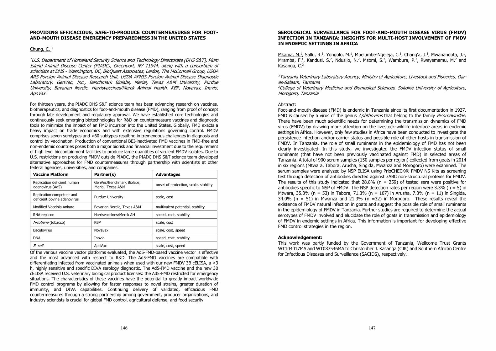
146
PROVIDING EFFICACIOUS, SAFE-TO-PRODUCE COUNTERMEASURES FOR FOOT-AND-MOUTH DISEASE EMERGENCY PREPAREDNESS IN THE UNITED STATES Chung, C. 1
1U.S. Department of Homeland Security Science and Technology Directorate (DHS S&T), Plum Island Animal Disease Center (PIADC), Greenport, NY 11944, along with a consortium of scientists at DHS - Washington, DC, BioQuest Associates, Leidos, The McConnell Group, USDA ARS Foreign Animal Disease Research Unit, USDA APHIS Foreign Animal Disease Diagnostic Laboratory, GenVec, Inc., Benchmark Biolabs, Merial, Texas A&M University, Purdue University, Bavarian Nordic, Harrisvaccines/Merck Animal Health, KBP, Novavax, Inovio, ApoVax. For thirteen years, the PIADC DHS S&T science team has been advancing research on vaccines, biotherapeutics, and diagnostics for foot-and-mouth disease (FMD), ranging from proof of concept through late development and regulatory approval. We have established core technologies and continuously seek emerging biotechnologies for R&D on countermeasure vaccines and diagnostic tools to minimize the impact of an FMD incursion into the United States. Globally, FMD exacts a heavy impact on trade economics and with extensive regulations governing control. FMDV comprises seven serotypes and >60 subtypes resulting in tremendous challenges in diagnosis and control by vaccination. Production of conventional BEI-inactivated FMD vaccines in FMD-free and non-endemic countries poses both a major biorisk and financial investment due to the requirement of high level biocontainment facilities to produce large quantities of virulent FMDV isolates. Due to U.S. restrictions on producing FMDV outside PIADC, the PIADC DHS S&T science team developed alternative approaches for FMD countermeasures through partnership with scientists at other federal agencies, universities, and companies.
Vaccine Platform Partner(s) Advantages
Replication deficient human adenovirus (Ad5)
GenVec/Benchmark Biolabs, Merial, Texas A&M onset of protection, scale, stability
Replication competent and deficient bovine adenovirus Purdue University scale, cost
Modified Vaccinia Ankara Bavarian Nordic, Texas A&M multivalent potential, stability
RNA replicon Harrisvaccines/Merck AH speed, cost, stability
Nicotiana (tobacco) KBP scale, cost
Baculovirus Novavax scale, cost, speed
DNA Inovio speed, cost, stability
E. coli ApoVax scale, cost, speed
Of the various vaccine vector platforms evaluated, the Ad5-FMD-based vaccine vector is effective and the most advanced with respect to R&D. The Ad5-FMD vaccines are compatible with differentiating infected from vaccinated animals when used with our new FMDV 3B cELISA, a <3 h, highly sensitive and specific DIVA serology diagnostic. The Ad5-FMD vaccine and the new 3B cELISA received U.S. veterinary biological product licenses: the Ad5-FMD restricted for emergency situations. The characteristics of these vaccines have the potential to greatly impact worldwide FMD control programs by allowing for faster responses to novel strains, greater duration of immunity, and DIVA capabilities. Continuing delivery of validated, efficacious FMD countermeasures through a strong partnership among government, producer organizations, and industry scientists is crucial for global FMD control, agricultural defense, and food security.
147
SEROLOGICAL SURVEILLANCE FOR FOOT-AND-MOUTH DISEASE VIRUS (FMDV) INFECTION IN TANZANIA: INSIGHTS FOR MULTI-HOST INVOLVEMENT OF FMDV IN ENDEMIC SETTINGS IN AFRICA Mkama, M.1, Sallu, R.1, Yongolo, M.1, Mpelumbe-Ngeleja, C.1, Chang’a, J.1, Mwanandota, J.1, Mramba, F.1, Kandusi, S.2, Ndusilo, N.2, Msomi, S.2, Wambura, P.2, Rweyemamu, M.2 and Kasanga, C.2 1Tanzania Veterinary Laboratory Agency, Ministry of Agriculture, Livestock and Fisheries, Dar-es-Salaam, Tanzania 2College of Veterinary Medicine and Biomedical Sciences, Sokoine University of Agriculture, Morogoro, Tanzania Abstract: Foot-and-mouth disease (FMD) is endemic in Tanzania since its first documentation in 1927. FMD is caused by a virus of the genus Aphthovirus that belong to the family Picornaviridae. There have been much scientific needs for determining the transmission dynamics of FMD virus (FMDV) by drawing more attention on the livestock-wildlife interface areas in endemic settings in Africa. However, only few studies in Africa have been conducted to investigate the persistence infection and/or carrier status and possible role of other hosts in transmission of FMDV. In Tanzania, the role of small ruminants in the epidemiology of FMD has not been clearly investigated. In this study, we investigated the FMDV infection status of small ruminants (that have not been previously vaccinated against FMD) in selected areas of Tanzania. A total of 900 serum samples (150 samples per region) collected from goats in 2014 in six regions (Mtwara, Tabora, Arusha, Singida, Mwanza and Morogoro) were examined. The serum samples were analyzed by NSP ELISA using PrioCHECK® FMDV NS Kits as screening test through detection of antibodies directed against 3ABC non-structural proteins for FMDV. The results of this study indicated that 28.8% (n = 259) of tested sera were positive for antibodies specific to NSP of FMDV. The NSP detection rates per region were 3.3% (n = 5) in Mtwara, 35.3% (n = 53) in Tabora, 71.3% (n = 107) in Arusha, 7.3% (n = 11) in Singida, 34.0% (n = 51) in Mwanza and 21.3% (n =32) in Morogoro. These results reveal the existence of FMDV natural infection in goats and suggest the possible role of small ruminants in the epidemiology of FMDV in Tanzania. Further studies are required to determine the actual serotypes of FMDV involved and elucidate the role of goats in transmission and epidemiology of FMDV in endemic settings in Africa. This information is important for developing effective FMD control strategies in the region. Acknowledgement: This work was partly funded by the Government of Tanzania, Wellcome Trust Grants WT104017MA and WT087546MA to Christopher J. Kasanga (CJK) and Southern African Centre for Infectious Diseases and Surveillance (SACIDS), respectively.
148
POTENTIAL USE OF NOVEL SINGLE-CHAIN ANTIBODY FRAGMENTS AGAINST FOOT-AND-MOUTH DISEASE SAT SEROTYPE VIRUSES IN VACCINE DEVELOPMENT AND DIAGNOSTICS Chitray, M.1, 2, Frischmuth, J.3, Maree, F.1, 2 and Opperman, P.1
1Agricultural Research Council, Onderstepoort Veterinary Research, Vaccines and Diagnostic Development, Private Bag X05, Onderstepoort 0110, South Africa. 2University of Pretoria, Faculty of Natural and Agricultural Sciences, Department of Microbiology and Plant Pathology, Pretoria 0002, South Africa. 3National Bioproducts Institute, Biotechnology division, Private Bag X9043, Pinetown 3600, South Africa. Foot-and-mouth disease virus (FMDV) populations are quasispecies, which is due to the lack of the proofreading mechanism of the RNA-dependent RNA polymerase during FMDV replication resulting in genomes that are related but non-identical. Thus, the impediment that arises is a broad spectrum of antigenic and genetic FMDV variants within each serotype. The location and constitutive epitopic residues of the antigenic sites tend to differ even though there are several structural and functional similarities. This poses serious implications in vaccine design and efficacy where an effective vaccine should include multiple independent neutralising epitopes to elicit an immune response. The hypervariable regions on surface-exposed loops of the FMDV that link β-sheets in the capsid proteins (VP1-3) have been shown to contribute to immunogenic variation. Further investigation of the residues that comprise the antigenic determinants of the virus will allow the identification of mutations in outbreak strains that potentially lessen the efficacy of a vaccine. Thus, if sufficient epitopes are identified, it may be possible to predict the protection afforded by a vaccine against a specific outbreak strain. Neutralising antigenic sites have been identified through several studies for serotype A, O, C and Asia-1. However, information regarding the epitopes of the SAT serotypes, which are confined geographically to occur in Africa, is scarce. In this study, we used phage display technology where the Nkuku® library [a single chain variable fragment (scFv) phage display naïve library] was panned against FMDV SAT1 and SAT3. Unique, novel scFvs were obtained and were used in virus neutralisation assays for epitope prediction. Phage libraries have additionally proven useful for the generation of diagnostic reagents for the detection of a wide variety of viruses such as influenza virus, noroviruses and malaria. The scFvs resulting from the biopanning process were further investigated for their prospective use as FMDV diagnostic reagents. ScFvs were tested in an indirect and a sandwich ELISA format and proved that they can be used in initial screening assays. Furthermore, the potential use of these scFvs in point of care tests must not be ruled out. Through the use of the SAT1 and SAT3 scFvs in this study, ELISA and structural data was utilised to predict potential SAT1 and SAT3 epitopes. These epitopes corresponded to previously identified antigenic sites for SAT1 but were novel for SAT3. Such knowledge can be used in the design of chimeric FMDV vaccines to afford better immunological protection. Additionally, the use of the scFvs as diagnostic reagents in an ELISA format for SAT1 and SAT3 has proven beneficial for potential use in improved FMD diagnostic assays.
149
RISK FACTORS FOR THE CONTAMINATION OF STOMOXYS NIGER NIGER MACQUART 1851 (DIPTERA: MUSCIDAE) WITH THE FOOT-AND-MOUTH DISEASE VIRUS Lendzele, S S.1, 6, Abdoulmoumini, M.2, Timoléon, T.1, Garabed, R.3, Renz, A.4, 6, Dickmu, S.5 1Vector-Borne Infectious Disease Unit, Laboratory of Applied Biology and Ecology (VBID-LABEA), Department of Animal Biology, Faculty of Science, University of Dschang, P.O. Box 067, Dschang, Cameroon. 2University of Ngaoundéré, School of Veterinary Medicine and Sciences, Department of Parasitology and Parasitological Diseases, Ngaoundéré, Cameroon. 3Department of Veterinary Preventative Medicine, College of Veterinary Medicine, Ohio State University, OH, USA. 4Institute of Evolution and Ecology, Department of Comparative Zoology, University of Tübingen, Tübingen, Germany. 5Laboratoire National Véterinaire (LANAVET), B.P. 503 Garoua Cameroun. 6Programme Onchocercoses field station of the University of Tübingen, Ngaoundéré, Cameroon.
The frequent contact of blood-feeding Stomoxys niger niger with cattle during an epidemic of FMDV in a herd around the Ngaoundere cattle market motivated us to identify the risk factors for their involvement in the transmission of the disease. Flies were collected using a Vavoua trap pitched 50m from the center of the herd and by net-catches on clinically sick (n=5) and symptomless cattle (n=5). Vesicular Epithelia Tissues (VET), collected from the mouth and foot of the clinically sick group were analysed by semi-quantitative real time RT-PCR and serum from randomly selected animals without clinical signs was examined by serological test (NS-ELISA Kit), both tests were applied in order to screen for FMDV. Of a total of 568 Stomoxes trapped using the Vavoua trap, four species were identified into S. niger niger 356 (62.7 %, 17, 0 Snn/t/d), S. niger bilineatus 108 (19.01%, 5.14 snb/t/d), S. calcitrans 82 (14.44%, 3.90 sc/t/d) and S. omega 22 (3.87%, 1.05 so/t/d). The dissected mouth or legs of each fly caught were screened for FMDV using the sq rRT-PCR. FMD-Virus RNA was found in the epithelial vesicles of all clinically sick animals (n=5, with mean CT of 27.98456) and 3 out of 5 were serologically positive in the clinically in-apparent cattle group. The overall S. niger niger (most abundant species) contamination rate with the FMDV irrespective of collection method was 40.3 % with females (49.0 %) being slightly more contaminated than males (21.7%) (OR =0.4471614, P= 0.1506) and legs being more often positive than mouth-parts (P=0.02002). Flies were contaminated less frequently on animals without clinical signs than those on animals with clinical signs, but this difference was not significant (P=0.69680). From the present findings we conclude that S. niger niger landing on and biting clinical and non-clinical cattle are usually contaminated during FMDV outbreaks. They may pose a risk of FMD transmission, spreading the virus to livestock within the range of their active (˃8 km) or passive flights. Fly-traps and insecticide treated screens strategically positioned may help to prevent spreading the disease.
150
A FIELD STUDY OF RISK FACTORS FOR FOOT-AND-MOUTH DISEASE IN BANGLADESH Osmani, M G.1, Ward, MP. 2, Giasuddin, M.3, Chawdhury E H. 1 and Islam, M R.1
1 Department of Pathology, Bangladesh Agricultural University, Mymensingh 2 Sydney School of Veterinary Science, C01A - Jl Shute - Camden , The University of Sydney, Australia 3 Bangladesh Livestock Research Institute, Savar, Dhaka Foot and mouth disease (FMD) is endemic in Bangladesh. Three serotypes O, A and Asia-1 are currently prevalent in the country. A baseline cross-sectional study was conducted to identify risk factors for FMD outbreaks in three regions of Bangladesh with different husbandry practices: Shahjadpur upazila in Sirajganj district; Savar upazila in Dhaka district; and Ghatail upazila in Tangail district. In Shahjadpur mostly cross-bred cattle are reared indoors with no access to grazing. In Ghatail the cattle are mostly indigenous type and allowed to graze in the fields. In Savar both cross-bred and indigenous cattle are reared with mixed type husbandry practices. A total of 458 farms (both organized commercial farms and household subsistence farms) from 50 clusters were selected through a multistage cluster sampling technique. A semi-structured pretested questionnaire was used to collect historical data on 43 variables from the field for the period from June 2013 to May 2014. Descriptive analyses were performed and then univariable and multivariable logistic regression was undertaken to understand the risk factors for FMD outbreaks. During the study period, 22.5% (103 out of 458) of farms in the three study areas experienced FMD outbreaks. A total of 1,019 cattle (18%) out of 5,663 were affected. The number of FMD affected farms varied significantly (p<0.05) among the three study areas: 28.3% in Savar, 23.3% in Shahjadpur and 12.3% in Ghatail. This suggests a possible relationship between FMD outbreaks and husbandry practices and the raising of cross-bred cattle, which needs further investigation. In univariable analysis, FMD outbreaks were significantly (p<0.05) associated with (i) absence of vaccination, (ii) proximity to long roads (< 3 kM), (iii) proximity to cattle markets (< 3 kM), (iv) contact with feed sellers, (v) milking of cows by hired persons and (vi) introduction of new cattle. Logistic regression analysis showed that FMD vaccination reduced the risk of outbreaks by 74% (odd ratio=0.264) while contact with feed sellers increased outbreak risk 9 times (odd ratio= 9.36) and introduction of new cattle doubled the risk of outbreaks (odd ratio= 2.01). However, proximity to long roads only had relatively little impact (odd ratio=1.76) whilst proximity to cattle markets and milking by hired persons had no impact outbreak risk. We conclude that inadequate vaccination, contact with feed sellers and the introduction of new animals are strong risk factors for FMD outbreaks in parts of Bangladesh.
151
FOOT-AND-MOUTH DISEASE VIRUS PERSISTENCE IN EQUATORIAL AFRICA: DOES THE VIRUS LIVE TO MID-20th CENTURY STANDARDS?
Mielke, S. MPH-VPH1, Garabed, R. VMD, PhD
1
1Ohio State University College of Veterinary Medicine, 1920 Coffey Road Columbus, OH 43210,
USA Since being identified in 1898 Foot and Mouth Disease virus has grasped our intellectual, societal, and economic attention for over a century. While many advances in knowledge have been made, there remain gaps in our understanding of how this virus evades elimination. The enormity of damage rendered by continued outbreaks and the need to prevent these events have led many to investigate how the virus is transmitted among and between populations. How the virus persists in the environment, what are the optimal conditions, seasons, and ecological factors contributing to the persistence, remains an unanswered question. Through a meta-analysis of historically relied upon data and the most current information on environmental transmission, we summarize what is known and unknown regarding environmental survival of FMDv. The 18 major studies that address environmental persistence date back to the early to mid-20th century and consist of both laboratory and field studies of pH, relative humidity (RH), temperature, and fomite assessments of FMDv survival. These studies incorporate what will be defined, for the purpose of this review, as an environmental study of FMDv survival. The factors that clearly relate to FMDv survival are RH, pH, and temperature. These parameters influence the virus’s ability to persist outside of the host and remain infective. Serotypes A, O, and C are the dominant experimental strains in these studies with most studies using one serotype during each experiment. Survival ranges anywhere from 1 day up to 16 weeks, dependent on temperature, RH, and fomite within ranges commonly seen in Europe. Current global research and modeling endeavors continue to delineate the underlying mechanisms of persistent FMDv outbreaks not only in regions that experience sporadic outbreaks but in endemic regions of Africa and Asia. Model development relies on parameter estimation using values determined from these past experiments. However, how to generalize this information to transmission in the African pastoralist settings, where multiple serotypes of FMDv are endemic and environmental parameters vary widely is unclear. To address the global issue of FMD control, the collaboration of global alliances that build models of disease transmission is of great importance. However, if these models rely on values from studies designed and executed 70 years ago in very limited settings, the parameter values are not likely to be robust.
152
LESSONS FROM FOOT-AND-MOUTH DISEASE OUTBREAKS IN ZIMBABWE AND PROSPECTIVE SOLUTIONS TOWARDS ITS ERADICATION Mbiriri, D.1. and Mutibvu, T.1 1Department of Animal Science, Faculty of Agriculture, University of Zimbabwe, P.O. Box MP 167, Mount Pleasant, Harare, Zimbabwe Foot and mouth disease (FMD) is endemic to Zimbabwe and the Southern African region. Despite this fact, Zimbabwe exported large quantities of beef to European markets through the 1980s to the 1990s, making the beef industry one of the largest foreign exchange earners for the country. This was made possible by stringent European Union (EU) disease control regulations particularly around FMD and anthrax. Unfortunately, these control measures, including veterinary cordon fences, favored large-scale commercial ranches mostly located in the northern high potential areas of the country. The southern parts, on the other hand, which are drier and would economically be more beneficial to raising cattle for beef are close to game parks where it would be virtually impossible to create disease free zones as dictated by the EU regulations. Zimbabwe is home to large herds of buffalo and other wildlife species that harbor the FMD virus. Fencing in these areas would also restrict wildlife movements, a concern to the countries’ future wildlife populations. The situation was made worse by the disputed land redistribution exercise that took place in the year 2000, when white-owned large-scale farms were compulsorily acquired redistribution to local black farmers. In the course of this exercise, fencing barriers were broken down and the large ranches were parceled-out as smaller pieces of land. EU funding that subsidized the beef industry and financed the erection of fences was withdrawn leaving the industry vulnerable to FMD spread. FMD outbreaks in Zimbabwe are a serious concern to its bordering neighbors, particularly Botswana, South Africa and Zambia. There has been incessant outbreaks since the year 2000 but the industry is making strides in controlling the disease through vaccinations. Zimbabwe has, however, since lost its EU market due to violation of the disease control agreement, leading to the loss of significant foreign exchange in potential revenue. In this paper, we retrace the history of FMD and its impact on the Zimbabwean beef industry. We also explore alternative FMD control measures that could work with the current land ownership status that has drastically shifted from the 1990s. We also highlight potential beef markets, other than the traditional EU markets, that Zimbabwe can target as it revives its industry under the harsh economic conditions prevailing in the country.
153
ANTIGENIC AND GENOMIC CHARACTERIZATION OF FMDV TYPE O IN SOUTH EAST ASIA IN 2015-2016 Seeyo, K.1; Ungvanijban, S.1; Thongtha, P.1; Choonnassard, A.1; Samanit, J.1; Puntumart, A.1; Deeplang, C.1; Linchongsubongkoch, W.2; Nishi, T.3; Yamada, M.3 and Sakamoto, K.3 1 Regional Reference Laboratory for FMD in South East Asia, National Institute Animal Health, Department of Livestock development, Pakchong, Nakhon Ratchasima, Thailand 2 Expert OIE and Consultant of Department of Livestock Development for foot and mouth disease, National Institute of Animal Health, Bangkok 3 Exotic Diseases Research Division, National Institute of Animal Health, NARO, Kodaira, Tokyo, 187-0022, Japan Foot and mouth disease (FMD) have been one of the most important economic disease of livestock animals in South East Asia (SEA) region. Recently FMDV type O are dominant outbreaks in this region and the major control measure is vaccination by using an appropriate vaccine. Hence, the antigenic and genomic characterisation of FMDV type O are investigated with the aim to study the serological relationship (r-value) between vaccine strain and field outbreak strain and to study the molecular epidemiology of FMDV for tracing back to the original virus causing outbreak in the field. Total 71 samples of FMDV type O isolated viruses (Thailand, Lao PDR, Myanmar and Cambodia) that submitted to the Regional Reference Laboratory (RRL), Pakchong, Thailand during 2015-2016 were used for this study. The result found the total of 47 sample of field isolated viruses in 2015 gave an r-value greater than 0.4 or good matching, indicating no antigenically change from vaccine strain of O/189/87. As well as the total 24 sample of field isolated viruses in 2016 were also gave r-value greather that 0.4 or good matching to O/189/87 vaccine strain indicating no antigenically change. In addition, the genomic characteristic of type O field isolated were investigated by nucleotide sequencing at VP1 encoding region. The phylogenetic tree of all viruses in 2015-2016 were almost showed viral lineage to O/SEA topotype/Mya-98 strain. Interestingly, the phylogenetic tree of isolated viruses in 2016 were showed viral lineage to O/ME-SA topotype/Ind2001d strain in 5 of 24 samples. In conclusion, the antigenic characteristic of type O field isolated viruses in SEA region indicated that the serological relationship close to O/189/87 vaccine strain. For the genomic characteristic found that most of the viral lineage were O/SEA topotype/ Mya-98 strain and some of them were O/ME-SA topotype/Ind2001d strain which were the viruses causing outbreak in 2016. Although, the genomic characteristic of those viruses were changed but the antigenic characteristic were not changed, that meant, the recent vaccine of O/189/87 would be suitable to use to protect the field outbreak viruses. Additionally, one isolated of whole L-fragment sequences of FMDV serotype O in 2015 had been comparatively analyzed that showed more than 92% Pairwise identity rates without any genetic deletion or insertion.These investigations could provide the useful information for seed vaccine selection and tracing back to the origin of virus causing outbreak in the region and support the strategic plan for the control and eradication of FMD in SEA region.
154
FMD VIRUS ECOLOGY AND ITS LANDSCAPE EPIDEMIOLOGY IN CATTEL AND BUFFALO UNDERNATURAL CONDITION IN INDIA Ranjan, R.1; Biswal, J.1; Subramaniam, S.1; Biswajit, D.1; Rodriguez, L.2; Arzt, J.2 & Pattnaik, B.1
1ICAR-Directorate of Foot and Mouth Disease, Mukteshwar, Nainital, Uttarakhand, PIN: 263138, India 2Foreign Animal Disease Research Unit, USDA/ARS Plum Island Animal Disease Center, P.O. Box 848, Greenport New York, ZIP: 11944, USA Foot and Mouth Disease (FMD) is endemic in India. There is a need to improve the understanding of FMD Virus (FMDV) natural ecology and epidemiology in endemic setting to provide the basis for effective control strategies. The possible role of FMDV persistently infected cattle and buffalo in initiating new outbreaks remains controversial and the inability to quantify the real risk posed by such animals could preclude control of FMD in India. This report reflects collaborative research activities between USDA-ARS Foreign Animal Disease Research Unit at PIADC, USA and the ICAR-Directorate of FMD, Mukteshwar, India. Here we report duration of persistence, genetic and antigenic variations of FMDV serotype O/ME-SA/Ind2001d lineage that have been analyzed in consecutive isolates recovered over a period of 24 months from cattle and buffalo under natural condition. Longitudinal study of FMD Virus circulating in regularly vaccinated cattle and Indian buffalo in organized dairy farm, where FMD outbreak was recorded in the month of December 2013, and it was caused by serotype O virus of the O/ME-SA/Ind2001d lineage. Oesophageal-pharyngeal fluid (OPF) samples were collected from convalescent (n=32) and asymptomatic (n=22) cattle and buffaloes at 2-3 months interval from March 2014 for a period up to 24 months. Viruses were recovered from the OPF in LFBK-αVβ6 cells and the entire capsid coding (P1) sequences were determined from the cell culture supernatant at low passage level (3-4). In cattle and buffalo, the genome positive probang samples varied from 7.4 to 100%. The herd revealed high prevalence of persistently infected animals (genome positive probang samples beyond 28 days). Between, cattle and buffalo, considerable difference was noticed in the proportion, and duration of VI positive animals post outbreak. Cattle and buffalo were VI positive up to 7 and 13 months post outbreak, respectively. Analysis of the P1 sequences from acutely infected animals (n=4), convalescent carriers (27 sequences from 19 animals) and asymptomatic carrier (10 sequences from 8 animals), revealed point mutations that represented fixation of mutations at the rate of 1.816 x 10⁻² substitution/site/year (s/s/y) with a 95% credible interval of 1.362-2.31 x 10⁻² s/s/y. Two codons in VP1 (138 and 148) and one codon each in VP2 (78) and VP3 (76) was found to be under positive selection with statistical significance. Though fixation of nucleotide and amino acid changes were observed at some position, majority were not conserved in consecutive isolates. Between different animals, mean dN/dS (ω) value of the entire capsid coding region varies from 0.076 to 0.357, which indicates that the selection pressure acting on viral genomes differ between individual animals. From the statistical parsimony analysis, it was evident that all the virus isolates from carrier animals were originated from acute virus except six isolates. This may be attributed to either silent re-introduction of virus or very high rate of mutation in some individual animals or coexistence of heterogeneous populations in which variants evolve independently of each other. The antigenic relationship value as determined by 2D-MNT assay indicates fluctuation of antigenic variants in some of the carrier animals. The genetic and antigenic variations observed in the carrier viruses differ largely between individual animals. Our study indicates evidence of viral activity in the persistently infected animals under field scenario and probable role of host factor in shaping their evolution.
155
DEVELOPMENT OF DIVA STRATEGY FOR FMD VIRUS BY USING LATERAL FLOW IMMUNOCHROMATOGRAPHIC STRIP IN EGYPT Sayed, R.1. and Abousenna, M.1. 1Central Laboratory for Evaluation of Veterinary Biologics,131 El-Seka El-Beda St., Abassia,13181, Cairo, Egypt. Foot-and-mouth disease (FMD) is a highly contagious disease in cloven-hoofed animals and the most important disease economically of livestock worldwide. The our work is aimed to applying DIVA test (Differentiating infected from vaccinated animals test ) that’s can differentiate well between FMD-infected animals and non infected vaccine animals using the serum sample from native and imported animals by using lateral flow immunochromatographic strip (LFIS) that is detect non structure proteins (NSPs) antibody of FMDV within 5 mins . The inactivated FMDV vaccines is concentrated and purified by industrial ultrafiltration in order to removing NSPs. NSPs was conjugated with nanogold was laid on the conjugate pad. Staphylococcus aureus protein A was used the capture antibody at the test line (T). Anti bovine rabbit antibodies was used as the capture antibody at the control line (C) of nitrocellulose membrane and this collected material called (LFIS). The minimal amount of antibodies against NSPs in bovine serum sample can be detected was 60% ELISA titer / 100 µl that was mean 0.1 µg (Ab) /100 µl sample. One hundred suspected bovine serum samples were collected from different cow flocks then tested with the prepared (LFIS) and 3ABC antibody ELISA test kit(gold standard test). The sensitivity, specificity and accuracy of LFIS as compared to 3ABC antibody ELISA reached to 94.4% , 90% and 94% respectively which were depending on control positive and control negative sera were provided by viral large animal vaccines evaluation department . The ideal diagnostic tool should be able to detect the NSPs Ab in the shortest possible time, simple, sensitive, specific and inexpensive. Also it should be suitable as field test or laboratory test and can be applied on large scale of animals. The LFIS was low cast, fast, no requirements for skilled technicians and applicable in field and quarantine condition gives results with in 5 mins that is help in large flock, also it can be used in prevention and control strategy against FMD virus infection and eradication of FMDV endemic areas.
156
PRESERVED IMMUNOGENICITY OF AN INACTIVATED VACCINE BASED ON FOOT-AND-MOUTH DISEASE VIRUS PARTICLES WITH IMPROVED STABILITY Caridi, F.1, Vázquez-Calvo, A.2, Borrego, B.3, McCullough, K.4, Summerfield, A.4, Sobrino, F.1, Martín-Acebes, M.2 1Department of Virology and Microbiology, Centro de Biología Molecular “Severo Ochoa” (CSIC-UAM), 28049 Madrid, Spain 2Department of Biotechnology, INIA, 28040 Madrid, Spain 3Centro de Investigación en Sanidad Animal, Instituto Nacional de Investigación y Tecnología Agraria y Alimentaria (INIA), Valdeolmos, 28130 Madrid, Spain 4Institute of Virology and Inmunology, 3147 Mittelhausern, Switzerland Stability of foot-and-mouth disease virus (FMDV) particles and empty capsids is a major issue for development of improved FMD vaccines, as long term conservation at 4 °C (the temperature for vaccine storage) or ruptures of the coldchain, provoke the dissociation of virions, reducing the immunogenicity of the vaccine. We previously reported the isolation of a FMDV mutant (VP1N17D+VP2 H145Y) that produced virions with increased resistance to acidic pH that were also more resistant to dissociation at 4 °C. We have evaluated the immunogenicity in swine (a natural FMDV host) of a chemically inactivated vaccine based on this mutant. The presence of amino acid substitutions VP1N17D and VP2 H145Y did not compromise the immunological potential, including its ability to elicit neutralizing antibodies that was similar to that of a WT vaccine. These results support the feasibility of this kind of mutants with increased capsid stability as suitable as suitable candidates for the development of novel, improved FMD vaccines.
157
FMD OUTBREAK IN LUKULU DISTRICT OF WESTERN ZAMBIA, EVIDENCE OF VIRAL SPREAD OUTSIDE THE KNOWN ENDEMIC AREAS Banda, F.1, Lebea, P.2, Fana, E.3, Sinkombe, T.1, Dautu, G.1 Sinkala, Y.4 1Central Veterinary Research Institute, Lusaka, Zambia. 2Councial for Scientific and Industrial Research, Pretoria, South Africa 3Botswana Vaccine Institute, Gaborone, Botswana 4Department of Veterinary Services, Lusaka, Zambia Foot-and-mouth disease (FMD) viruses are usually confined to specific geographical regions and spread to new areas may lead to significant epidemics. In Zambia, the disease is endemic around the three known high risk areas of Kafue Flats, lower Zambezi basin and Mbala-Isoka area. However, in 2015 there was an outbreak of the disease in areas outside the traditional endemic areas involving Western Zambia attributed to SAT 3. A quiescence of the disease followed from May 2016 until April 2017. The precipitating factors that contributes to the changing epidemiology of disease in this region is unknown. Herein we describe investigations conducted of the latest disease foci in Lukulu District of Western Province which historically has never recorded FMD. In May 2017, reports were received of suspected FMD in Lukulu district. Clinical examination of six kraals was conducted to determine the presence of FMD lesions. Five epithelial tissues and 22 blood samples were collected. Lab investigations involved Cell culture, RT-PCR, Antigen ELISA and NSP ELISA. A total of 75 animals (76.5%) out 102 animals from the kraals examined manifested clinical signs and lesions suggestive of FMD. Examination of epithelial samples showed five with 100% CPE result on cell cultures whilst typing by antigen ELISA classified the virus as SAT 3. RT-PCR detected the Virus Genome in all the tissue samples. Twelve (54.5 %) out of 22 blood samples were positive on NSP ELISA with percentage inhibition ranging from 60.8 % - 95.9 %. The outbreak of disease in Lukulu District after a quiescence of 11 months indicates that virus must have been circulating undetected in carrier animals. This is the first time FMD is being reported in this part of the country. A total of 75 cattle (76.5) % out of 102 examined had typical signs and lesions of FMD. This high positivity proportion shows the typical characteristics of an epidemic in a naive population. Factors that have led to epidemiology change of the disease require further investigation although preliminary investigations revealed uncontrolled movements as one of the predisposing factors. The focus of this recorded outbreak is close to North Western Province where demand for cattle is high. Its anticipated the disease might spread to this region due to high demand of beef and beef products instigated by expanding consumer base brought about by new mines. The earlier outbreak was controlled through strategic vaccination of cattle around outbreak areas and even though spread was abated, this outbreak suggests circulating of virus within the province despite vaccinations. It is therefore imperative that further studies are conducted to evaluate vaccine efficacy and vaccination strategies of FMD control in Zambia and use the outcome to inform policy.

158
EVALUATION OF CIRCULATING FOOT-AND-MOUTH DISEASE VIRUS (FMDV) SEROTYPES IN NIGERIA AS CANDIDATES FOR INDIGENOUS VACCINE DEVELOPMENT Ularamu, H.1; Lazarus, D.1; Wungak, Y.1; Ibu, J.2; Abenga, J.2 & Adah, M.2
1National Veterinary Research Institute P.M.B.01 Vom, Plateau State, Nigeria 2College of Veterinary Medicine University of Agriculture Makurdi, Nigeria
Foot and mouth disease (FMD) is an endemic transboundary animal disease (TAD) in Nigeria with severe economic implications. The study evaluated the current foot-and-mouth disease virus (FMDV) serotypes circulating in Nigeria for indigenous vaccine development. Of the forty-two (42) epithelial and oesophageal/pharyngeal samples collected from suspected cases of FMD outbreaks in cattle between 2011 and 2014, FMDV was isolated from twenty-five (25) samples and subjected to antigen ELISA serotyping, conventional reverse transcriptase polymerase chain reaction (Rt-PCR) and sequencing analysis. Phylogenetic analysis of the complete viral protein (VP1) sequences showed that three (3) recent serotype O isolates clustered within the EAST AFRICA 3 (EA-3) topotype and six (6) with WEST AFRICA (WA) topotype. The serotype A isolates fell within the G-IV genotype of the AFRICA topotype, while serotype SAT 2 viruses circulating in Nigeria belonged to topotype VII. The tested candidate vaccine strains (A Nig 03/13; A Nig 07/13; O Nig 03/14; O Nig 07/13; SAT 2 Nig 03/12 and SAT 2 Nig 17/11) showed strong antigenic match suggesting a close relationship between the field isolates and vaccine strains. A potent vaccine containing the virus strains was formulated into a multivalent vaccine and following challenge studies all vaccinated animals (n=12) showed no clinical signs typical of FMD lesions, however, the unvaccinated animals (n=3) manifested typical clinical signs of FMD.
159
DETECTION OF FMDV THROUGH RT-LAMP ASSAY IN PAKISTAN Waheed, U.1; Haider, F.1
1University of Veterinary & Animal Sciences, Lahore Sub-campus Jhang, Pakistan In Pakistan, historically livestock has been dominated by small holders to meet their needs of milk, food security and cash income on daily basis. Moreover, livestock is considered a source of employment generation at rural level, helping to reduce income variability. It is central to the livelihood of the rural poor in the country, due to its incremental potentials. The biggest threat to the livelihood of rural masses is the infectious diseases which end up with production losses and mortalities. Among infectious diseases, foot-and-mouth disease (FMD) is economically very important disease and causing billions of rupees direct and indirect losses to the livestock industry of Pakistan. The disease is caused by FMD virus (FMDV), which is not a public health threat, but it is highly contagious to cloven-footed animals. The losses due to FMD can be reduced by early and cheaper diagnostic methods. The loop mediated isothermal amplification (LAMP) assay is relatively new technique and ruled out the use of highly sophisticated equipment. In present study, we used LAMP assay to detect the presence of FMDV in ELISA known FMD samples and then applied the test on samples collected from field outbreak occurred in Pakistan in 2017. Three primer sets were used in the amplification of FMDV genome including primer inner, outer and loop primer sets. Bsm polymerase was used in the assay. We were able to optimize the assay and then successfully applied on the field outbreak samples. The field samples were also confirmed with RT-PCR using universal primer set for detection of FMDV.
160
BUILDING CAPACITY FOR FMD LABORATORY DIAGNOSIS IN THE FRAMEWORK OF THE PCP-FMD IN NIGERIA (2011-2016) Wungak, Y.1; Ularamu, H.1; Lazarus, D.1,2; Ehizibolo, D.1; Agom, D.1; Onoja, M.1; Chukwuedo, A.1 & Shamaki, D.1
1National Veterinary Research Institute P.M.B.01 Vom, Plateau State, Nigeria 2University of Pretoria Faculty of Veterinary Science, Private Bag X04, 0110 Onderstepoort, South Africa The National Veterinary Research Institute, Vom, Nigeria was established in 1913 as a Veterinary Department by the Colonial masters to control the scourge of rinderpest which was a major cattle plague in the West African coast. Since its establishment as a full diagnostic laboratory in 1924, the institution has fulfilled its mandate by addressing the most devastating livestock diseases such as the eradication of rinderpest and the control of other economic livestock diseases. During the period 1980s to the late 1990s most of the research and diagnostic activities of the Institute concentrated on the deadly rinderpest and CBPP with little attention on Foot-and-mouth disease (FMD) and other diseases of low mortality.However, with the successful eradication of rinderpest in 2010 and the global efforts to control FMD using the Progressive Control Pathway (PCP), FMD research and diagnostic laboratory of the institution was repositioned to provide the needed capacity for the progressive control programme.During the period under reviewlaboratory capacity for FMDV antibody detection, antigen detection, virus isolation and reverse transcriptase polymerase chain reaction platform were established. Between 2011 and 2016, a total of 5,675 sera and 243 epithelial tissue specimens were received from field officers as laboratory submissions. Of the total sera received 50.19% (95%CI: 48.89-51.49) were from cattle, 10.31% (95%CI: 9.55-11.13) from sheep and goats, 32.65% (95%CI: 31.44-33.88) from swine, 6.35% (95%CI: 5.74-7.01) from camels and 0.50% (95%CI: 0.34-0.71) from wildlife species. The sera were screened for FMDV NSP antibodies since most of the samples originated from non-vaccinated animals, and 38.9% (95%CI: 37.60-40.13) of the samples were positive to FMDV NSP antibodies. Of the 243 epithelial specimens received 77 (31.68%) were positive for one serotype or the other by antigen capture ELISA, with serotypes O (34%), A (23%), SAT 1 (9%) and SAT 2 (34%). During the same period, 40 FMDV were isolated from clinical specimens using foetal goat tongue cells (ZZ-R 127) out of which serotype O was (45%) , A (20%), SAT 2 (25%) and SAT 1 (10%). Human capacity development involved several staff members obtaining PhD, MSc and other laboratory trainings over the same period. In 2016 a new BSL 3 laboratory was commissioned as part of intervention programme for TADs diagnosis with the support of the Canadian Government. The laboratory has also participated in several FMD Proficiency tests within this period and has published and presented research outputs at several scientific conferences and workshop. The laboratory also enjoys good working relationship with the WRLFMD, the Pirbright Institute, UK, and is currently a beneficiary of an OIE Laboratory Twinning Programme with the Veterinary and Agrochemical Research Centre (CODA-CERVA), Belgium. From this appraisal, the laboratory has the requisite capacity to confirm immediate suspectedcase of FMDin collaboration with partner institutions. The laboratory has also tested several samples from different susceptible species during the time under review to understand the risk of FMD and the prevalent serotypes circulating within the country.
161
AN EVOLUTIONARY HISTORY OF FOOT-AND-MOUTH DISEASE VIRUSES IN SOUTHEAST ASIA BASED UPON FULL OPEN READING FRAME SEQUENCE ANALYSES Brito, B.,2, Pauszek, S.1, Hartwig, E.1, Smoliga, G.1, Vu, L T.3, Vu, P P.3, Stenfeldt, C.1,4, Rodriguez, L.1, King, D.5, Knowles, N.5, Bachanek-Bankowska, K.5, Long, N T.3, Dung, D H.6 & Arzt, J.1 1 Foreign Animal Disease Research Unit, ARS/USDA-Plum Island Animal Disease Center, 40550 Route 25, 11957, Orient Point, NY USA 2 Oak Ridge Institute for Science and Education, PIADC Research Participation Program, Oak Ridge, TN, USA 3 Regional Animal Health Office No. 6, Department of Animal Health, Ministry of Agriculture and Rural Development, Ho Chi Minh City, Vietnam. 4 Department of Veterinary and Population Medicine, University of Minnesota, St. Paul, MN 5 The Pirbright Institute, Ash Road, Pirbright, Woking, Surrey, GU24 0NF, UK. 6 Department of Animal Health, Ministry of Agriculture and Rural Development, Hanoi, Vietnam. Studies of the molecular epidemiology and evolution of foot-and-mouth disease virus (FMDV) are widely conducted using genomic sequences encoding VP1, the capsid protein containing the most relevant antigenic domains. Although sequencing of the full viral genome is not used as a routine diagnostic or surveillance tool, availability in public repositories has increased over recent years, thereby enabling more detailed analyses. Using full open reading frame (ORF) consensus sequences, previous studies have suggested that recombination between FMDVs is not an infrequent event and that the phylogeny of non-structural proteins (NSP) coding sequence does not necessarily correlate with that of structural proteins coding sequence. Understanding the mechanisms that drive viral diversity, and ultimately the emergence of novel, highly fit viral lineages are critical to predict patterns of FMDV occurrence and cyclical changes in strain dominance that are observed in endemic regions. The role of recombination in the evolution of FMDV and its potential association with the dependence between lineages and serotypes that co-circulate in the same space and time can provide key aspects to understanding FMDV occurrence and emergence of new lineages. In the current study, we selected 96 full polyprotein ORF encoding sequences representing five FMDV lineages (A/ASIA/Sea-97, O/ME-SA/PanAsia, O/SEA/Mya-98, O/CATHAY, and Asia1/Group V) co-circulating in Southeast Asia to investigate whether there was evidence for intra- and inter-lineage recombination. We first determined putative recombination breakpoints detected by different algorithms. Subsequently, we used a Bayesian phylogenetic approach to estimate time divergence of the recombination-free genome regions. Phylogenetic analysis revealed a close relationship between O/Mya-98 and A/Sea-97 lineages in their NSP coding regions. Additionally, three specific recombinant viruses of lineage A/Sea97 and O/Mya98 were detected, suggesting that the close NSP relationship between these two lineages may facilitate genetic exchange. Contrastingly, O/CATHAY virus capsids and NSP-encoding sequences have evolved independently from other lineages. Analysis of intra-lineage recombination revealed different locations of breakpoint hotspot patterns within the genome of each lineage. This study provides new insights about the evolutionary interdependence of FMDV serotypes and lineages. Elucidating the evolutionary mechanisms of FMDV may contribute to understanding emergence of new lineages, and inform the risk posed by co-circulating lineages in FMD endemic countries and regions.
162
ALTERNATIVE METHOD FOR EVALUATION OF FOOT-AND-MOUTH VACCINE Shafik, N.1 1Central laboratories for Evaluation of animal Vaccines and Biologics (CLEVB), 131 Al Sekkaa Al bedda, Abbassia, Cairo, EGYPT Foot and Mouth (FMD) disease controlling is mainly relies on vaccination of cattle and other susceptible species. The quality control of vaccines at CLEVB is strictly regulated, according to the Egyptian protocol and animal challenge test are prescribed to show vaccine efficacy, Use virulent strains (A,O,SAT2) for challenge the vaccinated animals after 21 days from vaccination with FMD vaccines lead to waste time for patch release so no delay in vaccination field program or in case of emergency, waste effort in securing each animal for injection and inspection for tongue and feet, waste money when culling animals after the end of challenge test (14 cattle for each patch), contamination with virulent virus may be occur in the containment so infection to free animals ,and Mixing between strains due to low of a little experience of workers give a false results. Potency will be based on serology, Correlation between SNT, ELISA that done on serum that collected from vaccinated animals after 21 days, and the results of challenge tests. Conclution, This study was done on last ten FMD vaccine patches received by CLEVB, The results show us recommendation for replacement the challenge test by lap serological tests only and change the EGYPTIAN protocol for FMD evaluation . CLEVB will apply new protocol for the newly received patches
163
GENETIC DIVERSITY OF FOOT-AND-MOUTH DISEASE IN NORTHWESTERN PAKISTAN Jamal, S.1 et al.
1Department of Biotechnology, University of Malakand, Chakdara, Dir (L), Khyber Pakhtunkhwa, Pakistan Foot-and-mouth disease (FMD) is endemic in Pakistan and three serotypes (O, A and Asia-1) of the FMD virus are responsible for outbreaks in the country. Continuous surveillance of the disease is necessary for determination of sub-type(s) of the virus for vaccine selection. This study reports characterization of FMDV in samples collected clinically suspected cases of FMD in Khyber Pakhtunkhwa Province, Pakistan during 2012-2015. The samples were subjected to antigen-ELISA for FMDV detection and virus typing. The complete VP1 coding region of the selected samples were sequenced. The sequence data generated were analysed with other sequences from GenBank and phylogenetic trees were constructed. A total of 44 samples tested positive in Ag-ELISA. Of these, 17, 18 and 7 scored positive for serotypes O, A and Asia-1, respectively. Three samples tested positive for both serotypes O and A FMDV. The complete VP1 coding sequences were generated from the serotypes O, A and Asia-1 positive samples. Phylogenetic analysis of serotype O FMDVs showed circulation of two different lineages including O-PanAsia-II and Pak-98. Viruses belonging to the O-PanAsia-II lineage further diverged into two different sublineages i.e. O-PanAsia-IIANT-10 and O-PanAsia-IIBAL-09. Serotype A FMDVs in the present study belonged to four different sublineages within A-Iran05 lineage. These included three already known sublineages (i.e. A-Iran05FAR-11, A-Iran05SIS-10, A-Iran05SIS-12 and a new sublineage, designated here as A-Iran05KPK-14. Serotype Asia-1 FMDVs reported in this study all belonged to the earlier reported Group-VII (Sindh-08), which is currently a dominant strain in the West EurAsian region. Co-circulation of different sublineages of FMDVs in region shows complexity in epidemiology of FMD.
164
THE IMPACT OF BINARY ETHYLENIMINE (BEI) ON THE STABILITY, ANTIGENICITY, AND GENOMIC INTEGRITY OF FOOT-AND-MOUTH DISEASE VIRUS Rodriguez, Y.1; Hershey, B. 1; Tadassa, Y. 1; Xu, L. 1; Das, A. 1; Jia, W. 1 and Wu, P. 1 1Foreign Animal Disease Diagnostic Laboratory, NVSL, STAS, VS, APHIS, USDA, USA The complete inactivation of foot and mouth disease virus (FMDV) is a critical requirement for preparing FMD vaccine and reference serum. Ethylenimine can selectively target viral nucleic acids while retaining viral antigenicity. Oligomeric ethylenimine has been used to inactivate FMDV over the past 30 years. In this study, the inactivation kinetics of FMDV treated with 0.5mM, 1.0mM and 1.5mM binary ethylenimine (BEI) at both room temperature (23±2°C) and 37°C was investigated. Samples were collected hourly from hours 1 to 10, then at hours 24 and 30. To evaluate the effects of BEI treatment on the viability of FMDV, the samples were titrated in vitro with LFBKV6 cells in 96-well tissue culture plates. The complete inactivation was evaluated by blindly passing the samples in vitro with LFBKV6 cells in T25 tissue culture flasks for 3 consecutive passages. Each passage was incubated for 3-4 days at 37°C, 5% CO2. The results indicated that the best FMDV inactivation rate was achieved using 1.5mM BEI at 37°C. Recent studies indicate that trimeric ethylenimine (TEI) may modify viral proteins. To further evaluate the effects of BEI treatment, 140S FMDV antigen ELISA and qRT-PCR were used to assess antigenic modification and genomic damages, respectively. Preliminary results from FMDV serotype Asia 1 suggest that the concentration of 140S antigen is significantly higher (p<0.01) in samples treated at 37°C than those treated at room temperature. No significant change in 140S concentration was observed among time course samples treated at the same temperature. The qRT-PCR results from samples treated at 37°C also indicate that genomic damage is occurring over the course of treatment.
165
AUTHOR INDEX
A Abdoulmoumini, M. .................................................................... 149 Abenga, J. ....................................................................................... 158 Abila, R. ............................................................................... 25, 38, 41 Abousenna, M. ....................................................................135, 155 Adah, M. ........................................................................................... 158 Adams, S. ........................................................................................... 60 Ademun, R. ..................................................................................... 116 Afzal, M................................................................................................ 78 Agom, D. .......................................................................................... 160 Ahmed, Z. .............................................................. 36, 78, 116, 117 Akopian, J. ......................................................................................... 81 Aliyev, J. ........................................................................................... 113 Aliyeva, T......................................................................................... 113 Alkhamis, M. ..................................................................................... 33 Andreu, D. .......................................................................................... 43 Angubua Baluka, S. ................................................................... 115 Arinaitwe, E. ................................................................................... 116 Armson, B. ......................................................................................... 61 Arzt, J. ............... 33, 48, 70, 74, 75, 120, 137, 138, 154, 161 Asadov, K. ....................................................................................... 113 Asfor, A. .................................................................................... 87, 109 Ashley, K. ............................................................................................ 39 Ataliba, I. ......................................................................................... 116 Atim, S .................................................................................................. 84 Avaliani, L........................................................................................... 79 Ayebazibwe, C. ............................................................................. 114 Ayude, A. ............................................................................................ 68
B Bachanek-Bankowska, K. ............................................... 61, 161 Bakkali-Kassimi, L. ................................................................. 62, 83 Balasubramanian, G. ......................................................... 40, 142 Banda, F. .......................................................................................... 157 Bandaranayaka-Mudiyanselage, C. ...................................... 60 Barrera, J. ........................................................................................ 133 Barrionuevo, F. ........................................................................ 54, 68 Bartels, C. ................................................................................... 27, 69 Beechler, B. ....................................................................................... 76 Beer, M. ............................................................................................... 91 Bellinzoni, R. ..................................................................................... 57 Belsham, G. ....................................................................................... 74 Ben Arous, J. .................................................................................... 90 Bergmann, I...................................................................................... 56 Bertram, M. ............................................................... 120, 137, 138 Binney, B. ........................................................................................ 129 Birch, C. .................................................................................... 66, 108 Biswajit, D. ...................................................................................... 154 Biswal, J. .................................................................................. 44, 154 Blaise-Boisseau, S. ................................................................ 62, 83 Blakeney, S. ...................................................................................... 60 Blanco, E. ............................................................................................ 43 Borca, M. ............................................................................................. 68
Borrego, B. ...................................................................................... 156 Bøtner, A. ........................................................................................... 74 Bottini, R. ............................................................................................ 65 Bounpheng, M. ................................................................................ 60 Boyer, T. .......................................................................................... 108 Bradhurst, R. .................................................................................... 71 Brake, D. .................................................................................. 60, 133 Branan, M........................................................................................... 70 Brito, B. .............................................. 33, 70, 120, 137, 138, 161 Brocchi, E. .......................................................................................... 87 Bronsvoort, M. ................................................................................. 99 Bucafusco, D. ................................................................................... 68 Buckle, K. ......................................................................................... 129 Bun, C. ................................................................................................. 39 Burrage, T. ......................................................................................... 60 Bush, R. ............................................................................... 38, 39, 41 Buza, J. ................................................................................................ 37 Byun, J-W.......................................29, 103, 105, 106, 126, 128
C Cabezas, A. ................................................................................ 88, 89 Caignard, G. ...................................................................................... 83 Campagnola, G. ........................................................................... 131 Cañas, R. ............................................................................................. 43 Capozzo, A. ................................................................................ 54, 68 Caridi, F. ........................................................................................... 156 Carr, V. ...................................................................................... 46, 112 Celeste, T. .......................................................................................... 54 Chaligava, T. ..................................................................................... 79 Chang’a, J. ...................................................................................... 147 Charleston, B. ...............................................46, 76, 77, 111, 112 Chawdhury, E H. ................................................................. 73, 150 Chen, J. ................................................................................................ 67 Chen, R-S. .......................................................................................... 85 Chitray, M................................................................................ 34, 148 Cho, K-H. ................................................................................. 28, 122 Choi , J-H. ....................................................................................... 134 Choi, B S. ......................................................................................... 124 Choi, H Y. ..............................................................................123, 124 Choi, I S. ................................................................................123, 124 Choi, J C. ................................................................................123, 124 Choi, J. ................................................................................................. 30 Choi, J-H., ........................................................................................... 47 Choonnassard, A. ........................................................................ 153 Choung, K. ......................................................................................... 96 Chu, J-Q. ............................................................................................. 47 Chukwuedo, A. ...................................................................141, 160 Chung, C. ................................................................................. 60, 146 Clavijo, A. ........................................................................................... 60 Cleaveland, S. ............................................................................... 114 Cobbold, C. ........................................................................................ 72 Coco-Martin, J. ............................................................................. 130 Colby, M. .......................................................................................... 133 Cook, C. .................................................................................... 66, 108 Curet, M. .......................................................................................... 133
166
D D’Alessio, F. ....................................................................................... 65 Dacre, I. ........................................................................................... 129 D'Alessio, F. ................................................................................................ 26 Dancho, B. ......................................................................................... 60 Danelyan, H. ..................................................................................... 82 Darwish, M D. ............................................................................... 136 Das, A. ............................................................................................... 164 Das, B. .................................................................................................. 44 Dash, BB. ......................................................................................... 142 Dautu, G. ......................................................................................... 157 De Clercq, K. ..................................................................................... 35 de Klerk-Lorist L-M. ...................................................................... 34 de Klerk-Lorist, L-M. ..................................................................... 76 de la Torre, B. .................................................................................. 43 de León, P. ......................................................................................... 43 de los Santos, T. ........................................................... 49, 58, 131 De Vleeschauwer, A. .................................................................... 35 Deeplang, C. .................................................................................. 153 Defaus, S. ........................................................................................... 43 Dekker, A.......................................................... 43, 45, 52, 53, 130 Delgado, A. ............................................................................. 70, 137 Deville, S. ............................................................................................ 90 Di Giacomo, S. ................................................................................. 68 Di Giácomo, S. ................................................................................. 54 Di Nardo, A. ....................................................................................... 72 Diaz-San Segundo, F. ................................................ 49, 58, 131 Dickmu, S. .............................................................................138, 149 Diederich, S....................................................................................... 92 Dill, V. ................................................................................................... 91 Dixon, S. ........................................................................................... 132 Do, D V. ............................................................................................... 97 Dominick, J. .................................................................................... 144 Dominowski, P. ............................................................................. 132 Donato, E. .......................................................................................... 65 Donduashvili, M. ............................................................................. 80 Dong, P V. ....................................................................................... 120 Dorea, F. .......................................................................................... 108 Dubé, C. ................................................................................... 66, 108 Dung, D H. ................................................................. 120, 137, 161 Dupuis, L. ........................................................................................... 90
E East, I. ............................................................................................... 108 Eblé, P. ...................................................................................... 45, 130 Ehizibolo, D. ................................................................. 35, 141, 160 Ekwem, D. .......................................................................................... 37 Eschbaumer, M. .............................................................. 75, 91, 92 Espinoza, A. M. ................................................................................ 56
F Fana, E. ............................................................................................ 157 Farooq, U.1 ......................................................................................... 78 Fawzy, M. ........................................................................................ 139 Ferrari, G. ........................................................................................... 27
Ferretti, L. .................................................................................. 76, 77 Finny, I. ............................................................................................ 142 Fiorucci, M. ..................................................................................... 133 Firestone, S. ...................................................................................... 71 Fish, I H. ................................................................................120, 137 Forde Folle, K. .................................................................................. 66 Forner, M. ........................................................................................... 43 Fowler, V. ........................................................................................... 61 Fraisse, F. ........................................................................................ 130 Frischmuth, J................................................................................. 148 Fukai, K. .............................................................................................. 51 Furukawa, M. .................................................................................... 51
G Gabbert, L. ......................................................................................... 93 Gakuya, F. .......................................................................................... 33 Galdo Novo, S. ................................................................................. 56 Garabed, R. ............................................................... 138, 149, 151 Garner, G. ................................................................................ 66, 108 Gaude, H. ................................................................................ 53, 130 Gay, C. .............................................................................................. 132 Gazaryants, M. ................................................................................. 81 Gelashvili, L. ...................................................................................... 80 Giasuddin, M.......................................................................... 73, 150 Goginashvili, K. ................................................................................ 80 Gorna, K. ..................................................................................... 62, 83 Goutebroze, S. ...................................................................... 53, 130 Govindaraj, G N. .......................................................................... 142 Gray, A. ................................................................................................ 92 Grazioli, S. .......................................................................................... 87 Gubbins, S. ........................................................................................ 76 Guerra, B. ........................................................................................... 43
H Haas, B. ............................................................................................... 92 Habibur, R. ...................................................................................... 142 Haegeman, A. .................................................................................. 35 Hägglund, S. ..................................................................................... 83 Haider, F. ......................................................................................... 159 Ham, A. ................................................................................................ 56 Hamers, C. .............................................................................. 53, 130 Handel, I. ............................................................................................ 99 Hardham, J. .................................................................................... 132 Harmsen, M. ..................................................................................... 46 Hartwig, E. ............................................... 33, 120, 137, 138, 161 Harvey Y. ............................................................................................ 46 Hayama, Y. ........................................................................................ 71 Haydon, D. ...................................................................................... 114 He, F. ................................................................................................. 121 Hegde, R. ......................................................................................... 142 Henry, E. ............................................................................................. 92 Hershey, B. ..................................................................................... 164 Hoang, B H. ..........................................................................120, 137 Hoffmann, B. ............................................................................ 91, 92 Hok, C. ................................................................................................. 39 Honemann, H. .................................................................................. 59
167
Hopcraft, G. ....................................................................................... 37 Horsington, J. ......................................................... 45, 55, 59, 102 Hovari, M. ........................................................................................... 69 Howson, E. ...................................................................................... 144 Hudelet, P. .............................................................................. 53, 130 Huether, M. .................................................................................... 132 Hung, V V. ............................................................. 55, 97, 120, 137 Huy, H. ......................................................................................... 55, 97 Hwang, J-H. ...................................................................................... 95 Hyunil K. .............................................................................................. 31
I Ibu, J. ................................................................................................ 158 Inthavong, P. ................................................................................. 129 Islam, M R. ............................................................................. 73, 150
J Jackson, B. .............................................................................. 46, 112 Jaejo, K................................................................................................ 56 Jamal, S. .......................................................................................... 163 Jeong, J Y. ............................................................................123, 124 Jeong, W. ................................................................................ 28, 122 Jia, W. ................................................................................................ 164 Jida, C. ................................................................................................. 56 Jo, H. ..................................................................................................... 30 Jolles, A. .............................................................................................. 76 Juleff, N. ......................................................................... 76, 111, 112 Jung , S M. ...................................................................................... 100 Jung, S. ............................................................28, 94, 98, 107, 125
K Kameyama, K. .................................................................................. 51 Kamicker, B. ...................................................................................... 60 Kandusi, S. ....................................................... 143, 144, 145, 147 Kang, Y L. ..............................................................................123, 124 Kang, Y-M. .............................................................................. 28, 122 Kasanga, C. ..................................................... 143, 144, 145, 147 Kato, T. ................................................................................................ 51 Katsuhiko, F. ..................................................................................... 86 Kazeem, H. ........................................................................................ 35 Kazuki, M. ........................................................................................... 86 Keith, A. ............................................................................................ 131 Kelly, J............................................................................................... 109 Kerfua, S. ......................................................................................... 114 Khan, T A. ....................................................................................... 142 Kharatyan, S. .................................................................................... 82 Khounsy, S. ............................................................................... 38, 41 Kim, B. ........................................................................ 30, 47, 95, 134 Kim, C S. ........................................................... 105, 106, 126, 128 Kim, C W. ......................................................................................... 123 Kim, F F. ........................................................................................... 108 Kim, H Y. ................................................................................106, 128 Kim, J. .............................................................................. 30, 125, 127 Kim, M Y. ............................................................................................ 95 Kim, P-W. ......................................................................................... 122
Kim, R-H. ............................................................................................ 47 Kim, S K. .......................................................................................... 125 Kim, S-M. ................................................................................. 47, 134 Kim, T. ..............................................................63, 94, 98, 107, 134 Kim, Y-J. .............................................................................................. 30 King, D. ........ 24, 45, 46, 53, 61, 87, 92, 143, 144, 145, 161 Kloc, A. .............................................................................................. 131 Knowles, N. ................................................ 24, 46, 143, 145, 161 Ko, M-K. ............................................................................................... 47 Ko, Y-J.......................................................................................... 30, 47 Kreimeyer, L. ................................................................................. 133 Kreindel, S. ........................................................................................ 27 Krishnamohan ............................................................................... 142 Krug, P. ............................................................................................. 132 Ku, B. ........................... 29, 63, 94, 96, 98, 100, 101, 104, 107 Kumar, N. ........................................................................................ 142 Kundlacz C. ........................................................................................ 83 Kusiluka, L. ..................................................................................... 114 Kwak, C. ........................................................................................... 127
L Laloy, E. ....................................................................................... 62, 83 Lazarus, D. ......................................................... 35, 141, 158, 160 Le, A L T. ............................................................................................ 97 Lebea, P. .......................................................................................... 157 Lee , J-S. ............................................................................................. 47 Lee , S M. ........................................................................................ 100 Lee K-N. ............................................................................................... 47 Lee, B J. ..................................................................................123, 124 Lee, C-H. .......................................................................................... 122 Lee, H J. ............................................... 63, 96, 98, 100, 107, 122 Lee, H. .................................................................................................. 94 Lee, H-S. ............................................................................................. 47 Lee, J B. ..................................................................................123, 124 Lee, J-W. ................................................. 63, 94, 96, 98, 100, 107 Lee, K-N. ........................................................................... 30, 95, 134 Lee, M-H. ................................................................................. 47, 134 Lee, S H. ................................................................................123, 124 Lee, S W. ...............................................................................123, 124 Lee, S. ............................................................................. 94, 104, 107 Lee, S-Y. .............................................................................................. 47 Lee, T Y. .............................................................................................. 30 Lee, Y-J. .............................................................................................. 47 Lefebvre, D........................................................................................ 35 Lembo, T. ................................................................................... 37, 61 Lendzele, S S................................................................................. 149 León, E................................................................................................. 65 Li, D. ...................................................................................................... 90 Lin, X. ................................................................................................. 133 Lin, Y-L. ............................................................................................... 85 Linchongsubongkoch, W. ....................................................... 153 Liu Y. ..................................................................................................... 67 Lobel, L. ............................................................................................... 36 Lohse, L. .............................................................................................. 74 Long, N T. .................................................................. 120, 137, 161 Ludi, A .................................................................................................. 45 Ludi, A. ......................................................................................... 24, 53
168
Lutwama, J. ............................................................................ 36, 116 Lyons N. .............................................................................................. 61
M Magi, N. ............................................................................................... 57 Mahapatra, M. ............................................................................... 109 Mahmoud, M. ................................................................................ 139 Makoto, Y. .......................................................................................... 86 Malirat, V. ........................................................................................... 56 Mamisashvili, E................................................................................ 80 Manabu, Y. ......................................................................................... 86 Mansilla, F. ......................................................................................... 54 Maradei, E. ................................................................................. 56, 65 Marcos, A. .......................................................................................... 65 Maree, F. ........................................................... 34, 36, 76, 77, 148 Marion, G. ........................................................................................... 99 Markosyan, T. .................................................................................. 81 Martín-Acebes, M. ....................................................................... 156 Martinon, N. ................................................................................... 132 Masujin K. ........................................................................................... 51 Matthews, L. ..................................................................................... 72 Maud, J. ............................................................................................... 69 Mbiriri, D. ......................................................................................... 152 McCullough, K. .............................................................................. 156 McFadden, A. ................................................................................. 129 McKendrick, I. .................................................................................. 99 McLachlan, I. .................................................................................... 99 Medina, G. .......................................................................................... 58 Metwally, S. ....................................................................................... 27 Mielke, S. ......................................................................................... 151 Miller, C. .............................................................................................. 38 Milward, F. ...................................................................................... 133 Minh, P Q......................................................................................... 120 Mioulet, V. ................................................................ 24, 46, 92, 143 Misri, J. .............................................................................................. 142 Mitchell, S., ........................................................................................ 72 Mkama, M. ...................................................................................... 147 Mkrtchyan, H. ................................................................................... 81 Moir, R. ............................................................................................. 108 Montabord, D. .................................................................................. 27 Moore, B. ............................................................................................ 46 Moreno-Torres, K. .......................................................................... 70 Morioka, K. ......................................................................................... 51 Morrison, T. ....................................................................................... 37 Motroni, R. ...................................................................................... 133 Mouton, L. ............................................................................... 53, 130 Mpelumbe-Ngeleja, C. .............................................................. 147 Mramba, F. ...........................................................................143, 147 Msomi, A. .................................................................... 143, 144, 145 Msomi, S. ......................................................................................... 147 Munsey, A. .............................................................................. 36, 117 Murray, C. ........................................................................................ 132 Mutibvu, T....................................................................................... 152 Mwanandota, J. ............................................................................ 147 Mwebe, R. .............................................................................114, 115 Mwiine, F. ................................................................................ 36, 117 Myrian, V. ........................................................................................... 54
N Naeem, K. ........................................................................................... 78 Nah, J. ......................... 29, 63, 94, 96, 98, 100, 101, 104, 107 Nair, V S. .......................................................................................... 142 Nakanjako, G................................................................................. 116 Nampanya, S. ........................................................................... 38, 41 Nantima, N. ............................................................................ 84, 119 Näslund, K. ........................................................................................ 83 Naxon, B. ......................................................................................... 116 Ndusilo, N. ....................................................... 143, 144, 145, 147 Neilan, J. .................................................................................. 93, 133 Nfon, C...................................................................................... 59, 102 Ngo, L. .................................................................................................. 55 Nguyen, P T. ............................................................................. 55, 97 Nishi, T. ............................................................................................ 153 Nobuaki, S. ........................................................................................ 86 Nora, M. ............................................................................................... 56 Ntaate, A1 ........................................................................................... 84 Nwosuh, C. ..................................................................................... 141
O Obanda, V. ......................................................................................... 33 Ochwo, S. ................................................................................ 36, 117 Oien, N.............................................................................................. 132 Okolocha, E. ...................................................................................... 35 Okurut, A R. ...................................................................................... 84 Olesen, I. ............................................................................................ 60 Olmo, L. ............................................................................................... 39 Omondi, G. ........................................................................................ 33 Onoja, M. ......................................................................................... 160 Opperman, P. ........................................................................ 34, 148 Osmani, M G. ......................................................................... 73, 150
P Pacheco, J. .................................................................... 60, 132, 133 Pack, S H. ........................................................................................... 96 Pairetti, A. .......................................................................................... 57 Panjwani, A. ................................................................................... 109 Parekh, K. ........................................................................................ 109 Parida, S. ................................................................................. 87, 109 Park, D. ............................................................................................. 101 Park, G. ...................................................................................128, 134 Park, H-S. ................................................................................ 28, 122 Park, J-H. ............................................................................ 30, 47, 95 Park, J-N. ............................................................................................ 47 Park, J-W. ........................................................................................... 47 Park, M. ............................................................................................... 29 Park, M-E. ........................................................................................... 47 Park, M-Y. .............................................. 103, 105, 106, 126, 128 Park, S H............................................................. 98, 100, 104, 107 Park, S Y. ............................................... 103, 106, 123, 124, 128 Park, S-H. ................................................................................... 63, 94 Park, T. ............................................................................................. 101 Paton, D. ................................................................................143, 145 Pattnaik, B. ................................................................... 44, 142, 154
169
Patyk, K. ................................................................................... 66, 108 Pauszek, S. .............................................. 33, 120, 137, 138, 161 Pedemonte, A. ......................................................................... 56, 65 Peersen, O. ..................................................................................... 131 Perez, A. .................................................................... 26, 33, 36, 117 Pérez-Filgueira, M. ................................................................. 54, 68 Pérez-Martín, E. ..........................................46, 76, 77, 111, 112 Perumal ............................................................................................. 142 Phiri, B. ............................................................................................. 129 Phuong, N T.........................................................................120, 137 Poo, H. .............................................................................................. 127 Porphyre, T. ...................................................................................... 99 Prabhakaran, K. ........................................................................... 142 Probert, WJ M. ................................................................................. 73 Puntumart, A. ................................................................................ 153 Pyo, H M. ........................................29, 103, 105, 106, 126, 128
Q Qiu, Y. ................................................................................................... 25 Quang, L T.V. ................................................................................ 120
R Rai, D. ................................................................................................ 131 Raizman, E. ....................................................................................... 27 Rajeev, R. ........................................................................................... 44 Ramirez-Medina, E. ....................................................................... 58 Ramulongo, T. ................................................................................. 34 Ranjan, R......................................................................................... 154 Rasmussen, M. ..................................................................... 93, 133 Rawdon, T. ........................................................ 66, 108, 110, 129 Reeve, R. ............................................................................ 37, 61, 72 Régulier, E. ........................................................................................ 57 Relmy, A. .................................................................................... 62, 83 Renz, A. ............................................................................................ 149 Ribeca, P. .................................................................................... 76, 77 Rieder, E. .......................................... 36, 60, 116, 117, 131, 132 Rivera, A. ................................................................................................... 26 Roche, S. ............................................................................................. 66 Rodriguez, L.......... 36, 44, 48, 58, 70, 74, 75, 78, 120, 132,
137, 138, 154, 161 Rodriguez, Y. ................................................................................. 164 Rodríguez-Pulido, M. .................................................................... 43 Rodtian, P. ......................................................................................... 55 Romey, A. ................................................................................... 62, 83 Ronel, A............................................................................................... 42 Roque, K. ......................................................................................... 128 Roth, J. ................................................................................................. 64 Rukhadze, Z. ..................................................................................... 79 Rumich, N. ......................................................................................... 69 Rweyemamu, M. ........................................... 143, 144, 145, 147 Ryoo, S. ...................... 29, 63, 94, 96, 98, 100, 101, 104, 107
S Saber, S............................................................................................ 136 Sagong, M. ....................................................... 63, 94, 96, 98, 107
Sahle, M. .......................................................................................... 118 Sakamoto, K. ................................................................................. 153 Sala, J. .................................................................................................. 54 Sallu, R. ..................................................................................143, 147 Samanit, J. ...................................................................................... 153 Sanderson, M. .......................................................................... 88, 89 Sangula, A. ........................................................................................ 33 Sanson, R. ..................................................................... 66, 108, 110 Sanz-Bernardo, B. .................................................................. 45, 53 Sargsyan, K. ...................................................................................... 81 Sayed, A. ............................................................................................. 60 Sayed, M. ......................................................................................... 136 Sayed, R........................................................................................... 155 Schafer, E. ....................................................................................... 131 Schammas, J. ................................................................................... 68 Schutta, C. ...................................................................................... 133 Scott, K. ....................................................................................... 34, 76 Seago, J. ........................................................................... 46, 76, 111 Seeyo, K. .................................................................................. 55, 153 Sellam, K. ........................................................................................ 132 Seo, M-G. ............................................................................................ 47 Shafik, N. ...............................................................................136, 162 Shamaki, D. .................................................................................... 160 Sharma, A. ...................................................................................... 133 Shimada, N. ....................................................................................... 51 Shimazaki, T. .................................................................................... 51 Shirima, G. .............................................................................. 37, 114 Siger, L. ............................................................................................ 133 Singanallur, N. .............................................38, 45, 55, 102, 129 Sinkala, Y. ....................................................................................... 157 Sinkombe, T................................................................................... 157 Smith, J. .............................................................................................. 93 Smitsaart, E. ............................................................................. 56, 57 Smoliga, G. .............................................. 33, 120, 137, 138, 161 Smoliga, G. R. .................................................................................. 75 Smylie, T. ................................................................................. 66, 108 Sobrino, F. ............................................................................... 43, 156 Song, C S...............................................................................123, 124 Souley, A. ........................................................................................ 138 Spence, R. ....................................................................................... 129 Spitteler, M. ....................................................................................... 57 Ståhl, K. ............................................................................................ 108 Stegner, J. ....................................................................................... 132 Stenfeldt, C. ............................... 48, 70, 74, 75, 120, 137, 161 Stevenson, M. ................................................................ 66, 71, 108 Subramaniam, S. ................................................................. 44, 154 Sugar, F. ........................................................................................... 133 Summerfield, A. ........................................................................... 156 Sumption, K. ............................................................................. 27, 69 Sung H W. ....................................................................................... 106 Suon, S. ............................................................................................... 39 Sutar, A. ........................................................................................... 129 Swainsbury, R. .............................................................................. 129 Swanson, J. .................................................................................... 109 Syseng, K. ....................................................................................... 129
170
T Tadassa, Y. ..................................................................................... 164 Taffarel A............................................................................................ 56 Tark, D. ................................................................................................ 47 Tatsuya, N. ................................................................................ 51, 86 Tchilian, E. ......................................................................................... 50 Thapa, B. ............................................................................................ 92 Tho, N D. ....................................................................... 96, 100, 120 Thompson, J. ................................................................................. 132 Thongtha, P. .................................................................................. 153 Tigilauri, T.......................................................................................... 80 Tildesley, MJ. .................................................................................... 73 Timoléon, T. ................................................................................... 149 To, T L. ..................................................................................... 96, 100 Toklikishvili, N. ................................................................................. 80 Torres, G............................................................................................. 27 Toru, K. ................................................................................................ 86 Tran, T M. .......................................................................................... 97 Tri, V T. ................................................................................................ 97 Tsogzol, E. ......................................................................................... 51 Tsutsui, T. .......................................................................................... 71 Tumanyan, P. ................................................................................... 82 Tumusiime, D................................................................................ 119 Tun Tun W. .................................................................................... 129 Turmanidze, M. ............................................................................... 80 Tuthill, T. ................................................................................. 87, 109
U Uddowla, S. .................................................................................... 132 Udon, R. .............................................................................................. 55 Ularamu, H. ....................................................... 35, 141, 158, 160 Ulziibat, G. .......................................................................................... 51 Umoh, J. .............................................................................................. 35 Ungvanijban, S. ............................................................................ 153
V Valarcher J-F. ................................................................................... 83 van Andel, M. ........................................................... 108, 110, 129 Van Borm, S...................................................................................... 35 Van den Born, E. ............................................................................ 59 van Schalkwyk, L. .................................................................. 34, 76 VanderWaal, K.................................................... 33, 36, 117, 137 Vázquez-Calvo, A. ....................................................................... 156 Veal, E. .............................................................................................. 133 Velazquez-Salinas, L. ........................................................ 36, 117 Vink, D. ............................................................................................. 129 Vitour, D. ............................................................................................ 83 Volkova, V. ................................................................................. 88, 89 Vosloo, W. .............................................. 38, 45, 55, 59, 102, 129 Vu, L T. ........................................................................ 120, 137, 161
Vu, P P. ...................................................................................120, 161
W Wadsworth, J. ............................................................................... 143 Waheed, U. ..................................................................................... 159 Wambura, P. ................................................... 143, 144, 145, 147 Ward, MP. ................................................................................ 73, 150 Watanabe, T. .................................................................................... 51 Waters, R. .......................................................................................... 53 Weber-Vintzel, L. ............................................................................ 27 Wee, S. ........................................................................... 29, 101, 126 Wee, S-H. ..... 63, 94, 96, 98, 100, 103, 104, 105, 107, 128 Wei, X. .................................................................................................. 67 Wekesa, S. ...................................................................................... 140 Widener, J. ...................................................................................... 133 Windsor, P. ................................................................. 38, 39, 41, 42 Wood, B. ............................................................................................. 92 Woodyard, L. ................................................................................. 133 Wu, P. ................................................................................................ 164 Wungak, Y. ................................................................ 141, 158, 160
X Xu, L. .................................................................................................. 164
Y Yadav, S. ............................................................................................. 70 Yamada, M. ............................................................................ 51, 153 Yamaguchi, A. .................................................................................. 51 Yamakawa, M. ................................................................................. 51 Yamamoto, T. .................................................................................. 71 Yamazoe, R. ...................................................................................... 51 Yang, J. ............................................................................................. 127 Yiltawe, W. ......................................................................................... 35 Yongolo, M. .................................................................................... 147 Yoon, H. ................................................................................... 28, 122 Yoon, K-J. ................................................................................ 98, 104 You, S-H. .......................................................................................... 134 Young, J. ..................................................................................... 38, 39 Yu, Z. ............................................................................... 66, 108, 110
Z Zaari, S. ............................................................................................ 129 Zhang, F. ................................................................ 76, 77, 111, 112 Zhang, G. ............................................................................................ 53 Zhu, J. .................................................................................................. 75 Zientara, S. ................................................................................ 62, 83 Zimmer, A. ......................................................................................... 91
LIFE TECHNOLOGIES FINANCE
Sponsors
Funds were provided to support participation of delegates by a Global Challenges Research Fund award to the Pirbright Institute