Gestational Diabetes Mellitus
60
Presenter: Dr. P. Usha Rani Resident, ASRAMS, Eluru
-
Upload
sriharsha3690 -
Category
Health & Medicine
-
view
78 -
download
0
Transcript of Gestational Diabetes Mellitus

Presenter: Dr. P. Usha Rani Resident,ASRAMS, Eluru

Clinical syndrome characterized by
deficiency of insulin or insensitivity to insulin

Type Ia- immune mediated beta cell destruction Type Ib-idiopathic beta cell destruction Type II-insulin secretory defect with insulin
resistance Type III-other cause
A-genetic deficiency in beta cell fnB-genetic deficiency in insulin actionC-ds of exocrine pancreasD-endocrinopathiesE-drug inducedF-infectionG- Uncommon forms stiff man’s syndrome Anti - insulin receptor antibodyH- Other genetic syndrome associated with Diabetes Mellitus
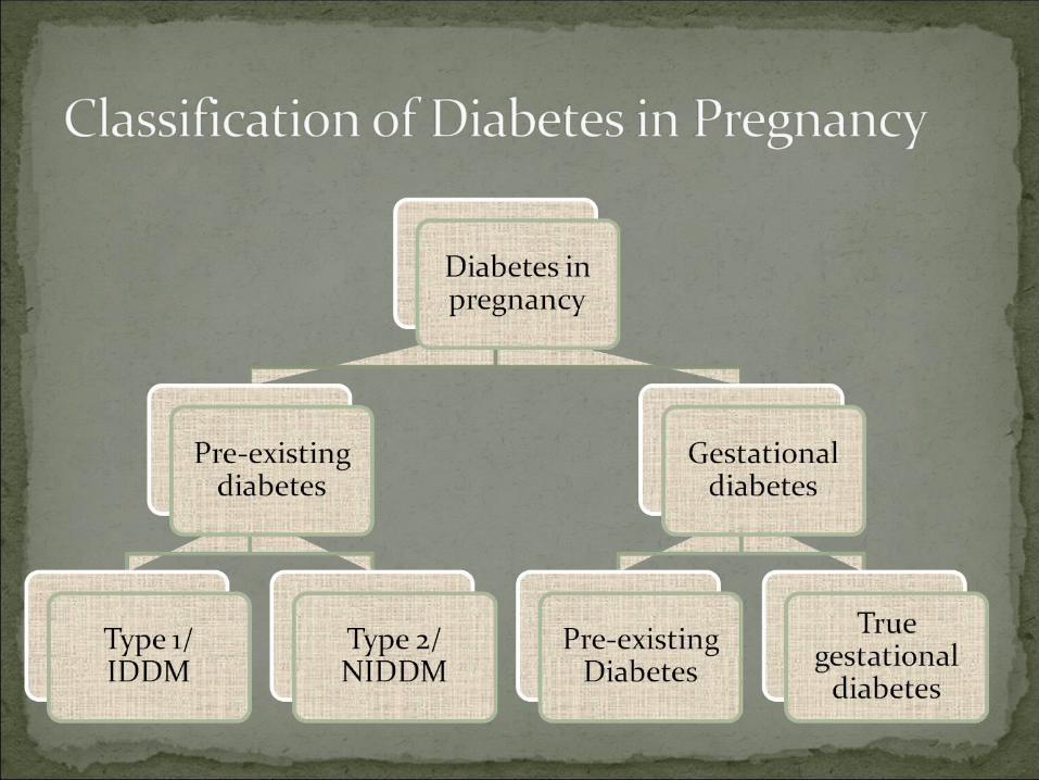
Type IV- Gestational Diabetes

It is defined as carbohydrate intolerance with onset or recognition during pregnancy.
The prevalence of gestational diabetes mellitus (GDM) in India varies from 3.8 to 21% in different parts of the country, depending on the geographical locations and diagnostic methods used.
GDM has been found to be more prevalent in urban areas than in rural areas.
• Women diagnosed to have GDM are at an increased risk of future diabetes predominantly type 2 DM and increase in the foetal/neonatal morbidity, future development of obesity, and diabetes in the offspring.
• The most common presentation of gestational diabetes in mothers is antenatal glycosuria.
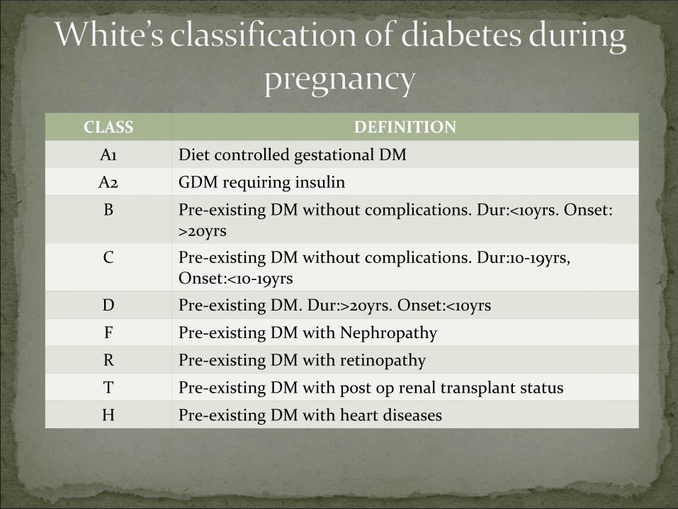
CLASS DEFINITION
A1 Diet controlled gestational DM
A2 GDM requiring insulin
B Pre-existing DM without complications. Dur:<10yrs. Onset: >20yrs
C Pre-existing DM without complications. Dur:10-19yrs, Onset:<10-19yrs
D Pre-existing DM. Dur:>20yrs. Onset:<10yrs
F Pre-existing DM with Nephropathy
R Pre-existing DM with retinopathy
T Pre-existing DM with post op renal transplant status
H Pre-existing DM with heart diseases
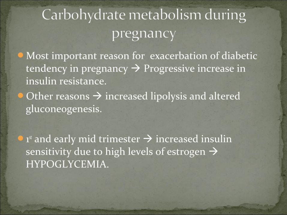
Most important reason for exacerbation of diabetic tendency in pregnancy Progressive increase in insulin resistance.
Other reasons increased lipolysis and altered gluconeogenesis.
1st and early mid trimester increased insulin sensitivity due to high levels of estrogen HYPOGLYCEMIA.

During 2nd trimester production of human placental lactogen, increase in cortisol, estriol and progesterone & due to increased destruction of insulin by kidney and placenta progressive insulin resistance and renal glycosuria HYPERGLYCEMIA
During 3rd trimester further insulin resistance decreased hypoglycemic effect of given dose of insulin HYPERGLYCEMIA ( reason for GDM to occur commonly after 26 wks of gestation).
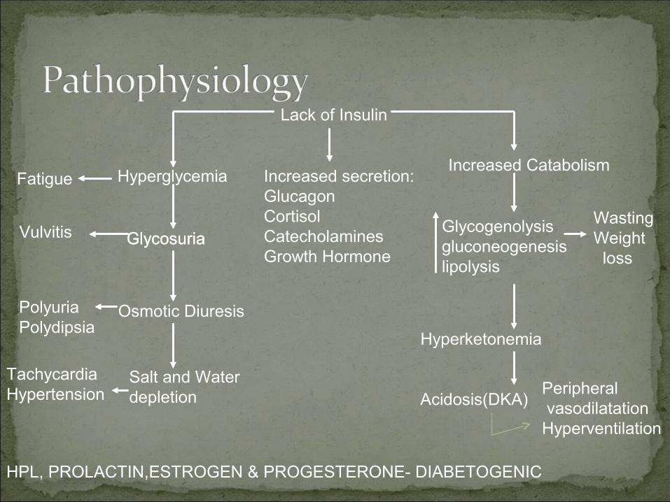
Lack of Insulin
Hyperglycemia
Glycosuria
Osmotic Diuresis
Salt and Water depletion
Increased secretion:GlucagonCortisolCatecholaminesGrowth Hormone
Increased Catabolism
Glycogenolysis gluconeogenesis lipolysis
Hyperketonemia
Acidosis(DKA)
Fatigue
Vulvitis
PolyuriaPolydipsia
TachycardiaHypertension
WastingWeight loss
Peripheral vasodilatationHyperventilation
Glycosuria
HPL, PROLACTIN,ESTROGEN & PROGESTERONE- DIABETOGENIC
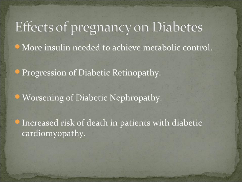
More insulin needed to achieve metabolic control.
Progression of Diabetic Retinopathy.
Worsening of Diabetic Nephropathy.
Increased risk of death in patients with diabetic cardiomyopathy.
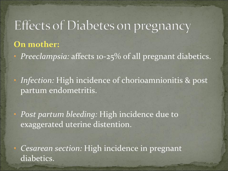
On mother:• Preeclampsia: affects 10-25% of all pregnant diabetics.
• Infection: High incidence of chorioamnionitis & post partum endometritis.
• Post partum bleeding: High incidence due to exaggerated uterine distention.
• Cesarean section: High incidence in pregnant diabetics.
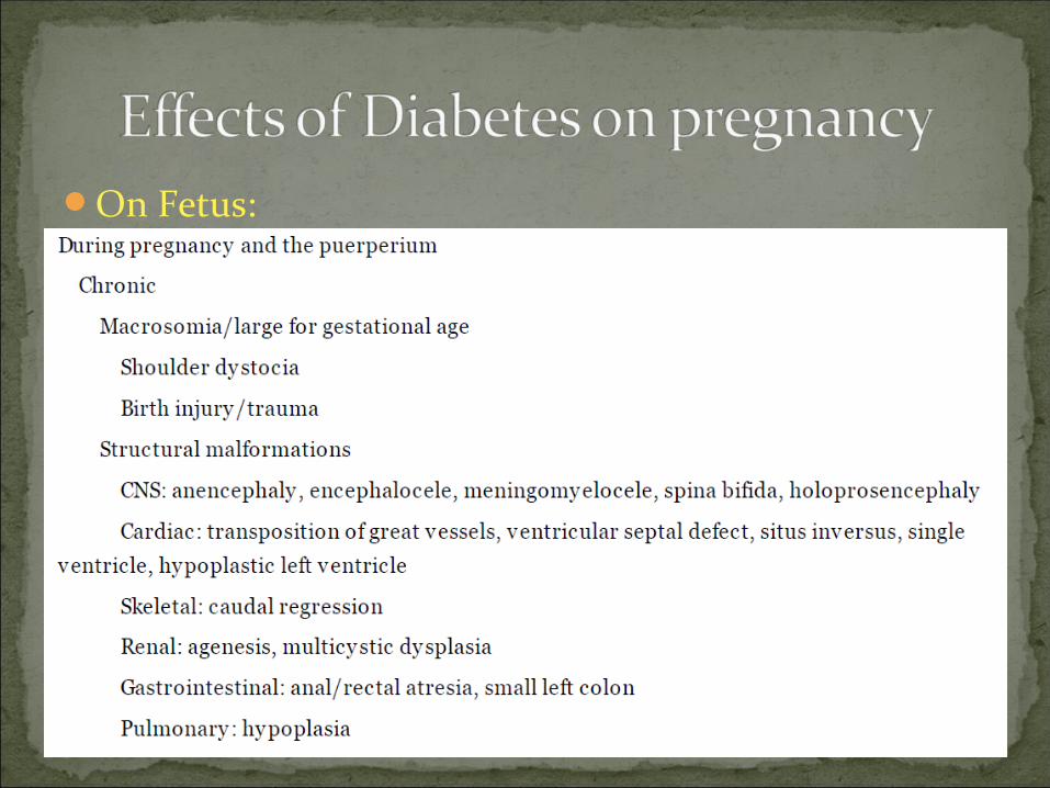
On Fetus:
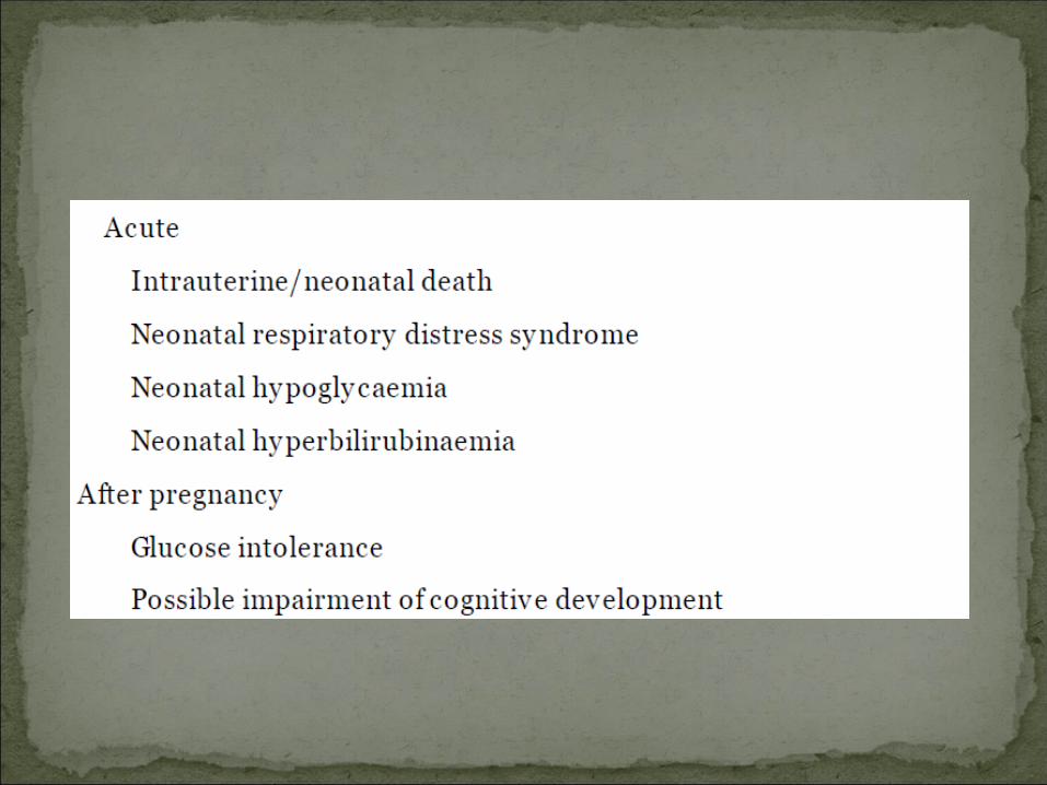
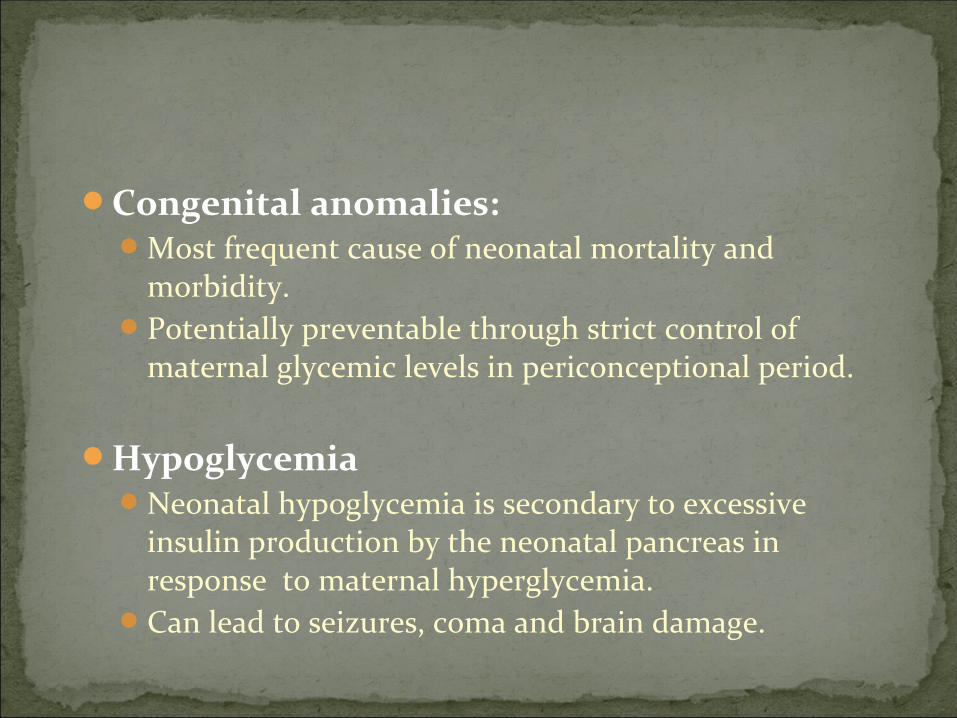
Congenital anomalies:Most frequent cause of neonatal mortality and
morbidity.Potentially preventable through strict control of
maternal glycemic levels in periconceptional period.
HypoglycemiaNeonatal hypoglycemia is secondary to excessive
insulin production by the neonatal pancreas in response to maternal hyperglycemia.
Can lead to seizures, coma and brain damage.

Postnatal hyperbilirubinemiaOccurs in appox. 25%, double that of normal.Due to immaturity of the neonate’s liver function.Phenobarbital administration antenatally may help
in preventing this condition.
Respiratory distress syndrome5-6 fold increased frequency.May be due to a delay in lung maturation or simply
due to the increased frequency of preterm deliveries
PolyhydramniosAmniotic fluid volume >2000 mLIncreased risk of placental abruption and preterm
labor.
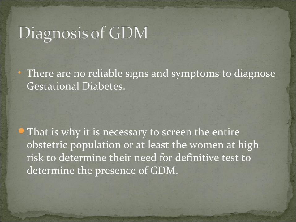
• There are no reliable signs and symptoms to diagnose Gestational Diabetes.
That is why it is necessary to screen the entire obstetric population or at least the women at high risk to determine their need for definitive test to determine the presence of GDM.

Low risk( Bl. Glucose screening not routinely required):Members of an ethnic group with a low prevalence of
GDM.No known 1st degree relatives with diabetes.Age < 25 yrs.Weight normal before pregnancy.Weight normal at birth.No history of abnormal glucose metabolism.

Average risk ( Bl. Glucose testing at 24 – 28 wks):Members of an ethnic group with high prevalence of
GDM.Diabetes in a 1st degree relative.Age >/= 25 yrs.Over weight before pregnancy.Weight high at birth.
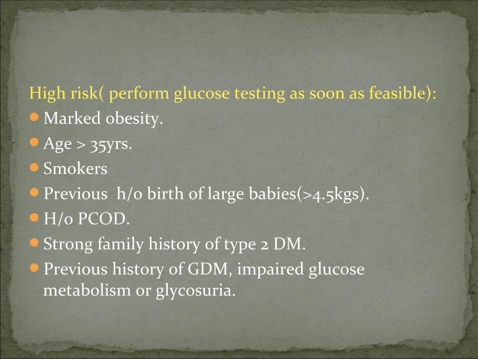
High risk( perform glucose testing as soon as feasible):Marked obesity.Age > 35yrs.SmokersPrevious h/o birth of large babies(>4.5kgs).H/o PCOD.Strong family history of type 2 DM.Previous history of GDM, impaired glucose
metabolism or glycosuria.
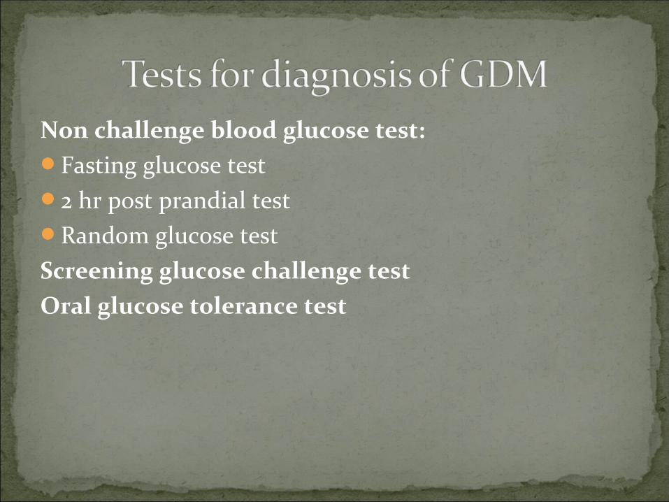
Non challenge blood glucose test:Fasting glucose test2 hr post prandial testRandom glucose testScreening glucose challenge testOral glucose tolerance test

Non challenge blood glucose test:
At the 1st antenatal Visit and on the subsequent day, if the fasting plasma glucose level is found to be > 126mg/dL or RBS > 200mg/dl, the diagnosis of GDM is confirmed.

Screening Glucose challenge test/ O’ Sullivan test:It is a simplified version of OGTT. Performed between 24-28wks.50g of oral glucose is given and blood sugar is
measured 1 hr later.If cut off point is set to 140 mg/dL, 80% of women with
GDM can be detected.If threshold is reduced to 130mg/dL, 90% of GDM
cases can be detected.No fasting is required for this test.

Oral Glucose Tolerance Test(OGTT):It requires over night fasting of 8-14 hrs.Usually 75g or 100 g of oral glucose is given. Blood glucose levels are measured at start of test and
then at set intervals of time.
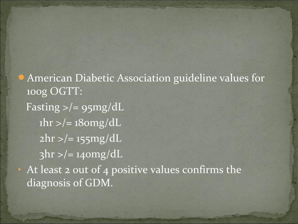
American Diabetic Association guideline values for 100g OGTT:
Fasting >/= 95mg/dL 1hr >/= 180mg/dL 2hr >/= 155mg/dL 3hr >/= 140mg/dL• At least 2 out of 4 positive values confirms the
diagnosis of GDM.
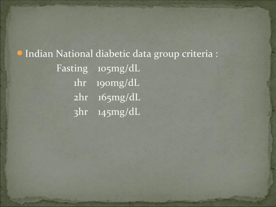
Indian National diabetic data group criteria : Fasting 105mg/dL 1hr 190mg/dL 2hr 165mg/dL 3hr 145mg/dL

1. Symptoms of diabetesPolyuriaPolydipsiaUnexpected weight loss
2. Fasting plasma glucose > 126 mg/dl3. 2 Hour plasma glucose >/= 200mg/dl after taking 75 gm
glucose load

Diet Provide adequate maternal &fetal nutrition Restricted fat & cholesterol Increase in dietary fiber 3 meal & 1-3 snacks , last snack being taken at bed time Desirable wt. gain 200-450 gm / week until full term Total wt gain 10-13 kg during normal & diabetic pregnancy is
recommended Calorie intake 25-35 kcal/kg
Monitoring Self monitoring of Bl.Sugar – 4 times/day Daily urine ketones testing when Bl.Sugar >200mg% or pt is unable to
eat.

ORAL AGENTSTransplacental passageTeratogenicProlonged hypoglycemiaSECOND GEN. SULFONYL UREA (glyburide) - Does
not cross placenta, achieve satisfactory glucose control

GLYBURIDE:Sulfonylurea. Primary mechanism of action is
stimulation of release of insulin from the storage granules of the pancreatic beta cells.
Also decreases insulin resistance.Non teratogenic, no noticed fetal effects and effective
control of maternal blood glucose levels are the reasons for its use in treating GDM.
Side effect: Hypoglycemia.Dose: 1.25 to 2.5 mg OD or BD. Daily max dose 20 mg.Peak plasma level: 2-4 hrs. Duration of action: 10-
12hrs
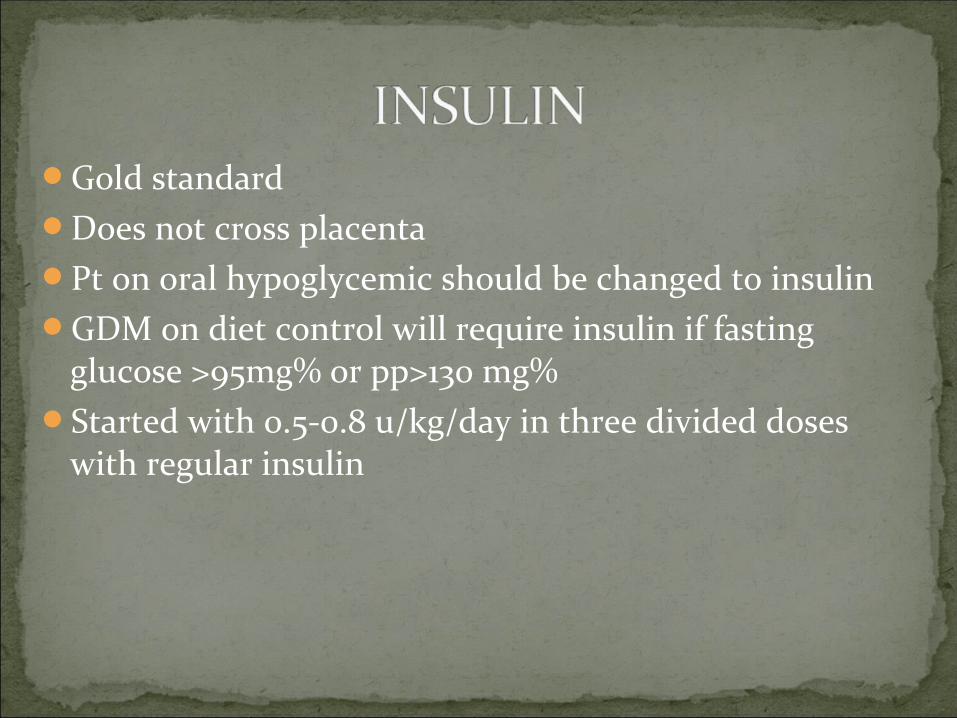
Gold standardDoes not cross placentaPt on oral hypoglycemic should be changed to insulinGDM on diet control will require insulin if fasting
glucose >95mg% or pp>130 mg%Started with 0.5-0.8 u/kg/day in three divided doses
with regular insulin
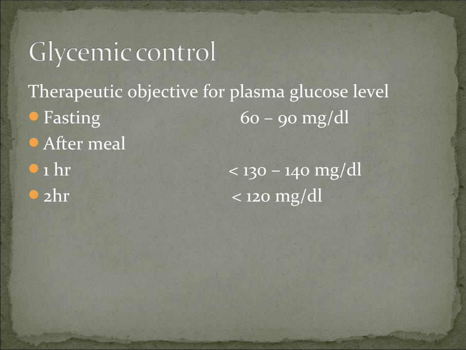
Therapeutic objective for plasma glucose levelFasting 60 – 90 mg/dlAfter meal 1 hr < 130 – 140 mg/dl2hr < 120 mg/dl

1. withhold a.m insulin2. start glucose infusion– 5% dextrose 125 ml/hr ( 6.25
g glucose/hr)Begin regular insulin infusion 0.5 U/hr(25 U Iin 250
ml NS)Monitor glucose every 1 – 2 hrAdjust insulin infusion
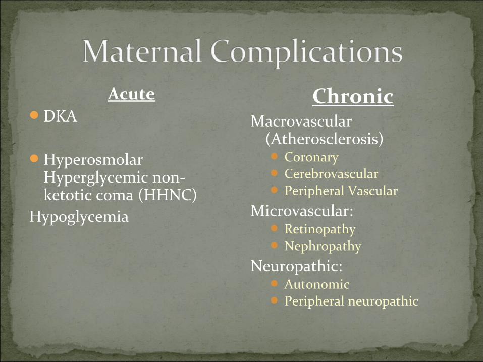
AcuteDKA
Hyperosmolar Hyperglycemic non-ketotic coma (HHNC)
Hypoglycemia
ChronicMacrovascular
(Atherosclerosis) Coronary Cerebrovascular Peripheral Vascular
Microvascular: Retinopathy Nephropathy
Neuropathic: Autonomic Peripheral neuropathic
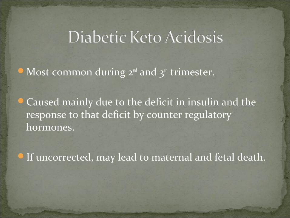
Most common during 2nd and 3rd trimester.
Caused mainly due to the deficit in insulin and the response to that deficit by counter regulatory hormones.
If uncorrected, may lead to maternal and fetal death.
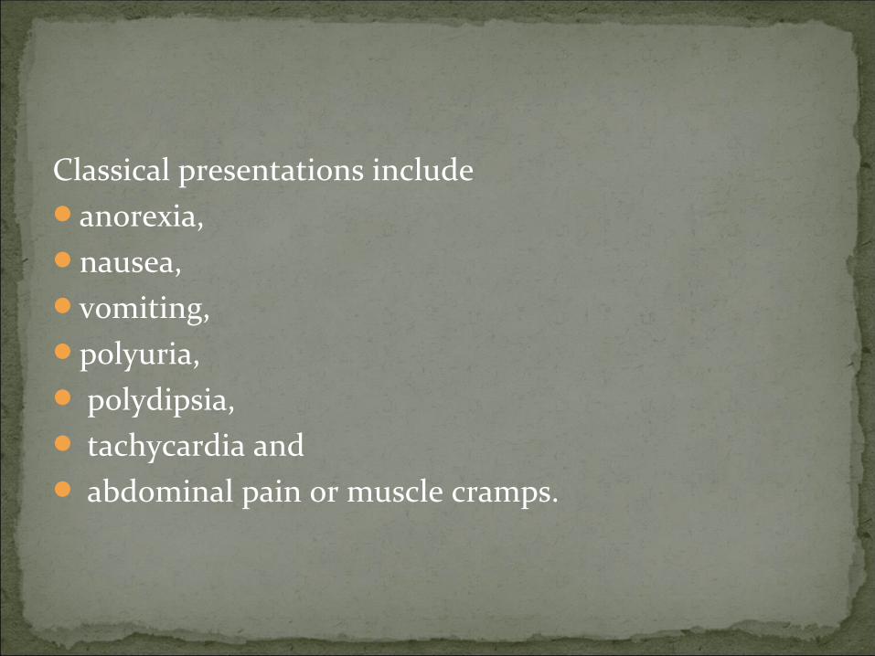
Classical presentations include anorexia, nausea, vomiting, polyuria, polydipsia, tachycardia and abdominal pain or muscle cramps.

If severe, the picture could include
Kussmaul hyperventilation, signs of volume depletion (e.g., hypotension and
oliguria), lethargy to coma, normal-to-cold body temperature and fruity odour noticeable in the patient’s breath.
Findings for diagnosing DKA:Bl. Glucose > 250mg/dL. DKA may develop in pregnancy with bl. Glucose
<250mg/dL and occasionally in the normal range ( EUGLYCEMIC KETOACIDOSIS).
Ketone bodies in urine and plasma.Arterial pH<7.3Sr. bicarbonate < 15mEq/L.Increased anion gap.
It is associated with the following factors: (1) bacterial infection; (2) omission of insulin doses in the presence of
gastroenteritis because of the parturient’s concern about the possibility of an insulin reaction due to anorexia, nausea, and vomiting;
(3) pump malfunction in patients receiving continuous subcutaneous insulin infusion therapy and
(4) tocolytic therapy with β- sympathomimetic agents, with or without concomitant glucocorticoid therapy
(5) decreased caloric intake,(6) poor medical management, (7) patient non-compliance.

i.v lineO2 by Face maskAsses level of glucose& electrolytesReplacement of fluid
Average fluid deficit 3-5 L 1-2 L in first hr- isotonic N saline followed by 250-500 ml/hr maintenance.
Insulin therapy The initial insulin dose is a continuous IV insulin infusion using an
infusion pump, at a rate of 0.1 U/kg/h. A mix of 24 units of regular insulin in 60 mL of isotonic sodium
chloride solution usually is infused at a rate of 15 mL/h (6 U/h) until the blood glucose level drops to less than 250 mg/dL; the rate of infusion then decreases to 5-7.5 mL/h (2-3 U/h) until the ketoacidotic state subsides.
Glucose administration– 5% dextrose(when plasma glucose reaches 250mg/dL, to prevent hypoglycemia).
K+ administration– after 3-4 hr of insulin therapy 10-20 mEq/hr.
Bicarbonate– if arterial ph <7.1 S. Hco3 <5 mEq/l
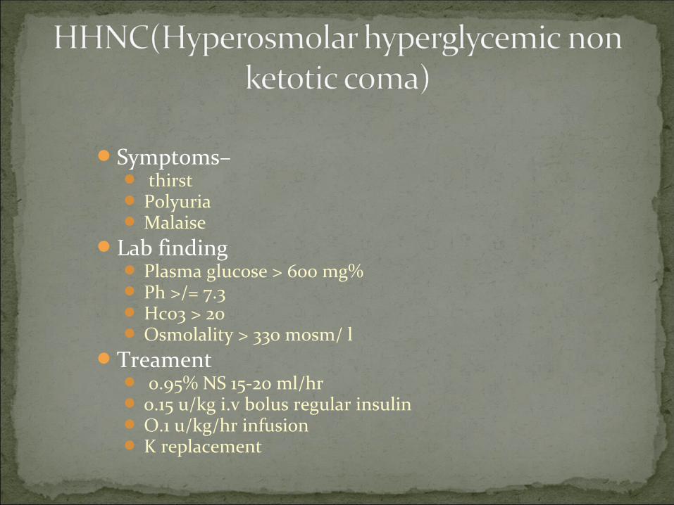
Symptoms– thirst Polyuria Malaise
Lab finding Plasma glucose > 600 mg% Ph >/= 7.3 Hco3 > 20 Osmolality > 330 mosm/ l
Treament 0.95% NS 15-20 ml/hr 0.15 u/kg i.v bolus regular insulin O.1 u/kg/hr infusion K replacement

Symptoms of rapidly decreasing blood glucoseSweating Increase heart rateTremorNervousnessIrritability WeaknessTingling
Symptoms of constantly low bl glucoseHeadacheVisual disturbancesMental dullnessConfusion Amnesiaseizure

Unconscious-100ml of 25% dextrose i.v.Or 0.5- 1 mg glucagon i.m/ s.c
ConsciousLiquid carbohydrate
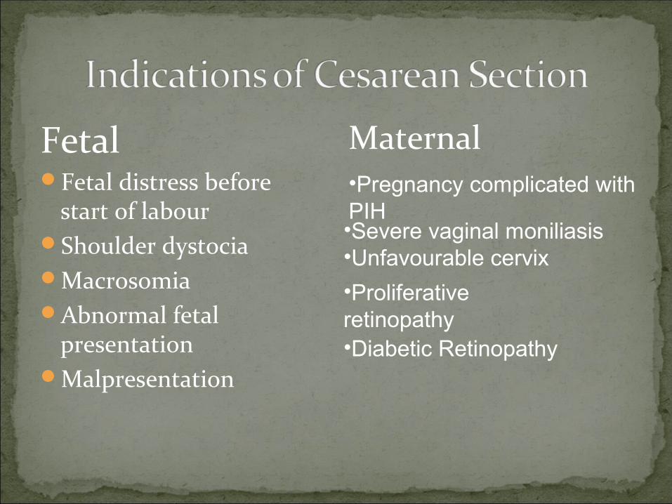
Fetal Fetal distress before
start of labourShoulder dystociaMacrosomiaAbnormal fetal
presentationMalpresentation
Maternal
•Diabetic Retinopathy
•Proliferative retinopathy
•Severe vaginal moniliasis•Unfavourable cervix
•Pregnancy complicated withPIH
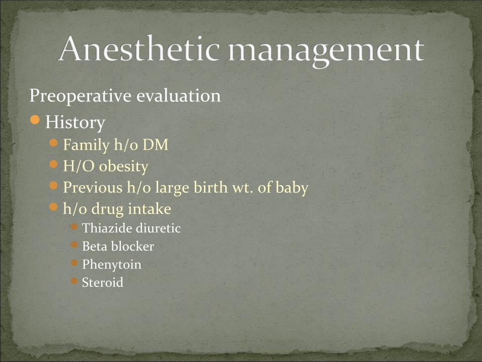
Preoperative evaluationHistory
Family h/o DMH/O obesityPrevious h/o large birth wt. of babyh/o drug intake
Thiazide diuretic Beta blockerPhenytoinSteroid
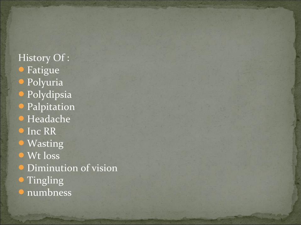
History Of :FatiguePolyuriaPolydipsiaPalpitationHeadacheInc RRWastingWt lossDiminution of visionTingling numbness

ExaminationPulse RateBlood PressureTemperatureJoint mobilityBed side test for autonomic function
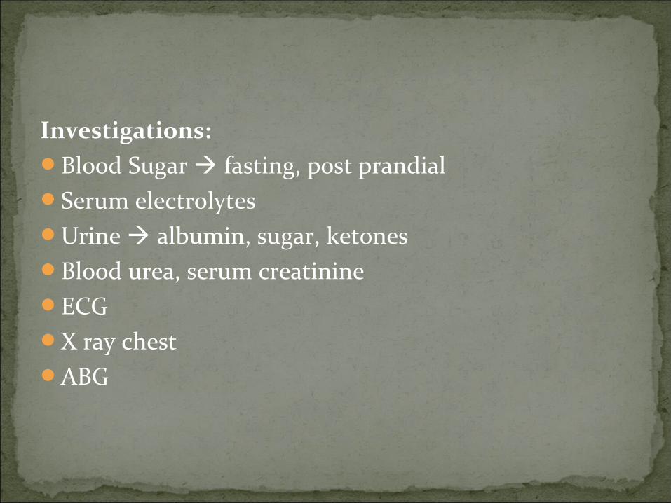
Investigations:Blood Sugar fasting, post prandialSerum electrolytesUrine albumin, sugar, ketonesBlood urea, serum creatinineECGX ray chestABG

Preop. AdviseNil Per Oral pt to be taken as first case in OTFasting Blood Sugar, Urine sugar ,ketones, Serum electrolytesAspiration prophylaxisPt is adviced to eat their usual evening meal and bed
time snack the evening prior to induction or cesarean delivery. Should also take their evening insulin dose.
Should omit their morning dose of insulin on the day of surgery.

To facilitate changes in insulin, electrolytes and fluid administration, it is ideal to have 2 I.V lines.
One to be used for the purpose of administering insulin and,
The other is connected to a 2 way system with one bag containing fluid and electrolytes and other containing fluid and electrolytes with 10% dextrose.
This allows a rapid response to manipulations in total fluids, insulin, electrolytes and dextrose administration.


Monitoring:Non Invasive Blood PressurePulse RateSPO2ECGUrine OutputBlood SugarTemperature- If autonomic dysfunction

Choice of AnaesthesiaDepends on status of mother and fetusRegional anaesthesia: If no evidence of peripheral
neuropathyEpidural labour analgesia:
Decreased Pain
Decreased plasma catecholamine level
Improved uteroplacental circulation
Minimises the need for GA in event of CS
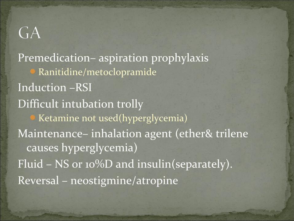
Premedication– aspiration prophylaxisRanitidine/metoclopramide
Induction –RSIDifficult intubation trolly
Ketamine not used(hyperglycemia)
Maintenance– inhalation agent (ether& trilene causes hyperglycemia)
Fluid – NS or 10%D and insulin(separately). Reversal – neostigmine/atropine

Conversion : 18 x mmol/L = mg/dL
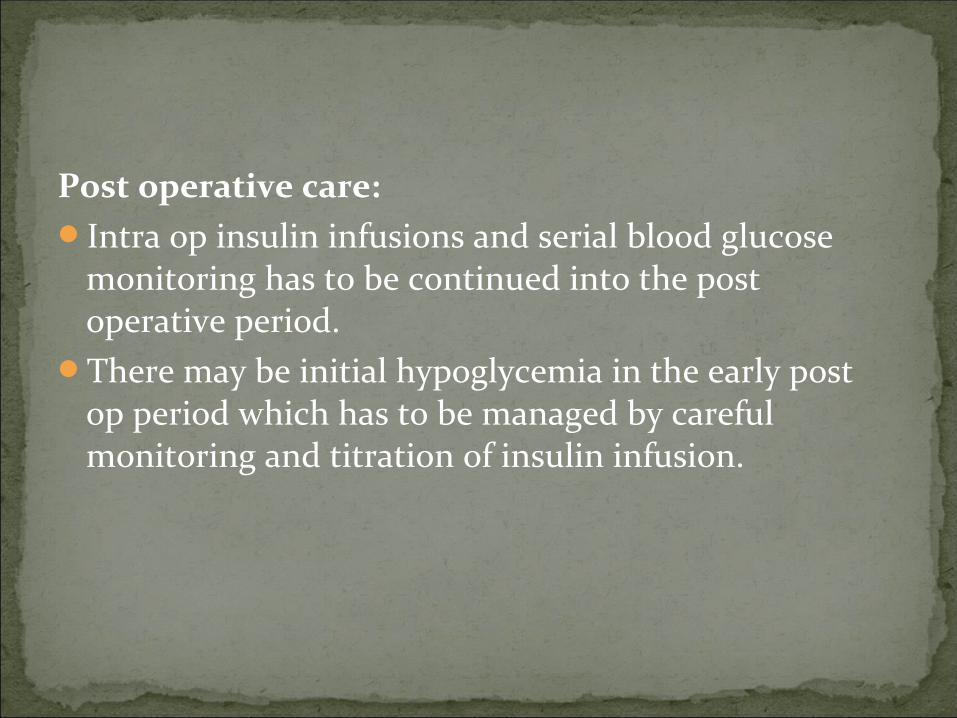
Post operative care:Intra op insulin infusions and serial blood glucose
monitoring has to be continued into the post operative period.
There may be initial hypoglycemia in the early post op period which has to be managed by careful monitoring and titration of insulin infusion.
References: Miller’s Anesthesia 7th edition.
Stoelting’s Anesthesia and co-existing disease 5th edition.
Practical guide to high risk pregnancy and delivery(Fernando Arias) 3rd edition.
Obstetric anesthesia(Dr. Sunanda Gupta) reprinted 1st edition.
Wylie and churchill Davidson principles and practice of anesthesia 5th edition.
www.en.wikipedia.org
www.ncbi.nlm.nih.gov
www.onlinelibrary.wiley.com
www.diabetes.co.uk
www.diabetes.org
www.diabetesindia.com
www.diabetesaustralia.com.au
www.diabetesupdate.blogspot.in
www.emedicine.medscape.com
www.apiindia.org