Person: Nanda, Sirish ([email protected]) Org: PHALLA Status ...

Gene Therapy for Gliomas
D. NANDA

D. Nanda – Gene Therapy for Malignant Gliomas Pagina 2 van 145 2
No part of this thesis may be reproduced or transmitted in any form by any means, electronic or mechanical, including photocopying, recording, or any information storage and retrieval system, without permission in writing from the publisher (Dharmin Nanda, Department of Neurology, Erasmus MC, P.O. Box 1738, 3000 DR, Rotterdam, The Netherlands)

D. Nanda – Gene Therapy for Malignant Gliomas Pagina 3 van 145 3
Gene Therapy for Gliomas
Gentherapie voor Gliomen
Proefschrift
ter verkrijging van de graad van doctor aan de Erasmus Universiteit Rotterdam
op gezag van de rector magnificus Prof.dr. S.W.J. Lamberts
en volgens het besluit van het College voor Promoties. De openbare verdediging zal plaatsvinden op woensdag 25 juni 2008 om 11.45 uur
door
Dharminderkoemar Nanda geboren te Suriname
D. Nanda – Gene Therapy for Malignant Gliomas Pagina 4 van 145 4
Promotiecommissie:
Promotor: Prof.dr. P.A.E. Sillevis Smitt Overige leden: Prof.dr. M.J. van den Bent
Prof.dr. R.C. Hoeben Prof.dr.ir. M. Hendriks-de Jong
The studies described in this thesis were performed at the Departments of Neurology and Nuclear Medicine from the Erasmus University Medical Center in Rotterdam, The Netherlands This research project was supported by the Revolving Fund from the Erasmus MC Rotterdam, The Netherlands and by KWF Kankerbestrijding, Amsterdam, The Netherlands.

D. Nanda – Gene Therapy for Malignant Gliomas Pagina 5 van 145 5
Aan: Shayra, Eshana & Eshita

D. Nanda – Gene Therapy for Malignant Gliomas Pagina 6 van 145 6

D. Nanda – Gene Therapy for Malignant Gliomas Pagina 7 van 145 7
Table of contents Chapter 1 Aim and scope of the thesis 9
Chapter 2 Gene Therapy for Gliomas
13
Chapter 3 Treatment of malignant gliomas with a replicating adenoviral vector expressing herpes simplex virus-thymidine kinase
27
Chapter 4 Imaging expression of adenoviral HSV1-tk suicide gene transfer using the nucleoside analogue FIRU
47
Chapter 5 [123I]FIRU, a tracer for the ‘molecular imaging’ of HSV1-tk suicide gene transfer
63
Chapter 6 Human adenovirus type 35 vector for gene therapy of brain cancer: improved transduction and bypass of pre-existing anti-vector immunity in cancer patients
79
Chapter 7 Clinical trials of adenoviral-mediated suicide gene therapy of malignant gliomas
99
Chapter 8 Summary and general discussion
119
Chapter 9 Samenvatting en discussie
131
Acknowledgements/ Dankwoord
143
List of publications Curriculum Vitae
144

D. Nanda – Gene Therapy for Malignant Gliomas Pagina 8 van 145 8

D. Nanda – Gene Therapy for Malignant Gliomas Pagina 9 van 145 9
CHAPTER 1 Aim and scope of the thesis

D. Nanda – Gene Therapy for Malignant Gliomas Pagina 10 van 145 10

D. Nanda – Gene Therapy for Malignant Gliomas Pagina 11 van 145 11
The overall median survival in glioblastoma multiforme (GBM) patients is less than one year and fewer than 5% of patients survive more than 5 years. The current standard of care for GBM patients involves neurosurgical resection of the tumor followed by radiotherapy with concomitant and adjuvant temozolomide chemotherapy. After initial treatment, all malignant gliomas eventually recur, mostly within a 2-3 cm margin of the original tumor on CT/MRI. The poor prognosis warrants research into new treatment modalities for malignant gliomas. Novel therapeutic approaches in the treatment of GBM include chemotherapy, targeted molecular agents, immunotherapy and virotherapy/gene therapy. Because malignant gliomas only rarely metastasize outside the skull, novel locoregional treatment modalities such as gene therapy are potentially interesting. The aim of this thesis was to identify bottlenecks limiting the efficacy of glioma gene therapy and address some of these bottlenecks in the laboratory. In chapter 2, a literature overview is presented of clinical trials in malignant gliomas and some of the hurdles identified in these trials are discussed. One of the most important issues is the limited distribution of viral particles compared to the size of malignant gliomas in patients. Various strategies to improve vector distribution and transduction efficiency are presented, including convection enhanced delivery, oncolytic virotherapy and genetic modification of adenoviral tropism. Finally, some of the molecular imaging tools are discussed that will allow the monitoring and quantification of transgene expression levels in patients. In chapter 3, we investigated the therapeutic potential of replication-competent adenoviral vectors which are a new and rapidly evolving platform for gene therapy. These viruses have a direct oncolytic effect (‘virotherapy’) and, following lysis of the target cells, can subsequently spread into the tumor. We studied the interaction of replication competent adenoviral vectors
and the HSV1-tk/GCV suicide in glioma cell lines and after intratumoral injection in a glioma xenograft model. Finally, the potential of a suicide gene as fail-safe in case of spread of the vector outside the tumor was evaluated. Non-invasive imaging and quantification of therapeutic transgene expression would provide important information on potential efficacy of glioma gene therapy in clinical trials. Because the HSV1-tk gene is the most frequently used therapeutic gene in clinical glioma gene therapy trials, we evaluated several nucleoside analogues, that are selectively phosphorylated by HSV1-tk, as tracers for monitoring HSV1-tk expression. In chapter 4, we described the biodistribution and imaging properties of 123I-FIRU in vitro and in vivo using constitutively HSV-tk expressing tumor cells. In chapter 5, we first described a method for radiolabeling and purification of FIRU in a clinical setting. Then we compared the in vitro toxicity of FIRU to the related compounds ganciclovir, FIAU and IVFRU in a number of cell lines. These studies demonstrated lower toxicity of FIRU as compared to FIAU in both constitutively HSV1-tk expressing and non-HSV1-tk expressing parental cell lines. The biodistribution and biokinetics of 123I-FIRU were further studied in HSV1-tk+ tumor bearing mice. Finally, the imaging properties of FIRU were examined using small animal SPECT. In chapter 6, we set out to identify an adenoviral vector better suited to infect primary glioma cells than Adenovirus serotype 5 (Ad5). To this end, we tested a library of fiber-chimeric Ad5-based adenoviral vectors on 12 fresh primary human glioma cell suspensions. We found significantly improved marker gene expression with several chimeric vectors, predominantly vectors carrying fiber molecules derived from B-serogroup viruses (Ad11, Ad16, Ad35 and Ad50). To explain this remarkable finding, we examined the expression of several molecules involved in adenoviral binding and entry on the primary glioma cells, including CAR, CD46

D. Nanda – Gene Therapy for Malignant Gliomas Pagina 12 van 145 12
and � v� integrins. Finally, we examined the seroprevalence of Ad35 among Dutch cancer patients, including glioma patients. In Chapter 7, we describe the preliminary results of a prospective, open label, multicenter dose escalation study in malignant glioma. This phase I trial was designed to assess the maximum tolerated dose (MTD) of IG.Ad.MPLI.TK followed by ganciclovir treatment in patients with relapsed glioblastoma assessing clinical and laboratory parameters. Treatment consisted of 10 ml of IG.Ad.MLPI.TK vector suspension injected on day 0 after optimal tumor resection into the surgical margins of the resection cavity, evenly distributed at approximately 50 sites 0.5-1 cm deep into the tissue. The dose levels were 4.6 x 10⁸, 4.6 x 10⁹, 4.6 x 10¹⁰, 4.6 x 10¹¹ vector particles. On day 2 ganciclovir was started and given by i.v. infusion over 1 hour, 5 mg/kg bid for 14 days. All patients had recurrent high grade glioma and all had received prior radiotherapy. The toxicity and efficacy data are presented in the first eleven patients that were enrolled in this study. In Chapter 8 we provide a summary and discussion of the chapters.
D. Nanda – Gene Therapy for Malignant Gliomas Pagina 13 van 145 13
CHAPTER 2 Gene Therapy for Glioma Based on: D. Nanda, S. Verwijnen and P. Sillevis Smitt Educational Book, XVth annual congress of the European Society of Gene and Cell Therapy (ESGCT), October 27-30, 2007, Rotterdam, Netherlands

D. Nanda – Gene Therapy for Malignant Gliomas Pagina 14 van 145 14

D. Nanda – Gene Therapy for Malignant Gliomas Pagina 15 van 145 15
Gliomas Primary brain tumors represent a diverse group of neoplasms arising from different cells of the nervous system. Gliomas are the most frequent primary brain tumors and present histological features of glial cells including astrocytes, oligodendrocytes and ependymal cells. Malignant or high-grade gliomas are classified by the WHO as either grade III anaplastic astrocytoma, anaplastic oligodendroglioma or mixed anaplastic oligoastrocytoma or as grade IV glioblastoma multiforme (GBM). At least 80% of malignant gliomas are GBM. The annual incidence of malignant gliomas varies between 3 – 7 per 100,000 people per year. Despite this relatively low incidence the burden of disease is immense due to the high morbidity and mortality of malignant gliomas. The prognosis of anaplastic gliomas varies by subtype and is most favourable in patients with an anaplastic oligodendroglioma (median survival 4 years from diagnosis). The overall median survival in GBM patients on the other hand is less than one year and fewer than 5% of patient survive more than 5 years. The current standard of care for GBM patients involves neurosurgical resection of the tumor (as extensively as safely possible) followed by radiotherapy with concomitant and adjuvant temozolomide chemotherapy 1. After initial treatment, all malignant gliomas eventually recur. Interestingly, 80% of these tumors recur within a 2-3 cm margin of the original tumor on CT/MRI 2. Also, these tumors only rarely metastasize outside the skull. The poor prognosis warrants research into new treatment modalities for malignant gliomas. Novel therapeutic approaches in the treatment of GBM include chemotherapy, targeted molecular agents, immunotherapy and virotherapy/gene therapy. Clinical Gene Therapy Trials in Malignant Glioma Due to the locoregional nature of malignant gliomas and their poor prognosis, recurrent malignant gliomas proved an excellent model to study gene therapy approaches. In fact, approximately 7% of the clinical gene therapy trials that were conducted between 1989 – 2007 included GBM patients (Figure 1). Many different vector systems and transgenes have been used for the treatment of malignant gliomas in clinical trials world wide, as depicted in Figure 2. The most frequently used vectors included retroviral, adenoviral and herpes simplex viral vectors. Almost 40% of GBM gene therapy trials used the suicide gene Herpes Simplex virus thymidine kinase (HSV-tk). After several promising phase II clinical trials using retroviral vectors encoding HSV-tk, a randomized controlled phase III clinical gene therapy trial was conducted in 248 newly diagnosed GBM patients 3. Patients received either standard therapy (surgical resection and radiotherapy) or standard therapy plus adjuvant gene therapy during surgery. Unfortunately, progression-free median survival, median survival and 12-months survival rates did not differ significantly between groups. Several phase I/II clinical trials were performed using a non-replicating Adenovirus serotype 5 (Ad5) expressing the HSV-tk gene in combination with ganciclovir in recurrent malignant gliomas. These trials have clearly demonstrated the feasibility and safety of the approach 4-9. Twenty-seven of 74 patients (37%) lived one year or more after gene therapy, which is longer than historical controls. Recently, a randomized controlled trial reported the successful adjuvant treatment of malignant glioma patients with Ad5-tk (Cerepro®) 9. Ad5-tk treatment produced a significant increase in mean survival from 39 to 71 weeks (P< 0.01) 9. The validity of this trial’s conclusions is compromised by the inclusion of newly diagnosed and recurrent glioma patients of different grades in both treatment and control groups and the imbalances between grade 3 and 4 patients between these groups. Despite these considerations, Ark Therapeutics Ltd. embarked on a phase III trial of Cerepro® in 250 newly diagnosed GBM patients, the results of which have not yet been reported.

D. Nanda – Gene Therapy for Malignant Gliomas Pagina 16 van 145 16
A different gene therapy approach tested in clinical trials consisted of tumor suppressor gene reconstitution, by using a non-replicating adenoviral vector encoding the p53 gene (Adp53). Replacement of p53 in gliomas is a rational approach because of the high frequency of p53 pathway abnormalities, including p53 mutations, overexpression of murine double minute 2 (mdm-2; the primary negative regulator of p53), inactivation of p14ARF (an inhibitor of mdm-2) or interference with p53 post-translational modifications 10. A phase I/II study of intratumoral Adp53 administration in 15 patients with recurrent glioma showed excellent tolerance and MTD was not reached at a dose of 3 x 10¹² viral particles. Despite the fact that the exogenous p53 was detected within tumor cells, these infected cells resided, on average, only within 5 mm of the needle tract 10. Figure 1.
Figure 1. The world wide clinical gene therapy trials from 1989 to 2007 divided by disease and by cancer type. This figure is adapted from http://www.wiley.co.uk/genmed/clinical/ and was last updated in January 2007.
Figure 2.
Figure 2. Vectors and genes used in clinical trials for GBM patients. This figure is adapted from http://www.wiley.co.uk/genmed/clinical/ and was last updated in January 2007. Hurdles Limiting the Efficacy of Glioma Gene Therapy One major problem relates to the size of gliomas in humans (Figure 3). These tumors cannot be entirely covered by the small radius of transgene dissemination that can be accomplished

D. Nanda – Gene Therapy for Malignant Gliomas Pagina 17 van 145 17
by using nonreplicating vectors 10. In addition, the limited success of adenovirus based clinical studies, which is in contrast with results obtained in animal models of gliomas clearly indicates additional challenges, including poor expression of the Coxsackie Adenovirus Receptor (CAR) on primary glioma cells resulting in low transduction efficiency compared to established cell lines 11, 12. In addition, the viral vectors elicit a strong immune response, which limits the efficacy of gene transfer, even in the relatively immune privileged brain 13, 14. The high prevalence of neutralizing antibodies (NAb) against Ad5 may further adversely affect transduction efficiency. All these issues that limit the efficacy of viral vectors have been addressed in the laboratory and will be discussed below. Improving Spatial Distribution – Alternative Routes of Administration To improve tissue penetration of the virus, new delivery methods have been explored. Recently, convection enhanced delivery (CED) was developed as a means to improve delivery of macromolecules throughout the brain 15, 16. CED is based on continuous infusion of drugs via intracranial catheters, enabling convective distribution of high drug concentrations over large volumes of the target tissue 17. In experimental models, delivery of viral vectors to the normal brain by CED improved distribution 18-21. CED of labelled adenovirus particles (80-90 nm diameter) to normal rat striatum resulted in a distribution volume (Vd) of approximately 30 mm3, better than a similarly sized nanosphere particle 22. CED of the much smaller adeno-associated virus (AAV) particle (23 nm diameter), however, only reached a Vd of 13 mm3. These results indicate that convective distribution can be used to distribute therapeutic viral vectors in the normal brain 22. In addition to size, surface properties have a major impact on the convective distribution of viruses and virus-sized particles in the brain. In a subcutaneous U87 glioma xenograft model in nude mice, CED of an adenoviral vector harbouring the sst2 reporter gene resulted in a Vd of 26 mm3, comparable to Vd after single injection (25 mm3). However, the maximum Vd was obtained with multiple intratumoral injections (Vd = 57 mm3) 23. CED has been successfully applied in clinical glioma trials to administer large molecules, including immunotoxins 24-27. Co-infusion of 123I labelled human serum albumin resulted in a broad distribution. However, target anatomy and catheter positioning had a significant influence on infusate distribution even within non-contrast enhancing regions of the brain. Intratumoral infusions tended to be anisotropic with accumulation of infusate in necrotic areas followed by eccentric efflux toward the peritumoral region through only a part of the tumor 27. In conclusion, CED holds promise to expand virus (in particular adenovirus) distribution in glioma patients provided optimal positioning of the catheter in the peritumoral region or around the resection cavity. Intravascular delivery is the least invasive method to deliver drugs to solid tissues, including the brain. However, the blood-brain barrier (BBB) does not allow passage of large and virus-sized molecules to the brain following intravascular administration. Following disruption with both mannitol and bradykinin, several groups have been able to transduce brain tumor cells growing orthotopically in rats with both replication defective adenoviruses and attenuated herpes viruses 28, 29. Gene transfer with the adenoviruses appeared less effective than with the herpes viruses, but it is not clear whether this is the result of herpes virus replication 28. Hower, intravascular virus delivery in combination with opening the BBB with these drugs may also increase the gene transfer to other areas of the brain, increasing the risk of toxicity. A more viable approach may be the targeting of tumor endothelial cells with viruses encoding anti-angiogenic molecules. An exciting new option is to use the homing properties of neural or mesenchymal stem cells to deliver gene therapy vectors to malignant gliomas 30, 31. Although the use of stem cells for this application remains confined to the basic science laboratory, though several groups are

D. Nanda – Gene Therapy for Malignant Gliomas Pagina 18 van 145 18
committed to translate this technology into clinical trials for patients with central nervous system neoplasms 32. The striking difference between the success of various gene therapy strategies in animal models versus clinical studies may be partly explained by the enormous difference in size of the tumor. To better address viral delivery and distribution, some authors have therefore proposed the use of larger animal models such as the dog 33, 34.
Figure 3. Vector distribution must be improved in glioma gene therapy. (A, B) MRI of a typical GBM tumor diagnosed in a 35-year old man. (A) On T1-weighted images after gadolinium administration, we see an enhancing mass with a diameter of 4.5 cm, corresponding to a tumor volume of approximately 43 ml. However, on T2-weighted images, the diameter is more than 10 cm, corresponding to a volume of over 600 ml. The treatment target, e.g. for radiotherapy, is generally the T2-weighted abnormality with a 2-3 cm margin, corresponding to a target volume of >1000 ml. The typical volume of an orthotopic mouse glioma in preclinical studies is maximally 100 � l, a 4 log difference. (C, D) Intratumoral injection of Ad5.tk.sstr by multiple injections resulted in a maximum volume of distribution of 57 � l, corresponding to a 4-5 mm penetration from the needle tract 23.
B
C

D. Nanda – Gene Therapy for Malignant Gliomas Pagina 19 van 145 19
Improving Spatial Distribution – Oncolytic Virotherapy Another approach to improve tissue penetration and oncolytic efficacy is the development of conditionally replicative viral vectors (virotherapy). The first replicating viruses employed in glioma clinical trial were two herpes virus vectors. Rampling et al. evaluated the toxicity of low doses of up to 105 pfu of an attenuated HSV named 1716 which has disruptions of the two copies of the RL1 gene 35. The other study, by Markert et al. tested a herpes virus (G207) with mutations in the RL1 gene and a lacZ insertion disabling the UL39 gene 36. Following injection of higher doses, up to 3x10⁹ pfu at five sites, no toxicity or serious adverse events could unequivocally be ascribed to G207. No patient developed HSV encephalitis. Radiographic and neuropathologic evidence suggestive of anti-tumor activity and long-term presence of viral DNA was found in some cases. The results of a phase I glioma trial with the E1B attenuated conditionally replicative adenovirus ONYX-015 were recently reported 37. None of the 24 patients experienced serious adverse events related to ONYX-015 and the MTD was not reached at 1010 pfu. The median time to progression was 46 days and the median survival time was 6.2 months. A direct side-by-side comparison of the anti-glioma activity of the oncolytic herpes simplex virus vector G47Delta with that of a conditionally replicative adenoviral vector for the treatment of glioblastoma demonstrated higher oncolytic efficacy and packaging capacity of the herpes virus vector compared to adenovirus 38. Reovirus replicates selectively in cells with an activated Ras signalling pathway, including glioma cells 39. In a Phase I trial of reovirus in malignant glioma patients, the MTD was not reached at 109 TICD50; however, there was no sign of antitumor activity either 40. CED of reovirus is currently undergoing clinical testing in recurrent glioma 41. Another virotherapy approach that is currently being tested in clinical trial in the treatment of recurrent GBM is the administration of an oncolytic measles virus (MV) vaccine strain, which is engineered to produce carcinoembryonic antigen (CEA) when the virus replicates in tumor cells 41, 42. CEA levels measured in the blood can subsequently be used as biomarkers to monitor viral gene expression and dose optimization. Another virus that is soon to be tested in the clinic is the intergenic poliovirus recombinant 41, 43. Many conditionally replicative adenoviral vectors (CRAd’s) have been developed for virotherapy of gliomas and other tumors. Two strategies have been used to restrict virus replication to tumor cells and spare normal cells; a) mutation-type CRAds; and b) promoter-controlled-type CRAds. Mutation-type CRAds have for instance deletions in the retinoblastoma gene (Rb)-binding region of E1A (e.g. the delta24 adenovirus 44) or in the p53 binding and inactivation region of E1B (e.g. ONYX-015) 45 in order to increase the tumor specificity of viral replication. A disadvantage of mutation-type CRADs is the attenuation of viral replication compared to wildtype 44, 46, 47. In addition, the stringency of replication control in these CRAds is still under discussion 48, 49. Another approach is the use of tissue or tumor specific promoters (TTSP) to drive viral genes critical for replication, such as E1A, E1B or E4. Many well-characterized TTSP have been investigated in CRAds for cancer gene therapy 46 50. Several CRAds controlled by TTSP have been examined in malignant glioma cells lines. These include the GFAP 51, the midkine (MK) 52, the cyclooxygenase-2 (COX-2) 53 and the hypoxia / hypoxia inducible factor (HIF) responsive promoters 54. A future development is the design and testing of adenoviral vectors for cancer virotherapy that replicate more efficiently on cancer cells than wildtype. Improving Transduction Efficiency by Adenoviral Vectors The limited clinical success of adenovirus-based delivery, which is in contrast with results obtained in many animal models of gliomas underlines additional challenges related to the

D. Nanda – Gene Therapy for Malignant Gliomas Pagina 20 van 145 20
low expression level of CAR in primary glioma cells and tumors as opposed to established cell lines 11, 12, 55. Because CAR expression apparently is a rate limiting factor for the infectivity with Ad5 50, modifications in adenoviruses are required to improve the infection efficiency. Currently, it is possible to use targeted viral vectors to direct gene transfer to specific receptors. The use of targeted adenoviruses is likely to increase safety and efficacy, reduce toxicity and may even permit systemic administration of these vectors 47. One of the first successful genetic modifications of adenoviral tropism was the insertion of the RGD motif into the hexon protein, HI loop or C-terminus of the viral fiber protein 56. This strategy improved adenoviral entry into cells, independent of CAR expression, but it did not enhance cancer specificity. Further attempts are being made to target adenovirus to receptors which are highly expressed on gliomas like epidermal growth factor receptor (EGFR), fibroblast growth factor receptor (FGFR) or urokinase-type plasminogen activator receptor (uPAR) 57, 58. Another strategy is the generation of chimeric adenoviral vectors by removing the fiber of Ad5 followed by the insertion of a PCR amplified DNA encoding for a fiber derived from an alternative serotype 59, 60. At present, 51 human Ad serotypes have been identified that are grouped into six species: A, B (subdivided in B1 and B2), C, D, E and F 61,
62. The association between serotype and clinical syndrome 63 suggests that diverse organs are targets for different serotypes. Although many adenoviral serotypes infect cells through CAR, other receptors have been described. Recently, CD46 was found to be the common receptor for subgroup B adenoviruses 64, 65. Since CD46 expression is very low on neurons 66 and because B-group adenoviral vectors infect glioma cells more efficiently than Ad5 11, 67, several chimeric adenoviral vectors, carrying B-serogroup fibers have been examined for the treatment of brain tumors. Ulasov et al.68 infected glioma cells with a chimeric Ad5/3 adenovirus, possessing the receptor binding fiber knob domain of Ad3 in the Ad5 capsid retargeting the virus to CD46, and showed increased transduction efficiency compared to Ad5 in glioma cells. Similarly, Brouwer et al. found increased transduction efficiency of Ad5-based chimeric vectors carrying B-serogroup fibers, following infection of primary glioma cells 11. Hoffmann et al. found improved GBM treatment with Ad5/35 fiber chimeric conditionally replicating adenoviruses 69. Ulasov et al. found potent anti-glioma activity when combining survivin-driven E1A expression with a pk7 fiber modification which selectively binds heparin sulfate proteoglycans, which are overespressed in glioma 70. The tropism enhanced oncolytic adenovirus Delta24-RGD-4C combines anchoring directly to integrins with E1A attenuation resulting in tumor selective replication 44. This virus will soon be tested in clinical trial in ovarian cancer and malignant gliomas. Molecular Imaging of Gene Transfer in GBM Non-invasive imaging of gene expression will give the possibility to quantify transgene expression, which may predict treatment outcome, as well as gain insight into vector distribution and, extent and duration of gene expression. At this moment, there is an inability to non-invasively measure transduction levels or functional enzyme activity in order to correlate this with clinical changes after ganciclovir (GCV) treatment. Jacobs et al. published a study in which five patients enrolled in a gene therapy procedure using a liposomal vector carrying an HSV-tk gene 71. They performed a dynamic PET scan using 124I-FIAU, a radioactive nucleoside analog, which is trapped in the same manner by HSV-tk as GCV. They showed a specific HSV-tk-related uptake of FIAU at the site of injection in one patient, who also showed a response to treatment. Unfortunately, in the other patients no increased FIAU uptake could be measured; they also failed to respond to treatment. This study shows that non-invasive imaging of HSV-tk gene expression is feasible and highly desirable in order to

D. Nanda – Gene Therapy for Malignant Gliomas Pagina 21 van 145 21
assess gene transfer. Dempsey et al. reported on eight GBM patients imaged with SPECT using 123I-FIAU prior to and after application of an oncolytic HSV virus 72. Unfortunately, no increased uptake of FIAU was determined after viral infection in these patients. A possible explanation for this is that FIAU might not be the ideal tracer for monitoring HSV-tk expression in subjects with intact BBB, as FIAU does not penetrate it 72, 73. Another approach is direct labeling of the viral particles which allows immediate visualization of vector distribution, regardless of transgene expression 23, 74. Conclusions The results of the initial clinical gene therapy trials in malignant gliomas have been disappointing. However, many of the reasons contributing to these failures have been identified and addressed in the laboratory. By lack of truly predictive animal models, new clinical trials are required to assess many of newly developed vectors for gene therapy / virotherapy. One of the most important issues is the limited distribution of viral particles compared to the size of malignant gliomas in patients. Hopefully, a combination of convection enhanced delivery and oncolytic virotherapy may lower this hurdle. Clearly, molecular imaging tools will provide important information for the evaluation of gene therapy in clinical glioma trials. References 1. Stupp R, Hegi ME, van den Bent MJ, et al. Changing paradigms--an update on the
multidisciplinary management of malignant glioma. Oncologist 2006; 11:165-80. 2. Wallner KE, Galicich JH, Krol G, Arbit E, Malkin MG. Patterns of failure following
treatment for glioblastoma multiforme and anaplastic astrocytoma. Int J Radiat Oncol Biol Phys 1989; 16:1405-9.
3. Rainov NG. A phase III clinical evaluation of herpes simplex virus type 1 thymidine kinase and ganciclovir gene therapy as an adjuvant to surgical resection and radiation in adults with previously untreated glioblastoma multiforme. Hum Gene Ther 2000; 11:2389-401.
4. Judy KD, Eck SL. The use of suicide gene therapy for the treatment of malignancies of the brain. In: Lattime EC, Stanton LG, eds. Gene Therapy of Cancer. San Diego: Academic Press, 2002.
5. Germano IM, Fable J, Gultekin SH, Silvers A. Adenovirus/herpes simplex-thymidine kinase/ganciclovir complex: preliminary results of a phase I trial in patients with recurrent malignant gliomas. J Neurooncol 2003; 65:279-89.
6. Sillevis Smitt P, Driesse MJ, Wolbers J, Kros JM, Bout A, Avezaat C. Treatment of relapsed malignant glioma with an adenoviral vector containing the herpes simplex thymidine kinase gene followed by ganciclovir. Molecular Therapy 2003; 7:851-8.
7. Trask TW, Trask RP, Aguilar-Cordova E, et al. Phase I study of adenoviral delivery of the HSV-tk gene and ganciclovir administration in patients with current malignant brain tumors. Mol Ther 2000; 1:195-203.
8. Sandmair AM, Loimas S, Puranen P, et al. Thymidine kinase gene therapy for human malignant glioma, using replication-deficient retroviruses or adenoviruses. Hum Gene Ther 2000; 11:2197-205.
9. Immonen A, Vapalahti M, Tyynela K, et al. AdvHSV-tk gene therapy with intravenous ganciclovir improves survival in human malignant glioma: a randomised, controlled study. Mol Ther 2004; 10:967-72.

D. Nanda – Gene Therapy for Malignant Gliomas Pagina 22 van 145 22
10. Lang FF, Bruner JM, Fuller GN, et al. Phase I trial of adenovirus-mediated p53 gene therapy for recurrent glioma: biological and clinical results. J Clin Oncol 2003; 21:2508-18.
11. Brouwer E, Havenga MJ, Ophorst O, et al. Human adenovirus type 35 vector for gene therapy of brain cancer: improved transduction and bypass of pre-existing anti-vector immunity in cancer patients. Cancer Gene Ther 2007; 14:211-9.
12. Miller CR, Buchsbaum DJ, Reynolds PN, et al. Differential susceptibility of primary and established human glioma cells to adenovirus infection: targeting via the epidermal growth factor receptor achieves fiber receptor-independent gene transfer. Cancer Res 1998; 58:5738-48.
13. Dewey RA, Morrissey G, Cowsill CM, et al. Chronic brain inflammation and persistent herpes simplex virus 1 thymidine kinase expression in survivors of syngeneic glioma treated by adenovirus-mediated gene therapy: implications for clinical trials. Nat Med 1999; 5:1256 - 1263.
14. Driesse MJ, Vincent AJPE, Sillevis Smitt PAE, et al. Intracerebral injection of adenovirus harboring the HSVtk gene combined with ganciclovir administration: toxicity study in nonhuman primates. Gene Therapy 1998; 5:1122-1130.
15. Bobo RH, Laske DW, Akbasak A, Morrison PF, Dedrick RL, Oldfield EH. Convection-enhanced delivery of macromolecules in the brain. Proc Natl Acad Sci USA 1994; 91:2076-2080.
16. Lieberman DM, Laske DW, Morrison PF, Bankiewicz KS, Oldfield EH. Convection-enhanced distribution of large molecules in gray matter during intersitial drug infusion. J Neurosurg 1995; 82:1021-1029.
17. Mardor Y, Rahav O, Zauberman Y, et al. Convection-enhanced drug delivery: increased efficacy and magnetic resonance image monitoring. Cancer Res 2005; 65:6858-63.
18. Bankiewicz KS, Eberling JL, Kohutnicka M, et al. Convection-enhanced delivery of AAV vector in parkinsonian monkeys; in vivo detection of gene expression and restoration of dopaminergic function using pro-drug approach. Exp Neurol 2000; 164:2-14.
19. Betz AL, Shakui P, Davidson BL. Gene transfer to rodent brain with recombinant adenoviral vectors: effects of infusion parameters, infectious titer, and virus concentration on transduction volume. Exp Neurol 1998; 150:136-42.
20. Nguyen JB, Sanchez-Pernaute R, Cunningham J, Bankiewicz KS. Convection-enhanced delivery of AAV-2 combined with heparin increases TK gene transfer in the rat brain. Neuroreport 2001; 12:1961-4.
21. Sanftner LM, Sommer JM, Suzuki BM, et al. AAV2-mediated gene delivery to monkey putamen: evaluation of an infusion device and delivery parameters. Exp Neurol 2005; 194:476-83.
22. Chen MY, Hoffer A, Morrison PF, et al. Surface properties, more than size, limiting convective distribution of virus-sized particles and viruses in the central nervous system. J Neurosurg 2005; 103:311-9.
23. ter Horst M, Verwijnen SM, Brouwer E, et al. Locoregional delivery of adenoviral vectors. J Nucl Med 2006; 47:1483-9.
24. Laske DW, Youle RJ, Oldfield EH. Tumor regression with regional distribution of the targeted toxin TF-CRM107 in patients with malignant brain tumors. Nat Med 1997; 3:1362-8.
25. Sampson JH, Raghavan R, Brady ML, et al. Clinical utility of a patient-specific algorithm for simulating intracerebral drug infusions. Neuro Oncol 2007; 9:343-53.

D. Nanda – Gene Therapy for Malignant Gliomas Pagina 23 van 145 23
26. Sampson JH, Raghavan R, Provenzale JM, et al. Induction of hyperintense signal on T2-weighted MR images correlates with infusion distribution from intracerebral convection-enhanced delivery of a tumor-targeted cytotoxin. AJR Am J Roentgenol 2007; 188:703-9.
27. Sampson JH, Brady ML, Petry NA, et al. Intracerebral infusate distribution by convection-enhanced delivery in humans with malignant gliomas: descriptive effects of target anatomy and catheter positioning. Neurosurgery 2007; 60:ONS89-98; discussion ONS98-9.
28. Ikeda K, Wakimoto H, Ichikawa T, et al. Complement depletion facilitates the infection of multiple brain tumors by an intravascular, replication-conditional herpes simplex virus mutant. J Virol 2000; 74:4765-75.
29. Nilaver G, Muldoon LL, Kroll RA, et al. Delivery of herpesvirus and adenovirus to nude rat intracerebral tumors after osmotic blood-brain barrier disruption. Proc Natl Acad Sci U S A 1995; 92:9829-33.
30. Aboody KS, Brown A, Rainov NG, et al. From the cover: neural stem cells display extensive tropism for pathology in adult brain: evidence from intracranial gliomas. Proc Natl Acad Sci U S A 2000; 97:12846-51.
31. Nakamizo A, Marini F, Amano T, et al. Human bone marrow-derived mesenchymal stem cells in the treatment of gliomas. Cancer Res 2005; 65:3307-18.
32. Badruddoja MA, Black KL. Improving the delivery of therapeutic agents to CNS neoplasms: a clinical review. Front Biosci 2006; 11:1466-78.
33. Candolfi M, Curtin JF, Nichols WS, et al. Intracranial glioblastoma models in preclinical neuro-oncology: neuropathological characterization and tumor progression. J Neurooncol 2007.
34. Candolfi M, Pluhar GE, Kroeger K, et al. Optimization of adenoviral vector-mediated transgene expression in the canine brain in vivo, and in canine glioma cells in vitro. Neuro Oncol 2007; 9:245-58.
35. Rampling R, Cruickshank G, Papanastassiou V, et al. Toxicity evaluation of replication-competent herpes simplex virus (ICP 34.5 null mutant 1716) in patients with recurrent malignant glioma. Gene Ther 2000; 7:859-66.
36. Markert JM, Medlock MD, Rabkin SD, et al. Conditionally replicating herpes simplex virus mutant, G207 for the treatment of malignant glioma: results of a phase I trial. Gene Ther 2000; 7:867-74.
37. Chiocca EA, Abbed KM, Tatter S, et al. A phase I open-label, dose-escalation, multi-institutional trial of injection with an E1B-Attenuated adenovirus, ONYX-015, into the peritumoral region of recurrent malignant gliomas, in the adjuvant setting. Mol Ther 2004; 10:958-66.
38. Hoffmann D, Wildner O. Comparison of herpes simplex virus- and conditionally replicative adenovirus-based vectors for glioblastoma treatment. Cancer Gene Ther 2007; 14:627-39.
39. Coffey MC, Strong JE, Forsyth PA, Lee PW. Reovirus therapy of tumors with activated Ras pathway. Science 1998; 282:1332-4.
40. Forsyth PA, Roldan G, George D. A Phase I trial of intratumoral (i.t.) administration of reovirus in patients with histologically confirmed recurrent malignant gliomas. J Clin Oncol 2006; 2006:S73 (Abstract 1563).
41. Simpson L, Galanis E. Recurrent glioblastoma multiforme: advances in treatment and promising drug candidates. Expert Rev Anticancer Ther 2006; 6:1593-607.
42. Phuong LK, Allen C, Peng KW, et al. Use of a vaccine strain of measles virus genetically engineered to produce carcinoembryonic antigen as a novel therapeutic agent against glioblastoma multiforme. Cancer Res 2003; 63:2462-9.

D. Nanda – Gene Therapy for Malignant Gliomas Pagina 24 van 145 24
43. Gromeier M, Lachmann S, Rosenfeld MR, Gutin PH, Wimmer E. Intergeneric poliovirus recombinants for the treatment of malignant glioma. Proc Natl Acad Sci U S A 2000; 97:6803-8.
44. Fueyo J, Alemany R, Gomez-Manzano C, et al. Preclinical characterization of the antiglioma activity of a tropism-enhanced adenovirus targeted to the retinoblastoma pathway. J Natl Cancer Inst 2003; 95:652-60.
45. Bischoff JR, Kirn DH, Williams A, et al. An adenovirus mutant that replicates selectively in p53-deficient human tumor cells. Science 1996; 274:373-377.
46. Kruyt FA, Curiel DT. Toward a new generation of conditionally replicating adenoviruses: pairing tumor selectivity with maximal oncolysis. Hum Gene Ther 2002; 13:485-95.
47. Jiang H, McCormick F, Lang FF, Gomez-Manzano C, Fueyo J. Oncolytic adenoviruses as antiglioma agents. Expert Rev Anticancer Ther 2006; 6:697-708.
48. Edwards SJ, Dix BR, Myers CJ, et al. Evidence that replication of the antitumor adenovirus ONYX-015 is not controlled by the p53 and p14(ARF) tumor suppressor genes. J Virol 2002; 76:12483-90.
49. Heise C, Hermiston T, Johnson L, et al. An adenovirus E1A mutant that demonstrates potent and selective systemic anti-tumoral efficacy. Nat Med 2000; 6:1134-9.
50. Rein DT, Breidenbach M, Curiel DT. Current developments in adenovirus-based cancer gene therapy. Future Oncol 2006; 2:137-43.
51. Ter Horst M, Rodijk M, Brouwer E, Hoeben R, De Leeuw B, Sillevis Smitt P. A GFAP-selective oncolytic adenovirus for locoregional treatment of malignant gliomas. Submitted 2005.
52. Kohno S, Nakagawa K, Hamada K, et al. Midkine promoter-based conditionally replicative adenovirus for malignant glioma therapy. Oncol Rep 2004; 12:73-8.
53. Ahmed A, Thompson J, Emiliusen L, et al. A conditionally replicating adenovirus targeted to tumor cells through activated RAS/P-MAPK-selective mRNA stabilization. Nat Biotechnol 2003; 21:771-7.
54. Post DE, Van Meir EG. A novel hypoxia-inducible factor (HIF) activated oncolytic adenovirus for cancer therapy. Oncogene 2003; 22:2065-72.
55. Fuxe J, Liu L, Malin S, Philipson L, Collins VP, Pettersson RF. Expression of the coxsackie and adenovirus receptor in human astrocytic tumors and xenografts. Int J Cancer 2003; 103:723-9.
56. Reynolds P, Dmitriev I, Curiel D. Insertion of an RGD motif into the HI loop of adenovirus fiber protein alters the distribution of transgene expression of the systemically administered vector. Gene Ther 1999; 6:1336-9.
57. van Beusechem VW, Mastenbroek DC, van den Doel PB, et al. Conditionally replicative adenovirus expressing a targeting adapter molecule exhibits enhanced oncolytic potency on CAR-deficient tumors. Gene Ther 2003; 10:1982-91.
58. Rancourt C, Rogers BE, Sosnowski BA, et al. Basic fibroblast growth factor enhancement of adenovirus-mediated delivery of the herpes simplex virus thymidine kinase gene results in augmented therapeutic benefit in a murine model of ovarian cancer. Clin Cancer Res 1998; 4:2455-61.
59. Havenga MJ, Lemckert AA, Ophorst OJ, et al. Exploiting the natural diversity in adenovirus tropism for therapy and prevention of disease. J Virol 2002; 76:4612-20.
60. Shayakhmetov DM, Lieber A. Dependence of adenovirus infectivity on length of the fiber shaft domain. J Virol 2000; 74:10274-86.
61. Wigand R, Mauss M, Adrian T. Chimpanzee adenoviruses are related to four subgenera of human adenoviruses. Intervirology 1989; 30:1-9.

D. Nanda – Gene Therapy for Malignant Gliomas Pagina 25 van 145 25
62. De Jong JC, Wermenbol AG, Verweij-Uijterwaal MW, et al. Adenoviruses from human immunodeficiency virus-infected individuals, including two strains that represent new candidate serotypes Ad50 and Ad51 of species B1 and D, respectively. J Clin Microbiol 1999; 37:3940-5.
63. Hierholzer JC. Adenoviruses in the immunocompromised host. Clin Microbiol Rev 1992; 5:262-74.
64. Gaggar A, Shayakhmetov DM, Lieber A. CD46 is a cellular receptor for group B adenoviruses. Nat Med 2003; 9:1408-12.
65. Segerman A, Atkinson JP, Marttila M, Dennerquist V, Wadell G, Arnberg N. Adenovirus type 11 uses CD46 as a cellular receptor. J Virol 2003; 77:9183-91.
66. Singhrao SK, Neal JW, Rushmere NK, Morgan BP, Gasque P. Spontaneous classical pathway activation and deficiency of membrane regulators render human neurons susceptible to complement lysis. Am J Pathol 2000; 157:905-18.
67. Skog J, Edlund K, Widegren B, Salford LG, Wadell G, Mei YF. Efficient internalization into low-passage glioma cell lines using adenoviruses other than type 5: an approach for improvement of gene delivery to brain tumours. J Gen Virol 2004; 85:2627-38.
68. Ulasov IV, Tyler MA, Zheng S, Han Y, Lesniak MS. CD46 Represents a Target for Adenoviral Gene Therapy of Malignant Glioma. Hum Gene Ther 2006.
69. Hoffmann D, Meyer B, Wildner O. Improved glioblastoma treatment with Ad5/35 fiber chimeric conditionally replicating adenoviruses. J Gene Med 2007; 9:764-78.
70. Ulasov IV, Zhu ZB, Tyler MA, et al. Survivin-driven and fiber-modified oncolytic adenovirus exhibits potent antitumor activity in established intracranial glioma. Hum Gene Ther 2007; 18:589-602.
71. Jacobs A, Voges J, Reszka R, et al. Positron-emission tomography of vector-mediated gene expression in gene therapy for gliomas. Lancet 2001; 358:727-9.
72. Dempsey MF, Wyper D, Owens J, et al. Assessment of 123I-FIAU imaging of herpes simplex viral gene expression in the treatment of glioma. Nucl Med Commun 2006; 27:611-7.
73. Jacobs A, Tjuvajev JG, Dubrovin M, et al. Positron emission tomography-based imaging of transgene expression mediated by replication-conditional, oncolytic herpes simplex virus type 1 mutant vectors in vivo. Cancer Res 2001; 61:2983-95.
74. Kristian Raty J, Liimatainen T, Unelma Kaikkonen M, Grohn O, Jumani Airenne K, Yla-Herttuala S. Non-invasive Imaging in Gene Therapy. Mol Ther 2007; 15:1579-86.

D. Nanda – Gene Therapy for Malignant Gliomas Pagina 26 van 145 26

D. Nanda – Gene Therapy for Malignant Gliomas Pagina 27 van 145 27
CHAPTER 3 Treatment of Malignant Gliomas with a Replicating Adenoviral Vector Expressing Herpes Simplex Virus-Thymidine Kinase D. Nanda, R. Vogels, M. Havenga, C. Avezaat, A. Bout and P. Sillevis Smitt CANCER RESEARCH 2001; 61: 8743-8750

D. Nanda – Gene Therapy for Malignant Gliomas Pagina 28 van 145 28

D. Nanda – Gene Therapy for Malignant Gliomas Pagina 29 van 145 29
ABSTRACT We evaluated the interaction between oncolytic, replication-competent adenoviral vectors and the herpes simplex virus-1 thymidine kinase (HSV1-tk) gene/ganciclovir (GCV) suicide system for the treatment of malignant gliomas. We constructed a panel of replication-competent adenoviral vectors in which the luciferase (IG.Ad5E1+. E3Luc) or HSV1-tk gene (IG.Ad5E1+.E3TK) replace the Mr 19,000 glycoprotein (gp19K) coding sequence in the E3 region. IG.Ad5E1. IG.Ad5.ClipLuc and IG.AdApt. TK are E1-deleted viruses that contain the luciferase or the HSV1-tk gene in the former E1 region driven by the human cytomegalovirus promoter. IG.Ad5.Sarcoma 1800HSA.E3Luc contains an irrelevant gene in the E1 region, whereas the gp19K coding sequence in the E3 region is replaced by the luciferase gene as in the replicating virus IG.Ad5E1_.E3Luc. For in vitro experiments, we used a panel of human glioma cell lines (U87 MG, T98G, A172, LW5, and U251), a rat gliosarcoma cell line (9 L), and human lung (A549) and prostate carcinoma (P3) cell lines. In vitro, GCV sensitivity (10 µg/ml) was studied in U87 MG cells after infection at a multiplicity of infection of 1 and 10. A s.c. U87 MG glioma xenograft model was established in NIH-bg-nu-xid mice. Tumors of 100–150 mm3 were treated with a single injection of adenovirus 109 IU suspended in 100 µl of PBS, and GCV 100 mg/kg was administered i.p. twice daily for 7 days. The cytopathic effect of all three replicationcompetent adenoviral vectors was similar to the cytopathic effect of wild-type adenovirus 5 on all human cell lines tested, indicating that deletion of the E3 gp19K sequences did not affect the oncolytic effect of the vectors. In vitro, luciferase expression was the same for both E1-deleted vectors (IG.Ad5.ClipLuc and IG.Ad5.Sarcoma 1800HSA.E3Luc), demonstrating the strength of the internal E3 promoter even in the absence of E1A. However, in vitro expression levels obtained with replicationcompetent IG.Ad5E1+. E3Luc were 3 log higher (allowing infection with a 2–3-log lower multiplicity of infection) in the human cell lines. In U87 MG glioma cells, the oncolytic effect of replication-competent IG.Ad5E1+.E3TK was significantly enhanced by the addition of GCV and greatly exceeded the cytotoxicity of replication-incompetent IG.AdApt.TK combined with GCV. In established s.c. U87 MG glioma xenografts, a single injection of IG.Ad5E1+.E3TK resulted in a significant slowing of tumor growth and prolonged survival compared with injection of IG.AdApt.TK. Addition of GCV slowed tumor growth, further adding to survival. In conclusion, the oncolytic effect of replicating adenoviral vectors and HSV1-tk/GCV have potent antitumor effects in gliomas. When combined, these two approaches are complementary, resulting in a significantly improved treatment outcome. In addition, replicationcompetent adenoviral vectors missing the E3 gp19K coding sequences, have oncolytic efficacy comparable with wild type. In combination with high expression levels obtained with the natural E3 promoter, such vectors are promising new anticancer agents. INTRODUCTION The incidence of primary brain tumors in the Western world is 9–10/100,000 persons and 7/100,000 die of brain tumors each year (1, 2). In children, brain tumors constitute a quarter of all cancer deaths, second only after leukemia (3). In adults, malignant gliomas are the most common primary brain tumors, and despite advances in neurosurgical techniques, radiation treatment, and chemotherapy, the prognosis of these tumors remains dismal, with a median survival of <1 year; and <5% survive for 5 years or more (4–6). Limitations of surgery,

D. Nanda – Gene Therapy for Malignant Gliomas Pagina 30 van 145 30
radiotherapy, and chemotherapy make the development of new treatment strategies necessary (7). Despite the fact that individual tumor cells spread through the brain at great distances from the primary site (8, 9), ~80% of malignant gliomas recur within a 2-cm margin of the contrast-enhancing rim on computed tomography (10, 11). This high rate of local recurrence within the region of the original tumor, combined with the very low incidence of distant metastases, warrants the additional pursuit of locoregional treatment strategies including gene therapy. Gene therapy for brain tumors has demonstrated efficacy in a variety of animal models using many different vector systems, including retrovirus (12), adenovirus (13) adeno-associated virus (14), herpes virus (15), and reovirus vectors (16). Despite promising results in experimental studies, clinical gene therapy trials in brain tumor patients have generally been disappointing. A Phase III study of adjuvant gene therapy in 248 patients with glioblastoma multiforme could not demonstrate any benefit of the injection of HSV-tk3 retrovirus vector-producing cells (17). A much smaller, Phase I, study of adeno.tk injection into malignant gliomas demonstrated that the approach was safe but not very effective (18). A small, uncontrolled trial of HSV-tk gene therapy in malignant gliomas demonstrated better efficiency of replication-deficient adenovirus vector compared with a retrovirus vector (19). Better efficacy is crucial for adenoviral cancer gene therapy to become clinically relevant. Most of the adenoviruses used in the clinical studies thus far carry a deletion in the E1 region which renders the virus replication defective or helper-dependent. The efficacy of adenoviral vectors could be greatly increased by using replicationcompetent vectors (20). The theoretical advantages for the use of replication-competent recombinant adenovirus vector are: (a) the cytolytic effect on infected cells; (b) the subsequent spread of virus to neighboring cells, resulting in additional tumor/tissue penetration; (c) an enhanced immune response; and (d) higher levels of expression of the therapeutic gene are achieved as a result of the replication of adenovirus DNA. For safety reasons, all replication competent adenoviral vectors are conditionally replicating, usually by small deletions in the E1A or E1B gene. These modifications may, however, decrease efficiency of viral replication, whereas, in the past, intratumoral injection of wild-type adenoviruses has not led to unwanted effects (21). To enhance further the tumoricidal effect of the replication-competent vectors and to provide a “fail-safe” in case of replication outside the tumor, we left E1 intact and placed the HSV1-tk suicide gene in E3 under control of the natural E3 promoter. We then examined the antitumor efficacy of the nonconditional replication-competent adenoviral vectors in a panel of glioma cell lines and in an animal model. We could demonstrate a direct lytic effect of the vectors in human cell lines and in vivo that was enhanced further by the suicide HSV1-tk/GCVsystem. MATERIALS AND METHODS Cell Lines The human glioma cell lines U87 MG, T98G, and A172 were obtained from the American Type Culture Collection (Manassas, VA). The LW5 and U251 human glioma cell lines were obtained from Dr. Langeveld (Department of Pharmacology, Free University Hospital Amsterdam, Amsterdam, the Netherlands; Ref. 13). The 9L-rat gliosarcoma brain tumor was a gift from Dr. Hebeda (Department of Experimental Neurosurgery, Free University Hospital Amsterdam; Ref. 13). The A549 human lung carcinoma cell line was purchased from Biowhittaker (Brussels, Belgium; Ref. 13). PC3, a human prostate cell line (22), and human embryonic kidney 293 cells (HEK-293) were purchased from the American Type Culture Collection. All cell lines were grown in DMEM (Life Technologies, Inc., Breda, the Netherlands) containing 10% fetal bovine serum (Life Technologies, Inc.), 100 units/ml

D. Nanda – Gene Therapy for Malignant Gliomas Pagina 31 van 145 31
penicillin, and 100 µg/ml streptomycin (Life Technologies, Inc.) and cultured at 37°C in a 5%-CO2 atmosphere. Virus Constructions Fig. 1 shows the structure of the different viruses used in this study.
Fig. 1. Scheme of nonconditional, replicationcompetent adenoviral vectors and replication-incompetent control adenoviral vectors. Nonconditional Replicating Viruses. IG.Ad5E1+. E3Luc and IG.Ad5E1+.- E3TK are replication-competent Ad5-based viruses in which the luciferase gene and the HSV1-tk gene, respectively, replace the gp19K coding sequence in the E3 region. IG.Ad5E1+. E3_is a replicating vector in which the gp19K gene in the E3 region is deleted. Coding sequences of the adenovirus death protein (E3–11.6K) were not disrupted (data not shown). The IG.Ad5E1+.E3TK vector was generated as follows: a 2.7 kb EcoRI fragment from wtAd5 containing the 5’ part of the E3 region was cloned into the EcoRI site of pBluescript (KS- ; Stratagene). Next, the HindIII site in the polylinker was removed by digestion with EcoRV and HincII and subsequent religation. The resulting clone, pBS.Eco-Eco/ad5∆HIII, was used to delete the gp19K coding region. Primers 1 (5’-GGG TAT TAG GCC AA AGG CGC A-3’) and 2 (5’-GAT CCC ATG GAA GCT TGG GTG GCG ACC CCA GCG-3’) were used to amplify a sequence from pBS.Eco-Eco/ad5∆HIII corresponding to sequences 28511–28734 in wtAd5 DNA. Primers 3 (5’-GAT CCC ATG GGG ATC CTT TAC TAA GTT ACA AAG CTA-3’) and 4 (5’-GTC GCT GTA GTT GGA CTG G-3’) were used on the same DNA to amplify Ad5 sequences from 29217 to 29476. The two resulting PCR fragments were ligated together by virtue of the new introduced NcoI site and subsequently digested with XbaI and MunI. This fragment was then ligated into the pBS. Eco-Eco/ad5_HIII vector that was digested with XbaI (partially) and MunIgenerating pBS.Eco-Eco/ad5_HIII._gp19K. To allow insertion of foreign genes into the HindIII and BamHI site, an XbaI deletion was made in pBS.Eco- Eco/ad5.∆HIII.∆gp19K to remove the BamHI site in the pBS polylinker. The resulting plasmid pBS.Eco-Eco/ad5∆HIII∆gp19K_XbaI, contains unique HindIII and BamHI sites corresponding to sequences 28733 (HindIII) and 29218 (BamHI) in Ad5. The HSV1-TK gene was then introduced as a HindIII-BamHI fragment into these sites, generating pBS.Eco-Eco/ Ad5.∆HindIII.∆gp19K.∆Xba.TK. After digestion of this construct with MunI and HindIII, the TK-containing fragment was then introduced into pBS.Eco-Eco/ad5∆HIII.∆gp19K,

D. Nanda – Gene Therapy for Malignant Gliomas Pagina 32 van 145 32
generating pBS.Eco-Eco/Ad5.∆HindIII∆gp19k/TK. The unique SrfI and NotI sites in this plasmid were used to transfer the region comprising the TK gene into the corresponding region of pBr/Ad.Bam-rITRsp, yielding construct pBr/Ad.Bam-rITR.gpTKsp. pBr/Ad.Bam-rITRsp is a pBr322-based vector containing adenoviral sequences from Ad nucl.21562 (BamHI site) to the end of the right ITR, the latter being flanked by a unique PacI site. The last step entailed the subcloning of the SpeI-PacI fragment from pBr/Ad.Bam-rITR.gpTKsp into the cosmid vector pWE.Ad5.AflII-rITRsp. pWE.Ad5.AflII-rITRsp is an pWE15- (CLONTECH) based cosmid clone containing Ad nucl.3534 to the right ITR flanked by PacI sites. The final construct, pWE.Ad5.AflII-rITRgpTKsp, was made by ligating the PacIdigested cosmid backbone with a PacI-SpeI fragment corresponding to the Ad nucl.3534–27082 from pWE.Ad5.AflII-rITRsp and the SpeI-PacI fragment from pBr/Ad.Bam-rITR.gpTKsp. The ligation mixture was packaged in λ-phage packaging extracts (Stratagene) according to the manufacturer’s protocol and introduced into DH5α cells to isolate cosmid DNA. To generate the IG.AdE1+.E3TK virus, one additional construct was made that contains the 5’ end of the Ad5, including the E1 region. Construct pBr.AdLITR-Sal(9.4) is a pBR322-based construct that contains adenovirus sequences from nucl. 1 to 9462 (SalI site). Transfection of pBr.AdLITRSal( 9.4) digested with SalI and pWE.Ad5.AflII-rITRgpTKsp digested with PacI into PER.C6 packaging cells generates viruses through recombination of the overlapping sequences in both constructs. E1-deleted Viruses. IG.Ad5.ClipLuc and IG.AdApt.TK are E1-deleted Ad5-based viruses containing the luciferase or the HSV-1 TK gene, respectively, driven by the human CMV promoter in the former E1 region. The CMV promoter in IG.AdApt.TK spans nt -735 to nt +95 (numbering according to Ref. 23), whereas in IG.Ad5.ClipLuc, a shorter version of the CMV promoter is present (nt -601 to nt +14). Furthermore, IG.AdApt.TK lacks the SV40 intron sequences that are present in IG.Ad5.ClipLuc. Both expression cassettes are terminated by the SV40 polyadenylation sequence. IG.Ad5.Sarcoma 1800HSA.E3Luc contains the mouse HSA gene (24) driven by a Molony murine leukemia virus long terminal repeat fragment (including the retroviral splice donor and splice acceptor site) in the former E1 region. In addition, the gp19K coding sequence in the E3 region is replaced by the luciferase gene as in the replicating virus IG.Ad5E1+.E3Luc. All viruses were produced on PER.C6 cells (25). After one round of plaque purification, viruses were amplified further on PER.C6 cells. Cells and viruses were harvested 2–3 days after the final amplification in triple-layer flasks, and viral particles were purified by a two-step CsCl gradient. Virus particles in purified virus batches were determined by high-performance liquid chromatography (26), and IU were determined by end-point titration on 911 cells (27). Virus particles:IU ratios were in all cases <30. m.o.i. is expressed as IU/cell. in Vitro Replication To assess replication, we determined the CPE in cultured cell lines after infection with the various vectors. Cells were plated in 6-well plates at a density of 105 cells/well. Twenty-four h after plating, the cells were infected with the vectors at m.o.i. of 0, 1, or 10. The number of days from infection to full CPE or 100% cell death was scored. As positive controls for replication, we used the constitutively Ad5 E1-expressing cell line 293 (28) and wtAd5. The nonpermissive rat gliosarcoma cell line 9L was used as negative control. All experiments were performed in triplicate. In a separate experiment, we tested the potential of HSV1-tk in combination with GCV as a “fail-safe” for adenoviral replication. A549 cells were seeded in 24-well plates at a density of 105 cells/well. Twenty-four h after plating, the cells were incubated with IG.Ad5E1+.E3TK or IG.Ad5E1+. E3_ for 2 h at m.o.i. of 0, 0.1, or 1. Cells were treated immediately with GCV (Roche, Mijdrecht, Netherlands) or PBS. The medium

D. Nanda – Gene Therapy for Malignant Gliomas Pagina 33 van 145 33
was refreshed every day, and GCV was kept at a concentration of 10 µg/ml. CPE was scored daily, and cell viability was assessed quantitatively at days 3, 6, and 8 using the XTT-assay following the manufacturer’s instructions (Sigma Chemical Co., St. Louis, MO). In Vitro Luciferase Expression Cells were plated in 6-well plates at a density of 105 cells/well. After 24 h, the cells were infected with the vectors at a m.o.i. of 0, 0.01, 0.05, 0.1, 0.5, 1, 5, 10, 50, or 100. Forty-eight h after infection, the cells were harvested using Trypsin-EDTA (Life Technologies, Inc.). The cells were washed twice with PBS (Life Technologies, Inc.). After the last wash, the cells were resuspended in 100 µl Reporter lysis buffer (Promega, Madison, WI). After centrifugation at 14,000 rpm for 5 min, the luciferase activity was measured in the supernatant. The protein content of the supernatant was measured, and the luciferase activity was expressed as relative light units/µg of protein. All experiments were performed in triplicate. In Vitro GCV Sensitivity U87 MG cells were plated in 6-well plates at a density of 105 cells/well. After 24 h, the cells were infected with the replication-competent and –incompetent vectors carrying the HSV1-tk transgene at m.o.i. of 1 and 10. Forty-eight h after infection, the cells were treated with GCV (Roche) or PBS for 7 days. Day 0 is the start of treatment with GCV or PBS. The medium was refreshed every day, and GCV was kept at a concentration of 10 µg/ml. At days 1–6, the living cells were counted using trypan blue exclusion (29). To calculate the percentage of living cells, we divided the number of cells in the experimental wells by the number of cells in the control wells (no vector, no GCV; x100%). All experiments were performed in triplicate. Animal Experiments All experimental protocols were approved by the Institutional Animal Care and Use Committee, in compliance with the Guide for the Care and Use of Laboratory Animals, University Hospital and Erasmus University Rotterdam, The Netherlands. Female Hsd:NIH-bg-nu-xid mice 5–6 weeks of age were purchased (Ref. 30; Harlan Sprague Dawley, Inc., Oxon, England). Mice were hosted 3–4/cage and allowed access to food and water ad libitum. After 1 week, 107 U87 MG cells were inoculated s.c. into both flanks in 500 µl of HBSS (Life Technologies, Inc.). The tumor growth was assessed by measuring bidimensional diameters three times a week with calipers. The tumor volume was determined by using the simplified formula of a rotational ellipse (l x width2 x 0.5; Ref. 31). When the tumor reached a volume of 100–150 mm3, animals were randomly assigned to treatment groups. Animals were treated with a single intratumoral injection of adenovirus 109 IU suspended in 100 µl of PBS or 100 µl of PBS alone as a control. GCV 100 mg/kg (or PBS) was administered i.p. twice daily for 7 days beginning 48 h after vector inoculation. The animals were killed by isoflurane when their tumors reached a volume of >4000 mm2 or after 60 days. Each treatment group consisted of a minimum of three animals (six tumors). Statistical Analysis Data were analyzed using GraphPad Prism version 3.0 software (GraphPad Software, Inc., San Diego, CA, 1999). The CPE induced by replicationcompetent IG.Ad5E1+. E3Luc and IG.Ad5E1+.E3TK and wtAd5 in the human cell lines were compared and analyzed by repeated-measures ANOVA, and posttest analysis was performed using Dunn’s multiple comparison test. The various treatment strategies testing in vitro U87 MG survival were

D. Nanda – Gene Therapy for Malignant Gliomas Pagina 34 van 145 34
compared side by side and analyzed for significance on days 2 and 6 by Student’s t test. Statistical significance of various vector and treatment combinations in vivo were analyzed by Student’s t test for normally distributed values and by nonparametric Mann-Whitney test in the case of non-normal distribution. All tests were performed two-sided, and P < 0.05 was considered statistically significant. RESULTS Replication of E1+ Adenoviral Vectors. Full CPE was scored on a variety of cell lines (Table 1). Application of all vectors containing the wild-type E1 sequences resulted in full CPE at m.o.i. of 1 and 10. This indicates that deletion of the gpigK coding sequences in the E3 region had not affected the oncolytic effect of the vectors. As expected, no CPE occurred on the nonpermissive rat gliosarcoma 9L cell line. On 293 cells, all vectors, replication competent and deficient, gave full CPE within 2 days after infection. The efficacy of replication of the E1-containing vectors is similar to wtAd5, as measured by the time after infection to full CPE at m.o.i. 1 (ANOVA; P =0.19) and m.o.i. 10 (ANOVA; P= 0.49). The duration to full CPE seems to be dose dependent (ANOVA; P< 0.002). In the posttest analysis, only infection with IG.Ad5E1+. E3Luc resulted in significantly shorter time to CPE at m.o.i. 10 than at m.o.i. 1 (P < 0.05). The shorter time to CPE after infection with IG.Ad5E1+.E3TK and wtAd5 at m.o.i. 10 versus 1 did not reach statistical significance (P < 0.05). The time to full CPE differed largely between cell lines. This may indicate more efficient infection or replication in certain cell lines. In the fail-safe experiment, the A549 cells that were treated with GCV, immediately after infection with IG.Ad5E1+.E3TK remained 96 (m.o.i., 0.1) to 98% (m.o.i., 1) viable whereas the cells treated with PBS were only 10 (m.o.i ,1; full CPE) to 38% (partial CPE) viable at day 8 (Fig. 2). The A549 cells infected with the replicating empty control vector IG.Ad5E1+. E3- had significantly reduced viability at day 8 (full CPE at m.o.i. 1 and partial CPE at m.o.i. 0.1) irrespective of treatment with GCV; Fig. 2). These results show that GCV can inhibit the replication and subsequent spread of IG.Ad5E1+.E3TK, and that the inhibition of adenoviral replication by GCV is HSV1-tkdependent. In Vitro Luciferase Expression. Luciferase expression using the E1+.E3luc replication-competent vector was compared with two nonreplicating vectors containing the luciferase transgene in either E1 (driven by a modified CMV-promoter) or in E3 (driven by the E3 promoter, as in the replication-competent vector). The two nonreplicating vectors gave similar luciferase expression levels in most cell lines, indicating that the internal E3-promoter is of comparable strength with the modified CMV-promoter (Fig. 3). However, at the same m.o.i., the replication-competent vector yielded luciferase expression levels that were ~3 log higher than with both nonreplicating vectors in all human glioma cell lines tested (U87 MG, T98G, A172, and LW5). Alternatively, to obtain a similar expression level, 2–3 log less IU of the replication-competent versus the nonreplicating vectors could be used. The A549 cell line showed the same pattern as the human glioma cell line. In the nonpermissive rat 9L cell line, expression levels with the replication- competent vector were ~1 log higher than with the nonreplicating vectors, indicating that some low-level replication may occur.

D. Nanda – Gene Therapy for Malignant Gliomas Pagina 35 van 145 35
In Vitro GCV Sensitivity of U87 MG Cells. Results of GCV sensitivity of U87 MG cells, after infection with m.o.i. 10 and 1, are summarized in Fig. 4. The addition of GCV at a concentration of 10 µg/ml in the absence of adenoviral vectors was not cytotoxic to U87 MG cells (data not shown). Infection with nonreplicating control vector IG.AdApt.TK and then the addition of PBS caused no significant cytotoxicity on days 2–6 at both m.o.i. (P > 0.1). However, infection with replication-competent IG.Ad5E1+.E3TK and then PBS resulted in a significant reduction of the percentage of surviving cells compared with IG.AdApt.TK at day 6 [m.o.i. of 1, P= 0.04 (Fig. 4A); m.o.i. of 10, P< 0.0001 (Fig. 4B)]. Addition of GCV further increased the cytotoxicity of IG.Ad5E1+.E3TK. After infection with IG.Ad5E1+.E3TK at a m.o.i. of 1, the percentage of surviving cells subsequent to GCV treatment was significantly lower than fter PBS, on both day 2 (P < 0.0002) and day 6 (P< 0.0001; Fig. 4A). After infection with the replicating vector at m.o.i. 10, the number of surviving cells was significantly lower with GCV than with PBS on day 2 (P < 0.004), whereas no surviving cells remained on day 6 in both groups (Fig. 4B). We then compared the percentage of surviving cells after infection with either the replication-incompetent or replicating HSV1-tkcontaining vectors in combination with GCV administration. After infection with m.o.i. 1, the percentage of surviving cells was significantly lower with the replication-competent IG.Ad5E1+.E3TK compared with the nonreplicating vector at both day 2 (P= 0.04) and day 6 (P = 0.0002; Fig. 4A). After infection with m.o.i. 10, the percentage of surviving cells was already low in both groups at day 2 (P = 0.3), and at day 6, no cells had survived either treatment (Fig. 4B). In Vivo Treatment of U87 MG Xenografts in bg-nu-xid Mice. Treatment results of s.c. U87 MG xenografts in bg-nu-xid mice are summarized in Fig. 5 and Table 2, A–C. The growth curve of tumors treated with PBS and then i.p. PBS was identical to the growth curve of tumors treated with IG.AdApt.TK and then PBS. Compared with these control curves, the intratumoral injection of replication-competent IG.Ad5E1+.E3TK gave slowing of tumor growth, resulting in significantly reduced tumor size at days 6, 11, 18, and 27 (Table 2A and Fig. 5). The oncolytic effect of IG.Ad5E1+.E3TK was strongly enhanced by the administration of GCV, resulting in additional slowing of tumor growth and smaller tumor size at days 6, 11, 18, and 27 (Table 2B and Fig. 5). Compared with the combination treatment of IG.AdApt.TK and then GCV, replication-competent IG.Ad5E1+.E3TK and then GCV was significantly more effective (Table 2C and Fig. 5). Survival curves demonstrate prolonged survival of mice treated with IG.Ad5E1+.E3TK in combination with GCV (Fig. 6).

D. Nanda – Gene Therapy for Malignant Gliomas Pagina 36 van 145 36
U87MG T98G A172 LW5 A549 PC3 293 9L m.o.i. = 10 IG.Ad5E1+.E3Luc 7 10 3 6 3 5 2 - IGAd5E1+.E3TK 6 10 4 7 3 7 2 - wtAd5 6 10 4 7 3 5 2 - IG.Ad5.S1800HAS.E3Luc - - - - - - 2 - IGAdApt.TK - - - - - - 2 - m.o.i. = 1 IG.Ad5E1+.E3Luc 8 11 4 10 6 6 2 - IGAd5E1+.E3TK 7 11 6 10 4 8 2 - wtAd5 7 11 4 7 4 6 2 - IG.Ad5.S1800HAS.E3Luc - - - - - - 2 - IGAdApt.TK - - - - - - 2 - Table 1 Cytopathogenic effect of replication-competent adenoviral vectors Cytopathogenic effect of replication-competent adenoviral vectors (IG.Ad5E1+.-E3Luc and IGAd5E1+.E3TK) is compared with wtAd5 and nonreplicating E1-deleted adenoviral vectors (IG.Ad5.S1800HAS.E3Luc and IGAdApt.TK) on human glioma cells (U87MG, T98G, A172, and LW5), human lung carcinoma cells (A549), human prostate cancer cells (PC3), and a rat glioma cell line (9L). The cells were infected with the adenoviral vectors at a m.o.i. of 10 or 1. The days from infection to full CPE are scored. - indicates that no full CPE was reached at the end of the experiment, 14 days after infection. Time to full CPE is comparable between wild-type and replicationcompetent adenoviral vectors. As expected, no CPE occurred on the nonpermissive rat 9L glioma cell line. On the constitutively E1- expressing 293 cell line, all vectors, including the E1-deleted vectors, reached full CPE.

D. Nanda – Gene Therapy for Malignant Gliomas Pagina 37 van 145 37
Fig. 2. Replication of adenovirus vectors containing the HSV1-tk gene is effectively blocked by GCV. A549 cells were infected at a m.o.i. of 0.1 and 1 with the replication-competent vectors IG.Ad5E1+.E3TK or IG.Ad5E1+. The cells infected with IG.Ad5E1+.E3TK and immediately treated with GCV remain almost 100% viable. Cells transfected with IG.Ad5E1+.E3TK and subsequently treated with PBS and the cells infected with IG.Ad5E1+ (and then either GCV or PBS) are significantly less viable.
. Fig. 3. Forty-eight h after infection, the luciferase expression was measured in the human glioma cell lines U87 MG, T98G, LW5, and A172; the human lung cancer cell line A549; and the nonpermissive rat glioma cell line 9L. Expression levels with the replication-competent vector IG.Ad5E1+. E3Luc are in the permissive cell lines 3 log higher than with the replication-incompetent vectors. The expression levels obtained with nonreplicating IG.Ad5.ClipLuc (luciferase in E1 driven by a modified CMV promoter) and IG.Ad5.Sarcoma 1800HSA.E3Luc (luciferase in E3 driven by the internal E3 promoter) are comparable with each other in most cell lines. , IG.Ad5.ClipLuc; , IG.Ad5.S1800HSA.E3Luc; , IG.AdE1+.E3Luc.

D. Nanda – Gene Therapy for Malignant Gliomas Pagina 38 van 145 38
Fig. 4. In vitro GCV sensitivity of U87 MG glioma cells. Forty-eight h after infection with m.o.i. of 1 (A) or 10 (B), cells were exposed to PBS or GCV (10 µg/ml). Surviving cells were counted on days 2 and 6 using trypan blue exclusion and expressed as a percentage of cells in the control wells. Compared with controls, infection with replication-competent vector expressing the HSV1-tk gene (IG.Ad5E1+.E3TK) is more toxic than the nonreplicating vector expressing HSV1-tk (IG.AdApt.TK) in combination with PBS. The addition of GCV significantly increases the cytotoxicity of both IG.Ad5E1+.E3TK and IG.AdApt.TK. At the lower m.o.i., IG.Ad5E1+.E3TK in combination with GCV is more toxic than IG.AdApt.TK. At m.o.i. = 10, both vectors are highly effective, killing almost all cells. , no vector/PBS; , IG.AdApt.TK/PBS; , IG.AdApt.TK/GCV; , IG.AD5E1+.E3TK/PBS; , IG.AD5E1+.E3TK/GCV. DISCUSSION To improve the efficacy of E1-deleted adenoviral vectors, we constructed several replication-competent adenoviral vectors carrying both E1A and E1B sequences and the HSV1-tk suicide gene. The HSV1-tk and luciferase genes replaced the coding sequence of the E3 region gp19K that binds to class I MHC in the endoplasmic reticulum, preventing antigen presentation on the cell surface (32). The transgenes were placed under control of the natural E3 promoter, because heterologous promoters were found to be silent when inserted in this area (33–35). To examine the transgene expression levels obtained from the internal E3 promoter, we also constructed an E1-deleted vector carrying the luciferase gene in E3. The luciferase expression obtained with this construct was similar to a first-generation, E1- deleted vector carrying the luciferase gene in E1 driven by a modified CMV-promoter, indicating the strength of the natural E3 promoter in these constructs, even in the absence of E1. Another concern was the potential disruption of the adenovirus death protein (E3–11.6K) by our cloning strategy. Adenovirus death protein is produced in large amounts at the late stage of infection and is required for effective cell lysis and virus release (36). However, the oncolytic effect of the gp19K-deleted, replicating vectors was similar to wtAd5, as measured by the time after infection to full CPE. The time to full CPE with both wtAd5 and the replication-competent vectors differed largely between the tested cell lines. The time to CPE seemed not

D. Nanda – Gene Therapy for Malignant Gliomas Pagina 39 van 145 39
to correlate with the level of luciferase expression, indicating that the efficacy of replication may not only depend on the efficacy of cell entry. Elucidating mechanisms affecting the efficiency of adenoviral replication in different cell lines requires additional study to enhance replication potentially (37). As expected, the replication-competent vectors and wtAd5 did not cause CPE in the nonpermissive 9L rat gliosarcoma cell line. We then compared the luciferase expression levels of the E1+luciferase vector with both E1-deleted vectors and found that luciferase expression was ~3 logs higher with the E1+ vector in all permissive (human) cell lines tested. Similar expression levels were obtained with ~2 log lower m.o.i. of the E1+ vector. Interestingly, infection of the nonpermissive 9L cell line with the E1+ vector resulted in significantly higher luciferase expression than with the E1-deleted vectors. This may indicate either low-level replication of the E1+ vector in the rodent cell line or transactivation of the E3 promoter by E1 proteins. Apart from the direct oncolytic effect on tumor cells (38), replication- competent vectors may enhance further the antitumor efficacy by increased expression levels of a tumoricidal transgene. To test this hypothesis, we examined the antitumor efficacy of E1+ and E1- deleted vectors carrying the HSV1-tk suicide gene. In vitro, the E1+ HSV1-tk vector demonstrated cytotoxicity to the U87 MG glioma cell line that was greatly enhanced by the addition of GCV. The latter combination was also significantly more toxic than infection with the E1-deleted HSV1-tk control vector and then GCV. In vivo, a single injection of the replicative E1+ HSV1-tk vector in established s.c. U87 MG xenografts resulted in a significant slowing of tumor growth. The addition of GCV resulted in additional slowing of tumor growth and significantly prolonged survival. Also, the E1+ HSV1-tk vector in combination with GCV was significantly more effective than the E1-deleted HSV1-tk vector with GCV. Replication-competent adenoviral vectors are a new and rapidly evolving platform for gene therapy (39). In the past, intratumoral injection of wild-type adenoviruses has not led to unwanted effects (21). However, “virotherapy” for cancer was abandoned because of only a few documented responses and because of the advent of more effective chemotherapeutic agents (20). Bischoff et al. (38) reintroduced the oncolytic effect of adenoviruses with vectors carrying mutations in the early region of the encoding E1B-Mr 55,000. These vectors only replicate in cells with a deficient p53 pathway and cause specific cytolysis after intratumoral and i.v. administration (40). Subsequently, an additive effect of HSV1-tk/GCV and radiotherapy to the oncolytic effect of E1B-deleted vectors was demonstrated (41, 42). The use of conditional replicating vectors that contain deletions in E1B (or E1A) carries the risk of reduced replication as compared with wtAd5. The present study demonstrates that even vectors replicating as well as wtAd5 don’t cause tumor eradication in some models. Therefore, addition of a therapeutic transgene is probably mandatory, and genes with improved bystander and cytotoxic effects may add further to the efficacy of replicating vectors (43). The toxic genes, such as the HSV1-tk gene, can also function as a fail-safe mechanism when replication outside the tumor occurs (41). Here, we demonstrated that adenoviral replication and subsequent spread can be effectively blocked by the immediate administration of GCV. The complex interactions between viral replication and GCV require additional in vivo studies to determine the optimal interval between vector injection and first GCV administration. In clinical studies, conditional replication will be required for patient- and biosafety. An alternative for selective replication is to drive the E1B or/and E1A genes by a tissue or tumor-specific promoter. Such vectors may replicate in target tissue (and tumor) even better than wtAd5. Examples include the use of enhancer/promoter sequences of the prostate specific antigen gene to drive E1A in prostate cancer (44), the α- fetoprotein promoter in hepatocellular carcinoma (45), and the DF3/ MUC1 promoter in breast cancer (46). In the present study, we have demonstrated that the addition of the HSV1-tk/GCV suicide system enhances the tumoricidal efficacy of replication competent adenoviral vectors in glioma cell lines and after intratumoral injection in a glioma xenograft model. The use of a

D. Nanda – Gene Therapy for Malignant Gliomas Pagina 40 van 145 40
suicide gene has the additional advantage of fail-safe in case of spread of the vector outside the tumor. Safety will be increased further by conditional replication, for instance by using a glia-specific promoter such as the gfa2 promoter, to drive the E1A gene (47).
Fig. 5. Growth of U87 MG glioma xenografts in nude mice. U87 MG cells (107) were inoculated s.c. into both flanks in 500 µl of HBSS. When the tumor reached a volume of 100–150 mm3, animals were treated with a single intratumoral injection with 109 IU of replication-competent IG.Ad5E1+.E3TK, 109 IU of nonreplicating IG.AdApt.TK, or PBS. GCV (or PBS) was administered i.p. twice daily for 7 days (days 2–9). No difference was observed in tumor growth in animals treated with PBS/PBS IG.AdApt.TK/PBS; addition of GCV to animals treated with IG.Ad5E1+.E3TK resulted in a significant slowing of tumor growth; addition of GCV to animals treated with IG.AdApt.TK also resulted in a slowing of tumor growth, although to a lesser extent than the combined treatment with replication-competent IG.Ad5E1+.E3TK and GCV. , PBS/PBS;
, IG.AdApt.TK/PBS; , IG.Ad5E1+.E3TK/PBS; , PBS/GCV; , IG.AdApt.TK/GCV; , IG.Ad5E1+.E3TK/GCV.

D. Nanda – Gene Therapy for Malignant Gliomas Pagina 41 van 145 41
Table 2 A. Replication-competent IG.Ad5E1+.E3TK and then PBS versus control Day PBS/PBS IG.Ad5E1+.E3TK/PBS P 1 85.3 ± 10.2 85.3 ± 10.2 1.0 6 198.3 ± 51.3 381.9 ± 160.0 0.3 11 381.6 ± 69.2 204.2 ± 28.7 0.04 18 723.8 ± 130.0 403.1 ± 71.1 0.05 27 4299 ± 635.0 2495 ± 517.4 0.05 B. Replication-competent IG.Ad5E1+.E3TK and then PBS or GCV Day IG.Ad5E1+.E3TK/PBS IG.Ad5E1+.E3TK/GCV P 1 85.3 ± 10.2 62.5 ± 0.6 0.2 6 381.9 ± 160.0 12.8 ± 4.9 0.002 11 204.2 ± 28.7 1.3 ± 0.8 0.002 18 403.1 ± 71.1 0.03 ± 0.02 0.002 27 2495 ± 517.4 124.7 ± 77.7 0.002 C. Replication-competent IG.Ad5E1+.E3TK versus nonreplicating IG.AdApt.TK and then GCV Day IG.AdApt.TK/GCV IG.Ad5E1+.E3TK/GCV P 1 85.4 ± 12.7 62.5 ± 0.6 0.2 6 217.0 ± 61.1 12.8 ± 4.9 0.002 11 223.0 ± 78.2 1.3 ± 0.8 0.002 18 345.7 ± 181.9 0.03 ± 0.02 0.002 27 855.3 ± 385.7 124.7 ± 77.7 0.06 31 2547 ± 689.1 614.2 ± 314.9 0.06 Table 2 U87MG xenograft tumor growth after adenoviral vector treatment Nude mice carrying s.c. U87MG glioma xenografts were treated with a single injection of 109 IU adenoviral vector or PBS and then by either GCV or PBS (days 2–9). Administration of replication-competent IG.Ad5E1_.E3TK and then PBS results in significant reduction in tumor growth, indicating a direct oncolytic effect of the vector (A). Administration of GCV significantly enhances the growth suppression by the replication- competent vector (B). The replication-competent vector is more effective than the nonreplicating vector in combination with GCV. Tumor volumes (mm3) are expressed as mean ±SE, and each treatment group was composed of at least six tumors.

D. Nanda – Gene Therapy for Malignant Gliomas Pagina 42 van 145 42
Fig. 6. Kaplan-Meier survival analysis of nude mice with s.c. U87 MG xenografts. To establish s.c. tumors, mice were inoculated s.c. in both flanks with U87 MG cells (107) in 500 µl of HBSS. The tumor growth was assessed by measuring bidimensional diameters, and when the tumor reached a volume of 100–150 mm3, animals were assigned randomly to a single intratumoral injection with 109 IU of replication-competent IG.Ad5E1+.E3TK, 109 IU of nonreplicating IG.AdApt.TK, or PBS. GCV 100 mg/kg (or PBS) was administered i.p. twice daily for 7 days beginning 48 h after vector inoculation. The animals were killed by isoflurane when the tumors reached a volume of >4000 mm2 or after 60 days. Animals treated with replication-competent IG.Ad5E1+.E3TK and GCV lived significantly longer than animals treated with IG.Ad5E1+.E3TK alone or with nonreplicating IG.AdApt.TK combined with GCV. ACKNOWLEDGMENTS We gratefully acknowledge Eric Brouwer for technical assistance References 1. Central Brain Tumor Registry of the United States. 1997 Annual Report. Chicago, IL:
CBTRUS, 1998. 2. Visser, O., Coebergh, J. W. W., Schouten, L. J., and van Dijck, J. A. A. M. Incidence
of Cancer in the Netherlands 1995. Utrecht, the Netherlands: Vereniging van Integrale Kankercentra, 1998.
3. Grovas, A., Fremgen, A., Rauck, A., Ruymann, F. B., Hutchinson, C. L., Winchester, D. P., and Menck, H. R. The National Cancer Data Base report on patterns of childhood cancers in the United States. Cancer (Phila.), 80: 2321–2332, 1997.
4. Surawicz, T. S., Davis, F., Freels, S., Laws, E. R., Jr., and Menck, H. R. Brain tumor survival: results from the National Cancer Data Base. J Neurooncol., 40: 151–160, 1998.
5. Davis, F. G., Freels, S., Grutsch, J., Barlas, S., and Brem, S. Survival rates in patients with primary malignant brain tumors stratified by patient age and tumor histological type: an analysis based on Surveillance, Epidemiology, and End Results (SEER) data, 1973–1991. J. Neurosurg., 88: 1–10, 1998.

D. Nanda – Gene Therapy for Malignant Gliomas Pagina 43 van 145 43
6. Davis, F. G., McCarthy, B. J., Freels, S., Kupelian, V., and Bondy, M. L. The
conditional probability of survival of patients with primary malignant brain tumors: surveillance, epidemiology, and end results (SEER) data. Cancer (Phila.), 85: 485– 491, 1999.
7. Perry, J. R., DeAngelis, L. M., Schold, S. C., Jr., Burger, P. C., Brem, H., Brown, M. T., Curran, W. J., Scott, C. B., Prados, M. D., Kaplan, R., and Cairncross, J. G. Challenges in the design and conduct of Phase III brain tumor therapy trials. Neurology, 49: 912–917, 1997.
8. Burger, P. C. Pathologic anatomy and CT correlations in the glioblastoma multiforme. Appl. Neurophysiol., 46: 180–187, 1983.
9. Kelly, P. J., Daumas-Duport, C., Kispert, D. B., Kall, B. A., Scheithauer, B. W., and Illig, J. J. Imaging-based stereotaxic serial biopsies in untreated intracranial glial neoplasms. J. Neurosurg., 66: 865–874, 1987.
10. Massey, V., and Wallner, K. E. Patterns of second recurrence of malignant astrocytomas. Int. J. Radiat. Oncol. Biol. Phys., 18: 395–398, 1990.
11. Wallner, K. E., Galicich, J. H., Krol, G., Arbit, E., and Malkin, M. G. Patterns of failure following treatment for glioblastoma multiforme and anaplastic astrocytoma. Int. J. Radiat. Oncol. Biol. Phys., 16: 1405–1409, 1989.
12. Culver, K. W., Ram, Z., Walbridge, S., Ishii, H., Oldfield, E. H., and Blaese, R. M. In vivo gene transfer with retroviral vector producer cells for treatment of experimental brain tumors. Science (Wash. DC), 256: 1550–1552, 1992.
13. Vincent, A. J. P. E., Vogels, R., Van Someren, G., Esandi, M. C., Noteboom, J. L., Avezaat, C. J. J., Vecht, C., Van Bekkum, D. W., Valerio, D., Bout, A., and Hoogerbrugge, P. M. Herpes simplex virus thymidine kinase gene therapy for rat malignant brain tumors. Human Gene Ther., 7: 197–205, 1996.
14. Okada, H., Miyamura, K., Itoh, T., Hagiwara, M., Wakabayashi, T., Mizuno, M., Colosi, P., Kurtzman, G., and Yoshida, J. Gene therapy against an experimental glioma using adeno-associated virus vectors. Gene Ther., 3: 957–964, 1996.
15. Kramm, C. M., Chase, M., Herrlinger, U., Jacobs, A., Pechan, P. A., Rainov, N. G., Sena-Esteves, M., Aghi, M., Barnett, F. H., Chiocca, E. A., and Breakefield, X. O. Therapeutic efficiency and safety of a second-generation replication-conditional HSV1 vector for brain tumor gene therapy. Hum. Gene Ther., 8: 2057–2068, 1997.
16. Coffey, M. C., Strong, J. E., Forsyth, P. A., and Lee, P. W. Reovirus therapy of tumors with activated Ras pathway. Science (Wash. DC), 282: 1332–1334, 1998.
17. Rainov, N. G. A Phase III clinical evaluation of herpes simplex virus type 1 thymidine kinase and ganciclovir gene therapy as an adjuvant to surgical resection and radiation in adults with previously untreated glioblastoma multiforme. Hum. Gene Ther., 11: 2389–2401, 2000.
18. Trask, T. W., Trask, R. P., Aguilar-Cordova, E., Shine, H. D., Wyde, P. R., Goodman, J. C., Hamilton, W. J., Rojas-Martinez, A., Chen, S. H., Woo, S. L., and Grossman, R. G. Phase I study of adenoviral delivery of the HSV-tk gene and ganciclovir administration in patients with current malignant brain tumors. Mol. Ther., 1: 195– 203, 2000.
19. Sandmair, A. M., Loimas, S., Puranen, P., Immonen, A., Kossila, M., Puranen, M., Hurskainen, H., Tyynela, K., Turunen, M., Vanninen, R., Lehtolainen, P., Paljarvi, L., Johansson, R., Vapalahti, M., and Yla-Herttuala, S. Thymidine kinase gene therapy for human malignant glioma, using replication-deficient retroviruses or adenoviruses. Hum. Gene Ther., 11: 2197–2205, 2000.

D. Nanda – Gene Therapy for Malignant Gliomas Pagina 44 van 145 44
20. Wildner, O., Morris, J. C., Vahanian, N. N., Ford, H., Ramsey, W. J., and Blaese, R. M. Adenoviral vectors capable of replication improve the efficacy of HSVtk/GCV suicide gene therapy of cancer. Gene Ther., 6: 57–62, 1999.
21. Smith, R. R., et al. Studies on the use of viruses in the treatment of carcinoma of the cervix. Cancer (Phila.), 9: 1211–1218, 1956.
22. Kaighn, M. E., Narayan, K. S., Ohnuki, Y., Lechner, J. F., and Jones, L. W. Establishment and characterization of a human prostatic carcinoma cell line (PC-3). Investig. Urol., 17: 16–23, 1979.
23. Boshart, M., Weber, F., Jahn, G., Dorsch-Hasler, K., Fleckenstein, B., and Schaffner, W. A very strong enhancer is located upstream of an immediate early gene of human cytomegalovirus. Cell, 41: 521–530, 1985.
24. Kay, R., Takei, F., and Humphries, R. K. Expression cloning of a cDNA encoding M1/69-J11d heat-stable antigens. J. Immunol., 145: 1952–1959, 1990.
25. Fallaux, F. J., Bout, A., van der Velde, I., van den Wollenberg, D. J., Hehir, K. M., Keegan, J., Auger, C., Cramer, S. J., van Ormondt, H., van der Eb, A. J., Valerio, D., and Hoeben, R. C. New helper cells and matched early region 1-deleted adenovirus vectors prevent generation of replication-competent adenoviruses. Hum. Gene Ther., 9: 1909–1917, 1998.
26. Shabram, P. W., Giroux, D. D., Goudreau, A. M., Gregory, R. J., Horn, M. T., Huyghe, B. G., Liu, X., Nunnally, M. H., Sugarman, B. J., and Sutjipto, S. Analytical anion-exchange HPLC of recombinant type-5 adenoviral particles. Hum. Gene Ther., 8: 453–465, 1997.
27. Fallaux, F. J., Kranenburg, O., Cramer, S. J., Houweling, A., Van Ormondt, H., Hoeben, R. C., and Van Der Eb, A. J. Characterization of 911: a new helper cell line for the titration and propagation of early region 1-deleted adenoviral vectors. Hum. Gene Ther., 7: 215–222, 1996.
28. Allard, A., and Wadell, G. Physical organization of the enteric adenovirus type 41 early region 1A. Virology, 164: 220–229, 1988.
29. Vincent, A. J., Esandi, M. C., Avezaat, C. J., Vecht, C. J., Sillevis Smitt, P., van Bekkum, D. W., Valerio, D., Hoogerbrugge, P. M., and Bout, A. Preclinical testing of recombinant adenoviral herpes simplex virus thymidine kinase gene therapy for central nervous system malignancies. Neurosurgery, 41: 442–451, 1997.
30. Kamel-Reid, S., and Dick, J. E. Engraftment of immune-deficient mice with human hematopoietic stem cells. Science (Wash. DC), 242: 1706–1709, 1988.
31. Dethlefsen, L. A., Prewitt, J. M., and Mendelsohn, M. L. Analysis of tumor growth curves. J. Natl. Cancer Inst. (Bethesda), 40: 389–405, 1968.
32. Rawle, F. C., Tollefson, A. E., Wold, W. S., and Gooding, L. R. Mouse antiadenovirus cytotoxic T lymphocytes. Inhibition of lysis by E3 gp19K but not E3 14.7K. J. Immunol., 143: 2031–2037, 1989.
33. Berkner, K. L. Expression of heterologous sequences in adenoviral vectors. Curr. Top. Microbiol. Immunol., 158: 39–66, 1992.
34. Johnson, D. C., Ghosh-Choudhury, G., Smiley, J. R., Fallis, L., and Graham, F. L. Abundant expression of herpes simplex virus glycoprotein gB using an adenovirus vector. Virology, 164: 1–14, 1988.
35. Morin, J. E., Lubeck, M. D., Barton, J. E., Conley, A. J., Davis, A. R., and Hung, P. P. Recombinant adenovirus induces antibody response to hepatitis B virus surface antigens. Proc. Natl. Acad. Sci. USA, 84: 4626–4630, 1987.
D. Nanda – Gene Therapy for Malignant Gliomas Pagina 45 van 145 45
36. Tollefson, A. E., Scaria, A., Hermiston, T. W., Ryerse, J. S., Wold, L. J., and Wold, W. S. The adenovirus death protein (E3-11.6K) is required at very late stages of infection for efficient cell lysis and release of adenovirus from infected cells. J. Virol., 70: 2296–2306, 1996.
37. Heise, C., Hermiston, T., Johnson, L., Brooks, G., Sampson-Johannes, A., Williams, A., Hawkins, L., and Kirn, D. An adenovirus E1A mutant that demonstrates potent and selective systemic anti-tumoral efficacy. Nat. Med., 6: 1134–1139, 2000.
38. Bischoff, J. R., Kirn, D. H., Williams, A., Heise, C., Horn, S., Muna, M., Ng, L., Nye, J. A., Sampson-Johannes, A., Fattaey, A., and McCormick, F. An adenovirus mutant that replicates selectively in p53-deficient human tumor cells. Science (Wash. DC), 274: 373–377, 1996.
39. Kirn, D. Selectively replicating viruses as therapeutic agents against cancer. In: E. C. Lattime and S. L. Gerson (eds.), Gene Therapy of Cancer, pp. 235–248. San Diego, CA: Academic Press, Inc., 1999.
40. Heise, C. C., Williams, A., Olesch, J., and Kirn, D. H. Efficacy of a replicationcompetent adenovirus (ONYX-015) following intratumoral injection: intratumoral spread and distribution effects. Cancer Gene Ther., 6: 499–504, 1999.
41. Wildner, O., Blaese, R. M., and Morris, J. C. Synergy between the herpes simplex virus tk/ganciclovir prodrug suicide system and the topoisomerase I inhibitor topotecan. Hum. Gene Ther., 10: 2679–2687, 1999.
42. Rogulski, K. R., Wing, M. S., Paielli, D. L., Gilbert, J. D., Kim, J. H., and Freytag, S. O. Double suicide gene therapy augments the antitumor activity of a replicationcompetent lytic adenovirus through enhanced cytotoxicity and radiosensitization. Hum. Gene Ther., 11: 67–76, 2000.
43. Erbs, P., Regulier, E., Kintz, J., Leroy, P., Poitevin, Y., Exinger, F., Jund, R., and Mehtali, M. In vivo cancer gene therapy by adenovirus-mediated transfer of a bifunctional yeast cytosine deaminase/uracil phosphoribosyltransferase fusion gene. Cancer Res., 60: 3813–3822, 2000.
44. Rodriguez, R., Schuur, E. R., Lim, H. Y., Henderson, G. A., Simons, J. W., and Henderson, D. R. Prostate attenuated replication competent adenovirus (ARCA) CN706: a selective cytotoxic for prostate-specific antigen-positive prostate cancer cells. Cancer Res., 57: 2559–2563, 1997.
45. Hallenbeck, P. L., Chang, Y. N., Hay, C., Golightly, D., Stewart, D., Lin, J., Phipps, S., and Chiang, Y. L. A novel tumor-specific replication-restricted adenoviral vector for gene therapy of hepatocellular carcinoma. Hum. Gene Ther., 10: 1721–1733, 1999.
46. Kurihara, T., Brough, D. E., Kovesdi, I., and Kufe, D. W. Selectivity of a replicationcompetent adenovirus for human breast carcinoma cells expressing the MUC1 antigen. J. Clin. Investig., 106: 763–771, 2000.
47. Brenner, M., Kisseberth, W. C., Su, Y., Besnard, F., and Messing, A. GFAP promoter directs astrocyte-specific expression in transgenic mice. J. Neurosci., 14: 1030–1037, 1994.

D. Nanda – Gene Therapy for Malignant Gliomas Pagina 46 van 145 46
D. Nanda – Gene Therapy for Malignant Gliomas Pagina 47 van 145 47
CHAPTER 4 Imaging expression of adenoviral HSV1-tk suicide gene transfer using the nucleoside analogue FIRU D. Nanda, M. de Jong, R. Vogels, M. Havenga, M. Driesse, W. Bakker, M. Bijster, C. Aveaat, P. Cox, K. Morin, E. Naimi, E. Knaus, L. Wiebe and P. Sillevis Smitt Eur J Nucl Med Mol Imaging 2002; 29: 939-47

D. Nanda – Gene Therapy for Malignant Gliomas Pagina 48 van 145 48

D. Nanda – Gene Therapy for Malignant Gliomas Pagina 49 van 145 49
Abstract. Substrates for monitoring HSV1-tk gene expression include uracil and acycloguanosine derivatives. The most commonly used uracil derivative to monitor HSV1-tk gene transfer is 1-(2-fluoro-2-deoxy- � -D-arabinofuranosyl)-5-[*I]iodouracil (fialuridine; I*-FIAU), where the asterisk denotes any of the radioactive iodine isotopes that can be used. We have previously studied other nucleosides with imaging properties as good as or better than FIAU, including 1-(2-fluoro-2-deoxy- � -Dribofuranosyl)-5-[*I]iodouracil (FIRU). The first aim of this study was to extend the biodistribution data of 123Ilabelled FIRU. Secondly, we assessed the feasibility of detecting differences in HSV1-tk gene expression levels following adenoviral gene transfer in vivo with 123IFIRU. 9L rat gliosarcoma cells were stably transfected with the HSV1-tk gene (9L-tk+). 123I-FIRU was prepared by radioiodination of 1-(2-fluoro-2-deoxy- � -D-ribofuranosyl)-5-tributylstannyl uracil (FTMRSU; recursor compound) and purified using an activated Sep-Pak column. Incubation of 9L-tk+ cells and the parental 9L cells with 123I-FIRU resulted in a 100-fold higher accumulation of radioactivity in the 9L-tk+ cells after an optimum incubation time of 4 h. NIH-bg-nu-xid mice were then inoculated subcutaneously with HSV1-tk (–) 9L cells or HSV1-tk (+) 9L-tk+ cells into both flanks. Biodistribution studies and gamma camera imaging were performed at 15 min and 1, 2, 4 and 24 h p.i. At 15 min, the tumour/muscle, tumour/blood and tumour/brain ratios were 5.2, 1.0 and 30.3 respectively. Rapid renal clearance of the tracer from the body resulted in increasing tumour/muscle, tumour/blood and tumour/brain ratios, reaching values of 32.2, 12.5 and 171.6 at 4 h p.i. A maximum specific activity of 22%ID/g tissue was reached in the 9L-tk+ tumours 4 h after 123I-FIRU injection. Two Ad5-based adenoviral vectors containing the HSV1-tk gene were constructed: a replication-incompetent vector with the transgene in the former E1 region, driven by a modified CMV promoter, and a novel replication-competent vector with the HSV1-tk gene in E3 driven by the natural E3 promoter. The human glioma cell lines U87MG and T98G were infected with a multiplicity of infection (m.o.i.) of 10. Forty-eight hours later the cells were incubated with 123I-FIRU and radioactivity was measured in a gamma counter. We found significantly higher levels of radioactivity in both cell lines following infection with the replication-competent vector (P<0.001). NIH-bg-nu-xid mice were then inoculated subcutaneously with U87MG cells. Tumours (approximately 1,000 mm3) were injected with 108 and 109 Infectious Units (I.U.) of either vector. After 48 h, the tracer was injected, followed by gamma camera imaging and direct measurement of radioactivity in the tumours at 4 h p.i. Images and direct measurements indicated increased uptake of tracer with higher I.U. and also demonstrated increased accumulation of tracer in the tumours treated with the replication-competent adenoviral vector (P=0.03). These results demonstrate that 123I-FIRU in combination with HSV1-tk is a valuable tracer for in vivo monitoring of adenoviral gene transfer.
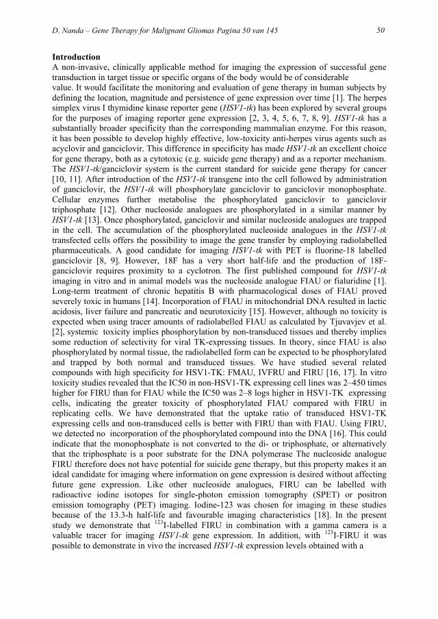
D. Nanda – Gene Therapy for Malignant Gliomas Pagina 50 van 145 50
Introduction A non-invasive, clinically applicable method for imaging the expression of successful gene transduction in target tissue or specific organs of the body would be of considerable value. It would facilitate the monitoring and evaluation of gene therapy in human subjects by defining the location, magnitude and persistence of gene expression over time [1]. The herpes simplex virus I thymidine kinase reporter gene (HSV1-tk) has been explored by several groups for the purposes of imaging reporter gene expression [2, 3, 4, 5, 6, 7, 8, 9]. HSV1-tk has a substantially broader specificity than the corresponding mammalian enzyme. For this reason, it has been possible to develop highly effective, low-toxicity anti-herpes virus agents such as acyclovir and ganciclovir. This difference in specificity has made HSV1-tk an excellent choice for gene therapy, both as a cytotoxic (e.g. suicide gene therapy) and as a reporter mechanism. The HSV1-tk/ganciclovir system is the current standard for suicide gene therapy for cancer [10, 11]. After introduction of the HSV1-tk transgene into the cell followed by administration of ganciclovir, the HSV1-tk will phosphorylate ganciclovir to ganciclovir monophosphate. Cellular enzymes further metabolise the phosphorylated ganciclovir to ganciclovir triphosphate [12]. Other nucleoside analogues are phosphorylated in a similar manner by HSV1-tk [13]. Once phosphorylated, ganciclovir and similar nucleoside analogues are trapped in the cell. The accumulation of the phosphorylated nucleoside analogues in the HSV1-tk transfected cells offers the possibility to image the gene transfer by employing radiolabelled pharmaceuticals. A good candidate for imaging HSV1-tk with PET is fluorine-18 labelled ganciclovir [8, 9]. However, 18F has a very short half-life and the production of 18F-ganciclovir requires proximity to a cyclotron. The first published compound for HSV1-tk imaging in vitro and in animal models was the nucleoside analogue FIAU or fialuridine [1]. Long-term treatment of chronic hepatitis B with pharmacological doses of FIAU proved severely toxic in humans [14]. Incorporation of FIAU in mitochondrial DNA resulted in lactic acidosis, liver failure and pancreatic and neurotoxicity [15]. However, although no toxicity is expected when using tracer amounts of radiolabelled FIAU as calculated by Tjuvavjev et al. [2], systemic toxicity implies phosphorylation by non-transduced tissues and thereby implies some reduction of selectivity for viral TK-expressing tissues. In theory, since FIAU is also phosphorylated by normal tissue, the radiolabelled form can be expected to be phosphorylated and trapped by both normal and transduced tissues. We have studied several related compounds with high specificity for HSV1-TK: FMAU, IVFRU and FIRU [16, 17]. In vitro toxicity studies revealed that the IC50 in non-HSV1-TK expressing cell lines was 2–450 times higher for FIRU than for FIAU while the IC50 was 2–8 logs higher in HSV1-TK expressing cells, indicating the greater toxicity of phosphorylated FIAU compared with FIRU in replicating cells. We have demonstrated that the uptake ratio of transduced HSV1-TK expressing cells and non-transduced cells is better with FIRU than with FIAU. Using FIRU, we detected no incorporation of the phosphorylated compound into the DNA [16]. This could indicate that the monophosphate is not converted to the di- or triphosphate, or alternatively that the triphosphate is a poor substrate for the DNA polymerase The nucleoside analogue FIRU therefore does not have potential for suicide gene therapy, but this property makes it an ideal candidate for imaging where information on gene expression is desired without affecting future gene expression. Like other nucleoside analogues, FIRU can be labelled with radioactive iodine isotopes for single-photon emission tomography (SPET) or positron emission tomography (PET) imaging. Iodine-123 was chosen for imaging in these studies because of the 13.3-h half-life and favourable imaging characteristics [18]. In the present study we demonstrate that 123I-labelled FIRU in combination with a gamma camera is a valuable tracer for imaging HSV1-tk gene expression. In addition, with 123I-FIRU it was possible to demonstrate in vivo the increased HSV1-tk expression levels obtained with a

D. Nanda – Gene Therapy for Malignant Gliomas Pagina 51 van 145 51
replication-competent adenoviral vector as compared with an older E1-deleted adenoviral vector. Materials and methods Cell culture. U87MG and T98G human glioma cell lines were purchased from the American Type Culture Collection (ATCC, Manassas, Va.). The rat 9L gliosarcoma brain tumour cell line was a gift from Dr. K.M. Hebeda (Dept. of Experimental Neurosurgery, Free University Hospital Amsterdam, The Netherlands). 9L cells were stably transfected using an HSV1-tk carrying retrovirus construct followed by G418 selection [19]. Clones were further selected for GCV sensitivity. All cell lines were grown in Dulbecco’s modified Eagle medium (Life Technologies, Breda, The Netherlands) containing 10% foetal bovine serum (Life Technologies), 100 units/ml penicillin and 100 µg/ml streptomycin (Life Technologies) at 37ºC in a 5% CO2 atmosphere. The 9L-tk+ cell line was grown in the presence of G418 (250 µg/ml) (Life Technologies). Vectors. IG.Ad5.E1+.E3TK is a replication-competent Ad5-based virus in which the HSV1-tk gene replaces the gp19K coding sequence in the E3 region (Fig. 1). IG.AdApt.TK is an E1-deleted Ad5-based virus containing the HSV1-tk gene driven by the human CMV promoter in the former E1 region. The CMV promoter in IG.AdApt.TK spans nt –735 to nt +95 (numbering according to [20]). Furthermore, IG.AdApt.TK lacks the SV40 intron sequences. The expression cassette is terminated by the SV40 polyadenylation sequence. Figure 1 shows the schematic structure of the two viruses used in this study. Expression levels obtained from the modified CMV promoter are comparable to expression with the natural E3 promoter [21]. Both viruses were produced on PER.C6 cells [22]. After one round of plaque purification, viruses were further amplified on PER.C6 cells. Cells and viruses were harvested 2–3 days following the final amplification in triple layer flasks and viral particles were purified by a two-step CsCl gradient. Virus particles (VP) in purified virus batches were determined by HPLC [23] and Infectious Units (I.U.) were determined by endpoint titration on 911 cells [24]. VP/I.U. ratios were lower than 30 in all cases. Synthesis of 1-(2-fluoro-2-deoxy- � -d-ribofuranosyl)-5-tributylstannyl uracil (FTBSRU) (precursor compound). 1-(2-Fluoro-2-deoxy- � -D-ribofuranosyl)-5-iodouracil (FIRU) was prepared as previously described [25]. (Ph3P)4Pd(0) (10 mg, 0.008 mmol) and Bu3SnSnBu3 (0.16 ml, 0.30 mmol) were added with stirring to a solution of FIRU (4.0 mg, 0.0107 mmol) in dry dioxane (5 ml), and the reaction mixture was stirred for 16 h at 100°C under an atmosphere of argon, at which time TLC analysis indicated the reaction was completed. The catalyst precipitated as a black solid. Removal of the solvent in vacuo and purification of the residue by silica gel column chromatography with methanol-chloroform (1:20, v/v) as eluent afforded FTBSRU as a colourless oil (23 mg, 40% yield). Carrier-added radioiodination and purification of FIRU. Labelling of FIRU was adapted from Wiebe et al. [16]. FTBSRU (100 µg) was dissolved in 10 µl ethanol/acetic acid (v/v; 50/50), vortexed and microcentrifuged. ICl was dissolved in ethanol/acetic acid (v/v; 50/50) at a concentration of 1 mg/ml, and 25 µl (25 µg ICl) was mixed with 12.5 µl (185 MBq) 123I. The FTBSRU precursor solution was added, the mixture was incubated at room temperature for 10 min and then the labelling mixture was pushed through an activated Sep-Pak column (activated with 5 ml 2-propanol, rinsed with 5 ml H2O).

D. Nanda – Gene Therapy for Malignant Gliomas Pagina 52 van 145 52
Free iodine is first eluted in 15 ml of H2O, followed by elution of FIRU in 50% ethanol (v/v ethanol/ H2O). The FTBSRU precursor eluted in 99% ethanol. The mean labelling yield was 56.4% and the radiochemical purity was >97% (data not shown). Cellular uptake of 123I-FIRU. 9L and 9L-tk+ cells were plated in six-well plates at a density of 105 cells per well. When cells had reached 80%–100% confluency, each well was incubated with 2 MBq of 123I-labelled FIRU for 1, 2, 4, 6, 8 and 24 h. Free radioactive 123I-FIRU was removed from the cells with two PBS (Life Technologies) washes. In a separate experiment, 123I-FIRU was washed off the cells 4 h after incubation, followed by collection of cells, 6, 8 and 24 h after incubation. The cells were then harvested and lysed with 0.1 M NaOH (Life Technologies). Radioactivity in the lysate was determined using a LKB Wallac 1282 Compugamma gamma counter (LKB Instruments, Inc., Gaithersburg, Md.) and corrected for decay. Total protein content was determined in the lysate [26] and radioactivity was expressed as counts per minute/mg protein. All experiments were performed at least in triplicate. The U87MG and T98G cells were plated in six-well plates at a density of 105 cells per well. After 24 h the cells were transfected with the replication-competent and replication-incompetent adenoviral vectors carrying the HSV1-tk transgene at a multiplicity of infection (m.o.i.) of 10. Forty-eight hours after infection, each well was incubated with 2 MBq of 123I-labelled FIRU for 4 h. Subsequently, we performed the same procedure as with the 9L and 9L-tk+ cells. Animal experiments. The Institutional Animal Care and Use Committee, in compliance with the Guide for the Care and Use of Laboratory Animals, approved all experimental protocols. Five- to 6-week-old female NIH-bg-nu-xid mice were purchased [27] (Harlan Sprague Dawley Inc., Oxon., UK). Mice were hosted three to four per cage and allowed access to food and water ad libitum. After 1 week, 9L, 9L-tk+ or U87MG cells (107 cells in 500 µL HBSS; Life Technologies) were inoculated subcutaneously into both flanks. Tumour growth was assessed by measuring bidimensional diameters three times a week with callipers. The tumour volume was determined using the simplified formula of a rotational ellipse (l x w2 x 0.5) [28]. When the 9L and the 9L-tk+ tumours had reached a volume of approximately 1,000 mm3, 3 MBq 123I-FIRU was injected intravenously into the tail vein. To correlate biodistribution results with gamma camera images, acquisition of images was performed at 15 min and 1, 2, 4 and 24 h after 123I-FIRU injection. At each time point at least four animals were examined. Mice were anaesthetized with pentobarbital sodium 15 min before acquisition and placed in a spread supine position under the camera. For imaging, a Siemens Rota II gamma camera system (Siemens, Erlangen, Germany) equipped with a low-energy high-resolution collimator was used. Biodistribution of 123I-FIRU. Biodistribution of 123I-FIRU was determined 15 min and 1, 2, 4 and 24 h after 3 MBq 123I-FIRU injections in mice carrying either 9L or 9L-tk+ subcutaneous xenografts. Four mice were examined at each time point. Tumour, brain, thyroid gland, heart, lungs, stomach, kidney, spleen, skin, muscle, femur and blood were collected and weighed, and radioactivity was determined in the gamma counter. The radioactivity is expressed as percentage of the injected dose/g organ (%ID/g) [6]. Imaging of adenoviral HSV1-tk gene transfer into U87MG xenograft. In vivo evaluation of adenoviral HSV1-tk gene transfer was performed in nude mice carrying U87MG xenografts in the right flank. Once the tumour had reached a volume of 1,000– 1,500

D. Nanda – Gene Therapy for Malignant Gliomas Pagina 53 van 145 53
mm3, animals were injected with replication-competent or -incompetent adenoviral vectors carrying the HSV1-tk gene, or with PBS. The adenoviral vectors (108 or 109 I.U.) were injected into the tumour in 100 µl of PBS. After 48 h, 3 MBq 123I-FIRU was injected intravenously and images were acquired 4 h later using the gamma camera with 10 min acquisition time. The animals were sacrificed, the tumour was taken out and weighed, and radioactivity was determined in the gamma counter. Each treatment group consisted of at least four animals. Statistical analysis. Data were analysed using GraphPad Prism version 3.0 software (GraphPad Software, Inc., San Diego, Calif., 1999). The uptake of 123I-FIRU by the HSV1-tk expressing cell line 9L-tk+ and the parental 9L cell line was compared at all time points with and without removal of 123I-FIRU from the medium, using ANOVA. 123I-FIRU uptake in U87MG and T98G cells following transfection with replication-competent and non-replicating adenoviral vectors was compared with controls using ANOVA. Post-hoc analysis was performed with Tukey’s Multiple Comparison Test. In vivo uptake of 123I-FIRU by 9L-tk+ tumours was compared with uptake by 9L tumours at all time points by unpaired Student’s t test. The medians of in vivo uptake of 123I-FIRU by U87MG xenografts following infection with replication-competent and non-replicating were compared with each other and with controls using the Kruskal-Wallis test.
Fig. 1. Scheme of replication-competent and non-replicating adenoviral vectors. Ad5 is a wild-type adenovirus 5 vector. IG.Ad5.E1+.E3TK is a replication-competent adenoviral vector carrying the HSV1-tk gene in E3, replacing the gp19K coding sequence, and is driven by the native E3 promoter. IG.AdApt.TK is an E1-deleted Ad5-based vector containing the HSV1-tk gene in the former E1 region driven by an adapted CMV promoter. Results Cellular uptake of 123I-FIRU in constitutively HSV1-tk expressing cells Figure 2 demonstrates the selective uptake and retention of 123I-FIRU by 9L-tk+ cells as compared with the parental 9L cells (ANOVA, P<0.001). The maximum difference between the two cell lines occurs at around 4 h of incubation (>2 log difference). Removal of 123I-FIRU from the medium after 4 h of incubation has no effect on the 123I-FIRU retention in HSV1-TK expressing cells atthe later time points (6, 8 and 24 h), indicating entrapment of labelled FIRU in the cells, probably by phosphorylation (Tukey’s Multiple Comparison Test, P>0.05). In vivo uptake of 123I-FIRU by constitutively HSV1-tk expressing tumours Animals carrying established tumours (either 9L or 9Ltk+) were injected with 123I-FIRU, and after imaging (Fig. 3) the animals were sacrificed and the radioactivity in the tumours was measured at various time points (Table 1). At all time points, the accumulation of 123I-FIRU was significantly higher in the HSV1-tk expressing 9Ltk+ tumours as compared with the 9L

D. Nanda – Gene Therapy for Malignant Gliomas Pagina 54 van 145 54
tumours (t test: 15 min, P=0.02; 1 h, P=0.0002; 2 h, P=0.0002; 4 h, P=0.0007; 24 h, P=0.03). The specific activity in the 9Ltk+ tumours was calculated by subtracting the background activity in HSV1-tk negative 9L tumours (Table 1). A maximum specific activity of 22%ID/g tissue was reached 4 h after 123I-FIRU injection. 123I-FIRU biodistribution Biodistribution studies again show the high and selective uptake of 123I-FIRU in HSV1-tk expressing 9L tumours as compared with the parental 9L tumours and other tissues (Table 1). In the first hour after injection, 123I-FIRU levels are highest in peripheral blood and in the kidney (renal excretion). Also, the radioactivity levels in stomach and thyroid are relatively high, indicating in vivo deiodination. The calculated ratios of 123I-FIRU uptake in 9L-tk+ tumours compared with muscle, blood and brain are depicted in Table 2. Cellular uptake of 123I-FIRU following adenoviral transfection of the HSV1-tk gene Infection of U87MG and T98G cells with replicationcompetent IG.Ad5.E1+.E3TK at the relatively low m.o.i. of 10 resulted in strong labelling of both cell types with 123I-FIRU (ANOVA, both cell lines, P<0.0001) (Fig. 4). In contrast, labelling was significantly lower with the replication-incompetent vector IG.AdApt.TK when compared directly with the replication-competent vector in both U87MG and T98G cells (Tukey’s Multiple Comparison Test, P<0.001 for both cell lines). In vivo uptake of 123I-FIRU following adenoviral intratumoural HSV1-tk gene transfer in U87MG Xenografts Two days after the injection of both adenoviral vectors into large U87MG s.c. xenograft tumours, we could identify the tumours with a gamma camera (Fig. 5). Following imaging, the animals were sacrificed and the radioactivity in the tumours was determined and expressed as %ID/g tumour tissue. The results of the directly measured radioactivity accumulation in the tumours correlated with the gamma camera images (Fig. 5, Table 3). We then analysed the dose of the adenoviral vectors used on the one hand and the use of replication-competent versus non-replicating virus on the other hand. The differences (Table 3) were significant (Kruskal-Wallis test, P=0.03), with higher median 123I-FIRU uptake in the tumours injected with the replication-competent vector versus the non-replicating vectors, and higher uptake in the tumours injected with the higher dose (m.o.i. 9 vs m.o.i. 8).

D. Nanda – Gene Therapy for Malignant Gliomas Pagina 55 van 145 55
Fig. 2. Results of in vitro uptake of 123I-FIRU in rat 9L glioma cells that constitutively express the HSV1-tk gene (9L-tk+) compared with uptake in non-expressing parental cells (9L). Each well was incubated with 2 MBq of 123I-labelled FIRU for up to 24 h. Cells were harvested and lysed, and the radioactivity was measured in a gamma counter. In a separate experiment, the FIRU was removed from the medium after 4 h of incubation. Uptake of radioactive FIRU was >2 log higher in the 9L-tk+ ells (P<0.001). Removal of 123I-FIRU from the medium after 4 h of incubation did not affect sequestration of radioactivity in the cells (P>0.05)
Fig. 3. Gamma camera images of NIH-bg-nu-xid mice bearing 9L and 9L-tk+ xenografts, 4 h after i.v. administration of 3 MBq 123I-FIRU. The 9L-tk+ tumours are clearly visualized whereas the 9L control tumours are at background level. Th, Thyroid; St, stomach; Bl, bladder

D. Nanda – Gene Therapy for Malignant Gliomas Pagina 56 van 145 56
Table 1. Biodistribution of 123I-FIRU in NIH-bg-nu-xid mice carrying HSV1-tk positive (9L-tk+) or HSV1-tk negative (9L) tumours
Time p.i.
9L-tk+ tumour
9L tumour
Specific activitya Liver Spleen Lung Skin Kidney
15 min 13.5±3.2 3.5±0.1 10.0±3.3 2.9±0.3 3.3±0.7 4.6±0.4 3.7±0.8 6.4±0.9 1 h 20.1±2.2 2.3±0.2 17.8±2.4 1.4±0.04 1.4±0.1 2.3±0.2 2.1±0.3 2.9±0.2
2 h 20.8±2.4 1.4±0.2 19.4±2.6 1.3±0.2 2.1±0.8 1.7±0.4 2.0±0.8 2.5±0.6 4 h 23.3±3.4 1.3±0.3 22.0±3.7 1.0±0.3 1.5±0.3 1.3±0.3 1.2±0.3 1.5±0.3
24 h 3.1±0.6 0.06±0.01 3.0±0.6 0.04±0.007 0.05±0.004 0.07±0.005 0.08±0.008 0.07±0.01
Time p.i. Heart Femur Muscle Thyroid Stomach Blood Brain 15 min 3.5±0.3 2.2±0.2 2.6±0.3 4.1±0.7 4.6±0.6 13.7±6.8 0.5±0.2
1 h 2.5±0.4 1.0±0.1 1.2±0.1 3.1±0.4 5.5±0.7 13.4±3.8 0.6±0.3 2 h 2.0±0.4 0.9±0.2 1.1±0.4 1.8±0.4 4.6±1.2 2.1±0.5 0.2±0.05
4 h 1.0±0.2 0.7±0.2 0.7±0.2 2.2±0.3 6.0±1.0 1.2±0.4 0.1±0.04 24 h 0.05±0.01 0.04±0.005 0.04±0.005 0.3±0.07 0.7±0.14 0.1±0.05 0.03±0.02
Values are expressed as mean of the %ID/g tissue ±SE (n=4)
a"Specific activity" is calculated by subtracting the background activity in HSV1-tk negative 9L tumour from the activity in the 9L-tk+ tumour
Fig. 4. Results of in vitro uptake of 123I-FIRU in human glioma cells U87MG and T98G. Cells were transfected with replication-competent adenoviral vector IG.Ad5.E1+.E3TK or the non-replicating IG.AdApt.TK at a multiplicity of infection (m.o.i.) of 10. Forty-eight hours after infection, each well was incubated with 2 MBq of 123I-labelled FIRU for 4 h. Cells were

D. Nanda – Gene Therapy for Malignant Gliomas Pagina 57 van 145 57
then harvested and lysed, and radioactivity was measured in a gamma counter. Differences in radioactivity between the three treatment groups were highly significant in both cell lines (P<0.0001). Uptake of 123I-FIRU was significantly higher following infection with the replication-competent adenoviral vector as compared with the non-replicating vector (P<0.001, both cell lines)
Table 2. Ratios of 123I-FIRU uptake in NIH-bg-nu-xid mice carrying HSV1-tk positive (9L-tk+) tumours. Tumour/blood, tumour/muscle and tumour/brain ratios were calculated at various time points
Tumour/muscle Tumour/blood Tumour/brain
15 min 5.2 1.0 30.3
1 h 16.3 1.5 32.9 2 h 18.2 10.0 114.3
4 h 32.3 19.4 171.6 24 h 78.7 31.9 122.1
Table 3. Tumour uptake of 123I – FIRU, two days after adenoviral injection. Values are the ratio of the %ID/gram tissue of tumour divided by %ID/gram of skin and are expressed as median (range) of at least 4 tumours per group. Tumours were injected with a multiplicity of infection (moi) of 8 or 9 of either the replication competent or nonreplicating adenoviral vectors containing the HSV1-tk gene.
Ad.tk
vector:
Nonreplicating Replication
competent
Nonreplicating Replication
competent
Control P
Vector dose 108 I.U. 108 I.U. 109 I.U. 109 I.U. -
median
(range)
1.2
(0.9 – 2.3)
3.0
(1.9 – 9.7)
3.0
(1.6 – 3.8)
4.2
(1.6 – 8.5)
1.1
(1.1 – 1.5)
0.03

D. Nanda – Gene Therapy for Malignant Gliomas Pagina 58 van 145 58
Fig. 5. Gamma camera images of tumour-bearing NIH-bg-nuxid mice, 4 h after i.v. administration of 3 MBq 123I-FIRU. Two days before imaging, the tumours had been injected with PBS (right), 108 I.U. replication-competent HSV1-tk adenovirus (left) or 109 I.U. of nonreplicating HSV1-tk adenovirus (middle). The tumour is better visualized in animals treated with the lower dose (108 I.U.) of replication-competent HSV1-tk adenovirus than in animals injected with a higher dose (109 I.U.) of non-replicating virus. Th, Thyroid; Tu, tumour; St, stomach; Bl, bladder Discussion Interest in non-invasive imaging for measuring therapeutic transgene expression is growing with the increasing clinical application of gene therapy. Methods used for “molecular imaging” include the use of techniques based on bioluminescence [29] and magnetic resonance imaging and spectroscopy [30], and the use of radionuclides followed by PET or SPET scanning. So far, radionuclide imaging of transgene expression makes use of marker genes encoding cell surface receptors such as the type 2 somatostatin [31] and dopamine type 2 receptors [32], or genes encoding enzymes that selectively metabolise the radionuclide tracer to cause intracellular sequestration [2, 3, 4, 5, 6, 7, 8, 9]. The most frequently used therapeutic gene in cancer gene therapy is the HSV1-tk gene [11], and various radiotracers based on uracil nucleosides and acycloguanosine derivatives have been proposed for the non-invasive imaging of HSV1-tk gene expression. In a comparative study, we found that FIRU provided optimal performance in terms of selectivity for HSV1-tk expressing cells compared with the nucleoside analogues FIAU and IVFRU [16]. In this study we determined the value of 123I-FIRU for imaging of HSV1-tk expression, and found a highly significant accumulation of 123I-FIRU in constitutively HSV1-tk expressing tumours. Following adenoviral transfer of the HSV1-tk gene in tumour xenografts, expression of the transgene could be visualised in

D. Nanda – Gene Therapy for Malignant Gliomas Pagina 59 van 145 59
vivo with a gamma camera. The accumulation of 123I-FIRU increased with larger amounts of virus injected and the use of a replication-competent adenovirus. Using the 9L rat glioma cell line stably transfected with the HSV1-tk gene, a 100-fold increase in the uptake of 123I-FIRU compared with the parental cell line was reached at an optimum of 4 h of incubation. The subsequent removal of 123I-FIRU from the medium did not result in substantial loss of intracellular 123I-FIRU, indicating efficient sequestration probably caused by effective phosphorylation. Biodistribution studies demonstrate selective accumulation of 123I-FIRU in HSV1-tk expressing tumours with 23.3% ID/g of 9L-tk+ tumour tissue, 4 h after tracer injection. This accumulation compares favourably with the 10.5% ID/g of 125I-FIAU and 0.15% ID/g of 18F-FHPG [(3-fluoro-1-hydroxy-2-propoxy) methyl guanine] reported for a similar model using the TK expressing murine fibrosarcoma cell line CMS-STK in BALB/c mice [33]. The specific accumulation of radioactivity at 4 h is 22% ID/g (calculated by subtracting the non-specific accumulation of radioactivity in non-transduced 9L tumours from the total radioactivity measured in the 9L-TK tumours) and is higher than the specific accumulation obtained with either 125I-FIAU (9.8% ID/g) or 18F-FHPG (0.08% ID/g) [33]. Four hours after tracer injection, the ratios of 123I-FIRU uptake in tumour to blood, muscle and brain were 19.4, 32.3 and 171.6 respectively. Using 125I-FIAU, Haubner et al. [6] reported tumour to blood and tumour to muscle ratios at 4 h p.i. of 32.0 and 88.3. In a more recent paper by the same group [33] 125I-FIAU ratios at 4 h p.i., calculated from Table 1, reveal a tumour to blood ratio of 24.4 and a tumour to muscle ratio of 21. These results indicate that a direct comparison of FIAU and FIRU as substrate for gene imaging is warranted. The high ratio with normal brain may make FIRU and similar compounds suitable for imaging gene transfer in brain tumours. However, Jacobs et al. [34] demonstrated that iodinated FIAU does not penetrate the intact bloodbrain barrier in cats. In a patient with recurrent glioblastoma, substantial levels of 124I-FIAU-derived radioactivity were found in brain, indicating that the blood-brain barrier in these tumours is disrupted [35]. This study demonstrates the feasibility of obtaining relevant levels of radioactivity to image HSV1-tk gene expression with SPET or PET following gene therapy of glioblastomas. However, the demonstrated uptake in non-transduced tumour also indicates that increased background activity may be expected. The high early radioactivity levels in the kidneys demonstrate the rapid clearance of 123I-FIRU through the kidneys, similar to 124/125I-FIAU [33, 35]. Early radioactivity levels in thyroid and stomach indicate some systemic de-iodination of 123I-FIRU. The in vivo stability of FIRU relative to related 5-substituted nucleosides that do not contain the 2� -fluoro group has been demonstrated before [25]. We then studied the sequestration of 123I-FIRU in the human glioma cell lines U87MG and T98G following infection with two adenoviral vectors carrying the HSV1-tk transgene. IG.Ad5.E1+.E3TK is a replication-competent Ad5-based virus that we constructed to amplify the antitumour efficacy of adenovirus-based gene therapy [36]. IG.Ad5.E1+.E3TK results in more efficient cell kill than IG.AdApt.TK, an E1-deleted Ad5-based virus containing the HSV1-tk gene driven by the human CMV promoter in the former E1 region [36]. Because rodent cells are not permissive for adenoviral replication, the evaluation of the performance of replication-competent adenoviral vectors requires the use of human cell lines and tumour xenograft models. Here, we have demonstrated that the accumulation of 123I-FIRU in U87MG and T98G cells was significantly higher following infection with the replication- competent adenovirus compared to the non-replicating vector. To investigate the possibility of detecting differences in HSV1-tk gene expression levels in vivo we injected U87MG xenografts with either 108 or 109 I.U. of replication- competent IG.Ad5.E1+.E3TK and non-replicating IG.AdApt.TK. Following injection of 123I-FIRU, we found that the accumulation of radioactivity was higher in the tumours injected with 109 I.U. of either virus compared to the tumours injected with 108 I.U. Also, the tumours injected with the replication-competent virus showed higher radioactivity

D. Nanda – Gene Therapy for Malignant Gliomas Pagina 60 van 145 60
levels than tumours injected with the non-replicating vector. These findings correlate with images obtained with the clinical gamma camera and demonstrate the feasibility of detecting differences in HSV1-tk expression levels using 123I-FIRU in vivo following adenoviral gene therapy. In conclusion, 123I-FIRU accumulates highly selectively in tumour expressing the HSV1-tk gene either constitutively or following adenoviral gene transfection. In vivo imaging of the level of HSV1-tk expression is possible and will be valuable in both animal studies as in ongoing clinical trials. Future studies will compare the imaging characteristics of tracers that accumulate in the cell following HSV1-tk transfection with those obtained with tracers directed at transfected cell surface receptors such as the type 2 somatostatin receptor. References 1. Tjuvajev JG, Stockhammer G, Desai R, et al. Imaging the expression of transfected
genes in vivo. Cancer Res 1995; 55:6126–6132. 2. Tjuvajev JG, Finn R, Watanabe K, et al. Noninvasive imaging of herpes virus
thymidine kinase gene transfer and expression: a potential method for monitoring clinical gene therapy. Cancer Res 1996; 56:4087–4095.
3. Tjuvajev JG, Avril N, Oku T, et al. Imaging herpes virus thymidine kinase gene transfer and expression by positron emission tomography. Cancer Res 1998; 58:4333–4341.
4. Tjuvajev JG, Chen SH, Joshi A, et al. Imaging adenoviral-mediated herpes virus thymidine kinase gene transfer and expression in vivo. Cancer Res 1999; 59:5186–5193.
5. Tjuvajev JG, Joshi A, Callegari J, et al. A general approach to the non-invasive imaging of transgenes using cis-linked herpes simplex virus thymidine kinase. Neoplasia 1999; 1:315–320.
6. Haubner R, Avril N, Hantzopoulos PA, Gansbacher B, Schwaiger M. In vivo imaging of herpes simplex virus type 1 thymidine kinase gene expression: early kinetics of radiolabelled FIAU. Eur J Nucl Med 2000; 27:283–291.
7. Gambhir SS, Barrio JR, Wu L, et al. Imaging of adenoviral-directed herpes simplex virus type 1 thymidine kinase reporter gene expression in mice with radiolabeled ganciclovir. J Nucl Med 1998; 39:2003–2011.
8. Gambhir SS, Barrio JR, Phelps ME, et al. Imaging adenoviraldirected reporter gene expression in living animals with positron emission tomography. Proc Natl Acad Sci U S A 1999; 96:2333–2338.9. de Vries EF, van Waarde A, Harmsen MC, Mulder NH, Vaalburg W, Hospers GA. [(11)C]FMAU and [(18)F]FHPG as PET tracers for herpes simplex virus thymidine kinase enzyme activity and human cytomegalovirus infections. Nucl Med Biol 2000; 27:113–119.
10. Sobol RE, Shawler D, Dorigo O, Gold D, Royston I, Fakhrai H. Immunogene therapy of cancer. In: Sobol RE, Scanlon KJ, eds. The Internet book of gene therapy. Stamford, Conn.: Appleton & Lange; 1995:175–180.
11. Nanda D, Driesse MJ, Sillevis Smitt PAE. Clinical trials of adenoviral- mediated suicide gene therapy of malignant gliomas. Progr Brain Res 2001; 132:709–720.
12. Moolten FL. Drug sensitivity (“suicide”) genes for selective cancer chemotherapy. Cancer Gene Therapy 1994; 1:279–287.
13. Balzarini J, Bohman C, Walker RT, De Clercq E. Comparative cytostatic activity of different antiherpetic drugs against herpes simplex virus thymidine kinase gene-transfected tumor cells. Mol Pharmacol 1994; 45:1253–1258.

D. Nanda – Gene Therapy for Malignant Gliomas Pagina 61 van 145 61
14. McKenzie R, Fried MW, Sallie R, et al. Hepatic failure and lactic acidosis due to fialuridine (FIAU), an investigational nucleoside analogue for chronic hepatitis B [see comments]. N Engl J Med 1995; 333:1099–1105.
15. Cui L, Yoon S, Schinazi RF, Sommadossi JP. Cellular and molecular events leading to mitochondrial toxicity of 1-(2-deoxy- 2-fluoro-1-beta-D-arabinofuranosyl)-5-iodouracil in human liver cells. J Clin Invest 1995; 95:555–563.
16. Wiebe LI, Knaus EE, Morin KW. Radiolabelled pyrimidine nucleosides to monitor the expression of HSV-1 thymidine kinase in gene therapy. Nucleosides Nucleotides 1999; 18:1065–1066.
17. Morin KW, Atrazheva ED, Knaus EE, Wiebe LI. Synthesis and cellular uptake of 2 � -substituted analogues of (E)-5-(2- [125I]iodovinyl)-2 � -deoxyuridine in tumor cells transduced with the herpes simplex type-1 thymidine kinase gene. Evaluation as probes for monitoring gene therapy. J Med Chem 1997; 40:2184–2190.
18. Bakker WH, Breeman WA, van der Pluijm ME, de Jong M, Visser TJ, Krenning EP. Iodine-131 labelled octreotide: not an option for somatostatin receptor therapy. Eur J Nucl Med 1996; 23:775–781.
19. Vincent AJ, Vogels R, Someren GV, et al. Herpes simplex virus thymidine kinase gene therapy for rat malignant brain tumors. Hum Gene Ther 1996; 7:197–205. 20. Boshart M, Weber F, Jahn G, Dorsch-Hasler K, Fleckenstein B, Schaffner W. A very strong enhancer is located upstream of an immediate early gene of human cytomegalovirus. Cell 1985; 41:521–530.
21. Nanda D, Vogels R, Havenga M, Avezaat C, Bout A, Sillevis Smitt P. Treatment of malignant gliomas with a replication competent adenoviral vectors. In: The Fourth Annual Meeting of the American Society of Gene Therapy, 2001. Seattle: Molecular Therapy; 2001:abstract 988.
22. Fallaux FJ, Bout A, van der Velde I, et al. New helper cells and matched early region 1-deleted adenovirus vectors prevent generation of replication-competent adenoviruses. Hum Gene Ther 1998; 9:1909–1917.
23. Shabram PW, Giroux DD, Goudreau AM, et al. Analytical anion- exchange HPLC of recombinant type-5 adenoviral particles. Hum Gene Ther 1997; 8:453–465.
24. Fallaux FJ, Kranenburg O, Cramer SJ, et al. Characterization of 911: a new helper cell line for the titration and propagation of early region 1-deleted adenoviral vectors. Hum Gene Ther 1996; 7:215–222.
25. Mercer JR, Xu LH, Knaus EE, Wiebe LI. Synthesis and tumor uptake of 5-82Br- and 5-131I-labeled 5-halo-1-(2-fluoro-2-deoxy- beta-D-ribofuranosyl)uracils. J Med Chem 1989; 32:1289– 1294.26. Bradford MM. A rapid and sensitive method for the quantitation of microgram quantities of protein utilizing the principle of protein-dye binding. Anal Biochem 1976; 72:248–254.
27. Kamel-Reid S, Dick JE. Engraftment of immune-deficient mice with human hematopoietic stem cells. Science 1988; 242:1706–1709.
28. Wildner O, Blaese RM, Morris JC. Synergy between the herpes simplex virus tk/ganciclovir prodrug suicide system and the topoisomerase I inhibitor topotecan. Hum Gene Ther 1999; 10:2679–2687.
29. Contag CH, Spilman SD, Contag PR, et al. Visualizing gene expression in living mammals using a bioluminescent reporter. Photochem Photobiol 1997; 66:523–531.
30. Bell JD, Taylor-Robinson SD. Assessing gene expression in vivo: magnetic resonance imaging and spectroscopy. Gene Ther 2000; 7:1259–1264.
31. Zinn KR, Buchsbaum DJ, Chaudhuri TR, Mountz JM, Grizzle WE, Rogers BE. Noninvasive monitoring of gene transfer using a reporter receptor imaged with a high-affinity peptide radiolabeled with 99mTc or 188Re. J Nucl Med 2000; 41:887–895.

D. Nanda – Gene Therapy for Malignant Gliomas Pagina 62 van 145 62
32. MacLaren DC, Gambhir SS, Satyamurthy N, et al. Repetitive, non-invasive imaging of the dopamine D2 receptor as a reporter gene in living animals. Gene Ther 1999; 6:785–791.
33. Brust P, Haubner R, Friedrich A, et al. Comparison of [18F]FHPG and [124/125I]FIAU for imaging herpes simplex virus type 1 thymidine kinase gene expression. Eur J Nucl Med 2001; 28:721–729.
34. Jacobs A, Tjuvajev JG, Dubrovin M, et al. Positron emission tomography-based imaging of transgene expression mediated by replication-conditional, oncolytic herpes simplex virus type 1 mutant vectors in vivo. Cancer Res 2001; 61:2983–2995.
35. Jacobs A, Braunlich I, Graf R, et al. Quantitative kinetics of 124I-FIAU in cat and man. J Nucl Med 2001; 42:467–475.
36. Nanda D, Vogels R, Havenga M, Avezaat C, Bout A, Sillevis Smitt P. Treatment of malignant gliomas with a replicating adenoviral vector expressing herpes simplex virus-thymidine kinase. Cancer Res 2001; 61:8743–8750.

D. Nanda – Gene Therapy for Malignant Gliomas Pagina 63 van 145 63
CHAPTER 5 [123I]FIRU, a tracer for the ‘molecular imaging’ of HSV1-tk suicide gene transfer M. ter Horst*, D. Nanda*, K. Morin, S. Verwijnen, M. Kros, F. Forrer, M. de Jong, E. Knaus, L. Wiebe and P. Sillevis Smitt *These authors contributed equally Submitted

D. Nanda – Gene Therapy for Malignant Gliomas Pagina 64 van 145 64

D. Nanda – Gene Therapy for Malignant Gliomas Pagina 65 van 145 65
Abstract Several radiopharmaceuticals have been proposed for in vivo imaging of the Herpes simplex virus-1 thymidine kinase (HSV1-tk) gene expression. We compared the in vitro toxicity of FIRU to the related compounds GCV (ganciclovir), FIAU and IVFRU. Furthermore, in vivo images using small animal SPECT, biodistribution, early kinetics and toxicity of FIRU were examined. Methods: The cytotoxicity of the compounds was determined in cell lines constitutively expressing the HSV1-tk gene and non-expressing maternal cell lines, using the MTT assay. Small animal SPECT images, biodistribution and early kinetics of [123I]FIRU were examined in nude mice carrying 9L and 9L-tk subcutaneous tumors. The in vivo toxicity of FIRU was tested in mice and rats. Results: In vitro toxicity studies demonstrated lower toxicity of FIRU as compared to FIAU in both non-HSV1-TK expressing cells (IC50 ratio 3.8–500) and in HSV1-TK expressing cells (IC50 ratio 1.7-2 x 10⁷). SPECT images using [123I]FIRU were able to visualize HSV-tk expression in mouse xenografts in vivo. Biodistribution and early kinetics studies of [123I]FIRU in tumor bearing mice showed initial phase and terminal phase half-lives of respectively 0.8 h and 1.3 h (blood); 0.1 h and 4 h (muscle); and 0.5 h and 6.7 h (9L tumor). In 9L-tk+ tumors, [123I]FIRU accumulated for about one hour, followed by a slower decrease of activity with a half-life of 11.3 h. Mice and rats receiving up to 200 times the projected clinical dose showed no toxicity. Conclusions: The favorable toxicity and imaging profile make radiolabeled FIRU a promising agent for imaging of HSV1-tk gene transfer in clinical studies. Introduction Interest in non-invasive imaging and quantification of therapeutic transgene production is growing with increasing clinical applications of gene therapy1-3. The most commonly used methods for ‘molecular imaging’ of gene transfer are based on radionuclide’s followed by PET or SPECT scanning4. Radionuclide imaging of transgene expression makes use of marker genes such as the cell surface gene of the human somatostatin receptor type 2 (hSSTR2)5,6, genes encoding transporter proteins including the sodium/iodide symporter7, or genes encoding enzymes that selectively metabolize the radionuclide tracer resulting in intracellular sequestration such as the herpes simplex virus type 1 thymidine kinase (HSV1-tk) gene8-13. The HSV1-tk gene is the most frequently used therapeutic gene in cancer gene therapy14-16. Various radiotracers based on uracil nucleosides and acycloguanosine derivatives that are selective substrates for this virus encoded enzyme have been proposed for the non-invasive imaging of HSV1-tk gene expression. The most commonly used radioiodinated substrate for monitoring HSV1-tk gene expression is 1-(2-fluoro-2-deoxy-β-D-arabinofuranosyl)-5-iodouracil (FIAU; fialuridine)17. Another nucleoside with high sensitivity for HSV-TK is 1-(2-fluoro-2-deoxy-β-D-arabinofuranosyl)-thymine (FMAU). Because of their severe mitochondrial toxicity in normal cells and fatal toxicity to neurons and liver in clinical studies18-23, FMAU and FIAU are not suitable for antiviral therapeutic applications but can be used as tracer molecules. Interestingly, the toxicity of FIAU reflects its phosphorylation by mammalian nucleoside kinases and subsequent incorporation into mammalian DNA21,24,25. Thus, radioiodinated FIAU uptake reflects not only activation by the viral kinase (HSV1-TK), but also uptake by mammalian cells not expressing HSV1-TK. This limits its selectivity, and may influence sensitivity, depending on the relative activity of the viral and mammalian enzymes. Other nucleosides with high specificity for HSV1-TK include 1-(2-fluoro-2-deoxy-β-D-ribofuranosyl)-5-(E)-(2-iodovinyl)-uracil (IVFRU), 1-(2-fluoro-2-deoxy-β-D-ribofuranosyl)-5-iodouracil (FIRU)11 and 9-(4-fluoro-3-hydroxy-methyl-butyl)guanine (FHGB)26. In a comparative study, uptake of FIRU in HSV1-tk expressing cells was similar to uptake of

D. Nanda – Gene Therapy for Malignant Gliomas Pagina 66 van 145 66
FIAU and IVFRU. Importantly, incorporation of FIRU into nucleic acids was only about one-tenth the incorporation of FIAU23. Biodistribution studies with [123I]FIRU have demonstrated a maximum uptake of 22 % of the injected dose per gram tissue in mice carrying HSV1-tk expressing tumor xenografts, and favorable imaging characteristics of [123I]FIRU following adenoviral HSV1-tk adenoviral gene transfer in a xenograft tumor model has been demonstrated12. The relative performance of several radioiodinated nucleosides in HSV1-tk-expressing cells depends to some degree on the cell type and the design of the gene cassette23. In the present study, we describe a Sep-Pak column-based method for purification of radioiodine labeled FIRU that can be easily applied to manufacturing of clinical grade batches. We subsequently present small animal SPECT images using [123I]FIRU as a reporter probe to visualize HSV-tk expression in mouse xenografts in vivo. Finally, we examined the early kinetics of iodine labeled FIRU and its toxicity profile in vitro and in vivo. Materials and Methods Compounds Ganciclovir (Cymevene; GCV) was purchased from Roche (Mijdrecht, Netherlands). IVFRU, FIAU and FIRU were synthesized using literature methods27. The structure formulae of the 4 compounds are depicted in Fig. 1. Carrier-added (radio)iodination was achieved by electrophilic iodination/destannylation of 1-(2-fluoro-2-deoxy-β-D-ribofuranosyl)-5-tributylstannyluracil (FTBSRU), the FIRU precursor, as previously described12,28. For recovery and purification of [123I]FIRU, the labeling mixture was passed through an activated Sep-Pak column; iodide was first eluted with H2O, followed by elution of [123I]FIRU in 50 % aqueous ethanol and recovery of unreacted FTBSRU precursor in 96 % ethanol. For the in vitro and in vivo toxicity experiments, ‘cold’ FIRU was synthesized by a similar procedure, but omitting [123I]NaI from the reaction mixture. HPLC analysis of [123I]FIRU An aliquot of eluted [123I]FIRU was diluted with HPLC-solvent, and an aliquot of this solution was injected into the HPLC-system (Waters-Instruments (Etten-Leur, The Netherlands) using a Partisil 10 column (ODS 4.6 mm x 250 mm; Whatman, Maidstone, England). UV-absorbance was measured at 254 nm. The outlet of the UV detector was connected to a well-type NaI crystal (Canberra Packard Benelux sa, Zellik, Belgium). Isocratic elution (1 mL/min) was carried out using acetonitrile/milliQ-water 10/90 vol %. Cell culture Murine fibrosarcoma KBALB, human osteosarcoma R-970-5 (a TK- mutant of R-970-5) and human 143B cell lines were purchased from American Type Culture Collection (ATCC, Manassas, VA). The rat 9L gliosarcoma brain tumor cell line was a gift from Dr. K.M. Hebeda (Dept. Experimental Neurosurgery, Free University Hospital Amsterdam, The Netherlands). The EMT-6 mouse mammary tumor cell line was obtained from the Cross Cancer Institute (Edmonton, Canada). The KBALB-STK cell line, which expresses HSV1-tk, was obtained from Dr. Scott Freeman (Tulane University Medical Center, New Orleans, USA.)29. The 143B and 9L cell lines were stably transfected using an HSV1-tk-carrying retrovirus construct, followed by G418 selection (250 � g – 1 mg/mL) (Life Technologies, Breda, The Netherlands). The corresponding HSV-tk positive cell lines were further selected for GCV sensitivity and were named 143B-LTK and 9L-tk+ respectively30,31. Cell lines were maintained in Dulbecco’s modified Eagles medium (DMEM; Life Technologies) supplemented with 10 % fetal bovine serum (FBS; Life Technologies), penicillin (100 U/mL) and streptomycin (100 � g/mL). Only EMT-6 cells were routinely maintained in Waymouth

D. Nanda – Gene Therapy for Malignant Gliomas Pagina 67 van 145 67
medium, and 143B cells were maintained in DMEM containing 5-bromo-2'-deoxyuridine (15 � g/mL). KBALB-STK, 143B-LTK and 9L-tk+ cells were maintained in medium that had 250 � g - 1 mg/mL G-418 (Life Technologies) added in addition to FBS and antibiotics. Cells were cultured at 37 °C in a 5 % CO2 atmosphere. MTT Cytotoxicity Assay Method Exponentially growing cells were trypsinized, centrifuged and suspended in growth medium and readjusted to 8x10³ cells/mL. The cells were seeded into 96-well plates at 8x10² cells/well and incubated at 37 oC, 5 % CO2 for 24 h. The compounds were dissolved in DMEM medium and incubated with the cells in 96-well plates at a volume of 100 µL to produce a final concentration of design. Control wells were filled with 100 µL of DMEM medium. The plates were incubated for 72 h at 37 oC in a humidified atmosphere containing 95 % air and 5 % CO2. At the end of incubation, 3-(4,5-dimethyl-2-thiazolyl)-2,5-diphenyl-2H-tetrazolium bromide (MTT, Sigma, Zwijndrecht, The Netherlands) was added, and the plates were incubated at 37 0C for 4 h. Dimethylsulfoxide was added to dissolve the formazan crystals, and the plate wells were then read at 540 nm on a scanning multi-well spectrophotometer. Animal experiments The Institutional Animal Care and Use Committee, in compliance with the Guide for the care and Use of Laboratory Animals, approved all experimental protocols. For the biodistribution studies, five- to six-week old female NIH-bg-nu-xid mice were purchased32 from Harlan Sprague Dawley Inc., Oxon, England. For the animal SPECT experiments two, five- to six-week old male nu/nu NMRI mice (Charles River, Les Oncins, France) were purchased. Mice were hosted 3-4 per cage and allowed access to food and water ad libitum. After one week, 9L or 9L-tk+ cells (107 cells in 500 � L HBSS; Life Technologies) were inoculated subcutaneously into both flanks. Tumor growth was monitored by measuring bidimensional diameters three times a week with calipers, and tumor volume was determined by applying the simplified formula of a rotational ellipse (l x w2 x 0.5)33. The experiments were performed when the 9L and the 9L-tk + tumors had reached a volume of approximately 1000 mm3. For the biodistribution experiments, [123I]FIRU (3 MBq) was injected intravenously into the tail vein. Biodistribution of [123I]FIRU was determined 15 min, and 1, 2, 4 and 24 h post injection (p.i.). Four mice were examined at each time point. Tumor, brain, thyroid gland, heart, lungs, stomach, kidney, spleen, skin, muscle, femur and blood were collected, the weight was measured and radioactivity was determined in the gamma counter. The radioactivity is expressed as percentage of the injected dose / gram organ (%ID/g). For the SPECT experiment [123I]FIRU (40 MBq) was injected intravenously into the penis vein and the animals were scanned 4 hr later. SPECT and Software SPECT imaging was performed with the four-headed multiplexing multi-pinhole small animal SPECT (Bioscan Inc., Washington D.C.). Each head is outfitted with an application specific tungsten collimator. For this study we imaged with the 9-pinhole-mouse apertures that are comprised of a total of 36 1.3 mm diameter pinholes imaging a cylindrical field of view that is 32 mm in diameter by 14 mm in length. These mouse apertures provide a reconstruction resolution below 1.0 mm at 159 keV with an average sensitivity of 1100 cps/MBq across the field of view (FOV). The axial FOV is extended using a step-and-shoot helical scan to perform a total body scan of the animal (total distance 85 mm). The energy-peak for the camera was set at 159 keV. The window width was ± 10%. An acquisition time of 20 sec. per projection was chosen. Acquisition time was 20 min. per animal. After the acquisition the data were reconstructed iteratively with the HiSPECT© software (Bioscan Inc., Washington DC),

D. Nanda – Gene Therapy for Malignant Gliomas Pagina 68 van 145 68
a dedicated ordered subset-expectation maximization (OSEM) software package for multiplexing multi-pinhole reconstruction. The SPECT is calibrated with a phantom such that voxel values in the reconstruction provide a proper estimate of the activity level without further calculation. In vivo toxicity testing Twenty-four male Wistar rats (weight approximately 50 g) and 24 male BALB-c mice (5-6 weeks old) were purchased from Harlan Sprague Dawley Inc., Oxon, England. Non-radioactive precursor was iodinated with non-radioactive ICl, followed by Sep-Pak purification, as described for [123I]FIRU. For the animal experiments, the ethanol fraction was air dried and dissolved in PBS. The FIRU was subsequently administered by tail vein injection into the animals, based on the projected dose to be administered to human subjects (100 µg of radiolabelled FIRU; nominally 1.4 µg/kg). Rats and mice were divided in 3 groups of 8 animals receiving 0, 20 or 200 times the anticipated clinical dose. Animals were observed daily and euthanized by pentobarbital sodium injection seven days after FIRU administration. Blood was drawn by cardiac puncture for hematological (Hb, Ht, leukocytes and platelets) and chemical tests (sodium, potassium, creatinin, ASAT, ALAT, alkaline phosphatase, αGT, LDH and amylase). Liver, spleen, pancreas, heart, lungs, kidneys, esophagus, stomach, duodenum, bladder, lymph nodes, striated muscle, salivary glands and sciatic nerve were dissected out immediately and fixed in formalin. After paraffin embedding, sections were cut and stained with H&E and examined by a licensed pathologist (M.K.). Statistical analysis The hematological and chemistry parameters in the three dose groups of rats and mice were compared using ANOVA. When a significant difference between groups was detected, we used post-hoc Dunnett's Multiple Comparison Test to determine the groups that differed significantly. All P-values are two-sided and a significance level � = 0.05 was used. All statistical analyses were performed using GraphPad Prism version 4.0 software (GraphPad Software, Inc., San Diego, CA). Results Analytic HPLC chromatograms before (a) and after Sep-Pak purification (b) are shown in Fig. 2. In profile (a), unbound radioactivity, presumably as radioiodide, elutes at the column void volume (~1.5 min), and [123I]FIRU is detected at ~ 4 min. Profile (b) is a chromatogram of the 50 % ethanol Sep-Pak eluant, showing only a single radioactive peak at the FIRU elution time. No unreacted FTBSRU was evident in this fraction (by UV detection; data not shown). The radiochemical purity was >97 % with a labeling yield of 56 ±10 %; specific activity not less than 690 MBq/µmol. This specific activity is lower than achievable using a ‘no carrier’ oxidant such as hydrogen peroxide34, but there is no evidence to suggest that this specific activity negatively influences uptake. In studies using radioactive IUdR and FIAU, it has been reported that uptake in the presence of respective carrier (10 µM) will reduce uptake of radioactivity by 3 - 10 fold over a 100 min incubation period34; consequently, in the present studies, in which FIRU concentrations are approximately 0.004 µM, little if any effect is to be anticipated.

D. Nanda – Gene Therapy for Malignant Gliomas Pagina 69 van 145 69
In vitro toxicity of FIRU Fig. 3 shows the cytotoxicity of increasing concentrations of ganciclovir, FIRU, FIAU and IVFRU in the cell lines tested. The relative inhibitory concentration of 50% (IC50)of FIRU compared to FIAU and ganciclovir is shown in Table 1. When the compounds are phosphorylated (in HSV1-tk expressing cells), FIAU is 7 logs more toxic than FIRU. This finding most probably reflects the incorporation of phosphorylated FIAU into cellular DNA as opposed to FIRU27 . However, in non-HSV1-tk expressing dividing cells, FIAU is also more toxic than FIRU (1-2 logs), presumably reflecting some phosphorylation of FIAU by endogenous TK24,25 In vivo imaging of [123I]-FIRU distribution using small animal SPECT Animals carrying established 9L tumors in the left flank and 9L-tk+ tumors in the right flank were intravenously injected with [123I]-FIRU (40MBq). Four hours later, the animals were sacrificed and scanned (Fig. 4). The high-resolution SPECT images clearly visualize the uptake of the radiopharmaceutical in the 9L-tk+ tumor, while the 9L tumor remains at background level. Biodistribution and early kinetics of [123I]FIRU The biodistribution of [123I]FIRU in 9L-tk+ tumors, 9L tumors, muscle, thyroid, stomach, blood and brain at 15 min and 1, 2, 4 and 24 h p.i. (Fig. 5) is compatible with rapid renal clearance of the tracer from the body, resulting in increased muscle/muscle, blood/blood and brain/brain ratios over time. A maximum tissue uptake of 23 % ID/g tissue was reached in the 9L-tk+ tumors 4 h after [123I]FIRU injection compared to muscle (<2 %), thyroid (2.2 %), stomach (6.2 %), blood (<2 %) and brain (<2 %). The accumulation of radioactivity in thyroid and stomach indicates, as reported previously28, some partial de-iodination of [123I]FIRU. Blood, muscle and HSV1-tk negative tumors clearly showed bi-exponential elimination characteristics (Fig. 6). The half-lives for the initial phase were 0.8 h (blood), 0.1 h (muscle) and 0.5 h (9L tumor). The half-lives for the terminal phase were 1.3 h (blood), 4 h (muscle) and 6.7 h (wild-type 9L tumor). After an initial accumulation of about one hour of 123I-FIRU in the HSV-tk positive 9L-tk+ tumors, the activity decreases more slowly with a half-life of 11.3 h (Fig 6). In vivo toxicity of FIRU None of the rats and mice showed clinical signs of disease. Hematological and biochemical parameters tested did not differ between animals receiving PBS, 20x or 200x the projected human dose of FIRU. Pathological examination of all organs did not demonstrate any abnormality indicative of toxicity. Discussion This paper describes a simple Sep-Pak based method for the purification of radio-iodinated FIRU allowing large scale, clinical grade production. The described carrier-mediated labeling method is simple, and purification over a Sep-Pak column is much more convenient than preparative HPLC, in particular for the production of clinical grade under GMP conditions. Using the described labeling and purification strategy, [123I]FIRU was routinely produced with a radiochemical purity > 97 %, a labeling yield of ~ 56 % and a specific activity not less than 690 MBq/µmol. The in vitro toxicity of FIRU, FIAU and related compounds was studied in a number of cell lines. In constitutively HSV1-tk expressing cell lines, the toxicity of FIRU

D. Nanda – Gene Therapy for Malignant Gliomas Pagina 70 van 145 70
was significantly lower than FIAU. This lack of toxicity of the phosphorylated FIRU compound is most likely due to the fact that FIRU is not incorporated into the DNA of the mammalian dividing cell27, as opposed to FIAU24,25. In dividing cells that don’t express HSV1-tk, FIRU was also less toxic than FIAU. This difference probably reflects a lower affinity of the endogenous TK enzyme for FIRU than for FIAU, but in neither case is uptake sufficient to warrant use as a cell proliferation marker35. Uptake of FIAU was reported to be about 20-fold higher than that of IUdR in D-247 MG cells, leading those authors to conclude that [125I]FIAU might have greater cytotoxicity for this cell line than [125I]IUdR34. In the past decade significant progress has been made in using PET and SPECT imaging methods to detect the expression of specific genes, such as HSV1-tk, in living animals36-38. Recently, a multiplexing multi-pinhole small-animal SPECT system has been described with greatly improved sensitivity while achieving high resolutions39,40. Using this system, we could clearly demonstrate the uptake of FIRU in HSV-tk expressing xenografts (Fig. 4). Choi et al41 earlier showed microSPECT images of [123I]FIAU uptake in HSV-tk expressing xenografts. These high-resolution SPECT images clearly demonstrated inhomogeneous tracer uptake in one of the xenografts, suggesting necrosis of the interior portion of the tumor. In our study we did not observe inhomogenous uptake nor did we see necrotic areas on pathological examination, probably due to limited tumor size. Progress in nuclear technology and fusion of multiple imaging modalities, such as SPECT and CT, will further enhance the sensitivity and accuracy of molecular imaging. The biodistribution (Table 1) and biokinetics (Fig. 5) studies show selective accumulation of 123I-FIRU in HSV1-tk+ tumors in the first hour after administration. Subsequently, the radioactivity incorporated in these tumors decreases with a much longer half-life than in the surrounding tissues. The optimal imaging time with [123I]FIRU is 4 h post-injection12,27. Finally, in preparation of clinical studies, we performed toxicity studies with ‘cold’ FIRU in rats and mice. The injected amount of FIRU per animal was 1x, 20x or 200x fold (mg/kg) of the intended human dose for imaging (100 � g (0.27 � mol) / 70 kg). Because the mitochondrial toxicity associated with FIAU manifests mainly as hepatotoxicity, pancreatitis, neuropathy or myopathy, we focused on these organs and the hematopoietic system 21. This multisystem mitochondrial toxicity is probably caused by the high rate at which FIAU is incorporated into mitochondrial DNA42,43. We could detect no clinical, laboratory or pathological signs of toxicity due to FIRU. In particular, all liver function tests and amylase levels were normal. Pathological examination of liver and pancreas and of the peripheral nervous system and muscle was completely normal. We conclude that radiolabeled FIRU can be easily produced in clinical grade batches. In addition, the safety profile and clear imaging properties make FIRU a good candidate tracer for clinical imaging of HSV1-tk gene transfer.

D. Nanda – Gene Therapy for Malignant Gliomas Pagina 71 van 145 71
Figures
Fig. 1 Structural formula of ganciclovir (GCV: Mw 255), FIRU (Mw 372), FIAU (Mw 372) and IVFRU (Mw 398). Note that the only difference between FIAU and FIRU is the 2’-fluoro-arabino (“up”) substitution in FIAU versus the 2’-fluoro-ribo (“down”) substitution in FIRU.
Fig. 2 HPLC analysis of [123I]FIRU. Radiolabeled FIRU elutes from the HPLC column at 4 min. Panel a depicts the HPLC result of [123I]FIRU before Sep-Pak purification. Panel b shows HPLC analysis of the 50 % ethanol fraction (v/v ethanol/H2O) of the Sep-Pak column, which contains only [123I]FIRU.
a b

D. Nanda – Gene Therapy for Malignant Gliomas Pagina 72 van 145 72
Fig. 3 In vitro toxicity of GCV, FIRU, FIAU and IVFRU. At increasing doses, all compounds become toxic in all cells. In the HSV1-TK expressing cells, FIRU is clearly less toxic than FIAU, GCV and FIAU.
GCV
-11 -10 -9 -8 -7 -6 -5 -4 -3 -2 -10
25
50
75
100
125
concentration (M)
% cells
FIAU
-11 -10 -9 -8 -7 -6 -5 -4 -3 -2 -1
0
25
50
75
100
125
concentration (M)
% cells
FIRU
-11 -10 -9 -8 -7 -6 -5 -4 -3 -2 -10
25
50
75
100
125
c onc entration (M)
% cells
IVFRU
-11 -10 -9 -8 -7 -6 -5 -4 -3 -2 -1
0
2 5
5 0
7 5
1 0 0
1 2 5
concentration (M)
% cells
K B A L B KBALB-ST 143B
143B-LTK EMT-6 R-970-5
9L
9 L - T K

D. Nanda – Gene Therapy for Malignant Gliomas Pagina 73 van 145 73
Fig. 4 Small animal SPECT image using [123I]FIRU as reporter probe. 3D-reconstruction of the whole mouse acquired by multipinhole SPECT is shown in (a). The transaxial (b), coronal (c) and saggital (d) slices of the mouse clearly visualize the HSV-tk positive tumor (9L-tk+) in the left flank. The HSV-tk negative tumor (9L) in the right flank remained at back-ground level. Th thyroid; B bladder.
Th
B 9L-tk 9L
a b
c d

D. Nanda – Gene Therapy for Malignant Gliomas Pagina 74 van 145 74
Fig. 5 Biodistribution of [123I]FIRU. The percentage of the injected dose per gram of tissue (%ID/g tissue) is measured at 15 min and 1, 2, 4 and 24 h after injection. The maximum tumor uptake of 23 %ID/g tissue in HSV1-tk expressing tumors is reached four hours after injection. Fig. 6 Early biokinetics of [123I]FIRU. While the elimination of [123I]FIRU from blood, muscle and HSV1-tk negative tumor (9L) is clearly biphasic, the activity accumulates in HSV1-tk positive tumor (9L-tk+) followed by a much slower decrease of the incorporated activity.
9L-tk+ tumor 9L tumormusc le thy roi dsto ma chbloo d brai n0.0
2.5
5.0
7.5
10.0
12.5
15.0
17.5
20.0
22.5
25.0
27.5
15 min
1 hour
2 hour
4 hour
24 hour
%ID/g tissue (SE)
0 10 20
0
10
20
30
9L-tk+ tumor 9L tumor
muscle blood
t (h)
%ID/g

D. Nanda – Gene Therapy for Malignant Gliomas Pagina 75 van 145 75
Table 1. Summary of cytotoxicity (IC50 , M) of ganciclovir, FIRU and FIAU in non-HSV1-tk and in HSV1-tk expressing cells.
IC50 of GCV IC50 of FIAU
IC50 of FIRU
Ratio IC50 FIRU:FIAU
Non-HSV1-TK expressing cells: KBALB
4.8 x 10⁻⁴ M
1.0 x 10⁻⁵ M
1.5 x 10⁻³ M
150
143B 1.0 x 10⁻⁴ M 2.0 x 10⁻³ M 7.5 x 10⁻³ M 3.8 R-970-5 1.2 x 10⁻³ M 1.0 x 10⁻⁴ M 7.0 x 10⁻³ M 70 EMT-6 2.0 x 10⁻⁴ M 6.5 x 10⁻⁶ M 3.0 x 10⁻³ M 500 HSV1-TK expressing cells: KBALB-STK
3.0 x 10⁻⁸ M
1.0 x 10⁻¹⁰ M
2.0 x 10⁻³ M
2 x 10⁷
143B-LTK 6.5 x 10⁻⁸ M 3.0 x 10⁻¹⁰ M 5.0 x 10⁻³ M 1.7 x 10⁷ Acknowledgments The authors gratefully thank Willem Bakker and Linda de Jong, Department of Nuclear Medicine, Erasmus University Medical Center, Rotterdam, for their technical support. References 1. Nanda D, Driesse MJ, Sillevis Smitt PA. Clinical trials of adenoviral-mediated suicide
gene therapy of malignant gliomas. Prog Brain Res 2001; 132: 699-710. 2. Min JJ, Gambhir SS. Gene therapy progress and prospects: noninvasive imaging of
gene therapy in living subjects. Gene Ther 2004; 11: 115-125. 3. Blasberg RG, Tjuvajev JG. Molecular-genetic imaging: current and future
perspectives. J Clin Invest 2003; 111: 1620-1629. 4. Ray P, Bauer E et al. Monitoring gene therapy with reporter gene imaging. Semin
Nucl Med 2001; 31: 312-320. 5. Zinn KR, Buchsbaum DJ et al. Noninvasive monitoring of gene transfer using a
reporter receptor imaged with a high-affinity peptide radiolabeled with 99mTc or 188Re. J Nucl Med 2000; 41: 887-895.
6. Ter Horst M, Verwijnen SM et al. Locoregional Delivery of Adenoviral Vectors. J Nucl Med 2006; 47: 1483-1489.
7. Carlin S, Akabani G, Zalutsky MR. In vitro cytotoxicity of (211)at-astatide and (131)I-iodide to glioma tumor cells expressing the sodium/iodide symporter. J Nucl Med 2003; 44: 1827-1838.
8. Tjuvajev JG, Finn R et al. Noninvasive imaging of herpes virus thymidine kinase gene transfer and expression: a potential method for monitoring clinical gene therapy. Cancer Res 1996; 56: 4087-4095.
9. Haubner R, Avril N et al. In vivo imaging of herpes simplex virus type 1 thymidine kinase gene expression: early kinetics of radiolabelled FIAU. Eur J Nucl Med 2000; 27: 283-291.
10. Gambhir SS, Barrio JR et al. Imaging of adenoviral-directed herpes simplex virus type 1 thymidine kinase reporter gene expression in mice with radiolabeled ganciclovir. J Nucl Med 1998; 39: 2003-2011.

D. Nanda – Gene Therapy for Malignant Gliomas Pagina 76 van 145 76
11. de Vries EF, van Waarde A et al. [(11)C]FMAU and [(18)F]FHPG as PET tracers for herpes simplex virus thymidine kinase enzyme activity and human cytomegalovirus infections. Nucl Med Biol 2000; 27: 113-119.
12. Nanda D, de Jong M et al. Imaging expression of adenoviral HSV1-tk suicide gene transfer using the nucleoside analogue FIRU. Eur J Nucl Med Mol Imaging 2002; 29: 939-947.
13. Verwijnen SM, Sillevis Smith PA et al. Molecular imaging and treatment of malignant gliomas following adenoviral transfer of the herpes simplex virus-thymidine kinase gene and the somatostatin receptor subtype 2 gene. Cancer Biother Radiopharm 2004; 19: 111-120.
14. St Clair MH, Lambe CU, Furman PA. Inhibition by ganciclovir of cell growth and DNA synthesis of cells biochemically transformed with herpesvirus genetic information. Antimicrob Agents Chemother 1987; 31: 844-849.
15. Moolten FL, Wells JM. Curability of tumors bearing herpes thymidine kinase genes transferred by retroviral vectors. J Natl Cancer Inst 1990; 82: 297-300.
16. Pulkkanen KJ, Yla-Herttuala S. Gene therapy for malignant glioma: current clinical status. Mol Ther 2005; 12: 585-598.
17. Tjuvajev JG, Stockhammer G et al. Imaging the expression of transfected genes in vivo. Cancer Res 1995; 55: 6126-6132.
18. McLaren C, Chen MS et al. Drug resistance patterns of herpes simplex virus isolates from patients treated with acyclovir. Antimicrob Agents Chemother 1985; 28: 740-744.
19. Pankiewicz KW, Nawrot B et al. Nucleosides. 146. 1-Methyl-5-(2-deoxy-2-fluoro-beta-D-arabinofuranosyl)uracil, the C-nucleoside isostere of the potent antiviral agent 1-(2-deoxy-2-fluoro-beta-D-arabinofuranosyl)thymine (FMAU). Studies directed toward the synthesis of 2'-deoxy-2'-substituted arabino nucleosides. 6. J Med Chem 1987; 30: 2314-2316.
20. Abbruzzese JL, Schmidt S et al. Phase I trial of 1-(2'-deoxy-2'-fluoro-1-beta-D-arabinofuranosyl)-5-methyluracil (FMAU) terminated by severe neurologic toxicity. Invest New Drugs 1989; 7: 195-201.
21. Cui L, Yoon S, Schinazi RF, Sommadossi JP. Cellular and molecular events leading to mitochondrial toxicity of 1-(2-deoxy-2-fluoro-1-beta-D-arabinofuranosyl)-5-iodouracil in human liver cells. J Clin Invest 1995; 95: 555-563.
22. Wang J, Eriksson S. Phosphorylation of the anti-hepatitis B nucleoside analog 1-(2'-deoxy-2'-fluoro-1-beta-D-arabinofuranosyl)-5-iodouracil (FIAU) by human cytosolic and mitochondrial thymidine kinase and implications for cytotoxicity. Antimicrob Agents Chemother 1996; 40: 1555-1557.
23. Morin KW, Duan W et al. Cytotoxicity and cellular uptake of pyrimidine nucleosides for imaging herpes simplex type-1 thymidine kinase (HSV-1 TK) expression in mammalian cells. Nucl Med Biol 2004; 31: 623-630.
24. Klecker RW, Katki AG, Collins JM. Toxicity, metabolism, DNA incorporation with lack of repair, and lactate production for 1-(2'-fluoro-2'-deoxy-beta-D-arabinofuranosyl)-5-iodouracil in U-937 and MOLT-4 cells. Mol Pharmacol 1994; 46: 1204-1209.
25. Grant AJ, Feinberg A et al. Incorporation of metabolites of 2'-fluoro-5-iodo-1-beta-D-arabinofuranosylcytosine into deoxyribonucleic acid of neoplastic and normal mammalian tissues. Biochem Pharmacol 1982; 31: 1103-1108.
26. Green LA, Nguyen K et al. A tracer kinetic model for 18F-FHBG for quantitating herpes simplex virus type 1 thymidine kinase reporter gene expression in living animals using PET. J Nucl Med 2004; 45: 1560-1570.

D. Nanda – Gene Therapy for Malignant Gliomas Pagina 77 van 145 77
27. Wiebe LI, Knaus EE, Morin KW. Radiolabelled pyrimidine nucleosides to monitor the expression of HSV-1 thymidine kinase in gene therapy. Nucleosides Nucleotides 1999; 18: 1065-1066.
28. Mercer JR, Xu LH, Knaus EE, Wiebe LI. Synthesis and tumor uptake of 5-82Br- and 5-131I-labeled 5-halo-1-(2-fluoro-2-deoxy-beta-D-ribofuranosyl)uracils. J Med Chem 1989; 32: 1289-1294.
29. Ramesh R, Munshi A et al. Expression of costimulatory molecules: B7 and ICAM up-regulation after treatment with a suicide gene. Cancer Gene Ther 1996; 3: 373-384.
30. Vincent AJ, Vogels R et al. Herpes simplex virus thymidine kinase gene therapy for rat malignant brain tumors. Hum Gene Ther 1996; 7: 197-205.
31. Wang ZX, Duan W et al. Synthesis of 1-(2-deoxy-beta-D-ribofuranosyl)-2,4-difluoro-5-substituted-benzenes: "thymine for evalation as anticancer and antiviral agents. Nucleosides Nucleotides 2001; 20: 41-58.
32. Kamel-Reid S, Dick JE. Engraftment of immune-deficient mice with human hematopoietic stem cells. Science 1988; 242: 1706-1709.
33. Dethlefsen LA, Prewitt JM, Mendelsohn ML. Analysis of tumor growth curves. J Natl Cancer Inst 1968; 40: 389-405.
34. Vaidyanathan G, Zalutsky MR. Preparation of 5-[131I]iodo- and 5-[211At]astato-1-(2-deoxy-2-fluoro-beta-D-arabinofuranosyl) uracil by a halodestannylation reaction. Nucl Med Biol 1998; 25: 487-496.
35. Tjuvajev JG, Joshi R et al. Noninvasive imaging of HSV1-tk marker gene with FIAU for monitoring transfer and expression of other therapeutic genes by multi-gene delivery vectors. J. Nucl med 1998; 39: 130P.
36. Shah K, Jacobs A, Breakefield XO, Weissleder R. Molecular imaging of gene therapy for cancer. Gene Ther 2004; 11: 1175-1187.
37. Blasberg RG. Molecular imaging and cancer. Mol Cancer Ther 2003; 2: 335-343. 38. Sharma V, Luker GD, Piwnica-Worms D. Molecular imaging of gene expression and
protein function in vivo with PET and SPECT. J Magn Reson Imaging 2002; 16: 336-351.
39. Schramm NU, Ebel G et al. High-resolution SPECT using multipinhole colliation. IEEE Trans Nucl Sci 2003; 50: 315-320.
40. Lackas C, Schramm NU et al. T-SPECT: a novel imaging technique for small animal research. IEEE Trans Nucl Sci 2005; 52: 181-187.
41. Choi SR, Zhuang ZP et al. SPECT imaging of herpes simplex virus type1 thymidine kinase gene expression by [(123)I]FIAU(1). Acad Radiol 2005; 12: 798-805.
42. Lewis W, Griniuviene B et al. Depletion of mitochondrial DNA, destruction of mitochondria, and accumulation of lipid droplets result from fialuridine treatment in woodchucks (Marmota monax). Lab Invest 1997; 76: 77-87.
43. Lewis W, Levine ES et al. Fialuridine and its metabolites inhibit DNA polymerase gamma at sites of multiple adjacent analog incorporation, decrease mtDNA abundance, and cause mitochondrial structural defects in cultured hepatoblasts. Proc Natl Acad Sci U S A 1996; 93: 3592-3597.

D. Nanda – Gene Therapy for Malignant Gliomas Pagina 78 van 145 78

D. Nanda – Gene Therapy for Malignant Gliomas Pagina 79 van 145 79
CHAPTER 6 Human adenovirus type 35 vector for gene therapy of brain cancer: improved transduction and bypass of pre-existing antivector immunity in cancer patients E. Brouwer, M. Havenga, O. Ophorst, B. de Leeuw, L. Gijsbers, G. Gillissen, R. Hoeben, M. ter Horst, D. Nanda, C. Dirven, C. Avezaat, J. Goudsmit and P. Sillevis Smitt Cancer Gene Therapy 2007; 14: 211–219

D. Nanda – Gene Therapy for Malignant Gliomas Pagina 80 van 145 80

D. Nanda – Gene Therapy for Malignant Gliomas Pagina 81 van 145 81
Abstract Clinical trials in malignant glioma have demonstrated excellent safety of recombinant adenovirus type 5 (Ad5) but lack of convincing efficacy. The overall low expression levels of the Coxsackie and Adenovirus receptor and the presence of high anti-Ad5- neutralizing antibody (NAb) titers in the human population are considered detrimental for consistency of clinical results. To identify an adenoviral vector better suited to infect primary glioma cells, we tested a library of fiber-chimeric Ad5-based adenoviral vectors on 12 fresh human glioma cell suspensions. Significantly improved marker gene expression was obtained with several Ad5- chimeric vectors, predominantly vectors carrying fiber molecules derived from B-group viruses (Ad11, Ad16, Ad35 and Ad50). We next tested Ad35 sero prevalence in sera derived from 90 Dutch cancer patients including 30 glioma patients and investigated the transduction efficiency of this vector in glioma cell suspensions. Our results demonstrate that the sero prevalence and the titers of NAb against Ad35 are significantly lower than against Ad5. Also, recombinant Ad35 has significantly increased ability to transfer a gene to primary glioma cells compared to Ad5. We thus conclude that Ad35 represents an interesting candidate vector for gene therapy of malignant glioma. Introduction The prognosis of malignant gliomas is dismal with an overall median survival of less than 1 year and few if any long-term survivors.1 After initial treatment, the tumors always recur and the residual glioma cells cannot be radically excised and are resistant to radiotherapy and most chemotherapeutic agents. However, these tumor cells may be eradicated by administration of viral vectors carrying suicide genes such as thymidine kinase (TK) or cytosine deaminase (CD).2,3 Adenoviral vectors are among the most promising gene delivery vehicles currently available because: (1) intratumoral administration of adenovirus type 5 (Ad5)-based vectors in malignant glioma patients has demonstrated excellent safety;4–7 (2) high titer batches of Ad5 can be produced at large scale; (3) large segments of foreign DNA can be incorporated into Ad5 and (4) Ad5 does not integrate into the host genome. In a recently published randomized controlled study, Ad5-TK increased the median survival time from 38 to 62 weeks in malignant glioma patients.4 However, the antitumor efficacy observed thus far remains limited and requires further improvement to become clinically relevant.4–6,8 At least two parameters have been identified that may contribute to the limited efficacy of Ad5-based vectors in malignant glioma trials. The first hurdle is the poor infection of primary glioma cells by Ad5, which is most likely caused by the low expression levels of the high affinity Coxsackie and Adenovirus receptor (CAR) on these cells. The second parameter is the high sero prevalence and antibody titers against Ad5 in the human population9 that may significantly hamper gene transfer, at least in experimental animals.10,11 Various strategies have been employed to achieve a CAR-independent infection capacity for Ad vectors. These include the incorporation of heterologeous-binding proteins at the fiber carboxy terminus or the fiber-knob HI-loop.12,13 Another approach is the genetic replacement of Ad5 fiber with its structural counterpart from another human or nonhuman serotype.14–17 Resultant vectors have shown CAR-independent tropism owing to the natural diversity in receptor recognition and identified chimeric vectors with superior infectivity to Ad5 in several clinically relevant cell lines.14–17 In pursuit of a better adenoviral vector to treat malignant glioma, we screened an Ad5- based fiber-chimeric vector library (n=16) to identify chimeric vectors with an increased ability to transfer nucleic acids to primary glioma cells. We observed that Ad5 vectors carrying fibers derived from B-group adenoviruses (Ad11, Ad16, Ad35 and Ad50)

D. Nanda – Gene Therapy for Malignant Gliomas Pagina 82 van 145 82
transferred marker genes more efficiently than Ad5 to primary glioma cells derived from 12 different patients. The highly prevalent immunity against Ad5 is, however, not circumvented by Ad5-based fiber-chimeric vectors.18 Recent studies have reported that the sero prevalence of human Ad35 is significantly lower than Ad5 in healthy blood donors.9 However, B-group adenoviruses, including Ad35, have been isolated relatively frequently from immunocompromised patients.19 Because the immune system is variably suppressed in cancer, we studied the sero prevalence in 90 Dutch cancer patients including 30 patients with a malignant glioma. Our results demonstrated that the sero prevalence and neutralizing antibody (NAb) titers against Ad35 in cancer patients were low compared to Ad5. Finally, we generated a recombinant Ad35 vector and showed that this virus infected primary glioma cells significantly better than Ad5. Based on the low sero prevalence of Ad35 and the significantly improved infection efficiency as compared to Ad5, we conclude that Ad35-based vectors represent a promising new gene transfer vehicle for the treatment of malignant gliomas. Materials and methods Generation of fiber-chimeric viruses All known human wild-type adenoviruses (types 1–51) were propagated on PER.C6 cells,20 and viral DNA was isolated as described previously.21 Fiber amplification and generation of fiber-chimeric viruses have been described. 16,22 In short, fiber genes were polymerase chain reaction (PCR) amplified with distinct sets of ‘subgroupspecific’ oligonucleotides. The two amplified products from each fiber were sequenced and confirmed as genuine adenovirus fiber molecules by phylogenetic analysis at the DNA and amino-acid level.16 Fiber sequences were cloned in an NdeI- and NsiI-digested pBr/Ad.BamR∆FIB plasmid, thus generating a library of plasmids coded pBr/ Ad.BamR∆FIBXX (where XX represents the serotype from which the fiber was amplified). Plasmid pBr/ Ad.BamR∆FIB contains the Ad5 viral genome sequence from the BamHI site (nucleotide [nt] 21562) until the 30 end of the viral genome. The Ad5 fiber sequence (nt31042–32787) was deleted by PCR, while simultaneously introducing unique restriction sites (NdeI and NsiI) to allow for the insertion of fiber molecules derived from other serotypes. Virus was then produced on four T175 triple-layer tissue culture flasks and purified with a twostep cesium chloride purification protocol.15 After purification, virus was aliquoted and stored at -80 0C. The virus titer expressed in virus particles (VP) per milliliter was determined by high-pressure liquid chromatography. 23 Plaque purifications and end point titrations were performed as described previously.20 A total of 16 recombinant Ad5-based chimeric viruses were produced with the luciferase marker gene driven by a modified cytomegalovirus promoter (Table 1, Clip. Luc).24 A limited number of recombinant Ad5-based chimeric virus was produced that contained the enhanced green fluorescent protein marker gene (Fibers 5, 16 and 35). An E1- lacking Ad35-based vector encoding the luciferase gene was constructed as described elsewhere.9

D. Nanda – Gene Therapy for Malignant Gliomas Pagina 83 van 145 83
Preparation and propagation of single-cell suspensions from glial tumors Brain tumor tissue was processed immediately after surgery or was stored in a 50ml tube containing Rosewell Park Memorial Institute medium (RPMI) 1640 (Invitrogen, Breda, The Netherlands) at 41C until further handling. Primary tumor cell cultures were prepared by cutting the tissue into small pieces and 1–3 1-h digestion steps in hepatocyte liver digest medium (Invitrogen) at 371C. After every digestion step, the suspension was pipetted up and down using a 5 or 10 ml pipet to remove loose cells from the tissue. The suspension was then placed at 37 0C again to let the debris sink down and the single cells in the suspension were transferred to a new 50 ml tube. After digestions, the single-cell suspension was passed through a 70 mm filter (Cell Strainer, Becton Dickinson, Franklin Lakes, NJ) to remove remaining undigested tissue parts. After centrifugation for 5 min at 1000 r.p.m. and removing the supernatant, erythrocytes were lysed for 10 min on ice using 2 ml of shock buffer containing 1 vol Shocker Aþ1 vol Shocker Bþ8 vol H2O. Shocker A: 1.55M NH4Cl (Sigma-Aldrich, Zwijndrecht, The Netherlands), 0.001M Na2-ethylenediaminetetraacetic acid (Sigma-Aldrich). Shocker B: 0.1M KHCO3. H2O (Sigma-Aldrich): distilled water (Invitrogen). Twenty-four milliliters of phosphate-buffered saline (PBS) (Invitrogen) were added and the suspension was centrifuged for 5 min at 1000 r.p.m. Depending on the cell pellet size, cell culture medium was added and the singlecell suspension was transferred to a cell culture plate. Cell culture medium: RPMI 1640, heat-inactivated fetal bovine serum 10% (Invitrogen), penicillin 1% and streptomycin 1% (Invitrogen). Depending on the yield, cells

D. Nanda – Gene Therapy for Malignant Gliomas Pagina 84 van 145 84
were plated on 96-multiwell plates (Corning, Schiphol-Rijk, The Netherlands) or on 75 cm2 flasks (Corning). The cells were used for experiments between passage number 6 and 10. Immunohistochemical characterization of cells cultured from primary gliomas Cytospins of the primary glioma cell cultures were incubated for 30 min with monoclonal anti-S100 (Dako, Glostrup, Denmark) diluted 1:300 or polyclonal anti-glial fibrillary acidic protein (GFAP) (Dako) diluted 1:100 in PBS/bovine serum albumin (BSA) 1%. After washing in PBS, the samples were incubated with a secondary antibody for 30 min at room temperature (rat antimouse- biotin (Dako) or swine anti-rabbit-biotin (Dako), diluted 1:200 in PBS/BSA 1%). After washing in PBS, the samples were incubated with StreptABComplex/AP (Dako) for 30 min and flushed with PBS and Tris/HCl, pH 8.0. The cytospins were incubated for 30 min at room temperature in the dark with the chromate (Naphtol ASMX (Sigma-Aldrich), New Fuchsine (Sigma-Aldrich), NaNO2, Levamisol (Across Organic, Geel, Belgium) in Tris/HCl, pH 8), flushed with water and counterstained with hematoxylin (Klinipath, Duiven, The Netherlands). Sections were mounted with Imsolmount (Klinipath) and dried at 421C. Negative control samples were treated the same with the exception that the primary antibody was replaced by the diluent. The cytospins were examined under the microscope and cells were scored as positive or negative. CAR, integrin and CD46 expression on primary glioma tumor cells Tumor cells were trypsinized and resuspended in 10 ml medium. After centrifugation for 5 min at 450 x g at 4 0C, the medium was removed and the cells were taken up in 5ml of PBS/BSA (0.5% BSA in PBS). Cells were counted and 105 cells were transferred to a fluorescence-activated cell sorting (FACS) tube. After centrifugation and discarding the supernatant, 100 ml of PBA was added. Tumor cells were then stained on ice with mouse monoclonal antibodies directed against CAR, against integrins (avb3 or avb5) or against CD46 for 30 min in PBA. Anti-CAR was used in a dilution of 1:100 (kind gift from JM Bergelson, Dana-Farber Cancer Institute, Boston, MA), anti-avb3 (Chemicon International, Temecula, CA) 1:100, anti-avb5 (Chemicon) 1:100 and anti- CD46 1:50 (Biotrend, Cologne, Germany). After washing, cells were incubated for 25 min at 4 0C with phycoerythrinconjugated rat anti-mouse immunoglobulin (IgG (5 mg/ sample, Becton Dickinson) or fluorescein isothiocyanateconjugated goat-anti mouse IgG (BD Pharmingen, San Diego, CA). After centrifugation and removal of supernatant, 100 ml of PBA was added and the samples were analyzed on a FACS-Calibur (Becton Dickinson). Infection of primary glioma tumor cells with Ad5, Ad5.FibXX and Ad35 Cells were trypsinized and plated out into 96-well plates at 104 cells/well in 100 ml medium. A549 cells were used as control cell line. After 24 h, viruses were added in triplicate at concentrations of 1000 and 5000 VP per cell (10 ml of 1x10⁹ and 5x 10⁹ VP/ml). Two hours after starting the incubation, the medium was removed, and fresh medium was added. After 48 h of incubation at 37 0C the medium was removed, the cells were washed with PBS and 100 ml of Reporter Lysis Buffer (Promega Benelux BV, Leiden, The Netherlands) was added. The plates were stored at -80 0C until analysis. To determine luciferase activity, the plates were thawed and 20 ml of cell lysate was transferred into a white 96-well flat bottom plate. After addition of 100 ml of reagent (Promega BeneluxBV), the relative light units were measured in an Ascent plate reader (Thermo Labsystems, Vantaa, Finland). Quantitation of adenovirus-NAbs To determine the titer of Ad-NAbs in patients’ sera, we used the recently validated luciferase-based Ad neutralization assay.25 Sera were collected from 90 patients with active cancer, 30

D. Nanda – Gene Therapy for Malignant Gliomas Pagina 85 van 145 85
of whom had a malignant glioma. Sera were heat inactivated at 56 0C for 60 min before a serial doubling dilution was performed in a 96-well culture plate. Sera were diluted in Dulbecco’s modified Eagle’s medium eventually resulting in dilutions ranging from 1/16 to 1/32 768 in a volume of 200 ml. No serum was added to the controls, which resulted in maximum luciferase activity. This value was used to calculate the 90% neutralization value. Patients with ≥90% neutralization at a titer of ≥32 were considered positive for NAbs. Quantitation of Ad genomes by real-time PCR Forty-eight hours after infection with Ad5.Luc and Ad35.Luc (multiplicity of infection (MOI) 1000), the adenoviral copy number per cell was determined with a multiplexreal-time PCR reaction.16 Total DNA was extracted by using a DNeasy Tissue Kit (Qiagen, Valencia, CA). To amplify the adenoviral DNA, specific primers Ad5Clip-F (5’-CGACGGATGTGGCAAAAGT-3’) and Ad5Clip-R (5’-CCTAAAACCGCGCGAAAA-3’) were used. Amplification is detected by a fluorogenic probe (Ad5Clip-Pr; 5’-VIC-CACCGGCGCACACCAAAAACG TAMRA-3’). A second pair of primers and a FAM (6- carboxy-fluorescein) probe directed to 18S rDNA determined the number of Ad genomes per cell. The PCR mixture contains 1 x buffer A, 3mM MgCl2, 200 mM deoxynucleoside triphosphate, 90 nM of each adenovirus primer, 100 nM of each 18S rDNA primer, 200Nm of each probe, 0.6U AmpliTaq Gold polymerase (Perkin-Elmer, Wellesley, MA) and 5 ml of total DNA sample. To determine the amount of adenoviral genomes and cellular DNA, human genomic DNA (Promega) was mixed with plasmid DNA as a standard. The PCR was initiated with a hot start at 95 0C for 10 min. Amplification occurred during 45 cycles, with each cycle consisting of 15 s at 95 0C and 1 min at 60 0C. Results are expressed as adenoviral DNA copy number per cell. Statistical analysis Data were analyzed using GraphPad Prism version 4.0 software (GraphPad Software, Inc., San Diego, CA, 2002). We used analysis of variance to analyze the differences in mean luciferase expression between Ad5 and the fiber chimeras taking all 12 tumors together and for each primary glioma culture, followed by Dunnett’s multiple comparison test. NAb prevalence and titers were compared with Fisher’s exact test and Mann–Whitney test, respectively. Following infection of five selected primary glioma cells (# 1, 2, 4, 9 and 11) with Ad5.Luc or Ad35.Luc, Ad genome copy number and luciferase expression were compared using t-test. Po0.05 was considered statistically significant. Results Characterization of primary human glioma cells Single-cell suspensions were obtained from 12 primary human gliomas. Of the 12 gliomas, two were diagnosed (WHO26) as Grade I, four as Grade II, two as grade III and four as Grade IV (Table 2). All primary glioma cells expressed the glia-specific intermediate filament GFAP demonstrating origin and purity of cell populations. As Ad5 binds to the CAR receptor and subsequently enters the cell via avb3 and avb5 integrins, we analyzed the primary tumor cell suspensions for expression of these proteins by FACS analysis. In 11 out of the 12 glioma cell suspensions tested, we found very low CAR expression (o5% positive cells). Also, avb3 integrin expression proved low, whereas expression levels of avb5 were readily detectable (410% of cells) on 10 of 11 glioma cell suspensions (Table 2). One cell suspension, clearly of glioma origin (#11), displayed high CAR expression (430% positive cells) indicating the high variability between cell preparations obtained from different tumors.

D. Nanda – Gene Therapy for Malignant Gliomas Pagina 86 van 145 86
Infection of primary glioma cells with the Ad5-based fiber chimeric vector library Following infection of the primary glioma cells suspensions with the Ad5-based fiber-chimeric vector library (Table 1), we compared the mean luciferase expression of Ad5.FibXX with Ad5 in all 12 tumors. Results demonstrated a statistically significant difference in luciferase activity (Figure 1a) between Ad5 and the Ad5-chimeric vectors carrying fiber molecules derived from other serotypes (MOI 1000, P¼0.0001; MOI 5000, Po0.0001). Post-analysis demonstrated that Ad5-chimeric vectors carrying fiber molecules from subgroup B serotypes 11, 16, 35 and 50 and the short fiber of the F-group serotype 40 performed significantly better than Ad5 following infection with MOI 1000 and MOI 5000. For each vector, considerable differences in luciferase expression levels were observed between tumors as reflected by the error bars in Figure 1a. We next analyzed, for each tumor separately, which of the Ad5.FibXX chimeras gave a significantly higher luciferase expression level than Ad5. From these data (Figure 1b), we conclude that in almost all tumors tested the Ad5.Fib11, Ad5.Fib16, Ad5.Fib35, Ad5.Fib40S and Ad5.Fib50 performed significantly better than Ad5. Only in tumor #11, Ad5 infection resulted in higher expression levels than the chimeras with exclusion of Ad5.Fib40S. Four of the five fiber-chimeric vectors that performed better than Ad5 belonged to subgroup B (Ad5.Fib11, Ad5.Fib16, Ad5.Fib35 and Ad5.Fib50). In Figure 2, the transduction efficiency of these four Bgroup chimeric vectors is presented and shows that the mean luciferase expression relative to Ad5 is similar for each of these four vectors in all primary glioma cultures tested. A more than 10-fold increase in luciferase expression relative to Ad5 was obtained with all four Bgroup chimeric vectors in five of 12 primary tumor cell cultures. This similarity may indicate that Fib11, 16, 35 and 50 have similar CD46 receptor affinity or CD46 isoform recognition. Figure 3 demonstrates that the BC-1 isoform of the CD46 receptor is readily expressed on the primary glioma cell suspensions, although at variable levels.

D. Nanda – Gene Therapy for Malignant Gliomas Pagina 87 van 145 87

D. Nanda – Gene Therapy for Malignant Gliomas Pagina 88 van 145 88

D. Nanda – Gene Therapy for Malignant Gliomas Pagina 89 van 145 89
Sero prevalence of Ad5 and Ad35 in Dutch cancer patients The finding that B-group fiber-chimeric Ad5 vectors infect primary glioma cells better than Ad5 raised the possibility of using a vector backbone completely derived from a B-group serotype. Recent reports indicate that the sero prevalence of Ad35 is low in blood derived from healthy donors.9 To further explore the potential of an Ad35-based vector, we compared the sero prevalence of Ad5 and Ad35 in a panel of sera derived from Dutch cancer patients. Ninety sera from cancer patients were collected, including 30 sera from patients suffering from malignant glioma. Screening of the sera (Figure 4a and b) demonstrates that Ad5 has high sero prevalence (66%) within this patient group which is significantly higher (Po0.0001) than the sero prevalence of Ad35 (31%). Also, NAb titers proved significantly higher for Ad5 as compared to Ad35 (Po0.0001). In addition, the prevalence of NAbs against Ad35 in patients suffering from malignant glioma was significantly (Po0.02) lower (17%) than in the other cancer patients (44%). These results demonstrate that the overall sero prevalence of Ad35 is low. Moreover, Ad35 seems as prevalent in sera derived from cancer patients as in sera derived from healthy blood donors.9

D. Nanda – Gene Therapy for Malignant Gliomas Pagina 90 van 145 90

D. Nanda – Gene Therapy for Malignant Gliomas Pagina 91 van 145 91
Recombinant Ad35 vector transduction of primary glioma cells The increased luciferase marker gene expression levels in primary glioma cells obtained with the B-group fiberchimeric Ad5 vectors stems from improved binding of the vector to the cells because the only difference between Ad5 and Ad5.Fib35 is the fiber molecule. However, when switching from Ad5.Fib35 to Ad35, the improved transduction efficiency owing to increased binding might be obscured because of differences in intracellular trafficking between adenovirus serotypes.9 To investigate whether recombinant Ad35 retains its superior infection and increased marker gene expression levels as compared to Ad5, we infected primary glioma cells and determined the luciferase activity as well as the number of vector genome copies per cell using quantitative PCR. Figure 5a demonstrates that the luciferase marker gene expression obtained with recombinant Ad35 is significantly higher than with Ad5 (P<0.05), as observed with Ad5.Fib35. Figure 5b shows that the increased marker gene expression results from better delivery of vector genomes into glioma cancer cells by Ad35 than by Ad5 (P<0.01).

D. Nanda – Gene Therapy for Malignant Gliomas Pagina 92 van 145 92
Discussion We tested a panel of 12 different primary human glioma cell preparations for the presence and expression level of several molecules associated with attachment and entry of Ad5. Although each cell preparation clearly expressed glioma specific markers, significant differences were found between tumor isolates in CAR and integrin expression. In vitro culture artifacts are an unlikely explanation for these differences, because the tumor cell preparations were screened early after tumor isolation. These data confirm an earlier report by Fuxe et al.27 who described

D. Nanda – Gene Therapy for Malignant Gliomas Pagina 93 van 145 93
variable CAR expression in 34 gliomas of various grade and demonstrated patient-to-patient variation that at present is not well understood. Although the exact function of the Ad5 high-affinity receptor CAR is not known, it is thought to be associated with adhesion and tight junction formation between cells. Furthermore, CAR expression may have a tumor suppressing effect28–30 and correlates inversely with the aggressiveness of a tumor, explaining the low expression levels of CAR in malignant gliomas and primary glioma cells.27,30 In an attempt to identify a vector that uses attachment molecules other than CAR, we tested a library of 16 different Ad5-based fiber-chimeric adenoviral vectors16 and identified five that gave significantly higher luciferase expression than Ad5 in 11 out of the 12 primary glioma cultures tested. The most efficient fiber-chimeric vectors contained a fiber molecule derived from the B-group adenoviruses Ad5.Fib11, Ad5.Fib16, Ad5.Fib35 and Ad5.Fib50, and the F-group Ad5.Fib40S. The receptor utilized by Ad40S serotype is currently unknown. The Bgroup adenoviruses were recently found to use CD46 as a high-affinity cellular attachment molecule.31,32 Skog et al.33 studied the infection of seven low-passage glioma cell lines by sixwild-type adenovirus serotypes, including Ad5. As determined by hexon expression, the B-group viruses Ad16 and Ad21 infected the glioma cells most efficiently, confirming the improved binding and entrance by B-group viruses as compared to CAR-binding Ad5. The CD46 molecule is ubiquitously expressed in primates and functions to protect ‘self’-cells against complement lysis by binding and inactivating C3b and C4b.34 The expression level of the BC-1 isoform of CD46 was significantly higher than the CAR expression level on primary glioma cells, although both attachment molecules were expressed at highly variable levels. The expression of the BC1-isoform (one of four known isoforms of CD46) did not closely correlate with marker gene expression levels obtained after infection with the B-group fiberchimeric vectors. This may indicate that other isoforms of CD46 are perhaps expressed by these cells and may contribute to binding and internalization of the B-group fiber-chimeric vectors. As CD46 expression is very low on neurons35 and because B-group chimeric vectors infect glioma cells more efficiently than Ad5, a recombinant vector that uses CD46 as attachment molecule might have a more favorable targeting ratio in the brain than Ad5. In animal models, the efficiency of adenoviral gene transfer by intratumoral injection is seriously hampered by the presence of NAbs in serum.10,11 Although the normal brain is relatively protected from circulating IgG’s, the disruption of the blood–brain barrier in malignant gliomas results in the accumulation of IgG and other large molecules in the interstitial space.36 Therefore, circumventing the highly prevalent anti-Ad5- neutralizing activity would be important to improve adenoviral gene transfer in malignant gliomas. The recent reports of the low prevalence and serum titers of anti- Ad35-neutralizing activity world wide further directed attention to Ad5.Fib35. However, vaccine studies in mice demonstrated that induced anti-Ad5-neutralizing activity significantly diminished the potency of an Ad5.Fib35 vector carrying a measles antigen as compared to naı¨ve animals. This study concluded that pre-existing immunity against Ad5 is not circumvented by Ad5.Fib35.18 On the other hand, the induced anti-Ad5-neutralizing activity could not diminish the efficiency of an Ad35-based Simian immunodeficiency virus-gag vaccine in mice.37 Thus, recombinant Ad35 is not affected by the high Ad5 sero prevalence and, in combination with improved transduction of primary glioma cells, may represent a better vector than Ad5. Because B-group viruses, including Ad35, are frequently isolated from immune compromised patients,19
the sero prevalence of Ad35 might be higher in such patient groups, including patients suffering from cancer, than in the general population. Therefore, we tested sera from 90 cancer patients. In cancer patients, the Ad35 sero prevalence was significantly lower than the sero prevalence of Ad5, as in the general population. In addition, Ad35 NAb titers were generally low if present. These results are supported by recent data demonstrating that in patients progressing towards AIDS and thus becoming more immune compromised, Ad35

D. Nanda – Gene Therapy for Malignant Gliomas Pagina 94 van 145 94
sero prevalence was low as compared to Ad5 whereas the number of Ad35 positive individuals did not increase with progressive immune suppression.38 Finally, we compared the transduction efficiency of recombinant Ad35 and Ad5 on primary glioma cells. Human adenoviruses, including B-group viruses, differ substantially in intracellular trafficking, nuclear transfer efficiency and subsequent transcription and translation of the gene of interest.39–41 We therefore examined both the Ad genome copy number per cell and the luciferase expression levels after infection of the primary glioma cells with Ad35 and Ad5. Following infection with Ad35, the Ad genome copy number per cell proved significantly higher than after Ad5 infection. Also, the luciferase marker gene expression was significantly higher following infection with Ad35 than with Ad5. These findings indicate that the high expression levels obtained by B-group fiber chimeras are due to improved entry. In summary, we have shown that primary glioma cells express low levels of the Ad5 receptor CAR, seriously reducing the susceptibility of these cells to Ad5-mediated gene transfer. Also, we identified Ad5-based vectors carrying fiber molecules, predominantly of B-group origin, that improved gene transfer as indicated by significantly increased luciferase marker gene activity. We further demonstrate that Ad35 has low sero prevalence among Dutch cancer patients and in particular in glioma patients, circumventing the high Ad5 sero prevalence. Finally, we demonstrate that recombinant Ad35 is well suited to infect primary glioma cells. Taken together, we conclude that recombinant Ad35 represents an interesting new candidate vector for gene therapy of malignant glioma. References 1. Surawicz TS, Davis F, Freels S, Laws Jr ER, Menck HR. Brain tumor survival: results
from the National Cancer Data Base. J Neurooncol 1998; 40: 151–160. 2. Miller CR, Williams CR, Buchsbaum DJ, Gillespie GY. Intratumoral 5-fluorouracil
produced by cytosine deaminase/ 5-fluorocytosine gene therapy is effective for experimental human glioblastomas. Cancer Res 2002; 62: 773–780.
3. Vincent AJ, Esandi MC, Avezaat CJ, Vecht CJ, Sillevis Smitt P, van Bekkum DW et al. Preclinical testing of recombinant adenoviral herpes simplexvirus thymidine kinase gene therapy for central nervous system malignancies. Neurosurgery 1997; 41: 442–451.
4. Immonen A, Vapalahti M, Tyynela K, Hurskainen H, Sandmair A, Vanninen R et al. AdvHSV-tk gene therapy with intravenous ganciclovir improves survival in human malignant glioma: a randomised, controlled study. Mol Ther 2004; 10: 967–972.
5. Lang FF, Bruner JM, Fuller GN, Aldape K, Prados MD, Chang S et al. Phase I trial of adenovirus-mediated p53 gene therapy for recurrent glioma: biological and clinical results. J Clin Oncol 2003; 21: 2508–2518.
6. Sillevis Smitt P, Driesse MJ, Wolbers J, Kros JM, Bout A, Avezaat C. Treatment of relapsed malignant glioma with an adenoviral vector containing the herpes simplexthymidine kinase gene followed by ganciclovir. Mol Ther 2003; 7: 851–858.
7. Chiocca EA, Abbed KM, Tatter S, Louis DN, Hochberg FH, Barker F et al. A phase I open-label, dose-escalation, multi-institutional trial of injection with an E1B-Attenuated adenovirus, ONYX-015, into the peritumoral region of recurrent malignant gliomas, in the adjuvant setting. Mol Ther 2004; 10: 958–966.
8. Trask TW, Trask RP, Aguilar-Cordova E, Shine HD, Wyde PR, Goodman JC et al. Phase I study of adenoviral delivery of the HSV-tk gene and ganciclovir administration in patients with current malignant brain tumors. Mol Ther 2000; 1: 195–203.
D. Nanda – Gene Therapy for Malignant Gliomas Pagina 95 van 145 95
9. Vogels R, Zuijdgeest D, van Rijnsoever R, Hartkoorn E, Damen I, de Bethune MP et al. Replication-deficient human adenovirus type 35 vectors for gene transfer and vaccination: efficient human cell infection and bypass of preexisting adenovirus immunity. J Virol 2003; 77: 8263–8271.
10. Esandi MC, Verlinden SF, Van Bekkum DW. The effect of neutralizing antibodies on adenovirus-mediated gene transfer in tumors. Mol Ther 2000; 1: S267.
11. Verlinden SF, Esandi C, Zurcher C, Vogels R, Van Bekkum DW. Hurdles to overcome for successfull adenoviral in vivo gene therapy. Mol Ther 2000; 1: 456.
12. Wickham TJ, Carrion ME, Kovesdi I. Targeting of adenovirus penton base to new receptors through replacement of its RGD motif with other receptor-specific peptide motifs. Gene Therapy 1995; 2: 750–756.
13. Dmitriev I, Krasnykh V, Miller CR, Wang M, Kashentseva E, Mikheeva G et al. An adenovirus vector with genetically modified fibers demonstrates expanded tropism via utilization of a coxsackievirus and adenovirus receptor-independent cell entry mechanism. J Virol 1998; 72: 9706–9713.
14. Glasgow JN, Kremer EJ, Hemminki A, Siegal GP, Douglas JT, Curiel DT. An adenovirus vector with a chimeric fiber derived from canine adenovirus type 2 displays novel tropism. Virology 2004; 324: 103–116.
15. Havenga MJ, Lemckert AA, Grimbergen JM, Vogels R, Huisman LG, Valerio D et al. Improved adenovirus vectors for infection of cardiovascular tissues. J Virol 2001; 75: 3335–3342.
16. Havenga MJ, Lemckert AA, Ophorst OJ, van Meijer M, Germeraad WT, Grimbergen J et al. Exploiting the natural diversity in adenovirus tropism for therapy and prevention of disease. J Virol 2002; 76: 4612–4620.
17. Kanerva A, Mikheeva GV, Krasnykh V, Coolidge CJ, Lam JT, Mahasreshti PJ et al. Targeting adenovirus to the serotype 3 receptor increases gene transfer efficiency to ovarian cancer cells. Clin Cancer Res 2002; 8: 275–280.
18. Ophorst OJ, Kostense S, Goudsmit J, De Swart RL, Verhaagh S, Zakhartchouk A et al. An adenoviral type 5 vector carrying a type 35 fiber as a vaccine vehicle: DC targeting, cross neutralization, and immunogenicity. Vaccine 2004; 22: 3035–3044.
19. De Jong JC, Wermenbol AG, Verweij-Uijterwaal MW, Slaterus KW, Wertheim-Van Dillen P, Van Doornum GJ et al. Adenoviruses from human immunodeficiency virusinfected individuals, including two strains that represent new candidate serotypes Ad50 and Ad51 of species B1 and D, respectively. J Clin Microbiol 1999; 37: 3940–3945.
20. FallauxFJ, Bout A, van der Velde I, van den Wollenberg DJ, Hehir KM, Keegan J et al. New helper cells and matched early region 1-deleted adenovirus vectors prevent generation of replication-competent adenoviruses. Hum Gene Ther 1998; 9: 1909–1917.
21. Zhang WW, Koch PE, Roth JA. Detection of wild-type contamination in a recombinant adenoviral preparation by PCR. Biotechniques 1995; 18: 444–447.
22. Rea D, Havenga MJ, van Den Assem M, Sutmuller RP, Lemckert A, Hoeben RC et al. Highly efficient transduction of human monocyte-derived dendritic cells with subgroup B fiber-modified adenovirus vectors enhances transgeneencoded antigen presentation to cytotoxic T cells. J Immunol 2001; 166: 5236–5244.
23. Shabram PW, GirouxDD, Goudreau AM, Gregory RJ, Horn MT, Huyghe BG et al. Analytical anion-exchange HPLC of recombinant type-5 adenoviral particles. Hum Gene Ther 1997; 8: 453–465.

D. Nanda – Gene Therapy for Malignant Gliomas Pagina 96 van 145 96
24. Nanda D, Vogels R, Havenga M, Avezaat C, Bout A, Sillevis Smitt P. Treatment of malignant gliomas with a replicating adenoviral vector expressing Herpes simplex virus-thymidine kinase. Cancer Res 2001; 61: 8743–8750.
25. Sprangers MC, Lakhai W, Koudstaal W, Verhoeven M, Koel BF, Vogels R et al. Quantifying adenovirus-neutralizing antibodies by luciferase transgene detection: addressing preexisting immunity to vaccine and gene therapy vectors. J Clin Microbiol 2003; 41: 5046–5052.
26. Kleihues P, Cavenee WK (eds) World Health Organization Classification of Tumours. Pathology and Genetics of Tumours of the Nervous System. IARC Press: Lyon, 2000.
27. Fuxe J, Liu L, Malin S, Philipson L, Collins VP, Pettersson RF. Expression of the coxsackie and adenovirus receptor in human astrocytic tumors and xenografts. Int J Cancer 2003; 103: 723–729.
28. Honda T, Saitoh H, Masuko M, Katagiri-Abe T, Tominaga K, Kozakai I et al. The coxsackievirus-adenovirus receptor protein as a cell adhesion molecule in the developing mouse brain. Brain Res Mol Brain Res 2000; 77: 19–28.
29. Cohen CJ, Shieh JT, Pickles RJ, Okegawa T, Hsieh JT, Bergelson JM. The coxsackievirus and adenovirus receptor is a transmembrane component of the tight junction. Proc Natl Acad Sci USA 2001; 98: 15191–15196.
30. Okegawa T, Pong RC, Li Y, Bergelson JM, Sagalowsky AI, Hsieh JT. The mechanism of the growth-inhibitory effect of coxsackie and adenovirus receptor (CAR) on human bladder cancer: a functional analysis of car protein structure. Cancer Res 2001; 61: 6592–6600.
31. Gaggar A, Shayakhmetov DM, Lieber A. CD46 is a cellular receptor for group B adenoviruses. Nat Med 2003; 9: 1408–1412.
32. Segerman A, Atkinson JP, Marttila M, Dennerquist V, Wadell G, Arnberg N. Adenovirus type 11 uses CD46 as a cellular receptor. J Virol 2003; 77: 9183–9191.
33. Skog J, Edlund K, Widegren B, Salford LG, Wadell G, Mei YF. Efficient internalization into low-passage glioma cell lines using adenoviruses other than type 5: an approach for improvement of gene delivery to brain tumours. J Gen Virol 2004; 85: 2627–2638.
34. Clemenza L, Dieli F, Cicardi M, Salerno A. Research on complement: old issues revisited and a novel sphere of influence. Trends Immunol 2003; 24: 292–296.
35. Singhrao SK, Neal JW, Rushmere NK, Morgan BP, Gasque P. Spontaneous classical pathway activation and deficiency of membrane regulators render human neurons susceptible to complement lysis. Am J Pathol 2000; 157: 905–918.
36. Wolff M, Boker DK. Immunohistochemical demonstration of immunoglobulins and albumin in human brain tumors. Clin Neuropathol 1989; 8: 72–78.
37. Barouch DH, Pau MG, Custers JH, Koudstaal W, Kostense S, Havenga MJ et al. Immunogenicity of recombinant adenovirus serotype 35 vaccine in the presence of pre-existing anti-Ad5 immunity. J Immunol 2004; 172: 6290–6297.
38. Kostense S, Koudstaal W, Sprangers M, Weverling GJ, Penders G, Helmus N et al. Adenovirus types 5 and 35 seroprevalence in AIDS risk groups supports type 35 as a vaccine vector. AIDS 2004; 18: 1213–1216.
39. Miyazawa N, Crystal RG, Leopold PL. Adenovirus serotype 7 retention in a late endosomal compartment prior to cytosol escape is modulated by fiber protein. J Virol 2001; 75: 1387–1400.
40. Shayakhmetov DM, Li ZY, Ternovoi V, Gaggar A, Gharwan H, Lieber A. The interaction between the fiber knob domain and the cellular attachment receptor determines the intracellular trafficking route of adenoviruses. J Virol 2003; 77: 3712–3723.

D. Nanda – Gene Therapy for Malignant Gliomas Pagina 97 van 145 97
41. Kawakami Y, Li H, Lam JT, Krasnykh V, Curiel DT, Blackwell JL. Substitution of the adenovirus serotype 5 knob with a serotype 3 knob enhances multiple steps in virus replication. Cancer Res 2003; 63: 1262–1269.

D. Nanda – Gene Therapy for Malignant Gliomas Pagina 98 van 145 98

D. Nanda – Gene Therapy for Malignant Gliomas Pagina 99 van 145 99
CHAPTER 7 Clinical trials of adenoviral-mediated suicide gene therapy of malignant gliomas D. Nanda, M. Driesse and P. Sillevis Smitt Progress in Brain Research, 2001, Vol. 132, 709-720

D. Nanda – Gene Therapy for Malignant Gliomas Pagina 100 van 145 100

D. Nanda – Gene Therapy for Malignant Gliomas Pagina 101 van 145 101
Introduction Almost 10 years ago, the first clinical gene therapy trial treated patients suffering from a form of severe combined immunodeficiency (SCID) caused by a deficiency in the enzyme adenosine deaminase (ADA) with disappointing results (Blaese et al., 1995; Hoogerbrugge et al., 1996). Despite the lack of efficacy in the early clinical studies, the 1990s saw a large number of clinical protocols. The negative results of many if not all of these trials resulted in the Orkin–Motulsky report in 1995. This report provided recommendations to the NIH director Harold Varmus regarding the current status of gene therapy and future gene therapy studies (www.nih.gov/news/panelrep.html). The report concluded that clinical efficacy had not been demonstrated in any gene therapy protocol and that results of laboratory and clinical studies had been oversold by investigators and their sponsors. The report recommended a greater focus on basic aspects of gene transfer and gene expression to improve vectors for gene delivery with enhanced level and duration of expression and to achieve tissue-specific and regulated expression of the transferred genes. Also, the importance of high standards of excellence in clinical protocols was stressed. Now, 5 years later, the first two patients with an X-linked form of SCID (SCID-X1) have been successfully treated with gene therapy (for at least up to 10 months) (Cavazzana- Calvo et al., 2000). At the same time, vectors for the treatment of hemophilia have greatly improved and cardiovascular disorders are treated with some success (Isner and Asahara, 1999; Kay et al., 2000). Finally, clinical efficacy has been demonstrated in some of the hundreds of cancer patients treated with adenoviral gene therapy. Brain tumors: scope of treatment The overall incidence of primary brain tumors in the Western world is 9–10 per 100,000 persons and 7/100,000 die of brain tumors each year (Central Brain Tumor Registry, 1998; Visser et al., 1998). Approximately 50% of all primary brain tumors are derived from glial cells and if 65% of these are malignant, the incidence of malignant gliomas is about 3 per 100,000 people (450 per year in The Netherlands). Despite advances in neurosurgical techniques, radiation treatment and chemotherapy, the prognosis for patients with malignant gliomas remains dismal. The overall median survival is less than 1 year and fewer than 5% survive for 5 years or longer (Surawicz et al., 1998). Initial treatment consists of surgery followed by radiotherapy and sometimes adjuvant chemotherapy. Eventually all malignant gliomas recur and at recurrence, the median survival is 2–3 months. The results of chemotherapy depend on the histology subtype of the tumor. Response rates vary from 60% in the anaplastic oligodendrogliomas (~10% of malignant gliomas); 40% in anaplastic astrocytomas (~15% of malignant gliomas); and 10% in glioblastomas multiforme (75% of malignant gliomas) (Friedman et al., 1999; Salcman, 1995; Yung et al., 1999). Limitations of surgery, radiotherapy and chemotherapy make the development of new treatment strategies necessary. Despite the fact that individual tumor cells spread through the brain at great distances from the primary site (Burger, 1983), approximately 80% of recurrences of malignant gliomas are within a 2-cm margin of the contrast enhancing rim on CT (Wallner et al., 1989). This high rate of local recurrence within the region of the original tumor combined with the very low incidence of distant metastases and the poor prognosis of the patients warrant the further pursuit of locoregional treatment strategies including gene therapy. Indeed, while primary brain tumors constitute approximately 1.5–3% of all cancers, 13% of human cancer gene therapy protocols are in brain tumor patients (Central Brain Tumor Registry, 1998; Visser et al., 1998).
D. Nanda – Gene Therapy for Malignant Gliomas Pagina 102 van 145 102
Gene therapy: a new treatment strategy for brain tumors An Internet search revealed a total of 252 humancancer gene therapy protocols, 33 of which were in nervous system tumors. The 29 clinical protocols involving primary brain tumors are summarized in Table 1. More than 400 patients with primary brain tumors have been treated with gene therapy. Gene therapy for brain tumors has demonstrated efficacy in a variety of animal models using many different vector systems including retrovirus, adenovirus, adeno-associated virus and herpes virus vectors (Culver et al., 1992; Vincent et al., 1996; Kramm et al., 1997). Transduction of tumor cells with the herpes simplex virus thymidine kinase (HSV-tk) gene, which activates the nucleoside prodrug ganciclovir (GCV) has been one of the most effective approaches in treating experimental brain tumors (Culver et al., 1992; Chen et al., 1994; Vincent et al., 1996). The efficacy of this treatment is enhanced by the ‘bystander effect’, whereby nontransduced tumor cells are also killed by transfer of toxic metabolites through gap junctions (Touraine et al., 1998), induction of apoptosis (Hamel et al., 1996), killing of tumor endothelial cells (Ram et al., 1994) and activation of a host immune response (Vile et al., 1997). HSV-tk is the most frequently used therapeutic gene in clinical brain tumor protocols (Table 1). For the immunotherapy approach, antisense insulin-like growth factor 1, antisense transforming growth factor-β2, Interleukin-2, Interleukin-4, Interferon-β and granulocyte-macrophage colony stimu-lating factor (G-MCSF) are used. Introduction of wild-type p53 is achieved by an adenoviral vector. One protocol studies the in vitro transfection of CD34C peripheral blood cells with the gene encoding O6-methylguanine DNA methyltransferase for chemoprotection in patients treated with intensified PCV (procarbazine, CCNU, vincristine) for relatively chemosensitive oligodendroglial tumors (Table 1).

D. Nanda – Gene Therapy for Malignant Gliomas Pagina 103 van 145 103

D. Nanda – Gene Therapy for Malignant Gliomas Pagina 104 van 145 104
Clinical retrovirus HSV-tk trials Phase I–II clinical trials in glioblastoma patients first demonstrated the feasibility of intratumoral injections of HSV-tk retrovirus vector producer cells (VPC) followed by ganciclovir and showed some clinical activity (Ram et al., 1997; Klatzmann et al., 1998; Shand et al., 1999). Shand et al. (1999) found a median survival time of 8.6 months, while the 12-month survival rate was 13 of 48 (27%). This study led to the only randomized controlled trial in cancer gene therapy sofar. This phase III trial evaluated adjuvant suicide gene therapy in 240 patients with newly diagnosed glioblastomas. Standard treatment consisted of ‘gross-total’ resection followed by 55–60 Gy radiotherapy. 120 patients received gene therapy consisting of woundbed injections of HSV.tk retrovirus VPCs followed by i.v. ganciclovir. Kaplan–Meier analysis did not indicate significant differences in progression or survival times between the groups while safety was comparable in both groups (Rainov, 2000). Factors involved in failure of the study included the method of delivery, the use of VPC and retroviruses, the size and mitotic rate of GBM and ganciclovir delivery. Also, interpretation of the results of phase II studies such as those by Shand et al. (1999) requires extreme caution due to the variability of survival after re-resection of glioblastomas based on patient selection (Table 2).
Clinical adenovirus HSV-tk trials More recently, six clinical phase I/II brain tumor protocols started using adenoviral vectors that were recombinant for HSV-tk in four, including our study in Rotterdam (Eck et al., 1996; Trask et al., 2000). The advantages of adenovirus vectors are the relatively efficient entry into many cell types, ease of high titer vector production, and high levels of expression and transduction of stationary cells. We started our study of adeno.tk in recurrent glioblastomas after the successful treatment of experimental brain tumors with this and similar adeno.tk vectors (Chen et al., 1994; Vincent et al., 1996, 1997). Toxicity studies of intracerebral injection of adeno.tk followed by GCV administration in cotton rats, which are permissive for adenoviral infection, and in other rodent models did not result in clinical toxicity nor systemic pathological changes (Shine et al., 1997; Driesse et al., 1998). Similar toxicity studies in primates resulted in dose-dependent clinical and pathological toxicity in two studies (Goodman et al., 1996; Smith et al., 1997) while no clinical toxicity occurred in the study conducted at our institution (Driesse et al., 1998). In addition, the injection of high doses of adeno.tk into the cerebrospinal fluid (as may occur during intratumoral injection) followed by ganciclovir was evaluated in nonhuman primates. Despite the absence of clinical symptoms, analysis of the cerebrospinal fluid and histopathological examination were consistent with a viral meningitis (Driesse et al., 2000). Based on these preclinical studies, approval was

D. Nanda – Gene Therapy for Malignant Gliomas Pagina 105 van 145 105
obtained to treat a maximum of 15 patients with a recurrent glioblastoma in a phase I study with adeno.tk (IG.Ad.MLPI.TK) followed by ganciclovir. Objectives The primary objective of the study was to determine the maximum tolerated dose (MTD) of adeno.tk followed by ganciclovir treatment in patients with relapsed glioblastoma assessing clinical and laboratory parameters. The secondary objective was to determine if there is evidence of anti-tumor efficacy assessing clinical and neuroradiological parameters. Patient selection Key inclusion criteria were: patients aged 18–75 years with relapsing histologically confirmed glioblastoma multiforme, Karnofsky performance status ≥60, any anticancer therapy completed .4 weeks prior to study entry, ability to give informed consent, men and women of child-bearing potential must practice a medically approved method of contraception for the duration of the 28-day treatment phase. The key exclusion criteria were: patients with multifocal tumors or tumors involving the brainstem, both hemispheres, cerebellum; plan for any concomitant anticancer therapy (e.g. chemotherapy), other than that foreseen in this protocol, active infection. Treatment plan Sequential cohorts of three patients are entered at each of the four planned dose levels, three patients more are entered at the highest planned dose level for additional safety and efficacy data. Should the highest planned dose not be reached, then additional patients are entered at the maximum tolerated dose, up to a total sample size of 15 patients in the entire study. Treatment consists of 10 ml of adeno.tk vector suspension injected on day 0 after optimal tumor resection into the surgical margins of the resection cavity, evenly distributed at approximately 50 sites 0.5–1 cm deep into the tissue (Fig. 1). The dose levels are 4.6 x 10⁸, 4.6 x 10⁹, 4.6 x 10¹⁰, 4.6 x 10¹¹ vector particles. On day 2 ganciclovir will start and will be given by i.v. infusion over 1 h, 5 mg/kg b.i.d. for 14 days. End of treatment evaluation will be performed on day 28, thereafter follow up will be initiated and evaluations will be performed until the patient dies. Treatment evaluations. Safety and tolerability will be assessed by physical examination, vital signs laboratory evaluations, neuroradiological examinations, adverse events and concomitant medication usage. Dose escalation will proceed until 4.6 x 10¹¹ particles. If, however, at any dose level, grade 3 or 4 (severe) adverse events judged to be probably or definitely related to study treatment occur in two patients within 15 days following the application, then the maximum tolerated dose of adeno.tk shall be defined as the dose below the one where these adverse events occurred. The main efficacy criteria are: patients’ survival time, time to a Karnofsky functional score below 60, time to tumor progression and tumor response.

D. Nanda – Gene Therapy for Malignant Gliomas Pagina 106 van 145 106
Resu l t s Eleven patients have been enrolled since December 1998 in the study with four patients to follow (Table 3). All patients had recurrent glioblastoma (WHO) and all had received prior radiotherapy. The patients were treated with four adeno.tk dose levels ranging from 4.6 x 10⁸ to 4.6 x 10¹¹ particles. No dose-limiting toxicity was observed indicating the safety of intratumoral injection of adeno.tk into the woundbed of glioblastomas. In particular the early

D. Nanda – Gene Therapy for Malignant Gliomas Pagina 107 van 145 107
postoperative MRI did not show new bleeding or edema in any of the patients. Severe thrombocytopenia caused by ganciclovir occurred in one patient. This was the only patient who did not finish GCV treatment. Four patients had mild and transient increases in liver function tests for 1–3 weeks following vector administration. No shedding of recombinant adenovirus was demonstrated by culture on 293 and Hep2 cells (see Table 1). Assay for titers of neutralizing antibodies will follow completion of the study. The survival time and time to progression are summarized in Table 3. All four patients who died had documented tumor progression on MRI. None of these patients were autopsied. Discussion The trial was designed to assess the safe/toxic dose levels of adeno.tk that can be injected into the woundbed of glioblastomas in combination with ganciclovir. In our study, we have not yet encountered dose-limiting toxicity at dose levels up to 4.6 x 10¹¹ viral particles, indicating the safety of multiple woundbed injections of adeno.tk followed by GCV. The results of two other reported phase I adeno.tk studies in brain tumors are summarized in Table 4. In the Baylor study, patients were treated with a single stereotactic injection of adeno.tk. In the University of Pennsylvania study, the patients were treated with stereotactic injections followed by resection and a second injection into the residual tumor (Eck et al., 1996). In the Rotterdam study, patients received only woundbed injections (50). Only the Baylor study encountered dose-limiting toxicity in two patients treated with 2 x 10¹² viral particles and MTD was set at 2 x 10¹¹ viral particles (Trask et al., 2000). The central nervous system toxicity consisted of confusion, hyponatremia, seizures and fever. Neuropathological examination of six autopsy brains revealed minimal to moderate intratumoral inflammation and foci of coagulative necrosis in all specimens, related to the needle tract in some suggesting an anti-tumor immune response. In none of the specimens was inflammation or demyelination found in the normal brain such as described by Dewey et al. (1999) in the normal brain of rats successfully treated for experimental glioma with an adenoviral vector. Ten of 12 patients had post-treatment elevations of neutralizing adenoviral antibody titers relative to the pretreatment level (Trask et al., 2000). Safety of adenoviral gene therapy Recently, the safety of adenovirus gene therapy was seriously questioned after a fatal incident (Lehrman, 1999). An 18-year-old patient developed diffuse intravascular coagulation, ARDS and multiple organ failure within hours of treatment to correct partial ornithine transcarbamylase (OTC) deficiency. The patient had received 3.8 x 10¹³ particles of an E1-deleted adenovirus vector carrying the normal OTC gene by injection into the hepatic artery. This first gene therapy death led to FDA and RAC hearings. During the hearings it became clear that over 450 cancer patients have been treated with replication deficient adenovirus vectors and analyzed (Chang, 2000; Fernandez-Chacon, 2000). Most patients were enrolled in phase I/II trials and treated with adeno.p53 for either head and neck cancer or ovarian cancer. For intratumoral and intraperitoneal administration the maximum tolerated dose (MTD) has not been reached (> 7.5 x 10¹³ viral particles). For administration into the hepatic artery (IHA) one vector-related serious adverse event occurred at 7.5 d 1013 viral particles and MTD was set one dose level lower for IHA administration. The most common related adverse events at high doses (phase II) were fever/chills/flu-like syndrome (60%), and pain at the injection site (39%). The general conclusion is that high doses of adenoviral vectors can be safely administered by intratumoral and intraperitoneal injection, whereas injection into the hepatic artery should be performed with more stringent monitoring (Brenner, 2000; Chang, 2000). Although the exact cause of death may never be determined, it is likely to relate to one or

D. Nanda – Gene Therapy for Malignant Gliomas Pagina 108 van 145 108
more of the toxicities seen in animal models. During the first few hours of infection, adenoviral vectors provoke an aspecific acute phase response with rapid release of inflammatory cytokines including IL-1, IL-6, IL-8 and TNF-α (Crystal et al., 1994; Bruder and Kovesdi, 1997; Cartmell et al., 1999; Driesse et al., 2000). Over the next 24–96 h, most of the adenoviral toxicity can be attributed to the production of viral proteins. Subsequently, the immune system specifically recognizes and destroys adenovirus vectors and infected cells (Byrnes et al., 1996; Kajiwara et al., 1997). The large amounts of adenoviral vectors used in current clinical trials result in strong inflammatory reactions, short duration of expression and a narrow therapeutic index. Therefore, adenoviral vectors are not suitable for gene replacement strategies that require long-term expression and no inflammatory reactions. However, for cancer gene therapy, the immune reaction that adenoviral vectors elicit may be advantageous and the short-term expression of toxic and/or immunostimulatory genes may be favorable (Brenner, 2000). Targeting may increase the specificity of adenoviral vectors resulting in the need for lower doses or increased efficacy at a similar dose. The use of replication competent adenoviral vectors may further increase their effectivity. Although the presence of neutralizing antibodies in patient serum is an obstacle to systemic administration, it also protects against adenoviral hepatotoxicity. Most patients harbor neutralizing antibodies against adenovirus serotype 5. In patients without these neutralizing antibodies, unexpected severe hepatotoxicity might occur (Verlinden et al., 2000). Unfortunately, we do not know the antibody status in the fatal incident.

D. Nanda – Gene Therapy for Malignant Gliomas Pagina 109 van 145 109
The human glioma cell lines U87MG, T98G, A172 and LW5, 293 cells and the nonpermissive rat 9L glioma cells were transfected at m.o.i. 10. Replication competent adenoviral vectors had E1 sequences in situ while the HSV.tk (Ad5RepTK) or luciferase (Ad5RepLuc) gene were cloned in E3, driven by the natural E3 promoter. Helper-dependent E1A deleted vectors had the HSV.tk gene in E1A driven by a modified CMV promoter (Ad5APTTK) or the luciferase in E3, driven by the natural E3 promoter (Ad5E3Luc). The replicating recombinant vectors caused full cytopathogenic effect (CPE) similar to wild-type adenovirus 5 in the human glioma and 293 cells but not in the nonpermissive rat 9L cells. CPE was scored as follows: ++++, full CPE �2 days; +++, �4 days; ++, �7 days;+, �1 0 days.
Future developments of adenoviral glioma gene theray Clearly, the efficacy of the clinically tested adenoviral vectors has been disappointing and recently, toxicity has been added to the disappointment. A major obstacle in the clinical application of adenoviral vectors is the limited specificity necessitating the use of very high doses that results in a strong immune responses. For cancer gene therapy, conditionally replicating adenoviral vectors (CRAds) are currently developed to enhance effectivity and specificity is increased by targeting. Replication competent adenoviral vectors Most of the adenoviruses used in clinical studies sofar, carry a deletion in the E1 region which renders the virus replication defective (helper dependent). Heterologous genes are cloned in place of the E1 genes, usually under the direction of a heterologous promoter. In addition, viruses have been constructed and clinically applied that are replication competent or helper independent (Berkner, 1992; Tacket et al., 1992). Those vectors necessarily contain and express the E1 region, usually in its ‘natural position’ (Berkner, 1992). The theoretical advantages for the use of replication competent recombinant adenovirus vectors in cancer gene therapy are: (a) cytolytic effect on infected cells (Table 5); (b) subsequent spread of virus to neighboring cells, resulting in further tumor/tissue penetration; (c) enhanced immune response; and (d) higher levels of expression of the therapeutic gene (Fig. 2). In the past, intratumoral injection of wild-type adenoviruses has not led to unwanted effects (Smith et al., 1956). Bischoff et al. (1996) reintroduced the oncolytic effect of adenoviruses with vectors carrying mutations in the early region of the gene encoding E1B-55kD. These vectors replicate in cells with a deficient p53 pathway and cause tumor specific cytolysis after intratumoral and intravenous administration (Heise et al., 1999). Subsequently, an additive effect of HSV-TK/GCV and radiotherapy to the oncolytic effect of E1B deleted vectors was demonstrated (Wildner et al., 1999; Rogulski et al., 2000). An alternative approach to obtain tumor specific cell lysis is to place genes that are essential for adenoviral replication under control of a tissue- or tumor- specific promoter. Examples include the use of enhancer/promoter sequences of the PSA (prostate specific antigen) to drive the E1A gene in prostate cancer (Rodriguez et al., 1997) and the α-
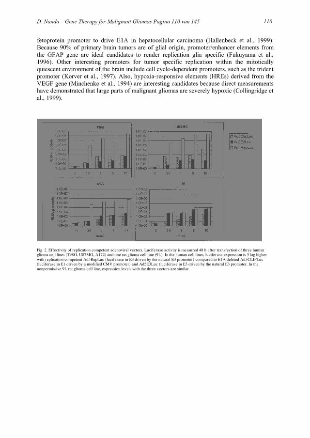
D. Nanda – Gene Therapy for Malignant Gliomas Pagina 110 van 145 110
fetoprotein promoter to drive E1A in hepatocellular carcinoma (Hallenbeck et al., 1999). Because 90% of primary brain tumors are of glial origin, promoter/enhancer elements from the GFAP gene are ideal candidates to render replication glia specific (Fukuyama et al., 1996). Other interesting promoters for tumor specific replication within the mitotically quiescent environment of the brain include cell cycle-dependent promoters, such as the trident promoter (Korver et al., 1997). Also, hypoxia-responsive elements (HREs) derived from the VEGF gene (Minchenko et al., 1994) are interesting candidates because direct measurements have demonstrated that large parts of malignant gliomas are severely hypoxic (Collingridge et al., 1999).
Fig. 2. Effectivity of replication competent adenoviral vectors. Luciferase activity is measured 48 h after transfection of three human glioma cell lines (T98G, U87MG, A172) and one rat glioma cell line (9L). In the human cell lines, luciferase expression is 3 log higher with replication competent Ad5RepLuc (luciferase in E3 driven by the natural E3 promoter) compared to E1A deleted Ad5CLIPLuc (luciferase in E1 driven by a modified CMV promoter) and Ad5E3Luc (luciferase in E3 driven by the natural E3 promoter. In the nonpermissive 9L rat glioma cell line, expression levels with the three vectors are similar.
D. Nanda – Gene Therapy for Malignant Gliomas Pagina 111 van 145 111
Receptor targeting increases the number of infected tumor cells Adenoviral vectors are dependent for their entry into target cells on binding to specific receptors on the cell membrane. This a two-step process that offers additional opportunities for targeting the vector to glioma cells and increasing transfection rates. The first step is normally mediated by attachment of the adenoviral fiber protein to the coxsackie adenoviral receptor (CAR) (Bergelson et al., 1998). The 3-dimensional structure of CAR and the regions binding to the COOH-terminal knob domain of the adenovirus fiber protein have recently been defined (Bewley et al., 1999; Roelvink et al., 1999). After virus attachment, internalization via receptor-mediated endocytosis occurs through the binding of RGD motifs in the viral penton base protein to cellular αvβ3 and αvβ5-integrins (Wickham et al., 1993). In high grade gliomas, low CAR expression limited the utility of adenoviral vectors for gene transfer to primary glioma cultures in vitro. However, retargeting the vector to the epidermal growth factor receptor (EGFR) proved valuable for increased and tumor specific gene transfer (Miller et al., 1998). One method of receptor targeting (redirecting) of adenoviruses involves the development of bispecific antibodies that block the adenoviral knob (that normally recognizes CAR) and are chemically conjugated to an antibody or the ligand reactive with the membrane receptor of choice (Douglas et al., 1996). However, to obtain maximum benefit from a receptor targeted vector, genetic modification of the adenoviral knob is mandatory. The conserved receptor binding sites of CAR binding Adenoviridae within the COOH-terminal knob domain of the fiber protein have been defined (Bewley et al., 1999; Roelvink et al., 1999). Mutations in the AB loop of the Ad5 fiber and introduction of a HA tag into the HI loop of the fiber resulted in a vector that no longer binds to CAR but is, instead, redirected to a new receptor. Subsequent removal of the RGD (Arg–Gly–Asp) sequence from the penton base protein resulted in vector that has no affinity to either CAR or αv-integrins. Krasnykh et al. (1996) demonstrated that the incorporation of an RGD-containing peptide in the HI loop of the fiber knob domain resulted in the ability of the virus to utilize an alternative receptor during the cell entry process. Due to its expanded tissue tropism, this vector was capable of efficient transduction of primary tumor cells resulting in a 2–3 log increase in gene transfer (Dmitriev et al., 1998). Soon, the methods will be available to genetically modify the knob and to produce vectors with stable expression of a target molecule on their surface, avoiding the need of a chemical conjugate (Douglas et al., 1999). The advantages of receptor targeting are two-fold: (a) transfection efficiency of tumor cells is increased; and (b) the liver and other non-target organ and CAR-mediated toxicity is reduced. As a result, lower doses of the vector are required for a similar or better result. Conclusion In conclusion, the first clinical trials with adenoviral vectors in cancer gene therapy demonstrated disappointing efficacy and, when administered at very high doses, toxicity. Ongoing vector development has led to major clinical improvements in other fields of gene therapy. For the treatment of glioma, new carefully designed small clinical studies are required to optimally assess conditionally replicating and targeted adenoviral vectors. Abbreviations SCID severe combined immunodeficiency ADA adenosine deaminase HSV-tk herpes simplex virus thymidine kinase SCID-X1 X-linked form of SCID GCV glanciclovir G-MCSF granulocyte-macrophage colony stimulating factor
D. Nanda – Gene Therapy for Malignant Gliomas Pagina 112 van 145 112
VPC vector producer cells MTD maximum tolerated dose OTC ornithine transcarbamylase IHA into hepatic artery CRAds conditionally replicating adenoviral vectors PSA prostate specific antigen HREs hypoxia-responsive elements CAR coxsackie adenoviral receptor EGFR epidermal growth factor receptor RGD Arg–Gly–Asp References Ammirati, M., Vick, N. and Liao, Y. et al. (1987) Effect of the extent of surgical resection on survival and quality of life in patients with supratentorial glioblastomas and anaplastic astrocytomas. Neurosurgery, 21: 201–206. Barker, F.G. II, Chang, S.M., Gutin, P.H., Malec, M.K., McDermott, M.W., Prados, M.D. and Wilson, C.B. (1998) Survival and functional status after resection of recurrent glioblastoma multiforme. Neurosurgery, 42: 709–720; discussion 720–723. Bergelson, J.M., Krithivas, A., Celi, L., Droguett, G., Horwitz, M.S., Wickham, T., Crowell, R.L. and Finberg, R.W. (1998) The murine CAR homolog is a receptor for coxsackie B viruses and adenoviruses. J. Virol., 72: 415–419. Berger, M.W., Tucker, A., Spence, A. and Winn, H.R. (1992) Reoperation for glioma. Clin. Neurosurg., 39: 177–186. Berkner, K.L. (1992) Expression of heterologous sequences in adenoviral vectors. Curr. Top. Microbiol. Immunol., 158: 39– 66. Bewley, M.C., Springer, K., Zhang, Y.B., Freimuth, P. and Flanagan, J.M. (1999) Structural analysis of the mechanism of adenovirus binding to its human cellular receptor, CAR. Science, 286: 1579–1583. Bischoff, J.R., Kirn, D.H., Williams, A., Heise, C., Horn, S., Muna, M., Ng, L., Nye, J.A., Sampson-Johannes, A., Fattaey, A. and McCormick, F. (1996) An adenovirus mutant that replicates selectively in p53-deficient human tumor cells. Science, 274: 373–377. Blaese, R.M., Culver, K.W., Miller, A.D., Carter, C.S., Fleisher, T., Clerici, M., Shearer, G., Chang, L., Chiang, Y. and Tolstoshev, P. et al. (1995) T lymphocyte-directed gene therapy for ADA- SCID: initial trial results after 4 years. Science, 270: 475–480. Brenner, M. (2000) Reports of adenovector ‘death’ are greatly exaggerated. Mol. Ther., 1: 205. Bruder, J.T. and Kovesdi, I. (1997) Adenovirus infection stimulates the Raf/MAPK signaling pathway and induces interleukin- 8 expression. J. Virol., 71: 398–404.

D. Nanda – Gene Therapy for Malignant Gliomas Pagina 113 van 145 113
Burger, P.C. (1983) Pathologic anatomy and CT correlations in the glioblastoma multiforme. Appl. Neurophysiol., 46: 180– 187. Byrnes, A.P., Wood, M.J. and Charlton, H.M. (1996) Role of T cells in inflammation caused by adenovirus vectors in the brain. Gene Ther., 3: 644–651. Cartmell, T., Southgate, T., Rees, G.S., Castro, M.G., Lowenstein, P.R. and Luheshi, G.N. (1999) Interleukin-1 mediates a rapid inflammatory response after injection of adenoviral vectors into the brain. J. Neurosci., 19: 1517–1523. Cavazzana-Calvo, M., Hacein-Bey, S., de Saint Basile, G., Gross, F., Yvon, E., Nusbaum, P., Selz, F., Hue, C., Certain, S., Casanova, J.L., Bousso, P., Deist, F.L. and Fischer, A. (2000) Gene therapy of human severe combined immunodeficiency (SCID)-X1 disease [see comments]. Science, 288: 669–672. Central Brain Tumor Registry of the United States (1998) 1997 Annual Report, Chicago, IL. Chang, S.M. (2000) The p53 tumor suppressor gene from the bench top to the clinic. First International Symposium on Genetic Anticancer Agents, Amsterdam. Chen, S.-H., Shine, H.D., Goodman, J.C., Grossman, R.G. and Woo, S.L.C. (1994) Gene therapy for brain tumors: regression of experimental gliomas by adenovirus-mediated gene transfer in vivo. Proc. Natl. Acad. Sci. USA, 91: 3054–3057. Collingridge, D.R., Piepmeier, J.M., Rockwell, S. and Knisely, J.P. (1999) Polarographic measurements of oxygen tension in human glioma and surrounding peritumoural brain tissue. Radiother. Oncol., 53: 127–131. Crystal, R.G., McElvaney, N.G., Rosenfeld, M.A., Chu, C.S., Mastrangeli, A., Hay, J.G., Brody, S.L., Jaffe, H.A., Eissa, N.T. and Danel, C. (1994) Administration of an adenovirus containing the human CFTR cDNA to the respiratory tract of individuals with cystic fibrosis. Nat. Genet., 8: 42–51. Culver, K.W., Ram, Z., Walbridge, S., Ishii, H., Oldfield, E.H. and Blaese, R.M. (1992) In vivo gene transfer with retroviral vector producer cells for treatment of experimental brain tumors. Science, 256: 1550–1552. Dewey, R.A., Morrissey, G., Cowsill, C.M., Stone, F., Bolognani, F., Dodd, N.J.F., Southgate, T.D., Klatzmann, D., Lassmann, H., Castro, M.G. and Lowenstein, P. (1999) Chronic brain inflammation and persistent herpes simplex virus 1 thymidine kinase expression in survivors of syngeneic glioma treated by adenovirus-mediated gene therapy: implications for clinical trials. Nat. Med., 5: 1256–1263. Dirks, P., Bernstein, M., Muller, P.J. and Tucker, W.S. (1993) The value of reoperation for recurrent glioblastoma. Can. J. Surg., 36: 271–275. Dmitriev, I., Krasnykh, V., Miller, C.R., Wang, M., Kashentseva, E., Mikheeva, G., Belousova, N. and Curiel, D.T. (1998) An adenovirus vector with genetically modified fibers demonstrates expanded tropism via utilization of a coxsackievirus and adenovirus receptor-independent cell entry mechanism. J. Virol., 72: 9706–9713.

D. Nanda – Gene Therapy for Malignant Gliomas Pagina 114 van 145 114
Douglas, J.T., Rogers, B.E., Rosenfeld, M.E., Michael, S.I., Feng, M. and Curiel, D.T. (1996) Targeted gene delivery by tropism-modified adenoviral vectors. Nat. Biotechnol., 14: 1574–1578. Douglas, J.T., Miller, C.R., Kim, M., Dmitriev, I., Mikheeva, G., Krasnykh, V. and Curiel, D.T. (1999) A system for the propagation of adenoviral vectors with genetically modified receptor specificities. Nat. Biotechnol., 17: 470–475. Driesse, M.J., Vincent, A.J.P.E., Sillevis Smitt, P.A.E., Kros, J.M., Hoogerbrugge, P.M., Avezaat, C.J.J., Valerio, D. and Bout, A. (1998) Intracerebral injection of adenovirus harboring the HSVtk gene combined with ganciclovir administration: toxicity study in nonhuman primates. Gene Ther., 5: 1122– 1130. Driesse, M.J., Esandi, M.C., Kros, J.M., Avezaat, C.J.J., Vecht, C.J., Zurcher, C., van der Velde, I., Valerio, D., Bout, A. and Sillevis Smitt, P.A.E. (2000) Intra-CSF administered recombinant adenovirus causes an immune response-mediated toxicity. Gene Ther., in press. Eck, S.L., Alavi, J.B., Alavi, A., Davis, A., Hackney, D., Judy, K., Mollman, J., Phillips, P.C., Wheeldon, E.B. and Wilson, J.M. (1996) Treatment of advanced CNS malignancies with the recombinant adenovirus H5.010RSVTK: a phase I trial. Hum. Gene Ther., 7: 1465–1482. Fernandez-Chacon, C. (2000) Gene therapy targeting overexpression of the tumor suppressor gene p53 represents a novel approach acting at the molecular level. First International Symposium on Genetic Anticancer Agents, Amsterdam. Friedman, H.S., Petros, W.P., Friedman, A.H., Schaaf, L.J., Kerby, T., Lawyer, J., Parry, M., Houghton, P.J., Lovell, S., Rasheed, K., Cloughsey, T., Stewart, E.S., Colvin, O.M., Provenzale, J.M., McLendon, R.E., Bigner, D.D., Cokgor, I., Haglund, M., Rich, J., Ashley, D., Malczyn, J., Elfring, G.L. and Miller, L.L. (1999) Irinotecan therapy in adults with recurrent or progressive malignant glioma. J. Clin. Oncol., 17: 1516–1525. Fukuyama, K., Matsuzawa, K., Hubbard, S.L., Dirks, P.B., Murakami, M. and Rutka, J.T. (1996) Analysis of glial fibrillary acidic protein gene methylation in human malignant gliomas. Anticancer Res., 16: 1251–1257. Goodman, J.C., Trask, T.W., Chen, S.H., Woo, S.L., Grossman, R.G., Carey, K.D., Hubbard, G.B., Carrier, D.A., Rajagopalan, S., Aguilar-Cordova, E. and Shine, H.D. (1996) Adenoviralmediated thymidine kinase gene transfer into the primate brain followed by systemic ganciclovir: pathologic, radiologic, and molecular studies. Hum. Gene Ther., 7: 1241–1250. Hallenbeck, P.L., Chang, Y.N., Hay, C., Golightly, D., Stewart, D., Lin, J., Phipps, S. and Chiang, Y.L. (1999) A novel tumor-specific replication-restricted adenoviral vector for gene therapy of hepatocellular carcinoma. Hum. Gene Ther., 10: 1721–1733. Hamel, W., Magnelli, L., Chiarugi, V.P. and Israel, M.A. (1996) Herpes simplex virus thymidine kinase/ganciclovir-mediated apoptotic death of bystander cells. Cancer Res., 56: 2697–2702.

D. Nanda – Gene Therapy for Malignant Gliomas Pagina 115 van 145 115
Harsh, G.R., Levin, V.A., Gutin, P.H., Seager, M., Silver, P. and Wilson, C.B. (1987) Reoperation for recurrent glioblastoma and anaplastic astrocytoma. Neurosurgery, 21: 615. Heise, C.C., Williams, A., Olesch, J. and Kirn, D.H. (1999) Efficacy of a replication-competent adenovirus (ONYX-015) following intratumoral injection: intratumoral spread and distributioneffects. Cancer Gene Ther., 6: 499–504. Hoogerbrugge, P.M., van Beusechem, V.W., Fischer, A., Debree, M., le Deist, F., Perignon, J.L., Morgan, G., Gaspar, B., Fairbanks, L.D., Skeoch, C.H., Moseley, A., Harvey, M., Levinsky, R.J. and Valerio, D. (1996) Bone marrow gene transfer in three patients with adenosine deaminase deficiency. Gene Ther., 3: 179–183. Isner, J.M. and Asahara, T. (1999) Angiogenesis and vasculogenesis as therapeutic strategies for postnatal neovascularization. J. Clin. Invest., 103: 1231–1236. Kajiwara, K., Byrnes, A.P., Charlton, H.M., Wood, M.J. and Wood, K.J. (1997) Immune responses to adenoviral vectors during gene transfer in the brain. Hum. Gene Ther., 8: 253– 265. Kay, M.A., Manno, C.S., Ragni, M.V., Larson, P.J., Couto, L.B., McClelland, A., Glader, B., Chew, A.J., Tai, S.J., Herzog, R.W., Arruda, V., Johnson, F., Scallan, C., Skarsgard, E., Flake, A.W. and High, K.A. (2000) Evidence for gene transfer and expression of factor IX in haemophilia B patients treated with an AAV vector [see comments]. Nat. Genet., 24: 257– 261. Klatzmann, D., Valery, C.A., Bensimon, G., Marro, B., Boyer, O., Mokhtari, K., Diquet, B., Salzmann, J.L. and Philippon, J. (1998) A phase I/II study of herpes simplex virus type 1 thymidine kinase ‘suicide’ gene therapy for recurrent glioblastoma. Study Group on Gene Therapy for Glioblastoma. Hum Gene Ther. 2595–2604. Korver, W., Roose, J., Heinen, K., Weghuis, D.O., de Bruijn, D., van Kessel, A.G. and Clevers, H. (1997) The human TRIDENT/HFH-11/FKHL16 gene: structure, localization, and promoter characterization. Genomics, 46: 435–442. Kramm, C.M., Chase, M., Herrlinger, U., Jacobs, A., Pechan, P.A., Rainov, N.G., Sena-Esteves, M., Aghi, M., Barnett, F.H., Chiocca, E.A. and Breakefield, X.O. (1997) Therapeutic efficiency and safety of a second-generation replication-conditional HSV1 vector for brain tumor gene therapy. Hum. Gene Ther., 8: 2057–2068. Krasnykh, V.N., Mikheeva, G.V., Douglas, J.T. and Curiel, D.T. (1996) Generation of recombinant adenovirus vectors with modified fibers for altering viral tropism. J. Virol., 70: 6839– 6846. Lehrman, S. (1999) Virus treatment questioned after gene therapy death. Nature, 401: 517–518. Miller, C.R., Buchsbaum, D.J., Reynolds, P.N., Douglas, J.T.,Gillespie, G.Y., Mayo, M.S., Raben, D. and Curiel, D.T. (1998) Differential susceptibility of primary and established human glioma cells to adenovirus infection: targeting via the epidermal growth factor receptor achieves fiber receptor-independent gene transfer. Cancer Res., 58: 5738–5748.

D. Nanda – Gene Therapy for Malignant Gliomas Pagina 116 van 145 116
Minchenko, A., Salceda, S., Bauer, T. and Caro, J. (1994) Hypoxia regulatory elements of the human vascular endothelial growth factor gene. Cell. Mol. Biol. Res., 40: 35–39. Rainov, N.G. (2000) (on behalf of the multicenter N. American- European study group). Herpes-simplex thymidine kinase gene/ganciclovir gene therapy for primary malignant glioma. Results of a phase III study. First International Symposium on Genetic Anticancer Agents, Amsterdam. Ram, Z., Walbridge, S., Shawker, T., Culver, K.W., Blaese, R.M. and Oldfield, E.H. (1994) The effect of thymidine kinase transduction and ganciclovir therapy on tumor vasculature and growth of 9L gliomas in rats. J. Neurosurg., 81: 256–260. Ram, Z., Culver, K.W., Oshiro, E.M., Viola, J.J., DeVroom, H.L., Otto, E., Long, Z., Chiang, Y., McGarrity, G.J., Muul, L.M., Katz, D., Blaese, R.M. and Oldfield, E.H. (1997) Therapy of malignant brain tumors by intratumoral implantation of retroviral vector-producing cells. Nat. Med., 3: 1354–1361. Rodriguez, R., Schuur, E.R., Lim, H.Y., Henderson, G.A., Simons,J.W. and Henderson, D.R. (1997) Prostate attenuated replication competent adenovirus (ARCA) CN706: a selective cytotoxic for prostate-specific antigen-positive prostate cancer cells. Cancer Res., 57: 2559–2563. Roelvink, P.W., Mi Lee, G., Einfeld, D.A., Kovesdi, I. and Wickham, T.J. (1999) Identification of a conserved receptor-binding site on the fiber proteins of CAR-recognizing adenoviridae. Science, 286: 1568–1571. Rogulski, K.R., Wing, M.S., Paielli, D.L., Gilbert, J.D., Kim, J.H. and Freytag, S.O. (2000) Double suicide gene therapy augments the antitumor activity of a replication-competent lytic adenovirus through enhanced cytotoxicity and radiosensitization. Hum. Gene Ther., 11: 67–76. Salcman, M., (1995) Glioblastoma and malignant astrocytoma. In: A.H. Kaye and E.R. Laws (Eds.), Brain Tumors, Churchill Livingstone, New York, pp. 449–479. Shand, N., Weber, F., Mariani, L., Bernstein, M., Gianella-Borradori, A., Long, Z., Sorensen, A.G. and Barbier, N. (1999) A phase 1–2 clinical trial of gene therapy for recurrent glioblastoma multiforme by tumor transduction with the herpes simplex thymidine kinase gene followed by ganciclovir. GLI328 European-Canadian Study Group. Hum. Gene Ther., 10: 2325– 2335. Shine, H.D., Wyde, P.R., Aguilar-Cordova, E., Chen, S.-H., Woo, S.L.C., Grossman, R.G. and Goodman, J.C. (1997) Neurotoxicity of intracerebral injection of a replication-defectiveadenoviral vector in a semipermissive species (cotton rat). Gene Ther., 4: 275–279. Smith, J.G., Raper, S.E., Wheeldon, E.B., Hackney, D., Judy, K., Wilson, J.M. and Eck, S.L. (1997) Intracranial administration of adenovirus expressing HSV-TK in combination with ganciclovir produces a dose-dependent, self-limiting inflammatory response. Hum. Gene Ther., 8: 943–954.

D. Nanda – Gene Therapy for Malignant Gliomas Pagina 117 van 145 117
Smith, R.R. et al. (1956) Studies on the use of viruses in the treatment of carcinoma of the cervix. Cancer, 9: 1211–1218. Surawicz, T.S., Davis, F., Freels, S., Laws Jr., E.R. and Menck, H.R. (1998) Brain tumor survival: results from the National Cancer Data Base. J. Neurooncol., 40: 151–160. Tacket, C.O., Losonsky, G., Lubeck, M.D., Davis, A.R., Mizutani, S., Horwith, G., Hung, P., Edelman, R. and Levine, M.M. (1992) Initial safety and immunogenicity studies of an oral recombinant adenohepatitis B vaccine. Vaccine, 10: 673–676. Touraine, R.L., Ishii-Morita, H., Ramsey, W.J. and Blaese, R.M. (1998) The bystander effect in the HSVtk/ganciclovir system and its relationship to gap junctional communication. Gene Ther., 5: 1705–1711. Trask, T.W., Trask, R.P., Aguilar-Cordova, E., Shine, H.D., Wyde, P.R., Goodman, J.C., Hamilton, W.J., Rojas-Martinez, A., Chen, S.-H., Woo, S.L.C. and Grossman, R.G. (2000) Phase I study of adenoviral delivery of the HSV-tk gene and ganciclovir administration in patients with recurrent malignant brain tumors. Mol. Ther., 1: 195–203. Verlinden, S.F., Esandi, C., Zurcher, C., Vogels, R. and Van Bekkum, D.W., (2000) Hurdles to Overcome for Successful Adenoviral in vivo Gene Therapy, American Society of Gene Therapy, Denver. Vile, R.G., Castleden, S., Marshall, J., Camplejohn, R., Upton, C. and Chong, H. (1997) Generation of an anti-tumour immune response in a non-immunogenic tumour: HSVtk killing in vivo stimulates a mononuclear cell infiltrate and a Th1-like profile of intratumoural cytokine expression. Int. J. Cancer, 71: 267– 274. Vincent, A.J.P.E., Vogels, R., Van Someren, G., Esandi, M.C., Noteboom, J.L., Avezaat, C.J.J., Vecht, C., Van Bekkum, D.W., Valerio, D., Bout, A. and Hoogerbrugge, P.M. (1996) Herpes simplex virus thymidine kinase gene therapy for rat malignant brain tumors. Hum. Gene Ther., 7: 197–205. Vincent, A.J., Esandi, M.C., Avezaat, C.J., Vecht, C.J., Sillevis Smitt, P., van Bekkum, D.W., Valerio, D., Hoogerbrugge, P.M. and Bout, A. (1997) Preclinical testing of recombinant adenoviral herpes simplex virus thymidine kinase gene therapy for central nervous system malignancies. Neurosurgery, 41: 442–451. Visser, O., Coebergh, J.W.W., Schouten, L.J. and van Dijck, J.A.A.M. (1998) Incidence of Cancer in the Netherlands 1995, Vereniging van Integrale Kankercentra, Utrecht. Wallner, K.E., Galicich, J.H., Krol, G., Arbit, E. and Malkin, M.G. (1989) Patterns of failure following treatment for glioblastoma multiforme and anaplastic astrocytoma. Int. J. Radiat. Oncol. Biol. Phys., 16: 1405–1409. Wickham, T.J., Mathias, P., Cheresh, D.A. and Nemerow, G.R. (1993) Integrins alpha v beta 3 and alpha v beta 5 promote adenovirus internalization but not virus attachment. Cell, 73: 309–319.

D. Nanda – Gene Therapy for Malignant Gliomas Pagina 118 van 145 118
Wildner, O., Morris, J.C., Vahanian, N.N., Ford, H., Ramsey, W.J. and Blaese, R.M. (1999) Adenoviral vectors capable of replication improve the efficacy of HSVtk/GCV suicide gene therapy of cancer. Gene Ther., 6: 57–62. Yung, W.K., Prados, M.D., Yaya-Tur, R., Rosenfeld, S.S., Brada, M., Friedman, H.S., Albright, R., Olson, J., Chang, S.M., O’Neill, A.M., Friedman, A.H., Bruner, J., Yue, N., Dugan, M., Zaknoen, S. and Levin, V.A. (1999) Multicenter phase II trial of temozolomide in patients with anaplastic astrocytoma or anaplastic oligoastrocytoma at first relapse. Temodal Brain Tumor Group. J. Clin. Oncol., 17: 2762–2771.

D. Nanda – Gene Therapy for Malignant Gliomas Pagina 119 van 145 119
CHAPTER 8 Summary and general discussion

D. Nanda – Gene Therapy for Malignant Gliomas Pagina 120 van 145 120

D. Nanda – Gene Therapy for Malignant Gliomas Pagina 121 van 145 121
Due to the locoregional nature of malignant gliomas and their poor prognosis, recurrent malignant gliomas proved an excellent model to study gene therapy approaches. Several phase I/II clinical trials were performed using non-replicating Adenovirus serotype 5 (Ad5) based vectors, expressing the HSV-tk gene in combination with ganciclovir in recurrent malignant gliomas. These trials have clearly demonstrated the feasibility and safety of the approach 1-6. However, the results of the initial clinical gene therapy trials in malignant gliomas have been disappointing. The limited clinical success of adenovirus-based gene therapy is in strong contrast with results obtained in many animal models of gliomas . In chapter 2, the many hurdles that are implicated in the limited success of clinical gene therapy trials in gliomas are discussed. These include the low expression level of CAR on primary glioma cells and tumors as opposed to established cell lines 7,8, the limited distribution of adenoviral vectors following intratumoral injection 5,9 and the high Ad5 seroprevalence in the general population 10-12. Identification of these hurdles in carefully conducted clinical trials 9 led to dramatic improvements in vector design as outlined in chapter 2. An important development has been the construction of adenoviral vectors that replicate selectively in glioma cells (recently reviewed by Jiang et al. 13). The selectivity and efficacy of adenoviral vectors is further increased by genetic modification of their tropism 10,13. Hopefully these modifications will not only increase the safety and efficacy of these vectors but also reduce their toxicity and even permit systemic administration. To further improve vector distribution, new methods of administration have been pioneered, including convection enhanced delivery and intravascular administration 14. Molecular imaging techniques are being developed that may provide surrogate endpoints in clinical glioma trials when they predict treatment outcome. Ideally, it will become feasible to non-invasively measure transduction levels or functional enzyme activity in order to correlate this with clinical changes after e.g. ganciclovir (GCV) treatment. Direct labeling of viral particles with radioactive molecules allows immediate visualization of vector distribution 14. In chapter 3, we attempted to improve the efficacy of E1-deleted adenoviral vectors, by constructing several replication-competent adenoviral vectors carrying both E1A and E1B sequences and the HSV1-tk suicide gene. In these vectors, the luciferase (IG.Ad5E1+. E3Luc) or HSV1-tk gene (IG.Ad5E1+.E3TK) replaced the coding sequence of the E3 region gp19K that binds to class I MHC in the endoplasmic reticulum, preventing antigen presentation on the cell surface 15. The transgenes were placed under control of the natural E3 promoter, because heterologous promoters were found to be silent when inserted in this area 16-18. To examine the transgene expression levels obtained from the internal E3 promoter, we also constructed an E1-deleted vector carrying the luciferase gene in E3 (IG.Ad5.Sarcoma 1800HSA.E3Luc). For in vitro experiments, we used a panel of human glioma cell lines (U87 MG, T98G, A172, LW5, and U251), a rat gliosarcoma cell line (9 L), and human lung (A549) and prostate carcinoma (P3) cell lines. The luciferase expression obtained with IG.Ad5.Sarcoma 1800HSA.E3Luc was similar to a first-generation, E1- deleted vector carrying the luciferase gene in E1 driven by a modified CMV-promoter (IG.Ad5.ClipLuc), indicating the strength of the natural E3 promoter in these constructs, even in the absence of E1. We then compared the luciferase expression levels of the E1+luciferase vector with both E1-deleted vectors and found that luciferase expression was ~3 logs higher with the E1+ vector in all permissive (human) cell lines tested. Similar expression levels were obtained with ~2 log lower m.o.i. of the E1+ vector. Interestingly, infection of the nonpermissive 9L cell line with the E1+ vector resulted in significantly higher luciferase expression than with the E1-deleted vectors. This may indicate either low-level replication of the E1+ vector in the rodent cell line or transactivation of the E3 promoter by E1 proteins. Apart from the direct oncolytic effect on

D. Nanda – Gene Therapy for Malignant Gliomas Pagina 122 van 145 122
tumor cells 19, replication- competent vectors may enhance further the antitumor efficacy by increased expression levels of a tumoricidal transgene. A concern was the potential disruption of the adenovirus death protein (E3–11.6K) by our cloning strategy. Adenovirus death protein is produced in large amounts at the late stage of infection and is required for effective cell lysis and virus release 20. However, the oncolytic effect of the gp19K-deleted, replicating vectors was similar to wtAd5, as measured by the time after infection to full CPE on our panel of cell lines. Interestingly, the time to CPE seemed not to correlate with the level of luciferase expression, indicating that the efficacy of replication may not only depend on the efficacy of cell entry. Elucidating mechanisms affecting the efficiency of adenoviral replication in different cell lines requires additional study to enhance replication potentially 21. As expected, the replication-competent vectors and wtAd5 did not cause CPE in the nonpermissive 9L rat gliosarcoma cell line. In vitro, GCV sensitivity (10 µg/ml) was studied in U87 MG cells after infection at a multiplicity of infection of 1 and 10. The oncolytic effect of replication-competent IG.Ad5E1+.E3TK was significantly enhanced by the addition of GCV and greatly exceeded the cytotoxicity of replication-incompetent IG.AdApt.TK combined with GCV. We then treated s.c. U87 MG glioma xenografts (volume 100–150 mm3) in NIH-bg-nu-xid mice with a single injection of adenovirus 109 IU suspended in 100 µl of PBS, and GCV 100 mg/kg was administered i.p. twice daily for 7 days. A single injection of IG.Ad5E1+.E3TK resulted in a significant slowing of tumor growth and prolonged survival compared with injection of IG.AdApt.TK. Addition of GCV slowed tumor growth, further adding to survival. Also, the E1+ HSV1-tk vector in combination with GCV was significantly more effective than the E1-deleted HSV1-tk vector with GCV. Replication-competent adenoviral vectors are a new and rapidly evolving platform for gene therapy 13,22. In the past, intratumoral injection of wild-type adenoviruses has not led to unwanted effects 23. However, “virotherapy” for cancer was abandoned because of only a few documented responses and because of the advent of more effective chemotherapeutic agents 24. Bischoff et al. 19 reintroduced the oncolytic effect of adenoviruses with vectors carrying mutations in the early region of the encoding E1B-Mr 55,000. Subsequently, an additive effect of HSV1-tk/GCV and radiotherapy to the oncolytic effect of E1B-deleted vectors was demonstrated 25,26. The use of conditionally replicating vectors that contain deletions in E1B (or E1A) carries the risk of reduced replication as compared with wtAd5. The present study demonstrates that even vectors replicating as well as wtAd5 don’t cause tumor eradication in some models. Therefore, addition of a therapeutic transgene is probably mandatory, and genes with improved bystander and cytotoxic effects may add further to the efficacy of replicating vectors 27. The toxic genes, such as the HSV1-tk gene, can also function as a fail-safe mechanism when replication outside the tumor occurs 26. Here, we demonstrated that adenoviral replication and subsequent spread can be effectively blocked by the immediate administration of GCV. The complex interactions between viral replication and GCV require additional in vivo studies to determine the optimal interval between vector injection and first GCV administration. In clinical studies, conditional replication will be required for patient- and biosafety. An alternative for selective replication is to drive the E1B or/and E1A genes by a tissue or tumor-specific promoter for instance by using a glia-specific promoter such as the gfa2 promoter, to drive the E1A gene 28,29. In chapter 4, we examined the value of 123I-FIRU for imaging of HSV1-tk expression. In vitro incubation of 9L-tk+ cells and the parental 9L cells with 123I-FIRU resulted in a 100-fold higher accumulation of radioactivity in the 9L-tk+ cells after an optimum incubation time of 4 h. The subsequent removal of 123I-FIRU from the medium did not result in substantial loss of intracellular 123I-FIRU, indicating efficient sequestration probably caused by effective

D. Nanda – Gene Therapy for Malignant Gliomas Pagina 123 van 145 123
phosphorylation. NIH-bg-nu-xid mice were then inoculated subcutaneously with HSV1-tk (–) 9L cells or HSV1-tk (+) 9L-tk+ cells into both flanks. Biodistribution studies and gamma camera imaging were performed at 15 min and 1, 2, 4 and 24 h p.i. At 15 min, the tumour/muscle, tumour/blood and tumour/brain ratios were 5.2, 1.0 and 30.3 respectively. Rapid renal clearance of the tracer from the body resulted in increasing tumour/muscle, tumour/blood and tumour/brain ratios, reaching values of 32.2, 12.5 and 171.6 at 4 h p.i. A maximum specific activity of 22%ID/g tissue was reached in the 9L-tk+ tumours 4 h after 123I-FIRU injection. This specific accumulation of radioactivity is higher than the specific accumulation obtained with either 125I-FIAU (9.8% ID/g) or 18F-FHPG (0.08% ID/g) 30. Using 125I-FIAU, Haubner et al. 31 reported tumour to blood and tumour to muscle ratios at 4 h p.i. of 32.0 and 88.3. In a more recent paper by the same group 30 125I-FIAU ratios at 4 h p.i., calculated, reveal a tumour to blood ratio of 24.4 and a tumour to muscle ratio of 21. These results indicate that a direct comparison of FIAU and FIRU as substrate for gene imaging is warranted. We then studied the sequestration of 123I-FIRU in the human glioma cell lines U87MG and T98G following infection with two adenoviral vectors carrying the HSV1-tk transgene. IG.Ad5.E1+.E3TK is a replication-competent Ad5-based virus that we constructed to amplify the antitumour efficacy of adenovirus-based gene therapy 32. IG.Ad5.E1+.E3TK results in more efficient cell kill than IG.AdApt.TK, an E1-deleted Ad5-based virus containing the HSV1-tk gene driven by the human CMV promoter in the former E1 region 32. The accumulation of 123I-FIRU in U87MG and T98G cells was significantly higher following infection with the replication- competent adenovirus compared to the non-replicating vector. To investigate the possibility of detecting differences in HSV1-tk gene expression levels in vivo we injected U87MG xenografts with either 108 or 109 I.U. of replication- competent IG.Ad5.E1+.E3TK and non-replicating IG.AdApt.TK. Following injection of 123I-FIRU, we found that the accumulation of radioactivity was higher in the tumours injected with 109 I.U. of either virus compared to the tumours injected with 108 I.U. Also, the tumours injected with the replication-competent virus showed higher radioactivity levels than tumours injected with the non-replicating vector. These findings correlate with images obtained with the clinical gamma camera and demonstrate the feasibility of detecting differences in HSV1-tk expression levels using 123I-FIRU in vivo following adenoviral gene therapy. The high ratio with normal brain may make FIRU and similar compounds suitable for imaging gene transfer in brain tumours. Jacobs et al. published a study in which five patients enrolled in a gene therapy procedure using a liposomal vector carrying an HSV-tk gene 33. They performed a dynamic PET scan using 124I-FIAU, similar to radiolabeled FIRU. They showed a specific HSV-tk-related uptake of FIAU at the site of injection in one patient, who also showed a response to treatment. Unfortunately, in the other patients no increased FIAU uptake could be measured; they also failed to respond to treatment. This study shows that non-invasive imaging of HSV-tk gene expression is feasible and highly desirable in order to assess gene transfer. Dempsey et al. reported on eight GBM patients imaged with SPECT using 123I-FIAU prior to and after application of an oncolytic HSV virus 34. However, no increased uptake of FIAU was determined after viral infection in these patients. A possible explanation is that FIAU might not be the ideal tracer for monitoring HSV-tk expression in subjects with an intact blood brain barrier, as FIAU does not penetrate it 34,35. In conclusion, 123I-FIRU accumulates highly selectively in tumor expressing the HSV1-tk gene either constitutively or following adenoviral gene transfection. In vivo imaging of the level of HSV1-tk expression is possible and will be valuable in both animal studies as in ongoing clinical trials. Future studies will compare the imaging characteristics of tracers that accumulate in the cell following HSV1-tk transfection with those obtained with tracers

D. Nanda – Gene Therapy for Malignant Gliomas Pagina 124 van 145 124
directed at transfected cell surface receptors such as the type 2 somatostatin receptor 36or dopamine type 2 receptor 37, or the sodium-iodide symporter (NIS) 38,39. Chapter 5 describes a simple Sep-Pak based method for the purification of radio-iodinated FIRU allowing large scale, clinical grade production. The described carrier-mediated labeling method is simple, and purification over a Sep-Pak column is much more convenient than preparative HPLC, in particular for the production of clinical grade under GMP conditions. Using the described labeling and purification strategy, [123I]FIRU was routinely produced with a radiochemical purity > 97 %, a labeling yield of ~ 56 % and a specific activity not less than 690 MBq/µmol. The in vitro toxicity of FIRU, FIAU and related compounds was studied in a number of cell lines, using the MTT assay. These studies demonstrated lower toxicity of FIRU as compared to FIAU in both non-HSV1-TK expressing cells (IC50 ratio 3.8–500) and in HSV1-TK expressing cells (IC50 ratio 1.7-2 x 10⁷). The lack of toxicity of the phosphorylated FIRU compound is most likely due to the fact that FIRU is not incorporated into the DNA of the mammalian dividing cell40, as opposed to FIAU 41,42. In dividing cells that don’t express HSV1-tk, FIRU was also less toxic than FIAU. This difference probably reflects a lower affinity of the endogenous TK enzyme for FIRU than for FIAU, but in neither case is uptake sufficient to warrant use as a cell proliferation marker 43. Uptake of FIAU was reported to be about 20-fold higher than that of IUdR in D-247 MG cells, leading those authors to conclude that [125I]FIAU might have greater cytotoxicity for this cell line than [125I]IUdR 44. Recently, a multiplexing multi-pinhole small-animal SPECT system has been described with greatly improved sensitivity while achieving high resolutions 45. Using this system, we could clearly demonstrate the uptake of FIRU in HSV-tk expressing xenografts. Choi et al46 earlier showed microSPECT images of [123I]FIAU uptake in HSV-tk expressing xenografts. These high-resolution SPECT images clearly demonstrated inhomogeneous tracer uptake in one of the xenografts, suggesting necrosis of the interior portion of the tumor. Progress in nuclear technology and fusion of multiple imaging modalities, such as SPECT and CT, will further enhance the sensitivity and accuracy of molecular imaging. Biodistribution and early kinetics studies of [123I]FIRU in tumor bearing mice showed initial phase and terminal phase half-lives of respectively 0.8 h and 1.3 h (blood); 0.1 h and 4 h (muscle); and 0.5 h and 6.7 h (9L tumor). In 9L-tk+ tumors, [123I]FIRU accumulated for about one hour. Subsequently, the radioactivity incorporated in these tumors decreased with a much longer half-life (11.3 h) than in the surrounding tissues. The optimal imaging time with [123I]FIRU is 4 h post-injection 40,47. Finally, in preparation of clinical studies, we performed toxicity studies with ‘cold’ FIRU in rats and mice. The injected amount of FIRU per animal was 1x, 20x or 200x fold (mg/kg) of the intended human dose for imaging (100 � g (0.27 � mol) / 70 kg). Because the mitochondrial toxicity associated with FIAU manifests mainly as hepatotoxicity, pancreatitis, neuropathy or myopathy, we focused on these organs and the hematopoietic system 48. This multisystem mitochondrial toxicity is probably caused by the high rate at which FIAU is incorporated into mitochondrial DNA49,50. We could detect no clinical, laboratory or pathological signs of toxicity due to FIRU. In particular, all liver function tests and amylase levels were normal. Pathological examination of liver and pancreas and of the peripheral nervous system and muscle was completely normal. In conclusion, radiolabeled FIRU can be easily produced in clinical grade batches. In addition, the safety profile and clear imaging properties make FIRU an attractive candidate tracer for clinical imaging of HSV1-tk gene transfer.

D. Nanda – Gene Therapy for Malignant Gliomas Pagina 125 van 145 125
In chapter 6, we set out to identify an adenoviral vector better suited to infect primary glioma cells than Adenovirus serotype 5 (Ad5). The overall low expression levels of the Coxsackie and Adenovirus receptor (CAR) and the presence of high anti-Ad5- neutralizing antibody (NAb) titers in the general population are considered detrimental for consistency of clinical results. We tested a library of 16 fiber-chimeric Ad5-based adenoviral vectors on 12 primary human glioma cell suspensions. Compared to Ad5, we found significantly improved marker gene expression with several chimeric vectors, predominantly vectors carrying fiber molecules derived from B-serogroup viruses (Ad11, Ad16, Ad35 and Ad50). To explain this remarkable finding, we examined the expression of several molecules involved in adenoviral binding and entry on the primary glioma cells, including CAR, CD46 and � v � integrins. We found significant differences between tumor isolates in CAR and integrin expression. In vitro culture artefacts are an unlikely explanation for these differences, because the tumor cell preparations were screened early after tumor isolation. These data confirm an earlier report by Fuxe et al. 51 who also described variable CAR expression in 34 gliomas of various grade and demonstrated patient-to-patient variation that at present is not well understood. Although the exact function of the Ad5 high affinity receptor CAR is not known, CAR expression may have a tumor suppressing effect 52-54 and correlates inversely with the aggressiveness of a tumor, explaining the low expression levels of CAR in malignant gliomas and primary glioma cells 51,54. The B-group adenoviruses were recently found to use CD46 as a high affinity cellular attachment molecule 55,56. The CD46 molecule is ubiquitously expressed in primates and functions to protect “self”-cells against complement lysis by binding and inactivating C3b and C4b 57. The expression level of the BC-1 isoform of CD46 was significantly higher than the CAR expression level on primary glioma cells although both attachment molecules were expressed at highly variable levels. The expression of the BC1-isoform (one of four known isoforms of CD46) did not closely correlate with marker gene expression levels obtained after infection with the B-group fiber-chimeric vectors. This may indicate that other isoforms of CD46 are perhaps expressed by these cells and may contribute to binding and internalization of the B-group fiber-chimeric vectors. Since CD46 expression is very low on neurons 58 and because B-group chimeric vectors infect glioma cells more efficiently than Ad5, a recombinant vector that uses CD46 as attachment molecule might have a more favorable targeting ratio in the brain than Ad5. Skog et al 59 studied the infection of seven low-passage glioma cell lines by six wildtype adenovirus serotypes, including Ad5. As determined by hexon expression, the B-group viruses Ad16 and Ad21 infected the glioma cells most efficiently, confirming the improved binding and entrance by B-group viruses as compared to CAR-binding Ad5. Hoffmann et al. 60 recently compared several conditionally replicative adenoviral vectors, including Ad5 based chimeric vectors with an Ad35 fiber (Ad5/35). Animals treated with the Ad5/35-based vectors showed significantly smaller tumors and longer survival than those treated with the homologous Ad5 vectors and no significant toxicity was observed in the intracranial model. These data confirm that Ad5/35-based vectors are promising tools for glioblastoma treatment 60. We next tested Ad35 sero prevalence in sera derived from 90 Dutch cancer patients including 30 glioma patients and investigated the transduction efficiency of this vector in glioma cell suspensions. Our results demonstrated that the sero prevalence and the titers of NAb against Ad35 were significantly lower than against Ad5. Also, recombinant Ad35 has significantly increased ability to transfer a gene to primary glioma cells compared to Ad5. We thus conclude that Ad35 represents an interesting candidate vector for gene therapy of malignant glioma.

D. Nanda – Gene Therapy for Malignant Gliomas Pagina 126 van 145 126
Between November 1998 and December 2001, we treated 14 patients with advanced recurrent high grade gliomas with a total dose of 4.6 x 10⁸, 4.6 x 10⁹, 4.6 x 10¹⁰ or 4.6 x 10¹¹ viral particles (VP) of a replication incompetent adenoviral vector harboring the herpes simplex virus thymidine kinase (HSV-tk) gene driven by the adenoviral major late promoter (IG.Ad.MLPI.TK), followed by ganciclovir (GCV) treatment 61. Inclusion was temporarily suspended following the tragic death of Mr. Gelsinger in the fall of 1999 62. In the spring of 2000, CCMO approved continued inclusion of patients and the study was reopened. Chapter 7 describes the preliminary results in the first eleven patients that were enrolled before the fall of 1999. The VP to infectious unit ratio was 40 and the vector was administered by 50 intra-operative woundbed injections of 0.2 mL each (total volume 10 mL). The study’s primary objective was to determine the safety of this treatment and establish the maximum tolerated dose (MTD). Injection of all doses of IG.Ad.MLPI.TK followed by GCV was safely tolerated and MTD was not reached. All patients had recurrence or progression of the tumor 1-24 months (median 3.5 months) after gene therapy 61. The overall median survival was 4 months 61. Four patients survived longer than one year following gene therapy and three of these patients had non-GBM pathology upon review (anaplastic oligodendroglioma in two and anaplastic mixed oligoastrocytoma in one 61). Ten patients died within 8 months of treatment, all from progression of the tumor 61. In five patients residual and measurable tumor was visible on the direct (<48 hours) postoperative MRI. No objective radiological response was documented on subsequent MRI 61. Recently, a randomized controlled trial reported the successful adjuvant treatment of malignant glioma patients with Ad5-tk (Cerepro®). Ad5-tk treatment produced a significant increase in mean survival from 39 to 71 weeks (P< 0.01) 6. The validity of this trial’s conclusions is compromised by the inclusion of newly diagnosed and recurrent glioma patients of different grades in both treatment and control groups and the imbalances between grade 3 and 4 patients between these groups. Nevertheless, based on these results a phase III trial in 250 newly diagnosed GBM has been conducted by Ark Therapeutics Ltd. The results of this study have not yet been reported. Future studies. Based on promising results in many animal models, the first clinical gene therapy trials in malignant gliomas were initiated with unrealistic optimism. As a result, the outcome of these first clinical trials was mostly disappointing. However, the careful conduct of many clinical trials has allowed us to pinpoint many of the barriers that limited the clinical efficacy and most of these barriers have been more or less successfully addressed in the laboratory. It is now time to assess the improved viral vectors in clinical trial. Adenoviral vectors are still very promising candidates to further explore in malignant glioma trials because of their proven safety in thousands of cancer patients. In addition, the adenoviral genome has been well studied and the function of most adenoviral genes are understood. A new adenoviral vector to test in the clinic should a) be (conditionally) replication competent; b) have a genetically altered tropism; c) be armed with a therapeutic gene; d) allow in vivo imaging of vector distribution and duration and extent of transgene expression; and e) be able to evade naturally existing immunity in the general population. An interesting candidate vector that will soon be studied in GBM patients in the Department of Neurosurgery of Erasmus MC (Principal Investigator, Prof.dr. C. Dirven). This Delta-24-RGD adenovirus encompasses an E1A deletion in the retinoblastoma (Rb) protein-binding region and has an RGD-4C peptide motif inserted into the adenoviral fiber, which allows the adenovirus to anchor directly to integrins 63. The vector will be administered by convection enhanced delivery into the woundbed following resection of the tumor.

D. Nanda – Gene Therapy for Malignant Gliomas Pagina 127 van 145 127
Another interesting candidate vector to study would by Ad35 based replication competent adenoviral vector ´armed´ with the HSV1-tk suicide gene that can also be used to image the extent and duration of transgene expression following administration of the vector and ganciclovir treatment. In addition, a proportion of the vector can be radioactively labelled to allow direct visualization of the vector distribution 14,64. Other vector systems that are promising for glioma ´virotherapy´ include conditionally replicating Herpes simplex virus based vectors and reovirus vectors 65-68. References 1. Judy KD, Eck SL. The use of suicide gene therapy for the treatment of malignancies
of the brain. In: Lattime EC, Stanton LG (eds). Gene Therapy of Cancer. Academic Press: San Diego, 2002.
2. Germano IM, Fable J, Gultekin SH, Silvers A. Adenovirus/herpes simplex-thymidine kinase/ganciclovir complex: preliminary results of a phase I trial in patients with recurrent malignant gliomas. J Neurooncol 2003; 65: 279-289.
3. Sillevis Smitt P et al. Treatment of relapsed malignant glioma with an adenoviral vector containing the herpes simplex thymidine kinase gene followed by ganciclovir. Molecular Therapy 2003; 7: 851-858.
4. Trask TW et al. Phase I study of adenoviral delivery of the HSV-tk gene and ganciclovir administration in patients with current malignant brain tumors. Mol Ther 2000; 1: 195-203.
5. Sandmair AM et al. Thymidine kinase gene therapy for human malignant glioma, using replication-deficient retroviruses or adenoviruses. Hum Gene Ther 2000; 11: 2197-2205.
6. Immonen A et al. AdvHSV-tk gene therapy with intravenous ganciclovir improves survival in human malignant glioma: a randomised, controlled study. Mol Ther 2004; 10: 967-972.
7. Brouwer E et al. Improved adenoviral vectors for gene therapy of malignant glioma: 95th Annual Meeting of the American Association for Cancer Research; Orlando, 2004, pp. P1182.
8. Miller CR et al. Differential susceptibility of primary and established human glioma cells to adenovirus infection: targeting via the epidermal growth factor receptor achieves fiber receptor-independent gene transfer. Cancer Res 1998; 58: 5738-5748.
9. Lang FF et al. Phase I trial of adenovirus-mediated p53 gene therapy for recurrent glioma: biological and clinical results. J Clin Oncol 2003; 21: 2508-2518.
10. Brouwer E et al. Human adenovirus type 35 vector for gene therapy of brain cancer: improved transduction and bypass of pre-existing anti-vector immunity in cancer patients. Cancer Gene Ther 2007; 14: 211-219.
11. Dewey RA et al. Chronic brain inflammation and persistent herpes simplex virus 1 thymidine kinase expression in survivors of syngeneic glioma treated by adenovirus-mediated gene therapy: implications for clinical trials. Nat Med 1999; 5: 1256 - 1263.
12. Driesse MJ et al. Intracerebral injection of adenovirus harboring the HSVtk gene combined with ganciclovir administration: toxicity study in nonhuman primates. Gene Therapy 1998; 5: 1122-1130.
13. Jiang H et al. Oncolytic adenoviruses as antiglioma agents. Expert Rev Anticancer Ther 2006; 6: 697-708.
14. ter Horst M et al. Locoregional delivery of adenoviral vectors. J Nucl Med 2006; 47: 1483-1489.

D. Nanda – Gene Therapy for Malignant Gliomas Pagina 128 van 145 128
15. Rawle FC, Tollefson AE, Wold WS, Gooding LR. Mouse anti-adenovirus cytotoxic T lymphocytes. Inhibition of lysis by E3 gp19K but not E3 14.7K. J Immunol 1989; 143: 2031-2037.
16. Berkner KL. Expression of heterologous sequences in adenoviral vectors. Curr Topics Microbiol Immunol 1992; 158: 39-66.
17. Johnson DC et al. Abundant expression of herpes simplex virus glycoprotein gB using an adenovirus vector. Virology 1988; 164: 1-14.
18. Morin JE et al. Recombinant adenovirus induces antibody reponse to hepatitis B virus surface antigens. Proc. Natl. Acad. Sci. USA 1987; 84: 4626-4630.
19. Bischoff JR et al. An adenovirus mutant that replicates selectively in p53-deficient human tumor cells. Science 1996; 274: 373-377.
20. Tollefson AE et al. The adenovirus death protein (E3-11.6K) is required at very late stages of infection for efficient cell lysis and release of adenovirus from infected cells. J Virol 1996; 70: 2296-2306.
21. Heise C et al. An adenovirus E1A mutant that demonstrates potent and selective systemic anti-tumoral efficacy. Nat Med 2000; 6: 1134-1139.
22. Kirn D. Selectively replicating viruses as therapeutic agents against cancer. In: Lattime EC, Gerson SL (eds). Gene therapy of cancer. Academic Press: San Diego, 1999, pp. 235-248.
23. Smith RR, al. e. Studies on the use of viruses in the treatment of carcinoma of the cervix. Cancer 1956; 9: 1211-1218.
24. Wildner O et al. Adenoviral vectors capable of replication improve the efficacy of HSVtk/GCV suicide gene therapy of cancer. Gene Therapy 1999; 6: 57-62.
25. Rogulski KR et al. Double suicide gene therapy augments the antitumor activity of a replication-competent lytic adenovirus through enhanced cytotoxicity and radiosensitization. Hum Gene Ther 2000; 11: 67-76.
26. Wildner O, Blaese RM, Morris JC. Synergy between the herpes simplex virus tk/ganciclovir prodrug suicide system and the topoisomerase I inhibitor topotecan. Hum Gene Ther 1999; 10: 2679-2687.
27. Erbs P et al. In vivo cancer gene therapy by adenovirus-mediated transfer of a bifunctional yeast cytosine deaminase/uracil phosphoribosyltransferase fusion gene. Cancer Res 2000; 60: 3813-3822.
28. de Leeuw B et al. Increased glia-specific transgene expression with glial fibrillary acidic protein promoters containing multiple enhancer elements. J Neurosci Res 2006; 83: 744-753.
29. Horst MT et al. Targeting malignant gliomas with a glial fibrillary acidic protein (GFAP)-selective oncolytic adenovirus. J Gene Med 2007.
30. Brust P et al. Comparison of [18F]FHPG and [124/125]FIAU for imaging herpes simplex virus type 1 thymidine kinase gene expression. Eur J Nucl Med 2001; 28: 721-729.
31. Haubner R et al. In vivo imaging of herpes simplex virus type 1 thymidine kinase gene expression: early kinetics of radiolabelled FIAU. Eur J Nucl Med 2000; 27: 283-291.
32. Nanda D et al. Treatment of malignant gliomas with a replicating adenoviral vector expressing Herpes simplex virus-thymidine kinase. Cancer Res 2001; 61: 8743-8750.
33. Jacobs A et al. Positron-emission tomography of vector-mediated gene expression in gene therapy for gliomas. Lancet 2001; 358: 727-729.
34. Dempsey MF et al. Assessment of 123I-FIAU imaging of herpes simplex viral gene expression in the treatment of glioma. Nucl Med Commun 2006; 27: 611-617.
D. Nanda – Gene Therapy for Malignant Gliomas Pagina 129 van 145 129
35. Jacobs A et al. Positron emission tomography-based imaging of transgene expression mediated by replication-conditional, oncolytic herpes simplex virus type 1 mutant vectors in vivo. Cancer Res 2001; 61: 2983-2995.
36. Zinn KR, Chaudhuri TR. The type 2 human somatostatin receptor as a platform for reporter gene imaging. Eur J Nucl Med 2002; 29: 388-399.
37. MacLaren DC et al. Repetitive, non-invasive imaging of the dopamine D2 receptor as a reporter gene in living animals. Gene Ther 1999; 6: 785-791.
38. Dwyer RM et al. Adenovirus-mediated and targeted expression of the sodium-iodide symporter permits in vivo radioiodide imaging and therapy of pancreatic tumors. Hum Gene Ther 2006; 17: 661-668.
39. Buchsbaum DJ, Chaudhuri TR, Zinn KR. Radiotargeted gene therapy. J Nucl Med 2005; 46 Suppl 1: 179S-186S.
40. Wiebe LI, Knaus EE, Morin KW. Radiolabelled pyrimidine nucleosides to monitor the expression of HSV-1 thymidine kinase in gene therapy. Nucleosides Nucleotides 1999; 18: 1065-1066.
41. Klecker RW, Katki AG, Collins JM. Toxicity, metabolism, DNA incorporation with lack of repair, and lactate production for 1-(2'-fluoro-2'-deoxy-beta-D-arabinofuranosyl)-5-iodouracil in U-937 and MOLT-4 cells. Mol Pharmacol 1994; 46: 1204-1209.
42. Grant AJ et al. Incorporation of metabolites of 2'-fluoro-5-iodo-1-beta-D-arabinofuranosylcytosine into deoxyribonucleic acid of neoplastic and normal mammalian tissues. Biochem Pharmacol 1982; 31: 1103-1108.
43. Tjuvajev JG et al. Imaging herpes virus thymidine kinase gene transfer and expression by positron emission tomography. Cancer Res 1998; 58: 4333-4341.
44. Vaidyanathan G, Zalutsky MR. Preparation of 5-[131I]iodo- and 5-[211At]astato-1-(2-deoxy-2-fluoro-beta-D-arabinofuranosyl) uracil by a halodestannylation reaction. Nucl Med Biol 1998; 25: 487-496.
45. Ostendorf B et al. High-resolution multipinhole single-photon-emission computed tomography in experimental and human arthritis. Arthritis Rheum 2006; 54: 1096-1104.
46. Lee JK et al. Inhibition of growth and angiogenesis of human neurofibrosarcoma by heparin and hydrocortisone. J Neurosurg 1990; 73: 429-435.
47. Nanda D et al. Imaging expression of adenoviral HSV1-tk suicide gene transfer using the nucleoside analogue FIRU. Eur J Nucl Med Mol Imaging 2002; 29: 939-947.
48. Cui L, Yoon S, Schinazi RF, Sommadossi JP. Cellular and molecular events leading to mitochondrial toxicity of 1-(2-deoxy-2-fluoro-1-beta-D-arabinofuranosyl)-5-iodouracil in human liver cells. J Clin Invest 1995; 95: 555-563.
49. Lewis W et al. Depletion of mitochondrial DNA, destruction of mitochondria, and accumulation of lipid droplets result from fialuridine treatment in woodchucks (Marmota monax). Lab Invest 1997; 76: 77-87.
50. Lewis W et al. Fialuridine and its metabolites inhibit DNA polymerase gamma at sites of multiple adjacent analog incorporation, decrease mtDNA abundance, and cause mitochondrial structural defects in cultured hepatoblasts. Proc Natl Acad Sci U S A 1996; 93: 3592-3597.
51. Fuxe J et al. Expression of the coxsackie and adenovirus receptor in human astrocytic tumors and xenografts. Int J Cancer 2003; 103: 723-729.
52. Honda T et al. The coxsackievirus-adenovirus receptor protein as a cell adhesion molecule in the developing mouse brain. Brain Res Mol Brain Res 2000; 77: 19-28.
53. Cohen CJ et al. The coxsackievirus and adenovirus receptor is a transmembrane component of the tight junction. Proc Natl Acad Sci U S A 2001; 98: 15191-15196.

D. Nanda – Gene Therapy for Malignant Gliomas Pagina 130 van 145 130
54. Okegawa T et al. The mechanism of the growth-inhibitory effect of coxsackie and adenovirus receptor (CAR) on human bladder cancer: a functional analysis of car protein structure. Cancer Res 2001; 61: 6592-6600.
55. Gaggar A, Shayakhmetov DM, Lieber A. CD46 is a cellular receptor for group B adenoviruses. Nat Med 2003; 9: 1408-1412.
56. Segerman A et al. Adenovirus type 11 uses CD46 as a cellular receptor. J Virol 2003; 77: 9183-9191.
57. Clemenza L, Dieli F, Cicardi M, Salerno A. Research on complement: old issues revisited and a novel sphere of influence. Trends Immunol 2003; 24: 292-296.
58. Singhrao SK et al. Spontaneous classical pathway activation and deficiency of membrane regulators render human neurons susceptible to complement lysis. Am J Pathol 2000; 157: 905-918.
59. Skog J et al. Efficient internalization into low-passage glioma cell lines using adenoviruses other than type 5: an approach for improvement of gene delivery to brain tumours. J Gen Virol 2004; 85: 2627-2638.
60. Hoffmann D, Meyer B, Wildner O. Improved glioblastoma treatment with Ad5/35 fiber chimeric conditionally replicating adenoviruses. J Gene Med 2007; 9: 764-778.
61. Smitt PS et al. Treatment of relapsed malignant glioma with an adenoviral vector containing the herpes simplex thymidine kinase gene followed by ganciclovir. Mol Ther 2003; 7: 851-858.
62. Marshall E. Gene therapy death prompts review of adenovirus vector. Science 1999; 286: 2244-2245.
63. Fueyo J et al. Preclinical characterization of the antiglioma activity of a tropism-enhanced adenovirus targeted to the retinoblastoma pathway. J Natl Cancer Inst 2003; 95: 652-660.
64. Kristian Raty J et al. Non-invasive Imaging in Gene Therapy. Mol Ther 2007; 15: 1579-1586.
65. Markert JM et al. Conditionally replicating herpes simplex virus mutant, G207 for the treatment of malignant glioma: results of a phase I trial. Gene Ther 2000; 7: 867-874.
66. Smakman N et al. Sensitization to apoptosis underlies KrasD12-dependent oncolysis of murine C26 colorectal carcinoma cells by reovirus T3D. J Virol 2005; 79: 14981-14985.
67. Forsyth PA, Roldan G, George D. A Phase I trial of intratumoral (i.t.) administration of reovirus in patients with histologically confirmed recurrent malignant gliomas. J Clin Oncol 2006; 2006: S73 (Abstract 1563).
68. Wilcox ME et al. Reovirus as an oncolytic agent against experimental human malignant gliomas. J Natl Cancer Inst 2001; 93: 903-912.

D. Nanda – Gene Therapy for Malignant Gliomas Pagina 131 van 145 131
Chapter 9 Samenvatting en discussie

D. Nanda – Gene Therapy for Malignant Gliomas Pagina 132 van 145 132

D. Nanda – Gene Therapy for Malignant Gliomas Pagina 133 van 145 133
Vanwege het locoregionale groeipatroon van maligne gliomen en hun slechte prognose, zijn deze tumoren een uitstekend model gebleken om gentherapie te bestuderen. Verscheidene fase I/II klinische trials zijn uitgevoerd, gebruik makend van niet-replicerende op Adenovirus serotype 5 (Ad5) gebaseerde vectoren, welke het HSV-tk gen tot expressie brengen in combinatie met ganciclovir, in recidief maligne gliomen. Deze trials hebben duidelijk de haalbaarheid en veiligheid van de benadering gedemonstreerd 1-6. Echter, de resultaten van de initiële klinische gentherapie trials van maligne gliomen zijn teleurstellend. Het beperkte succes van deze klinische studies contrasteert sterk met de resultaten verkregen in vele glioom diermodellen. In hoofdstuk 2 worden enkele obstakels besproken die bijdragen aan het beperkte succes van klinische glioom gentherapie trials. Tot deze obstakels behoren het lage expressieniveau van CAR op primaire glioomcellen en tumoren, in tegenstelling tot cellijnen 7,8, de matige distributie van adenovirale vectoren na intratumorale injectie 5,9 en de hoge Ad5 sero-prevalentie in de algemene populatie 10-12. Identificatie van deze obstakels in zorgvuldig ontworpen klinische trials9 heeft geleid tot verbeteringen in vectorontwerp, als behandeld in hoofdstuk 2. Een belangrijke ontwikkeling is de constructie van adenovirale vectoren die selectief repliceren in glioomcellen (recente review door Jiang et al. 13). De selectiviteit en effectiviteit van adenovirale vectoren is verder toegenomen door genetische aanpassing van hun tropisme 10, 13. Hopelijk zullen deze aanpassingen niet alleen de veiligheid en effectiviteit van de vectoren doen toenemen, maar ook de toxiciteit reduceren en wellicht zelfs systemische toediening mogelijk maken. Om de vectordistributie verder te verbeteren, worden nieuwe toedienings methoden uitgetest, waaronder “convection enhanced delivery” en intravasculaire toediening 14. Moleculaire ‘imaging’ technieken worden ontwikkeld die als ‘surrogate’ eindpunten in klinische glioomtrials zouden kunnen dienen. Idealiter zal het mogelijk zijn op een non-invasieve manier transductieniveau of functionele enzymactiviteit te meten, om deze te correleren met klinische veranderingen na bijvoorbeeld ganciclovir (GCV) behandeling. Direct labelen van virale partikels met radioactieve moleculen maakt onmiddellijke visualisatie van vectordistributie mogelijk 14. In hoofdstuk 3 hebben we getracht om de doeltreffendheid van de E1- gedeleteerde adenovirale vectoren te verbeteren, door verscheidene replicatiecompetente adenovirale vectoren te construeren die zowel E1A en E1B-sequenties als het HSV1-tk suïcide-gen dragen. In deze vectoren, vervangen het luciferase (IG.Ad5E1+. E3Luc) of HSV1-tk-gen (IG.Ad5E1+E3TK) coderingssequentie van gp19K, in deE3 regio, dat in het endoplasmatisch reticulum klasse I MHC bindt en zo antigenpresentatie op het celoppervlak voorkomt 15. De transgenen werden geplaatst onder controle van de natuurlijke E3 promoter, omdat heterologe promoters in deze regio geen expressie geven 16-18. Om de transgen expressieniveaus met de interne E3 promoter te onderzoeken, hebben we ook een E1-gedeleteerde vector geconstrueerd met het luciferase gen in E3 gekloneerd (IG.Ad5.Sarcoma 1800HSA.E3Luc). Voor in vitro experimenten gebruikten we een panel van menselijke glioomcellijnen (U87MG, T98G, A172, LW5, en U251), een rat gliosarcoomcellijn (9 L), en menselijke long- (A549) en prostaatcarcinoom (P3) cellijnen. De luciferase expressie verkregen met IG.Ad.Sarcoma 1800HSA.E3Luc was gelijk aan een eerste generatie, E1-gedeleteerde vector, met het luciferase gen in E1, aangedreven door een CMV-promoter (IG.Ad5.ClipLuc), waaruit de sterkte van de natuurlijke E3 promoter in deze constructen blijkt, zelfs in de afwezigheid van E1. Daarna hebben we de luciferase expressieniveaus van de E1+luciferase vector met beide E1-gedeleteerde vectoren vergeleken en vonden dat de luciferase expressie ~3 logs hoger was met de E1+vector, in alle geteste permissieve (menselijke) cellijnen. Gelijke expressieniveaus werden verkregen met ~2 log lagere m.o.i.’s van de E1+vector. Infectie van de niet-

D. Nanda – Gene Therapy for Malignant Gliomas Pagina 134 van 145 134
permissieve 9L cellijn met de E1+ vector resulteerde in significant hogere luciferase expressie dan met de E1-gedeleteerde vectoren. Dit kan duiden op low-level replicatie van de E1+vector in de rattencellijn of transactivatie van de E3 promoter door E1 eiwitten. Los van het directe oncolytische effect op tumorcellen19, kunnen replicatiecompetente vectoren de antitumor effectiviteit versterken door toegenomen transgen expressie. Een punt van zorg was dat de kloneerstrategie het ‘adenovirus death protein’ (ADP; E3-11.6K) had ontwricht. ADP wordt geproduceerd in het late stadium van infectie en is nodig voor effectieve cellysis en virusrelease 20. Echter, het oncolytische effect van de gp19K-gedeleteerde, replicerende vectoren was gelijk aan wtAd5, gemeten aan de tijd van infectie tot volledige CPE in ons panel van cellijnen. Interessant is, dat de CPE-tijd niet bleek te correleren met het niveau van luciferase expressie, wat aanduidt dat de doeltreffendheid van replicatie niet alleen afhangt van de efficiency van infectie. Het verhelderen van de mechanismen welke invloed hebben op de efficiency van adenovirale replicatie, vereist additionele studies21. Zoals verwacht veroorzaakten de replicatiecompetente vectoren en wtAd5 geen CPE in de niet-permissieve 9L rat gliosarcoom cellijn. In vitro werd de GCV gevoeligheid (10 µg/ml) bestudeerd in U87 MG cellen, na infectie met een m.o.i. van 1 en 10. Het oncolytische effect van het replicatiecompetente IG.Ad5E1+.E3TK werd significant verhoogd door de toevoeging van GCV en overtrof de cytotoxiteit van replicatie-incompetente IG.AdApt.TK gecombineerd met GCV. Vervolgens hebben we s.c. U87 MG glioom xenograften (volume 100-150 mm3) in NIH-bg-nu-xid muizen behandeld met een enkele injectie van 109 IU adenovirus in 100 µl PBS en GCV 100 mg/kg intraperitoneaal 2 maal daags, gedurende 7 dagen. Een enkele injectie van IG.Ad5E1+.E3TK resulteerde in een significante vertraging van de tumorgroei en verlenging van overleving vergeleken met injectie van IG.AdApt.TK. Toevoeging van GCV vertraagde de tumorgroei en verlengde de overleving. Tevens was de E1+HSV1-tk vector in combinatie met GCV significant effectiever dan de E1-gedeleteerde HSV1-tk met GCV. Replicatiecompetente adenovirale vectoren vormen een nieuw en snel evoluerend platform voor gentherapie13,22. In het verleden hebben intratumorale injecties van wild-type adenovirussen niet geleid tot ernstige bijwerkingen 23. Echter, deze “virotherapie” voor kanker werd niet verder ontwikkeld doordat er maar enkele gedocumenteerde responses waren en door de komst van meer effectieve chemotherapeutische behandelingen 24. Bischoff et al. 19 herintroduceerden het oncolytische effect van adenovirussen met vectoren welke mutaties droegen in de E1B-Mr 55,000 regio. Vervolgens werd een additief effect van HSV1-tk/GCV en radiotherapie aan het oncolytische effect van E1B-gedeleteerde vectoren gedemonstreerd 25,26. Het gebruik van conditioneel-replicerende vectoren met deleties in E1B (of E1A) vermindert het risico op ongecontroleerde replicatie, in vergelijking met wtAd5. De huidige studie demonstreert dat in sommige modellen zelfs wtAd5 geen tumor eradicatie tot stand kan brengen. Daarom is toevoeging van een therapeutisch transgen waarschijnlijk vereist. Genen met verbeterde bystander en cytotoxische effecten kunnen de doeltreffendheid van replicerende vectoren verder vergroten 27. Toxische genen, zoals het HSV1-tk-gen, kunnen tevens functioneren als een “failsafe” mechanisme wanneer zich replicatie buiten de tumor voordoet 26. Hier demonstreerden we dat adenovirale replicatie en daarop volgende verspreiding effectief geblokkeerd kan worden door onmiddellijke toediening van GCV. De complexe interacties tussen virale replicatie en GCV vereisen additionele in vivo studies om het optimale interval tussen vectorinjectie en de eerste GCV toediening vast te stellen. In klinische studies is het gebruik van conditioneel replicerende vectoren waarschijnlijk noodzakelijk uit het oogpunt van patiëntveiligheid en biosafety. Een alternatief voor selectieve replicatie is het aandrijven van het E1B en/of E1A-gen door een weefsel of tumorspecifieke promoter, bijvoorbeeld de glia-specifieke gfa2 promoter 28,29.

D. Nanda – Gene Therapy for Malignant Gliomas Pagina 135 van 145 135
In hoofdstuk 4 hebben we de waarde van 123I-FIRU onderzocht, om de HSV1-tk expressie in beeld te kunnen brengen. In vitro incubatie van 9L-tk+cellen en 9L cellen met 123I-FIRU resulteerde in een 100 keer hogere accumulatie van radioactiviteit in de 9L-tk+ cellen, na een optimale incubatietijd van 4h. Verwijdering van 123I-FIRU van het medium na 4h incubatie, resulteerde niet in substantieel verlies van intracellulair 123I-FIRU, waarschijnlijk veroorzaakt door effectieve fosforylatie. NIH-bg-nu-xid muizen werden toen subcutaan geïnjecteerd met HSV1-tk (-) 9L cellen of HSV1-tk (+) 9L-tk cellen in beide flanken. Biodistributiestudies en gamma camera beeldvorming werden uitgevoerd 15 min en 1, 2, 4 en 24 h na injectie van 3 MBq 123I-FIRU. Na 15 min waren de tumor/spier, tumor/bloed en tumor/brein- ratio’s respectievelijk 5.2, 1.0 en 30.3. Snelle renale klaring van de tracer resulteerde in toenemende tumor/spier, tumor/bloed en tumor/brein- ratio’s, die waarden bereikten van 32.2, 12.5 en 171.6 op 4 h p.i. Een maximum specifieke activiteit van 22%ID/g weefsel werd bereikt in de 9L-tk+tumoren 4 h na 123I-FIRU-injectie. Deze specifieke accumulatie van radioactiviteit is hoger dan de specifieke accumulatie verkregen met 125I-FIAU (9.8% ID/g) of 18FFHPG (0.08% ID/g) 30. Haubner et al. 31 bereikten met 125I-FIAU tumor/bloed en tumor/spier ratio’s van 32.0 en 88.3, 4h p.i. In een recentere studie meldde dezelfde groep 30 125I-FIAU ratio’s, 4 h p.i. van 24.4 (tumor/bloed) en van 21 (tumor/spier), als berekend uit Tabel 1. Deze resultaten geven aan dat een directe vergelijking van FIAU en FIRU als substraat voor ”gene imaging” op zijn plaats is. Daarna hebben we de sequestratie van 123I-FIRU onderzocht in de humane glioomcellijnen U87MG en T98G, na infectie met twee adenovirale vectoren, die het HSV1-tk transgen droegen. IG.Ad5.E1+.E3TK is een replicatiecompetente, op Ad5 gebaseerd vector, die we hebben geconstrueerd om de antitumor-effectiviteid van adenovirale gentherapie te vergroten32. IG.Ad5.E1+.E3TK heeft een sterker oncolytisch effect dan IG.AdApt.TK, een op Ad5 gebaseerde vector met het HSV1-tk-gen aangedreven door de CMV promoter in de voormalige E1-regio 32. De accumulatie van 123I-FIRU in U87MG en T98G cellen, was significant hoger na infectie met het replicatiecompetente adenovirus, vergeleken met de niet-replicerende vector. Om de verschillen in HSV1-tk-gen expressie in vivo te bestuderen, injecteerden we U87MG xenograften met 108 en 109 I.U. van het replicatiecompetente IG.Ad5.E1+.E3TK en het niet-replicerende IG.AdApt.TK. Na injectie van 123I-FIRU, vonden we dat de accumulatie van radioactiviteit hoger was in de tumoren die met 109 I.U. waren geïnjecteerd, vergeleken met de tumoren geïnjecteerd met 108 I.U. Tevens toonden de tumoren die geïnjecteerd waren met het replicatiecompetente virus hogere radioactiviteitniveaus dan tumoren geïnjecteerd met de niet-replicerende vector. Deze bevindingen correleren met de beelden verkregen met de klinische gamma camera. De hoge ratio met normaal brein, zou FIRU en zijn analogen geschikt maken voor imaging van gentransfer in hersentumoren. Jacobs et al. behandelden vijf hersentumor patiënten met HSV1-tk-gentherapie middels een liposomale vector 33. Zij verrichtten dynamische PET-scans, gebruik makend van 124I-FIAU, en toonden specifieke HSV-tk-gerelateerd opname van FIAU aan op de plaats van injectie bij een patiënt, die ook een response liet zien op de behandeling. Helaas kon bij de andere patiënten geen toegenomen FIAU-opname worden gemeten; dezen respondeerden echter ook niet op de behandeling. Deze studie laat zien dat het mogelijk is, met non-invasieve beeldvorming, expressie van HSV1-tk-gen vast te stellen. Dempsey et al. rapporteerden SPECT-studies gebruik makend van 123I-FIAU bij acht GBM patiënten, voorafgaand aan en na toediening van een oncolytisch HSV virus34. Bij deze patiënten vonden zij geen toegenomen opname van FIAU. Een mogelijke verklaring is dat FIAU niet de ideale tracer is om HSV-tk expressie te monitoren, bij patiënten met een intacte bloedhersen barrière omdat FIAU de bloedhersen barrière niet zou penetreren 34,35. Concluderend accumuleert 123I-FIRU zeer selectief in tumoren die het HSV1-tk-gen tot expressie brengen, zowel constitutief als na adenovirale transfectie. In vivo imaging van

D. Nanda – Gene Therapy for Malignant Gliomas Pagina 136 van 145 136
HSV1-tk-expressie is mogelijk en kan waardevol zijn in zowel preklinische studies als in klinische trials. Toekomstige studies zullen de beeldkarakteristieken vergelijken van tracers welke in de cel accumuleren na HSV1-tk transfectie met die van tracers gericht op getransfecteerde celoppervlak receptoren, zoals het type 2 somatostatin receptor 36 of dopamine type 2 receptor 37, of de natrium-iodide symporter (NIS) 38,39. Hoofdstuk 5 beschrijft een eenvoudige Sep-Pak gebaseerde methode voor de zuivering van radiogeïodeerd FIRU, welke grootschalige productie toelaat voor klinische doeleinden. De beschreven ”carier-mediated labeling” is eenvoudig en zuivering over een Sep-Pak column is voor klinische toepassing meer geschikt dan preparatieve HPLC. Gebruik makend van de beschreven labeling en zuiveringstrategie, werd [123I]FIRU routinematig geproduceerd met een radiochemische zuiverheid > 97%, een labeling opbrengst van ~56% en een specifieke activiteit van niet minder dan 690 MBq/µmol. De in vitro toxiciteit van FIRU, FIAU en de gerelateerde producten werden bestudeerd in een aantal cellijnen, met behulp van de MTT-assay. Deze studies demonstreerden de lagere toxiciteit van FIRU, vergeleken met FIAU in zowel HSV1-tk negatieve cellen (IC50 ratio 3.8-500) als in de HSV1-tk tot expressie brengende cellen (IC50 ratio 1.7-2 x 10⁷). Het ontbreken van toxiciteit van gefosforyleerd FIRU is hoogstwaarschijnlijk te danken aan het het feit dat FIRU niet geïncorporeerd wordt in het DNA van delende zoogdiercellen 40, in tegenstelling tot FIAU 41,42. In delende cellen die HSV1-tk niet tot expressie, is FIRU ook minder toxisch dan FIAU. Dit verschil reflecteert waarschijnlijk een lagere affiniteit van het endogene TK-enzym voor FIRU, dan voor FIAU, maar in beide gevallen is de opname onvoldoende als cel proliferatiemarker te gebruiken43. In D-247 cellen is de gerapporteerde opname van FIAU ongeveer 20-keer hoger dan van IUdR; hierdoor zou [125I]FIAU een grotere cytotoxiciteit kunnen hebben voor deze cellijn, dan [125I]IUdR 44. Onlangs is een “multiplexing multi-pinhole SPECT-systeem” beschreven voor kleine proefdieren met een sterk verbeterde sensitiviteit en hoge resoluties 45. Gebruik makend van dit systeem, kunnen we duidelijk de opname van FIRU in HSV-tk tot expressie brengende xenografts demonstreren. Choi et al46 toonde eerder micro-SPECT-beelden van [123I]FIAU-opname in HSV-tk tot expressie brengende xenografts aan. Deze hoge resolutie SPECT-beelden lieten duidelijk inhomogene tracer opname in een van het xenografts zien, suggestief voor necrose van het interne deel van de tumor. Vooruitgang in nucleaire technologie en fusie van veelvoudige beeldmodaliteiten, zoals SPECT en CT, zullen de sensitiviteit en accuraatheid van moleculaire beeldvorming verder doen toenemen. Biodistributie en vroege kinetische studies van [123I]FIRU in tumor-dragende muizen, toonden een initiële fase en terminale fase halfwaardetijd van respectievelijk 0.8 en 1.3 h (bloed); 0.1 h en 4 h (spier); en 0.5 h en 6.7 h (9L tumor). In 9L-tk+tumoren, accumuleerde [123I]FIRU gedurende ongeveer een uur. Daarna verdween de radioactiviteit in deze tumoren met een langere halfwaardetijd (11.3 h) dan in de omgevende weefsels. De optimale beeldtijd met [123I]FIRU is 4 h post-injectie 40, 47. Uiteindelijk, ter voorbereiding op klinische studies, hebben we toxiciteitsstudies met “koude” FIRU op ratten en muizen uitgevoerd. De geïnjecteerde hoeveelheid FIRU per dier was 1x, 20x of 200x hoger (mg/kg) van de bedoelde dosering op mensen voor beeldvorming (100 µg (0.27 µmol)/70 kg). Omdat de door FIRU veroorzaakte mitochondriële toxiciteit zich hoofdzakelijk manifesteert als hepatotoxiciteit, pancreatitis, neuropathie of myopathie, hebben we op deze organen en het hematopoietische systeem gefocust 48. Deze multisysteem mitochondriële toxiciteit wordt waarschijnlijk veroorzaakt door de hoge mate waarin FIAU wordt ingecorporeerd in mitochondrieel DNA 49, 50. We konden geen door FIRU veroorzaakte klinische, laboratorium of pathologische tekenen van toxiciteit detecteren. Met name waren alle levertesten en amylaseniveaus normaal. Pathologisch onderzoek van lever en pancreas en

D. Nanda – Gene Therapy for Malignant Gliomas Pagina 137 van 145 137
van het perifere zenuw- en spierstelsel was geheel normaal. Concluderend, radioactieve FIRU batches kunnen gemakkelijk worden geproduceerd voor klinische doeleinden. Bovendien maken het veiligheidsprofiel en de duidelijke beeldeigenschappen FIRU tot een aantrekkelijke kandidaat tracer voor klinische beeldvorming van HSV1-tk-gen transfer. In hoofdstuk 6 trachtten wij een adenovirale vector te identificeren die beter geschikt is om primaire gliomacellen te infecteren dan Adenovirus serotype 5 (Ad5).. De lage expressieniveaus van de Coxsackie en Adenovirus receptor (CAR) op tumorcellen en de aanwezigheid van hoge anti-Ad5-neutraliserende antilichaam (Nab) titers in de algemene populatie, lijken goede klinische resultaten in de weg te staan. We hebben een bibliotheek van 16 fiber-chimere Ad5-gebaseerde adenovirale vectoren op 12 primaire menselijke glioomcelsuspensies getest. Vergeleken met Ad5, hebben we met verscheidene chimere vectoren significant verbeterde markergenexpressie gevonden. Het betrof vooral vectoren met fiber moleculen die afgeleid zijn van B-serogroep adenovirussen (Ad11, Ad16, Ad35 en Ad50). Om deze opmerkelijke bevinding te verklaren, hebben we de expressie van verscheidene moleculen onderzocht, betrokken bij adenovirale binding en entry in de primaire glioma cellen, inclusief CAR, CD46 en αvβ integrins. We vonden significante verschillen tussen tumorcellen in CAR en integrine expressie. In vitro kweekartefacten vormen geen waarschijnlijke verklaring voor deze verschillen, omdat de primaire tumorcellen vroeg na isolatie getest zijn. Deze data bevestigen een eerdere studie van Fuxe et al. 51 die ook variabele CAR-expressie beschreef in 34 gliomen van verschillende graderingen. Hoewel de exacte functie van de Ad5 hoge affiniteitreceptor CAR niet bekend is, zou CAR expressie een tumoronderdrukkingseffect hebben52-54 en omgekeerd evenredig correleren met de agressiviteit van een tumor, de lage CAR expressieniveaus verklarend in maligne gliomen en de primaire glioomcellen 51,54. Recent is gebleken dat de B-groep adenovirussen CD46 gebruiken als een hoge affiniteit cellulair bindingsmolecuul 55,56. Het CD46 molecuul wordt uitgebreid tot expressie gebracht in primaten en beschermt “zelf”-cellen tegen complement lysis door binding en inactiveren van C3b en C4b 57. Het expressieniveau van de BC-1 isoform van CD46 was significant hoger dan het CAR-expressieniveau op primaire glioomcellen. De expressie van de BC1-isoform (een van vier bekende isoforms van CD46) correleerde echter slecht met markergenexpressieniveaus, verkregen na infectie met de B-groep fiber-chimere vectoren. Dit kan erop wijzen dat andere isoformen van CD46 misschien bijdragen aan binding en entry van de B-groep fiber-chimere vectoren. Omdat CD46-expressie erg laag is op neuronen 58 en omdat B-groep chimere vectoren efficiënter glioomcellen infecteren dan Ad5, zou een recombinante vector, die CD46 als bindingsmolecuul gebruikt, een meer gunstige doelratio in het brein kunnen hebben, dan Ad5. Skog et al 59 bestudeerde de infectie van zeven ”low-passage” glioomcellijnen door zes wildtype adenovirale serotypes, inclusief wtAd5. Gebaseerd op hexon expressie bleken de B-groep virussen Ad16 en Ad21 de glioomcellen het meest efficiënt te infecteren. Deze bevinding bevestigt de verbeterde binding en entry door de B-groepvirussen, vergeleken met Ad5. Hoffmann et al. 60 heeft onlangs verscheidene conditioneel-replicerende adenovirale vectoren vergeleken, inclusief Ad5-gebaserde chimere vectoren met een Ad35 fiber (Ad5/35). Proefdieren, behandeld met de Ad5/35 vectoren, vertoonden significant kleinere tumoren en langere overleving dan degene die behandeld werden met de homologe Ad5 vectoren. Bovendien werd in het intracraniële model geen significante toxiciteit geobserveerd. Deze data bevestigen dat Ad5/35-gebaseerde vectoren veelbelovend kunnen zijn voor glioblastoma behandeling 60. Vervolgens testten we de seroprevalentie van Ad35 in serum van 90 Nederlandse kankerpatiënten, waaronder 30 glioompatiënten en onderzochten de transductie efficiency van

D. Nanda – Gene Therapy for Malignant Gliomas Pagina 138 van 145 138
deze vector in glioomcelsuspensies. Onze resultaten demonstreerden dat de seroprevalentie en de NAb-titers tegen Ad35, significant lager waren dan tegen Ad5. Omdat Ad35 primaire glioomcellen ook nog efficiënter transfecteert dan Ad5 kunnen we concluderen dat Ad35 een interessante kandidaat-vector is voor gentherapie van maligne gliomen. Tussen november 1998 en december 2001 hebben we 14 patiënten behandeld met recidief hooggradige gliomen, met een totaaldosering van 4.6 x 10⁸, 4.6 x 10⁹, 4.6 x 10¹⁰ of 4.6 x 10¹¹ virale partikels (VP) van een replicatie-incompetente adenovirale vector, die het herpes simplex virus thymidine kinase (HSV-tk) gen bevat, gestuurd door de adenovirale ‘major late promoter’ (IG.Ad.MLPI.TK), gevolgd door ganciclovir (GCV) behandeling 61. Inclusie werd tijdelijk opgeschort na de tragische dood van dhr. Gelsinger in de herfst van 1999 62. In de lente van 2000, heeft de CCMO de verdere inclusie van patiënten weer toegestaan en is de studie heropend. Hoofdstuk 7 beschrijft de voorlopige resultaten van de eerste elf patiënten, die werden geïncludeerd voor de herfst van 1999. De VP:infectious unit ratio was 40 en de vector werd toegediend middels 50 intra-operatieve wondbed injecties van elk 0.2 mL (totaal volume 10 mL). Het primaire doel van de studie was om de veiligheid van deze behandeling vast te stellen en om de maximaal getolereerde dosis bepalen (MTD). Injectie van alle doses IG.Ad.MLPI.TK gevolgd door GCV werd goed verdragen en MTD werd niet bereikt. Echter alle patiënten hadden een recidief of progressie van de tumor 1-24 maanden (gemiddeld 3.5 maanden) na gentherapie 61. De overall mediane overleving was 4 maanden 61. Vier patiënten overleefden langer dan 1 jaar, drie van deze patiënten hadden non-GBM pathologie bij revisie (anaplastische oligodendroglioom bij twee en anaplastisch gemengd oligoastrocytoom bij één 61). Tien patiënten overleden binnen acht maanden na de behandeling, allen door progressie van de tumor 61. Bij vijf patiënten was een meetbare resttumor zichtbaar op de direct (<48 uur) postoperatieve MRI. Er werd geen objectieve radiologische respons op de daaropvolgende MRI gedocumenteerd 61. Onlangs zijn de resultaten gerapporteerd van een kleine gerandomiseerde, gecontroleerde trial welke de adjuvante behandeling van maligne glioompatiënten met Ad5-tk (Cerepro ®) bestudeerde. Ad5-tk behandelde patienten toonden een significante toename van de gemiddelde overleving van 39 naar 71 weken (P<0.01)6. De resultaten van deze trial zijn echter niet goed te interpreteren door de inclusie van zowel nieuw gediagnosticeerde als recidief glioompatiënten van verschillende graden en de onbalans tussen graad 3 en 4-patiënten tussen de behandel- en controlegroep. Desalniettemin is er, gebaseerd op deze resultaten, een fase III trial gestart bij 250 nieuw gediagnosticeerde GBM-patiënten. De trial wordt geleid door Ark Therapeutics Ltd en de resultaten van deze studie zijn nog niet bekend. Toekomstige studies. Gebaseerd op veelbelovende resultaten in vele diermodellen, werden de eerste klinische gentherapietrials bij maligne gliomen geïnitieerd met onrealistisch optimisme. De uitkomst van deze eerste klinische trials waren dan ook ronduit teleurstellend. Door de gedegen opzet van vele klinische trials is het mogelijk gebleken enkele barrières nauwkeurig aan te wijzen, die de klinische doeltreffendheid beperkt hebben. De meeste barrières zijn min of meer succesvol aan de orde gesteld in het laboratorium. Nu is de tijd om de verbeterde virale vectoren in klinische trials te beoordelen. Adenovirale vectoren zijn nog steeds veelbelovende kandidaten om verder te onderzoeken in maligne glioomtrials door hun bewezen veiligheid bij duizenden kankerpatiënten. Bovendien is het adenovirale genoom goed bestudeerd en wordt de functie van de meeste adenovirale genen begrepen. Een nieuwe adenovirale vector om in de kliniek te testen zou a) (conditioneel) replicatiecompetent moeten zijn; b) een genetisch gewijzigd tropisme moeten hebben; c) gewapend moeten zijn met een therapeutisch gen; d) in vivo beeldvorming van

D. Nanda – Gene Therapy for Malignant Gliomas Pagina 139 van 145 139
vectordistributie en duur en mate van transgenexpressie moeten toestaan; en e) in staat moeten zijn om natuurlijk voorkomende immuniteit in de algemene populatie te omzeilen. Een interessante vectorkandidaat zal binnenkort bestudeerd worden bij GBM patiënten op de Afdeling Neurochirurgie van het Erasmus MC (Hoofdonderzoeker, Prof.dr. C. Dirven). Dit Delta-24-RGD adenovirus is gekenmerkt door een E1A-deletie in de retinoblastoma (Rb) eiwit-bindende regio en heeft een RGD-4C peptidenmotief in het adenovirale fiber 63. De vector zal worden toegediend via de zogenaamde “convection enhanced delivery” na tumor resectie. Een andere interessante vectorkandidaat om te bestuderen zou zijn een Ad35-gebaseerde replicatiecompetente vector, ‘gewapend’ met het HSV1-tk suïcide-gen, dat ook gebruikt kan worden om de mate en de duur van transgen expressie in beeld te brengen na toediening van de vector en ganciclovirbehandeling. Bovendien kan een deel van de vector radioactief gelabeld worden om de vectordistributie direct na injectie te visualiseren 14,64. Andere veelbelovende vectorsystemen voor glioom ‘virotherapie’ zijn conditioneel replicerende Herpes simplex virus-gebaseerde vectoren en reovirus vectoren 65-68. Referenties 1. Judy KD, Eck SL. The use of suicide gene therapy for the treatment of malignancies
of the brain. In: Lattime EC, Stanton LG (eds). Gene Therapy of Cancer. Academic Press: San Diego, 2002.
2. Germano IM, Fable J, Gultekin SH, Silvers A. Adenovirus/herpes simplex-thymidine kinase/ganciclovir complex: preliminary results of a phase I trial in patients with recurrent malignant gliomas. J Neurooncol 2003; 65: 279-289.
3. Sillevis Smitt P et al. Treatment of relapsed malignant glioma with an adenoviral vector containing the herpes simplex thymidine kinase gene followed by ganciclovir. Molecular Therapy 2003; 7: 851-858.
4. Trask TW et al. Phase I study of adenoviral delivery of the HSV-tk gene and ganciclovir administration in patients with current malignant brain tumors. Mol Ther 2000; 1: 195-203.
5. Sandmair AM et al. Thymidine kinase gene therapy for human malignant glioma, using replication-deficient retroviruses or adenoviruses. Hum Gene Ther 2000; 11: 2197-2205.
6. Immonen A et al. AdvHSV-tk gene therapy with intravenous ganciclovir improves survival in human malignant glioma: a randomised, controlled study. Mol Ther 2004; 10: 967-972.
7. Brouwer E et al. Improved adenoviral vectors for gene therapy of malignant glioma: 95th Annual Meeting of the American Association for Cancer Research; Orlando, 2004, pp. P1182.
8. Miller CR et al. Differential susceptibility of primary and established human glioma cells to adenovirus infection: targeting via the epidermal growth factor receptor achieves fiber receptor-independent gene transfer. Cancer Res 1998; 58: 5738-5748.
9. Lang FF et al. Phase I trial of adenovirus-mediated p53 gene therapy for recurrent glioma: biological and clinical results. J Clin Oncol 2003; 21: 2508-2518.
10. Brouwer E et al. Human adenovirus type 35 vector for gene therapy of brain cancer: improved transduction and bypass of pre-existing anti-vector immunity in cancer patients. Cancer Gene Ther 2007; 14: 211-219.
11. Dewey RA et al. Chronic brain inflammation and persistent herpes simplex virus 1 thymidine kinase expression in survivors of syngeneic glioma treated by adenovirus-mediated gene therapy: implications for clinical trials. Nat Med 1999; 5: 1256 - 1263.

D. Nanda – Gene Therapy for Malignant Gliomas Pagina 140 van 145 140
12. Driesse MJ et al. Intracerebral injection of adenovirus harboring the HSVtk gene combined with ganciclovir administration: toxicity study in nonhuman primates. Gene Therapy 1998; 5: 1122-1130.
13. Jiang H et al. Oncolytic adenoviruses as antiglioma agents. Expert Rev Anticancer Ther 2006; 6: 697-708.
14. ter Horst M et al. Locoregional delivery of adenoviral vectors. J Nucl Med 2006; 47: 1483-1489.
15. Rawle FC, Tollefson AE, Wold WS, Gooding LR. Mouse anti-adenovirus cytotoxic T lymphocytes. Inhibition of lysis by E3 gp19K but not E3 14.7K. J Immunol 1989; 143: 2031-2037.
16. Berkner KL. Expression of heterologous sequences in adenoviral vectors. Curr Topics Microbiol Immunol 1992; 158: 39-66.
17. Johnson DC et al. Abundant expression of herpes simplex virus glycoprotein gB using an adenovirus vector. Virology 1988; 164: 1-14.
18. Morin JE et al. Recombinant adenovirus induces antibody reponse to hepatitis B virus surface antigens. Proc. Natl. Acad. Sci. USA 1987; 84: 4626-4630.
19. Bischoff JR et al. An adenovirus mutant that replicates selectively in p53-deficient human tumor cells. Science 1996; 274: 373-377.
20. Tollefson AE et al. The adenovirus death protein (E3-11.6K) is required at very late stages of infection for efficient cell lysis and release of adenovirus from infected cells. J Virol 1996; 70: 2296-2306.
21. Heise C et al. An adenovirus E1A mutant that demonstrates potent and selective systemic anti-tumoral efficacy. Nat Med 2000; 6: 1134-1139.
22. Kirn D. Selectively replicating viruses as therapeutic agents against cancer. In: Lattime EC, Gerson SL (eds). Gene therapy of cancer. Academic Press: San Diego, 1999, pp. 235-248.
23. Smith RR, al. e. Studies on the use of viruses in the treatment of carcinoma of the cervix. Cancer 1956; 9: 1211-1218.
24. Wildner O et al. Adenoviral vectors capable of replication improve the efficacy of HSVtk/GCV suicide gene therapy of cancer. Gene Therapy 1999; 6: 57-62.
25. Rogulski KR et al. Double suicide gene therapy augments the antitumor activity of a replication-competent lytic adenovirus through enhanced cytotoxicity and radiosensitization. Hum Gene Ther 2000; 11: 67-76.
26. Wildner O, Blaese RM, Morris JC. Synergy between the herpes simplex virus tk/ganciclovir prodrug suicide system and the topoisomerase I inhibitor topotecan. Hum Gene Ther 1999; 10: 2679-2687.
27. Erbs P et al. In vivo cancer gene therapy by adenovirus-mediated transfer of a bifunctional yeast cytosine deaminase/uracil phosphoribosyltransferase fusion gene. Cancer Res 2000; 60: 3813-3822.
28. de Leeuw B et al. Increased glia-specific transgene expression with glial fibrillary acidic protein promoters containing multiple enhancer elements. J Neurosci Res 2006; 83: 744-753.
29. Horst MT et al. Targeting malignant gliomas with a glial fibrillary acidic protein (GFAP)-selective oncolytic adenovirus. J Gene Med 2007.
30. Brust P et al. Comparison of [18F]FHPG and [124/125]FIAU for imaging herpes simplex virus type 1 thymidine kinase gene expression. Eur J Nucl Med 2001; 28: 721-729.
31. Haubner R et al. In vivo imaging of herpes simplex virus type 1 thymidine kinase gene expression: early kinetics of radiolabelled FIAU. Eur J Nucl Med 2000; 27: 283-291.

D. Nanda – Gene Therapy for Malignant Gliomas Pagina 141 van 145 141
32. Nanda D et al. Treatment of malignant gliomas with a replicating adenoviral vector expressing Herpes simplex virus-thymidine kinase. Cancer Res 2001; 61: 8743-8750.
33. Jacobs A et al. Positron-emission tomography of vector-mediated gene expression in gene therapy for gliomas. Lancet 2001; 358: 727-729.
34. Dempsey MF et al. Assessment of 123I-FIAU imaging of herpes simplex viral gene expression in the treatment of glioma. Nucl Med Commun 2006; 27: 611-617.
35. Jacobs A et al. Positron emission tomography-based imaging of transgene expression mediated by replication-conditional, oncolytic herpes simplex virus type 1 mutant vectors in vivo. Cancer Res 2001; 61: 2983-2995.
36. Zinn KR, Chaudhuri TR. The type 2 human somatostatin receptor as a platform for reporter gene imaging. Eur J Nucl Med 2002; 29: 388-399.
37. MacLaren DC et al. Repetitive, non-invasive imaging of the dopamine D2 receptor as a reporter gene in living animals. Gene Ther 1999; 6: 785-791.
38. Dwyer RM et al. Adenovirus-mediated and targeted expression of the sodium-iodide symporter permits in vivo radioiodide imaging and therapy of pancreatic tumors. Hum Gene Ther 2006; 17: 661-668.
39. Buchsbaum DJ, Chaudhuri TR, Zinn KR. Radiotargeted gene therapy. J Nucl Med 2005; 46 Suppl 1: 179S-186S.
40. Wiebe LI, Knaus EE, Morin KW. Radiolabelled pyrimidine nucleosides to monitor the expression of HSV-1 thymidine kinase in gene therapy. Nucleosides Nucleotides 1999; 18: 1065-1066.
41. Klecker RW, Katki AG, Collins JM. Toxicity, metabolism, DNA incorporation with lack of repair, and lactate production for 1-(2'-fluoro-2'-deoxy-beta-D-arabinofuranosyl)-5-iodouracil in U-937 and MOLT-4 cells. Mol Pharmacol 1994; 46: 1204-1209.
42. Grant AJ et al. Incorporation of metabolites of 2'-fluoro-5-iodo-1-beta-D-arabinofuranosylcytosine into deoxyribonucleic acid of neoplastic and normal mammalian tissues. Biochem Pharmacol 1982; 31: 1103-1108.
43. Tjuvajev JG et al. Imaging herpes virus thymidine kinase gene transfer and expression by positron emission tomography. Cancer Res 1998; 58: 4333-4341.
44. Vaidyanathan G, Zalutsky MR. Preparation of 5-[131I]iodo- and 5-[211At]astato-1-(2-deoxy-2-fluoro-beta-D-arabinofuranosyl) uracil by a halodestannylation reaction. Nucl Med Biol 1998; 25: 487-496.
45. Ostendorf B et al. High-resolution multipinhole single-photon-emission computed tomography in experimental and human arthritis. Arthritis Rheum 2006; 54: 1096-1104.
46. Lee JK et al. Inhibition of growth and angiogenesis of human neurofibrosarcoma by heparin and hydrocortisone. J Neurosurg 1990; 73: 429-435.
47. Nanda D et al. Imaging expression of adenoviral HSV1-tk suicide gene transfer using the nucleoside analogue FIRU. Eur J Nucl Med Mol Imaging 2002; 29: 939-947.
48. Cui L, Yoon S, Schinazi RF, Sommadossi JP. Cellular and molecular events leading to mitochondrial toxicity of 1-(2-deoxy-2-fluoro-1-beta-D-arabinofuranosyl)-5-iodouracil in human liver cells. J Clin Invest 1995; 95: 555-563.
49. Lewis W et al. Depletion of mitochondrial DNA, destruction of mitochondria, and accumulation of lipid droplets result from fialuridine treatment in woodchucks (Marmota monax). Lab Invest 1997; 76: 77-87.
50. Lewis W et al. Fialuridine and its metabolites inhibit DNA polymerase gamma at sites of multiple adjacent analog incorporation, decrease mtDNA abundance, and cause mitochondrial structural defects in cultured hepatoblasts. Proc Natl Acad Sci U S A 1996; 93: 3592-3597.

D. Nanda – Gene Therapy for Malignant Gliomas Pagina 142 van 145 142
51. Fuxe J et al. Expression of the coxsackie and adenovirus receptor in human astrocytic tumors and xenografts. Int J Cancer 2003; 103: 723-729.
52. Honda T et al. The coxsackievirus-adenovirus receptor protein as a cell adhesion molecule in the developing mouse brain. Brain Res Mol Brain Res 2000; 77: 19-28.
53. Cohen CJ et al. The coxsackievirus and adenovirus receptor is a transmembrane component of the tight junction. Proc Natl Acad Sci U S A 2001; 98: 15191-15196.
54. Okegawa T et al. The mechanism of the growth-inhibitory effect of coxsackie and adenovirus receptor (CAR) on human bladder cancer: a functional analysis of car protein structure. Cancer Res 2001; 61: 6592-6600.
55. Gaggar A, Shayakhmetov DM, Lieber A. CD46 is a cellular receptor for group B adenoviruses. Nat Med 2003; 9: 1408-1412.
56. Segerman A et al. Adenovirus type 11 uses CD46 as a cellular receptor. J Virol 2003; 77: 9183-9191.
57. Clemenza L, Dieli F, Cicardi M, Salerno A. Research on complement: old issues revisited and a novel sphere of influence. Trends Immunol 2003; 24: 292-296.
58. Singhrao SK et al. Spontaneous classical pathway activation and deficiency of membrane regulators render human neurons susceptible to complement lysis. Am J Pathol 2000; 157: 905-918.
59. Skog J et al. Efficient internalization into low-passage glioma cell lines using adenoviruses other than type 5: an approach for improvement of gene delivery to brain tumours. J Gen Virol 2004; 85: 2627-2638.
60. Hoffmann D, Meyer B, Wildner O. Improved glioblastoma treatment with Ad5/35 fiber chimeric conditionally replicating adenoviruses. J Gene Med 2007; 9: 764-778.
61. Smitt PS et al. Treatment of relapsed malignant glioma with an adenoviral vector containing the herpes simplex thymidine kinase gene followed by ganciclovir. Mol Ther 2003; 7: 851-858.
62. Marshall E. Gene therapy death prompts review of adenovirus vector. Science 1999; 286: 2244-2245.
63. Fueyo J et al. Preclinical characterization of the antiglioma activity of a tropism-enhanced adenovirus targeted to the retinoblastoma pathway. J Natl Cancer Inst 2003; 95: 652-660.
64. Kristian Raty J et al. Non-invasive Imaging in Gene Therapy. Mol Ther 2007; 15: 1579-1586.
65. Markert JM et al. Conditionally replicating herpes simplex virus mutant, G207 for the treatment of malignant glioma: results of a phase I trial. Gene Ther 2000; 7: 867-874.
66. Smakman N et al. Sensitization to apoptosis underlies KrasD12-dependent oncolysis of murine C26 colorectal carcinoma cells by reovirus T3D. J Virol 2005; 79: 14981-14985.
67. Forsyth PA, Roldan G, George D. A Phase I trial of intratumoral (i.t.) administration of reovirus in patients with histologically confirmed recurrent malignant gliomas. J Clin Oncol 2006; 2006: S73 (Abstract 1563).
68. Wilcox ME et al. Reovirus as an oncolytic agent against experimental human malignant gliomas. J Natl Cancer Inst 2001; 93: 903-912.
D. Nanda – Gene Therapy for Malignant Gliomas Pagina 143 van 145 143
Acknowledgements/Dankwoord Prof. dr. P.A.E. Sillevis Smitt: Beste Peter, zonder jouw intensieve begeleiding was mij dit nooit gelukt. Door jou heb ik niet alleen klinisch, maar ook wetenschappelijk leren denken. Ik bewonder je doorzettingsvermogen (drive) waaraan ik nooit zal kunnen tippen. Mijn dank gaat niet alleen hiernaar uit, maar ook naar de blijvende steun in de periode na mijn wetenschappelijke stage bij jou. Mijn uiteindelijke doel om een goede neurochirurg te worden had ik zonder jouw hulp nimmer bereikt. Je bent niet alleen een uiterst ambitieuze wetenschapper en clinicus, maar ook een goed mens. Die eigenschap is lang niet iedereen gegeven. Prof. dr. C.J.J. Avezaat: Na een AGNIO-periode van 6 maanden zag u mij als mogelijke kandidaat om neurochirurg te worden. Zodoende gaf u mij de mogelijkheid dit onderzoek te doen. Mijn grote dank hiervoor. Menzo Havenga, Bram Bout, Ronald Vogels en Ietje van der Velde: Als AGNIO was het niet gemakkelijk een weg te vinden in het laboratorium. Mede door jullie professionele begeleiding heb ik me dit in korte tijd eigen gemaakt. Deze ervaring heeft geresulteerd in bagage die ik in mijn verdere wetenschappelijke carrière met me mee zal dragen. Prof. dr. ir. M. Hendriks-de Jong en Dr. W. Bakker: Bedankt voor het aanleren van de benodigde vaardigheden om in het nucleaire laboratorium te kunnen werken. Zonder jullie goede ideeen hadden we nooit zulke resultaten bereikt. Magda Bijster: De bekende proefdierencursus had een zekere basis gelegd, maar mede dankzij jouw hulp heb ik mijzelf de omgang met de proefdieren eigen gemaakt volgens de wettelijke voorschriften. Mijn dank hiervoor. Tevens dank ik je voor alle hulp tijdens de proeven. Prof. dr. J.A. Grotenhuis: Beste André, hartelijk dank voor al het vertrouwen in mijn capaciteiten. Ik ben ontzettend trots op het feit dat ik mijn opleiding tot neurochirurg in Nijmegen mocht doen. Mede dankzij jou ben ik zover gekomen. Hiervoor ben ik je enorm dankbaar. Jeroen Boogaarts en Iain Haitsma: Mijn paranimfen bedankt voor alle steun. Alle coauteurs, medewerkers van de neuro-oncologie laboratorium en nucleaire geneeskunde bedankt voor alle steun. Verder dank aan een ieder die op een of ander manier bij hebben gedragen aan dit proefschrift. Letitia Nanda-Balai, Grayson, Danisha en Anisha: Dank voor alle steun en begrip. Meisjes, ik doe mijn best om voortaan wat meer thuis te zijn.

D. Nanda – Gene Therapy for Malignant Gliomas Pagina 144 van 145 144
List of publications 1. Nanda, D., Driesse, M.J., Sillevis Smitt, P.A.E., Clinical trials of adenoviral-mediated
suicide gene therapy of malignant gliomas. Prog Brain Res 2001;132:699-710. 2. Nanda, D., Vogels, R., Havenga, M., Avezaat, C., Bout, A., Sillevis Smitt, P.,
Treatment of malignant gliomas with replicating adenoviral vector expressing herpes Simplex thymidine kinase. Cancer research, 2001 Dec 15;61(24):8743-50.
3. Nanda, D., de Jong, M., Driesse, M.J., Bakker, W., Bijster, M., Avezaat, C., Cox, P.,
Wiebe,L., Sillevis Smitt, P. Imaging expression of adenoviral HSV1-tk suicide gene transfer using the nucleoside analogue FIRU. Eur J Nucl Med Mol Imaging. 2002 jul; 29(7): 939-47.
4. Brouwer E, Havenga MJ, Ophorst O, de Leeuw B, Gijsbers L, Gillissen G, Hoeben
RC, Ter Horst M, Nanda D, Dirven C, Avezaat CJ, Goudsmit J, Sillevis Smitt P. Human adenovirus type 35 vector for gene therapy of brain cancer: improved transduction and bypass of pre-existing anti-vector immunity in cancer patients. Cancer Gene Ther, 2006; 14: 211-9
5. D. Nanda, S. Verwijnen and P. Sillevis Smitt. Gene Therapy for Glioma. Educational
Book, XVth annual congress of the European Society of Gene and Cell Therapy (ESGCT), October 27-30, 2007, Rotterdam, Netherlands
6. Maarten ter Horst, Dharmin Nanda, Kevin Morin, Suzanne Verwijnen, Max Kros,
Flavio Forrer, Marion de Jong, Edward Knaus, Leonard Wiebe, and Peter Sillevis Smitt. [123I]FIRU, a tracer for the ‘molecular imaging’ of HSV1-tk suicide gene transfer. Submitted.

D. Nanda – Gene Therapy for Malignant Gliomas Pagina 145 van 145 145
Curriculum Vitae Op 15 maart 1970 is Dharmin Nanda geboren in Suriname waar hij zijn eerste levensjaren heeft doorgebracht. Nadat hij twee jaar geneeskunde heeft gestudeerd in Suriname, is hij in 1991 gestart met de studie geneeskunde aan de Erasmus Universiteit te Rotterdam. In juni 1997 heeft hij daar zijn artsexamen gehaald. Hierna heeft hij 15 maanden als AGNIO bij de neurochirurgie gewerkt in het Academisch Medisch Centrum te Amsterdam (met als hoofd prof. dr. D.A. Bosch) en in het Dijkzigt ziekenhuis te Rotterdam (met als hoofd prof. dr. C.J.J. Avezaat). Hierna heeft hij onder leiding van prof. dr. P.A.E. Sillevis Smitt 28 maanden aan dit wetenschappelijk onderzoek gewerkt in het Josephine Nefkens Instituut te Rotterdam. Op 1 januari 2001 is Dharmin begonnen met de opleiding neurochirurgie in het Universitair Medisch Centrum Sint Radboud te Nijmegen. Zijn opleider was prof. dr. J.A. Grotenhuis. Sinds 1 januari 2007 is hij werkzaam als neurochirurg in het Neurochirurgisch Centrum Nijmegen met als aandachtsgebieden vasculaire neurochirurgie en neuro-oncologie (in het bijzonder schedelbasischirurgie).


![Gene Expression Profiles Associated with Treatment ...cancerres.aacrjournals.org/content/canres/65/24/11335.full.pdf · hexyl-L-nitrosourea, and vincristine], whereas other gliomas](https://static.fdocuments.us/doc/165x107/5e0e2e01e805302fe233ae8d/gene-expression-profiles-associated-with-treatment-hexyl-l-nitrosourea-and.jpg)