Gastritis - University of Louisville
50
Gastritis Hannah Sheinin MD 6/21/07
Transcript of Gastritis - University of Louisville

Gastritis
Hannah Sheinin MD6/21/07

What is gastritis?
• A symptom complex• Endoscopic appearance of the stomach• Microscopic inflammation of the
stomach

Gastritis: definitions
• Gastritis: inflammation associated with epithelial cell damage and regeneration
• Gastropathy: mucosal injury without inflammation
• Atrophy: loss of normal mucosal glands• Metaplasia: change in epithelial cell
types

gastritis
• There is not a close relationship between clinical symptoms and histologic gastritis
• Although gastritis may not produce symptoms, its complications do.
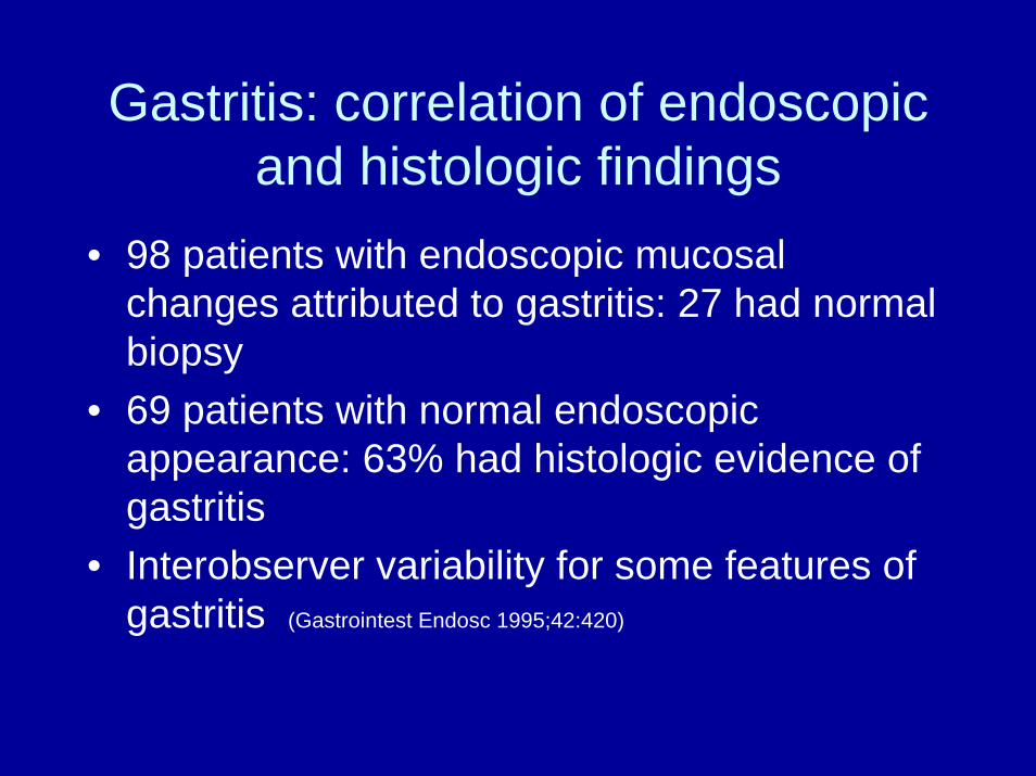
Gastritis: correlation of endoscopic and histologic findings
• 98 patients with endoscopic mucosal changes attributed to gastritis: 27 had normal biopsy
• 69 patients with normal endoscopic appearance: 63% had histologic evidence of gastritis
• Interobserver variability for some features of gastritis (Gastrointest Endosc 1995;42:420)


Chronic nonspecific Gastritis classification
• Nonatrophic- type B
• atrophic - type A
• H. pylori• Antrum > corpus• Diffuse antral predominant
gastritis
• AUTOIMMUNE (body, fundus)– Diffuse corporal atrophic
gastritis– Autoimmune metaplastic
atrophic gastritis• MULTIFOCAL (H. pylori, antrum
= corpus)– Multifocal atrophic gastritis

Chronic nonspecific gastritis

Natural history of H pylori

H. Pylori gastritis
• Acute• Chronic
– Antral predominant gastritis, also called type B, nonatrophic, diffuse antral predominant gastritis
– Atrophic gastritis, also called type A, multifocal atrophic gastritis, metaplastic atrophic gastritis

Clinical significance of H pylori gastritis
• Acute– May be symptomatic with epigastric pain, nausea and
vomiting• Antral predominant gastritis
– Duodenal ulcer• Atrophic gastritis
– Intestinal metaplasia– Gastric ulcer– Gastric adenocarcinoma

Where to biopsy for H. pylori
• 2 from antrum• 2 from gastric body• 1 from incisura: site most likely to show
atrophic gastritis and premalignant dysplasia

Where to biopsy for H. pylori

Endoscopic findings of H. pylori
• No distinct endoscopic pattern
• Normal• Red streaks in antrum• Erosions and ulcerations• Hypertrophy, atrophy

Autoimmune metaplastic atrophic gastritis
• Immune response directed against parietal cells and intrinsic factor
• 3x more common in women• Autosomal dominant disorder• Northern European• Associated with other autoimmune
disorders: Hashimotos thyroiditis and vitiligo

Autoimmune metaplastic atrophic gastritis: endoscopic findings
• Appearance of multiple polyps• Absent or inconspicous rugae in body
and fundus• Submucosal blood vessels visible
through thin atrophic overlying mucosa• Usually no antral involvement

Chronic atrophic gastritis associated with pernicious anemia, fundus

Intestinal metaplasia
• Eventually atrophic glands are replaced by metaplastic epithelium
• H. pylori: Intestinal metaplasia develops at a rate of 1-2% per annum to yield a lifetime risk of 50-75%

Patchy intestinal metaplasia, paler than surrounding mucosa, in the antrum of 52 year old woman with dyspepsia. Test for H.
pylori was positive

Intestinal metaplasia of the gastric antrum


H. Pylori and peptic ulcer disease
• Gastritis is found in virtually all patients infected with H. pylori
• In the United States, 80% of pts with DU and 60% with GU are associated with H. pylori
• Fewer than 20% of people with H. p ever develop PUD
• H. p. Rx with antibiotics dramatically decreases ulcer recurrance rate

Gastric adenocarcinoma

Gastritis and gastric cancer
• Adenocarcinoma– H pylori– autoimmune
• MALT: H. pylori• Carcinoid: autoimmune

Gastritis and gastric cancer: H pylori and adenocarcinoma
• 2nd most common cancer worldwide• in US: 8th cancer related mortality in men and 10th in
women• Decreased incidence of gastric adenocarcinoma in western
populations parallels decrease in prevalence of H pylori• H pylori infected individuals have 2-10x increased incidence
of gastric cancer• 36 and 47% of all gastric cancers in developed and
developing countries respectively are attributable to H pylori• Multifactorial

H. Pylori and Gastric adenocarcinoma

Chronic gastritis and gastric adenocarcinoma
• Increased risk with intestinal metaplasia; not known if cancer arises from intestinal metaplasia or whether it represents a marker of increased risk
• Autoimmune gastritis: 3-18x increased risk of gastric adenocarcinoma

Gastritis and gastric cancerMALToma
• Low grade B cell lymphoma • Mucosal associated lymphoid tissue

MALToma
• associated with chronic H. pylori infection in more than 90% of cases
• Primary gastric lymphoma accounts for 3% of gastric neoplasms and 10% of lymphomas
• 50% of gastric lymphomas are MALT• H. pylori induces mucosal inflammatory reaction,
lymphoid follicles -> B cell monoclonal cells -> autonomous uncontrolled growth

• Gastric MALToma: dense monotonous lymphoid infiltrate in the lamina propria

Autoimmune gastritis and carcinoid tumors
• Loss of parietal cell mass - > hypochlorhydria -> G cell hyperplasia and hypergastrinemia -> chronic stimulation of enterochromaffin like cells by gastrin

Gastric carcinoid

What to do about gastric cancer and gastritis: look for H pylori ?
Unknown if treatment for H pylori decreases risk of gastric cancer
• Studies are difficult because of long cancer development process that may take several decades

What to do about gastritis and gastric cancer: look for H pylori ?
• some studies show improvement in inflammation and intestinal metaplasia
• 2 studies show improvement in gastritis and superficial epithelial damage but no improvement in intestinal metaplasia or atrophy
• 1 study from China: healthy H. pylori carriers, treated and followed for 7.5 years. No overall decrease in gastric cancer. Subgroup of patients with no precancerous lesions on presentation did have decreased gastric cancer risk. (JAMA 2004;291:187)

What to do about gastritis and cancer risk:look for H. pylori
• If you find H. pylori: eradication should be considered because it is a carcinogen (ASGE)
• Insufficient evidence to recommend screening asymptomatic patients for H. pylori to prevent gastric cancer (up to date)
• Consider testing first degree relatives of patients with noncardia gastric cancer (Mayo)

What to do about gastritis and cancer risk: surveillance scope for dysplasia or cancer?
ASGE guidelines• Pernicious anemia: Single endoscopy should be
performed to identify carcinoid and gastric cancer• Endoscopic surveillance of gastric intestinal
metaplasia has not been extensively studied in the United states and therefore cannot be uniformly recommended
• Patients at increased risk for gastric cancer due to ethnic background or family history may benefit from surveillance

What to do about gastritis and cancer risk:dysplasia
• Low grade dysplasia: surveillance EGD every 3 months for at least 1 year with topographic mapping biopsy strategy
• High grade dysplasia: consider endoscopic resection or gastrectomy

MALToma
• H. pylori therapy is useful in patients with localized, mucosal or submucosal, nonbulky, flat disease (without metastasis, LN, or diffuse large B cell lymphoma)
• Only 10% of lymphoma patients • 50-90% complete remission

Infectious gastritis:CMV
• Immunocompromised patient• Epigastric pain, fever, atypical
lymphocytosis• Endoscopic findings: congested,
edematous mucosa, erosions, ulcerations, nodular mucosa

CMV gastritis

CMV gastritis: pathology
• Cytomegalic cells with intranuclear and intracytoplasmic inclusions of cytomegalovirus

Granulomatous gastritis: Crohn’s disease
• Crohns disease of the stomach is uncommon• Almost always associated with intestinal
disease• Nausea, vomiting, epigastric pain, anorexia
and weight loss• Endoscopy: reddened mucosa, irregularly
shaped ulcers, erosions, nodular lesions and cobblestone pattern

Crohn’s gastritis
• Severe nodular gastritis in 18 year old male with crohnsdisease

Gastropathy
• Hemorrhagic or erosive lesions• Caused by irritants such as medications
or reduction in mucosal blood flow• NSAIDs, alcohol, trauma, sepsis• Disruption of normal protective barrier:
mucin, bicarbonate, epithelium, PG

Gastropathy
• Endoscopic findings– Acute: may be diffuse (NSAIDs and
alcohol) or confined to body and fundus (stress)
– Chronic: usually antrum

NSAID gastropathy

NSAID gastropathy

gastropathy
• Pathology:– Acute: subtle changes– Chronic: foveolar hyperplasia, edema,
increased smooth muscle fibers, vascular dilatation and congestion
– Few inflammatory cells

Foveolar hyperplasia
• Tortuous, corkscrew appearance
• Marker of increased epithelial cell turnover
• Chemical injury and H pylori gastritis

Bile reflux gastropathy• Often occurs after gastric
surgery• Asymptomatic or
abdominal pain, emesis and weight loss
• Erosions, redness, bile staining of gastric mucosa
• Treatment: sulcralfate, ursodeoxycholic acid, surgery