
Gandage Dhananjay et al., Anat Physiol 2013, 3:2 … · 2 1 0011 9100,3 Case Report Open Access...
4
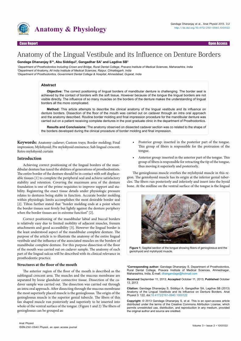
Volume 3 • Issue 2 • 1000122 Anat Physiol ISSN:2161-0940 Physiol, an open access journal Open Access Case Report Anatomy & Physiology Gandage Dhananjay et al., Anat Physiol 2013, 3:2 http://dx.doi.org/10.4172/2161-0940.1000122 Keywords: Anatomy cadaver; Custom trays; Border molding; Final impression; Mylohyoid; Pre mylohyoid eminence; Sub lingual crescent; Retro mylohyoid curtain Introduction Achieving correct positioning of the lingual borders of the man- dibular denture has taxed the abilities of generations of prosthodontists. e entire border of the denture should be in contact with soſt displace- able tissues [1] to complete the peripheral seal and achieve satisfactory stability and retention. Covering the maximum area of the denture foundation is one of the prime requisites to improve support and sta- bility. Registering the exact tissue details under physiologic pressure relates to dentures being stable in function. Accurate border molding within physiologic limits accomplishes the most desirable border seal [2]. Tilton further stated that “border molding ends at a point where the border tissues seat firmly but lightly against the denture periphery when the border tissues are in extreme function” [2]. Correct positioning of the mandibular labial and buccal borders is relatively easy due to limited mobility of adjacent muscles, frenum attachments and good accessibility [3]. However the lingual border is the least understood aspect of the mandibular complete denture. e purpose of the article is to illustrate the anatomy of the entire lingual vestibule and the influence of the associated muscles on the borders of mandibular complete denture. For this purpose dissection of the floor of the mouth was carried out on cadaver sample. e anatomy of each part of the lingual sulcus will be described with its clinical relevance in prosthodontic practice. Structures at the floor of the mouth e anterior region of the floor of the mouth is described as the sublingual crescent area. e muscles and the mucous membrane are separated by loose glandular connective tissue. Dissection of the ca- daver sample was carried out. e dissection was carried out through an intra oral approach. Aſter dissecting through the mucous membrane the most superiorly placed muscle is the genioglossus. e origin of the genioglossus muscle is the superior genial tubercle. e fibers of this fan shaped muscle run posteriorly and superiorly to be inserted into whole of the ventral surface of the tongue. (Figure 1 and 2) e fibers of genioglossus can be grouped as: • Posterior group: inserted in the posterior part of the tongue. is group of fibers is responsible for the protrusion of the tongue. • Anterior group: inserted in the anterior part of the tongue. is group of fibers is responsible for retracting the tip of the tongue, thus moving it superiorly and posteriorly. e genioglossus muscle overlies the mylohyoid muscle in this re- gion. e geniohyoid muscle has its origin at the inferior genial tuber- cles. e fibers run posteriorly and inferiorly and insert into the hyoid bone. At the midline on the ventral surface of the tongue is the lingual *Corresponding author: Gandage Dhananjay S, Department of Prosthodontics, Rural Dental College, Pravara Institute of Medical Sciences, Ahmednagar, Maharashtra, India, E-mail: [email protected] Received September 11, 2013; Accepted October 11, 2013; Published October 13, 2013 Citation: Gandage Dhananjay S, Siddiqui A, Gangadhar SA, Lagdive SB (2013) Anatomy of the Lingual Vestibule and its Influence on Denture Borders. Anat Physiol 3: 122. doi:10.4172/2161-0940.1000122 Copyright: © 2013 Gandage Dhananjay S, et al. This is an open-access article distributed under the terms of the Creative Commons Attribution License, which permits unrestricted use, distribution, and reproduction in any medium, provided the original author and source are credited. Abstract Objective: The correct positioning of lingual borders of mandibular denture is challenging. The border seal is achieved by the contact of borders with the soft tissue. However because of the tongue the lingual borders are not visible directly. The influence of so many muscles on the borders of the denture makes the understanding of lingual borders all the more complicated. Method: This article attempts to describe the clinical anatomy of the lingual vestibule and its influence on denture borders. Dissection of the floor of the mouth was carried out on cadaver through an intra oral approach and the anatomy described. Routine border molding and final impression procedure for the mandibular denture was carried out on a patient receiving complete dentures in the post graduate clinic in the department of Prosthodontics. Results and Conclusions: The anatomy observed on dissected cadaver section was co related to the shape of the borders developed during the clinical procedure of border molding and final impression. Anatomy of the Lingual Vestibule and its Influence on Denture Borders Gandage Dhananjay S 1 *, Abu Siddiqui 2 , Gangadhar SA 1 and Lagdive SB 3 1 Department of Prosthodontics Including Crown and Bridge, Rural Dental College, Pravara Institute of Medical Sciences, Maharashtra, India 2 Department of Anatomy, All India Institute of Medical Sciences, Raipur, Chhattisgarh, India 3 Department of Prosthodontics, Government Dental College & Hospital, Ahmedabad, Gujarat, India Figure 1: Sagittal section of the tongue showing fibers of genioglossus and the geniohyoid and mylohyoid muscle.
Transcript of Gandage Dhananjay et al., Anat Physiol 2013, 3:2 … · 2 1 0011 9100,3 Case Report Open Access...
Volume 3 • Issue 2 • 1000122Anat PhysiolISSN:2161-0940 Physiol, an open access journal
Open AccessCase Report
Anatomy & Physiology Gandage Dhananjay et al., Anat Physiol 2013, 3:2
http://dx.doi.org/10.4172/2161-0940.1000122
Keywords: Anatomy cadaver; Custom trays; Border molding; Final impression; Mylohyoid; Pre mylohyoid eminence; Sub lingual crescent; Retro mylohyoid curtain
IntroductionAchieving correct positioning of the lingual borders of the man-
dibular denture has taxed the abilities of generations of prosthodontists. The entire border of the denture should be in contact with soft displace-able tissues [1] to complete the peripheral seal and achieve satisfactory stability and retention. Covering the maximum area of the denture foundation is one of the prime requisites to improve support and sta-bility. Registering the exact tissue details under physiologic pressure relates to dentures being stable in function. Accurate border molding within physiologic limits accomplishes the most desirable border seal [2]. Tilton further stated that “border molding ends at a point where the border tissues seat firmly but lightly against the denture periphery when the border tissues are in extreme function” [2].
Correct positioning of the mandibular labial and buccal borders is relatively easy due to limited mobility of adjacent muscles, frenum attachments and good accessibility [3]. However the lingual border is the least understood aspect of the mandibular complete denture. The purpose of the article is to illustrate the anatomy of the entire lingual vestibule and the influence of the associated muscles on the borders of mandibular complete denture. For this purpose dissection of the floor of the mouth was carried out on cadaver sample. The anatomy of each part of the lingual sulcus will be described with its clinical relevance in prosthodontic practice.
Structures at the floor of the mouthThe anterior region of the floor of the mouth is described as the
sublingual crescent area. The muscles and the mucous membrane are separated by loose glandular connective tissue. Dissection of the ca-daver sample was carried out. The dissection was carried out through an intra oral approach. After dissecting through the mucous membrane the most superiorly placed muscle is the genioglossus. The origin of the genioglossus muscle is the superior genial tubercle. The fibers of this fan shaped muscle run posteriorly and superiorly to be inserted into whole of the ventral surface of the tongue. (Figure 1 and 2) The fibers of genioglossus can be grouped as:
• Posterior group: inserted in the posterior part of the tongue. This group of fibers is responsible for the protrusion of the tongue.
• Anterior group: inserted in the anterior part of the tongue. This group of fibers is responsible for retracting the tip of the tongue, thus moving it superiorly and posteriorly.
The genioglossus muscle overlies the mylohyoid muscle in this re-gion. The geniohyoid muscle has its origin at the inferior genial tuber-cles. The fibers run posteriorly and inferiorly and insert into the hyoid bone. At the midline on the ventral surface of the tongue is the lingual
*Corresponding author: Gandage Dhananjay S, Department of Prosthodontics, Rural Dental College, Pravara Institute of Medical Sciences, Ahmednagar, Maharashtra, India, E-mail: [email protected]
Received September 11, 2013; Accepted October 11, 2013; Published October 13, 2013
Citation: Gandage Dhananjay S, Siddiqui A, Gangadhar SA, Lagdive SB (2013) Anatomy of the Lingual Vestibule and its Influence on Denture Borders. Anat Physiol 3: 122. doi:10.4172/2161-0940.1000122
Copyright: © 2013 Gandage Dhananjay S, et al. This is an open-access article distributed under the terms of the Creative Commons Attribution License, which permits unrestricted use, distribution, and reproduction in any medium, provided the original author and source are credited.
AbstractObjective: The correct positioning of lingual borders of mandibular denture is challenging. The border seal is
achieved by the contact of borders with the soft tissue. However because of the tongue the lingual borders are not visible directly. The influence of so many muscles on the borders of the denture makes the understanding of lingual borders all the more complicated.
Method: This article attempts to describe the clinical anatomy of the lingual vestibule and its influence on denture borders. Dissection of the floor of the mouth was carried out on cadaver through an intra oral approach and the anatomy described. Routine border molding and final impression procedure for the mandibular denture was carried out on a patient receiving complete dentures in the post graduate clinic in the department of Prosthodontics.
Results and Conclusions: The anatomy observed on dissected cadaver section was co related to the shape of the borders developed during the clinical procedure of border molding and final impression.
Anatomy of the Lingual Vestibule and its Influence on Denture BordersGandage Dhananjay S1*, Abu Siddiqui2, Gangadhar SA1 and Lagdive SB3
1Department of Prosthodontics Including Crown and Bridge, Rural Dental College, Pravara Institute of Medical Sciences, Maharashtra, India2Department of Anatomy, All India Institute of Medical Sciences, Raipur, Chhattisgarh, India3Department of Prosthodontics, Government Dental College & Hospital, Ahmedabad, Gujarat, India
Figure 1: Sagittal section of the tongue showing fibers of genioglossus and the geniohyoid and mylohyoid muscle.

Citation: Gandage Dhananjay S, Siddiqui A, Gangadhar SA, Lagdive SB (2013) Anatomy of the Lingual Vestibule and its Influence on Denture Borders. Anat Physiol 3: 122. doi:10.4172/2161-0940.1000122
Page 2 of 6
Volume 3 • Issue 2 • 1000122Anat PhysiolISSN:2161-0940 Physiol, an open access journal
frenum. Lateral to the lingual frenum are two mucosal folds one on either side known as the sublingual fold. The sublingual folds carry the openings of the corresponding Wharton’s ducts of the submandibular salivary glands. The other structures at the floor of the mouth are the sublingual gland along with the hypoglossal nerve, lingual nerve, lin-gual vein and lingual artery.
Prosthodontic application
For the stability and retention of a complete denture; border seal in the floor of the mouth can compliment seals in the vestibular space [3]. To achieve adequate degree of tongue freedom and tissue reflection; accurate border molding in the vestibular spaces is a prime requisite. It should also be emphasized that the soft tissues at the base of the tongue is not directly supported by bone. This region is defined in GPT [4] as the sublingual crescent area. “It is the crescent shaped area on the floor of the mouth formed by the lingual wall of the mandible and the adjacent sublingual fold. It is the region of the anterior alveolingual sulcus”. The sub lingual crescent area influences the anterior lingual border of the denture. Pendelton advocated permitting the tissues to establish their own association by forming impression materials to their own indi-vidual requirements [5]. Lawson suggested thickening the sublingual region of the denture results in increased retention [1]. Lott and Levin [6] also demonstrated the clinical advantages of thick borders.
Tongue position and the degree of freedom provided for tongue movements during border molding procedures also play an important role in positioning of the denture borders, design of the denture flange thus influencing stability of the mandibular denture. On completion of the border seal in the anterior lingual vestibule the borders should:
1. Extend over the resting tissues of the sublingual crescent area
2. Exert minimal pressure on the tissues
Tongue should be in resting position with the tip just passively touching the lingual surface of the mandibular anterior teeth and the lateral surface touching the mandibular posterior teeth.
Developing the border seal
1. The custom tray should be 2 mm short of the vestibular reflec-tion. Add border molding material (either tempered low fusing compound or poly ether material) over the borders of the tray and place the tray in the patients mouth.
2. Ask the patient to protrude the tongue. This movement will activate the posterior fibers of the genioglossus muscle. The an-terior region of the floor of the mouth is raised to determine the length (height) of the lingual flange in the anterior lingual sulcus.
3. Ask the patient to retract his tongue. This will activate the ante-rior fibers of genioglossus muscle. The border molding material will be compressed between the ventral surface of the tongue on one side and the lingual surface of the mandible on the oth-er. Thus the width of the border in the anterior lingual sulcus will be determined.
It can be demonstrated that the border of the mandibular denture is influenced by the genioglossus muscle in the anterior lingual sulcus.
The Pre mylohyoid eminence
In the region of the premolars on the lingual surface of the man-dible lies the sub lingual gland over the mylohyoid muscle between the mandible and the genioglossus muscle. The flange of the mandibular denture should provide adequate space for the gland. This is achieved by the flange sloping inwards, medially away from the lingual surface of the mandible. The border should rest on the floor of the mouth be-low the tongue to accommodate the sublingual gland. The sub lingual gland region is recorded as the pre mylohyoid eminence while making impression with a low viscosity material [7].
The mylohyoid region
The region of the mylohyoid extends from the pre mylohyoid fossa to the distal end of the mylohyoid ridge (Figure 3 and 4). The mylohyoid
Figure 2: Coronal view of the floor of the mouth showing the anterior fibers of genioglossus (AG), Posterior fibers of genioglossus (PG) and underlying mylohyoid.
Figure 3: Coronal view of the structures at the floor of the mouth with the mylohyoid (green), anterior fibers of genioglossus (white) and posterior fibers of genioglossus (red).
Figure 4: Lingual aspect of mandible showing the relation of mylohyoid, sub mandibular gland and superior constrictor.
Citation: Gandage Dhananjay S, Siddiqui A, Gangadhar SA, Lagdive SB (2013) Anatomy of the Lingual Vestibule and its Influence on Denture Borders. Anat Physiol 3: 122. doi:10.4172/2161-0940.1000122
Page 3 of 6
Volume 3 • Issue 2 • 1000122Anat PhysiolISSN:2161-0940 Physiol, an open access journal
directly influences the border of the mandibular denture this region. The posterior fibers of mylohyoid muscle attach more superiorly on the lingual aspect of the mandible and descend more vertically downwards to insert into the hyoid bone [8]. The fibers extend medio inferiorly when contracted. The border of the mandibular denture extends below the mylohyoid ridge and turns medially away from the lingual surface of the mandible; parallel to the mylohyoid muscle fibers to avoid the undercut underneath the mylohyoid ridge and rest over the soft tissues below the tongue. Thus the tongue rests over the flange. When the floor of the mouth is raised, the mylohyoid muscle is activated and contact is established between the borders of the mandibular denture and the soft tissues on the floor of the mouth.
Developing the border seal
1. With the custom tray being 2 mm short; add the border mold-ing material over the borders, and place the tray in the mouth.
2. Ask the patient to protrude the tongue, followed by swallowing action.
3. The mylohyoid muscle is activated and the floor of the mouth is raised to contact the material.
4. The border is molded by the action of the mylohyoid muscle on the borders of the tray
Retro mylohyoid space
The term ‘retro mylohyoid space’ was explained by Edwards and Boucher [9]. Though there are no sharp boundaries; this region, is di-vided into two areas: glandular triangle and constrictor square [10].
The triangle includes the deep part of the submandibular gland and the underlying mylohyoid muscle and the square includes the superior constrictor muscle, pterygomandibular raphae and a small part of buc-cinator muscle [10]. The sub mandibular gland lies between the distal fibers of the mylohyoid muscle and the superior constrictor muscle (Figure 4). The mucous membrane and the superior constrictor form the retromylohyoid curtain of Edwards and Boucher [9]. The region of the retromylohyoid curtain influences the disto lingual flange of the mandibular denture. Two muscles that influence the denture border in the region of the retro mylohyoid curtain are:
• Superior constrictor of pharynx
• Medial pterygoid
The medial pterygoid muscle lies posterior to the superior constric-tor. When the mandible is elevated the contracting medial pterygoid will push against the fibers of the superior constrictor and will create a bulge in the wall of the retro mylohyoid curtain.
Developing Contour of the border in RMC region
1. Add border molding material on the disto lingual aspect of the custom tray.
2. Place the tray in the mouth and ask the patient to protrude the tongue and then close the lower jaw.
3. Protruding the tongue activates the superior constrictor muscle which molds the disto lingual border of the denture.
4. It should be borne in mind that the medial pterygoid is one of the elevators of the mandible. When the patient closes his jaw, the medial pterygoid contracts against the superior con-strictor of pharynx which is immediately anterior to it which in turn forms the postero lateral aspect of the retro mylohyoid
curtain. Thus the superior constrictor is pushed anteriorly by the contracting medial pterygoid which molds the border of the mandibular denture in the region of the retromylohyoid fossa.
This completes the border seal on the lingual aspect of the man-dibular denture.
The region of the retro molar pad
The retromolar pad region and the associated structures form a very stable anatomical landmark. The retromolar pad does not resorb. The mucosa is thin, non keratinized with the submucosa composed of loose areolar tissues and glandular tissues. The region is important for it is under the influence of many muscles viz:
• Laterally by the buccinator
• Postero superiorly by the terminal fibers of temporalis
• Pterygo mandibular raphae
• Medially by the superior constrictor of the pharynx (Figure 5)
The action of these muscles limits the extent of the denture and pre-vents extra pressure during the impression procedure the denture base should extend approximately half to two thirds of the retro molar pad [7]. Extending the borders to include the retro molar pad completes the soft tissue border seal and the S shaped flange on the lingual aspect of the denture is thus achieved (Figure 6).
Figure 5: Showing buccinator, pterygo-mandibualr raphae and the superior constrictor muscle.
Figure 6: Showing final impression of mandibular denture foundation. 1. Anterior lingual sulcus2. Region of Pre mylohyoid eminence3. Region of the mylohyoid4. Retro mylohyoid region5. Region of the retromolar pad6. Disto buccal region
Citation: Gandage Dhananjay S, Siddiqui A, Gangadhar SA, Lagdive SB (2013) Anatomy of the Lingual Vestibule and its Influence on Denture Borders. Anat Physiol 3: 122. doi:10.4172/2161-0940.1000122
Page 4 of 6
Volume 3 • Issue 2 • 1000122Anat PhysiolISSN:2161-0940 Physiol, an open access journal
ConclusionThe final lingual border should be so shaped that it guides the tongue
into the same position it will occupy in relation to the finished denture. The tray should not dislodge when the tip of the tongue is in contact with the vermilion border of the lips. Border molding is achieved either by incremental technique using low fusing impression compound or by one step technique using elastomeric impression material like a polyether. In relation to the anatomic variations; guidelines for placing the borders should be customized for each patient. For e.g. in a case of resorbed ridge; molding a thick border and wide sub lingual region often helps to achieve a border seal because it aids continuity with the floor of the mouth [1,6]. The borders should be uniform and round to minimize trauma or sore spots. The shape of the final lingual borders should be such that the pa-tient should be able to wipe the tip of the tongue to the vermilion border of the upper lip without noticeable displacement of the tray [7]. The final borders of the denture should be in harmony with the anatomy of the floor of the mouth in rest and in function.
References
1. Alan Lawson W (1961) Influence of the sublingual Fold on the retention of complete lower dentures. J Prosthet Dent 11: 1038-1044.
2. Tilton GE (1952) Denture periphery. J Prosthet Dent 2: 290-306.
3. Azzam MK, Yurkstas AA, Kronman J (1992) The sublingual crescent extension and its relation to the stability and retention of mandibular complete dentures. J Prosthet Dent 67: 205-210.
4. St Louis (1987) Glossary of prosthodontic terms: (5thedn), CV Mosby Co.
5. Pendelton EC (1942) American textbook of prosthetic dentistry: (7thedn), Lea & Febiger Publication, Philadelphia.
6. Lott F, Levin B (1966) Flange technique: an anatomic and physiologic approach to increased retention, function, comfort, and appearance of dentures. J Prosthet Dent 16: 394-413.
7. Zarb, Bolender. Prosthodontic Treatment of Edentulous Patients. (12thedn) Elesvier.
8. Jacobson TE, Krol AJ (1983) A contemporary review of the factors involved in complete dentures. Part II: stability. J Prosthet Dent 49: 165-172.
9. Edwards LF, Boucher CO (1942) Anatomy of the mouth in relation to Complete Dentures. The Journal of Prosthetic Dentistry 29: 331-345.
10. Sidney G Barrett, Wheeler Haines (1962) Structure of the mouth in the mandibular molar region and its relation to the denture. J Prosthet dent 12: 835-847.
Submit your next manuscript and get advantages of OMICS Group submissionsUnique features:
• Userfriendly/feasiblewebsite-translationofyourpaperto50world’sleadinglanguages• AudioVersionofpublishedpaper• Digitalarticlestoshareandexplore
Special features:
• 250OpenAccessJournals• 20,000editorialteam• 21daysrapidreviewprocess• Qualityandquickeditorial,reviewandpublicationprocessing• IndexingatPubMed(partial),Scopus,EBSCO,IndexCopernicusandGoogleScholaretc• SharingOption:SocialNetworkingEnabled• Authors,ReviewersandEditorsrewardedwithonlineScientificCredits• Betterdiscountforyoursubsequentarticles
Submityourmanuscriptat:www.omicsonline.org/submission
Citation: Gandage Dhananjay S, Siddiqui A, Gangadhar SA, Lagdive SB (2013) Anatomy of the Lingual Vestibule and its Influence on Denture Borders. Anat Physiol 3: 122. doi:10.4172/2161-0940.1000122


![Am J Physiol Heart Circ Physiol 2011[1]](https://static.fdocuments.us/doc/165x107/577ce0031a28ab9e78b28109/am-j-physiol-heart-circ-physiol-20111.jpg)


