Gallbladder histopathology during murine … histopathology during murine gallstone formation:...
10
Gallbladder histopathology during murine gallstone formation: relation to motility and concentrating function Karel J. van Erpecum, 1,2, * David Q-H. Wang, 1,† Antonio Moschetta, § Domenico Ferri,** Maria Svelto, †† Piero Portincasa, §§ Jan-Jaap Hendrickx,*** Margue ´rite Schipper,*** and Giuseppe Calamita, †† Department of Gastroenterology* and Department of Pathology,*** University Medical Center Utrecht, Utrecht, The Netherlands; Gastroenterology Division, † Beth Israel Deaconess Medical Center, Harvard Medical School, and Harvard Digestive Diseases Center, Boston, MA; Howard Hughes Medical Institute and Department of Pharmacology, § University of Texas Southwestern Medical Center, Dallas, TX; and Department of Zoology, Laboratory of Histology and Comparative Anatomy,** Department of General and Environmental Physiology, †† and Section of Internal Medicine, Department of Internal and Public Medicine, §§ University of Bari, Bari, Italy Abstract C57L mice are susceptible and AKR mice are resistant to gallstone formation. We studied in male mice of both strains gallbladder histopathology, cholecystokinin- induced emptying, and concentrating function at 0, 14, 28, and 56 days on a lithogenic diet. Gallbladder wall thick- ness increased on the diet, with stromal granulocyte in- filtration, progressive fibrosis, edema, and epithelial cell indentation, particularly in C57L. Strong basal cholecys- tokinin octapeptide-induced gallbladder emptying (70% of fasting volumes) occurred in both strains, but fasting gall- bladder volumes were significantly larger in C57L (14.8 6 2.2 ml vs. 8.8 6 1.0 ml). On the diet, fasting volumes in- creased exclusively in C57L (28.6 6 2.9 ml on day 56), with progressively decreased emptying (27% of fasting volumes on day 56). Gallbladder emptying remained normal in AKR. Gallbladder concentrating function decreased on the lithogenic diet (especially in C57L), coinciding with de- creased aquaporin-1 (AQP1) and AQP8 expression at the mRNA and protein levels. In additional experiments, similar downregulation of AQP1 and AQP8 mRNA expression oc- curred in farnesoid X receptor (FXR)-deficient mice after 1 week on the lithogenic diet, without any difference from corresponding wild-type mice. In conclusion, during murine lithogenesis, altered gallbladder histology is associated with impaired motility, reduced concentrating function, and de- creased AQP1 and AQP8 expression, the latter without the involvement of the FXR.—van Erpecum, K. J., D. Q-H. Wang, A. Moschetta, D. Ferri, M. Svelto, P. Portincasa, J-J. Hendrickx, M. Schipper, and G. Calamita. Gallbladder histopathology during murine gallstone formation: relation to motility and concentrating function. J. Lipid Res. 2006. 47: 32–41. Supplementary key words aquaporin . cholesterol . farnesoid X re- ceptor . gallbladder emptying . water channel C57L and AKR inbred mice exhibit different suscept- ibilities to cholesterol gallstone formation, depending on Lith genes. Susceptibility is high in C57L males (gallstones in 80% of mice after 56 days on a lithogenic diet) and low in AKR males (gallstones in 15% after 56 days on a litho- genic diet) (1). Based on their time course during the earliest stages of lithogenesis, biliary cholesterol super- saturation, the hydrophobic bile salt deoxycholate, and high concentrations of crystallization-promoting mucin are thought to play crucial roles in murine gallstone for- mation (1, 2). In humans, the gallbladder is thought to be another key player in gallstone pathogenesis. Impaired postprandial and interdigestive gallbladder emptying are often found in gallstone patients, providing time for nu- cleation of cholesterol crystals and their aggregation into macroscopic stones (3). Also in animal models, gallblad- der contractility is decreased in the earliest stages of gall- stone formation, even before gallstones have formed (4). Furthermore, in the fasting gallbladder, hepatic bile is concentrated 4- to 5-fold by absorption of water, thereby enhancing cholesterol crystallization (5, 6). Aquaporins (AQP0 to AQP10) are a family of transmembrane channels mediating the movement of water through the lipid bi- layer. AQP1 (7–12) and AQP8 (12) have recently been detected in gallbladder epithelial cells. Virtually no infor- mation is available about the gallbladder in murine gall- stone formation, despite the obviously crucial role of this organ in lithogenesis. In the present study, we describe gallbladder histopa- thology (including reduced AQP expression), motility, and Manuscript received 9 May 2005 and in revised form 6 October 2005. Published, JLR Papers in Press, October 13, 2005. DOI 10.1194/jlr.M500180-JLR200 Abbreviations: CCK, cholecystokinin octapeptide; FXR, farnesoid X receptor. 1 K. J. van Erpecum and D. Q-H. Wang contributed equally to this work. 2 To whom correspondence should be addressed. e-mail: [email protected] Copyright I 2006 by the American Society for Biochemistry and Molecular Biology, Inc. This article is available online at http://www.jlr.org 32 Journal of Lipid Research Volume 47, 2006 by guest, on July 11, 2018 www.jlr.org Downloaded from
Transcript of Gallbladder histopathology during murine … histopathology during murine gallstone formation:...

Gallbladder histopathology during murine gallstone
formation: relation to motility and concentrating function
Karel J. van Erpecum,1,2,* David Q-H. Wang,1,† Antonio Moschetta,§
Domenico Ferri,** Maria Svelto,†† Piero Portincasa,§§ Jan-Jaap Hendrickx,***Marguerite Schipper,*** and Giuseppe Calamita,††
Department of Gastroenterology* and Department of Pathology,*** University Medical Center Utrecht,Utrecht, The Netherlands; Gastroenterology Division,† Beth Israel Deaconess Medical Center, HarvardMedical School, and Harvard Digestive Diseases Center, Boston, MA; Howard Hughes Medical Institute andDepartment of Pharmacology,§ University of Texas Southwestern Medical Center, Dallas, TX; andDepartment of Zoology, Laboratory of Histology and Comparative Anatomy,** Department of General andEnvironmental Physiology,†† and Section of Internal Medicine, Department of Internal and PublicMedicine,§§ University of Bari, Bari, Italy
Abstract C57L mice are susceptible and AKR mice areresistant to gallstone formation. We studied in male miceof both strains gallbladder histopathology, cholecystokinin-induced emptying, and concentrating function at 0, 14, 28,and 56 days on a lithogenic diet. Gallbladder wall thick-ness increased on the diet, with stromal granulocyte in-filtration, progressive fibrosis, edema, and epithelial cellindentation, particularly in C57L. Strong basal cholecys-tokinin octapeptide-induced gallbladder emptying (70% offasting volumes) occurred in both strains, but fasting gall-bladder volumes were significantly larger in C57L (14.8 62.2 ml vs. 8.8 6 1.0 ml). On the diet, fasting volumes in-creased exclusively in C57L (28.6 6 2.9 ml on day 56), withprogressively decreased emptying (27% of fasting volumeson day 56). Gallbladder emptying remained normal inAKR. Gallbladder concentrating function decreased on thelithogenic diet (especially in C57L), coinciding with de-creased aquaporin-1 (AQP1) and AQP8 expression at themRNA and protein levels. In additional experiments, similardownregulation of AQP1 and AQP8 mRNA expression oc-curred in farnesoid X receptor (FXR)-deficient mice after1 week on the lithogenic diet, without any difference fromcorresponding wild-type mice. In conclusion, during murinelithogenesis, altered gallbladder histology is associated withimpaired motility, reduced concentrating function, and de-creased AQP1 and AQP8 expression, the latter without theinvolvement of the FXR.—van Erpecum, K. J., D. Q-H. Wang,A. Moschetta, D. Ferri, M. Svelto, P. Portincasa, J-J. Hendrickx,M. Schipper, and G. Calamita. Gallbladder histopathologyduring murine gallstone formation: relation to motility andconcentrating function. J. Lipid Res. 2006. 47: 32–41.
Supplementary key words aquaporin . cholesterol . farnesoid X re-ceptor . gallbladder emptying . water channel
C57L and AKR inbred mice exhibit different suscept-ibilities to cholesterol gallstone formation, depending onLith genes. Susceptibility is high in C57L males (gallstonesin 80% of mice after 56 days on a lithogenic diet) and lowin AKR males (gallstones in 15% after 56 days on a litho-genic diet) (1). Based on their time course during theearliest stages of lithogenesis, biliary cholesterol super-saturation, the hydrophobic bile salt deoxycholate, andhigh concentrations of crystallization-promoting mucinare thought to play crucial roles in murine gallstone for-mation (1, 2). In humans, the gallbladder is thought tobe another key player in gallstone pathogenesis. Impairedpostprandial and interdigestive gallbladder emptying areoften found in gallstone patients, providing time for nu-cleation of cholesterol crystals and their aggregation intomacroscopic stones (3). Also in animal models, gallblad-der contractility is decreased in the earliest stages of gall-stone formation, even before gallstones have formed (4).Furthermore, in the fasting gallbladder, hepatic bile isconcentrated 4- to 5-fold by absorption of water, therebyenhancing cholesterol crystallization (5, 6). Aquaporins(AQP0 to AQP10) are a family of transmembrane channelsmediating the movement of water through the lipid bi-layer. AQP1 (7–12) and AQP8 (12) have recently beendetected in gallbladder epithelial cells. Virtually no infor-mation is available about the gallbladder in murine gall-stone formation, despite the obviously crucial role of thisorgan in lithogenesis.
In the present study, we describe gallbladder histopa-thology (including reducedAQP expression),motility, and
Manuscript received 9 May 2005 and in revised form 6 October 2005.
Published, JLR Papers in Press, October 13, 2005.DOI 10.1194/jlr.M500180-JLR200
Abbreviations: CCK, cholecystokinin octapeptide; FXR, farnesoidX receptor.
1 K. J. van Erpecum and D. Q-H. Wang contributed equally to thiswork.
2 To whom correspondence should be addressed.e-mail: [email protected]
Copyright I 2006 by the American Society for Biochemistry and Molecular Biology, Inc.
This article is available online at http://www.jlr.org32 Journal of Lipid Research Volume 47, 2006
by guest, on July 11, 2018w
ww
.jlr.orgD
ownloaded from
concentrating function on a lithogenic diet. Farnesoid Xreceptor (FXR: NR1H4) functions as a bile salt receptorregulating the transcription of numerous genes involved incholesterol and bile salt homeostasis (13). This nuclearreceptor may be relevant for gallstone formation: FXR-deficient (FXR�/�) mice were recently found to exhibithigh susceptibility to cholesterol gallstone formation (14).In addition, in gallstone-susceptible C57L mice ona lithogenic diet, biliary cholesterol supersaturation andgallstone formation were prevented by treatment with asynthetic FXR agonist: FXR-dependent increases in biliarysecretion of cholesterol-solubilizing bile salt and phospha-tidylcholine molecules restored cholesterol solubility andthereby prevented gallstone formation (14). Because thelithogenic diet contains cholic acid [a high-affinity ligandfor FXR (15)], we investigated in the FXR�/� murinemodel whether the FXR pathway could play a direct role inreducing AQP gene expression.
MATERIALS AND METHODS
Animals
Homozygous male C57L/J and AKR/J mice were obtainedfrom the Jackson Laboratory (Bar Harbor, ME). They werehoused in a temperature-controlled room (226 1jC) with a 12 hlight cycle (6 AM–6 PM), with free access to water and Purinalaboratory chow containing traces (,0.02%) of cholesterol (Har-lan Teklad Laboratory Animal Diets, Madison WI). Once animalsachieved 8 weeks of age, they were fed a semisynthetic lithogenicdiet containing (per 100 g) 15 g of dairy fat, 1 g of cholesterol,0.5 g of cholic acid, 2 g of corn oil, 50 g of sucrose, 20 g of casein,and essential vitamins and minerals (16). Cholecystectomy wasperformed and gallbladder bile was aspirated completely at 9 AMbefore (day 0) and after feeding the lithogenic diet for 14, 28,and 56 days (five mice of each strain at each time point). Gall-bladders were immediately placed in formalin. Cholecystectomyand gallbladder bile collection were followed by cannulationof the common bile duct and hepatic bile collection for 1 h (1,17, 18). Pooled gallbladder biles and individual hepatic biles (fivemice at each time point) were stored at �20jC until further an-alysis. In a second and a third series of experiments, the effects ofa lithogenic diet on AQP1 and AQP8 mRNA expression and ongallbladder motility were examined (five mice of each strain ateach time point in all experimental groups). Gallbladder em-ptying was determined in fasted mice by intravenous injection ofsulfated cholecystokinin octapeptide (CCK-8; 17 nmol/kg bodyweight dissolved in 100 ml of PBS), identical volumes of PBS, orno injection. Exactly 15 min after the injection, cholecystectomywas performed and gallbladder volume was determined (19, 20).
A fourth series of experiments was performed in male FXR�/�
mice (21). FXR�/� and wild-type mice were on the same mixedbackground (C57BL/6N3129/SVJ3FVB, backcross 3C57BL/6N). Experimental conditions were the same as described above.Mice were maintained on a low-cholesterol (0.02%) Purina chowdiet until 9 weeks of age, followed by only 1 week of the lithogenicdiet, given the sensitivity of FXR�/� mice to cholic acid (21).After 12 h of fasting, cholecystectomies were performed, andgallbladders were frozen in liquid nitrogen and stored at �80jCuntil further analysis. Protocols were approved by the Institu-tional Animal Care and Use Committee of Harvard Universityand by the Institutional Animal Care and Research AdvisoryCommittee of the Southwestern Medical Center at Dallas andwere consistent with euthanasia recommendations of theAmerican Veterinary Medical Association.
Lipid analyses
Bile samples were lipid-extracted (22) after a short centri-fugation (5 min at 3,000 g) to spin down cholesterol crystals.Cholesterol (23) and phospholipid (24) concentrations weremeasured enzymatically in the extracted biles, and total bile saltconcentrations in whole biles were measured by the 3a-hydroxy-steroid dehydrogenase method (25). The cholesterol saturationindex was calculated according to the critical tables (26). Gall-bladder bile samples were examined for the presence of choles-terol crystals using direct and polarizing light microscopy (1003and 4003 magnification). Conjugated bile salt species were an-alyzed by HPLC (27) using a C-18 Waters Bondapack 10 mmcolumn. Methanol-phosphate buffer was used as an eluent (pH 5.2,flow rate of 1 ml/min).
Histology
Histological evaluations were performed by two independentpathologists unaware of mouse strains or time on the lithogen-ic diet (5 mm sections from paraffin-embedded tissue), withhigh interobserver agreement. For each gallbladder, numbersof granulocytes were scored after hematoxylin-eosin staining in20–40 high-power fields (4003 magnification). For each gall-bladder, wall thickness is the mean value of six determinations inthe fundic area. Relative amounts of smooth muscle cells com-pared with relative amounts of fibrosis were scored in Azan-stained sections.
Immunohistochemistry and Western blots
Endogenous peroxidase was preliminarily blocked and gall-bladder sections were then incubated overnight at 4jC with pu-rified monospecific primary antibodies directed against the Cterminus of AQP1 or AQP8 (anti-AQP1 or anti-AQP8; Alpha Di-agnostic International) at a final concentration of 5 mg/ml inblocking buffer (1% normal goat serum in phosphate-bufferedsaline) (28). Thereafter, sections were washed and incubated with
Fig. 1. Gallbladder histology of resistant AKR (A)and susceptible C57L (B) mice at baseline (hematox-ylin-eosin; 4003magnification). Black arrows indicatethe epithelial layer, and the white arrow indicatesgranulocyte infiltrate in C57L.
Murine gallbladder and lithogenic diet 33
by guest, on July 11, 2018w
ww
.jlr.orgD
ownloaded from
goat anti-rabbit IgG (Sigma, St. Louis, MO). After several washes,the sections were incubated with horseradish peroxidase-antiper-oxidase, and immunolabeling was visualized by incubation with3,39-diaminobenzidine-H2O2 (29). Controls performed using an-tibodies preadsorbed with immunizing peptides or by omittingthe primary antibodies were always negative. Images were cap-tured using a Nikon E600 photomicroscope equipped with a dig-ital camera (Nikon DMX1200; Nikon Instruments S.p.a., SestoFiorentino, Italy). The intensity of AQP staining was scored withthe aid of a semiquantitative score (05 absent; 115minor; 215
moderate; 31 5 appreciable; 41 5 strong) by one investigator(M. Svelto) unaware of mouse strain or time on the lithogenicdiet. To quantitate gallbladder protein concentrations of AQP1and AQP8, Western blot analysis was performed in separate setsof mice from both strains (n 5 35 per group) before and after28 and 56 days of feeding the lithogenic diet with the aid ofanti-AQP1 or anti-AQP8 (Alpha Diagnostic Int.) and b-actin asa control.
Quantitative real-time PCR assays of Aqp1 and Aqp8 genes
Total RNA was extracted from C57L and AKR gallbladdersusing RNeasy Midi (Qiagen, Valencia, CA). Reverse-transcrip-tion reaction was performed using the SuperScript II First-Strand Synthesis System (Invitrogen, Carlsbad, CA) with 5 mg oftotal RNA and random hexamers to generate cDNA. Primer
Express Software (Applied Biosystems, Foster City, CA) was usedto design the following primers: mouse Aqp1 (NM_007472)forward (59-TTCCCCTTTGGTCTGACTTACC-39), reverse (59-TCAGCACAGGGACAATTCCA-39), and probe (59-AGGACCCTT-CCCCTTGAACTCACTCTAAGACC-39); mouse Aqp8 (NM_007474)forward (59-CAGTCTGTGACCTAGAGATAAGTGAGTACA-39),reverse (59-GATCCGAGCCAGAGCTACCA-39), and probe (59-AGGGCAGCCGGCGAACGTC-39). Real-time PCR assays for allsamples were performed in triplicate on the GeneAmp 5700 Se-quence Detection System (Applied Biosystems). Relative mRNAlevels were calculated using the threshold cycle of an unknownsample against a standard curve with known copy numbers. Datawere normalized using endogenous GAPDH as the invariant con-trol (part 4308313; Applied Biosystems). For FXR�/� mice, RNAextraction from gallbladder was performed using the RNA STAT-60 reagent (Tel-Test B, Inc., Friendswood, TX). RNA was treatedwith RNase-free DNase (Roche, Diagnostics Corp., Indianapolis,IN) and reverse-transcribed (SuperScript II; Invitrogen) usingrandom hexamers (Roche) to a final concentration of 20 ng/ml.Primer sequences were as follows: AQP1 forward (59-CCTGCT-GGCGATTGACTACA-39) and reverse (59-GCACAGCAGAGCCAAAT-GAC-39); AQP8 forward (59-GTAGCTCTGGCTCGGATCTTC-39)and reverse (59-CCTTGACCTCAGGTAGGTCCAT-39). Real-timequantitative PCR was performed as described previously (30)using SYBRGreen I chemistry (SYBRGreen PCRMasterMix; ABI)
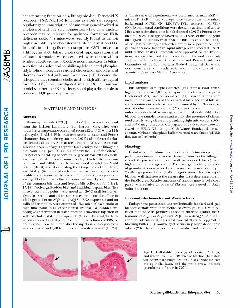
Fig. 2. Gallbladder wall thickness as a function of time on thelithogenic diet. Gallbladder wall thickness increases progressivelyduring the diet period (P , 0.0001) and is more pronounced inC57L than in AKR (P , 0.02). Closed circles, C57L; open squares,AKR. Results are shown as means 6 SEM.
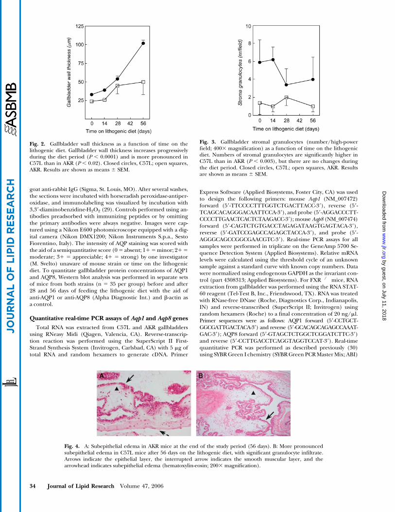
Fig. 3. Gallbladder stromal granulocytes (number/high-powerfield; 4003 magnification) as a function of time on the lithogenicdiet. Numbers of stromal granulocytes are significantly higher inC57L than in AKR (P , 0.003), but there are no changes duringthe diet period. Closed circles, C57L; open squares, AKR. Resultsare shown as means 6 SEM.
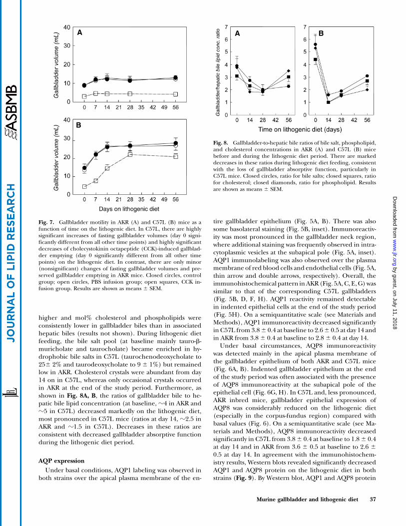
Fig. 4. A: Subepithelial edema in AKR mice at the end of the study period (56 days). B: More pronouncedsubepithelial edema in C57L mice after 56 days on the lithogenic diet, with significant granulocyte infiltrate.Arrows indicate the epithelial layer, the interrupted arrow indicates the smooth muscular layer, and thearrowhead indicates subepithelial edema (hematoxylin-eosin; 2003 magnification).
34 Journal of Lipid Research Volume 47, 2006
by guest, on July 11, 2018w
ww
.jlr.orgD
ownloaded from

on the ABI Prism 7900HT Sequence Detection System. Each sam-ple was run in triplicate. Relative fold changes were calculatedusing the comparative cycle times method with cyclophilin as thereference gene and the wild-type mice as the calibrators.
Statistical analysis
Results are shown as means 6 SEM. Differences betweengroups and time effects were analyzed for statistical significanceby Mann-Whitney U-tests or ANOVA with Fisher’s least significantdifference as the posthoc test. A two-tailed P value of ,0.05 wasconsidered significant.
RESULTS
Gallbladder histology
At baseline, gallbladder wall thickness was greater, andepithelial cells appeared more elongated, in C57L than
in AKR (Figs. 1, 2). On the lithogenic diet, gallbladderwall thickness increased markedly, but this was most pro-nounced in C57L (Fig. 2). Also, there was moderate in-filtration of neutrophilic granulocytes in gallbladder wallstroma and some intraepithelial granulocytes in C57Lduring the entire study period (Figs. 1B, 3). Granulocyteswere infrequent in AKR (Fig. 1A, 3), but occasional lym-phoid aggregates were noted during the diet period. Fromday 28 on, progressive fibrosis (C57L > AKR) and, morepronounced, increased smooth muscle thickness werenoted in the gallbladder wall. Also from day 28, sporadicmacrophages loaded with debris and fat were seen inC57L. These abnormalities were progressive on day 56 inC57L, with concomitant edema in the subepithelial layer(Fig. 4B). Similar changes were not noted before day 56 inAKR and were less pronounced than for C57L (Fig. 4A).Also, progressive epithelial indentation was seen in C57Lfrom day 28 of lithogenic feeding (Figs. 5F, H, 6F, H).
Fig. 5. Immunohistochemical distribution of aqua-porin-1 (AQP1) in the gallbladders of AKR (A, C, E,G) and C57L (B, D, F, H) mice on the lithogenicdiet. A, B: Day 0. Considerable AQP1 immunoreac-tivity (brown staining) is observed over the apical(arrowheads) and lateral (insets; open arrowheads)membranes of AKR (A) and C57L (B) gallbladderepithelia. Staining is also observed over the plasmamembrane of endothelial cells (A,B; double arrows)and red blood cells (A; thin arrow). Additional label-ing is often present in the neck region of both AKRand C57L gallbladders, where intracellular stainingis seen over vesicles located at the subapical pole ofsurface epithelial cells (A, inset; arrows). Lateral spacesbetween adjacent epithelial cells appear enlarged asa morphological consequence of the process of fluidabsorption (48) (B, inset; dashed arrows). C, D: Day14. Immunohistochemical patterns of AQP1 in AKR(C) and C57L (D) gallbladder epithelial cells andplasma membranes of endothelial or red blood cells(apical staining, arrowheads; endothelial staining,double arrows; red blood cell staining, small arrows).E, F: Day 28. Considerable AQP1 expression (arrow-heads) is seen at the apical pole of the gallbladderepithelium of both AKR (E) and C57L (F) mice. Sub-apical staining is frequently observed in the neck re-gion of gallbladders (arrows). A striking indentationof the surface epithelial layer (red dashed areas) isobserved in the C57L gallbladders (F). G, H: Day 56.Apical (arrowheads) and subapical (arrows) AQP1labeling is present in the epithelial layer of bothAKR (G) and C57L (H) gallbladders. Indentation ofthe epithelium is now also observed in the neck regionof the AKR gallbladder (G; red dashed areas). AQP1immunoreactivity is observed even in epithelial sheetsdetached away from the C57L gallbladder as a con-sequence of advanced lithogenesis (H, inset). Originalmagnifications,3400.
Murine gallbladder and lithogenic diet 35
by guest, on July 11, 2018w
ww
.jlr.orgD
ownloaded from

Although less marked than in C57L, indented epitheliumwas also observed in AKR gallbladders at day 56 on thelithogenic diet (Figs. 5G, 6G).
Gallbladder motility
Basal fasting gallbladder volumes were significantlylarger in C57L than in AKR inbred mice (14.8 6 2.2ml vs. 8.86 1.0 ml). PBS infusion did not affect gallbladdervolumes before or during diet treatment (Fig. 7). Beforethe lithogenic diet, low gallbladder volumes were observedafter CCK infusion in both strains (4.4 6 0.5 ml and 3.0 6
0.3 ml in C57L and AKR, respectively, corresponding to70% emptying). As shown in Fig. 7B, fasting gallbladdervolumes increased progressively on the diet in C57L (from14.8 6 2.2 ml on day 0 to 28.6 6 2.9 ml on day 56), withprogressively decreased CCK-induced emptying (70% and
27% of fasting volumes on day 0 and day 56, respectively).In contrast, there were only minor (nonsignificant) in-creases of fasting gallbladder volumes in AKR during thelithogenic diet feeding (from 8.86 1.0 ml on day 0 to 1361.3 ml on day 56), and CCK-induced gallbladder emptyingremained largely preserved (Fig. 7A).
Biliary lipids and gallbladder concentrating function
The cholesterol saturation index in hepatic biles (basal,0.91 6 0.12 and 0.83 6 0.12 for C57L and AKR, res-pectively) and associated gallbladder biles (basal, 0.53 and0.52 for C57L and AKR, respectively) was supersaturatedfrom day 14 in both strains. At baseline, mol% bile salts,cholesterol, and phospholipids in gallbladder biles andassociated hepatic biles were identical. In contrast, duringlithogenic diet feeding, mol% bile salts was consistently
Fig. 6. Immunohistochemical expression of AQP8in AKR and C57L gallbladders during lithogenic dietfeeding. A, B: Day 0. Considerable AQP8 immunore-activity (arrowheads) is seen at the apical pole of bothAKR (A) and C57L (B) gallbladder. Some staining isalso observed over the plasma membrane of endo-thelial cells (double arrows). C, D: Day 14. Althoughmuch less pronounced than on day 0, AQP8 reactivityis observed at the apical pole of both AKR (C) andC57L (D) gallbladders (arrowheads). The decreasein apical AQP8 expression is more pronounced inC57L (D; arrowheads) than in AKR gallbladders. Ex-pression of AQP8 in both AKR and C57L gallbladderendothelial cells (double arrows) appears unchangedcompared with basal values. E, F: Day 28. Apicallabeling of AQP8 in AKR gallbladder epithelium(E; arrowheads) and C57L gallbladder epithelium(F; arrowheads). Note the intracellular reactivity (F,inset; arrows) often associated with the characteristicindentation of the C57L gallbladder epithelium (F;red dashed areas). G, H: Day 56. Considerable AQP8reactivity is seen at the apical pole of both AKR (G)and C57L (H) gallbladders (arrowheads). Subapicalreactivity (insets; arrows) is seen below the indentation(red dashed areas). Original magnifications, 3400.
36 Journal of Lipid Research Volume 47, 2006
by guest, on July 11, 2018w
ww
.jlr.orgD
ownloaded from
higher and mol% cholesterol and phospholipids wereconsistently lower in gallbladder biles than in associatedhepatic biles (results not shown). During lithogenic dietfeeding, the bile salt pool (at baseline mainly tauro-b-muricholate and taurocholate) became enriched in hy-drophobic bile salts in C57L (taurochenodeoxycholate to256 2% and taurodeoxycholate to 9 6 1%) but remainedlow in AKR. Cholesterol crystals were abundant from day14 on in C57L, whereas only occasional crystals occurredin AKR at the end of the study period. Furthermore, asshown in Fig. 8A, B, the ratios of gallbladder bile to he-patic bile lipid concentration (at baseline, z4 in AKR andz5 in C57L) decreased markedly on the lithogenic diet,most pronounced in C57L mice (ratios at day 14, z2.5 inAKR and z1.5 in C57L). Decreases in these ratios areconsistent with decreased gallbladder absorptive functionduring the lithogenic diet period.
AQP expression
Under basal conditions, AQP1 labeling was observed inboth strains over the apical plasma membrane of the en-
tire gallbladder epithelium (Fig. 5A, B). There was alsosome basolateral staining (Fig. 5B, inset). Immunoreactiv-ity was most pronounced in the gallbladder neck region,where additional staining was frequently observed in intra-cytoplasmic vesicles at the subapical pole (Fig. 5A, inset).AQP1 immunolabeling was also observed over the plasmamembrane of red blood cells and endothelial cells (Fig. 5A,thin arrow and double arrows, respectively). Overall, theimmunohistochemical pattern inAKR(Fig. 5A,C, E,G)wassimilar to that of the corresponding C57L gallbladders(Fig. 5B, D, F, H). AQP1 reactivity remained detectablein indented epithelial cells at the end of the study period(Fig. 5H). On a semiquantitative scale (see Materials andMethods), AQP1 immunoreactivity decreased significantlyin C57L from 3.86 0.4 at baseline to 2.66 0.5 at day 14 andin AKR from 3.86 0.4 at baseline to 2.86 0.4 at day 14.
Under basal circumstances, AQP8 immunoreactivitywas detected mainly in the apical plasma membrane ofthe gallbladder epithelium of both AKR and C57L mice(Fig. 6A, B). Indented gallbladder epithelium at the endof the study period was often associated with the presenceof AQP8 immunoreactivity at the subapical pole of theepithelial cell (Fig. 6G, H). In C57L and, less pronounced,AKR inbred mice, gallbladder epithelial expression ofAQP8 was considerably reduced on the lithogenic diet(especially in the corpus-fundus region) compared withbasal values (Fig. 6). On a semiquantitative scale (see Ma-terials and Methods), AQP8 immunoreactivity decreasedsignificantly in C57L from 3.86 0.4 at baseline to 1.86 0.4at day 14 and in AKR from 3.6 6 0.5 at baseline to 2.6 6
0.5 at day 14. In agreement with the immunohistochem-istry results, Western blots revealed significantly decreasedAQP1 and AQP8 protein on the lithogenic diet in bothstrains (Fig. 9). By Western blot, AQP1 and AQP8 protein
Fig. 7. Gallbladder motility in AKR (A) and C57L (B) mice as afunction of time on the lithogenic diet. In C57L, there are highlysignificant increases of fasting gallbladder volumes (day 0 signi-ficantly different from all other time points) and highly significantdecreases of cholecystokinin octapeptide (CCK)-induced gallblad-der emptying (day 0 significantly different from all other timepoints) on the lithogenic diet. In contrast, there are only minor(nonsignificant) changes of fasting gallbladder volumes and pre-served gallbladder emptying in AKR mice. Closed circles, controlgroup; open circles, PBS infusion group; open squares, CCK in-fusion group. Results are shown as means 6 SEM.
Fig. 8. Gallbladder-to-hepatic bile ratios of bile salt, phospholipid,and cholesterol concentrations in AKR (A) and C57L (B) micebefore and during the lithogenic diet period. There are markeddecreases in these ratios during lithogenic diet feeding, consistentwith the loss of gallbladder absorptive function, particularly inC57L mice. Closed circles, ratio for bile salts; closed squares, ratiofor cholesterol; closed diamonds, ratio for phospholipid. Resultsare shown as means 6 SEM.
Murine gallbladder and lithogenic diet 37
by guest, on July 11, 2018w
ww
.jlr.orgD
ownloaded from
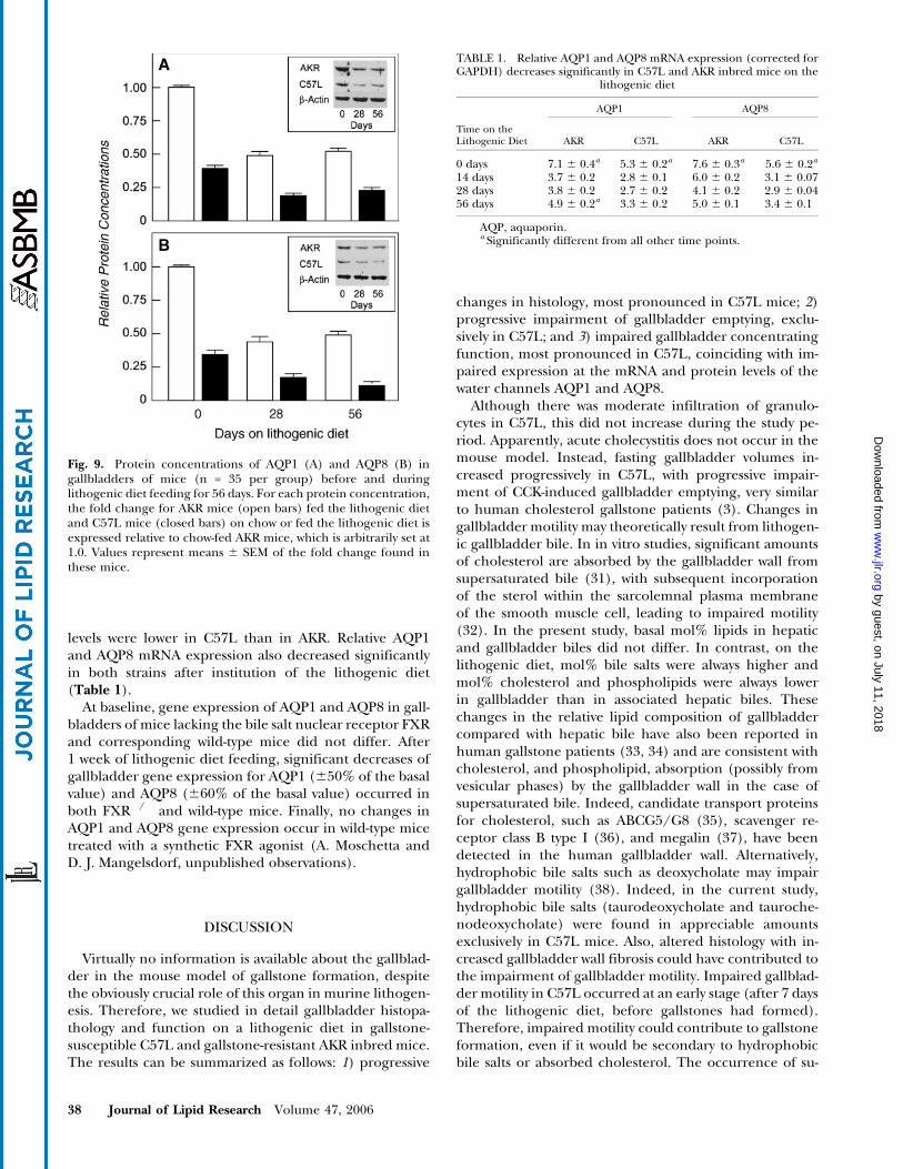
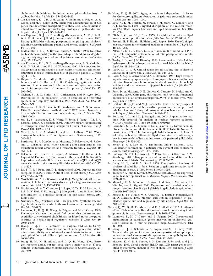
levels were lower in C57L than in AKR. Relative AQP1and AQP8 mRNA expression also decreased significantlyin both strains after institution of the lithogenic diet(Table 1).
At baseline, gene expression of AQP1 and AQP8 in gall-bladders of mice lacking the bile salt nuclear receptor FXRand corresponding wild-type mice did not differ. After1 week of lithogenic diet feeding, significant decreases ofgallbladder gene expression for AQP1 (650% of the basalvalue) and AQP8 (660% of the basal value) occurred inboth FXR�/� and wild-type mice. Finally, no changes inAQP1 and AQP8 gene expression occur in wild-type micetreated with a synthetic FXR agonist (A. Moschetta andD. J. Mangelsdorf, unpublished observations).
DISCUSSION
Virtually no information is available about the gallblad-der in the mouse model of gallstone formation, despitethe obviously crucial role of this organ in murine lithogen-esis. Therefore, we studied in detail gallbladder histopa-thology and function on a lithogenic diet in gallstone-susceptible C57L and gallstone-resistant AKR inbred mice.The results can be summarized as follows: 1) progressive
changes in histology, most pronounced in C57L mice; 2)progressive impairment of gallbladder emptying, exclu-sively in C57L; and 3) impaired gallbladder concentratingfunction, most pronounced in C57L, coinciding with im-paired expression at the mRNA and protein levels of thewater channels AQP1 and AQP8.
Although there was moderate infiltration of granulo-cytes in C57L, this did not increase during the study pe-riod. Apparently, acute cholecystitis does not occur in themouse model. Instead, fasting gallbladder volumes in-creased progressively in C57L, with progressive impair-ment of CCK-induced gallbladder emptying, very similarto human cholesterol gallstone patients (3). Changes ingallbladder motility may theoretically result from lithogen-ic gallbladder bile. In in vitro studies, significant amountsof cholesterol are absorbed by the gallbladder wall fromsupersaturated bile (31), with subsequent incorporationof the sterol within the sarcolemnal plasma membraneof the smooth muscle cell, leading to impaired motility(32). In the present study, basal mol% lipids in hepaticand gallbladder biles did not differ. In contrast, on thelithogenic diet, mol% bile salts were always higher andmol% cholesterol and phospholipids were always lowerin gallbladder than in associated hepatic biles. Thesechanges in the relative lipid composition of gallbladdercompared with hepatic bile have also been reported inhuman gallstone patients (33, 34) and are consistent withcholesterol, and phospholipid, absorption (possibly fromvesicular phases) by the gallbladder wall in the case ofsupersaturated bile. Indeed, candidate transport proteinsfor cholesterol, such as ABCG5/G8 (35), scavenger re-ceptor class B type I (36), and megalin (37), have beendetected in the human gallbladder wall. Alternatively,hydrophobic bile salts such as deoxycholate may impairgallbladder motility (38). Indeed, in the current study,hydrophobic bile salts (taurodeoxycholate and tauroche-nodeoxycholate) were found in appreciable amountsexclusively in C57L mice. Also, altered histology with in-creased gallbladder wall fibrosis could have contributed tothe impairment of gallbladder motility. Impaired gallblad-der motility in C57L occurred at an early stage (after 7 daysof the lithogenic diet, before gallstones had formed).Therefore, impaired motility could contribute to gallstoneformation, even if it would be secondary to hydrophobicbile salts or absorbed cholesterol. The occurrence of su-
TABLE 1. Relative AQP1 and AQP8 mRNA expression (corrected forGAPDH) decreases significantly in C57L and AKR inbred mice on the
lithogenic diet
AQP1 AQP8
Time on theLithogenic Diet AKR C57L AKR C57L
0 days 7.1 6 0.4a 5.3 6 0.2a 7.6 6 0.3a 5.6 6 0.2a
14 days 3.7 6 0.2 2.8 6 0.1 6.0 6 0.2 3.1 6 0.0728 days 3.8 6 0.2 2.7 6 0.2 4.1 6 0.2 2.9 6 0.0456 days 4.9 6 0.2a 3.3 6 0.2 5.0 6 0.1 3.4 6 0.1
AQP, aquaporin.a Significantly different from all other time points.
Fig. 9. Protein concentrations of AQP1 (A) and AQP8 (B) ingallbladders of mice (n = 35 per group) before and duringlithogenic diet feeding for 56 days. For each protein concentration,the fold change for AKR mice (open bars) fed the lithogenic dietand C57L mice (closed bars) on chow or fed the lithogenic diet isexpressed relative to chow-fed AKR mice, which is arbitrarily set at1.0. Values represent means 6 SEM of the fold change found inthese mice.
38 Journal of Lipid Research Volume 47, 2006
by guest, on July 11, 2018w
ww
.jlr.orgD
ownloaded from

persaturated bile (and possibly also hydrophobic bile saltcomposition) could relate to certain Lith genes. A signi-ficant number of hepatic lipid regulatory genes, hepaticlipoprotein receptors and related genes, hepatic and in-testinal membrane and intracellular lipid transporters,and hepatic lipid regulatory transcription factors thatcould be involved in the occurrence of biliary cholesterolsupersaturation colocalize with various Lith genes thatwere identified by quantitative trait locus studies in themouse model (for an overview, see 39). In addition, theCCK-A receptor controls to a large extent murine gall-bladder contractility, the encoding gene Cckar colocalizeswith a Lith gene onmouse chromosome (39), and targeteddisruption of the murine CCK receptor increases suscep-tibility to gallstones (40). Therefore, Lith genes could beinvolved in the marked differences in gallbladder motilitybetween C57L and AKR mice found in this study.
As inferred from gallbladder-to-hepatic ratios of lipidconcentrations, we found a strong decrease of gallbladderabsorptive function after institution of the lithogenic diet,particularly in C57L inbred mice. We hypothesize thatdecreased gallbladder concentrating function could alsobe secondary to the absorption of cholesterol by the gall-bladder wall from supersaturated bile and altered histol-ogy with increased gallbladder wall thickness. Our findingsof water channel AQP1 expression in apical and basolat-eral epithelial cell membranes, particularly in the gall-bladder neck, are in agreement with previous data (7, 8,10, 12). The presence of AQP1 in murine gallbladder mi-crovessels (9, 10, 12) has also been reported previously.Recently, AQP8 was detected in the apical gallbladderepithelial plasma membrane of various species, both at themRNA and protein levels (12). Reduction of AQP expres-sion in the gallbladder epithelial plasma membrane maynot be the consequence of a local effect exerted by thelithogenic diet, because concomitant AQP expression ingallbladder endothelial cells appeared not to change.Also, similar AQP8 downregulation was reported recentlyin the liver of cholesterol-fed mice (41) and during extra-hepatic cholestasis (42). We also found, in the currentstudy, significant downregulation of relative AQP1 mRNAexpression (from 1 6 0.12 to 0.48 6 0.09) and relativeAQP8 mRNA expression (from 1 6 0.08 to 0.57 6 0.1) inlivers of C57L mice after 1 week on the lithogenic diet(results not shown). These findings all suggest that thelithogenic diet exerts a more general effect on AQP ex-pression, possibly because of its high fat and cholesterolcontents. With regard to the gallbladder, concordantchanges of AQP expression and the absorptive function ofthis organ on the lithogenic diet, as found in this study,suggest the involvement of these water channels in gall-bladder water transport, possibly secondary to changes inelectrolyte transport. Gallbladder Na1 absorption in thefasting state is mostly mediated through apically restrictedNa1/H1 exchange. This protein kinase C-a-regulatedmembrane protein facilitates the electroneutral exchangeof extracellular Na1 for intracellular H+, with subsequentbasolateral Na1 extrusion and secondary osmosis-drivenwater transport (43). Limited data in the prairie dog
model suggest dysfunctional regulation of Na1/H1 ex-change by protein kinase C-a during gallstone formation(43, 44). Associated Cl� transfer probably involves Cl�/HCO3
� exchangers and chloride channels. In contrastwith the fasting state, gallbladder water secretion seems topredominate after meal ingestion, possibly influenced bygastrointestinal hormones such as secretin and vasoactiveintestinal polypeptide (45–47). These hormones act oncystic fibrosis-transmembrane conductance regulator inthe epithelial cell (47). Increases of intracellular cAMPlevels subsequently inhibit Cl�/HCO3
� and Na1/H1 ex-changers at the apical membrane, with the result that netNaCl entry is inhibited (10). Although one may speculatethat AQP1 and AQP8 exhibit different functions in thefasting versus the fed state, this remains to be explored infurther studies.
Because the lithogenic diet contains significant amountsof the FXR ligand cholic acid, we tested whether AQP1 andAQP8 downregulation could occur through the FXR sig-naling pathway. Similar AQP downregulation in FXR�/�
and corresponding wild-type mice after 1 week of the lith-ogenic diet argues against a direct role of FXR in drivingthe lithogenic diet-induced decreases of AQP1 and AQP8gene expression in gallbladder or liver. This contentionis further supported by the absence of any changes inAQP1 and AQP8 gene expression in wild-type mice treatedwith a synthetic FXR agonist (A. Moschetta and D. J. Man-gelsdorf, unpublished observations). In summary, pro-gressive alterations in gallbladder histology during murinelithogenesis are associated with progressively impairedgallbladder motility, particularly in C57L. A temporal as-sociation between decreased gallbladder concentratingfunction and decreased AQP1 or AQP8 expression sug-gests the involvement of these water channels in gall-bladder water transport.
This study was supported by research grants from the Fondoper gli Investimenti della Ricerca di Base (Grant RBAU01R-ANB), Centro di Eccellenza di Genomica in Campo Biomedicoed Agrario, and Progetto Laboratorio Analisi del Gene Studiodi Geni di Interesse Biomedico ed Agroalimentare (to G.C. andM. Svelto), by the Howard Hughes Medical Institute (A.M.), bya grant from the Ellison Medical Foundation (to D.Q-H.W.),and by research Grant DK-54012 (to D.Q-H.W.) from theNational Institutes of Health (U.S. Public Health Service). Theauthors thank Prof. Giuseppa E. Liquori for her skillful con-tribution to the immunohistochemical studies. The authorsalso thank Dr. Frank Gonzalez for supplying the FXR�/� miceused in this study, Dr. David Mangelsdorf for allowing theFXR�/� mouse experiments to be performed in his labora-tory, and Angie Bookout for contributing to the studies in theFXR�/�mouse. The authors are greatly indebted to Helen H-F.Wang (Beth Israel Deaconess Medical Center, Boston, MA) forexcellent technical assistance.
REFERENCES
1. Wang, D. Q. H., B. Paigen, and M. C. Carey. 1997. Phenotypiccharacterization of Lith genes that determine susceptibility to
Murine gallbladder and lithogenic diet 39
by guest, on July 11, 2018w
ww
.jlr.orgD
ownloaded from
cholesterol cholelithiasis in inbred mice: physical-chemistry ofgallbladder bile. J. Lipid Res. 38: 1395–1411.
2. van Erpecum, K. J., D. Q-H. Wang, F. Lammert, B. Paigen, A. K.Groen, and M. C. Carey. 2001. Phenotypic characterization of Lithgenes that determine susceptibility to cholesterol cholelithiasis ininbred mice: soluble pronucleating proteins in gallbladder andhepatic biles. J. Hepatol. 35: 444–451.
3. van Erpecum, K. J., G. P. vanBerge-Henegouwen, M. F. J. Stolk,W. P. M. Hopman, J. B. M. J. Jansen, and C. B. H. W. Lamers. 1992.Fasting gallbladder volume, postprandial emptying and cholecys-tokinin release in gallstone patients and normal subjects. J. Hepatol.14: 194–202.
4. Fridhandler, T. M., J. S. Davison, and E. A. Shaffer. 1983. Defectivegallbladder contractility in the ground squirrel and prairie dog dur-ing the early stages of cholesterol gallstone formation. Gastroenter-ology. 85: 830–836.
5. van Erpecum, K. J., G. P. vanBerge-Henegouwen, B. Stoelwinder,Y. M. G. Schmidt, and F. L. H. Willekens. 1990. Bile concentrationis a key factor for nucleation of cholesterol crystals and cholesterolsaturation index in gallbladder bile of gallstone patients. Hepatol-ogy. 11: 1–6.
6. Halpern, Z., M. A. Dudley, M. P. Lynn, J. M. Nader, A. C.Breuer, and R. T. Holzbach. 1986. Vesicle aggregation in modelsystems of supersaturated bile: relation to crystal nucleationand lipid composition of the vesicular phase. J. Lipid Res. 27:295–306.
7. Nielsen, S., B. L. Smith, E. I. Christensen, and P. Agre. 1993.Distribution of the aquaporin CHIP in secretory and resorptiveepithelia and capillary endothelia. Proc. Natl. Acad. Sci. USA. 90:7275–7279.
8. Hasegawa, H., S. C. Lian, W. E. Finkbeiner, and A. S. Verkman.1994. Extrarenal tissue distribution of CHIP28 water channels byin situ hybridization and antibody staining. Am. J. Physiol. 266:C893–C903.
9. Ma, T., S. Jayaraman, K. S. Wang, Y. Song, B. Yang, J. Li, J. A.Bastidas, and A. S. Verkman. 2001. Defective dietary fat processingin transgenic mice lacking aquaporin-1 water channels. Am. J.Physiol. 280: C126–C134.
10. Masyuk, A. I., R. A. Marinelli, and N. F. LaRusso. 2002. Watertransport by epithelia of the digestive tract. Gastroenterology. 122:545–562.
11. Portincasa, P., A. Moschetta, A. Mazzone, G. Palasciano, M. Svelto,and G. Calamita. 2003. Water handling and aquaporins in bileformation: recent advances and research trends. J. Hepatol. 39:864–874.
12. Calamita, G., D. Ferri, C. Bazzini, A. Mazzone, G. Botta, G. E.Liquori, M. Paulmichl, P. Portincasa, G. Meyer, andM. Svelto. 2005.Expression and subcellular localization of the AQP8 and AQP1water channels in the mouse gallbladder epithelium. Biol. Cell. 97:415–423.
13. Lu, T. T., J. J. Repa, and D. J. Mangelsdorf. 2001. Orphan nuclearreceptors as eLiXiRs and FiXeRs of sterol metabolism. J. Biol. Chem.276: 37735–37738.
14. Moschetta, A., A. L. Bookout, and D. J. Mangelsdorf. 2004. Pre-vention of cholesterol gallstone disease by FXR agonists in a mousemodel. Nat. Med. 10: 1352–1358.
15. Makishima, M., A. Y. Okamoto, J. J. Repa, H. Tu, R. M. Learned, A.Luk, M. V. Hull, K. D. Lustig, D. J. Mangelsdorf, and B. Shan. 1999.Identification of a nuclear receptor for bile acids. Science. 284:1362–1365.
16. Nishina, P. M., J. Verstuyft, and B. Paigen. 1990. Synthetic low andhigh fat diets for the study of atherosclerosis in the mouse. J. LipidRes. 31: 859–869.
17. Lammert, F., D. Q. H. Wang, B. Paigen, and M. C. Carey. 1999.Phenotypic characterization of Lith genes that determine sus-ceptibility to cholesterol cholelithiasis in inbred mice: integratedactivities of hepatic lipid regulatory enzymes. J. Lipid Res. 40:2080–2090.
18. Wang, D. Q. H., F. Lammert, B. Paigen, and M. C. Carey.1999. Phenotypic characterization of Lith genes that deter-mine susceptibility to cholesterol cholelithiasis in inbred mice:pathophysiology of biliary lipid secretion. J. Lipid Res. 40:2066–2079.
19. Wang, H. H., N. H. Afdhal, and D. Q. H. Wang. 2004. Estro-gen receptor alpha, but not beta, plays a major role in 17beta-estradiol-inducedmurine cholesterol gallstones.Gastroenterology. 127:239–249.
20. Wang, D. Q. H. 2002. Aging per se is an independent risk factorfor cholesterol gallstone formation in gallstone susceptible mice.J. Lipid Res. 43: 1950–1959.
21. Sinal, C. J., M. Tohkin, M. Miyata, J. M. Ward, G. Lambert, andF. J. Gonzalez. 2000. Targeted disruption of the nuclear recep-tor FXR/BAR impairs bile acid and lipid homeostasis. Cell. 102:731–744.
22. Bligh, E. G., and W. J. Dyer. 1959. A rapid method of total lipidextraction and purification. Can. J Biochem. Physiol. 37: 911–917.
23. Fromm, H., P. Hamin, H. Klein, and I. Kupke. 1980. Use of a simpleenzymatic assay for cholesterol analysis in human bile. J. Lipid Res.21: 259–261.
24. Allain, C. C., L. S. Poon, C. S. G. Chan, W. Richmond, and P. C.Fu. 1974. Enzymatic determination of total phospholipids in bile.J. Clin. Chem. 20: 470–475.
25. Turley, S. D., and J. M. Dietschy. 1978. Re-evaluation of the 3 alpha-hydroxysteroid dehydrogenase assay for total bile acids in bile. J.Lipid Res. 19: 924–928.
26. Carey, M. C. 1978. Critical tables for calculating the cholesterolsaturation of native bile. J. Lipid Res. 19: 945–955.
27. Rossi, S. S., J. L. Converse, and A. F. Hofmann. 1987. High pressureliquid chromatographic analysis of conjugated bile acids in humanbile: simultaneous resolution of sulfated and unsulfated lithocholylamidates and the common conjugated bile acids. J. Lipid Res. 28:589–595.
28. Ferri, D., A. Mazzone, G. E. Liquori, G. Cassano, M. Svelto, and G.Calamita. 2003. Ontogeny, distribution, and possible functionalimplications of an unusual aquaporin, AQP8, in mouse liver. Hepa-tology. 38: 947–957.
29. Graham, R. C., Jr., and M. J. Karnovsky. 1966. The early stages ofabsorption of injected horseradish peroxidase in the proximaltubules of mouse kidney: ultrastructural cytochemistry by a newtechnique. J. Histochem. Cytochem. 14: 291–302.
30. Bookout, A. L., and D. J. Mangelsdorf. 2003. A quantitative real-time PCR protocol for analysis of nuclear receptor pathways.NURSA e-Journal. Vol. 1 doi: 10.1621/nrs.01012.
31. Corradini, S. G., C. Ripani, P. Della Guardia, L. Giovannelli, W.Elisei, A. Cantafora, M. C. Pisanelli, G. D. Tebala, G. Nuzzo, A.Corsi, et al. 1998. The human gallbladder increases cholesterolsolubility in bile by differential lipid absorption: a study using anew in vitro model of isolated intra-arterially perfused gallbladder.Hepatology. 28: 314–322.
32. Behar, J., K. Y. Lee, W. R. Thompson, and P. Biancani. 1989.Gallbladder contraction in patients with pigment and cholesterolstones. Gastroenterology. 97: 1479–1484.
33. Gallinger, S., P. R. C. Harvey, C. N. Petrunka, R. G. Ilson, and S. M.Strasberg. 1987. Biliary proteins and the nucleation defect in cho-lesterol cholelithiasis. Gastroenterology. 92: 867–875.
34. Carey, M. C., and D. M. Small. 1978. The physical chemistry ofcholesterol solubility in bile. Relation to gallstone formation anddissolution in man. J. Clin. Invest. 61: 998–1026.
35. Tauscher, A., and R. Kuver. 2003. ABCG5 and ABCG8 are expressedin gallbladder epithelial cells. Biochem. Biophys. Res. Commun. 307:1021–1028.
36. Miquel, J. F., M. Moreno, L. Amigo, H. Molina, P. Mardones, I. I.Wistuba, and A. Rigotti. 2003. Expression and regulation of sca-venger receptor class B type I (SR-BI) in gall bladder epithelium.Gut. 52: 1017–1024.
37. Erranz, B., J. F. Miquel, W. S. Argraves, J. L. Barth, F. Pimentel, andM. P. Marzolo. 2004. Megalin and cubilin expression in gall-bladder epithelium and regulation by bile acids. J. Lipid Res. 45:2185–2198.
38. Xu, Q. W., S. M. Freedman, and E. A. Shaffer. 1997. Inhibitoryeffect of bile salts on gallbladder smooth muscle contractility in theguinea pig in vitro. Gastroenterology. 112: 1699–1706.
39. Lammert, F., M. C. Carey, and B. Paigen. 2001. Chromosomalorganization of candidate genes involved in cholesterol gall-stone formation: a murine gallstone map. Gastroenterology. 120:221–238.
40. Wang, D. Q., F. Schmitz, A. S. Kopin, and M. C. Carey. 2004.Targeted disruption of the murine cholecystokinin-1 receptor pro-motes intestinal cholesterol absorption and susceptibility to cho-lesterol cholelithiasis. J. Clin. Invest. 114: 521–528.
41. Maxwell, K. N., R. E. Soccio, E. M. Duncan, E. Schayek, and J. L.Breslow. 2003. Novel putative SREBP and LXR target genes iden-tified by microarray analysis in liver of cholesterol-fed mice. J. LipidRes. 44: 2109–2119.
40 Journal of Lipid Research Volume 47, 2006
by guest, on July 11, 2018w
ww
.jlr.orgD
ownloaded from
42. Carreras, F. I., S. A. Gradilone, A. Mazzone, F. Garcia, B. Q. Huang,J. E.Ochoa, P. S. Tietz,N. F. LaRusso,G.Calamita, andR.A.Marinelli.2003. Rat hepatocyte aquaporin-8 water channels are down-regulatedin extrahepatic cholestasis. Hepatology. 37: 1026–1033.
43. Narins, S. C., R. Ramakrishnan, E. H. Park, P. B. Bolno, D. A.Haggerty, P. R. Smith, W. C. Meyers, and M. Z. Abedin. 2005.Protein kinase C-alpha regulation of gallbladder Na1 transport be-comes progressively more dysfunctional during gallstone forma-tion. J. Lab. Clin. Med. 146: 227–237.
44. van Erpecum, K. J., and D. Q. H. Wang. 2005. The gallbladder:innocent bystander or major factor in cholesterol-gallstone forma-tion? J. Lab. Clin. Med. 146: 202–204.
45. Igimi, H., F. Yamamoto, and S. P. Lee. 1992. Gallbladder mucosal
function: studies in absorption and secretion in humans and in doggallbladder epithelium. Am. J. Physiol. 263: G69–G74.
46. Sweeting, J. G. 1993. Does the gallbladder secrete? Gastroenterology.104: 329–330.
47. Peters, R. H., J. H. van Doorninck, P. J. French, R. Ratcliff, M. J.Evans, W. H. Colledge, J. Bijman, and B. J. Scholte. 1997. Cysticfibrosis transmembrane conductance regulator mediates the cyclicadenosine monophosphate-induced fluid secretion but not the in-hibition of resorption in mouse gallbladder epithelium. Hepatology.25: 270–277.
48. Whitlock, R. T., and H. O. Wheeler. 1964. Coupled transport ofsolute and water across rabbit gallbladder epithelium. J. Clin. Invest.43: 2249–2265.
Murine gallbladder and lithogenic diet 41
by guest, on July 11, 2018w
ww
.jlr.orgD
ownloaded from