Functional Assessments and Histopathology of …article.aascit.org/file/pdf/9220770.pdf ·...
14
American Journal of Pharmacy and Pharmacology 2017; 4(5): 21-34 http://www.aascit.org/journal/ajpp ISSN: 2375-3900 Keywords Carbon Tetrachloride, Histopathology, Hydrothermal, Kidney, Liver Received: March 16, 2017 Accepted: May 3, 2017 Published: August 8, 2017 Functional Assessments and Histopathology of Hepatorenal Tissues of Wistar Rats Infused with Carbon Tetrachloride and Treated with Raw and Hydrothermal Processed Herbs Okey Alphonsus Ojiako 1 , Paul Chidoka Chikezie 2, * , Doris Ifeyinwa Ukairo 1 , Chiedozie Onyejiaka Ibegbulem 1 , Reginald Nwazue Nwaoguikpe 1 1 Department of Biochemistry, Federal University of Technology, Owerri, Nigeria 2 Department of Biochemistry, Imo State University, Owerri, Nigeria Email address [email protected] (P. C. Chikezie) * Corresponding author Citation Okey Alphonsus Ojiako, Paul Chidoka Chikezie, Doris Ifeyinwa Ukairo, Chiedozie Onyejiaka Ibegbulem, Reginald Nwazue Nwaoguikpe. Functional Assessments and Histopathology of Hepatorenal Tissues of Wistar Rats Infused with Carbon Tetrachloride and Treated with Raw and Hydrothermal Processed Herbs. American Journal of Pharmacy and Pharmacology. Vol. 4, No. 5, 2017, pp. 21-34. Abstract The present study ascertained the functional integrity of hepatic and renal tissues, concurrently with blood lipid patterns, of Wistar rats infused with CCl 4 and treated with raw and hydrothermal processed herbs, namely, Monodora myristica, Chromolaena odorata, Buccholzia coriacea and Sphenostylis stenocarpa. Measurement of phytochemical contents of the herbs was according to standard methods. The rats were randomly designated on the bases of diets and treatments received for 28 consecutive days. Fibrosis was induced in the Wistar rats by single dose intra-peritoneal injection of CCl 4 for 2 consecutive days. Liver and kidney function tests and serum lipid profile were measured using spectrophotometric methods. Renal and hepatic tissues were subjected to histopathological examinations. The concentrations of alkaloids in the four herbal extracts were within the range of 4.83 ± 0.03 – 31.33 ± 0.29 mg/100 g sample, whereas the concentrations of saponins varied within a relatively narrow range: 0.33 ± 0.09 – 4.33 ± 0.02 mg/100 g dry sample; p > 0.05. The activity ratios of AST to ALT of the rat groups were generally less than 1.0 unit. Atherogenic indices of fibrotic rats were within the following ranges: TAG/HDL-C ratio (3.59 ± 0.03 – 6.76 ± 0.06), TC/HDL-C ratio (3.72 ± 0.02 – 6.94 ± 0.05) and LDL-C/HDL-C ratio (2.00 ± 0.01 – 4.59 ± 0.02). Losses in phytochemical contents following hydrothermal processing of the herbs did not substantially affect their overall therapeutic scores against morphological and functional impairments of hepatic and renal tissues following CCl 4 intoxication of the rats. 1. Introduction The metabolic concerns of hepatic tissues, among several physiologic functions, ensure the detoxification of endogenously formed toxic molecules and xenobiotics through concerted processes of functional enzyme systems and facilitated by adaptable
Transcript of Functional Assessments and Histopathology of …article.aascit.org/file/pdf/9220770.pdf ·...
American Journal of Pharmacy and Pharmacology 2017; 4(5): 21-34
http://www.aascit.org/journal/ajpp
ISSN: 2375-3900
Keywords Carbon Tetrachloride,
Histopathology,
Hydrothermal,
Kidney,
Liver
Received: March 16, 2017
Accepted: May 3, 2017
Published: August 8, 2017
Functional Assessments and Histopathology of Hepatorenal Tissues of Wistar Rats Infused with Carbon Tetrachloride and Treated with Raw and Hydrothermal Processed Herbs
Okey Alphonsus Ojiako1, Paul Chidoka Chikezie
2, *,
Doris Ifeyinwa Ukairo1, Chiedozie Onyejiaka Ibegbulem
1,
Reginald Nwazue Nwaoguikpe1
1Department of Biochemistry, Federal University of Technology, Owerri, Nigeria 2Department of Biochemistry, Imo State University, Owerri, Nigeria
Email address [email protected] (P. C. Chikezie) *Corresponding author
Citation Okey Alphonsus Ojiako, Paul Chidoka Chikezie, Doris Ifeyinwa Ukairo, Chiedozie Onyejiaka
Ibegbulem, Reginald Nwazue Nwaoguikpe. Functional Assessments and Histopathology of
Hepatorenal Tissues of Wistar Rats Infused with Carbon Tetrachloride and Treated with Raw and
Hydrothermal Processed Herbs. American Journal of Pharmacy and Pharmacology.
Vol. 4, No. 5, 2017, pp. 21-34.
Abstract The present study ascertained the functional integrity of hepatic and renal tissues,
concurrently with blood lipid patterns, of Wistar rats infused with CCl4 and treated with
raw and hydrothermal processed herbs, namely, Monodora myristica, Chromolaena
odorata, Buccholzia coriacea and Sphenostylis stenocarpa. Measurement of
phytochemical contents of the herbs was according to standard methods. The rats were
randomly designated on the bases of diets and treatments received for 28 consecutive
days. Fibrosis was induced in the Wistar rats by single dose intra-peritoneal injection of
CCl4 for 2 consecutive days. Liver and kidney function tests and serum lipid profile were
measured using spectrophotometric methods. Renal and hepatic tissues were subjected to
histopathological examinations. The concentrations of alkaloids in the four herbal
extracts were within the range of 4.83 ± 0.03 – 31.33 ± 0.29 mg/100 g sample, whereas
the concentrations of saponins varied within a relatively narrow range: 0.33 ± 0.09 –
4.33 ± 0.02 mg/100 g dry sample; p > 0.05. The activity ratios of AST to ALT of the rat
groups were generally less than 1.0 unit. Atherogenic indices of fibrotic rats were within
the following ranges: TAG/HDL-C ratio (3.59 ± 0.03 – 6.76 ± 0.06), TC/HDL-C ratio
(3.72 ± 0.02 – 6.94 ± 0.05) and LDL-C/HDL-C ratio (2.00 ± 0.01 – 4.59 ± 0.02). Losses
in phytochemical contents following hydrothermal processing of the herbs did not
substantially affect their overall therapeutic scores against morphological and functional
impairments of hepatic and renal tissues following CCl4 intoxication of the rats.
1. Introduction
The metabolic concerns of hepatic tissues, among several physiologic functions,
ensure the detoxification of endogenously formed toxic molecules and xenobiotics
through concerted processes of functional enzyme systems and facilitated by adaptable
22 Okey Alphonsus Ojiako et al.: Functional Assessments and Histopathology of Hepatorenal Tissues of Wistar Rats
Infused with Carbon Tetrachloride and Treated with Raw and Hydrothermal Processed Herbs
anatomical architecture [1, 2]. Additionally, administered
drugs and metabolic waste products are eliminated by a
combination of hepatic metabolism and renal excretion [3].
The kidneys, which are located in the posterior abdominal
wall, are the principal organs for osmoregulation, electrolytes
balance and regulation of blood pH levels as well as
elimination of detoxified xenobiotics from the vascular
system [4].
Biochemical investigations using hepatic and renal
functional indices, the so-called liver function test (LFT) and
kidney function test (KFT), coupled with histopathological
studies have revealed that dysfunctional and morphological
damage to hepatic tissues predispose animal models to
developing acute renal failure when exposed, in acute way, to
chemical toxicants [5-9]. Carbon tetrachloride (CCl4) is a
chemical agent noted to be toxic to hepatorenal tissues in
experimental animals and human subjects [5, 6, 8, 10-13].
Reports showed that CCl4 toxicity is elicited by microsomal
CYP2E1–induced formation of reactive trichloromethyl
(CCl3•−
) radical and the highly reactive oxygenated derivative,
trichloromethylperoxyl (Cl3COO•−
) radical, which initiates
lipid peroxidation and accumulation of lipid peroxidation
products, engendering inflammation and fibrotic changes to
hepatic and renal tissues via activation of the macrophages [1,
9, 11-14].
Herbal extracts have been reported to ameliorate hepatic
and renal disorders in humans and animal models [2, 9, 13,
15]. There are vast array of Nigerian indigenous medicinal
plants that are used for the treatment and management of
pathologic conditions linked with overwhelming levels of
reactive metabolic radicals [16, 17]. Monodora myristica,
also called calabash Nutmeg, is an edible plant of the
Annonaceae family. Phytochemical screening showed that
the plant is rich in flavonoids, saponins and sterols but with
minimal anti-nutrients such as cyanogenic glycosides,
tannins, oxalates and phytates [18]. The medicinal usefulness
of M. myristica has been reported elsewhere [19-22].
Chromolaena odorata (Linn) is commonly referred to as
Siam weed, ‘Elizabeth’, ‘Independence leaf’, whereas in
South-Eastern Nigeria, it is called ‘Enugu plantation weed’
and ‘Awolowo’ [23]. The proximate composition and
phytochemical contents as well as the medicinal usefulness
of the herb have been reported elsewhere [23-26]. Buccholzia
coriacea, commonly called ‘Wonderful Kola’, is frequently
use in traditional medicine practice [27]. The phytochemical
contents and proximate composition as well as the medicinal
usefulness of B. coriacea have been reported elsewhere [27-
31]. Sphenostylis stenocarpa is commonly referred to as
African yam bean. The nutraceutical usefulness of the
African yam bean is associated with its antioxidant and low
hepatotoxic properties [32, 33]. Furthermore, Ndidi et al., [34]
reported the proximate, antinutrients and mineral
composition of raw and processed African yam bean.
Studies have shown that CCl4 infusion or inhalation elicits
chemical-induced multiple organ lesions such as fibrosis,
cirrhosis and hepatocarcinoma [12, 13, 35, 36], and by
extension, renal dysfunction in animal models [5, 6, 12].
Organ lesion and ensuing pathology elicit wide variations in
concentrations of metabolites and elevation of non-functional
plasma enzymes in blood samples of experimental animals.
Specifically, because plasma lipoproteins, for the most part,
are biosynthesized in the hepatocytes, chemical and biotic
agents that impact negatively on liver functional integrity
distort plasma lipid patterns [2, 37, 38]. Serum lipid profile
(SLP) describes the propensity to developing atherogenic
plague [2, 39]. Accordingly, functional assessments of
hepatic and renal tissues, using blood indicators in
conjunction with inspection of tissue morphology and
integrity tests using histopathological technique, describe the
level of chemical-induced organ lesions and measure of
success of therapeutic interventions.
There are growing interests in the quest for multi-
dimensional applications of herbs; particularly those
commonly used among traditional herbal medicine
practitioners, for the alleviation of pathologic conditions [13,
17, 40]. Thus, ethno-botanical bio-prospecting offers another
strategy for new herbal remedy discoveries, including those
used for alleviation of co-existing liver and kidney pathologic
conditions. Additionally, in ethno-medicinal practices, herbs
are either administered raw or subjected to hydrothermal
processing prior their application. The present study sought
to ascertain the functional integrity of hepatic and renal
tissues, concurrently with blood lipid patterns, of Wistar rats
infused with CCl4 and treated with raw and hydrothermal
processed Nigerian indigenous herbs, namely, M. myristica,
C. odorata, B. coriacea and S. stenocarpa.
2. Materials and Methods
2.1. Collection and Preparation of Samples
High-grade raw seeds of M. myristica, B. coriacea and S.
stenocarpa were purchased from Relief and Obazu-Mbieri
Markets located in Owerri Capital Territory, Imo State,
Nigeria. Fresh leaves of C. odorata were harvested from a
private garden in Amakihia, Owerri-North Local
Government Area, Imo State, Nigeria. The samples were
transported to the laboratory, identified and authenticated by
Dr. E. S. Willie at the Herbarium of the Department of
Agronomy, Michael Okpara University of Agriculture,
Umudike, Abia State, Nigeria. All samples were collected
between the months of February and March, 2015. Voucher
specimens were deposited at the Herbarium for reference
purposes.
The various samples were washed separately in continues
flow of distilled water for 15 min and allowed to dry at
laboratory ambient temperature (T = 24 ± 5°C). The samples
were divided into two portions on equal weight basis and
designated as follows:
Group R: Raw samples
Group H: Hydrothermal processed samples
Appropriate separate quantities of Group R samples were
American Journal of Pharmacy and Pharmacology 2017; 4(5): 21-34 23
pulverized using Thomas-Willey milling machine (ASTM D-
3182, India). The ground samples were transferred into
corresponding vacuum desiccators and allowed to dry at
laboratory ambient temperature until a constant weight was
achieved. Appropriate separate quantities of Group H
samples were boiled in distilled water in corresponding
conical flasks (sample/water ratio = 1:4 w/v). According to
local traditional medicine practice, hydrothermal processing
of the seed samples was in the following durations: M.
myristica = 10 min, B. coriacea = 1 h and S. stenocarpa = 1.5
h, whereas leaves of C. odorata were subjected to
hydrothermal processing for 5 min. Next, the Group H
samples were dried separately in an oven (Gallenkamp Oven
300 plus series, England) at 50°C until a constant weight was
achieved. Finally, Group H samples were ground using the
Thomas-Willey milling machine (ASTM D-3182; India),
after which the samples were stored in separate air-tight
plastic bottles with screw caps pending extraction.
2.2. Extraction of Samples
Extraction of Group R and Group H samples was
according to the methods previously described [15]. Portion
of 10 g each of the ground and dried Group R and Group H
samples were subjected to repeated soxhlet extraction cycles
for 2 h using 96% CH3OH (BDH, U.K) as solvent to obtain
final volume of 250 mL of corresponding extracts. The
volumes of the extracts were concentrated and recovered in a
rotary evaporator (Büch Rotavapor R-200) for 12 h at 50°C
under reduced pressure. The extracts were dried in vacuum
desiccators for 24 h, wrapped in aluminum foil and stored in
air-tight plastic bottles with screw caps at ≤ 4 °C. The yields
were calculated to be as follows:
a) Extract R1; Raw seeds of M. myristica = 8.94% (w/w).
b) Extract R2; Raw leaves of C. odorata = 6.22% (w/w).
c) Extract R3; Raw seeds of B. coriacea = 8.07% (w/w).
d) Extract R4; Raw seeds of S. stenocarpa = 14.02% (w/w).
e) Extract H1; Hydrothermal processed seeds of M.
myristica = 6.41% (w/w).
f) Extract H2; Hydrothermal processed leaves of C.
odorata = 4.39% (w/w).
g) Extract H3; Hydrothermal processed seeds of B.
coriacea = 7.12% (w/w).
h) Extract H4; Hydrothermal processed seeds of S.
stenocarpa = 13.51% (w/w).
Portion of the each extract was measured for
phytochemical contents. Also, the each extract was
reconstituted in phosphate buffered saline (PBS) solution that
was osmotically equivalent to 100 g/L PBS (90.0 g NaCl,
17.0 Na2HPO4.2H2O and 2.43 g NaH2PO4.2H2O) and
appropriate dose was administered to corresponding
experimental animals.
2.3. Phytochemicals
Quantitative compositions of some phytochemicals,
namely, alkaloids, flavonoids, tannins and saponins were
measured using standard methods. The concentration of
alkaloids and saponins were measured using the methods of
Harborne, [41]. The flavonoids content was according the
methods of Boham and Kocipal, [42]. The concentration of
tannins was measured using the methods of Van-Burden and
Robinson, [43] as reported by Belonwu et al., [44].
2.4. Experimental Animals
Healthy male Wistar rats (90 days old) weighing between
150-260 g were maintained at laboratory ambient
temperature of 30–55% relative humidity on a 12-h light/12-
h dark cycle, with access to water and standard commercial
feeds (SCF) (Ewu Feed Mill, Edo State, Nigeria) ad libitum,
for 2 weeks acclimatization period. The Institutional Review
Board of the Department of Biochemistry, Federal University
of Technology, Owerri, Nigeria, granted approval for this
study. The care and handling of the animals conformed to the
standard principles of laboratory animal care of the United
States National Institutes of Health (NIH, 1978).
2.5. Infusion of Carbon
Tetrachloride/Experimental Design
A total of 72 male Wistar rats were allotted into 12 groups
of 6 rats each. Fibrosis was induced in the Wistar rats by
single dose intra-peritoneal (i.p) injection of CCl4 in paraffin
oil as vehicle {1:1 v/v; dose = 1.0 mL/kg body weight (b.wt.)}
for 2 consecutive days [45]. The animals were deprived of
feeds only for additional 16 h before commencement of
treatment as described [1, 46]. The rats were randomly
designated on the bases of diets and treatments (dose = 250
mg/kg b.wt.; i.p. of the extracts and silymarin or otherwise
1.0 mL/kg b.wt.; i.p. of PBS, paraffin oil and CCl4/paraffin
oil mixture) received for 28 consecutive days. Silymarin
(Medical Union Pharmaceuticals Company) was used as the
standard drug for reference treatment of fibrotic rats [1].
a) Group 1: Normal rats received SCF + water ad libitum
+ PBS.
b) Group 2: Normal rats received SCF + water ad libitum
+ paraffin oil.
c) Group 3: Fibrotic rats received SCF + water ad libitum
+ CCl4/paraffin oil mixture.
d) Group 4: Fibrotic rats received SCF + water ad libitum
+ silymarin.
e) Group 5: Fibrotic rats received SCF + water ad libitum
+ Extract R1.
f) Group 6: Fibrotic rats received SCF + water ad libitum
+ Extract R2.
g) Group 7: Fibrotic rats received SCF + water ad libitum
+ Extract R3.
h) Group 8: Fibrotic rats received SCF + water ad libitum
+ Extract R4.
i) Group 9: Fibrotic rats received SCF + water ad libitum
+ Extract H1.
j) Group 10: Fibrotic rats received SCF + water ad libitum
+ Extract H2.
k) Group 11: Fibrotic rats received SCF + water ad libitum
+ Extract H3.
24 Okey Alphonsus Ojiako et al.: Functional Assessments and Histopathology of Hepatorenal Tissues of Wistar Rats
Infused with Carbon Tetrachloride and Treated with Raw and Hydrothermal Processed Herbs
l) Group 12: Fibrotic rats received SCF + water ad libitum
+ Extract H4.
At the end of the feeding and treatment period, the rats
were subjected to fasting for 12 h, after which the animals
were sacrificed and blood samples were drawn from the
orbital sinus [47] and measured for serum biomolecules of
interest. Autopsy samples of the renal and hepatic tissues
were excised for histopathological examinations.
2.6. Liver Function Test (LFT)
2.6.1. Aspartate Aminotransferase (AST) and
Alanine Aminotransferase (ALT)
Activities
Measurement of serum AST and ALT activities were
according to the methods of Reitman and Frankel, [48].
2.6.2. Alkaline Phosphatase (ALP) Activity
Serum ALP activity was assayed by the methods described
by Glogowski et al., [49], but with minor modifications
according to Njoku et al., [50].
2.6.3. Bilirubin
Serum total bilirubin concentration (TBC) was measured
using diazotized sulphanilic acid methods as previously
described [51].
2.6.4. Total Protein
Serum total protein concentration (TPC) was measured
using the biuret methods as described by Gornall et al., [52].
2.6.5. Albumin
Measurement of serum albumin concentration (AlC) was
by the methods described by Doumas et al., [53].
2.7. Kidney Function Test (KFT)
2.7.1. Creatinine
Measurement of serum creatinine concentration (CrC) was
according to the methods as described by Bonsnes and
Taussky, [54].
2.7.2. Urea
Estimation of serum urea concentration (UrC) was by the
rapid methods as described by Fawcett and Scott, [55].
2.8. Lipid Profiling
Blood samples were obtained from the various
experimental animal groups and measured for SLP according
to the methods previously described [56]. Serum total
cholesterol (TC), triacylglycerol (TAG) and high-density
lipoprotein cholesterol (HDL-C) concentrations were
measured using commercial kits (Randox Laboratory Ltd.,
UK). Serum low-density lipoprotein cholesterol (LDL-C)
concentration was estimated according to the formula of
Friedewald et al., [57]: LDL-C = TC – (HDL-C) – (TAG/5),
as reported by Shaker et al., [1]. Atherogenic index (AI) was
calculated as previously reported [58].
2.9. Hepatorenal Tissue Morphology
Organ histology was according to the methods described
by Banchroft et al., [59] as previously reported [15, 60].
Autopsy samples of the renal and hepatic tissues of the
different animal groups, fixed in 10% formolsaline (pH = 7.2)
for 24 h and washed with continuous flow of distilled water.
The specimens were cleared in xylene embedded in paraffin
in hot air oven at 56°C for 24 h. Paraffin bees wax tissue
blocks were prepared for sectioning at 4-mm thickness using
a semi-automated rotatory microtome. The obtained tissue
sections were collected on glass slides, dehydrated by
immersing in serial dilutions of ethyl alcohol-water mixture,
cleaned in xylene and embedded in paraffin wax. Next, the
specimens were deparaffinized and stained with hematoxylin
and eosin (H&E) dye for histopathological examinations.
Photomicrographs of the tissue sections were captured using
chare-couple device (CCD) camera under light microscope
(Olympus BX51TF; Olympus Corporation, Tokyo, Japan) at
× 400 magnification power.
2.10. Statistical Analysis
The data collected were analyzed by the analysis of
variance procedure while treatment means were separated by
the least significance difference (LSD) incorporated in the
statistical analysis system (SAS) package of 9.1 version,
(2006).
3. Results
Table 1 showed that the concentrations of alkaloids in the
four herbal extracts were within the range of 4.83 ± 0.03 –
31.33 ± 0.29 mg/100 g sample, in which Extract R1 gave the
highest concentration of alkaloids. The concentration of
alkaloids in Extract R2 was approximately 1.9 fold lower
than that of Extract R1; p < 0.05. Furthermore, the
concentration of alkaloids in Extract R3 was 2.36 folds lower
than that of Extract R2; p < 0.05. The Extract R4 gave the
lowest concentration of alkaloids and was not significantly
different (p > 0.05) from that of Extract R3.
The concentration of alkaloids in Extract H1 was
significantly different (p < 0.05) from that of Extract H2.
Conversely, the concentration of alkaloids in Extract H3 was
not significantly different (p > 0.05) from that of Extract H4.
The concentrations of alkaloids in Extract H3 and Extract H4
were significantly different (p < 0.05) from those of Extract
H1 and Extract H2. An overview of Table 1 showed that the
concentrations of alkaloids in Extracts (R1-R4) were
significantly higher (p < 0.05) than those of Extracts (H1-H4),
except the corresponding concentrations of alkaloids between
Extract R4 and Extract H4; p > 0.05.
American Journal of Pharmacy and Pharmacology 2017; 4(5): 21-34 25
Table 1. Some phytochemical contents of raw and hydrothermal processed herbal extracts.
Concentration (mg/100 g dry sample)
Extract Alkaloids Flavonoids Tannins Saponins*
R1 31.33 ± 0.29a 14.17 ± 0.10b 13.28 ± 0.11a 1.67 ± 0.04
R2 16.50 ± 0.10b,c 21.00 ± 0.18a 12.63 ± 0.09a,b 2.23 ± 0.02
R3 7.00 ± 0.09d,e 6.33 ± 0.08d,e 7.39 ± 0.07c,d 2.33 ± 0.03
R4 4.83 ± 0.03d,e,f 6.23 ± 0.07d,e,f 8.10 ± 0.12c 2.83 ± 0.03
H1 20.01 ± 0.21b 6.50 ± 0.07d 0.62 ± 0.01e 1.33 ± 0.09
H2 7.33 ± 0.07d 12.33 ± 0.09b,c 0.42 ± 0.01e,f,g,h 1.00 ± 0.01
H3 1.50 ± 0.01e,f,g,h 4.33 ± 0.03d,e,f,g 0.46 ± 0.01e,f,g 1.67 ± 0.02
H4 2.17 ± 0.03e,f,g 3.17 ± 0.03d,e,f,g,h 0.52 ± 0.01e,f 2.33 ± 0.02
The mean (X) ± S.D of six (n = 6) determinations. Means in the column with the same letter are not significantly different at p > 0.05 according to LSD.
*Concentrations of saponins showed no significant difference p > 0.05 according to LSD.
Table 1 showed that Extract R2 gave the highest
concentration of flavonoids, which was significantly different
(p < 0.05) from that of Extract R1. The concentrations of
flavonoids in Extract R3 and Extract R4 showed no
significant difference (p > 0.05). In a corresponding manner,
the concentration of flavonoids in Extract H2 was
significantly higher (p < 0.05) than that of Extract H1.
Likewise, the concentration of flavonoids in Extract H3 was
not significantly different (p > 0.05) from that of Extract H4.
An overview of Table 1 showed that the concentrations of
flavonoids in Extracts (R1-R4) were significantly higher (p <
0.05) than those of Extracts (H1-H4), except the
corresponding concentrations of flavonoids in Extract R3 and
Extract H3 as well as Extract R4 and Extract H4.
The concentration of tannins in Extract R1 was not
significantly different (p > 0.05) from that of Extract R2.
However, the concentration of tannins in Extract R3 was 1.79
fold lower than that of Extract R1; p < 0.05. The Extract R4
gave marginal higher concentration of tannins than that of
Extract R3; p > 0.05. Table 1 showed that the concentrations
of tannins in Extracts (H1-H4) were significantly lower (p <
0.05) than their corresponding Extracts (R1-R4).
Additionally, the concentrations of tannins in Extracts (H1-
H4) was within a relatively narrow range of 0.42 ± 0.01 –
0.62 ± 0.01 mg/100 g dry sample; p > 0.05.
The concentrations of saponins in the herbal extracts
varied within a relatively narrow range: 0.33 ± 0.09 – 4.33 ±
0.02 mg/100 g dry sample; p > 0.05. Although the
concentrations of saponins in Extracts (R1-R4) were
comparatively higher than those of Extracts (H1-H4), the
values showed no corresponding significant difference (p >
0.05). Finally, a general survey of Table 1 showed that the
concentrations of the phytochemicals analyzed in the raw and
hydrothermal processed herbs were in the order: alkaloids >
flavonoids > tannins > saponins.
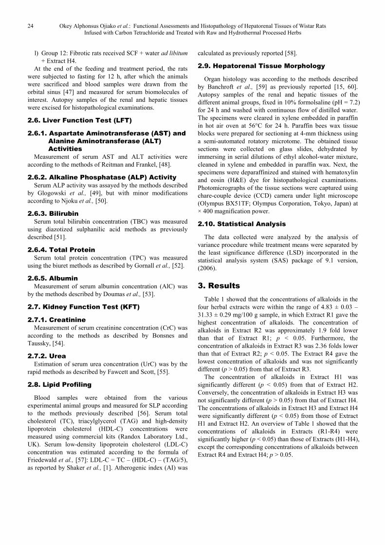
Figure 1. Serum aspartate aminotransferase, alanine aminotransferase and alkaline phosphatase activities of experimental rat groups.
Serum ALT activity of Group 1 was not significantly
different (p > 0.05) from that of Group 2 (Figure 1). However,
Group 3 exhibited comparatively raised level of serum ALT
activity, which represented 4.44 folds increase in the enzyme
activity compared with that of Group 1; p < 0.05. Conversely,
serum ALT activities of Groups (4-12) were significantly
lower (p < 0.05) than that of Group 3, but significantly higher
(p < 0.05) than those of Group 1 and Group 2. Specifically,
serum ALT activities of Group 6, Group 10, Group 11 and
Group 12 were significantly higher (p < 0.05) than that of
group 4, whereas serum ALT activities of Group 5, Group 7,
Group 8 and Group 9 were comparable with that of Group 4;
p > 0.05. Serum AST activities of the various experimental
rat groups followed the same pattern with their serum ALT
activities (Figure 1). However, the activity ratios of AST to
ALT of the various experimental rat groups were generally
less than 1.0 unit.
Serum ALP activity of Group 1 was not significantly
26 Okey Alphonsus Ojiako et al.: Functional Assessments and Histopathology of Hepatorenal Tissues of Wistar Rats
Infused with Carbon Tetrachloride and Treated with Raw and Hydrothermal Processed Herbs
different (p > 0.05) from that of Group 2 (Figure 1). Serum
ALP from Group 3 gave the highest activity, which was
significantly different (p < 0.05) from other experimental rat
groups. Additionally, serum ALP activity of Group 3 was
comparable with those of group 5, Group 6 and Group 10 (p >
0.05), whereas serum ALP activities of Group 7, Group 8,
Group 9, Group 11 and Group 12 were significantly higher (p
< 0.05) than that of Group 3.
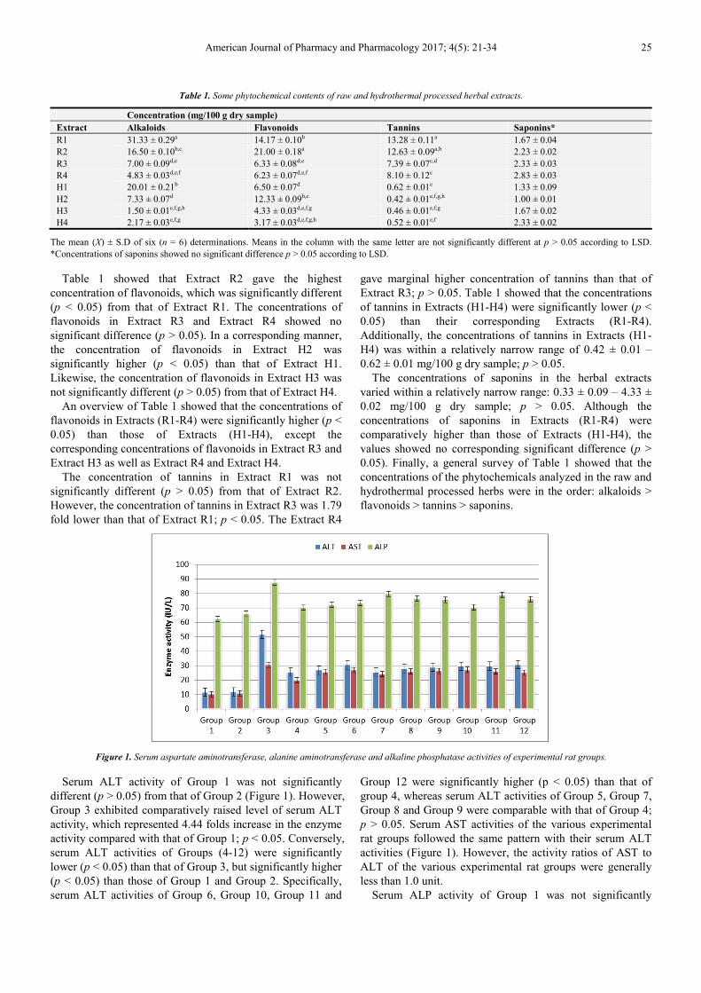
Figure 2. Serum total bilirubin concentration of experimental rat groups.
Figure 2 showed that serum TBCs of Group 1 and Group 2
were less than 1.0 mg/mL and showed no significant
difference (p > 0.05). Contrary, serum TBCs of other
experimental groups, except Group 11, were above 1.0
mg/dL. Specifically, Group 3 gave the highest serum TBC,
which was significantly different (p < 0.05) from other
experimental rat groups. Furthermore, serum TBC of Group
11 was comparable with those of Group 4, Group 7, Group 9
and Group 12; p > 0.05.
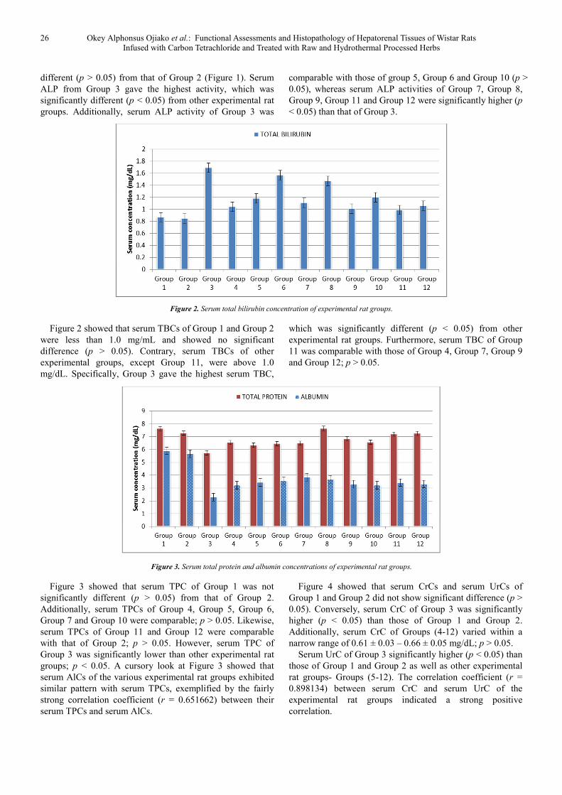
Figure 3. Serum total protein and albumin concentrations of experimental rat groups.
Figure 3 showed that serum TPC of Group 1 was not
significantly different (p > 0.05) from that of Group 2.
Additionally, serum TPCs of Group 4, Group 5, Group 6,
Group 7 and Group 10 were comparable; p > 0.05. Likewise,
serum TPCs of Group 11 and Group 12 were comparable
with that of Group 2; p > 0.05. However, serum TPC of
Group 3 was significantly lower than other experimental rat
groups; p < 0.05. A cursory look at Figure 3 showed that
serum AlCs of the various experimental rat groups exhibited
similar pattern with serum TPCs, exemplified by the fairly
strong correlation coefficient (r = 0.651662) between their
serum TPCs and serum AlCs.
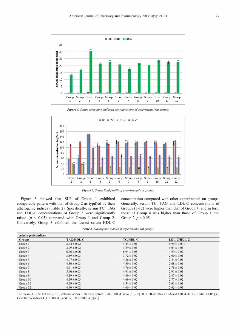
Figure 4 showed that serum CrCs and serum UrCs of
Group 1 and Group 2 did not show significant difference (p >
0.05). Conversely, serum CrC of Group 3 was significantly
higher (p < 0.05) than those of Group 1 and Group 2.
Additionally, serum CrC of Groups (4-12) varied within a
narrow range of 0.61 ± 0.03 – 0.66 ± 0.05 mg/dL; p > 0.05.
Serum UrC of Group 3 significantly higher (p < 0.05) than
those of Group 1 and Group 2 as well as other experimental
rat groups- Groups (5-12). The correlation coefficient (r =
0.898134) between serum CrC and serum UrC of the
experimental rat groups indicated a strong positive
correlation.
American Journal of Pharmacy and Pharmacology 2017; 4(5): 21-34 27
Figure 4. Serum creatinine and urea concentrations of experimental rat groups.
Figure 5. Serum lipid profile of experimental rat groups.
Figure 5 showed that SLP of Group 1 exhibited
comparable pattern with that of Group 2 as typified by their
atherogenic indices (Table 2). Specifically, serum TC, TAG
and LDL-C concentrations of Group 3 were significantly
raised (p < 0.05) compared with Group 1 and Group 2.
Conversely, Group 3 exhibited the lowest serum HDL-C
concentration compared with other experimental rat groups.
Generally, serum TC, TAG and LDL-C concentrations of
Groups (5-12) were higher than that of Group 4, and in turn,
those of Group 4 was higher than those of Group 1 and
Group 2; p < 0.05.
Table 2. Atherogenic indices of experimental rat groups.
Atherogenic indices
Groups TAG/HDL-C TC/HDL-C LDL-C/HDL-C
Group 1 2.74 ± 0.02 1.44 ± 0.01 0.99 ± 0.001
Group 2 2.99 ± 0.02 1.59 ± 0.01 1.01 ± 0.01
Group 3 6.76 ± 0.06 6.94 ± 0.05 4.59 ± 0.02
Group 4 3.59 ± 0.03 3.72 ± 0.02 2.00 ± 0.01
Group 5 4.07 ± 0.03 4.26 ± 0.02 2.44 ± 0.01
Group 6 4.56 ± 0.03 4.59 ± 0.02 2.68 ± 0.01
Group 7 4.91 ± 0.03 4.76 ± 0.02 2.78 ± 0.02
Group 8 5.40 ± 0.03 4.91 ± 0.02 2.91 ± 0.01
Group 9 4.59 ± 0.03 4.39 ± 0.02 2.47 ± 0.01
Group 10 4.59 ± 0.03 4.69 ± 0.02 2.77 ± 0.02
Group 11 4.69 ± 0.02 4.54 ± 0.02 2.61 ± 0.01
Group 12 4.96 ± 0.02 4.66 ± 0.02 2.65 ± 0.01
The mean (X) ± S.D of six (n = 6) determinations. Reference values: TAG/HDL-C ratio [61, 62], TC/HDL-C ratio < 1.66 and LDL-C/HDL-C ratio < 1.06 [39].
Castelli risk indices I (TC/HDL-C) and II (LDL-C/HDL-C) [63].
28 Okey Alphonsus Ojiako et al.: Functional Assessments and Histopathology of Hepatorenal Tissues of Wistar Rats
Infused with Carbon Tetrachloride and Treated with Raw and Hydrothermal Processed Herbs
An overview of Table 2 showed that atherogenic indices of
Group 1 and Group 2 were below the critical values, whereas
those of Groups (3-12) were within the following ranges:
TAG/HDL-C ratio (3.59 ± 0.03 – 6.76 ± 0.06), TC/HDL-C
ratio (3.72 ± 0.02 – 6.94 ± 0.05) and LDL-C/HDL-C ratio
(2.00 ± 0.01 – 4.59 ± 0.02).
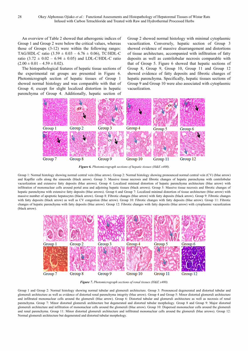
The histopathological features of hepatic tissue sections of
the experimental rat groups are presented in Figure 6.
Photomicrograph section of hepatic tissues of Group 1
showed normal histology and was comparable with that of
Group 4; except for slight localized distortion in hepatic
parenchyma of Group 4. Additionally, hepatic section of
Group 2 showed normal histology with minimal cytoplasmic
vacuolization. Conversely, hepatic section of Group 3
showed evidence of massive disarrangement and distortions
of tissue architecture, accompanied with infiltration of fatty
deposits as well as centrilobular necrosis comparable with
that of Group 5. Figure 6 showed that hepatic sections of
Group 8, Group 9, Group 10, Group 11 and Group 12
showed evidence of fatty deposits and fibrotic changes of
hepatic parenchyma. Specifically, hepatic tissues sections of
Group 9 and Group 10 were also associated with cytoplasmic
vacuolization.
Figure 6. Photomicrograph sections of hepatic tissues (H&E x400).
Group 1: Normal histology showing normal central vein (blue arrow). Group 2: Normal histology showing pronounced normal central vein (CV) (blue arrow)
and Kupffer cells along the sinusoids (black arrow). Group 3: Massive tissue necrosis and fibrotic changes of hepatic parenchyma with centrilobular
vacuolization and extensive fatty deposits (blue arrows). Group 4: Localized minimal distortion of hepatic parenchyma architecture (blue arrow) with
infiltration of mononuclear cells around portal area and adjoining hepatic tissues (black arrows). Group 5: Massive tissue necrosis and fibrotic changes of
hepatic parenchyma with extensive fatty deposits (blue arrows). Group 6 and Group 7: Localized minimal distortion of tissue architecture (blue arrow) with
massive number of apoptotic hepatocytes (black arrow). Group 8: Fibrotic changes (blue arrow) with fatty deposits (black arrow). Group 9: Fibrotic changes
with fatty deposits (black arrow) as well as CV congestion (blue arrow). Group 10: Fibrotic changes with fatty deposits (blue arrow). Group 11: Fibrotic
changes of hepatic parenchyma with fatty deposits (blue arrow). Group 12: Fibrotic changes with fatty deposits (blue arrow) with cytoplasmic vacuolization
(black arrow).
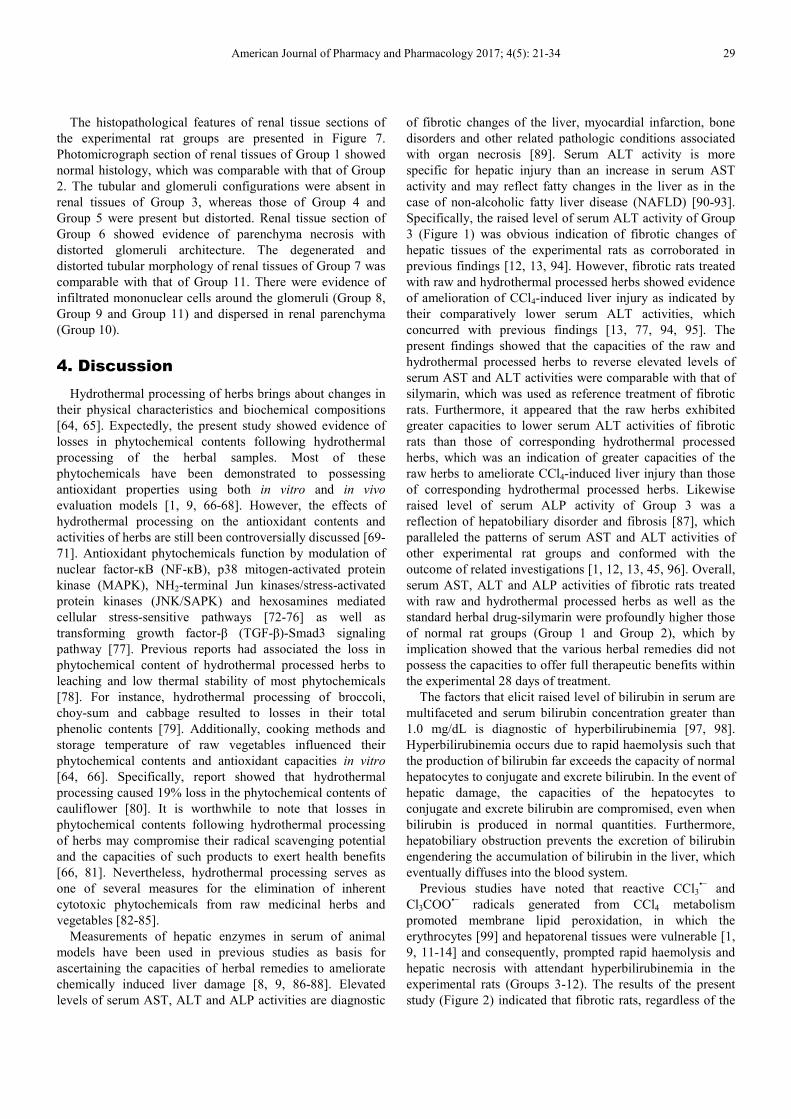
Figure 7. Photomicrograph sections of renal tissues (H&E x400).
Group 1 and Group 2: Normal histology showing normal tubular and glomeruli architecture. Group 3: Pronounced degenerated and distorted tubular and
glomeruli architecture as well as evidence of distorted renal parenchyma integrity (blue arrow). Group 4 and Group 5: Minor distorted glomeruli architecture
and infiltrated mononuclear cells around the glomeruli (blue arrow). Group 6: Distorted tubular and glomeruli architecture as well as necrosis of renal
parenchyma. Group 7: Minor distorted glomeruli architecture but degenerated and distorted tubular morphology. Group 8 and Group 9: Major distorted
glomeruli architecture and infiltration of mononuclear cells around the glomeruli (blue arrow). Group 10: Dispersed mononuclear cells around the glomeruli
and renal parenchyma. Group 11: Minor distorted glomeruli architecture and infiltrated mononuclear cells around the glomeruli (blue arrows). Group 12:
Normal glomeruli architecture but degenerated and distorted tubular morphology.
American Journal of Pharmacy and Pharmacology 2017; 4(5): 21-34 29
The histopathological features of renal tissue sections of
the experimental rat groups are presented in Figure 7.
Photomicrograph section of renal tissues of Group 1 showed
normal histology, which was comparable with that of Group
2. The tubular and glomeruli configurations were absent in
renal tissues of Group 3, whereas those of Group 4 and
Group 5 were present but distorted. Renal tissue section of
Group 6 showed evidence of parenchyma necrosis with
distorted glomeruli architecture. The degenerated and
distorted tubular morphology of renal tissues of Group 7 was
comparable with that of Group 11. There were evidence of
infiltrated mononuclear cells around the glomeruli (Group 8,
Group 9 and Group 11) and dispersed in renal parenchyma
(Group 10).
4. Discussion
Hydrothermal processing of herbs brings about changes in
their physical characteristics and biochemical compositions
[64, 65]. Expectedly, the present study showed evidence of
losses in phytochemical contents following hydrothermal
processing of the herbal samples. Most of these
phytochemicals have been demonstrated to possessing
antioxidant properties using both in vitro and in vivo
evaluation models [1, 9, 66-68]. However, the effects of
hydrothermal processing on the antioxidant contents and
activities of herbs are still been controversially discussed [69-
71]. Antioxidant phytochemicals function by modulation of
nuclear factor-ĸB (NF-ĸB), p38 mitogen-activated protein
kinase (MAPK), NH2-terminal Jun kinases/stress-activated
protein kinases (JNK/SAPK) and hexosamines mediated
cellular stress-sensitive pathways [72-76] as well as
transforming growth factor-β (TGF-β)-Smad3 signaling
pathway [77]. Previous reports had associated the loss in
phytochemical content of hydrothermal processed herbs to
leaching and low thermal stability of most phytochemicals
[78]. For instance, hydrothermal processing of broccoli,
choy-sum and cabbage resulted to losses in their total
phenolic contents [79]. Additionally, cooking methods and
storage temperature of raw vegetables influenced their
phytochemical contents and antioxidant capacities in vitro
[64, 66]. Specifically, report showed that hydrothermal
processing caused 19% loss in the phytochemical contents of
cauliflower [80]. It is worthwhile to note that losses in
phytochemical contents following hydrothermal processing
of herbs may compromise their radical scavenging potential
and the capacities of such products to exert health benefits
[66, 81]. Nevertheless, hydrothermal processing serves as
one of several measures for the elimination of inherent
cytotoxic phytochemicals from raw medicinal herbs and
vegetables [82-85].
Measurements of hepatic enzymes in serum of animal
models have been used in previous studies as basis for
ascertaining the capacities of herbal remedies to ameliorate
chemically induced liver damage [8, 9, 86-88]. Elevated
levels of serum AST, ALT and ALP activities are diagnostic
of fibrotic changes of the liver, myocardial infarction, bone
disorders and other related pathologic conditions associated
with organ necrosis [89]. Serum ALT activity is more
specific for hepatic injury than an increase in serum AST
activity and may reflect fatty changes in the liver as in the
case of non-alcoholic fatty liver disease (NAFLD) [90-93].
Specifically, the raised level of serum ALT activity of Group
3 (Figure 1) was obvious indication of fibrotic changes of
hepatic tissues of the experimental rats as corroborated in
previous findings [12, 13, 94]. However, fibrotic rats treated
with raw and hydrothermal processed herbs showed evidence
of amelioration of CCl4-induced liver injury as indicated by
their comparatively lower serum ALT activities, which
concurred with previous findings [13, 77, 94, 95]. The
present findings showed that the capacities of the raw and
hydrothermal processed herbs to reverse elevated levels of
serum AST and ALT activities were comparable with that of
silymarin, which was used as reference treatment of fibrotic
rats. Furthermore, it appeared that the raw herbs exhibited
greater capacities to lower serum ALT activities of fibrotic
rats than those of corresponding hydrothermal processed
herbs, which was an indication of greater capacities of the
raw herbs to ameliorate CCl4-induced liver injury than those
of corresponding hydrothermal processed herbs. Likewise
raised level of serum ALP activity of Group 3 was a
reflection of hepatobiliary disorder and fibrosis [87], which
paralleled the patterns of serum AST and ALT activities of
other experimental rat groups and conformed with the
outcome of related investigations [1, 12, 13, 45, 96]. Overall,
serum AST, ALT and ALP activities of fibrotic rats treated
with raw and hydrothermal processed herbs as well as the
standard herbal drug-silymarin were profoundly higher those
of normal rat groups (Group 1 and Group 2), which by
implication showed that the various herbal remedies did not
possess the capacities to offer full therapeutic benefits within
the experimental 28 days of treatment.
The factors that elicit raised level of bilirubin in serum are
multifaceted and serum bilirubin concentration greater than
1.0 mg/dL is diagnostic of hyperbilirubinemia [97, 98].
Hyperbilirubinemia occurs due to rapid haemolysis such that
the production of bilirubin far exceeds the capacity of normal
hepatocytes to conjugate and excrete bilirubin. In the event of
hepatic damage, the capacities of the hepatocytes to
conjugate and excrete bilirubin are compromised, even when
bilirubin is produced in normal quantities. Furthermore,
hepatobiliary obstruction prevents the excretion of bilirubin
engendering the accumulation of bilirubin in the liver, which
eventually diffuses into the blood system.
Previous studies have noted that reactive CCl3•−
and
Cl3COO•−
radicals generated from CCl4 metabolism
promoted membrane lipid peroxidation, in which the
erythrocytes [99] and hepatorenal tissues were vulnerable [1,
9, 11-14] and consequently, prompted rapid haemolysis and
hepatic necrosis with attendant hyperbilirubinemia in the
experimental rats (Groups 3-12). The results of the present
study (Figure 2) indicated that fibrotic rats, regardless of the

30 Okey Alphonsus Ojiako et al.: Functional Assessments and Histopathology of Hepatorenal Tissues of Wistar Rats
Infused with Carbon Tetrachloride and Treated with Raw and Hydrothermal Processed Herbs
type of herbal treatment administered, presented evidence of
hyperbilirubinemia. However, the hydrothermal processed
herbs appeared to exhibit relatively higher tendencies to
lower the severity of hyperbilirubinemia than those of
corresponding raw herbs. In that regard, the results appeared
to suggest that hydrothermal processing of the herbs caused
the elimination or reduction in the amount of cytotoxic
components in the herbs that probably interfered with the
capacity of the herbs to ameliorate hyperbilirubinemia.
The plasma proteins (e.g. albumin) are synthesized by the
liver and therefore low circulating level of plasma proteins
indicates hepatic dysfunction. Previous studies have shown
that mild perturbation of hepatic tissues integrity did not
profoundly affect the capacity of the liver to biosynthesize
plasma proteins [58, 100]. The present study showed that
serum total protein and albumin concentrations of Group 3
were lower than those of Group 1 and Group 2 (Figure 3),
which confirmed compromised hepatic function. However,
the administration of raw and hydrothermal processed herbs
caused limited improvement in the capacity of hepatocytes of
fibrotic rats to biosynthesize plasma proteins.
The renal tissues are primarily concerned with the
clearance of nitrogenous waste products and other blood low
threshold substances. Creatinine is mainly the catabolic waste
product from tissue protein turnover, whereas urea is derived
from oxidative deamination of dietary amino acids [1, 101,
102]. Elevated blood creatinine and urea concentrations are
diagnostic of impaired renal function. The present study
showed evidence of renal dysfunction following infusion of
the experimental rats with CCl4, exemplified by
comparatively raised serum creatinine and urea
concentrations of Group 3 (Figure 4). The present findings
confirmed previous research outcomes, which noted that
acute chemical intoxication that caused morphological and
functional damages to hepatic tissues predisposed animal
models to developing acute renal dysfunction [1, 5, 6, 8, 9].
Fibrotic rats treated with raw and hydrothermal processed
herbs showed evidence of limited amelioration of impaired
renal function, typified by their lowered serum creatinine and
urea concentrations compared to that of Group 3. In a related
study, Elgazar and AboRaya, [103], using serum urea and
creatinine concentrations as biomarkers, noted that single and
combinatorial formulations of Petroselinum sativum, Eruca
sativa and Curcuma longa ameliorated gentamicin-induced
renal tubular necrosis in adult male Sprague Dawley rats.
The metabolic concerns of the liver, amongst other
functions, are to regulate the mobilization, biosynthesis and
catabolism of lipoproteins in vertebrates. Previous reports
showed that CCl4 interferes with the metabolism of
lipoproteins in hepatic smooth endoplasmic reticulum,
engendering alterations in SLP patterns and associated
dyslipidemia [12, 104, 105], whereby blood lipid
concentrations are elevated as typified by raised levels of
serum LDL-C and TC of Group 3 compared to that of Group
1 (Figure 5). Additionally, the SLP patterns of fibrotic rats
administered with raw and hydrothermal processed herbs
were identical and therapeutic scores of the herbs were not
profoundly different from that of the standard herbal drug-
silymarin. Overall, the present study showed that the herbs
did not offer full therapeutic benefits to the fibrotic rats, in
terms of their capacities to ameliorate dyslipidemia. The
predisposition of the experimental rat groups to
arteriosclerosis and associated cardiovascular morbidity and
mortality were defined by their atherogenic indices (Table 2).
According to the TAG/HDL-C and LDL-C/HDL-C ratios of
the present study, adjustments of SLP patterns in the fibrotic
rats, irrespective of the type of herbal treatment they received,
indicated incidences of atherogenicity as defined elsewhere
[39, 58, 61-63].
The massive disarrangement of hepatic tissues architecture
of untreated fibrotic rats (Group 3) correlated with the levels
of alterations of serum indicators, namely, serum ALT, AST
and ALP activities as well as serum TBC as previously
described [12, 13, 98, 99]. Additionally, photomicrograph
section of hepatic tissues of fibrotic rats (Figure 6) revealed
and confirmed CCl4-induced necrosis and steatosis as
previously described [12, 13, 88, 106]. The localized
distortions in hepatic architecture, persistence of hepatic
steatosis and hydropic degenerations in fibrotic rats
following treatment with raw and hydrothermal processed
herbs were indications, which confirmed limited capacities of
the herbs to ameliorate CCl4-induced morphological and
functional impairments of hepatic tissues within the
experimental period of 28 days. According to Sokol et al.
[107], manifestation of hepatic steatosis was as a result of
low availability of tissue α-tocopherol and ascorbic acid,
rather than glutathione (GSH), which also predisposed the
liver to oxidative injuries as exemplified by the presence of
hepatic necrosis in tissue sections of the present report.
Rincón et al. [5] suggested that the effect of CCl4 on
kidney tissue morphology and function depended on the
functional state of the liver. Similar to the histopathological
status of the liver, photomicrograph sections of renal tissues
(Figure 7) showed that the distortions in renal tissue
architecture correlated with the levels of alterations in their
blood indicators; serum creatinine and urea concentrations as
previously described [60]. Furthermore, the tissue
architecture of fibrotic rats administered with raw and
hydrothermal processed herbs confirmed the limited
capacities of the herbs to ameliorate renal dysfunction within
the experimental period of 28 days.
5. Conclusion
The losses in phytochemical contents following
hydrothermal processing of the herbs did not substantially
affect their overall therapeutic scores against morphological
and functional impairments of hepatic and renal tissues
following CCl4 intoxication of the experimental rats.
However, herbal intervention against CCl4-induced
hyperbilirubinemia showed that hydrothermal processed
herbs possessed greater capacity to lower the severity of
hyperbilirubinemia than their corresponding raw herbs.
Nevertheless, a more rewarding bio-prospecting exercise for

American Journal of Pharmacy and Pharmacology 2017; 4(5): 21-34 31
the alleviation of CCl4-induced hepatorenal impairment could
be achieved by subjecting the herbs to sequential multi-
solvent extraction process, which perhaps, will provide
improved and better therapeutic benefits than the present
outcomes.
Conflict of Interests
The authors declare that there is no conflict of interest
regarding the publication of this article.
Acknowledgement
The authors are grateful for the technical assistance offered
by Dr. E. S. Willie of the Department of Agronomy, Michael
Okpara University of Agriculture, Umudike, Abia State,
Nigeria.
References
[1] Shaker E, Mahmoud H, Mnaa S 92010). Silymarin, the antioxidant component and Silybum marianum extracts prevent liver damage. Food and Chemical Toxicology. 48: 803-806.
[2] Chikezie PC, Uwakwe AA (2014). Protective effect of Allium Sativa extract against carbon tetrachloride-induced hepatic oxidative stress and hyperlipidemia in rats. African Journal of Biotechnology. 13: 1671-1678.
[3] Katzung BG (1998). Basic and Clinical Pharmacology. Appleton and Lange. 7th Edition, Stamford CT. pp. 372-375.
[4] Manahan S (2003). Toxicological Chemistry and Biochemistry. 3rd Edition, CRC Press, Boca Raton, FL., USA.
[5] Rincón AR, Covarrubias A, Pedraza-Chaverrí J, Poo JL, Armendáriz-Borunda J, Panduro A, (1999). Differential effect of CCl4 on renal function in cirrhotic and non-cirrhotic rats. Experimental and Toxicologic Pathology. 51: 199-205.
[6] Jaramillo-Juárez F, Rodríguez-Vázquez ML, Rincón-Sánchez AR, Martínez MC, Ortiz GG, Llamas J (2008). Acute renal failure induced by carbon tetrachloride in rats with hepatic cirrhosis. Annals of Hepatology. 7: 331-338.
[7] Slack A, Yeoman A, Wendon J (2010). Renal dysfunction in chronic liver disease. Critical Care. 14: 10 pages.
[8] Alqasoumi S (2010). Carbon tetrachloride-induced hepatotoxicity: Protective effect of 'Rocket' Eruca sativa L. in rats. American Journal of Chinese Medicine. 38: 75-88.
[9] Khan RA, Khan MR, Sahreen S (2012). Protective effect of Sonchus asper extracts against experimentally induced lung injuries in rats: a novel study. Experimental and Toxicologic Pathology. 64: 725-731.
[10] Ruprah H, Mant TGK, Flanagan RJ (1985). Acute carbon tetrachloride poisoning in 19 patients: implications for diagnosis and treatment. Lancet. 1: 1027-1029.
[11] Adewole SO, Salako AA, Doherty OW, Naicker T (2007). Effect of melatonin on carbon tetrachloride-induced kidney injury in Wistar rats. African Journal of Biomedical Research. 10: 153-164.
[12] Althnaian T, Albokhadaim I, El-Bahr SM (2013). Biochemical and histopathological study in rats intoxicated with carbon tetrachloride and treated with camel milk. Spring Open Journal of Research. 2: 57.
[13] Gonçalves RV, da Matta SLP, Novaes RD, Leite JPV, Peluzio MCG, Vilela EF (2014). Bark extract of Bathysa cuspidata in the treatment of liver injury induced by carbon tetrachloride in rats. Brazilian Archives of Biology and Technology. 57: 504-513.
[14] Saile B, Ramadori G (2007). Inflammation, damage repair and liver fibrosis-role of cytokines and different cell types. Zeitschrift für Gastroenterologie. 45: 77-86.
[15] Ojiako AO, Chikezie PC, Ogbuji CA (2015). Histopathological studies of renal and hepatic tissues of hyperglycemic rats administered with traditional herbal formulations. International Journal of Green Pharmacy. 9: 184-191.
[16] Mensah J, Okoli R, Ohaju-Obodo J, Eifediyi K (2008). Phytochemical, nutritional and medical properties of some leafy vegetables consumed by Edo people of Nigeria. African Journal of Biotechnology. 7: 2304-2309.
[17] Chikezie PC, Ojiako AO, Nwufo KC (2015). Overview of anti-diabetic medicinal plants: The Nigerian research experience. Journal of Diabetes and Metabolism. 6: 7 pages.
[18] Middleton E, Kandaswami C, Theohardes TC (2000). The effects of plant flavonoids on mammalian cells, implication for inflammation, heart disease and cancer. Pharmacological Review. 52: 673-751.
[19] Okafor JC (1987). Development of forest tree crops for food supply in Nigeria. Forest Ecology and Management. 1: 235-247.
[20] Udeala OK (2000). Preliminary evaluation of dike fat a new tablet lubricant. Journal Pharmacy and Pharmacology. 32: 6-9.
[21] Iwu MM (2002). Evaluation of the anti-hepatotoxic activity of the bioflavonoids of Garcina kola seeds. Journal of Ethnopharmacology. 21: 14-19.
[22] Akinwunmi KF, Oyedapo OO (2013). Evaluation of antioxidant potentials of Monodora myristica (Gaertn) dunel seeds. African Journal of Food Science. 7: 317-324.
[23] Alisi CS, Nwogu LA, Ibegbulem CO, Ujowundu CU (2011). Antimicrobial action of methanol extract of Chromolaena odoranta Linn is logistic and exerted by inhibition of dehydrogenase enzymes. Journal of Research in Biology. 3: 209-216.
[24] Anyasor GN, Aina DA, Olushola M, Aniyikaye AF (2011). Phytochemical constituent, proximate analysis, antioxidant, antibacterial and wound healing properties of leaf extracts of Chromolaena odorata. Annals of Biological Research. 2: 441.
[25] Chakraborty JB, Oakley F, Walsh MJ (2011). Chromolaena odorata L: An overview. Journal of Pharmacy Research. 4: 573.
[26] Pandith H, Zhang X, Liggert J, Min K, Gritsanaptan W, Baek SJ (2013). Hemostatic and wound healing properties of Chromolaena odorata leaf extract. ISRN Dermatology. 2013: 8 pages.
32 Okey Alphonsus Ojiako et al.: Functional Assessments and Histopathology of Hepatorenal Tissues of Wistar Rats
Infused with Carbon Tetrachloride and Treated with Raw and Hydrothermal Processed Herbs
[27] Nwaehujor CO, Ode OJ, Nwinyi FC, Udeh NE (2012). Effect of methanol extract of Buchholzia coriacea fruits on streptozotocin-induced diabetic rats. Journal of Pharmacology and Toxicology. 7: 181-191.
[28] Amaechi NC (2009). Nutritive and anti-nutritive evaluation of wonderful kola (Buccholzia coricea) seeds. Pakistan Journal of Nutrition. 8: 1120-1122.
[29] Mbata TI, Duru CM, Onwumelu HA (2009). Antibacterial activity of crude seed extracts of Buchholzia coriacea E. on some pathogenic bacteria. Journal of Developmental Biology and Tissue Engineering. 1: 1-5.
[30] Adisa RA, Choudhary MI, Olorunsogo OO (2011). Hypoglycemic activity of Buchholzia coriacea (Capparaceae) seed in streptozotocin-induced diabetic rats and mice. Experimental and Toxicological Pathology. 63: 619-625.
[31] Ibrahim TA, Fagboun ED (2013). Phytochemical and nutritive quality of dried seed of Buchholzia coricea. Greener Journal of Physical Sciences. 2: 185-191.
[32] Ajibola C, Fashakin JB, Fagbemi TN, Aluko RE (2011). Effect of peptide size on antioxidant properties of African yam bean seed (Sphenostylis stenocarpa) protein hydrolysate fractions. International Journal of Molecular Science. 12: 6685-6702.
[33] Okonkwo CC, Njoku OU, Ikevude CT, Odo CE (2013). Hepatoprotective effect of methanol exytact of Sphenostylis stenocarpa (Hoschst ex. A. Rich. Harms) against carbon tetrachloride-induced liver toxicity in Wistar rats. Journal of Pharmacy Research. 6: 293-298.
[34] Ndidi US, Ndidi CU, Olagunju A, Muhammad A, Billy FG, Okpe O (2014). Proximate, anti-nutrition, and mineral composition of raw and processed (boiled and roasted) Sphenostylis stenocarpa seeds from Southern Kaduna, Northwest Nigeria. ISRN Nutrition. 2014: 9 pages.
[35] Hwang YP, Choi JH, Jeong HG (2009). Protective effect of the Aralia continentalis root extract against carbon tetrachloride-induced hepatotoxicity in mice. Food Chemistry and Toxicology. 47: 75-81.
[36] Karakus E, Karadeniz A, Simsek N (2011). Protective effect of Panax ginseng against serum biochemical changes and apoptosis in liver of rats treated with carbon tetrachloride (CCl4). Journal of Hazard Materials. 195: 208-213.
[37] Wolf P (1999). Biochemical diagnosis of liver diseases. International Journal of Clinical Biochemistry. 14: 59-90.
[38] Ramcharran D, Wahed AS, Conjeevaram HS, Evans RW, Wang T, Belle SH et al., (2011). Serum lipids and their associations with viral levels and liver disease severity in a treatment-naïve chronic hepatitis C type 1-infected cohort. Journal of Viral Hepatitis. 18: 144-152.
[39] Ibegbulem CO, Chikezie PC (2012). Serum lipid profile of rats (Rattus norvegicus) fed with palm oil and palm kernel oil-containing diets. Asian Journal of Biochemistry. 7: 46-53.
[40] Wojcikowski K, Stevenson L, Leach D, Wohlmuth H, Gobe G (2007). Antioxidant capacity of 55 medicinal herbs traditionally used to treat the urinary system: A comparison using a sequential three-solvent extraction process. Journal of Alternative and Complementary Medicine. 13: 103-109.
[41] Harborne JB (1973). Phytochemical Methods: A Guide to
Modern Techniques of Plant Analysis, 1st Edition, London, Chapman and Hall Ltd. pp. 278.
[42] Boham AB, Kocipal AC (1994). Flavonoid and condensed tannins from leaves of Hawaiian vaccininum, vaticulum and vicalycinium. Pacific Science. 48: 458-463.
[43] Van-Burden TP, Robinson WC (1981). Formation of complex between protein and tannic acid. Journal of Agriculture and Food Chemistry. 1: 77-82.
[44] Belonwu DC, Ibegbulem CO, Nwokocha MN, Chikezie PC (2014). Some phytochemicals and hydrophilic vitamins of Anacardium occidentale. Research Journal of Phytochemistry. 8: 78-91.
[45] Khan AA, Alzohairy M (2011). Hepatoprotective effects of camel milk against CCl4-induced hepatotoxicity in rats. Asian Journal of Biochemistry. 6: 171-181.
[46] Ibegbulem CO, Chikezie PC (2013). Hypoglycemic properties of ethanolic extracts of Gongronema latifolium, Aloe perryi, Viscum album and Allium sativum administered to alloxan-induced diabetic albino rats (Rattus norvegicus). Pharmacognosy Communications. 3: 12-16.
[47] Hoff N (2000). Methods of blood collection in the mouse. Laboratory Animal. 29: 47-53.
[48] Reitman S, Frankel S (1957). Colorimetric method for the determination of serum glutamic oxaloacetic and glutamic pyruvic transaminase. American Clinical Pathology. 28: 56-63.
[49] Glogowski J, Danforth DR, Ciereszko A (2002). Inhibition of alkaline phosphatase activity of boar semen by pentoxifylline, caffeine, and theophylline. Journal of Andrology. 23: 783-792.
[50] Njoku VO, Chikezie PC, Kaoje AM (2011). Kinetic studies of alkaline phosphatase extracted from rabbit (Lepus townsendii) liver. African Journal of Biotechnology. 10: 3157-3162.
[51] Pearlman FC, Lee RT (1974). Detection and measurement of total bilirubin in serum, with use of surfactants as solubilizing agents. Clinical Chemistry. 20: 447-453.
[52] Gornall AG, Bardawill CJ, David MM (1949). Determination of serum protein by means of the biuret reaction. Journal of Biological Chemistry. 177: 751-766.
[53] Doumas BT, Watson WA, Biggs HG (1971). Albumin standards and the measurement of serum albumin with bromcresol green. Clinical Chimica Acta. 31: 87-96.
[54] Bonsnes RW, Taussky HH (1945). On the colorimetric determination of creatinine by the Jaffe reaction. Journal of Biological Chemistry. 158: 581-591.
[55] Fawcett JK, Scott JE (1960). A rapid and precise method for the determination of urea. Journal of Clinical Pathology. 13: 156-159.
[56] Ojiako AO, Chikezie PC, Zedech UC (2013). Serum lipid profile of hyperlipidemic rabbits (Lepus townsendii) administered with leaf extracts of Hibiscus rosesinesis, Emilia coccinea, Acanthus montanus and Asystasia gangetica. Journal of Medicinal Plant Research. 7: 3226-3231.
[57] Friedewald W, Levy R, Fredrickson D (1972). Estimation of concentration of low-density lipoprotein in plasma, without use of the preparative ultracentrifuge. Clinical Chemistry. 18: 499-502.
American Journal of Pharmacy and Pharmacology 2017; 4(5): 21-34 33
[58] Ibegbulem CO, Chikezie PC (2016). Levels of acute blood indices disarrangement and organ weights of Wistar rats fed with flavour enhancer- and contraceptive-containing diets. Journal of Investigational Biochemistry. 5: 1-9.
[59] Banchroft JD, Stevens A, Turner DR (1996). Theory and Practice of Histological Techniques, 4th Edition, Churchill Livingstone, New York, London, San Francisco, Tokyo.
[60] Ibegbulem CO, Chikezie PC, Dike EC (2016). Pathological research and acute hepatic and renal tissue damage in Wistar rats induced by cocoa. Journal of Acute Disease. 5: 51-58.
[61] Gaziano JM, Hennekens CH, O’Donnell CJ, Breslow JL, Buring JE (1997). Fasting triglycerides, high-density lipoprotein, and risk of myocardial infarction. Circulation. 96: 2520-2525.
[62] Bittner V, Johnson D, Zineh I, Rogers WJ, Vido D, Marroquin OC et al., (2009). The triglyceride/high-density lipoprotein cholesterol ratio predicts all-cause mortality in women with suspected myocardial ischemia: A report from the Women's Ischemia Syndrome Evaluation (WISE). American Heart Journal. 157: 548-55.
[63] Asare GA, Santa S, Ngala RA, Asiedu B, Afriyie D, Amoah AGB (2014). Effect of hormonal contraceptives on lipid profile and the risk indices for cardiovascular disease in a Ghanaian community. International Journal of Women Health. 6: 597-603.
[64] Pellegrini N, Chiawato E, Gardama C, Mazzeo T, Continto D, Gallo M et al., (2010). Effect of different cooking methods in color, phytochemical concentration and antioxidant capacity of raw and frozen Brassica vegetables. Journal of Agriculture and Food Chemistry. 58: 4310-4321.
[65] Adefegha SA, Oboh GO (2011). Cooking enhances the antioxidant properties of some tropical leafy vegetables. African Journal of Biotechnology. 10: 632-639.
[66] Kaur P, Bains K, Kaur H (2012). Effect of hydrothermal treatment on free radical scavenging potential of selected green vegetables. Indian Journal of Natural Products and Resources. 3: 563-569.
[67] Saeed N, Khan M, Shabbir M (2012). Antioxidant activity, total phenolic and total flavonoid contents of whole plant extracts Torilis leptophylla L. BMC Complementary and Alternative Medicine. 12: 221.
[68] Ojiako AO, Chikezie PC, Ogbuji CA (2016). Radical scavenging potentials of single and combinatorial herbal formulations in vitro. Journal of Traditional and Complementary Medicine. 6: 153-159.
[69] Zhang D, Hamauzu Y (2004). Phenolics, ascorbic acid, carotenoids and antioxidant activity of broccoli and their changes during conventional and microwave cooking. Food Chemistry. 88: 503-509.
[70] Turkman N, Sari F, Velioglu YS (2005). The effect of cooking methods on total phenolics and antioxidant activity of selected green vegetables. Food Chemistry. 93: 713-718.
[71] Thi ND, Hwang E-S (2016). Effects of drying methods on contents of bioactive compounds and antioxidant activities of black chokeberries (Aronia melanocarpa). Food Science and Technology. 25: 55-61.
[72] Mate´s JM (2000). Effects of antioxidant enzymes in the
molecular control of reactive oxygen species toxicology. Toxicology. 153: 83-104.
[73] Evans JL, Goldfine ID, Maddux BA, Grodsky GM (2002). Oxidative stress and stress activated signaling pathways: a unifying hypothesis of Type 2 diabetes. Endocrinology Review. 23: 599-622.
[74] Hye-Lin H, Hye-Jun S, Mark AF, Dae-Yeul Y (2010). Oxidative stress and antioxidants in hepatic pathogenesis. World Journal of Gastroenterology. 16: 6035-6043.
[75] Rodríguez-Ramiro I, Ramos S, López-Oliva E, Agis-Torres A, Bravo L et al., (2012). Cocoa polyphenols prevent inflammation in the colon of azoxymethane-treated rats and in TNF-α-stimulated Caco-2 cells. British Journal of Nutrition. 28: 1-10.
[76] Ranneh Y, Ali F, Esa NM (2013). The protective effect of cocoa (Theobroma cacao L.) in colon cancer. Journal of Nutrition and Food Science. 3: 3 pages.
[77] Huang Q, Zhang S, Zhang L, He M, Huang R, Lin X (2012). Hepatoprotective effects of total saponins isolated from Turaphochlamys affiris against carbon tetrachloride-induced liver injury in rats. Food and Chemical Toxicology. 50: 713-718.
[78] Rungapamestry V, Duncan AJ, Fuller Z, Ratcliffe B (2007). Effect of cooking Brassica vegetables on the subsequent hydrolysis and metabolic fate of glucosinolates. Proceedings of Nutrition Society. 66: 69-81.
[79] Watchtel-Galor S, Wong KW, Benzie IFF (2008). The effect of cooking on Brassica vegetable. Food Chemistry. 110: 706-710.
[80] Volden J, Borge GIA, Hansen M, Wicklund T, Bengtsson GB (2009). Processing (blanching, boiling, steaming) effects on the content of glucosinolates and antioxidant-related parameters in cauliflower (Brassica oleracea L. ssp. botrytis). LWT-Food Science and Technology. 42: 63-73.
[81] Song A-S, Lim S-W, Kim S-J, Lee S-C (2013). Effect of hydrothermal treatment on the antioxidant activity of Sambaekcho (Saururus chinensis) leaves. Food Science and Biotechnology. 22: 825-829.
[82] Fagbemi TN, Oshodi AA, Ipinmoroti KO (2005). Processing effects on some anti-nutritional factors and in vitro multi-enzyme protein digestibility (ivpd) of three tropical seeds, bread nut (Artocarpus altilis), cashew nut (Anacardium occidentale) and fluted pumpkin (Telfairia occidentalis). Pakistan Journal of Nutrition. 4: 250-256.
[83] Hefnawy TH (2011). Effect of processing methods on nutritional composition and anti-nutritional factors in lentils (Lens culinaris). Annals of Agricultural Sciences. 56: 57-61.
[84] Chu C, Ho K, Hu A, Chiu C, Wu H, Ye S et al., (2012). Toxicity attenuation of Atractyloside in traditional Chinese medicinal herbs after hydrothermal processing. Botany Studies. 53: 459-465.
[85] Chen F, Xiong H, Wang J, Ding X, Shu G, Mei Z (2013). Antidiabetic effect of total flavonoids from Sanguis draxonis in Type 2 diabetic rats. Journal of Ethnopharmacology. 149: 729-736.
[86] Mayuren C, Reddy VV, Priya SV, Devi VA (2010). Protective effect of Livactine against CCl4 and paracetamol induced hepatotoxicity in adult Wistar rats. North American Journal of Medical Sciences. 2: 491-495.

34 Okey Alphonsus Ojiako et al.: Functional Assessments and Histopathology of Hepatorenal Tissues of Wistar Rats
Infused with Carbon Tetrachloride and Treated with Raw and Hydrothermal Processed Herbs
[87] Anusha M, Venkateswarlu M, Prabhakaran V, Taj SS, Kumari BP, Ranganayakulu D (2011). Hepatoprotective activity of aqueous extract of Portulaca oleracea in combination with lycopene in rats. Indian Journal of Pharmacology. 43: 563-567.
[88] Cordero-Pérez P, Torres-González L, Aguirre-Garza M, Camara-Lemarroy C, la Garza FG, Alarcón-Galván G et al., (2013). Hepatoprotective effect of commercial herbal extracts on carbon tetrachloride-induced liver damage in Wistar rats. Pharmacognosy Research. 5: 150-156.
[89] Rodwell VW, Kennelly PJ (2003). Enzymes: Mechanism of Action. In: Murray RK, Granner DK, Mayes PA, Rodwell VW. Harper’s Illustrated Biochemistry, 26th Edition, Lange Medical Books/McGraw-Hill, New York, USA. pp. 49-59.
[90] Vozarova B, Stefan N, Lindsay RS, Saremi A, Pratley RE, Bogardus C et al., (2002) High alanine aminotransferase is associated with decrease hepatic insulin sensitivity and predicts the development of Type 2 diabetes. Diabetes. 51: 1189-1895.
[91] Fraser A, Harris R, Sattar N, Ebrahim S, Smith GD, Lawlor DA (2009). Alanine aminotransferase, γ-glutamyltransferase, and incident diabetes: the British women's heart and health study and meta-analysis. Diabetes Care. 32: 741-750.
[92] Ghamar-Chehreh ME, Amini M, Khedmat H, Alavian SM, Daraei F, Mohtashami R et al., (2012). Elevated alanine aminotransferase activity is not associated with dyslipidemias, but related to insulin resistance and higher disease grades in non-diabetic non-alcoholic fatty liver disease. Asian Pacific Journal of Tropical Biomedicine. 2: 702-706.
[93] de Luis DA, Aller R, Izaola O, Sagrado MG, Conde R, de La Fuente B (2013). Role of insulin resistance and adipocytokines on serum alanine aminotransferase in obese patients with Type 2 diabetes mellitus. European Review for Medicine and Pharmacological Sciences. 17: 2059-2064.
[94] Teschke R, Vierke W, Goldermann L (1983). Carbon tetrachloride (CCl4) levels and serum activities of liver enzymes following acute CCl4 intoxication. Toxicological Letters. 17: 175-180.
[95] Taira Z, Yabe K, Hamaguchi Y, Hirayama K, Kishimoto M, Ishida S et al., (2004). Effects of Sho-saiko-to extract and its components, Baicalin, baicalein, glycyrrhizin and glycyrrhetic acid, on pharmacokinetic behavior of salicylamide in carbon tetrachloride intoxicated rats. Food Chemistry and Toxicology. 42: 803-807.
[96] Adewale OB, Adekeye AO, Akintayo CO, Onikanni A, Saheed S (2014). Carbon tetrachloride (CCl4)-induced hepatic damage in experimental Sprague Dawley rats: Antioxidant
potential of Xylopia aethiopica. The Journal of Phytopharmacology. 3: 118-123.
[97] Kochar DK, Singh P, Agarwal P, Kochar SK, Pokharna R, Sareen PK (2003). Malarial hepatitis. Journal of the Association of Physicians of India. 51: 1069-1072.
[98] Murray RK (2003). Porphyrins and Bile Pigments. In: Murray RK, Granner DK, Mayes PA, Rodwell VW. (Editors), Harper’s Illustrated Biochemistry, 26th Edition, California, Lange Medical Books/McGraw-Hill. pp. 270-285.
[99] da Rosa DP, Bona S, Simonetto D, Zettler C, Marroni CA, Marroni NP (2010). Melatonin protects the liver and erythrocytes against oxidative stress in cirrhotic rats. Experimental Gastroenterology. 47: 72-78.
[100] Ashafa AOT, Orekoya LO, Yakubu MT (2012). Toxicity profile of ethanolic extract of Azadirachta indica stem bark in male Wistar rats. Asian Pacific Journal of Tropical Biomedicine. 2: 811-817.
[101] Rodwell VW (2003) Catabolism of Proteins and of Amino Acids Nitrogen. In: Murray RK, Granner DK, Mayes PA, Rodwell VM. (Editors), Harper's Illustrated Biochemistry, 26th Edition, New York, McGraw-Hill Medical. pp. 242-248.
[102] Samra M, Abcar AC (2012). False estimates of elevated creatinine. The Permanente Journal. 16: 51-52.
[103] Elgazar AF, AboRaya AO (2013). Nephroprotective and diuretic effects of three medicinal herbs against gentamicin-induced nephrotoxicity in male rats. Pakistan Journal of Nutrition. 12: 715-722.
[104] Mayes PA, Botham KM (2003) Lipid Transport and Storage. In: Murray RK, Granner DK, Mayes PA, Rodwell VW. (Editors), Harper’s Illustrated Biochemistry. 26th Edition, California, Lange Medical Books/McGraw-Hill, pp. 205-218.
[105] Dani C, Pasquali MA, Oliveira MR, Umezu FM, Salvador M, Henriques JA et al., (2008). Protective effects of purple grape juice on carbon tetrachloride induced oxidative stress in brains of adult Wistar rats. Journal of Medicinal Food. 11: 55-56.
[106] Ismail RSA, El-Megeid AAA, Abdel-Moemin AR (2009). Carbon tetrachloride-induced liver disease in rats: The potential effect of supplement oils with vitamins E and C on the nutritional status. German Medical Science. 7: 8 pages.
[107] Sokol RJ, Twedt D, McKim Jr JM, Deveraux MW, Karrer FM, Kam I et al., (1994). Oxidant injury to hepatic mitochondria in patients with Wilson's disease and Bedlington terriers with copper toxicosis. Gastroenterology. 107: 1788-1798.
![[XLS] · Web viewHoney Honor Hoor Hooria Hoorya Hope Hosanna Hrileena Huda Humairah Humaya Ibhidh Ibinye Ida Ida-Rose Ife-Oluwa Ifeoluwa Ifeoluwakiisha Ifeoma Ifeyinwa Ifrah Iga Iiona](https://static.fdocuments.us/doc/165x107/5ad5e5077f8b9a571e8e18a0/xls-viewhoney-honor-hoor-hooria-hoorya-hope-hosanna-hrileena-huda-humairah-humaya.jpg)