Functional analysis of developmental control genes -...
37
Functional analysis of developmental control genes in California poppy (Eschscholzia californica Cham.) Funktionelle Analyse von Entwicklungsgenen im Kalifornischen Mohn (Eschscholzia californica Cham.) Dissertation zur Erlangung des Doktorgrades der Naturwissenschaften (Dr. rer. nat.) Dem Fachbereich Biologie/Chemie der Universität Bremen vorgelegt von Matthias Lange aus Bremen Bremen 2010
Transcript of Functional analysis of developmental control genes -...

Functional analysis of developmental control genes
in California poppy (Eschscholzia californica Cham.)
Funktionelle Analyse von Entwicklungsgenen
im Kalifornischen Mohn
(Eschscholzia californica Cham.)
Dissertation
zur Erlangung des Doktorgrades
der Naturwissenschaften
(Dr. rer. nat.)
Dem Fachbereich Biologie/Chemie
der Universität Bremen
vorgelegt von
Matthias Lange
aus Bremen
Bremen 2010

Reviewer: PProf. Dr. Annette Becker Evolutionary Developmental Genetics University of Bremen PProf. Dr. Uwe Nehls Section of Botany, Department of Ecology University of Bremen Examiner: PProf. Dr. Barbara Reinhold-Hurek Department of Micobe-Plant Interactions University of Bremen DDr. Andrea Krause
Department of Micobe-Plant Interactions University of Bremen Staff: SSabrina Lange University of Bremen Student: NNils Engelke University of Bremen

“I am a firm believer,
that without speculation there is
no good and original observation.”
Charles Darwin,
Letter to A. R. Wallace,
December 1857

Danksagungen
Ich möchte mich bei Prof. Dr. Annette Becker dafür bedanken, dass Sie es mir ermöglicht
hat, an einem spannenden Thema zu arbeiten! Ich möchte Ihr außerdem meinen Dank
aussprechen für die vielseitige Unterstützung meiner Arbeit sowie für die Förderung
meiner wissenschaftlichen Entwicklung.
Prof. Dr. Uwe Nehls möchte ich danken für die Übernahme des Zweitgutachtens meiner
Dissertation. Ebenfalls Dank geht an Frau Prof. Dr. Reinhold-Hurek, sowie an Dr. Andrea
Krause für die Einwilligung bei meinem Dissertationskolloquium als Mitglied des
Prüfungsausschusses teil zu nehmen.
Nochmals danken möchte ich Prof. Dr. Reinhold-Hurek für die Möglichkeiten in Ihrem
Labor zu arbeiten. Hier sei auch Janina Oetjen noch mal im Besonderen gedankt für Ihre
zuverlässige Hilfe bei den Experimenten.
Ein besonderer Dank geht an die Mitglieder der AG Becker für all die schönen Momente
während der langjährigen Arbeit im Labor, sowie während unserer schönen
Betriebsausflüge. Mein Dank geht an Svetlana, Sabrina, Aravinda und Robert, die die Zeit
hier mit mir geteilt haben, und immer für ein helfendes oder aufmunterndes Gespräch
bereit waren. Ich danke auch Anna, Dawit und den vielen Studenten, die hier bei uns im
Labor waren und zur guten Atmosphäre beigetragen haben. Vielen Dank an Tina, im
Besonderen für die Korrekturen an meiner schriftlichen Dissertation und diversen
Manuskripten.
Den unzähligen netten Mitarbeitern an der Universität Bremen möchte ich hiermit
meinen Dank aussprechen, denn alle zusammen ermöglichen es letzten Endes, dass wir
unsere Arbeit verrichten können.
Ich möchte im besonderen Svetlana danken, dass Sie alle Freude und alles Leid in dieser
Zeit mitgetragen hat.
Vielen Dank auch an meine Eltern, die mir immer eine Stütze im Leben wahren und mir
nicht nur einmal aus einer schwierigen Situation geholfen haben.

1
TTable of contents
Abbreviations .................................................................................................... 4
Summary ........................................................................................................... 8
Zusammenfassung ............................................................................................. 9
1. Introduction ................................................................................................ 11
1.1 Evolutionary development (evo-devo) ...................................................................... 111.1.1 Evolutionary developmental genetics of plants ................................................................. 11
1.1.2 Eschscholzia californica as a versatile model species ........................................................ 12
1.2 Molecular genetics of flower development ................................................................ 151.2.1 Floral organ specification and the ABCE model ................................................................ 15
1.2.2 MADS-box gene function at the molecular level .............................................................. 17
1.3 B-class MADS-box genes specify petals and stamens ................................................ 18
1.3.1 DEF and GLO -like genes ................................................................................................... 18
1.3.2 Evolution of functional interactions of B class proteins.................................................... 20
1.3.3 Evolution of petal and stamen developmental programs .................................................. 22
1.4 Carpel developmental genetics .................................................................................. 25
1.4.1 Fundamentals of carpel structure ....................................................................................... 25
1.4.2 CRABS CLAW -like YABBY transcription factors in carpel development ..................... 26
2. Generation of genetic tools for functional analysis of genes in E. californica ......................................................................................................................... 27
2.1 Objectives ................................................................................................................... 27
2.2 Material and Methods ................................................................................................ 282.2.1 Construction of a bacterial artificial chromosome library ................................................ 28
2.2.2 Estimation of BAC library genome coverage ..................................................................... 28
2.2.3 Estimation of organelle content in the library .................................................................. 29
2.2.4 Fast neutron mutagenesis .................................................................................................... 29
2.2.5 Plant crossings ..................................................................................................................... 30
2.3 Results and Discussion ............................................................................................... 302.3.1 Construction and characterization of the genomic BAC library ...................................... 30
2.3.2 Fast neutron mutagenesis and subsequent gene identification ......................................... 32
2.4 Conclusion and Outlook ............................................................................................ 35

2
33. The floral homeotic mutant sirene and the evolution of flower regulatory
networks .......................................................................................................... 36
3.1 Objectives ................................................................................................................... 36
3.2 Material and Methods ................................................................................................ 373.2.1 RT-PCR based expression analysis of homeotic B class genes .......................................... 37
3.2.2 Virus induced gene silencing (VIGS) of SIR and EScaDEF2 ............................................. 37
3.2.3 Characterization of the mutant locus by 3`RACE and RAGE .......................................... 38
3.2.4 Yeast-two and yeast-three hybrid analysis ........................................................................ 38
3.2.5 Electrophoretic mobility shift assay ................................................................................... 39
3.2.6 Bimolecular fluorescence complementation ...................................................................... 40
3.2.7 in situ hybridization ............................................................................................................ 41
3.3 Results and Discussion ............................................................................................... 413.3.1 sir shows characteristics of a class B floral homeotic mutant ........................................... 41
3.3.2 SIR but not EScaDEF gene expression is affected in the sir mutant ................................. 43
3.3.3 SIR encodes the B-class MADS-box protein sir1 ............................................................... 45
3.3.4 VIGS of SIR resembles a weak sir mutant .......................................................................... 47
3.3.5 Protein-protein interactions of floral homeotic genes in wild type and sir flowers ....... 49
3.3.6 Higher order B class protein complexes establish petal and stamen identity .................. 55
3.3.7 Evolution of floral homeotic gene regulatory interactions ............................................... 61
3.4 Conclusion and Outlook ............................................................................................ 67
4. Functional analysis of EcCRC-novel and conserved aspects of CRC-like gene function .................................................................................................. 72
4.1 Objectives ................................................................................................................... 72
4.2 Material and Methods ................................................................................................ 734.2.1 Cloning of EcCRC ............................................................................................................... 73
4.2.2 Phylogeny reconstruction of CRC-like genes .................................................................... 73
4.2.3 Expression analysis of EcCRC by RT-PCR and in situ hybridization .............................. 74
4.2.4 Vector construction for VIGS and plant inoculation ........................................................ 74
4.2.5 Histological staining of floral and fruit tissues .................................................................. 75
4.3 Results and Discussion ............................................................................................... 764.3.1 EcCRC expression is confined to the gynoecium and mature seeds ................................. 76
4.3.2 EcCRC and its regulatory roles in E. californica development ......................................... 79
4.3.3 Gain and loss of gene functions in CRC-like gene evolution ........................................... 85
4.4 Conclusion and Outlook ............................................................................................ 89

3
55. Concluding remarks and outlook ............................................................... 93References ....................................................................................................... 95Appendices .................................................................................................... 110
6.1 Supplemental tables and figures ............................................................................... 110
6.2 Manuscript I ............................................................................................................. 114
6.3 Manuscript II ........................................................................................................... 114
4
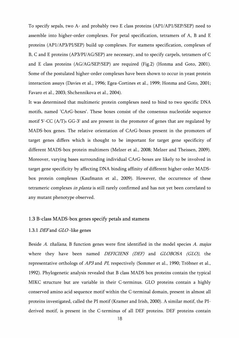
AAbbreviations
3-AT 3-amino-1,2,4-triazole
Aa Asparagus asparagoides
ACT ACTIN
A adenine
Ade adenine
AEKI Atomic Energy Research Institute
ANA Amborellales, Nymphaeales, Austrobaileyales
Af Aquilegia formosa
AG AGAMOUS
AGL AGAMOUS-like
Amb Amborella trichopoda
AP APETALA
APG Angiosperm Phylogeny Group
At Arabidopsis thaliana
BAC bacterial artificial chromosome
BNQ BANQUO
BiFC bimolecular fluorescence complementation
bp base pairs
ca carpels
CArG-box 5’-CC(A/T)6GG-3’ consensus sequence
cDNA complementary DNA
cfc cellulose fortified cells
CLV CLAVATA
cox3 CYTOCHROME OXIDASE 3
CRC CRABS CLAW
DEF DEFICIENS
DL DROOPING LEAF
DSB double strand break
Ec Eschscholzia californica

5
EcAuxRF1 Eschscholzia californica auxin response factor 1
EMSA Electrophoretic mobility shift assay
EREBP ethylene responsive element binding protein
ESca Eschscholzia californica
euAP3 motif eudicot APETALA3 motif
FAE formaldehyde-acetic acid-ethanol solution
FAR FARINELLI
FIL FILAMENTOUS FLOWER
g gynoecium
GGM Gnetum gnemon MADS
GLO GLOBOSA
GTR general time reversal
Gy gray
His histidine
HMG-box high mobility group-box
HMW high molecular weight
HOX homeobox transcription factor
ig inner gynoecium
INO INNER NO OUTER
Kb kilobase pairs
LB medium lysogeny broth medium
Leu leucine
LEU LEUNIG
lpc lignified parenchymatic cells
lr lateral ridges
MADS-box ‘MCM1-AGAMOUS-D
Mb megabase pairs
EFICIENS-SRF’-box transcription factor
MIKC MADS, Intervening, Keratin-like and C-terminal domain structure
min minutes
miRNA microRNA
mr middle ridges

6
MYA million years ago
og outer gynoecium
ONPG ortho-Nitrophenyl-�-galactoside
ORF open reading frame
OsMADS Oryza sativa MADS
ov ovule
p placental tissue
paleoAP3 motif paleo APETALA3 motif
PCR polymerase chain reaction
pe petals
PFGE pulse field gel electrophoresis
Ph Petunia hybrida
PLE PLENA
PI PISTILLATA
PI motif PISTILLATA- motif
PPI protein-protein interaction
QP glutamine-proline
r replum
RACE rapid amplification of cDNA ends
RAGE rapid amplification of genomic DNA ends
rbcL Ribulose-1,5-bisphosphate carboxylase oxygenase, large subunit
RBL REBELOTE
RT-PCR reverse transcriptase-polymerase chain reaction
SD synthetic dropout
se sepals
sec seconds
SEP SEPALLATA
SEU SEUSS
SHP1/2 SHATTERPROOF 1 and 2
si-1 silky-1
sir sirene

7
SLiM short linear interaction motif
spw-1 superwoman-1
SQUA SQUAMOSA
SQN SQUINT
SSR simple sequence repeat
st stamens
STK SEEDSTICK
SUP SUPERMAN
T thymine
ta-siRNA transacting-small interfering RNA
TBE buffer Tris Borate EDTA buffer
TM6 TOMATO MADSBOX GENE 6
Trp tryptophan
UPT ULTRAPETALA
UTR untranslated region
vb vascular bundle
VIGS virus induced gene silencing
wt wild type
WUS WUSCHEL
YAB YABBY

8
SSummary
The development of plant organs is organized by a hierarchy of regulatory genes
interacting with each other to build the plant’s structures. How exactly floral organs and
their morphology has evolved in angiosperms is investigated in the field of evolutionary
development (evo-devo). Evo-devo research requires model species from widely
distributed phylogenetic lineages, and the presented results were conducted in
Eschscholzia californica (California poppy), a member of the Papaveraceae in one of the
earliest diverging lineages of the basal eudicots. E. californica has recently been
established as a model system for evo-devo studies and for this purpose new genetic
resources were established.
A bacterial artificial chromosome library of California poppy genomic DNA was
constructed and characterized to facilitate the molecular analysis of developmental
control genes. Further, fast neutron mutagenesis was conducted to create a mutant
population of California poppy in order to discover unknown genes involved in flower
development of E. californica.
A new floral homeotic mutant, called sirene (sir), was uncovered within the help of the
mutant population. sir plants showed homeotic conversions of petals into sepals and
stamens are transformed into carpels. The molecular basis of the sir mutation was found to
be based on a mutation in the C-terminal domain of the B class gene SIR (EScaGLO). This
mutation altered the protein-protein interaction behaviour of SIR and multimeric
complexes composed of SIR, EScaAG1 (C class), and EScaAGL9 (E class) were not
assembled in the mutant. The failure to form those multimers most likely resulted in the
failure of organ specification in stamens and petals.
The E. californica ortholog of the carpel developmental control gene CRABS CLAW
(CRC) of Arabidopsis thaliana, EcCRC, was investigated in detail to uncover its function
in E. californica carpel development. It was shown that EcCRC has very specific functions
in regulating the development of carpel marginal tissues in the gynoecium, along with
widely conserved functions shared by most angiosperm species. The CRC-like genes in
general show an enormous diversity of functions in the different lineages of flowering
plants. To understand the causes of this functional diversity might be very important to
follow the evolution of the gynoecium, considered as being one the most important
inventions of angiosperms that laid the ground for their evolutionary success.

9
ZZusammenfassung
Die Entwicklung pflanzlicher Organe wird durch eine Hierarchie von Entwicklungs-
Genen gesteuert, die durch das Zusammenwirken untereinander die Pflanzenstrukturen
ermöglichen. Wie genau sich diese Blütenorgane und die Morphologie der Blüte als
Ganzes im Laufe der Evolution entwickelt haben, ist der Forschungsgegenstand der
Evolutionären Entwicklungsbiologie (Evo-Devo). Die Forschung im Bereich Evo-Devo
erfordert Modell-Organismen aus einem weiten Bereich der Angiospermen-Systematik.
Die vorliegenden Untersuchungen wurden deshalb mit Eschscholzia californica (Kalifornischer Mohn) ausgeführt, einem Mitglied der Familie der Papaveraceae, welche
wiederum zu den frühesten Linien der eudicotylen Pflanzen gehört. E. californica ist eine
neuartige Modellpflanze für evolutionäre Entwicklungsbiologie und für diese
Forschungen wurden neue genetische Methoden etabliert.
Eine genomische Bibliothek des Kalifornischen Mohns wurde hergestellt und
charakterisiert, um die molekulare Analyse von Entwicklungsgenen zu beschleunigen.
Zusätzlich wurde eine Mutantenpopulation mit Hilfe der Fast Neutron-Mutagenese
aufgebaut. Diese soll es ermöglichen, bisher unbekannte Kontrollgene der Blüten-
entwicklung in E. californica zu entdecken.
Mit Hilfe der etablierten Mutantenpopulation war es möglich, eine florale Mutante des
Kalifornischen Mohn zu entdecken, welche sirene (sir) genannt wurde. sir Mutanten
zeigen homeotische Transformationen der Kronblätter (Petalen) in Kelchblätter (Sepalen)
sowie eine Umwandlung der Staubblätter (Stamina) in Fruchtblätter (Karpelle). Die
Ursache der sir Mutation konnte im C-terminalen Ende des homeotischen Gens SIR (EScaGLO) lokalisiert werden. Diese spezifische Mutation hat zur Folge, das sich SIR
Proteine nicht mehr mit dem C-Klasse Protein EScaAG1 sowie dem E-Klasse Protein
EScaAGL9 zu höheren Proteinkomplexen zusammen lagern können. Dies hat vermutlich
zur Folge, dass die Identität von Kron- und Staubblättern in der sir Mutante nicht
festgelegt werden kann und sich deshalb diese Organe nicht bilden.
Ein dem CRABS CLAW (CRC) aus Arabidopsis thaliana orthologes Karpell-
entwicklungsgen, genannt EcCRC, wurde in E. californica detailliert funktionell unter-
sucht. Es konnte gezeigt werden, das EcCRC sehr spezifische Funktionen ausübt in der
Entwicklung jener Gewebe, die sich von den Karpell-Marginalen ableiten. Zusätzlich hat
EcCRC Aufgaben in der Entwicklung des Gynoeciums, die in vielen Blütenpflanzen
konserviert sind. Darüber hinaus zeigen die CRC-ähnlichen Gene eine besondere

10
funktionelle Vielfalt zwischen den verschiedenen Linien der Blütenpflanzen. Die Gründe
dieser Vielseitigkeit an Genfunktionen werden als wichtig erachtet für die Evolution des
Fruchtknotens (Gynoecium). Dieser stellt eine der entscheidenden Neuerungen der
Blütenpflanzen dar, denen sie einen Großteil ihres evolutionären Erfolges verdanken.

11
11. Introduction
1.1 Evolutionary development (evo-devo)
1.1.1 Evolutionary developmental genetics of plants The enormous variation of plant flower morphologies has always caught the attention of
botanists and evolutionary biologists (Darwin, 1862). Many hypotheses have been
launched to explain a particular astonishing phenomenon of plant structures, their great
diversity in morphology and simultaneously, the appearance of a common underlying
blueprint to all these structures. A modern attempt to understand this phenomenon comes
from the field of evolutionary development, or evo-devo (Hall, 2003). It tries to integrate
evolution and development, focusing on the function and molecular evolution of
developmental genes that organize the ontogeny of organisms. A key to the evo-devo
concept is that developmental control genes, often encoding highly conserved
transcription factors or signalling molecules, are organized in regulatory networks that
show hierarchical gene interactions. Genes with a higher hierarchical position in
development generally occupy more central roles in the regulatory network whereas
genes that are more peripherally located in the network have a lower hierarchical
position.
A good example of central regulatory genes is given by the vertebrate HOX genes which
specify the main body axis in the developing embryos in these organisms. An important
evolutionary characteristic of genes like the HOX genes is the degree of pleiotropy by
which these genes can be characterized. Mutational changes in the coding sequence can
affect several developmental pathways simultaneously. For instance, mutations in HOX
gene coding sequences can lead to homeotic transformation of one body part into another
and are often embryo-lethal (Lemons and McGinnis, 2006). On the other hand, mutations
in cis-regulatory regions are less likely to be deleterious because cis-regulatory mutations
usually affect only a subset of the overall expression domain of these genes which can lead
to altered expression patterns and allow phenotypic evolution (Stern and Orgogozo, 2008;
Gompel and Prud'homme, 2009).

12
Another aspect of evo-devo deals with the dynamics of gene duplications. In plant
evolutionary developmental studies, it became apparent that the majority of
developmental control genes belong to transcription factor families originated by
segmental or whole genome duplications (Bowers et al., 2003; Blanc and Wolfe, 2004;
Chapman et al., 2004). Gene duplication can lead to neofunctionalization or
subfunctionalization, i.e. the duplicated copy can evolve novel functions or become
restricted to a subset of functions of the progenitor gene (Lynch and Conery, 2000). Also,
duplicated genes usually retain partial redundancy in function, particularly directly after
duplication, but this redundancy can remain in duplicated gene pairs over very long
periods (Wagner, 1999; Moore and Purugganan, 2005). Gene duplications likely effect the
organization of regulatory networks. Sub- and neofunctionalization allow the
developmental networks to adjust to different environmental conditions whereas
functional redundancy buffers the genome against harmful mutations and adds stability to
regulatory networks (Blanc and Wolfe, 2004; Moore and Purugganan, 2005).
A prominent example of gene duplications in plants are the MADS-box transcription
factors which have expanded by gene and/or genome duplications to different extents
during the evolution of angiosperms (Theissen, 2001; Parenicova et al., 2003; Pinyopich et
al., 2003). The developmental function of duplicated MADS-box genes has been
elucidated in several species and ranges from total redundancy in the case of the
SHATTERPROOF1/2 (SHP1/2) duplicated genes in A. thaliana, to a clear subfunctionali-
zation between PLENA (PLE) and FARINELLI (FAR) genes in Antirrhinum majus,
although remnant overlapping functions still exist between these both paralogs (Davies et
al., 1999; Pinyopich et al., 2003).
1.1.2 Eschscholzia californica as a versatile model species The California poppy, Eschscholzia californica, is a species native to the western parts of
North America where it occupies a wide range of habitats, often living in naturally
disturbed, open environments. E. californica lives from the sea level up to 2000 m on
different soil formations, on river terraces and on the hillside, but also in human disturbed
environments and along roadsides in California. The plant has also been introduced to

13
Chile, Tasmania, Australia and New Zealand where it is now a natural part of ecosystems
(Cook, 1962). In these new locations, E. californica has proven to have a huge potential as
an invasive species and was therefore chosen as a model species for invasion biology of
plants (Leger and Rice, 2003, 2007).
E. californica, as a member of the family Papaveraceae, has long been known to contain
specific compounds that had already been used as medicine by North-American natives.
The secondary metabolites of California poppy belong to different types of alkaloids;
aporphines, pavines, protoberberines, protopines and benzophenanthridines (Hauschild et
al., 1998; Fabre et al., 2000). Many of these alkaloids have been characterized in detail, but
the biosyntheses of benzophenanthridine-type alkaloids have been completely elucidated
at the enzymatic level (Haider et al., 1997; Hauschild et al., 1998) and an alkaloid
detoxifying enzyme has also been characterized recently(Vogel et al., 2010). Due to the
pharmaceutical importance of their alkaloid contents, cell cultures of E. californica are
continuously engineered to produce commercially important agents like benzyl-
isoquinoline alkaloids (Pauli and Kutchan, 1998; Inui et al., 2007; Takemura et al., 2010).
E. californica also serves as a model for studying the molecular basis of development in
early diverging eudicots and for the comparative genetics of developmental processes. The
latter approach requires particularly informative taxa with respect to morphology or
phylogenetic position. E. californica, as a member of the earliest diverging lineage of the
Ranunculales, represents an important evolutionary link, and its position points in two
directions; i) towards the core eudicots and ii) to different groups of highly evolved
monocots like grasses or orchids (Fig.1)(APG, 2003). Moreover, the developmental
characteristics of E. californica can be compared to more basal angiosperm lineages like
the magnoliids or the species of the most basal angiosperm ANA grade (Amborellales,
Nymphaeales and Austrobaileyales) (Fig.1) (Soltis et al., 2004; Frohlich and Chase, 2007).
Besides these facts, the following characteristics are making E. californica a well-suited
model plant for evolutionary developmental genetics. It is an easily cultivated annual to
perennial plant with a life-cycle of three month under optimal growth conditions (Becker
et al., 2005; Wege et al., 2007).

14
Figure 1: SSummary tree of the angiosperm phylogeny In ((a), the angiosperm phylogeny is given with the ‘core’ eudicots composed of two major clades named rosids and asterids is highlighted, and known model species are additionally coloured. In the inset, a typical flower of E. californica is placed. In ((b) and ((c), the orders of Brassicales and Lamiales are shown. In ((d), maize and other Poaceae are part of the order Poales, which is nested within the commelinoid clade of monocots. Besides the model species, common names of exemplary agricultural important crops are shown for some plant groups but it is not considered a complete list. Modified from (Soltis and FGP, 2002).
The California poppy is amenable to genetic transformation and therefore allows a
detailed characterization of gene functions (Park and Facchini, 2000). E. californica has a
diploid genome consisting of 12 chromosomes with a size of 1115 Mb per haploid
chromosome set (Bennett et al., 2000). Additionally, the genome of E. californica has

15
independently duplicated after the split that separated higher eudicots and Ranunculales,
with the possibility to study the evolutionary dynamics of gene duplications (Cui et al.,
2006). The Floral Genome Project has made an extensive effort to compile large amounts
of expressed sequence tags and next-generation sequencing data that are publicly available
(Soltis and FGP, 2002; Carlson et al., 2006; Wall et al., 2009). Of particularly interest is the
fact that the miRNA content of different tissues has been surveyed in transcriptome
analysis. Several classes of miRNAs and ta-siRNAs have been identified and provide an
additional gateway for future evolutionary developmental studies (Barakat et al., 2007). In
the field of leaf development, E. californica has already been established as a model species
for evo-devo genetics (Busch and Gleissberg, 2003; Groot et al., 2005).
1.2 Molecular genetics of flower development
1.2.1 Floral organ specification and the ABCE model Most angiosperm flowers consist of four distinct organs arranged in whorls. The
outermost sterile perianth is composed of sepals and petals. The central flower whorls are
occupied by male and female reproductive organs which are stamens and the carpels. In
many cases, the carpels are fused and thereby build up the gynoecium which bears the
ovules and provides a cavity for the developing seeds. The genetics of flower development
is one of the best studied systems in plant developmental biology. Careful observations of
floral homeotic mutants and their molecular characterization revealed a model of how the
different organs of flowers are genetically determined. This is known as the ABC model
whose hallmarks are three distinct genetic functions, A, B, and C that specify each floral
organ (Fig.2) (Bowman et al., 1991; Coen and Meyerowitz, 1991).
In detail, the floral homeotic genes mainly belong to the MADS-box family of
transcription factors, a large gene family that is involved in many plant developmental
programs (Riechmann and Meyerowitz, 1997). In the following paragraphs, the floral
developmental program of A. thaliana will be described as it is the model organism from
which most of the data for the ABC model were obtained. The ABC functions act alone or
combinatorial to specify floral organ identity. In this manner, sepals are specified solely by
the action of the A class genes APETALA1 (AP1) and AP2, the latter belonging to the

16
AP2/EREBPs (APETALA2/ethylene-responsive element binding protein) family of
transcription factors (Okamuro et al., 1997) and being the only non-MADS-box gene in
the ABC model. Petals are specified by combinatorial action of the B function genes AP3
and PISTILLATA (PI) together with A function genes. The C class gene is AGAMOUS
(AG) which acts collectively with B class genes to specify stamen organ identity and AG
alone specifies the identity of carpels (Coen and Meyerowitz, 1991) (Fig.4). A major
extension of the classic ABC model was the discovery of the four redundantly acting E
class SEPALLATA (SEP) genes, which are additionally required to specify all four floral
organs (Pelaz et al., 2000; Theissen and Saedler, 2001; Ditta et al., 2004).
MADS-box genes other than AG also play an important role in carpel development and
these genes mainly belong to the AG subclass of MADS-box transcription factors. The two
genes that can independently of AG promote carpel development in A. thaliana are
SHP1/2. Recently, SHP genes were also recognized to be more specifically occupied for
the development of stigma, style and valve marginal tissues (Colombo et al., 2010). Both
SHP genes are also involved in the specification of ovule identity, and share this function
redundantly with AG and SEEDSTICK (STK), another member of this subclass (Pinyopich
et al., 2003). STK is required for the development of the funiculus, a stalk-like structure
that connects the ovules to the carpel wall within the gynoecium (Pinyopich et al., 2003).
Predictions of the originally described ABC model were comparatively tested in several
other angiosperm taxa. Extensive functional conservation, however, has only been found
for B and C class genes (e.g. (Pnueli et al., 1994; Samach et al., 1997; Kramer et al., 1998;
Ambrose et al., 2000; Drea et al., 2007). B and C function genes were also identified in
gymnosperms and, given their expression in the corresponding reproductive organs, it was
suggested that one of the ancestral functions of B and C genes included male and female
reproductive development. The evolutionary origin of the ABC model is likely to be
found in a sex determination system already present in the ancestors of angiosperms and
gymnosperms (Mouradov et al., 1999; Theissen and Becker, 2004).
Contrarily, clearly comparable A class function homologs of A. thaliana were difficult to
find in any other species surveyed (Gutierrez-Cortines and Davies, 2000; Litt, 2007).
Furthermore, it was suggested that a specific A function is not required for perianth organ

17
identity outside of the Brassicaceae and can therefore be omitted from the original ABC
model (Litt, 2007).
11.2.2 MADS-box gene function at the molecular level MADS-box proteins consist of a highly conserved N-terminal DNA-binding MADS
domain (M), a less conserved intervening domain (I) and a keratin-like domain (K) that
contains three amphipathic �-helices involved in protein-protein interactions. At the C-
terminal end of MADS box proteins, a variable C-terminal (C) domain is located which
are specific for the different subgroups of this gene family. The MADS box proteins of the
ABC model belong to the subgroup of MIKCC type of MADS-box proteins (Yang and Jack,
2004).
MADS-box proteins form protein dimers and higher-order multimeric complexes to carry
out their biological function as transcriptional regulators. The molecular mechanism of
floral organ specification in A. thaliana was hypothesized to be mediated by ‘floral
quartets’ consisting of MADS-box protein tetramers that act in a combinatorial manner
(Theissen and Saedler, 2001)..
Figure 2: ABCE model and floral quartets
Assumed molecular action of floral organ specification in which tetramers of MADS-box transcription factor proteins act in combination to specify the identity of the four floral organs. Protein complexes bind to DNA on CArG-boxes in the promoter of target genes. For details see text, modified from (Theissen and Saedler, 2001).
18
To specify sepals, two A- and probably two E class proteins (AP1/AP1/SEP/SEP) need to
assemble into higher-order complexes. For petal specification, tetramers of A, B and E
proteins (AP1/AP3/PI/SEP) build up complexes. For stamens specification, complexes of
B, C and E proteins (AP3/PI/AG/SEP) are necessary, and to specify carpels, tetramers of C
and E class proteins (AG/AG/SEP/SEP) are required (Fig.2) (Honma and Goto, 2001).
Some of the postulated higher-order complexes have been shown to occur in yeast protein
interaction assays (Davies et al., 1996; Egea-Cortines et al., 1999; Honma and Goto, 2001;
Favaro et al., 2003; Shchennikova et al., 2004).
It was determined that multimeric protein complexes need to bind to two specific DNA
motifs, named ’CArG-boxes‘. These boxes consist of the consensus nucleotide sequence
motif 5'-CC (A/T)6 GG-3‘ and are present in the promoter of genes that are regulated by
MADS-box genes. The relative orientation of CArG-boxes present in the promoters of
target genes differs which is thought to be important for target gene specificity of
different MADS-box protein multimers (Melzer et al., 2008; Melzer and Theissen, 2009).
Moreover, varying bases surrounding individual CArG-boxes are likely to be involved in
target gene specificity by affecting DNA binding affinity of different higher-order MADS-
box protein complexes (Kaufmann et al., 2009). However, the occurrence of these
tetrameric complexes in planta is still rarely confirmed and has not yet been correlated to
any mutant phenotype observed.
1.3 B-class MADS-box genes specify petals and stamens
1.3.1 DEF and GLO -like genes Beside A. thaliana, B function genes were first identified in the model species A. majus
where they have been named DEFICIENS (DEF) and GLOBOSA (GLO), the
representative orthologs of AP3 and PI, respectively (Sommer et al., 1990; Tröbner et al.,
1992). Phylogenetic analysis revealed that B class MADS box proteins contain the typical
MIKC structure but are variable in their C-terminus. GLO proteins contain a highly
conserved amino acid sequence motif within the C-terminal domain, present in almost all
proteins investigated, called the PI motif (Kramer and Irish, 2000). A similar motif, the PI-
derived motif, is present in the C-terminus of all DEF proteins. DEF proteins contain

19
additional conserved amino acid sequence motifs in the C-terminus, called the euAP3-
motif, present only in the core eudicots or an ancient paleoAP3 motif in all other
angiosperm DEF proteins (Fig.3) (Kramer and Irish, 2000).
These two different DEF gene lineages presumably arose by a gene duplication event just
before the radiation of the higher eudicots (Kramer and Irish, 2000). Not all higher
eudicot species have retained both duplicated DEF copies in their genome of which the
paleoAP3 containing genes have evolved into the TM6 lineage, named after the TOMATO
MADS BOX GENE 6 (Pnueli et al., 1991). TM6 genes are particularly present in the family
of Solanaceae; however, they are neither present in A. thaliana nor in A. majus (Fig.3).
FFigure 3: Simplified phylogenetic tree of DEF (AP3) and GLO (PI) proteins
(A) The estimated evolutionary history of AP3 and PI proteins from angiosperms and
gymnosperms are illustrated. The C-terminal motifs are shown on the right. Amino
acids in critical positions of AP3 and PI proteins that were subject to positive
selection are indicated by arrows. The estimated time of gene duplication events is
shown at the split of PI/AP3 and euAP3/paleoAP3 lineages. (B) Three amphipathic
helices K1-K3 are shown with the positions of amino acids under positive selection
and the region responsible for PI/AP3 protein heterodimerization. Modified
according to (Hernandez-Hernandez et al., 2007).

20
The reason why some species of higher eudicots, especially in asterids, have retained both
DEF and TM6 gene copies while others lost exclusively their paleoAP3 containing TM6
paralogous genes remains enigmatic. This is particularly remarkable when considering
that genes which contain the paleoAP3 motif are the functionally ancient DEF genes
present in all other angiosperm lineages.
The functional significance of either of the conserved motifs in the C-terminus of DEF
and GLO proteins could not definitely been shown to date, basically due to inconsistent
experimental results. In vitro protein interaction assays clearly showed that DEF and GLO
C-termini are required for higher-order protein complex formation (Egea-Cortines et al.,
1999). Furthermore, complementation experiments in the respective mutants of A.
thaliana with domain swapped and C-terminal truncated B class proteins revealed that the
C-terminus is essential for proper protein function (Lamb and Irish, 2003). On the other
hand, similar experiments did not confirm that the C-terminus of DEF- and GLO-like
proteins is required for floral organ identity function (Berbel et al., 2005; Piwarzyk et al.,
2007). Due to this incongruence, the evolutionary and functional importance of the
conserved C-terminal motifs of B class genes remain poorly understood.
11.3.2 Evolution of functional interactions of B class proteins
All floral homeotic MADS-box proteins have three amphipathic helices within their K
domain, named K1-K3 and these were shown to be important for DEF and GLO protein
heterodimerization via conserved hydrophobic positions within these helices (Yang et al.,
2003; Yang and Jack, 2004). Several studies on DEF/GLO protein interactions elucidated
that in higher eudicots like A. thaliana and A. majus no homodimers but only hetero-
dimers are formed (Schwarz-Sommer et al., 1992; Goto and Meyerowitz, 1994; Davies et
al., 1996; Krizek and Meyerowitz, 1996; McGonigle et al., 1996; Riechmann et al., 1996b).
DEF/GLO heterodimers bind to CArG boxes which are present in the promoters of
downstream target genes regulated by B genes in eudicots (Schwarz-Sommer et al., 1992;
Tröbner et al., 1992; Riechmann et al., 1996b; Riechmann et al., 1996a).
A specific feature of DEF/GLO heterodimers is that they control their own transcription
in a positive autoregulatory feedback mechanism during organ development (Jack et al.,
1992; Goto and Meyerowitz, 1994). This function was imposed to be direct in the case of

21
AP3 in A. thaliana due to the presence of two adjacent CArG boxes in the AP3 promoter
that are indeed bound by AP3/PI heterodimers (Riechmann et al., 1996a; Hill et al., 1998;
Tilly et al., 1998; Honma and Goto, 2000). In case of PI, the autoregulation was postulated
to be indirect, because the responsible PI promoter does not contain any CArG boxes
(Chen et al., 2000; Honma and Goto, 2000).
Besides their ability to form dimers, it is likely that DEF and GLO hetero- or homodimers
take part in higher-order transcription factor complexes in planta which are hypothesized
to regulate petal and stamen development (Fig.2) (Honma and Goto, 2001; Theissen and
Saedler, 2001). Indeed, the DNA binding ability of MADS-box protein complexes
consisting of the A. thaliana SEP3-SEP3-SEP3-SEP3 homotetramers and of SEP3-SEP3-
AP3-PI heterotetramers has been shown by protein-DNA binding assays (Melzer et al.,
2008). But how exactly multimer formation, promoter specificity and transcriptional
activation are realized in the cell is still poorly understood.
The dimerization capabilities of B class proteins were determined in several studies to
reveal the degree of functional conservation across divergent taxa. In basal eudicots and
monocots, the formation of DEF or GLO homodimers was uncovered with some of them
also showing DNA binding activity and even evidence of a GLO-like DNA binding
homomultimer exists in orchids (Tzeng and Yang, 2001; Hsu and Yang, 2002; Kramer et
al., 2003; Tzeng et al., 2004; Drea et al., 2007; Kramer et al., 2007; Tsai et al., 2008).
Furthermore, the ancestral B class proteins were hypothesized to carry out their
functional repertoire as obligate homodimers which was shown for GGM2 B class protein
homodimers in the gymnosperm Gnetum gnemon (Winter et al., 2002). This obligate
homodimerization has evolved into obligate heterodimerization of DEF/GLO proteins in
higher eudicots and monocot grasses with some homodimerization ability retained in
other monocots and basal eudicots (Riechmann et al., 1996b; Winter et al., 2002; Whipple
et al., 2004; Drea et al., 2007; Kramer et al., 2007; Tsai et al., 2008). The question whether
B class homodimers still have a residual function in eudicots or monocots remains open,
since the functional specialisation from homo- to heterodimerization most probably has
been accompanied by structural modifications in protein sequences of participating
protein partners and may also have influenced the expression domains of B class genes via

22
auto- or cross-regulatory mechanisms (Winter et al., 2002; Hernandez-Hernandez et al.,
2007; Lenser et al., 2009).
Addressing these questions, Kim and colleagues (2004) extensively studied the diversi-
fication of protein sequences during angiosperm evolution. Both, DEF and GLO proteins
in the most basal angiosperm Amborella trichopoda show several features that are unique
to the angiosperm DEF/GLO proteins, e. g. a prominent ‘DEAER’ amino acid motif in the
C-terminus and altered amino acid residues in otherwise highly conserved positions like
the KEN motif in GLO proteins. Also, critical residues for B class heterodimerization, like
97E and 98N in PI and 98N and 102R in AP3, were not found in the respective A.
trichopoda proteins. These results together led to the suggestion that protein interactions
among B class proteins might be very flexible in terms of homo- and heterodimerization
capabilities and dynamic with respect to higher-order protein formation in A. trichopoda
and to a lesser extent in other basal angiosperms (Kim et al., 2004). Deletions and other
modifications in the K domain and possibly also in the C-terminus of DEF and GLO
proteins might than have aided in the canalization of their functional properties, possibly
resulting in obligate heterodimerization of B class proteins in core eudicots and monocot
grasses. Furthermore, the regulation of B class higher-order protein interactions within
the sepal and petal developmental programs might have been facilitated by protein
sequence co-evolution of participating B class proteins (Vandenbussche et al., 2003; Kim
et al., 2004).
11.3.3 Evolution of petal and stamen developmental programs The B gene clade originated early in land plant evolution approximately 300-400 MYA in
the common ancestor of gymnosperms and angiosperms (Theissen et al., 2000). Ferns, the
sister group to seed plants, do not contain orthologs of floral homeotic genes, and
therefore it was suggested that the B clade arose in the common ancestor of seed plants
(Theissen et al., 2000). Putative B class gene orthologs have been isolated from
gymnosperms, e.g. from the gnetophyte G. gnemon (gnemon) and the conifers Picea abies
(Norway spruce), and Pinus radiata (Monterey pine) (Mouradov et al., 1999; Sundstrom et
al., 1999; Winter et al., 1999). Approximately 290 MYA, close at the base of the
angiosperms, but after gymnosperms and angiosperms have split, a duplication of B genes

23
was proposed that led to the present DEF and GLO lineages (Hernandez-Hernandez et al.,
2007). Within the angiosperms, DEF and GLO-like genes have duplicated several times
independently, e.g. GLO-like genes in monocot grasses and lilies and DEF-like genes in
orchids and in the basal eudicot order Ranunculales (Kramer and Irish, 1999, 2000;
Theissen et al., 2000; Munster et al., 2001; Kanno et al., 2003; Kramer et al., 2003; Tsai et
al., 2004). In case of the core eudicots, DEF gene duplication led to the aforementioned
differential AP3/TM6 gene lineages which contain either euAP3 or paleoAP3 motifs in
the C-terminal domain pointing towards functional deviations of the two duplicated gene
copies (Fig.3).
According to their role in specifying petal and stamen organ identity, DEF and GLO
expression can be found primarily in petals and stamens (Jack et al., 1992; Schwarz-
Sommer et al., 1992; Tröbner et al., 1992; Goto and Meyerowitz, 1994). In the eudicot
model species A. thaliana and A. majus, shortly after sepal primordia arise in the floral
meristem, DEF expression can be detected in places where petals and stamens will
develop later. The expression continues throughout stages of floral organ development.
An additional domain of low expression of DEF can be found at the base of the sepals and
in carpels of A. majus (Schwarz-Sommer et al., 1992). AP3 expression, on the other hand,
can also be found in developing A. thaliana ovules. The expression of GLO and PI genes is
very similar to DEF and AP3 in whorls 2 and 3, where petals and stamens will develop.
They are also expressed at low levels in early stages of developing carpels (Jack et al.,
1992). Other core eudicot species were found to have a similar spatially and temporally
restricted domain of DEF and GLO expression, e.g. tomato, petunia and primrose
(Tsuchimoto et al., 2000; de Martino et al., 2006; Li et al., 2008).
In the basal eudicot order Ranunculales, the expression patterns of B class genes have been
determined for a wide range of families and species. Here, the expression domains of DEF
and GLO genes appear generally broader, e.g. expression domains slide laterally from
petals into the sepals or centrally from stamens into the carpels (Kramer and Irish, 2000;
Kramer et al., 2003; Drea et al., 2007; Kramer et al., 2007). A comparable situation is found
in monocots, where the B gene expression has been explored in lineages of grasses, lilies
and orchids. Here, a generally less restricted domains of expression activity compared to
core eudicots can be observed for B class genes (Ambrose et al., 2000; Munster et al., 2001;

24
Tzeng and Yang, 2001; Hsu and Yang, 2002; Nagasawa et al., 2003; Tsai et al., 2004).
Further deviations from the conserved expression domain of B genes in petals and stamens
was evident in some magnoliid dicot species which revealed that DEF and GLO genes are
not universally expressed cooperatively in stamens as the classic ABC model would
predict (Kramer and Irish, 2000).
These observations led to the proposal that the conservation of B class gene expression
domains is less strict outside the core eudicots with their domains allowed to ‘slide’ or
‘shift’ over their boundaries as opposed to a more canalized expression pattern in higher
eudicots (Fig.4) (Bowman, 1997; Kramer and Irish, 2000). This scenario is supported by
expression analysis of B class genes in basal angiosperms, where they are expressed in all
four floral organs and sometimes even in vegetative leaves (Kim et al., 2005). While B
gene expression is not restricted to specific floral organs in A. trichopoda, Nuphar advena
(cow lily), and Illicum floridanum (Florida anisetree), they do form a gradient of
decreasing strength towards the margins of their domains (Fig.4). This results in an
intergrading morphology of spirally arranged floral organs (Buzgo et al., 2004; Kim et al.,
2005). Therefore, the ‘fading border’ model was launched as an explanation of how the
spatial structure of gene expression affect floral organ morphology in basal angiosperms
(Buzgo et al., 2004; Soltis et al., 2007).
Figure 4: Expression domains of ABCE function
regulatory genes in the four floral whorls of
different plant groups.
(A) In higher eudicots, B class genes show a clear
boundary of expression in the 2nd and 3rd whorl. ((B)
The sliding boundary of constricted or extended B
gene expression as derived from basal eudicots and
magnoliids is shown. (C) The fading border model of
class gene expression in basal angiosperms is
highlighted. Modified from (Soltis et al., 2007).

25
11.4 Carpel developmental genetics
1.4.1 Fundamentals of carpel structure The female reproductive organs of angiosperms are complex structures, called gynoecium
when composed of more than one carpel. Two types of gynoecia basically exist:
apocarpous gynoecia with unfused carpels that form independent pistils within the flower
and syncarpous gynoecia with fused carpels, which are considered a key innovation of
angiosperms (Endress and Igersheim, 1999; Endress, 2001). It is of major commercial
interest to understand the evolution of gynoecium development, since angiosperm carpels
and their derived structures are an important source of food and drugs for human living
and health. Most of the discoveries of carpel or gynoecium development were made in the
advanced higher eudicot plant A. thaliana and in the monocot grasses Oryza sativa (rice)
and Zea mays (maize) (e.g. (Mena et al., 1996; Alvarez and Smyth, 1999; Alvarez and
Smyth, 2002; Yamaguchi et al., 2004; Yamaguchi et al., 2006). But with respect to the
complexity of gynoecium structures and their development, these groups alone might not
be sufficient to explain the full evolutionary history of gynoecium development, and there
is need of more comparative data derived from model species covering a wider
phylogenetic range like E. californica.
The gynoecium can be divided into several parts based on an apical-basal axis. The most
basally located structure is the ovary which bears the ovules and can be separated by septa
into one or more locules or cavities. On top of the ovary, a structure called style is
positioned which contains the transmitting tract. And most apically, the stigma develops
with specialized tissue cells at its top. The pollen attaches to the stigmatic cells and the
pollen tubes subsequently grow via the transmitting tract to the ovules for fertilization.
The stages of prefertilization flower development and postfertilization fruit development
have been described in detail for California poppy by Becker and colleagues (Becker et al.,
2005).

26
11.4.2 CRABS CLAW -like YABBY transcription factors in carpel development The YABBY genes comprise a small family of plant specific transcriptional regulators
which are present only in seed plants (Floyd and Bowman, 2007). YABBY genes encode
for proteins that contain a C2C2 zinc finger domain near their N-terminal end and a helix-
loop-helix domain, with sequence homology to the known DNA binding HMG-box (high
mobility group), a common part of regulatory proteins in vertebrates (Bowman, 2000). Six
YABBY genes are known from A. thaliana, whereas eight members were discovered in
rice (Alvarez and Smyth, 1999; Bowman and Smyth, 1999; Eshed et al., 1999; Sawa et al.,
1999; Siegfried et al., 1999; Villanueva et al., 1999; Yamaguchi et al., 2004; Toriba et al.,
2007). All YABBY genes in eudicots are involved in promoting abaxial cell fate in lateral
organs (Bowman, 2000). In contrast to eudicots, YABBY genes in monocot grasses differ in
this respect, because maize YABBY genes are expressed adaxially and members in rice,
wheat and sorghum are not expressed in a polar manner at all (Jang et al., 2004; Juarez et
al., 2004; Zhao et al., 2006; Ishikawa et al., 2009).
The YABBY genes CRABS CLAW (CRC) and INNER NO OUTER (INO), initially
analysed in A. thaliana, are involved in the development of female reproductive organs.
INO acts exclusively in the specification of the outer integument of ovules in A. thaliana
(Villanueva et al., 1999). CRC has a particular function in carpel growth and fusion and is
the major determinant of nectary development (Alvarez and Smyth, 1999; Bowman and
Smyth, 1999; Eshed et al., 1999). CRC-like genes are also the most widely characterized
YABBY members with respect to phylogenetic distribution and number of species, where
representatives of higher eudicots as well as monocots and basal angiosperms were
analysed (Yamaguchi et al., 2004; Fourquin et al., 2005; Lee et al., 2005b). CRC-like genes
are consistently expressed at the abaxial side of developing carpels from basal angiosperms
to higher eudicots (Bowman and Smyth, 1999; Fourquin et al., 2005; Lee et al., 2005a).
Exceptions to this are the CRC-like genes in the highly specialized grass lineage, where
they have acquired additional roles. The CRC ortholog from rice, DROOPING LEAF (DL),
is a major determinant of carpel identity, floral meristem termination and leaf midrib
development. DL, like other YABBY genes in monocot grasses, has lost the polar

27
expression domain within the carpel and leaf primordia. Instead, the expression of DL is
extended to the adaxial side of carpels and the leaf midrib (Yamaguchi et al., 2004).
22. Generation of genetic tools for functional analysis of genes in E. californica
2.1 Objectives
Comparative developmental genetic studies require the precise analysis of gene functions.
In model plants like A. thaliana, diverse genetic tools to investigate and to manipulate
gene function are often available but less so in the new model organism E. californica.
We aimed to expand the repertoire of functional tools for E. californica by constructing a
bacterial artificial chromosome (BAC) library of nuclear DNA with the emphasis to aid
cloning strategies of coding sequences and in particular of cis-regulatory regions of
developmental genes. The latter are probably involved in the evolution of gene function
by mutational changes in orthologous genes whose function has been described in other
model plants. But since the focus of the thesis was not only to compare the functional
characteristics of genes already described in other species, a second project was launched.
It consists of a population of poppy plants, mutagenized with fast neutrons and subject to
subsequent mendelian crossing that can be screened for alterations of the flower and
organ morphology. With this approach it was expected to significantly enhance the
discovery rate of genes involved in flower and carpel development in E. californica which
are not known so far or very specific to this lineage.
Both projects were thought to strengthen the role of E. californica as a model plant for
evolutionary developmental genetics of flower organs by extending the experimental
repertoire. The mutant population was also expected to benefit from the BAC library, i.e.
that the molecular analysis of promising mutant phenotypes would be aided by the
analysis of the mutated genomic locus carried out with the help of BAC library screening.
28
22.2 Material and Methods
2.2.1 Construction of a bacterial artificial chromosome library E. californica variety ‘Aurantiaca Orange King’ were grown under conditions previously
described (Wege et al., 2007). The above-ground organs of three week old seedlings were
harvested, snap frozen and stored at -80°C. High molecular weight (HMW) DNA was used
for construction of the BAC library. HMW-DNA isolation and BAC library construction
was carried out by Rx Biosciences (Rockville, USA). For this, HMW-DNA of E. californica
was partially digested with HindIII and size separated by pulse field gel electrophoreses
(PFGE). DNA fragments in the size range between 100 kb and 250 kb were ligated into
the pINDIGO-BAC-5 HindIII cloning ready vector system (Epicentre Biotechnologies,
Hess. Oldendorf, Germany), transformed into EPI300™ cells (Epicentre Biotechnologies,
Hess. Oldendorf, Germany) and selected on 12,5 μg chloramphenicol/ml.
BAC clones were picked manually, cultured in LB media [13 mM KH2PO4, 36 mM
K2HPO4, 1.7 mM sodium citrate, 6.8 mM (NH4)2SO4, 0.4 mM MgSO4, 4.4 % v/v glycerol
and 12,5 μg chloramphenicol/ml], and subsequently stored at -80°C. For membrane
spotting, carried out by the ADIS facility at the Max-Planck Institute for Plant Breeding
Research (Cologne, Germany), the clones were picked by a QPix2 colony picker (Genetix,
New Milton, UK) and macroarrayed on Amersham Hybond™-N+ membranes (GE
Healthcare Europe GmbH) with the Biorobotics MircroGridII system (Isogen Life Science,
De Meern, NL).
2.2.2 Estimation of BAC library genome coverage BAC plasmids were isolated from bacterial cultures, grown in LB media containing 12,5 μg
chloramphenicol/ml, with the EasyPrepPro miniprep kit (Biozym Scientific GmbH, Hess.
Oldendorf, Germany). The inserts of the BAC vector were released by NotI digestion,
separated on a 1 % (w/v) agarose gel in 0,5X TBE buffer with PFGE on a CHEFII apparatus
(BioRAD Laboratories, Munich, Germany) using the following conditions: 5s-15s linear
switch ramp, 120° angle at 6V/cm for 16 h at 12°C, and documented.
29
22.2.3 Estimation of organelle content in the library The screening of the BAC library for organelle contamination was conducted by Anna
Menke, Developmental Genetics Group, University of Bremen, Germany. Hereafter is a
brief description of the methodological work.
The BAC library was screened with E. californica gene specific probes. A 316 bp fragment
of the mitochondrial gene cox3 (CYTOCHROME OXIDASE 3), a 392 bp fragment of the
chloroplast rbcL gene (large subunit of RuBisCO) and a 303 bp fragment of the nuclear
gene EcAuxRF1 were amplified from genomic DNA for probe generation. The PCR
fragments of cox3, rbcL and EcAuxRF1 were labeled with digoxigenin (DIG).
Hybridization of the BAC filter was done according to the Roche DIG Application Manual
for Filter Hybridization (Roche Applied Science, Mannheim, Germany). Hybridizations
were performed over night at 43,3°C for rbcL, 43,8°C for cox3, and 43,4°C for EcAuxRF1,
followed by low and high stringency washes. Signals were detected by CDP-star (Roche
Applied Science) and luminescence of the BAC filter was measured with a LAS-3000
chemiluminescence detector (Fuji Photo Film Europe GmbH, Düsseldorf, Germany).
(Primer sequences and accession numbers are given in chapter 6.1).
2.2.4 Fast neutron mutagenesis Dry seeds from E. californica were irradiated with fast neutrons at three dosages: 20 gray
(Gy), 40 Gy, and 60 Gy at the AEKI Irradiation Facility in Budapest, Hungary under the
expertise of Joe K. Palfalfi. Seeds exposed to fast neutron irradiation were surface sterilized
with 2% sodium hypochloride and transferred to Petri dishes with B5-agar. To ensure
synchronous germination, seeds were vernalized at 4°C for 4 days. Germination and
survival assays were carried out in growth rooms at 20 °C with 16 hours light at 70 μmol s-1
m-2. Seed germination and seedling growth was observed over a period of 21 days in 3-day
intervals, and the numbers of germinating seeds at each dose rate and the seedlings
survival over the three week period was recorded.

30
22.2.5 Plant crossings Seeds irradiated with 40 Gy were used for all further experiments and cultivated (Wege et
al., 2007). Two F0 plants were crossed with each other to produce the F1 generation. F1
sibling plants were than inbreeded to produce homozygous recessive genotypes in the F2
generation. Inbred lines of all F2 crosses were grown and screened for flower
developmental mutants.
2.3 Results and Discussion
2.3.1 Construction and characterization of the genomic BAC library To facilitate reverse genetic techniques a genomic bacterial artificial chromosome library
of E. californica was constructed which is intended to help in cloning and characterization
of open reading frames and regulatory regions of developmental control genes.
Additionally, mutant plants identified in the fast neutron mutant population will be
characterized with the help of the BAC library.
The construction of the BAC library was essentially done by Rx Biosciences (Rockville,
Maryland, USA). Genomic DNA of the above-ground of young plants was isolated as
high-molecular weight DNA and partially digested. Fragments were size selected with
pulse field gel electrophoresis and subsequently used to construct the BAC library. A total
of 22,565 BAC clones were obtained which were kept in 384 well plates at -80°C for long
term storage. The whole library was replicated from hand-picked 96-well-plates and
spotted on nylon membranes by the Automatic Sequencing Facility (ADIS) at the Max-
Planck Institute for Plant Breeding Research in Cologne, Germany. The identification of
particular clones can be facilitated by screening the spotted library with standard
hybridization techniques using sequence specific probes.
In order to characterize the E. californica BAC library, the average insert size of BAC
plasmids was assessed by picking 137 randomly selected BAC clones. The BAC plasmids
were digested with NotI to release the insert from the vector pINDIGO-BAC-5 (Fig.5A).
The average insert size of the BAC plasmids was calculated to be 131 kb. The insert size
31
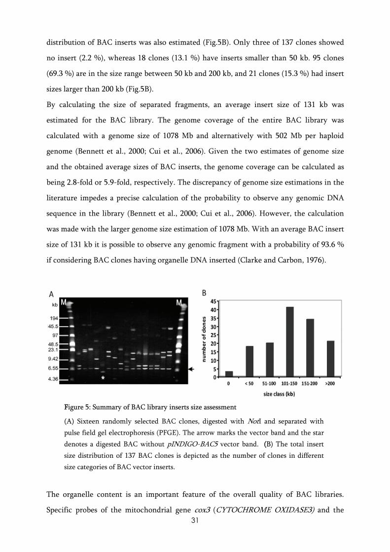
distribution of BAC inserts was also estimated (Fig.5B). Only three of 137 clones showed
no insert (2.2 %), whereas 18 clones (13.1 %) have inserts smaller than 50 kb. 95 clones
(69.3 %) are in the size range between 50 kb and 200 kb, and 21 clones (15.3 %) had insert
sizes larger than 200 kb (Fig.5B).
By calculating the size of separated fragments, an average insert size of 131 kb was
estimated for the BAC library. The genome coverage of the entire BAC library was
calculated with a genome size of 1078 Mb and alternatively with 502 Mb per haploid
genome (Bennett et al., 2000; Cui et al., 2006). Given the two estimates of genome size
and the obtained average sizes of BAC inserts, the genome coverage can be calculated as
being 2.8-fold or 5.9-fold, respectively. The discrepancy of genome size estimations in the
literature impedes a precise calculation of the probability to observe any genomic DNA
sequence in the library (Bennett et al., 2000; Cui et al., 2006). However, the calculation
was made with the larger genome size estimation of 1078 Mb. With an average BAC insert
size of 131 kb it is possible to observe any genomic fragment with a probability of 93.6 %
if considering BAC clones having organelle DNA inserted (Clarke and Carbon, 1976).
FFigure 5: Summary of BAC library inserts size assessment
(A) Sixteen randomly selected BAC clones, digested with NotI and separated with pulse field gel electrophoresis (PFGE). The arrow marks the vector band and the star denotes a digested BAC without pINDIGO-BAC5 vector band. ((B) The total insert size distribution of 137 BAC clones is depicted as the number of clones in different size categories of BAC vector inserts.
The organelle content is an important feature of the overall quality of BAC libraries.
Specific probes of the mitochondrial gene cox3 (CYTOCHROME OXIDASE3) and the

32
chloroplast gene rbcL (the large subunit of the enzyme RIBULOSE BIS-PHOSPHATE
CARBOXILASE/OXIDASE) were designed to assess the amount of organellar DNA
contamination in the library. A total of six hybridization signals for the mitochondrial
gene cox3 was detected which would suggest a contamination rate of 0.03% BAC plasmids
having DNA of mitochondrial origin. The chloroplast gene specific probe of rbcL revealed
57 signals when the library was probed. This approximately corresponds to 0.25% BAC
clones with chloroplast DNA content in the library.
Angiosperm chloroplast genomes are usually in the size range between 120 kb-160 kb
(reviewed in(Palmer, 1985) whereas plant mitochondrial genomes can be much larger and
vary in size more between species. The probability of finding any chloroplast and
mitochondrial sequence in the library was also estimated. Since the organelle genome
sizes of E. californica are not known, a genome size of 120 kb for chloroplasts was
assumed which is at the lower size limit of angiosperm chloroplast genomes (Palmer,
1985). With 57 BAC clones identified having chloroplast DNA inserted and an average
BAC insert size of 131 kb, it was assumed that almost any BAC clone with chloroplast
DNA can be detected using a single chloroplast gene-specific probe. The genome sizes of
mitochondria in flowering plants are more variable and range between 200 kb and 2400
kb. Six hybridization signals with a mitochondrial DNA specific probe were detected and
therefore, with a single mitochondrial gene specific probe, it is likely to find this
particular gene in the library with a rate between 65% to 5% of all mitochondria
containing BAC clones in the library, depending on the actual mitochondrial genome
sizes, when calculated with 200 kb to 2400 kb, respectively.
This low contamination rate of the library with organelle DNA shows that the method
used to isolate HMW-DNA from poppy tissue was extremely efficient in excluding
organelle DNA, specifically mitochondrial DNA, from further processing of the library.
22.3.2 Fast neutron mutagenesis and subsequent gene identification The identification of novel regulatory genes of floral organ development in poppy requires
the molecular analysis of mutant plants. In order to obtain mutants affected in flower
development, poppy seeds were mutagenized with fast neutron irradiation to saturate the
33
genome with mutations, and a strategy for screening the resultant mutant population for
relevant mutant phenotypes was developed.
It has long been known that ionizing radiation like fast neutrons induce DNA double
strand breaks (DSB) (McClintock, 1931). Such break points are targeted by the plant’s
endogenous DSB repair system (Britt, 1999; Gorbunova and Levy, 1999). Upon induction
of DSB, deletions and insertions are usually introduced into the genomic locus by the
endogenous double strand break repair system that mainly acts via non-homologous end-
joining, an error prone mechanism (Rinehart et al., 1997), and the genomic loci affected
by DSB frequently show a complex rearrangement pattern. The genomic alterations at the
DSB loci can span a range between a few base pairs up to 10 kb, thereby most likely
producing mutations such as whole gene deletion, mutations in coding genes that lead to
aberrant proteins or reorganized promoters that regulate the expression patterns of genes
(Shirley et al., 1992; Bruggemann et al., 1996).
The dosages that are applied to seeds in fast neutron treatments are given in gray (Gy),
which is the amount of radiation energy absorbed by any form of matter. This spanned
from 4,5 Gy in Lactuca sativa (lettuce), to 8 Gy in Lotus japonicus and Glycine max
(soybean) up to 60 Gy in A. thaliana (Okubara et al., 1994; Li et al., 2001; Men et al., 2002;
Hoffmann et al., 2007). Therefore, the optimal dosage of fast neutron irradiation on E.
californica seeds was experimentally assayed by observing seed germination and survival
of seeds treated with 20 Gy, 40 Gy, and 60 Gy. Seeds were plated onto plant agar and the
germination of seeds was recorded for a three week period. From the seeds that
germinated, between 80-90% did so within the first 3 days of the assay, the survival rate
was determined as the number of seedlings that were still growing after a three week
period.
The germination rate of seeds increased with irradiation intensity from 34.9% in control
plants to 54.7% in the 60 Gy treatments. This possibly reflects reduced seed dormancy in
mutagenized seeds since dormancy appears to be a quantitative trait genetically controlled
by many loci and also by environmental factors (Foley and Fennimore, 1998). On the
other hand, the survival of germinated seedlings was negatively correlated with increasing
fast neutron dosage. The high survival rate in the 20 Gy treated plants suggests mild