Fulminant gas gangrene in an adolescent with ... · Rev. Fac. Med. 2016 Vol. 64 No. 3: 555-9 555...
5
555 Rev. Fac. Med. 2016 Vol. 64 No. 3: 555-9 Fulminant gas gangrene in an adolescent with immunodeficiency. Case report and literature review Gangrena gaseosa fulminante en adolescente con inmunodeficiencia. Reporte de caso y revisión de la literatura Received: 24/03/2015. Accepted: 01/05/2015. Edna Karina García 1 • Pedro Alberto Sierra 1, 2 • Omar Quintero-Guevara 1, 2 • Lina Jaramillo 3 1 Universidad Nacional de Colombia - Sede Bogotá - Faculty of Medicine - Department of Pediatrics - Bogotá, D.C. - Colombia. 2 Fundación Hospital de La Misericordia - Emergency Department - Bogotá, D.C. - Colombia. 3 Universidad Nacional de Colombia - Bogotá Campus - Faculty of Medicine - Department of Pathology - Bogotá, D.C. - Colombia. Corresponding author: Pedro Alberto Sierra. Department of Pediatrics - Faculty of Medicine - Universidad Nacional de Colombia. Carrera 30 No. 45-03. Phone number: +57 13373842. Bogotá, D.C. Colombia. Email: [email protected]. DOI: http://dx.doi.org/10.15446/revfacmed.v64n3.49794 CASE REPORT | Abstract | Immunity defects are important predisposing factors to aggressive infections with high risk of mortality. The case of a teenager with a history of immunodeficiency, who developed gas gangrene infection originated in the left lower limb is reported here. The disease progressed in less than 24 hours, developed systemic involvement and led to multiple organ failure and death. Pathophysiological aspects and features of the agent are reviewed here, highlighting the importance of high index of clinical suspicion and immediate handling. Keywords: Gas Gangrene; Infection; Immunodeficiency; Subcutaneous Emphysema; Histiocytosis (MeSH). García EK, Sierra PA, Quintero O, Jaramillo L. Fulminant gas gangrene in an adolescent with immunodeficiency. Case report and literature review. Rev. Fac. Med. 2016;64(3):555-9. English. doi: http://dx.doi.org/10.15446/revfacmed.v64n3.49794. | Resumen | Los defectos de la inmunidad constituyen un importante factor predisponente a las infecciones agresivas de alto riesgo de mortalidad. Se presenta el caso de un adolescente con antecedente de inmunodeficiencia, quien de forma rápida desarrolla infección del tipo gangrena gaseosa. La infección inicia en miembro inferior izquierdo y en menos de 24 horas desarrolla compromiso sistémico con falla orgánica múltiple y el paciente fallece. Se revisan los aspectos fisiopatológicos y las características del agente causal, resaltando la importancia del diagnóstico y tratamiento oportuno y temprano. Palabras clave: Gangrena gaseosa; Inmunodeficiencia; Histiocitosis; (DeCS). García EK, Sierra PA, Quintero-Guevara O, Jaramillo L. [Gangrena gaseosa fulminante en adolescente con inmunodeficiencia. Reporte de caso y revisión de literatura]. Rev. Fac. Med. 2016;64(3):555-9. English. doi: http://dx.doi.org/10.15446/revfacmed.v64n3.49794. Introduction Gangrene means cell necrosis (1) and may be caused by various microorganisms: Pseudomonas aeruginosa, Estaphylococcus aureus, Streptococcus pyogenes (2), Clostridium spp and other anaerobic bacteria (3,4). When gas is present and evolution is fast, gangrene is caused by bacteria of the genus clostridium, whose spectrum of infection includes contamination and anaerobic cellulite before myonecrosis (5). Clostridial myonecrosis or gas gangrene is very rare in pediatric population; its origin can be traumatic (6) due to the continuity to the inoculum, or spontaneous due to hematogenous spread from the gastrointestinal tract. The first is mainly caused by Clostridium perfringens (7) and the second usually by Clostridium septicum (8). C.septicum is a Gram positive anaerobic bacillus that forms spores, found in 2% of healthy population at the cecum and the ileocecal area, where poor vascularization, pH and osmotic and electrolytic characteristics —associated with lymphocyte protection imbalance— (9) favor their proliferation (10). The microorganism has tolerance relative to oxygen, which facilitates its proliferation in healthy tissues, so that the inoculum required for infection is 300 times smaller than that for C. perfringens (11). The proliferation of Clostridium produces large amounts of hydrogen and carbon dioxide; these gases are distributed in tissue planes, are dissected and generate palpable emphysema. The necrotic process extends to adjacent healthy tissue, causing massive necrotizing gangrene within hours (12). The rapid proliferation and systemic toxicity generated by C. septicum is considered to be caused due to the production of four exotoxins —alpha toxin, cytolysin, β-toxin and neuraminidase —(13); alpha toxin (14), the most pathogenic of all, is responsible for intravascular hemolysis, tissue necrosis and increased capillary permeability —pore-forming cytolysin— (15). Other functions of this toxin include sphingomyelinase activity, favoring the increase of platelet aggregation, reducing the adhesion of polymorphonuclear (16) and suppressing muscle contraction (17).Tissue destruction results in edema
Transcript of Fulminant gas gangrene in an adolescent with ... · Rev. Fac. Med. 2016 Vol. 64 No. 3: 555-9 555...

555Rev. Fac. Med. 2016 Vol. 64 No. 3: 555-9
Fulminant gas gangrene in an adolescent with immunodeficiency. Case report and literature review
Gangrena gaseosa fulminante en adolescente con inmunodeficiencia. Reporte de caso y revisión de la literatura
Received: 24/03/2015. Accepted: 01/05/2015.
Edna Karina García1 • Pedro Alberto Sierra1, 2 • Omar Quintero-Guevara1, 2 • Lina Jaramillo3
1 Universidad Nacional de Colombia - Sede Bogotá - Faculty of Medicine - Department of Pediatrics - Bogotá, D.C. - Colombia.2 Fundación Hospital de La Misericordia - Emergency Department - Bogotá, D.C. - Colombia.3 Universidad Nacional de Colombia - Bogotá Campus - Faculty of Medicine - Department of Pathology - Bogotá, D.C. - Colombia.
Corresponding author: Pedro Alberto Sierra. Department of Pediatrics - Faculty of Medicine - Universidad Nacional de Colombia. Carrera 30 No. 45-03. Phone number: +57 13373842. Bogotá, D.C. Colombia. Email: [email protected].
DOI: http://dx.doi.org/10.15446/revfacmed.v64n3.49794
CASE REPORT
| Abstract |
Immunity defects are important predisposing factors to aggressive infections with high risk of mortality. The case of a teenager with a history of immunodeficiency, who developed gas gangrene infection originated in the left lower limb is reported here. The disease progressed in less than 24 hours, developed systemic involvement and led to multiple organ failure and death. Pathophysiological aspects and features of the agent are reviewed here, highlighting the importance of high index of clinical suspicion and immediate handling.
Keywords: Gas Gangrene; Infection; Immunodeficiency; Subcutaneous Emphysema; Histiocytosis (MeSH).
García EK, Sierra PA, Quintero O, Jaramillo L. Fulminant gas gangrene in an adolescent with immunodeficiency. Case report and literature review. Rev. Fac. Med. 2016;64(3):555-9. English. doi: http://dx.doi.org/10.15446/revfacmed.v64n3.49794.
| Resumen |
Los defectos de la inmunidad constituyen un importante factor predisponente a las infecciones agresivas de alto riesgo de mortalidad.
Se presenta el caso de un adolescente con antecedente de inmunodeficiencia, quien de forma rápida desarrolla infección del tipo gangrena gaseosa. La infección inicia en miembro inferior izquierdo y en menos de 24 horas desarrolla compromiso sistémico con falla orgánica múltiple y el paciente fallece.
Se revisan los aspectos fisiopatológicos y las características del agente causal, resaltando la importancia del diagnóstico y tratamiento oportuno y temprano.
Palabras clave: Gangrena gaseosa; Inmunodeficiencia; Histiocitosis; (DeCS).
García EK, Sierra PA, Quintero-Guevara O, Jaramillo L. [Gangrena gaseosa fulminante en adolescente con inmunodeficiencia. Reporte de caso
y revisión de literatura]. Rev. Fac. Med. 2016;64(3):555-9. English. doi: http://dx.doi.org/10.15446/revfacmed.v64n3.49794.
Introduction
Gangrene means cell necrosis (1) and may be caused by various microorganisms: Pseudomonas aeruginosa, Estaphylococcus aureus, Streptococcus pyogenes (2), Clostridium spp and other anaerobic bacteria (3,4). When gas is present and evolution is fast, gangrene is caused by bacteria of the genus clostridium, whose spectrum of infection includes contamination and anaerobic cellulite before myonecrosis (5).
Clostridial myonecrosis or gas gangrene is very rare in pediatric population; its origin can be traumatic (6) due to the continuity to the inoculum, or spontaneous due to hematogenous spread from the gastrointestinal tract. The first is mainly caused by Clostridium perfringens (7) and the second usually by Clostridium septicum (8).
C.septicum is a Gram positive anaerobic bacillus that forms spores, found in 2% of healthy population at the cecum and the ileocecal area, where poor vascularization, pH and osmotic and electrolytic characteristics —associated with lymphocyte protection imbalance— (9) favor their proliferation (10). The microorganism has tolerance relative to oxygen, which facilitates its proliferation in healthy tissues, so that the inoculum required for infection is 300 times smaller than that for C. perfringens (11).
The proliferation of Clostridium produces large amounts of hydrogen and carbon dioxide; these gases are distributed in tissue planes, are dissected and generate palpable emphysema. The necrotic process extends to adjacent healthy tissue, causing massive necrotizing gangrene within hours (12).
The rapid proliferation and systemic toxicity generated by C. septicum is considered to be caused due to the production of four exotoxins —alpha toxin, cytolysin, β-toxin and neuraminidase —(13); alpha toxin (14), the most pathogenic of all, is responsible for intravascular hemolysis, tissue necrosis and increased capillary permeability —pore-forming cytolysin— (15). Other functions of this toxin include sphingomyelinase activity, favoring the increase of platelet aggregation, reducing the adhesion of polymorphonuclear (16) and suppressing muscle contraction (17).Tissue destruction results in edema
556 Fulminant gas gangrene in an adolescent: 555-9
and ischemia, which consequently cause severe metabolic acidosis, fever, DIC and renal failure secondary to the effects of hypotension, myoglobinuria and direct toxin nephrotoxicity (10). Genomic studies of the bacterium show highly conserved nuclear sequences (18).
Case presentation
17 year old teenager with a history of Rosai-Dorfman histiocytosis, autoimmune lymphoproliferative syndrome, decreased T helper lymphocytes, with two episodes of febrile neutropenia, recurrent stomatitis and two episodes of pneumonia. The patient attended the emergency room due to a clinical picture of pain for 16 hours in the anterolateral side of the left leg, with limited mobility and gait; acetaminophen and optimal analgesia was administered for management of fever and pain at home. Two hours before consultation, a rapidly progressive edema had begun in the same location of the lower left limb. A diarrheal episode occurred a week before, which resolved spontaneously, and no traumas were referred.
On admission to the emergency room, the respiratory rate was 23 breaths/min, the heart rate of 112 beats/min, body temperature of
38°C and blood pressure of 105/74mmHg; the left inguinal region presented erythema, limbs with edema, cyanosis and erythema, crackles in the left lower limb in its entirety, capillary filling greater than three seconds, difficult palpation pulse and no other abnormalities on physical examination. Gas gangrene was considered and antibiotic treatment was administered with vancomycin and meropenem, as well as analgesia with morphine.
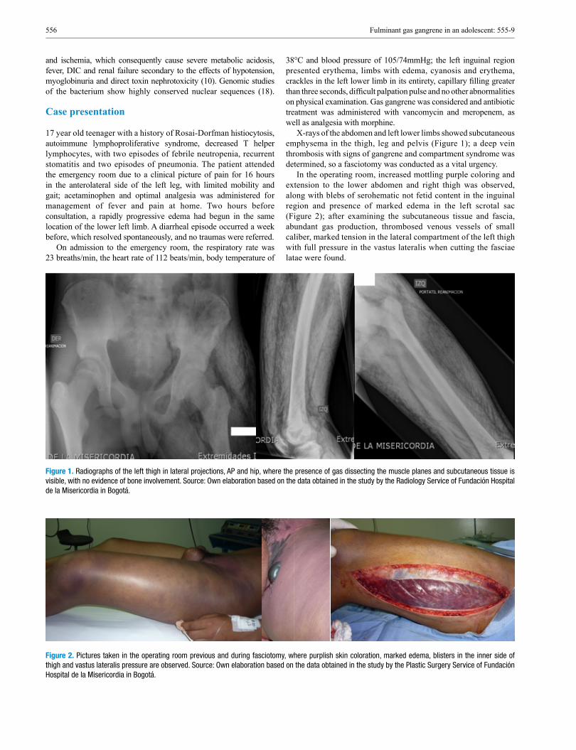
X-rays of the abdomen and left lower limbs showed subcutaneous emphysema in the thigh, leg and pelvis (Figure 1); a deep vein thrombosis with signs of gangrene and compartment syndrome was determined, so a fasciotomy was conducted as a vital urgency.
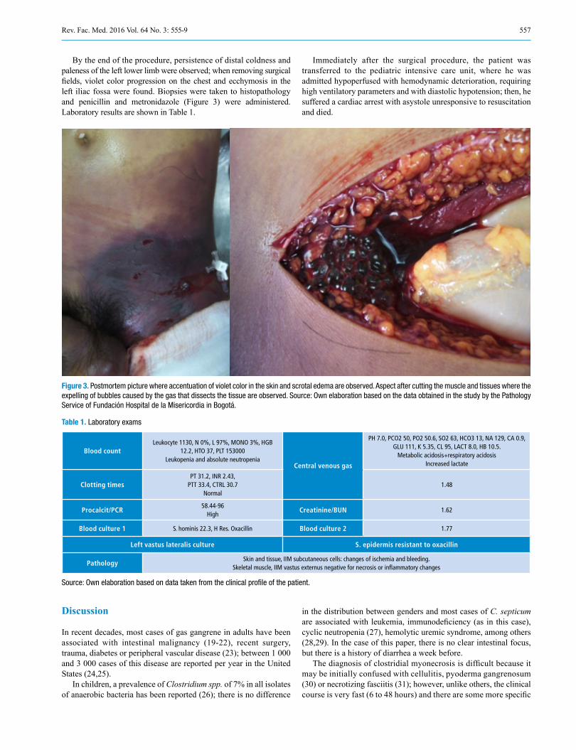
In the operating room, increased mottling purple coloring and extension to the lower abdomen and right thigh was observed, along with blebs of serohematic not fetid content in the inguinal region and presence of marked edema in the left scrotal sac (Figure 2); after examining the subcutaneous tissue and fascia, abundant gas production, thrombosed venous vessels of small caliber, marked tension in the lateral compartment of the left thigh with full pressure in the vastus lateralis when cutting the fasciae latae were found.
Figure 1. Radiographs of the left thigh in lateral projections, AP and hip, where the presence of gas dissecting the muscle planes and subcutaneous tissue is visible, with no evidence of bone involvement. Source: Own elaboration based on the data obtained in the study by the Radiology Service of Fundación Hospital de la Misericordia in Bogotá.
Figure 2. Pictures taken in the operating room previous and during fasciotomy, where purplish skin coloration, marked edema, blisters in the inner side of thigh and vastus lateralis pressure are observed. Source: Own elaboration based on the data obtained in the study by the Plastic Surgery Service of Fundación Hospital de la Misericordia in Bogotá.
557Rev. Fac. Med. 2016 Vol. 64 No. 3: 555-9
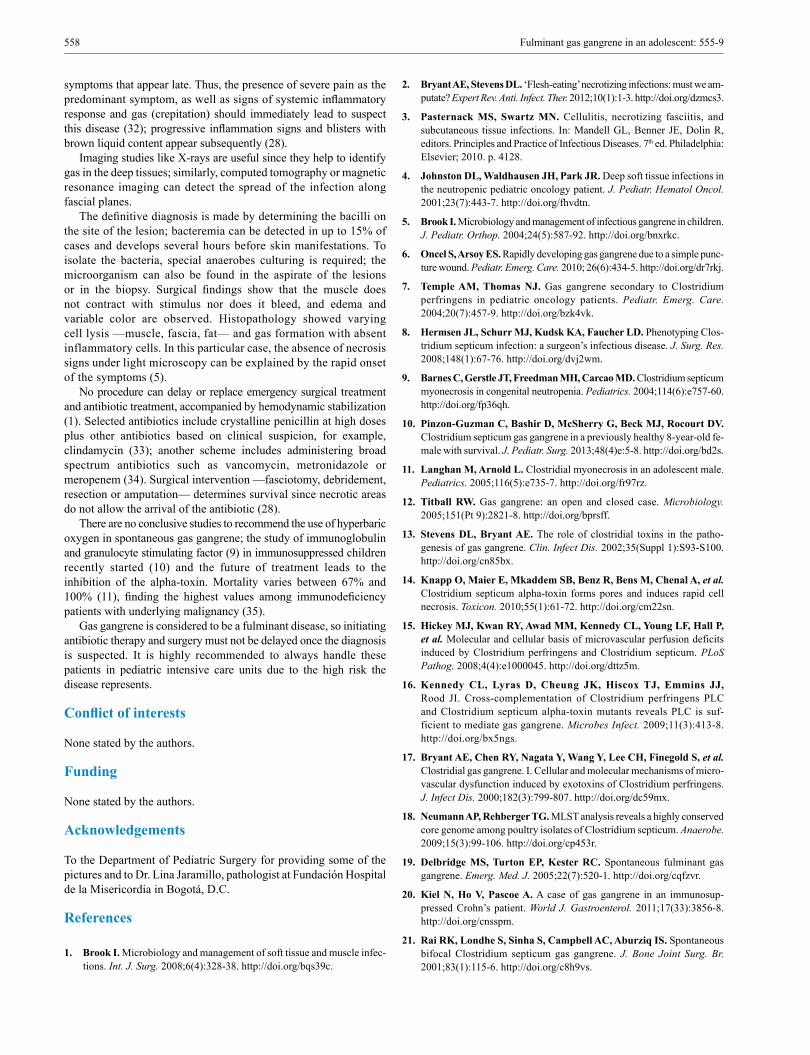
By the end of the procedure, persistence of distal coldness and paleness of the left lower limb were observed; when removing surgical fields, violet color progression on the chest and ecchymosis in the left iliac fossa were found. Biopsies were taken to histopathology and penicillin and metronidazole (Figure 3) were administered. Laboratory results are shown in Table 1.
Immediately after the surgical procedure, the patient was transferred to the pediatric intensive care unit, where he was admitted hypoperfused with hemodynamic deterioration, requiring high ventilatory parameters and with diastolic hypotension; then, he suffered a cardiac arrest with asystole unresponsive to resuscitation and died.
Figure 3. Postmortem picture where accentuation of violet color in the skin and scrotal edema are observed. Aspect after cutting the muscle and tissues where the expelling of bubbles caused by the gas that dissects the tissue are observed. Source: Own elaboration based on the data obtained in the study by the Pathology Service of Fundación Hospital de la Misericordia in Bogotá.
Table 1. Laboratory exams
Blood countLeukocyte 1130, N 0%, L 97%, MONO 3%, HGB
12.2, HTO 37, PLT 153000Leukopenia and absolute neutropenia
Central venous gas
PH 7.0, PCO2 50, PO2 50.6, SO2 63, HCO3 13, NA 129, CA 0.9, GLU 111, K 5.35, CL 95, LACT 8.0, HB 10.5.
Metabolic acidosis+respiratory acidosis Increased lactate
Clotting timesPT 31.2, INR 2.43,
PTT 33.4, CTRL 30.7Normal
1.48
Procalcit/PCR58.44-96
HighCreatinine/BUN 1.62
Blood culture 1 S. hominis 22.3, H Res. Oxacillin Blood culture 2 1.77
Left vastus lateralis culture S. epidermis resistant to oxacillin
PathologySkin and tissue, IIM subcutaneous cells: changes of ischemia and bleeding.
Skeletal muscle, IIM vastus externus negative for necrosis or inflammatory changes
Source: Own elaboration based on data taken from the clinical profile of the patient.
Discussion
In recent decades, most cases of gas gangrene in adults have been associated with intestinal malignancy (19-22), recent surgery, trauma, diabetes or peripheral vascular disease (23); between 1 000 and 3 000 cases of this disease are reported per year in the United States (24,25).
In children, a prevalence of Clostridium spp. of 7% in all isolates of anaerobic bacteria has been reported (26); there is no difference
in the distribution between genders and most cases of C. septicum are associated with leukemia, immunodeficiency (as in this case), cyclic neutropenia (27), hemolytic uremic syndrome, among others (28,29). In the case of this paper, there is no clear intestinal focus, but there is a history of diarrhea a week before.
The diagnosis of clostridial myonecrosis is difficult because it may be initially confused with cellulitis, pyoderma gangrenosum (30) or necrotizing fasciitis (31); however, unlike others, the clinical course is very fast (6 to 48 hours) and there are some more specific
558 Fulminant gas gangrene in an adolescent: 555-9
symptoms that appear late. Thus, the presence of severe pain as the predominant symptom, as well as signs of systemic inflammatory response and gas (crepitation) should immediately lead to suspect this disease (32); progressive inflammation signs and blisters with brown liquid content appear subsequently (28).
Imaging studies like X-rays are useful since they help to identify gas in the deep tissues; similarly, computed tomography or magnetic resonance imaging can detect the spread of the infection along fascial planes.
The definitive diagnosis is made by determining the bacilli on the site of the lesion; bacteremia can be detected in up to 15% of cases and develops several hours before skin manifestations. To isolate the bacteria, special anaerobes culturing is required; the microorganism can also be found in the aspirate of the lesions or in the biopsy. Surgical findings show that the muscle does not contract with stimulus nor does it bleed, and edema and variable color are observed. Histopathology showed varying cell lysis —muscle, fascia, fat— and gas formation with absent inflammatory cells. In this particular case, the absence of necrosis signs under light microscopy can be explained by the rapid onset of the symptoms (5).
No procedure can delay or replace emergency surgical treatment and antibiotic treatment, accompanied by hemodynamic stabilization (1). Selected antibiotics include crystalline penicillin at high doses plus other antibiotics based on clinical suspicion, for example, clindamycin (33); another scheme includes administering broad spectrum antibiotics such as vancomycin, metronidazole or meropenem (34). Surgical intervention —fasciotomy, debridement, resection or amputation— determines survival since necrotic areas do not allow the arrival of the antibiotic (28).
There are no conclusive studies to recommend the use of hyperbaric oxygen in spontaneous gas gangrene; the study of immunoglobulin and granulocyte stimulating factor (9) in immunosuppressed children recently started (10) and the future of treatment leads to the inhibition of the alpha-toxin. Mortality varies between 67% and 100% (11), finding the highest values among immunodeficiency patients with underlying malignancy (35).
Gas gangrene is considered to be a fulminant disease, so initiating antibiotic therapy and surgery must not be delayed once the diagnosis is suspected. It is highly recommended to always handle these patients in pediatric intensive care units due to the high risk the disease represents.
Conflict of interests
None stated by the authors.
Funding
None stated by the authors.
Acknowledgements
To the Department of Pediatric Surgery for providing some of the pictures and to Dr. Lina Jaramillo, pathologist at Fundación Hospital de la Misericordia in Bogotá, D.C.
References
1. Brook I. Microbiology and management of soft tissue and muscle infec-tions. Int. J. Surg. 2008;6(4):328-38. http://doi.org/bqs39c.
2. Bryant AE, Stevens DL. ‘Flesh-eating’ necrotizing infections: must we am-putate? Expert Rev. Anti. Infect. Ther. 2012;10(1):1-3. http://doi.org/dzmcs3.
3. Pasternack MS, Swartz MN. Cellulitis, necrotizing fasciitis, and subcutaneous tissue infections. In: Mandell GL, Benner JE, Dolin R, editors. Principles and Practice of Infectious Diseases. 7th ed. Philadelphia: Elsevier; 2010. p. 4128.
4. Johnston DL, Waldhausen JH, Park JR. Deep soft tissue infections in the neutropenic pediatric oncology patient. J. Pediatr. Hematol Oncol. 2001;23(7):443-7. http://doi.org/fhvdtn.
5. Brook I. Microbiology and management of infectious gangrene in children. J. Pediatr. Orthop. 2004;24(5):587-92. http://doi.org/bnxrkc.
6. Oncel S, Arsoy ES. Rapidly developing gas gangrene due to a simple punc-ture wound. Pediatr. Emerg. Care. 2010; 26(6):434-5. http://doi.org/dr7rkj.
7. Temple AM, Thomas NJ. Gas gangrene secondary to Clostridium perfringens in pediatric oncology patients. Pediatr. Emerg. Care. 2004;20(7):457-9. http://doi.org/bzk4vk.
8. Hermsen JL, Schurr MJ, Kudsk KA, Faucher LD. Phenotyping Clos-tridium septicum infection: a surgeon’s infectious disease. J. Surg. Res. 2008;148(1):67-76. http://doi.org/dvj2wm.
9. Barnes C, Gerstle JT, Freedman MH, Carcao MD. Clostridium septicum myonecrosis in congenital neutropenia. Pediatrics. 2004;114(6):e757-60. http://doi.org/fp36qh.
10. Pinzon-Guzman C, Bashir D, McSherry G, Beck MJ, Rocourt DV. Clostridium septicum gas gangrene in a previously healthy 8-year-old fe-male with survival. J. Pediatr. Surg. 2013;48(4)e:5-8. http://doi.org/bd2s.
11. Langhan M, Arnold L. Clostridial myonecrosis in an adolescent male. Pediatrics. 2005;116(5):e735-7. http://doi.org/fr97rz.
12. Titball RW. Gas gangrene: an open and closed case. Microbiology. 2005;151(Pt 9):2821-8. http://doi.org/bprsff.
13. Stevens DL, Bryant AE. The role of clostridial toxins in the patho-genesis of gas gangrene. Clin. Infect Dis. 2002;35(Suppl 1):S93-S100. http://doi.org/cn85bx.
14. Knapp O, Maier E, Mkaddem SB, Benz R, Bens M, Chenal A, et al. Clostridium septicum alpha-toxin forms pores and induces rapid cell necrosis. Toxicon. 2010;55(1):61-72. http://doi.org/cm22sn.
15. Hickey MJ, Kwan RY, Awad MM, Kennedy CL, Young LF, Hall P, et al. Molecular and cellular basis of microvascular perfusion deficits induced by Clostridium perfringens and Clostridium septicum. PLoS Pathog. 2008;4(4):e1000045. http://doi.org/dttz5m.
16. Kennedy CL, Lyras D, Cheung JK, Hiscox TJ, Emmins JJ, Rood JI. Cross-complementation of Clostridium perfringens PLC and Clostridium septicum alpha-toxin mutants reveals PLC is suf-ficient to mediate gas gangrene. Microbes Infect. 2009;11(3):413-8. http://doi.org/bx5ngs.
17. Bryant AE, Chen RY, Nagata Y, Wang Y, Lee CH, Finegold S, et al. Clostridial gas gangrene. I. Cellular and molecular mechanisms of micro-vascular dysfunction induced by exotoxins of Clostridium perfringens. J. Infect Dis. 2000;182(3):799-807. http://doi.org/dc59mx.
18. Neumann AP, Rehberger TG. MLST analysis reveals a highly conserved core genome among poultry isolates of Clostridium septicum. Anaerobe. 2009;15(3):99-106. http://doi.org/cp453r.
19. Delbridge MS, Turton EP, Kester RC. Spontaneous fulminant gas gangrene. Emerg. Med. J. 2005;22(7):520-1. http://doi.org/cqfzvr.
20. Kiel N, Ho V, Pascoe A. A case of gas gangrene in an immunosup-pressed Crohn’s patient. World J. Gastroenterol. 2011;17(33):3856-8. http://doi.org/cnsspm.
21. Rai RK, Londhe S, Sinha S, Campbell AC, Aburziq IS. Spontaneous bifocal Clostridium septicum gas gangrene. J. Bone Joint Surg. Br. 2001;83(1):115-6. http://doi.org/c8h9vs.
559Rev. Fac. Med. 2016 Vol. 64 No. 3: 555-9
22. Schade VL, Roukis TS, Haque M. Clostridium septicum necrotizing fasciitis of the forefoot secondary to adenocarcinoma of the colon: Case report and review of the literature. J. Foot Ankle Surg . 2010;49(2):159.e1-8. http://doi.org/bshxh7.
23. Dylewski J, Drummond R, Rowen J. A case of Clostridium septicum spontaneous gas gangrene. CJEM. 2007;9(2):133-5.
24. Abella BS, Kuchinic P, Hiraoka T, Howes DS. Atraumatic Clos-tridial myonecrosis: case report and literature review. J. Emerg. Med. 2003;24(4):401-5. http://doi.org/bn9pd5.
25. Rechner PM, Agger WA, Mruz K, Cogbill TH. Clinical features of clostridial bacteremia: a review from a rural area. Clin. Infect. Dis. 2001;33(3):349-53. http://doi.org/fjbscj.
26. Brook I. Clostridial infection in children. J. Med. Microbiol. 1995;42(2):78-82. http://doi.org/fbzr43.
27. Bar-Joseph G, Halberthal M, Sweed Y, Bialik V, Shoshani O, Etzioni A. Clostridium septicum infection in children with cyclic neutropenia. J. Pediatr. 1997;131(2):317-9. http://doi.org/cd2jp6.
28. Smith-Slatas CL, Bourque M, Salazar JC. Clostridium septicum in-fections in children: a case report and review of the literature. Pediatrics. 2006;117(4):e796-805. http://doi.org/fghgd2.
29. Elliott DC, Kufera JA, Myers RA. Necrotizing soft tissue infections. Risk factors for mortality and strategies for management. Ann. Surg. 1996;224(5):672-83. http://doi.org/b5457d.
30. Wangia MW, Mitchell CL, Wesson SK, Scott E, Glavin FL. Pyoderma gangrenosum or necrotizing fasciitis? A diagnostic conundrum. Case report and literature review. J. Pediatr. Surg. 2013;1(6):139-42. http://doi.org/bd2t.
31. Bingöl-Koloğlu M, Yildiz RV, Alper B, Yağmurlu A, Ciftçi E, Gökçora IH, et al. Necrotizing fasciitis in children: diagnostic and therapeutic aspects. J. Pediatr. Surg. 2007;42(11):1892-7. http://doi.org/cm46t7.
32. Schexnayder SM, Klein SG. Images in clinical medicine. Gas gangrene. N. Engl. J Med. 2004;350(25):2603. http://doi.org/dpw3nm.
33. Kuroda S, Okada Y, Mita M, Okamoto Y, Kato H, Ueyama S, et al. Fulminant massive gas gangrene caused by Clostridium perfringens. Intern. Med. 2005;44(5):499-502. http://doi.org/cv6rfx.
34. Chipp E, Phillips C, Rubin P. Successful management of spontaneous Clostridium septicum myonecrosis. J. Plast. Reconstr. Aesthet. Surg. 2009;62(10):e391-3. http://doi.org/bjr4kg.
35. Lehman TJ, Quinn MJ, Siegel SE, Ortega JA. Clostridium septicum infection in childhood leukemia: report of a case and review of the liter-ature. Cancer. 1977;40(2):950-3. http://doi.org/cnz8t2.