From Hopeless to Good Prognosis: Journey of a Failing...
5
53 Journal of International Oral Health 2015; 7(2):53-57 Interdisciplinary management of a failing tooth … Gupta S et al Case Report Received: 05 th August 2014 Accepted: 03 rd November 2014 Conflicts of Interest: None Source of Support: Nil From Hopeless to Good Prognosis: Journey of a Failing Tooth Saurabh Gupta 1 , Jeevanand Deshmukh 2 , Richa Khatri 3 , Vinaya Kumar Kulkarni 4 , B Karthik 5 Contributors: 1 Reader, Department of Periodontology, Rishi Raj College of Dental Sciences & Research Centre, Bhopal, Madhya Pradesh, India; 2 Professor & Head, Department of Periodontology, Rishi Raj College of Dental Sciences & Research Centre, Bhopal, Madhya Pradesh, India; 3 Postgraduate Student, Department of Periodontology, Rishi Raj College of Dental Sciences & Research Centre, Bhopal, Madhya Pradesh, India; 4 Professor, Department of Pedodontics, Modern Dental College & Research Centre, Indore, Madhya Pradesh, India; 5 Senoir Lecturer, Department of Oral Pathology, Rishi Raj College of Dental Sciences & Research Centre, Bhopal, Madhya Pradesh, India. Correspondence: Dr. Deshmukh J. Department of Periodontology, Rishi Raj College of Dental Sciences & Research Centre, Gandhinagar, Bhopal - 462 036, Madhya Pradesh, India. Phone: +91-9806088606. Email: [email protected] How to cite the article: Gupta S, Deshmukh J, Khatri R, Kulkarni VK, Karthik. From hopeless to good prognosis: Journey of a failing tooth. J Int Oral Health 2015;7(2):53-57. Abstract: Chronic periodontitis, along with associated clinical findings such as pathologic tooth migration, diastema, functional and aesthetic aberrations, poses an immense challenge to a dental professional. These findings convert clinical decision making into a daunting task and adversely affect the prognosis and the treatment plan for the presenting clinical problem. An interdisciplinary approach aimed at restoring functional and aesthetic needs of the affected individual within the limitations of such a compromised clinical scenario may be a viable alternative to any radical treatment causing loss of natural tooth structure such as extraction. This article reports the usefulness of the interdisciplinary route for managing an otherwise hopeless clinical situation of chronic periodontitis complicated with extreme mobility and pathologic tooth migration, which resulted in compromised function and aesthetics. Key Words: Chronic periodontitis, hydroxyapatite, pathologic tooth migration, platelet-rich fibrin, platelet-rich fibrin membrane Introduction Chronic periodontitis is one of the myriad challenges faced by a professional in dental practice. It presents as an infectious disease resulting in inflammation within the supporting tissues of the teeth, progressive attachment loss, and bone loss. 1 The predicament of the clinician is compounded by secondary factors coupled with chronic periodontitis such as pathologic tooth migration, 2 diastema, functional and aesthetic aberrations. These findings adversely affect the prognosis and the treatment planned and push it down from good or fair towards poor or hopeless, leading to an eventual loss of natural tooth structure. An interdisciplinary approach aimed at restoring functional and aesthetic needs of the affected individual within the limitations of the compromised clinical scenario may be a viable alternative to a radical treatment such as extraction. This article reports the usefulness of the interdisciplinary route for managing an otherwise hopeless clinical situation of chronic periodontitis complicated with extreme mobility and pathologic tooth migration, which resulted in compromised function and aesthetics. Case Report A 26-year old male individual came to visit the Department of Periodontology, Rishi Raj College of Dental Sciences, Bhopal with the chief complaint of loosening of a tooth in the front region of the upper arch, associated with swelling and bleeding from the gums since 1 year. He reported difficulty in chewing from the affected area, often associated with discomfort and less than acceptable frontal appearance. The individual was otherwise normal with no reported medical anomalies. Upon dental examination, oral cavity presented with poor oral hygiene (Figure 1a). There was an abundance of calculus and stains on the teeth, especially in the anterior region. There was generalized exudation present along with generalized erythematous gingiva. Tooth number 21 presented with erythematous and enlarged gingival tissue which was friable in nature, and there was extrusion along with Grade III mobility. A midline diastema was associated with tooth number 11 and 21. There were generalized periodontal pockets, with 21 presenting with a 10 mm deep periodontal pocket and overall anterior region appeared enlarged. Upon examination, 21 was found to be vital. Anterior deep bite was also evident. Considering the factors influencing individual tooth prognosis, tooth number 21 appeared to have a questionable to hopeless prognosis. A provisional diagnosis of chronic generalized periodontitis with inflammatory gingival enlargement in the anterior region was made. Upon investigation, an orthopantomograph (OPG) was taken, which confirmed the diagnosis of chronic generalized periodontitis. An intra-oral periapical radiograph (IOPA) was advised for tooth number 21 region, which subsequently revealed an extruded tooth (21) along with advanced bone loss in the interdental region, especially in
-
Upload
nguyendiep -
Category
Documents
-
view
213 -
download
0
Transcript of From Hopeless to Good Prognosis: Journey of a Failing...

53
Journal of International Oral Health 2015; 7(2):53-57Interdisciplinary management of a failing tooth … Gupta S et al
Case ReportReceived: 05th August 2014 Accepted: 03rd November 2014 Confl icts of Interest: None
Source of Support: Nil
From Hopeless to Good Prognosis: Journey of a Failing ToothSaurabh Gupta1, Jeevanand Deshmukh2, Richa Khatri3, Vinaya Kumar Kulkarni4, B Karthik5
Contributors:1Reader, Department of Periodontology, Rishi Raj College of Dental Sciences & Research Centre, Bhopal, Madhya Pradesh, India; 2Professor & Head, Department of Periodontology, Rishi Raj College of Dental Sciences & Research Centre, Bhopal, Madhya Pradesh, India; 3Postgraduate Student, Department of Periodontology, Rishi Raj College of Dental Sciences & Research Centre, Bhopal, Madhya Pradesh, India; 4Professor, Department of Pedodontics, Modern Dental College & Research Centre, Indore, Madhya Pradesh, India; 5Senoir Lecturer, Department of Oral Pathology, Rishi Raj College of Dental Sciences & Research Centre, Bhopal, Madhya Pradesh, India.Correspondence:Dr. Deshmukh J. Department of Periodontology, Rishi Raj College of Dental Sciences & Research Centre, Gandhinagar, Bhopal - 462 036, Madhya Pradesh, India. Phone: +91-9806088606. Email: [email protected] to cite the article:Gupta S, Deshmukh J, Khatri R, Kulkarni VK, Karthik. From hopeless to good prognosis: Journey of a failing tooth. J Int Oral Health 2015;7(2):53-57.Abstract:Chronic periodontitis, along with associated clinical fi ndings such as pathologic tooth migration, diastema, functional and aesthetic aberrations, poses an immense challenge to a dental professional. These fi ndings convert clinical decision making into a daunting task and adversely aff ect the prognosis and the treatment plan for the presenting clinical problem. An interdisciplinary approach aimed at restoring functional and aesthetic needs of the aff ected individual within the limitations of such a compromised clinical scenario may be a viable alternative to any radical treatment causing loss of natural tooth structure such as extraction. This article reports the usefulness of the interdisciplinary route for managing an otherwise hopeless clinical situation of chronic periodontitis complicated with extreme mobility and pathologic tooth migration, which resulted in compromised function and aesthetics.
Key Words: Chronic periodontitis, hydroxyapatite, pathologic tooth migration, platelet-rich fi brin, platelet-rich fi brin membrane
IntroductionChronic periodontitis is one of the myriad challenges faced by a professional in dental practice. It presents as an infectious disease resulting in inflammation within the supporting tissues of the teeth, progressive attachment loss, and bone loss.1 The predicament of the clinician is compounded by secondary factors coupled with chronic periodontitis such as pathologic tooth migration,2 diastema, functional and aesthetic aberrations. These fi ndings adversely aff ect the prognosis and the treatment planned and push it down from good or
fair towards poor or hopeless, leading to an eventual loss of natural tooth structure. An interdisciplinary approach aimed at restoring functional and aesthetic needs of the aff ected individual within the limitations of the compromised clinical scenario may be a viable alternative to a radical treatment such as extraction.
This article reports the usefulness of the interdisciplinary route for managing an otherwise hopeless clinical situation of chronic periodontitis complicated with extreme mobility and pathologic tooth migration, which resulted in compromised function and aesthetics.
Case ReportA 26-year old male individual came to visit the Department of Periodontology, Rishi Raj College of Dental Sciences, Bhopal with the chief complaint of loosening of a tooth in the front region of the upper arch, associated with swelling and bleeding from the gums since 1 year. He reported diffi culty in chewing from the aff ected area, often associated with discomfort and less than acceptable frontal appearance. The individual was otherwise normal with no reported medical anomalies.
Upon dental examination, oral cavity presented with poor oral hygiene (Figure 1a). There was an abundance of calculus and stains on the teeth, especially in the anterior region. There was generalized exudation present along with generalized erythematous gingiva. Tooth number 21 presented with erythematous and enlarged gingival tissue which was friable in nature, and there was extrusion along with Grade III mobility. A midline diastema was associated with tooth number 11 and 21. There were generalized periodontal pockets, with 21 presenting with a 10 mm deep periodontal pocket and overall anterior region appeared enlarged. Upon examination, 21 was found to be vital. Anterior deep bite was also evident. Considering the factors infl uencing individual tooth prognosis, tooth number 21 appeared to have a questionable to hopeless prognosis.
A provisional diagnosis of chronic generalized periodontitis with inflammatory gingival enlargement in the anterior region was made. Upon investigation, an orthopantomograph (OPG) was taken, which confi rmed the diagnosis of chronic generalized periodontitis. An intra-oral periapical radiograph (IOPA) was advised for tooth number 21 region, which subsequently revealed an extruded tooth (21) along with advanced bone loss in the interdental region, especially in

54
Journal of International Oral Health 2015; 7(2):53-57Interdisciplinary management of a failing tooth … Gupta S et al
the mesial interdental region where an angular defect could be appreciated (Figure 1b). There was buccal cortical bone dehiscence. However, distal interdental bone and palatal bony component were healthy. Relevant clinical parameters such as probing pocket depth and clinical attachment level were recorded. Routine blood investigations were performed, which did not reveal anything of relevance.
The prognosis for 21 was determined to be hopeless. Clinical and radiographic findings led to an initial treatment plan entailing full mouth fl ap surgery along with extraction of 21. The treatment plan was explained to the patient. However, the patient was insistent upon not sacrifi cing the tooth and desired every possible alternative for rehabilitation of the same. Eventually, the treatment plan was modifi ed, keeping in mind the patient’s need for rehabilitation without sacrifi cing the aff ected tooth, and the presenting clinical and radiographic evidence. The treatment plan included components of non-surgical therapy, regenerative periodontal surgery and subsequent aesthetic and functional rehabilitation, along with re-evaluation after every treatment phase.
On the fi rst visit to the department, phase I therapy was begun. A thorough scaling and root planing was performed, which was followed by explaining oral hygiene instructions to the patient. The patient was put on recall visits periodically (Figure 2). Upon stabilization of the periodontal condition and prior to regenerative periodontal surgery, an extra-coronal wire and composite splint was fabricated to manage the extreme mobility associated with 21.
Papilla preservation fl ap procedure was performed in relation to 11 and 21. Debridement was completed. Upon surgical exposure, a combined type of the osseous defect was evident in relation to 21 (Figure 3). The bone was intact on the palatal aspect. Root biomodifi cation was performed with tetracycline (500 mg capsule opened and mixed with 10 ml sterile water). Subsequently, hydroxyapatite containing bone graft (Sybograf- Eucare Pharmaceuticals) with particle size ranging between 600-700 μ was placed in the combined osseous defect (Figure 4a).
The platelet-rich fibrin (PRF) membrane was prepared according to the following protocol: 10 ml of intravenous blood was withdrawn from the antecubital fossa into a sterile tube via venipuncture. No anticoagulant was added to the tube, and it was immediately centrifuged at 3000 rpm for 10 min. It yielded a fi brin clot wedged in between the top layer of acellular plasma and the bottom layer of erythrocytes.3 The fi brin clot was subsequently separated using sterile tweezers and scissors and compressed with a glass slab to form a fl at membrane.
The bone graft was covered with the PRF membrane thus obtained (Figure 4b), fl ap sutured and a periodontal
Figure 2: Clinical view 2 weeks after scaling and root planing.
Figure 3: Papilla preservation fl ap refl ected and combined osseous defect in relation to mesiodistal of 21.
Figure 1: (a) Pre-operative view, (b) pre-operative intraoral periapical in relation to 21.
ba
Figure 4: (a) After bone graft placement, (b) platelet-rich fi brin membrane placed over the bone graft
ba
55
Journal of International Oral Health 2015; 7(2):53-57Interdisciplinary management of a failing tooth … Gupta S et al
dressing placed. Post-operative maintenance care included ibuprofen-twice a day for 3 days, amoxicillin-thrice daily for 5 days, soft diet for ≥2 weeks, to avoid anterior biting of food for 8 weeks, no brushing in surgical area for 2 weeks, no intrasulcular brushing for 8 weeks, and chlorhexidine 0.2% rinse for 2 weeks.
On recall visit after 10 days, periodontal pack and sutures were removed, and post-operative maintenance care was continued at regular intervals.
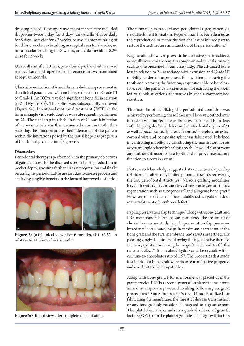
Clinical re-evaluation at 6 months revealed an improvement in the clinical parameters, with mobility reduced from Grade III to Grade I. An IOPA revealed signifi cant bone fi ll in relation to 21 (Figure 5b). The splint was subsequently removed (Figure 5a). Intentional root canal treatment (RCT) in the form of single visit endodontics was subsequently performed on 21. The fi nal step in rehabilitation of 21 was fabrication of a crown, which was then cemented onto the tooth, thus restoring the function and esthetic demands of the patient within the limitations posed by the initial hopeless prognosis of the clinical presentation (Figure 6).
DiscussionPeriodontal therapy is performed with the primary objectives of gaining access to the diseased sites, achieving reduction in pocket depth, arresting further disease progression and fi nally restoring the periodontal tissues lost due to disease process and achieving tangible benefi ts in the form of improved aesthetics.
The ultimate aim is to achieve periodontal regeneration via new attachment formation. Regeneration has been defi ned as the reproduction or reconstitution of a lost or injured part to restore the architecture and function of the periodontium.2
Regeneration, however, proves to be an elusive goal to achieve, especially when we encounter a compromised clinical situation such as one presented in our case study. The advanced bone loss in relation to 21, associated with extrusion and Grade III mobility rendered the prognosis for any attempt at saving the tooth and restoring the function, as questionable to hopeless. However, the patient’s insistence on not extracting the tooth led to a look at various alternatives in such a compromised situation.
The fi rst aim of stabilizing the periodontal condition was achieved by performing phase I therapy. However, orthodontic intrusion was not feasible as there was advanced bone loss with deep angular bone defect in the interdental region of 21 as well as buccal cortical plate dehiscence. Therefore, an extra-coronal wire and composite splint was fabricated. It helped in controlling mobility by distributing the masticatory forces across multiple relatively healthier teeth.4 It would also prevent any further extrusion of the tooth and improve masticatory function to a certain extent.4
Past research knowledge suggests that conventional open fl ap debridement off ers only limited potential towards recovering the lost periodontal structures.5 Various grafting modalities have, therefore, been employed for periodontal tissue regeneration such as autogenous6-7 and allogenic bone graft.8 However, none of them has been established as a gold standard in the treatment of intrabony defects.
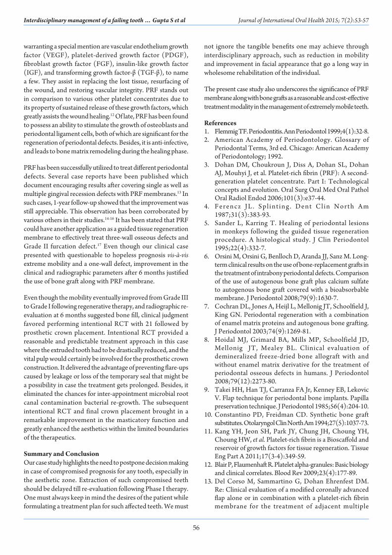
Papilla preservation fl ap technique9 along with bone graft and PRF membrane placement was considered the treatment of choice in our case study. Papilla preservation fl ap preserves interdental soft tissues, helps in maximum protection of the bone graft and the PRF membrane, and results in aesthetically pleasing gingival contours following the regenerative therapy. Hydroxyapatite containing bone graft was used to fill the osseous defect.10 It contained hydroxyapatite crystals with a calcium-to-phosphate ratio of 1.67. The properties that made it suitable as a bone graft were its osteoconductive property, and excellent tissue compatibility.
Along with bone graft, PRF membrane was placed over the graft particles. PRF is a second-generation platelet concentrate aimed at improving wound healing following surgical procedures.3 Since the patient’s own blood is utilized for fabricating the membrane, the threat of disease transmission or any foreign body reactions is negated to a great extent. The platelet-rich layer aids in a gradual release of growth factors (GFs) from the platelet granules.11 The growth factors Figure 6: Clinical view after complete rehabilitation.
Figure 5: (a) Clinical view after 6 months, (b) IOPA in relation to 21 taken after 6 months
ba
56
Journal of International Oral Health 2015; 7(2):53-57Interdisciplinary management of a failing tooth … Gupta S et al
warranting a special mention are vascular endothelium growth factor (VEGF), platelet-derived growth factor (PDGF), fi broblast growth factor (FGF), insulin-like growth factor (IGF), and transforming growth factor-β (TGF-β), to name a few. They assist in replacing the lost tissue, resurfacing of the wound, and restoring vascular integrity. PRF stands out in comparison to various other platelet concentrates due to its property of sustained release of these growth factors, which greatly assists the wound healing.12 Of late, PRF has been found to possess an ability to stimulate the growth of osteoblasts and periodontal ligament cells, both of which are signifi cant for the regeneration of periodontal defects. Besides, it is anti-infective, and leads to bone matrix remodeling during the healing phase.
PRF has been successfully utilized to treat diff erent periodontal defects. Several case reports have been published which document encouraging results after covering single as well as multiple gingival recession defects with PRF membranes.13 In such cases, 1-year follow-up showed that the improvement was still appreciable. This observation has been corroborated by various others in their studies.14-16 It has been stated that PRF could have another application as a guided tissue regeneration membrane to eff ectively treat three-wall osseous defects and Grade II furcation defect.17 Even though our clinical case presented with questionable to hopeless prognosis vis-à-vis extreme mobility and a one-wall defect, improvement in the clinical and radiographic parameters after 6 months justifi ed the use of bone graft along with PRF membrane.
Even though the mobility eventually improved from Grade III to Grade I following regenerative therapy, and radiographic re-evaluation at 6 months suggested bone fi ll, clinical judgment favored performing intentional RCT with 21 followed by prosthetic crown placement. Intentional RCT provided a reasonable and predictable treatment approach in this case where the extruded tooth had to be drastically reduced, and the vital pulp would certainly be involved for the prosthetic crown construction. It delivered the advantage of preventing fl are-ups caused by leakage or loss of the temporary seal that might be a possibility in case the treatment gets prolonged. Besides, it eliminated the chances for inter-appointment microbial root canal contamination bacterial re-growth. The subsequent intentional RCT and final crown placement brought in a remarkable improvement in the masticatory function and greatly enhanced the aesthetics within the limited boundaries of the therapeutics.
Summary and ConclusionOur case study highlights the need to postpone decision making in case of compromised prognosis for any tooth, especially in the aesthetic zone. Extraction of such compromised teeth should be delayed till re-evaluation following Phase I therapy. One must always keep in mind the desires of the patient while formulating a treatment plan for such aff ected teeth. We must
not ignore the tangible benefi ts one may achieve through interdisciplinary approach, such as reduction in mobility and improvement in facial appearance that go a long way in wholesome rehabilitation of the individual.
The present case study also underscores the signifi cance of PRF membrane along with bone grafts as a reasonable and cost-eff ective treatment modality in the management of extremely mobile teeth.
References1 . Flemmig TF. Periodontitis. Ann Periodontol 1999;4(1):32-8.2. American Academy of Periodontology. Glossary of
Periodontal Terms, 3rd ed. Chicago: American Academy of Periodontology; 1992.
3. Dohan DM, Choukroun J, Diss A, Dohan SL, Dohan AJ, Mouhyi J, et al. Platelet-rich fi brin (PRF): A second-generation platelet concentrate. Part I: Technological concepts and evolution. Oral Surg Oral Med Oral Pathol Oral Radiol Endod 2006;101(3):e37-44.
4. F e r e n c z J L . S p l i n t i n g . D e n t C l i n N o r t h A m 1987;31(3):383-93.
5. Sander L, Karring T. Healing of periodontal lesions in monkeys following the guided tissue regeneration procedure. A histological study. J Clin Periodontol 1995;22(4):332-7.
6. Orsini M, Orsini G, Benlloch D, Aranda JJ, Sanz M. Long-term clinical results on the use of bone-replacement grafts in the treatment of intrabony periodontal defects. Comparison of the use of autogenous bone graft plus calcium sulfate to autogenous bone graft covered with a bioabsorbable membrane. J Periodontol 2008;79(9):1630-7.
7. Cochran DL, Jones A, Heijl L, Mellonig JT, Schoolfi eld J, King GN. Periodontal regeneration with a combination of enamel matrix proteins and autogenous bone grafting. J Periodontol 2003;74(9):1269-81.
8. Hoidal MJ, Grimard BA, Mills MP, Schoolfield JD, Mellonig JT, Mealey BL. Clinical evaluation of demineralized freeze-dried bone allograft with and without enamel matrix derivative for the treatment of periodontal osseous defects in humans. J Periodontol 2008;79(12):2273-80.
9. Takei HH, Han TJ, Carranza FA Jr, Kenney EB, Lekovic V. Flap technique for periodontal bone implants. Papilla preservation technique. J Periodontol 1985;56(4):204-10.
10. Constantino PD, Freidman CD. Synthetic bone graft substitutes. Otolaryngol Clin North Am 1994;27(5):1037-73.
11. Kang YH, Jeon SH, Park JY, Chung JH, Choung YH, Choung HW, et al. Platelet-rich fi brin is a Bioscaff old and reservoir of growth factors for tissue regeneration. Tissue Eng Part A 2011;17(3-4):349-59.
12. Blair P, Flaumenhaft R. Platelet alpha-granules: Basic biology and clinical correlates. Blood Rev 2009;23(4):177-89.
13. Del Corso M, Sammartino G, Dohan Ehrenfest DM. Re: Clinical evaluation of a modifi ed coronally advanced fl ap alone or in combination with a platelet-rich fi brin membrane for the treatment of adjacent multiple

57
Journal of International Oral Health 2015; 7(2):53-57Interdisciplinary management of a failing tooth … Gupta S et al
gingival recessions: A 6-month study. J Periodontol 2009;80(11):1694-7.
14. Anilkumar K, Geetha A, Umasudhakar, Ramakrishnan T, Vijayalakshmi R, Pameela E. Platelet-rich-fi brin: A novel root coverage approach. J Indian Soc Periodontol 2009;13(1):50-4.
15. Shah M, Gujjari S, Gaekwad S, Dalal S. Double papilla fl ap with platelet rich fi brin in isolated gingival recession: A case
report. J Contemp Dent Sci 2012;2:36-40.16. Shah M, Gujjari S, Shah K, Patel V. Coronally advanced fl ap
with platelet rich fi brin: A novel root coverage approach. J Contemp Dent Sci 2013;2:40-4.
17. Sharma A, Pradeep AR. Treatment of 3-wall intrabony defects in patients with chronic periodontitis with autologous platelet-rich fi brin: A randomized controlled clinical trial. J Periodontol 2011;82(12):1705-12.