FRI BRIEFINGS BRIEFINGS Human Safety of Hormone Implants Used to Promote Growth in Cattle A Review...
24
FRI BRIEFINGS Food Research Institute, July 2000 Human Safety of Hormone Implants Used to Promote Growth in Cattle A Review of the Scientific Literature Ellin Doyle, Ph.D. Food Research Institute, University of Wisconsin Madison, WI 53706 TABLE OF CONTENTS Introduction and Historical Background ........................................... 2 Hormone Metabolism and Toxicity in Humans ................................ 4 Toxicity Studies in Animals and Cell Cultures ................................. 6 Genotoxicity Assays ........................................................ 6 Carcinogenicity Assays .................................................... 8 Other Toxicity Tests/Issues ............................................. 8 Hormone Concentrations in Cattle Tissues ....................................... 9 Estradiol ......................................................................... 10 Progesterone ................................................................... 11 Testosterone ................................................................... 11 Trenbolone ..................................................................... 11 Zeranol ........................................................................... 12 Assays for Determination of Hormone Levels ................................. 12 Hormone Concentrations in Other Foods ........................................ 14 References ....................................................................................... 15 Acknowledgments ........................................................................... 24 http://fri.wisc.edu/docs/pdf/hormone.pdf
Transcript of FRI BRIEFINGS BRIEFINGS Human Safety of Hormone Implants Used to Promote Growth in Cattle A Review...

FRI BRIEFINGS
������������� ������������������
Human Safety of Hormone ImplantsUsed to Promote Growth in Cattle
A Review of the Scientific Literature
Ellin Doyle, Ph.D.Food Research Institute, University of Wisconsin
Madison, WI 53706
TABLE OF CONTENTS
Introduction and Historical Background ........................................... 2Hormone Metabolism and Toxicity in Humans ................................ 4Toxicity Studies in Animals and Cell Cultures ................................. 6
Genotoxicity Assays ........................................................ 6Carcinogenicity Assays .................................................... 8Other Toxicity Tests/Issues ............................................. 8
Hormone Concentrations in Cattle Tissues ....................................... 9Estradiol ......................................................................... 10Progesterone................................................................... 11Testosterone ................................................................... 11Trenbolone ..................................................................... 11Zeranol ........................................................................... 12
Assays for Determination of Hormone Levels ................................. 12Hormone Concentrations in Other Foods ........................................ 14References ....................................................................................... 15Acknowledgments ........................................................................... 24
http://fri.wisc.edu/docs/pdf/hormone.pdf
������������� ������������������
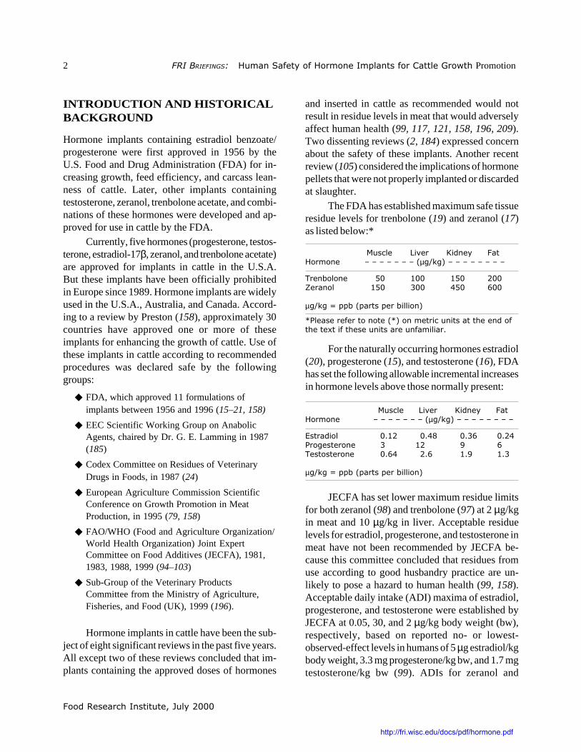
2 ������������������� ��������������� ������� �������������������Promotion
INTRODUCTION AND HISTORICALBACKGROUND
Hormone implants containing estradiol benzoate/progesterone were first approved in 1956 by theU.S. Food and Drug Administration (FDA) for in-creasing growth, feed efficiency, and carcass lean-ness of cattle. Later, other implants containingtestosterone, zeranol, trenbolone acetate, and combi-nations of these hormones were developed and ap-proved for use in cattle by the FDA.
Currently, five hormones (progesterone, testos-terone, estradiol-17β, zeranol, and trenbolone acetate)are approved for implants in cattle in the U.S.A.But these implants have been officially prohibitedin Europe since 1989. Hormone implants are widelyused in the U.S.A., Australia, and Canada. Accord-ing to a review by Preston (158), approximately 30countries have approved one or more of theseimplants for enhancing the growth of cattle. Use ofthese implants in cattle according to recommendedprocedures was declared safe by the followinggroups:
◆ FDA, which approved 11 formulations ofimplants between 1956 and 1996 (15–21, 158)
◆ EEC Scientific Working Group on AnabolicAgents, chaired by Dr. G. E. Lamming in 1987(185)
◆ Codex Committee on Residues of VeterinaryDrugs in Foods, in 1987 (24)
◆ European Agriculture Commission ScientificConference on Growth Promotion in MeatProduction, in 1995 (79, 158)
◆ FAO/WHO (Food and Agriculture Organization/World Health Organization) Joint ExpertCommittee on Food Additives (JECFA), 1981,1983, 1988, 1999 (94–103)
◆ Sub-Group of the Veterinary ProductsCommittee from the Ministry of Agriculture,Fisheries, and Food (UK), 1999 (196).
Hormone implants in cattle have been the sub-ject of eight significant reviews in the past five years.All except two of these reviews concluded that im-plants containing the approved doses of hormones
and inserted in cattle as recommended would notresult in residue levels in meat that would adverselyaffect human health (99, 117, 121, 158, 196, 209).Two dissenting reviews (2, 184) expressed concernabout the safety of these implants. Another recentreview (105) considered the implications of hormonepellets that were not properly implanted or discardedat slaughter.
The FDA has established maximum safe tissueresidue levels for trenbolone (19) and zeranol (17)as listed below:*
����� �!� "�� �� ������� � ���#�#�#�#�#�#�#�$µ%&'%(�#�#�#�#�#�#�#�#
)� *��� � +� ,�� ,+� ���-�� �� ,+� .�� /+� 0��
µ%&'%�1���*�$��������*����� (
23������������� ����$2(�� ������� ������������ ����������4������������ �������� �������5
For the naturally occurring hormones estradiol(20), progesterone (15), and testosterone (16), FDAhas set the following allowable incremental increasesin hormone levels above those normally present:
����� �!� "�� �� ������� � �#�#�#�#�#�#�#�$µ%&'%(�#�#�#�#�#�#�#�#
6������� �5,� �5/7 �5.0 �5�/3�%����� � . ,� 8 0)�������� � �50/ ��50 ,58 ,5.
µ%&'%�1���*�$��������*����� (
JECFA has set lower maximum residue limitsfor both zeranol (98) and trenbolone (97) at 2 µg/kgin meat and 10 µg/kg in liver. Acceptable residuelevels for estradiol, progesterone, and testosterone inmeat have not been recommended by JECFA be-cause this committee concluded that residues fromuse according to good husbandry practice are un-likely to pose a hazard to human health (99, 158).Acceptable daily intake (ADI) maxima of estradiol,progesterone, and testosterone were established byJECFA at 0.05, 30, and 2 µg/kg body weight (bw),respectively, based on reported no- or lowest-observed-effect levels in humans of 5 µg estradiol/kgbody weight, 3.3 mg progesterone/kg bw, and 1.7 mgtestosterone/kg bw (99). ADIs for zeranol and
http://fri.wisc.edu/docs/pdf/hormone.pdf
�������������������� ��������������� ������� �������������������3������ 3
������������� ������������������
trenbolone acetate were set at 0.5 and 0.02 µg/kg bwbased on feeding experiments with cynomolgusmonkeys (98) and pigs (103).
Safety of hormonal implants has been ques-tioned on the basis that residues from these implantsin beef may significantly increase exposure ofhumans, particularly children, to estrogens and otherhormones which may adversely affect health. Estro-gens are naturally present in all mammals, with higherconcentrations in females during the reproductivephase of their lives. According to evaluations by theInternational Agency for Research on Cancer (IARC;215), there is currently sufficient evidence for thecarcinogenicity of estradiol and limited evidence forthe carcinogenicity of testosterone to humans. Ex-periments with some of the other compounds used inimplants have shown that, under certain conditions,they may cause adverse effects in experimental ani-mals. Although some recent data indicate that estra-diol-17β has genotoxic potential, JECFA scientistspointed out that there are no data demonstrating thatconcentrations below the no-hormonal-effect level(NHEL) cause adverse effects in animals or humans(99).
Whether residues of hormone implants in beefconstitute a health risk remains a controversial issue.The 1999 JECFA analysis (99) estimated that aperson consuming 500 g (about 1.1 lb) of meat fromimplanted cattle would consume an extra 30–50 ngestradiol per day. This calculation utilized the highestresidue levels reported for implanted beef and consid-ered “meat” as a mixture of 300 g muscle, 100 g liver,50 g kidney, and 50 g fat. (Estradiol levels are higherin organ meats than in muscle.) This additional 50 ngestradiol consumed can be compared to the accept-able daily intake of 50 ng/kg/day or 3000 ng/day fora 60-kg (132-lb) person.
In 1999, the European Commission also issueda report that discussed available data on hormoneimplants and their potential toxicity and concludedthat elevated levels of hormones in meat from im-planted cattle might present a hazard, particularly tochildren (184). Invoking the precautionary principle,the Committee decided that beef from implanted cattleshould not be allowed in European markets. Theprecautionary principle is a risk management strategy
which “covers cases where scientific evidence isinsufficient, inconclusive, or uncertain and prelimi-nary scientific evaluation indicates that there arereasonable grounds for concern ...” (3). The con-cerns expressed by the Committee members include:
◆ Sensitivity of hormone assays. Theycontend that the routinely used radioimmuno-assay is not sensitive enough to accuratelymeasure low hormone levels in blood ofchildren and perhaps in some meat samples.Rather they propose that the very lowplasma hormone concentrations measured inchildren by the recombinant cell yeastbioassay (109) are more accurate.
◆ Effects of low concentrations of hormones.They argue that even though excess exposureto hormones in beef from implanted cattlemay amount to as little as 1 to severalhundred nanograms per person, pre-adolescent boys and girls and the fetusin utero may be very sensitive to slightincreases in hormone levels.
◆ Irrelevance of NHEL (no-hormonal-effectlevel). They point out that some recent dataindicate that estradiol is genotoxic andtherefore may have carcinogenic effects atconcentrations below the NHEL.
In response, the 1999 review by the UK’s Sub-Group of the Veterinary Products Committee (196)critically evaluated genotoxicity studies of estradioland concluded that “None of the publications (re-search papers) reviewed above provide any substan-tive evidence that oestradiol is mutagenic/genotoxic.”The recombinant yeast cell bioassay was criticallyevaluated and this Committee was concerned thatinterfering substances in the crude extracts of serumsamples could have caused artificially low serumestradiol measurements. Further validation is re-quired for this assay. Although high concentrationsof hormones can certainly disrupt normal metabolicprocesses, this Sub-group concluded that small in-creases in hormone intakes from implanted cattlewere unlikely to have a significant effect.
In a search of several databases, includingCurrent Contents, Medline, Food Science and
http://fri.wisc.edu/docs/pdf/hormone.pdf

������������� ������������������
4 ������������������� ��������������� ������� �������������������Promotion
Technology, and Agricola, and of government websites, a total of 217 references relevant to the issueof safety of hormone implants in cattle were identi-fied and collected. Although the topic of interest is,strictly speaking, whether implants used in cattleleave hormonal residues that present a hazard tohuman consumers of beef, there are few studies inhumans, and therefore regulators and consumersmust rely on:
◆ toxicity data from any human exposures,including effects of natural hormones;
◆ data from animal and cell culture research toindicate the possible toxicity of these com-pounds to humans;
◆ reports of hormone levels in cattle tissues,both endogenous (naturally occurring)hormones and those which originate fromimplants;
◆ information on total dietary exposure tocompounds with hormonal activity, includinghormones in meat from unimplanted as wellas implanted cattle and hormones in otherfoods, in order to put the hormone concen-trations in different foods in context of thetotal diet; and
◆ reports on the sensitivity of assays used todetermine hormone levels and assess potentialhuman exposure.
Information from relevant references on each ofthese topics is summarized in the following sections.Please refer to note (*) on metric units at the end ofthe text if these units are unfamiliar.
HORMONE METABOLISM ANDTOXICITY IN HUMANS
Estradiol, progesterone, and testosterone are natu-rally present in humans although the amounts varywith age, sex, diet, exercise, and, in females, withpregnancy and stage of the menstrual cycle. Thesecompounds may be present in serum as unbound“free” compounds or attached to hormone bindingproteins. Estradiol 17β is present in high levels innewborn males and females but concentrations drop
rapidly after birth so that young children of bothsexes have very low concentrations of this com-pound. In adult males, estradiol concentrations arein the range of 20–40 pg/ml, and most of this is in thebound form (161, 173). Estradiol levels in femalesvary between 40 and 400 pg/ml during the month(155). Some women are genetically predisposedto have higher endogenous hormone levels (67).After menopause estradiol concentrations decreaseto 5–20 pg/ml (155).
Children and the fetus in utero have been con-sidered at greater risk from exposure to hormonesbecause their normal physiological hormone levelsare low compared to adults. Results from one recentstudy using a radio-immunoassay indicated that es-tradiol levels in prepubertal boys and girls were 2.6and 4.5 pg/ml, respectively (154). However, using arecombinant yeast assay, other researchers reportedlevels of 0.6 pg/ml in girls and 0.08 pg/ml in boys(109). The yeast estrogen receptor binding assayappears to be more sensitive than the immunoassay;however, these very low concentrations have beenquestioned because the solvent system used to ex-tract hormones prior to assay is not very efficient(11). Inter- and intra-assay variation at these verylow concentrations of estradiol was reported to be50–60% (109).
Oral estrogens generally have poor bio-availability due to extensive metabolism afterabsorption from the gut. Only 0.1–12% of anoral micronized estradiol dose was found to bebioavailable. Hormone replacement therapy witha daily dose of 0.625 mg conjugated estrogensproduces serum estradiol levels of approximately40 pg/ml in postmenopausal women (143). Oraltestosterone and progesterone are also reported tohave low bioavailability due to gastrointestinal andhepatic inactivation.
Dietary constituents (other than hormones them-selves) are well known for influencing serum hor-mone concentrations. For example, total estradiollevels were significantly lower when women con-sumed a low fat (20%), high fiber (40 g/day) diet ascompared to a more typical high fat (40%), low fiber(12 g/day) diet (214). Caloric intake was directlycorrelated with serum progesterone levels in another
http://fri.wisc.edu/docs/pdf/hormone.pdf

�������������������� ��������������� ������� �������������������3������ 5
������������� ������������������
study of premenopausal women (35). When maleathletes consumed diets high in animal protein, totaltestosterone levels were significantly higher and es-tradiol levels were lower than when they consumedan isocaloric vegetarian diet with an equivalentamount of protein (161). Serum testosterone concen-trations were also directly associated with intake ofdietary fat and of saturated fats (207).
Although hormones are essential for variousphysiological processes in the body, excessiveamounts may have adverse effects. The most contro-versial and well documented of these is estradiol.Estradiol stimulates cell division in hormonally sen-sitive tissues thereby increasing the possibility foraccumulation of random errors during DNA duplica-tion. This increased cell proliferation also has theeffect of stimulating growth of mutant cells (67).Among post-menopausal women using hormone re-placement therapy, there appears to be an increasedincidence of breast cancer. Two recent references onepidemiological studies are included here as an intro-duction to the voluminous literature on this subject(171, 179). Results from epidemiological studiesand experiments with laboratory animals were con-sidered by IARC to be sufficient evidence for thecarcinogenicity of estradiol (215).
However, only a small proportion of post-menopausal women taking estrogen supplementsdevelop cancer so there are other factors necessaryfor the development of this disease. A recent reviewon hormonal carcinogenesis concluded that geneticsusceptibility plays a critical role in the developmentof hormone-related cancers (67).
Some research has also investigated the pos-sible influence of consuming estrogens and proges-terone in contraceptives or hormone replacementtherapy on the incidence or severity of asthma (48).Data reported from a variety of small studies wasinconsistent: In some cases the hormones appeared tomake asthma worse, whereas in other cases takinghormones appeared to improve asthmatic symptoms.Progesterone is also prescribed for some medicalconditions and usually has few serious side effectsalthough minor symptoms sometimes occur (211).
Some epidemiological studies have also founda link between prostate cancer and higher testoster-
one levels in males. Prostate cancer is rare before age40, but the increasing incidence of this cancer withage is much greater for some national populationsand ethnic groups with higher serum testosteroneconcentrations than for others. No environmental orlifestyle risk factors have been identified to explainthis difference. Rather, higher endogenous testoster-one levels along with a genetic susceptibility appearto determine development of prostate cancer (67,215). Elevated levels of testosterone have not beenassociated with any other type of cancer in humans.
Since trenbolone and zeranol are not naturallyoccurring compounds in humans there has been moreconcern about their possible toxicity. In a single trial,a dose of radioactively labeled trenbolone was givento a human volunteer. Results demonstrated that63% of the radioactivity was excreted in the urineduring the first 72 hours, with over half of theradioactivity present in glucuronides (191). This isin contrast to the primarily biliary and subsequentfecal excretion observed in the rat and the cow (156).
Some data on human exposure to zeranol areavailable from a study of workers in a pelletizingplant (6). Analyses of blood samples from exposedworkers showed no detectable residues of zeranoland no significant elevation or depression of serumconcentrations of estradiol or other hormones. It wasestimated that workers were exposed to airbornezeranol concentrations of 86–1554 µg/m3. Zeranolwas also detected in swabs from the lunchroom andon work clothes. Symptoms of breast irritation werereported by some workers but no other acute orchronic effects were evident. Young children of someworkers in the plant had also experienced breastsymptoms and this was traced to employees wearingtheir work clothes home.
Zeranol has been used in the past in Europe asan estrogen substitute for postmenopausal womenand has been shown to affect the activity of someenzymes in human prostate cell cultures (198).Human volunteers given a radioactively labeled doseof zeranol absorbed at least 55% of the dose and thenexcreted metabolites and glucuronides of this com-pound primarily in urine whereas dogs, monkeys,and rats excrete radioactive metabolites predomi-nantly in bile and feces (137).
http://fri.wisc.edu/docs/pdf/hormone.pdf

������������� ������������������
6 ������������������� ��������������� ������� �������������������Promotion
Two outbreaks, one of breast enlargement inyoung school children in Italy (45) and another ofprecocious sexual development in Puerto Rico (176),were suggestive of exposure to environmental estro-genic compounds, possibly zeranol. In the case ofthe Italian children, symptoms disappeared entirelyafter 8 months and researchers suspected that oneconsignment of meat or poultry might have containedhigh levels of some estrogenic compound. In thecase of the Puerto Rican children, high levels ofestrogenic compounds were said to be present insome local chicken and it was reported thatsymptoms gradually disappeared after the childrenstopped consuming local chicken, beef, and milk.However, subsequent investigation of the PuertoRican outbreak by the Centers for Disease Control(CDC), USDA, and FDA indicated that the outbreakcould have been the result of increased awarenessand reporting of cases by physicians (209).
Summary. Estradiol, progesterone, and testosteroneare hormones naturally present in humans at differ-ent concentrations depending on age, sex, diet,exercise, and stage in the reproductive cycle. Inadult males and premenopausal females, estradiolconcentrations are in the range of 20–40 pg/ml and40 and 400 pg/ml, respectively. After menopauseestradiol concentrations decrease to 5–20 pg/ml.Estradiol levels have been measured by radio-immunoassay in prepubertal boys and girls at2.6 and 4.5 pg/ml, respectively. A recombinant yeastassay detected only 0.6 pg/ml in girls and 0.08 pg/mlin boys.
Pharmacological studies have revealed that oraldoses of estradiol have low bioavailability — usually12% or less. Diets high in fat, calories, and animalprotein and low in fiber have been associated withhigher estradiol, progesterone, or testosterone levelsin several studies.
Epidemiological and experimental data exam-ined by International Agency for Research on Can-cer were judged to be sufficient evidence for thecarcinogenicity of estradiol to humans. Estradiolstimulates cell division thereby increasing the possi-bility for random errors during DNA duplication. Asmall proportion of women taking estrogen supple-
ments develop breast or uterine cancer, indicatingthat estradiol may be one of several factors importantin the development of cancer. Epidemiological stud-ies have also found a link between prostate cancerand higher testosterone levels in males.
Few human studies of the metabolism oftrenbolone and zeranol are available. In humantrials, radioactive doses of both compounds wereprimarily excreted in the urine. Zeranol has beenused in the past in Europe as an estrogen substitutefor postmenopausal women. Occupational exposureto zeranol has produced symptoms of breast irrita-tion but no other acute or chronic effects.
Two outbreaks, one of breast enlargement inyoung school children in Italy and another of preco-cious sexual development in Puerto Rico, were sug-gestive of exposure to environmental estrogeniccompounds, possibly zeranol. However, subsequentinvestigation of these outbreaks revealed no definiteevidence of exposure to estrogenic compounds infoods.
TOXICITY STUDIES IN ANIMALSAND CELL CULTURES
Genotoxicity assays
These assays assess damage to DNA and chromo-somes which can lead to mutations and heritablechanges in genetic information. Not all DNA changesare mutagenic because cells have efficient DNArepair systems. It is generally agreed that both invivo and in vitro assays should be used to determinethe genotoxicity of a compound. Many assays testhormone concentrations that are many-fold higherthan normal physiological concentrations, and onemust make a judgment as to their relevancy. Also,these hormones are metabolized to different com-pounds in the body and these metabolites may havegreater, lesser, or the same toxicity as the parentcompound.
17β-Estradiol
Negative results were obtained for estradiol inthe Ames test (bacterial mutagenicity), in the in vitro
http://fri.wisc.edu/docs/pdf/hormone.pdf

�������������������� ��������������� ������� �������������������3������ 7
������������� ������������������
hamster cell micronucleus assay for chromosomaldamage (167), in the in vivo mouse lymphoma for-ward mutation assay (89, 147, 167), and in the invivo male rat bone marrow germ cell assay (167).Estradiol was found to bind covalently to rat liverDNA, but bound residues were removed by repairmechanisms (10). Syrian hamsters dosed with estra-diol develop kidney tumors, and some changes inmicrosatellite DNA in kidney cells were observed intreated animals (74).
Other in vitro genotoxicity assays have yieldedpositive results but interpretation was not alwaysclear cut. In some cases changes induced were notstable or occurred at a low frequency so that theirsignificance was questionable. Estradiol inducedmethotrexate resistance in breast cancer cells in vitrobut when methotrexate was withdrawn, resistancewas lost, indicating that the change was not inherited(197). One report indicating that estradiol causedmutations in an in vitro hamster cell assay actuallyobserved only 5 mutants out of 2,000,000 cellscompared to 2 mutants in 2,000,000 cells of thecontrol (162). This small difference between experi-mental and control cultures makes any conclusionsquestionable. Other reports have demonstrated thatestradiol can cause oxidative DNA damage to calfthymus DNA (138) and induce transformation andaneuploidy (alteration of chromosome number) inhamster embryo cells in vitro (201). Both ofthese effects occurred at estradiol concentrationsseveral orders of magnitude above normal physi-ological concentrations of this hormone. Endogenousantioxidants were also observed to drastically de-crease oxidative damage to calf thymus DNA.Therefore, these genotoxic effects may not occurunder normal physiological conditions in vivo.
In the presence of 25 µg estradiol/ml (but notof 10 µg/ml), chromosomal aberrations and sisterchromatid exchanges were observed in humanblood peripheral lymphocytes (1). (Estradiol levelsin human female serum normally range up to400 pg/ml.)
Results from numerous genotoxicity assays havebeen summarized and discussed in recent publica-tions (114, 132, 184, 196). Overall it appears thatestradiol has a weak potential for genotoxic effects.
ProgesteroneNegative results were obtained for progester-
one in the Ames test (89) and in tests for inductionof chromosomal aberrations and aneuploidy in cul-tured Syrian hamster embryo cells (200). A highoral dose of 100 mg progesterone/kg body weightdid induce the formation of micronuclei in rat livercells (123).
TestosteroneNegative results were obtained for testosterone
in the Ames test (89), in the in vivo mouse lymphomaforward mutation assay (167), and in tests for theinduction of chromosomal aberrations and aneu-ploidy in cultured Syrian hamster embryo cells (181,200). Testosterone was found to bind covalently torat liver DNA but bound residues were removed byrepair mechanisms (9). Testosterone exhibited a weaktransforming effect on Syrian hamster embryo cellsin culture according to some reports (112, 200) butnot in another (182).
TrenboloneAlthough one research group reported positive
results for trenbolone in the Ames bacterial mutage-nicity assay (119), another scientist was unable toduplicate these results (124) and several other re-ports indicated that trenbolone is not an effectivemutagen in the Ames test (89, 125, 167, 182). Nega-tive results were also obtained in the SOS bacterialchromotest, the hamster V79 sister chromatid ex-change test, the rec bacterial assay (180), the test forunscheduled DNA synthesis (182), and the test forinduction of chromosomal aberrations and aneu-ploidy in cultured Syrian hamster embryo cells (167,200).
Trenbolone was found to bind covalently to ratliver DNA but bound residues were removed byrepair mechanisms (10, 119, 152, 153). Trenboloneexhibited a weak to moderate transforming effecton Syrian hamster embryo cells in culture (112, 125,182, 200) and did induce micronucleus formationin Syrian hamster embryo fibroblasts but not inmouse cells (167, 181). Richold (167), Lone (117),the European Commission (184), FDA (18, 21), andJECFA (101, 103) summarized much of the research
http://fri.wisc.edu/docs/pdf/hormone.pdf
������������� ������������������
8 ������������������� ��������������� ������� �������������������Promotion
done to assess the genotoxicity of trenbolone andconcluded that the preponderance of negative testsindicated that this compound does not cause DNAdamage or mutations.
Zeranol
Zeranol was found to bind covalently to ratliver DNA at much lower levels than estradiol, andbound residues were removed by repair mechanisms(10). Negative results were obtained for zeranol inthe Ames test (89), in the mouse bone marrow cyto-genetic assay, and in the in vivo mouse lymphomaforward mutation assay (147). Negative results werealso obtained in the SOS chromotest and the V79sister chromatid exchange test but results were posi-tive in the rec-assay (180). Zeranol also did notinduce micronuclei in mouse spermatids in vivo orin vitro (160). Nearly all the assays listed in theJECFA Toxicology report (100) and the review byLone (117) indicate that zeranol is not genotoxic andnone were positive for mutagenicity.
Carcinogenicity Tests
The carcinogenicity of estradiol and of testosteronein rodents is related to binding of the hormones toreceptors on the surface of cells in reproductiveorgans and stimulating growth. Testosterone cancause cervical-uterine tumors in female rats andprostate cancer in males but does not appear to becarcinogenic in other organs (67, 215). Estradiol iscarcinogenic to rodents and primarily affects repro-ductive organs. However, an increased incidence ofbone, pituitary, and lymphoid tumors has also ac-companied high doses of estradiol in some rodents(184, 215). There have been some reports that highdoses of progesterone administered over a long timeare correlated with an increase in tumor growth inanimals (117).
A great deal of research has demonstratedthat low estradiol concentrations, where no normalphysiological effects of the hormone are observed,do not promote tumor growth. This low hormoneconcentration has been called the no-hormonal-effectlevel (NHEL) or the no-observed-effect level(NOEL). In dose–response studies with women, the
NOEL was determined to be 5000 nanograms (ng)(59, 67).
Estimates of the estrogenic potency of zeranol(oral dose) indicate that it is 150 times less potentthan estradiol in rats (42, 199) and no estrogeniceffects were observed in male cynomolgus monkeysdosed with 5 mg zeranol/kg body weight/day (100).The hormone no-effect levels in ovariectomizedfemale rhesus and cynomolgus monkeys were deter-mined to be 0.225 and 0.05 mg/kg/day, respectively(8, 100, 147). Zeranol binds to rat hepatic estrogenreceptors with about 30% of the affinity exhibited byestradiol (157) and is capable of eliciting estrogeniceffects in liver in vivo (127). When injected at highconcentrations (36 mg), zeranol induced hepatictumors in hamsters; this effect was inhibited bytamoxifen, indicating that estrogen receptors wereinvolved in tumorigenesis (23). According to reportsof unpublished carcinogenicity studies in rats,dietary zeranol (1.25 or 0.8 mg/kg/day) administeredover a 3-year period did not enhance tumor risk atany site although estrogenic effects were observedin the rats (100, 170). Long-term carcinogenicitystudies involving oral administration of zeranol torats (2 yr), dogs (7 yr), and rhesus monkeys (10 yr)demonstrated no significant increase in tumors inany of the treated groups (8).
Data from unpublished carcinogenicity studiesof trenbolone in rodents indicate that 10 or 100 ppm(µg/g) trenbolone in the diet was associated with anincrease in liver cancer in mice and 50 ppm trenbolonemay have enhanced pancreatic cancer in rats (170).The no-hormonal-effect level was estimated to beat or near 0.5 µg/g for both rodent species. Datafrom castrated rhesus monkeys indicated that theno-hormonal-effect level for oral trenbolone wasbetween 2 and 50 µg/kg/day (73).
Other Toxicity Tests/Issues
Steroid hormones are active during the prenatalperiod and affect the programming and timing ofcritical events such as birth, puberty, growth rates,and sexual differentiation, including the differentia-tion of estrogen binding proteins (2, 56, 184, 189,196).
http://fri.wisc.edu/docs/pdf/hormone.pdf

�������������������� ��������������� ������� �������������������3������ 9
������������� ������������������
Exposure of pregnant rats to 4 mg zeranol/kgbody weight/day depressed maternal weight gain,prolonged gestation period, and decreased numberof live births in some animals (163, 164). However,zeranol did not appear to be teratogenic (cause birthdefects) (187, 210). Zeranol administered to miceduring pregnancy also decreased fetal size (150) andcaused some abnormalities in testicular development(149, 151, 160).
Estrogens are also known to affect functioningof the immune system. Progesterone and zeranol alsoaffect immune function to an extent related to theirestrogenicity (118).
Summary. Genotoxicity assays assess damage toDNA and chromosomes which can lead to mutationsand heritable changes in genetic information. Bothin vivo and in vitro assays are used to determine thegenotoxicity of a compound. Negative results wereobtained for estradiol, progesterone, testosterone,trenbolone, and zeranol in the Ames test of bacterialmutagenicity and in numerous other in vitro andin vivo tests for genotoxicity. These hormones didbind to DNA molecules but were later removed byrepair enzymes, and a few assays with estradiol andtrenbolone gave weak positive results. Assay resultsfor the other compounds indicated a lack ofgenotoxicity. Some scientists believe that the weaklypositive results in some tests with estradiol indicatethat it is potentially genotoxic to humans whileothers believe that these results indicate that estradiolis not genotoxic at reasonable concentrations undernormal physiological conditions.
The carcinogenicity of estradiol and of testos-terone in rodents is related to binding of the hor-mones to receptors on the surface of cells inreproductive organs. Testosterone and estradiol arecarcinogenic to rodents and primarily affect repro-ductive organs. An increased incidence of bone,pituitary, and lymphoid tumors has also accompa-nied high doses of estradiol in some rodents. Proges-terone has increased carcinogenesis in reproductivetissues in some animal studies. Long-term carcinoge-nicity studies involving oral administration of zeranolto rats (2 yr), dogs (7 yr), and rhesus monkeys (10 yr)demonstrated no significant increase in tumors in
any of the treated groups. Some unpublished carci-nogenicity studies of trenbolone in rodents indicatethat 10 or 100 ppm (µg/g) trenbolone in the diet wasassociated with an increase in liver cancer in miceand 50 ppm trenbolone may have enhanced pancre-atic cancer in rats.
A great deal of research has demonstrated thatlow estradiol concentrations, where no normal physi-ological effects of the hormone are observed, do notpromote tumor growth. This low hormone concen-tration has been called the no-observed-effect level(NOEL). In dose–response studies, the NOEL forestradiol is 5 µg/kg body weight in women, fortestosterone is 1.7 mg/kg bw in men, for zeranol is0.05 mg/kg/day for cynomolgus monkeys, and fortrenbolone is between 2 and 50 µg/kg/day for cas-trated rhesus monkeys.
Steroid hormones are active during the prenatalperiod and affect the programming and timing ofcritical events such as birth, puberty, growth ratesand sexual differentiation, including the differentia-tion of estrogen binding proteins. Exposure of preg-nant rats to 4 mg zeranol/kg body weight/daydepressed maternal weight gain but did not causebirth defects. Zeranol administered to mice duringpregnancy caused some abnormalities in testiculardevelopment in rats.
Estrogens are also known to affect functioningof the immune system. Progesterone and zeranol alsoaffect immune function to an extent related to theirestrogenicity.
HORMONE LEVELS IN CATTLETISSUES
Numerous research papers have reported data onhormone levels measured in tissues from untreatedand implanted cattle. Results are summarized belowfor cattle with and without hormone implants. Whenconsidering these data, one should be mindful ofvariations in sensitivity of different assays.
Endogenous (naturally occurring) hormonelevels in tissues vary with the sex (61), age (5, 26, 50,141), and breed (26) of cattle and whether the ani-mals have been castrated or are pregnant (66). Fat
http://fri.wisc.edu/docs/pdf/hormone.pdf

������������� ������������������
10 ������������������� ��������������� ������� �������������������Promotion
content also affects the levels of hormones in meat(52), and concentrations of some hormones are sig-nificantly higher in liver and kidney than in muscletissue.
Published results of an extensive analysis forchemical residues in beef from cattle slaughtered inthe U.S.A. in 1990 revealed that neither zeranol nortrenbolone was present in detectable levels (190).Another survey of beef in Ireland also demonstratedthat residue levels of zeranol and trenbolone were<0.05 µg/kg in all samples (144). However, off-labeluse of trenbolone has been detected from residues inliver of veal calves in Canada (105).
Concentrations of various hormones have beenmeasured in plasma, urine, muscle, kidney, liver, andfat of cattle that have received hormone implants.Most of these papers are included in the bibliogra-phy. Several review articles (121, 158, 178, 209)have also compiled data from a number of articleson hormone residue levels in cattle. Most of thesources cited in these reviews are included in thisreport but there are a few articles which could not beobtained.
Further data on hormone levels in tissues otherthan meat may be found in these references forestradiol (58, 63, 64, 68, 113, 116, 134, 139, 174,183); testosterone (58, 86, 87, 104, 113, 134, 186);trenbolone 63, 64, 68, 86, 90, 116, 183); zeranol(107, 159). Some articles in peer-reviewed journalslist hormone concentrations in meat. Additionaldata from unpublished research have been printedin reports by WHO/FAO (94–98, 102) and other
regulatory agencies. Some of these reports also dis-cuss metabolites of the hormones which may bepresent in cattle.
Estradiol
Calves: Natural estradiol levels are low, closeto the limits of detection, with maximum reportedlevels of 5–7 ng/kg in muscle (94) and 0.02 ng/ml inserum and 0.2 ng/ml in urine (5).
Bulls: Estradiol levels have been measured as5.8 ng/kg in muscle (94), 3.3 pg/ml in plasma (86),and 20.8 pg/g in fat (70).
Steers: Reported estradiol levels include: (a)1.8–7.1 pg/ml in plasma (86, 174); (b) 2.8–14.4pg/g in muscle (49, 70, 71, 94); (c) 12 pg/g in liver(70, 72); and (d) 12.6 pg/g in kidney (70, 72).
Heifers: Natural estradiol levels in female cattlevary greatly depending on cycling and pregnancy.Data on heifers (not pregnant or cycling) indicateestradiol levels in the following tissues: plasma,0.3–2.2 µg/ml (111, 139, 141); muscle, 12 pg/g (70,71); liver, 38.3 pg/g (72); kidney, 39.8 pg/g (72).Estradiol levels in fat vary from 9 pg/g in the lutealphase to 24.2 pg/g in the follicular phase and from20 to 68 pg/g in pregnant cows (70, 71). Estradiollevels in plasma from pregnant cows increase from52 pg/ml 20 days before giving birth to 277 pg/mlat 5 days before birth (66).
Implanted cattle: A study published in 1999demonstrated that beef from steers implanted withSynovex S contained concentrations of estradiol
����������� ��������������� ������������������������������������������������
����� 3�����
������ ����� � �� ��� ����� ��� �� ��� ����� ��� ����� �
���� �� �!�49� �57 .5� ��
���� �� �!�49�� �5+ 85� ��
���� )� &6��� ,57 ,75� ��
:��� )� &6��� .5. �.5� ��
����� 6��������,4 �5� ,.5, ��
����� 6���������4 �5� ,057 ��
����� )� &6��� �5� ,75. ��
����� ������������ �5/0 �58+
)� &6����1�,����%��� *��� ��;��/��%���������
6��������,<�1�,8��%���������=��<�1�.7��%���������
http://fri.wisc.edu/docs/pdf/hormone.pdf

�������������������� ��������������� ������� �������������������3������ 11
������������� ������������������
similar to those detected in meat from unimplantedsteers (49). Data in the FAO report (89) indicatedthat maximum estradiol levels in meat from im-planted steers and heifers were 9.7 pg/g (15 days)and 33 pg/g (30 days). At 61 days after implantation,estradiol concentrations declined to 7.3 and 11 pg/g,respectively. Low residue levels were also reportedin other studies (19, 99).
Progesterone
Progesterone levels are very low in calves,the concentrations ranging from non-detectable to1.4 mg/ml in plasma (5) and measuring 5.8 mg/kgin fat (79).
Progesterone levels in meat (muscle) from steershave been reported to range from 0.21 to 1.2 mg/kg(49–52, 61). However, hormone concentrationsare much lower in meat with the visible fatremoved (0.05 mg/kg) (52). Progesterone levels alsodecrease with age in steers from 0.21 to 0.13 mg/kg(50).
Progesterone levels in meat (muscle) from bullshave been reported to range from 0.16 to 0.3 mg/kg(50–52, 60). However, hormone concentrations aremuch lower in meat with the visible fat removed(0.06 mg/kg) (52). Progesterone levels also decreasewith age in bulls from 0.25 to 0.16 mg/kg (50).
Progesterone levels can be much higher in meatfrom adult heifers, with one report of 18.9 mg/kg(61). Hormone levels change during pregnancy,with a plasma concentration of 8.5 ng/ml at 20 daysbefore birth declining to 4.7 ng/ml 5 days beforebirth and to 0.7 ng/ml 5 days after birth (66).
Implanted cattle: A recent study demonstratedthat beef from steers implanted with Synovex Scontained significantly lower concentrations ofprogesterone than meat from unimplanted steers(49). An FAO report (95) stated that progesteronelevels in meat from unimplanted steers, calves,and pregnant heifers averaged 0.27, 0.9, and10.1 mg/kg, respectively. This was compared toconcentrations measured in implanted steers at120 days (0.58 mg/kg) and in implanted calves at50 days (0.77 mg/kg).
Testosterone
In male calves, testosterone levels have beenreported as 0.16 mg/kg in muscle, 1.8 mg/kg inkidney, 3.57 mg/kg in fat, and 0.3–6.3 mg/L inplasma. Corresponding values for female calvesare 0.078 mg/kg, 0.57 mg/kg, 0.49 mg/kg, and0.1 mg/L, respectively (5, 54).
Testosterone levels are low in heifers and steers,with concentrations of 0.069 mg/kg in muscle tissuefrom heifers and 0.01–0.14 mg/kg detected in musclefrom steers (49–52, 54, 61).
Testosterone levels in bulls vary with breedand age, with serum levels at 15 months of agemeasured as 5.9 mg/L in Simmental, 2.63 mg/L inCharolais, 1.43 mg/L in Hereford, and 2.54 mg/L inAngus bulls (26). At 12 months serum testosteronelevels for these 4 breeds ranged from 7.06 to10.13 mg/L. Testosterone levels in muscle weredetermined as 0.34–0.73 mg/kg while concentrationsin kidney (2.8 mg/kg) and fat (5.3 mg/kg) weremuch higher (49–52, 54, 61).
Implanted cattle: A recent study demonstratedthat beef from steers implanted with Synovex Scontained similar concentrations of testosterone asdetected in meat from unimplanted steers (49). Datareported by FAO (96) indicated that testosteronelevels in muscle of implanted heifers peaked at30 days at 0.102 mg/kg and then declined to0.03 mg/kg at 120 days. In female calves, the peakwas 0.36 mg/kg at 15 days and declined to0.225 mg/kg at 50 days. For this hormone, higherlevels were detected in kidney and lower levels inliver.
Trenbolone
According to several research reports,trenbolone residues in meat from implanted cattlerange from 0.01 to 0.3 mg/kg in most cases (65, 145,168, 169, 175). Higher levels were detected in tissuesof calves slaughtered after 50 days (44). Data fromFAO (97, 101, 102) indicated that trenbolone con-centrations in muscle from steers were highest on day15 (after implantation) at 0.25 mg/kg and decreasedto 0.07 mg/kg at 75 days. Higher concentrations
http://fri.wisc.edu/docs/pdf/hormone.pdf

������������� ������������������
12 ������������������� ��������������� ������� �������������������Promotion
were detected in liver and lower levels in kidney.Peak trenbolone levels in muscles of heifers weredetected at 30 days (0.65 mg/kg). Concentrationsdecreased to 0.18 mg/kg at 75 days.
Zeranol
Analyses of tissues from cattle implantedwith zeranol (Ralgro) revealed that highest residuelevels of zeranol present in muscle, liver, and kidneyafter 70 days were 0.23, 0.39, and 0.29 mg/kg,respectively. Zeranol was still detectable in sometissues after 120 days but concentrations werevery low (32, 33). According to other data citedin a FAO report (98), zeranol residues in beefpeaked at 0.13 mg/kg on day 5 following implanta-tion and then decreased to 0.044 mg/kg after65 days. When cattle were implanted with morethan four times the recommended dose, residuelevels were not significantly increased. Data fromthis report confirmed that much higher concentra-tions of zeranol were present in liver and kidneytissue.
Recent reports have indicated that zearalenone,a mycotoxin produced on grains and cereals byFusarium spp., can be converted to zeranol in therumen of cattle (107). It appears that some cattle thatnever had hormone implants do contain traces ofzeranol from eating moldy feed.
Summary. Estradiol levels in edible tissues of im-planted cattle are usually significantly higher than incontrols but the increases are small, in the ng/kgrange. The greatest increases reported in an FAOreport on estradiol residues were 0.002, 0.0065,0.005, and 0.0084 mg/kg for implanted bulls, steers,heifers, and calves, respectively. These increasesare well below the FDA recommended limits listedin the table on p. 2 and well below estradiol con-centrations in muscles of pregnant heifers (0.016 to0.033 mg/kg).
Progesterone levels in implanted steers areelevated somewhat above background levels,with reports of 0.4 mg/kg in muscle and 3.5 mg/kgin fat as compared to concentrations of 0.2 mg/kgin muscle and 2.5 mg/kg in fat of untreated steers.These increased levels are well below the FDA regu-
lations (see table on p. 2). Progesterone levels are50–100-fold higher in pregnant heifers.
Testosterone levels in implanted heifers peak atapproximately 30 days after implantation, with con-centrations of 0.1 mg/kg in muscle and 0.34 mg/kgin fat compared to 0.0.02 mg/kg in muscle and0.026 mg/kg in fat in unimplanted heifers. Theseincreases are well below the permitted levels set bythe FDA (see table on p. 2).
Trenbolone levels in muscle tissue of treatedcattle generally range from 0.01 to 0.3 mg/kg, morethan 100-fold lower than the FDA limits. Zeranolresidues in muscles of treated cattle were also wellbelow the FDA limits, with highest concentrationsreported as 0.23 mg/kg. Residues of both of thesegrowth promoters are higher in liver.
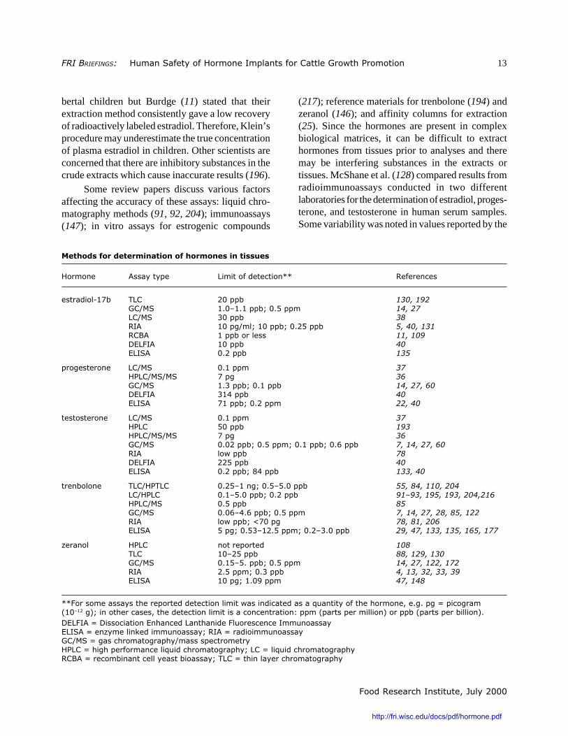
ASSAYS FOR DETERMINATION OFHORMONE LEVELS
A variety of methods have been developed forthe determination of hormone concentrations in bio-logical samples. Biological assays and thin layerchromatography procedures were developed sometime ago. More recently, in vitro assays with celllines sensitive to hormones and enzyme-linked andradioactive immunoassays have been used to deter-mine hormone concentrations. Several versions ofthese assays are commercially available. Gas chro-matography–mass spectrometry methods have alsobeen recently refined to detect very low levels ofhormones. In addition, yeast cells have been geneti-cally modified to contain genes for the human estro-gen receptor linked to reporter genes such as theβ-galactosidase gene. These cells have been used todetermine levels of 17β-estradiol in human serumsamples (109) and bovine serum samples (11). Al-though these assays may detect very low levels ofestrogenic compounds (0.02 pg/ml according torefernce 109), interassay variation is approximately65% at estradiol concentrations of <1 pg/ml (11).
Accuracy of all these assays depends on theefficiency of the methods used to extract hormonesfrom plasma or other tissues. Klein (109) reportedextremely low levels of estradiol in plasma of prepu-
http://fri.wisc.edu/docs/pdf/hormone.pdf
�������������������� ��������������� ������� �������������������3������ 13
������������� ������������������
bertal children but Burdge (11) stated that theirextraction method consistently gave a low recoveryof radioactively labeled estradiol. Therefore, Klein’sprocedure may underestimate the true concentrationof plasma estradiol in children. Other scientists areconcerned that there are inhibitory substances in thecrude extracts which cause inaccurate results (196).
Some review papers discuss various factorsaffecting the accuracy of these assays: liquid chro-matography methods (91, 92, 204); immunoassays(147); in vitro assays for estrogenic compounds
(217); reference materials for trenbolone (194) andzeranol (146); and affinity columns for extraction(25). Since the hormones are present in complexbiological matrices, it can be difficult to extracthormones from tissues prior to analyses and theremay be interfering substances in the extracts ortissues. McShane et al. (128) compared results fromradioimmunoassays conducted in two differentlaboratories for the determination of estradiol, proges-terone, and testosterone in human serum samples.Some variability was noted in values reported by the
������������������������������������������
���� � >��������� ��������������� 22 ����� ��
��������9,?* ) � �����* ���� ��
��&�� ,5�#,5,���*=��5+���� �����
�&�� .����* ��
��> ,���%&��=�,����*=��5�+���* ������� �
��:> ,���*������� �� ��
@6 ��> ,����* ��
6 ��> �5����* ��
��%����� � �&�� �5,���� ��
�3 �&��&�� ?��% ��
��&�� ,5.���*=��5,���* ���������
@6 ��> .,/���* ��
6 ��> ?,���*=��5����� ������
��������� � �&�� �5,���� ��
�3 � +����* ��
�3 �&��&�� ?��% ��
��&�� �5�����*=��5+����=��5,���*=��50���* ��� ���������
��> ������* ��
@6 ��> ��+���* ��
6 ��> �5����*=�7/���* ������
�� *��� � ) �&�3) � �5�+#,� %=��5+#+5����* �������� ������
�&�3 � �5,#+5����*=��5����* � ����� ���� ��������� �
�3 �&�� �5+���* ��
��&�� �5�0#/50���*=��5+���� ��� ��������������� ��
��> ������*=�A?���% ����� �����
6 ��> +��%=��5+.#,�5+����=��5�#.5����* �������� ���� ���� ���� ��
B�� �� �3 � ��������� ��
) � ,�#�+���* ���� ���� ��
��&�� �5,+#+5���*=��5+���� ������� ���� ��
��> �5+����=��5.���* ��� �������������
6 ��> ,���%=�,5�8���� ���� ��
22��������������������������������� ������������ ������������C�� ���������������� ����5%5��%�1����%��$,�#,��%(=�� ����������������������� ������������� � ����� D�����$�������������� (�����*�$��������*����� (5
@6 ��>�1�@��������� �6 �� ��� � ��� ����������� ������ ������6 ��>�1�� B������ '������� ������=���>�1��������� ��������&���1�%���������%����&���������������3 ��1���%�������� ����C����������%����=� ��1���C����������%������:>�1����*� � ������������*�������=�) ��1���� ������������%����
http://fri.wisc.edu/docs/pdf/hormone.pdf
������������� ������������������
14 ������������������� ��������������� ������� �������������������Promotion
two laboratories, and it appeared that the assaysoverestimated the amount of estradiol present in thesamples having the lowest concentrations. Fitzgeraldet al. (46) developed a sensitive electron-capturenegative-chemical-ionization gas chromatography–mass spectrometry method for the detection of tes-tosterone and compared it to a nonradioactiveimmunoassay. There was an excellent correlationbetween the results of the two procedures whenserum samples from males were analyzed but agree-ment was poor for samples from females. It wassuggested that the immunoassay was detecting othercross-reacting compounds (besides testosterone) thatcaused the discrepancy in the female samples.
A different approach to identify abuse of tes-tosterone in cattle was taken by Mason (126) in arecently developed method, gas chromatography–isotope ratio mass spectrometry, which can distin-guish between endogenous and external sources oftestosterone in cattle. Testosterone produced incattle will contain a certain ratio of 12C to 13Cdepending on the diet of the animals. This ratio islikely to be different in manufactured doses oftestosterone. Therefore, analyses of the isotope ratioin testosterone from an animal should allow thedetection of testosterone abuse.
The table above lists the reported detectionlimits (sensitivity) and types of assays developed fordetection of the five hormones of interest and theresearch papers reporting these results. The reporteddetection limits should not be considered strictlycomparable because of the variety of research proto-cols and test materials (muscle, blood serum, urine).
Summary. Assays for hormones in biological tis-sues have evolved and become more sensitive andaccurate during the past 30 years. The most sensitiveassays can now reliably detect hormone concentra-tions in the low parts per billion range. Enzyme-linked- and radio-immunoassays have been the mostwidely used methods in recent years, and severalversions of these assays are commercially available.Methods utilizing gas chromatography–mass spec-trometry and a recombinant yeast cell culture arebeing refined to yield even lower limits of detection.However, there is still significant variability among
measurements reported by different laboratories. Thisvariation may be due to differences in the antibodypreparations used in immunoassays and to differ-ences in extraction methods which may be more orless efficient in recovering all the hormone moleculesin a sample and in removing interfering substances.Controversy over the effects of minute concentra-tions of hormones and the true concentration ofestradiol in prepubertal children emphasizes the im-portance of repeatable, ultra-sensitive assays forhormones, particularly estradiol, in biologicalsamples.
HORMONE LEVELS IN OTHERFOODS
Estradiol, testosterone, and progesterone are natu-rally present in many foods of animal origin, andsome plant foods also contain these hormones.Hartmann et al. (59) present results from their ownexperiments as well as summarizing much of theprevious data from other researchers on hormonelevels in foods. A sampling of foods with significanthormone concentrations is presented in the followingtable. From the analytical results and information onaverage food consumption in Germany, Hartmann etal. estimate that the average daily intake of estradiolfor women, men, prepubertal girls, and prepubertalboys is 0.08, 0.1, 0.07, and 0.08 µg, respectively,with 60–70% supplied by milk products and15–20% each by eggs and meat/fish. [Accordingto JECFA, the acceptable daily intake of estradiolfor a 60-kg adult = 3.0 µg and for a 10-kg child =0.5 µg (99).] Milk products are also a major dietarysource of progesterone and testosterone, with eggsand meat also providing significant amounts. Manyplant foods contain other compounds which haveestrogenic activity and contribute significantly todietary exposure to estrogenic compounds. Morecomplete data can be found in references 12, 41, 57,59, 80, 82, 140, 142, 208, and 213.
Summary. Estradiol, progesterone, and testosteroneare present in many foods of animal origin includingbeef, pork, poultry, milk, eggs, and fish. Some plant
http://fri.wisc.edu/docs/pdf/hormone.pdf

�������������������� ��������������� ������� �������������������3������ 15
������������� ������������������
foods such as potatoes and wheat contain significantlevels of progesterone, and other foods, includingsome oils and wheat, have measurable levels oftestosterone. In addition, many plants contain othercompounds with estrogenic activity. It has beenestimated that milk products provide approximately80% of the progesterone, 30–40% of testosterone,and 60–70% of estrogens in the diet. Meat andfish provide about 5% of progesterone, 20–30% oftestosterone, and 15–20% of estrogens in the diet.Eggs and plant foods are responsible for the remain-der of the dietary hormone intake.
*Metric units may not be familiar to some readers.1 kilogram (kg) = 1,000 grams (g)
= approximately 2.2 lb
For units smaller than grams:
1 gram = 1,000 milligrams (mg)= 1,000,000 micrograms (mg)= 1,000,000,000 nanograms (ng)= 1,000,000,000,000 picograms (pg)
References1. Ahmad ME, Shadab GGHA, Hoda A, Afzal M.
Genotoxic effects of estradiol-17 beta on human lymphocytechromosomes. Mutat. Res. — Toxicol. Environ. Mutagen.2000; 466(1):109–115.
2. Andersson AM, Skakkebaek NE. Exposure toexogenous estrogens in food: possible impact on humandevelopment and health. Eur. J. Endocrinol. 1999;140(6):477–485.
3. Anonymous. Commission adopts communicationon precautionary principle. 2000. http://europa.eu.int/rapid/start/cgi/guesten.ksh?p_action.gettxt=gt&doc=IP/00/96|0|AGED&lg=EN [Search at http://europa.eu.int/rapid/start/cgi/guesten.ksh?qry for reference IP/00/96]
4. Arts CJ, Kemperman PT, van den Berg H. Oestro-gen radioreceptor assay for multi-residue screening of bo-vine urine for oestrogenic anabolic compounds. Food Additiv.Contam. 1989; 6(1):103–15.
5. Arts CJM, Baak MJ van, Berg H van den, Schilt R,Berende PLM, Hartog JMP den. Concentrations of the en-dogenous steroid hormones oestradiol-17 beta, testosteroneand progesterone in veal calves in connection with thecontrol for illegal administration. Arch. Lebensm. 1990;41(3):58–62.
6. Aw TC, Smith AB, Stephenson RL, Glueck CJ.Occupational exposure to zeranol, an animal growth pro-moter. Brit. J. Industr. Med. 1989; 46(5):341–346.
7. Bagnati R, Fanelli R. Determination of 19-nor-testosterone, testosterone and trenbolone by gas chromatog-raphy-negative-ion mass spectrometry after formation of thepentafluoro-benzylcarboxymethoxime-trimethylsilyl deriva-tives. J. Chromatogr. 1991; 547(1–2):325–334.
������� �� �����������������������������������������������
���� ,?*9�������� ��%����� � ��������� �
�'������' ,5/#�5�
E��������' �5�,#�5�. 85+#,,57 �5��#�5�+
:���� A�5�. ,/,#.�� A�5�+
������ �5�,#�5�. //5� �5/7#,5/,
6%%� A�5�.#�5�� ,�5+#/.50 �5�/#�5/8
���'� ����� A�5�.�#��5�� �5�/ A�5���#��5�.
:�������� �58, .5?,
:������ �5/. ,,580
:�����!� 850? ,5�
��� % A�5�. �5+, �5�?
3������� A�5�. +5�? A�5��
E���� A�5�? �570 �5�8
��� A�5�? �5.7
������������ A�5�. �5?, �5�,
http://fri.wisc.edu/docs/pdf/hormone.pdf
������������� ������������������
16 ������������������� ��������������� ������� �������������������Promotion
8. Baldwin RS, Williams RD, Terry MK. Zeranol: areview of the metabolism, toxicology, and analyticalmethods for detection of tissue residues. Reg. Toxicol.Pharmacol. 1983; 3(1):9–25.
9. Barraud B, Lugnier A, Dirheimer G. In vivo cova-lent binding to rat liver DNA of trenbolone as compared to17 β-estradiol, testosterone, and zeranol. In: Anabolics inAnimal Production, E. Meissonnier, J. Mitchell-Vigneron(eds.). Levallois, France: Soregraph. 1983; pp. 325–338.
10. Barraud B, Lugnier A, Dirheimer G. Determina-tion of the binding of trenbolone and zeranol to rat-liverDNA in vivo as compared to 17 beta-oestradiol and testos-terone. Food Additiv. Contam. 1984; 1(2):147–155.
11. Burdge GC, Coldham NG, Dave M, Sauer MJ,Bleach ECL. Determination of oestrogen concentrations inbovine plasma by a recombinant oestrogen receptor-reportergene yeast bioassay. Analyst 1998; 123:2585–2588.
12. Cantoni C, Aubert Sd’. 17beta estradiol, proges-terone and testosterone levels in cheeses. Indust. Aliment.1995; 34(335):257–258.
13. Carter AP, Dixon SN, Bew MH. Preparation andproperties of monoclonal antibodies to the anabolic agentzeranol. J. Vet. Pharmacol. Therapeut. 1984; 7(1):17–21.
14. Casademont G, Perez B, Regueiro JAG. Simulta-neous determination, in calf urine, of twelve anabolic agentsas heptafluorobutyryl derivatives by capillary gas chroma-tography mass spectrometry. J. Chromatogr. B: Biomed.Appl. 1996; 686(2):189–198.
15. Center for Veterinary Medicine, Food and DrugAdministration. Summary of NADA 009-576: Synovex®(estradiol benzoate and progesterone). 1994. http://www.fda.gov/cvm/efoi/section1/009576s81994.html[Search at http://www.fda.gov/cvm/efoi/foiglist.html for allNADA numbers.]
16. Center for Veterinary Medicine, Food and DrugAdministration. Summary of NADA 011-427: Synovex-H(estradiol benzoate and testosterone propionate). 1983.http://www.fda.gov/cvm/efoi/section1/011427s100583.html
17. Center for Veterinary Medicine, Food and DrugAdministration. Summary of NADA 038-233: RALGRO®(zeranol). 1995. http://www.fda.gov/cvm/efoi/section1/038233s040695.html
18. Center for Veterinary Medicine, Food and DrugAdministration. Summary of NADA 138-612: Finaplix®(trenbolone acetate). 1986. http://www.fda.gov/cvm/efoi/sec-tion1/138612.html
19. Center for Veterinary Medicine, Food and DrugAdministration. Summary of NADA 140-897: Revalor®-S(trenbolone acetate and estradiol). 1991. http://www.fda.gov/cvm/efoi/section2/140897.html
20. Center for Veterinary Medicine, Food and Drug
Administration. Summary of NADA 140-992: Revalor®-H(trenbolone acetate and estradiol). 1994. http://www.fda.gov/cvm/efoi/section2/140992.html
21. Center for Veterinary Medicine, Food and DrugAdministration. Summary of NADA 141-043: Synovex®Plus (trenbolone acetate and estradiol). 1996. http://www.fda.gov/cvm/efoi/section2/141043022296.html
22. Claycomb RW, Delwiche MJ, Munro CJ,BonDurant RH. Rapid enzyme immunoassay for measure-ment of bovine progesterone. Biosensors Bioelectronics1998; 13(11):1165–1171.
23. Coe JE, Ishak KG, Ward JM, Ross MJ. Tamoxifenprevents induction of hepatic neoplasia by zeranol, an estro-genic food contaminant. Proc. Natl. Acad. Sci. USA 1992;89(3):1085–1089.
24. Collins SS, Belk KE, Cross HR, Smith GC. TheEEC ban against growth promoting hormones. Nutr. Rev.1989; 47(8):238–246.
25. Crooks SRH, Elliott CT, Thompson CS,McCaughey WJ. Comparison and evaluation of the specific-ity and binding capacity of commercial and in house affinitycolumns used in sample preparation for analysis of growth-promoting drugs. J. Chromtogr.-Biomed. Appl. 1997; 690(1–2):161–172.
26. Cross HR, Schanbacher BD, Crouse JD. Sex, ageand breed related changes in bovine testosterone and intra-muscular collagen. Meat Sci. 1984; 10(3):187–195.
27. Daeseleire E, Vandeputte R, Van Peteghem C.Validation of multi-residue methods for the detection ofanabolic steroids by GC-MS in muscle tissues and urinesamples from cattle. Analyst 1998; 123(12):2595–2598.
28. de Boer D, Gainza Bernal ME, van Ooyen RD,Maes RA. The analysis of trenbolone and the human urinarymetabolites of trenbolone acetate by gas chromatography/mass spectrometry and gas chromatography/tandem massspectrometry. Biol. Mass Spectrom. 1991; 20(8):459–466.
29. Degand G, Schmitz P, Maghuin-Rogister G. En-zyme immunoassay screening procedure for the syntheticanabolic estrogens and androgens diethylstilbestrol, nor-testosterone, methyltestosterone and trenbolone in bovineurine. J. Chromatogr. 1989; 489(1):235–243.
30. Din N, Bartle KD, Clifford AA, Castle L. Aninvestigation of supercritical fluid extraction of trenbolonefrom beef. J. High Res. Chromatogr. 1996; 19(8):465–469.
31. Dixon SN. The efficacy, mode of action and safetyof non-steroidal non-antimicrobial growth promoters. Vet.Res. Com. 1983; 7(1–4):51–57.
32. Dixon SN, Russell KL. Radioimmunoassay of theanabolic agent zeranol. II. Zeranol concentrations in urine ofsheep and cattle implanted with zeranol (Ralgro). J. Vet.Pharmacol. Therapeut. 1983; 6(3):173–179.
http://fri.wisc.edu/docs/pdf/hormone.pdf
�������������������� ��������������� ������� �������������������3������ 17
������������� ������������������
33. Dixon SN, Russell KL. Radioimmunoassay of theanabolic agent zeranol. IV. The determination of zeranolconcentrations in the edible tissues of cattle implanted withzeranol (Ralgro). J. Vet. Pharmacol. Therapeut. 1986;9(1):94–100.
34. Dixon SN, Russell KL, Heitzman RJ, MallinsonCB. Radioimmunoassay of the anabolic agent zeranol. V.Residues of zeranol in the edible tissues, urine, faeces andbile of steers treated with Ralgro. J. Vet. Pharmacol.Therapeut. 1986; 9(4):353–358.
35. Dorgan JF, Reichman ME, Judd JT, Brown C,Longcope C, Schatzkin A, Forman M, Campbell WS,Franz C, Kahle L, Taylor PR. Relation of energy, fat, andfiber intakes to plasma concentrations of estrogens andandrogens in premenopausal women. Am. J. Clin. Nutr.1996; 64(1):25–31.
36. Draisci R, Giannetti L, Lucentini L, Pallesch L,Purificato I, Moretti G. Confirmation of anabolic hormoneresidues in bovine blood by micro-HPLC-ion spray tandemmass spectrometry. J. High Res. Chromatogr. 1997;20(8):421–426.
37. Draisci R, Palleschi L, Ferretti E, Lucentini L,Cammarata P. Quantitation of anabolic hormones and theirmetabolites in bovine serum and urine by liquid chromatog-raphy-tandem mass spectrometry. J. Chromatogr. 2000;870(1–2):511–522.
38. Draisci R, Palleschi L, Ferretti E, Marchiafava C,Lucentini L, Cammarata P. Quantification of 17 beta-estra-diol residues in bovine serum by liquid chromatographytandem mass spectrometry with atmospheric pressure chemi-cal ionization. Analyst 1998; 123(12):2605–2609.
39. Duchatel JP, Maghuin-Rogister G. Free and con-jugated zeranol residues determined by radio-immunoassayin urine and plasma of calves treated with forplix. Ann.Recher. Vet. 1985; 16(1):93–97.
40. Elliott CT, Francis KS, Shortt HD, McCaugheyWJ. Determination of the concentrations of the steroidsestradiol, progesterone and testosterone in bovine sera: com-parison of commercial dissociation enhanced lanthanidefluorescence immunoassay kits with conventional radio andenzyme immunoassays. Analyst 1995; 120(6):1827–1830.
41. Erb RE, Chew BP, Keller HF. Relative concen-trations of estrogen and progesterone in milk and blood,and excretion of estrogen in urine. J. Anim. Sci. 1977;45(3):617–626.
42. Everett DJ, Perry CJ, Scott KA, Martin BW, TerryMK. Estrogenic potencies of resorcylic acid lactones and 17β-esrtradiol in female rats. J. Toxicol. Environ. Health 1987;20:435–443.
43. Evrard P, Maghuin-Rogister G. In vitro metabo-lism of trenbolone: study of the formation of covalently
bound residues. Food Additiv. Contam. 1988; 5(1):59–65.
44. Evrard P, Maghuin-Rogister G, Rico AG. Fateand residues of trenbolone acetate in edible tissues fromsheep and calves implanted with tritium-labeled trenboloneacetate. J. Anim. Sci. 1989; 67(6):1489–1496.
45. Fara GM, Del Corvo G, Bernuzzi S, Bigatello A,DiPietro C, Scaglioni S, Chiumello G. Epidemic of breastenlargement in an Italian school. Lancet 1979; II:295–297.
46. Fitzgerald RL, Herold DA. Serum total testoster-one: immunoassay compared with negative chemical ioniza-tion gas chromatography-mass spectrometry. Clin. Chem.1996; 42(5):749–755.
47. Fodey TL, Elliott CT, Crooks SRH, McCaugheyWJ. The appraisal of an automated multi-immunoaffinitychromatography system to detect anabolic agents in bile andurine. Food Agric. Immunol. 1996; 8(3):157–167.
48. Forbes L. Do exogenous oestrogens and progester-one influence asthma? Thorax 1999; 54(3):265–267.
49. Fritsche S, Rumsey TS, Meyer HHD, Schmidt G,Steinhart H. Profiles of steroid hormones in beef from steersimplanted with synovex-s (estradiol benzoate and progester-one) in comparison to control steers. Z. Lebensm. Unters.Forsch. 1999; 208(5–6):328–331.
50. Fritsche S, Schmidt G, Schwarz FJ, KirchgesserM, Augustini C, Steinhart H. Natural hormone patternsof meat from steers and bulls depending on slaughter age.Z. Lebensm. Unters. Forsch. 1998; 207(3):183–188.
51. Fritsche S, Schmidt G, Steinhart H. Gas chromato-graphic-mass spectrometric determination of natural pro-files of androgens, progestogens, and glucocorticoids inmuscle tissue of male cattle. Eur. Food Res. Technol. 1999;209(6):393–399.
52. Fritsche S, Schwarz FJ, Kirchgessner M, AugustiniC, Steinhart H. Influence of sampling on steroid hormonepatterns of beef from bulls and steers. Meat Sci. 1998;50(2):257–264.
53. Fritsche S, Steinhart H. Differences in naturalsteroid hormone patterns of beef from bulls and steers. J.Anim. Sci. 1998; 76(6):1621–1625.
54. Gaiani R, Chiesa F. Physiological levels of andros-tenedione and testosterone in some edible tissues from calves,bulls and heifers. Meat Sci. 1986; 17(3):177–185. 1986.
55. Gernhard K, Lange WA. Simple semiquantitativedetermination of trenbolone acetate and trenbolone in bio-logical material from farm animals [German]. Arch. Exp.Veterinarmedizin. 1989; 43(6):863–866.
56. Gill JW, Hosking BJ, Egan AR. Prenatal program-ming of mammalian growth - a review of the role of steroids.Livestock Production Sci. 1998; 54(3):251–267.
57. Ginther OJ, Nuti LC, Garcia MC, Wentworth BC,
http://fri.wisc.edu/docs/pdf/hormone.pdf

������������� ������������������
18 ������������������� ��������������� ������� �������������������Promotion
Tyler WJ. Factors affecting progesterone concentration incow’s milk and dairy products. J. Anim. Sci. 1976; 42(1):155–159.
58. Gray DG, Unruh JA, Dikeman ME, Stevenson JS.Implanting young bulls with zeranol from birth to fourslaughter ages: iii. Growth performance and endocrineaspects. J. Anim. Sci. 1986; 63(3):747–756.
59. Hartmann S, Lacorn M, Steinhart H. Naturaloccurrence of steroid hormones in food. Food Chem. 1998;62(1):7–20.
60. Hartmann S, Steinhart H. Necessity of enzymatichydrolysis in steroid hormone analysis of beef. Arch.Lebensmittelhyg. 1997; 48(5):111–115.
61. Hartwig M, Hartmann S, Steinhart H. Physiologi-cal quantities of naturally occurring steroid hormones(androgens and progestogens), precursors and metabolitesin beef of differing sexual origin. Z. Lebensm. Unters. Forsch.1997; 205(1):5–10.
62. Hartwig M, Hartmann S, Steinhart H. Determina-tion of naturally occurring sex steroids (androgens andprogestogens) in beef. Z. Lebensm. Unters. Forsch. 1995;201(6):533–536.
63. Hayden JM, Bergen WG, Merkel RA. Skeletalmuscle protein metabolism and serum growth hormone,insulin, and cortisol concentrations in growing steersimplanted with estradiol-17 beta, trenbolone acetate, orestradiol-17 beta plus trenbolone acetate. J. Anim. Sci.1992; 70(7):2109–2119.
64. Heitzman RJ. The absorption, distribution and ex-cretion of anabolic agents. J. Anim. Sci. 1983; 57(1):233–238.
65. Heitzman RJ, Harwood DJ. Residue levels oftrenbolone and oestradiol-17 beta in plasma and tissues ofsteers implanted with anabolic steroid preparations. Brit.Vet. J. 1977; 133(6):564–571.
66. Heitzman RJ, Harwood DJ, Kay RM, Little W,Mallinson CB, Reynolds IP. Effects of implanting prepu-beral dairy heifers with anabolic steroids on hormonalstatus, puberty and parturition. J. Anim. Sci. 1979; 48(4):859–866.
67. Henderson BE, Feigelson HS. Hormonal carcino-genesis. Carcinogenesis 2000; 21(3):427–433.
68. Henricks DM, Brand RT, Titgemeyer EC, MiltonCT. Serum concentrations of trenbolone-17-beta and estra-diol-17-beta and performance of heifers treated withtrenbolone acetate, melengestrol acetate, or estradiol-17-beta. J. Anim. Sci. 1997; 75(10):2627–2633.
69. Henricks DM, Edwards RL, Champe KA, GettysTW, Skelley GC Jr, Gimenez T. Trenbolone, estradiol-17beta and estrone levels in plasma and tissues and liveweight gains of heifers implanted with trenbolone acetate.
J. Anim. Sci. 1982; 55(5):1048–1056.
70. Henricks DM, Gray SL, Hoover JL. Residue levelsof endogenous estrogens in beef tissues. J. Anim. Sci. 1983;57(1):247–255.
71. Henricks DM, Gray SL, Hoover JLB. Residuelevels of endogenous estrogens in beef tissues. In: Anabolicsin Animal Production, E. J. Meissonnier, J. Mitchell-Vigneron (eds.). Levallois, France: Soregraph. 1983; pp.233–248.
72. Henricks DM, Torrence AK. Endogenous estra-diol-17beta in bovine tissues. Assoc. Off. Anal. Chem.1978; 61(5):1280–1283.
73. Hess DL. Determination of the hormonal no-effectlevels of 17B-trenbolone and altrenogest in the macaque.In: Anabolics in Animal Production, E. J. Meissonnier,J. Mitchell-Vigneron (eds.). Levallois, France: Soregraph.1983; pp. 359–378.
74. Hodgson AV, Ayala-Torres S, Thompson EB, LiehrJG. Estrogen induced microsatellite DNA alterations areassociated with Syrian hamster kidney tumorigenesis.Carcinogenesis 1998; 19(12):2169–2172.
75. Hoffmann B. Use of radioimmunoassay formonitoring hormonal residues in edible animal products. J.Assoc. Off. Anal. Chem. 1978; 61(5):1263–1273.
76. Hoffmann B. Aspects on the formation and detec-tion of tissue levels of anabolic steroids in domestic animals.J. Steroid Biochem. 1979; 11(1C):919–922.
77. Hoffmann B. Natural occurrence of steroid hor-mones in food producing animals. In: Anabolics in AnimalProduction, E. J. Meissonnier, J. Mitchell-Vigneron (eds.).Levallois, France: Soregraph. 1983; pp. 215–231.
78. Hoffmann B. Use of radioimmunoassay proce-dures for the determination of sex hormones in animal tis-sues. J. Steroid Biochem. 1983; 19(1C):947–951.
79. Hoffman B. Problems of residues and health risksof anabolic agents with sex hormone-like activities. In:Proc. Sci. Conf. on Growth Promotion in Meat Production,European Comm. Dir. Gen. VI. Agric., Official Pub. Euro-pean Comm., Brussels. 1996; pp. 271–296.
80. Hoffmann B, Hamburger R, Karg H. Natural oc-currence of progesterone in commercial milk products. Z.Lebensm. Unters. Forsch. 1975; 158(5):257–259.
81. Hoffmann B, Oettel G. Radioimmunoassays forfree and conjugated trienbolone and for trienbolone acetatein bovine tissue and plasma samples. Steroids 1976;27(4):509–523.
82. Hoffmann B, Rattenberger E. Testosterone con-centrations in tissue from veal calves, bulls and heifers andin milk-samples. J. Anim. Sci. 1977; 45(3):635–641.
83. Hoffmann B, Schopper D, Karg H. Investigations
http://fri.wisc.edu/docs/pdf/hormone.pdf
�������������������� ��������������� ������� �������������������3������ 19
������������� ������������������
on the occurrence of non-extractable residues of trenboloneacetate in cattle tissues in respect to their bioavailability andimmunological reactivity. Food Additiv. Contam. 1984;1(3):253–259.
84. Hohls FW, Stan HJ. Detection of trenbolone resi-dues in meat by thin layer chromatography and fluorimetry.Z. Lebensm. Unters. Forsch. 1978; 167(4):252–255.
85. Hsu SH, Eckerlin RH, Henion JD. Identificationand quantitation of trenbolone in bovine tissue by gas chro-matography-mass spectrometry. J. Chromatogr. 1988;424(2):219–229.
86. Hunt DW, Henricks DM, Skelley GC, GrimesLW. Use of trenbolone acetate and estradiol in intact andcastrate male cattle: effects on growth, serum hormones, andcarcass characteristics. J. Anim. Sci. 1991; 69(6):2452–2462.
87. Hunter RA, Magner T, Berger KT. Sustainedgrowth promotion of steers, using anabolic steroids. Austr.J. Agric. Res. 1998; 49(4):589–596.
88. Ingerowski GH, Hellmann E, Stan HJ. Determina-tion of zeranol in meat. Z. Lebensm. Unters. Forsch. 1975;157(4):189–195.
89. Ingerowski GH, Scheutwinkel-Reich M, Stan HJ.Mutagenicity studies on veterinary anabolic drugs with theSalmonella/microsome test. Mutat. Res. 1981; 91:93–98.
90. Istasse L, Evrard P, Van Eenaeme C, Gielen M,Maghuin-Rogister G, Bienfait JM. Trenbolone acetate incombination with 17 beta-estradiol: influence of implantsupports and dose levels on animal performance and plasmametabolites. J. Anim. Sci. 1988; 66(5):1212–1222.
91. Jansen EH, Both-Miedema R, van Blitterswijk H,Stephany RW. Separation and purification of severalanabolics present in bovine urine by isocratic high-perfor-mance liquid chromatography. J. Chromatogr. 1984;299(2):450–455.
92. Jansen EH, Both-Miedema R, van den Berg RH.Application of optimization procedures for the separationof anabolic compounds by high-performance liquid chroma-tography. J. Chromatogr. 1989; 489(1):57–64.
93. Jansen EH, Zoontjes PW, Van Blitterswijk H,Both-Miedema R, Stephany RW. Fast high-performanceliquid chromatographic screening method for the presence oftrenbolone and its major metabolite in urine of slaughtercattle. J. Chromatogr. 1985; 319(3):436–439.
94. Joint FAO/WHO Expert Committee on Food Ad-ditives. Residues of some veterinary drugs in animals andfoods. Estradiol. FAO Food & Nutrition Paper 1988; 41:7–17.
95. Joint FAO/WHO Expert Committee on Food Ad-ditives. Residues of some veterinary drugs in animals andfoods. Progesterone. FAO Food & Nutrition Paper 1988;
41:18–23.
96. Joint FAO/WHO Expert Committee on Food Ad-ditives. Residues of some veterinary drugs in animals andfoods. Testosterone. FAO Food & Nutrition Paper 1988;41:24–28.
97. Joint FAO/WHO Expert Committee on Food Ad-ditives. Residues of some veterinary drugs in animals andfoods. Trenbolone acetate. FAO Food & Nutrition Paper1988; 41:29–37. 1988.
98. Joint FAO/WHO Expert Committee on Food Ad-ditives. Residues of some veterinary drugs in animals andfoods. Zeranol. FAO Food & Nutrition Paper 1988; 41:38–47. 1988.
99. Joint FAO/WHO Expert Committee on Food Addi-tives. Summary and Conclusions of the Fifty-second Meeting,Rome, 2–11 February 1999. http://www.fao.org/WAICENT/FAOINFO/ECONOMIC/ESN/Jecfa/jecfa52.pdf
100. Joint FAO/WHO Expert Committee on Food Ad-ditives. Toxicological evaluation of certain veterinary drugresidues in food: Zeranol. WHO Food Additives Series1988; 23:123–151.
101. Joint FAO/WHO Expert Committee on Food Ad-ditives. Toxicological evaluation of certain veterinary drugresidues in food: Trenbolone. WHO Food Additives Series.1988; 23:73–121.
102. Joint FAO/WHO Expert Committee on Food Ad-ditives. Residues of some veterinary drugs in animals andfoods. Trenbolone acetate. FAO Food & Nutrition Paper1990; 41(2):88–98.
103. Joint FAO/WHO Expert Committee on Food Ad-ditives. Toxicological evaluation of certain veterinary drugresidues in food: Trenbolone. WHO Food Additives Series1991; 25:101–109.
104. Jones SJ, Johnson RD, Calkins CR, Dikeman ME.Effects of trenbolone acetate on carcass characteristics andserum testosterone and cortisol concentrations in bulls andsteers on different management and implant schemes. J.Anim. Sci. 1991; 69(4):1363–1369.
105. Karg H, Meyer HHD. Update evaluation oftrenbolone acetate, zeranol and melengestrol acetate asgrowth promoters (considerations concerning the “hormoneissues” between EU and USA at the WTO [German]. Arch.Lebensmittelhyg. 1999; 50(2):28–37.
106. Karisto T, Tanhuanpaeae E, Uusi Rauva A,Antila M. Progesterone concentration of milk and somedairy products. Meijeritieteellinen Aikakauskirja 1982;40(2):20–31.
107. Kennedy DG, Hewitt SA, McEvoy JD, Currie JW,Cannavan A, Blanchflower WJ, Elliot CT. Zeranol is formedfrom Fusarium spp. toxins in cattle in vivo. Food Addit.Contam. 1998; 15(4):393–400.
http://fri.wisc.edu/docs/pdf/hormone.pdf

������������� ������������������
20 ������������������� ��������������� ������� �������������������Promotion
108. Kim HL, Ray AC, Stipanovic RD. Rapid separa-tion and identification of urinary metabolites of zeranolby HPLC-UV spectrophotometry. J. Agric. Food Chem.1986; 34:312–315.
109. Klein KO, Baron J, Colli MJ, McDonnell DP,Cutler GB. Estrogen levels in childhood determined by anultrasensitive recombinant cell bioassay. J. Clin. Invest.1994; 94:2475–2480.
110. Laitem L, Gaspar P, Bello I. Detection of trenboloneresidues in meat and organs of slaughtered animals by thin-layer chromatography. J. Chromatogr. 1978; 147:538–539.
111. Lammers BP, Heinrichs AJ, Kensinger RS. Theeffects of accelerated growth rates and estrogen implants inprepubertal holstein heifers on growth, feed efficiency, andblood parameters. J. Dairy Sci. 1999; 82(8):1746–1752.
112. Lasne C, Lu YP, Orfila L, Ventura L,Chouroulinkov I. Study of various transforming effects ofthe anabolic agents trenbolone and testosterone on Syrianhamster embryo cells. Carcinogenesis 1990; 11(4):541–547.
113. Lee CY, Henricks DM, Skelley GC, Grimes LW.Growth and hormonal response of intact and castrate malecattle to trenbolone acetate and estradiol. J. Anim. Sci. 1990;68(9):2682–2689.
114. Liehr JG. Is estradiol a genotoxic mutagenic car-cinogen? Endocrine Rev. 2000; 21(1):40–54.
115. Lindsay DG. Zeranol — a ‘nature-identical’ oestro-gen? Food Chem. Toxicol. 1985; 23(8):767–774.
116. Lobley GE, Connell A, Mollison GS, Brewer A,Harris CI, Buchan V, Galbraith H. The effects of a combinedimplant of trenbolone acetate and oestradiol-17 beta onprotein and energy metabolism in growing beef steers.British J. Nutr. 1985; 54(3):681–694.
117. Lone KP. Natural sex steroids and their xenobioticanalogs in animal production: growth, carcass quality,pharmacokinetics, metabolism, mode of action, residues,methods, and epidemiology. Crit. Rev. Food Sci. Nutr. 1997;37(2):93–209.
118. Luster MI, Hayes HT, Korach K, Tucker AN, DeanJH, Greenlee WF, Boorman GA. Estrogen immuno-suppres-sion is regulated through estrogenic responses in the thymus.J. Immunol. 1984; 133(1):110–116.
119. Lutz WK, Deuber R, Caviezel M, Sagelsdorff P,Friederich U, Schlatter C. Trenbolone growth promotant:covalent DNA binding in rat liver and in Salmonellatyphimurium, and mutagenicity in the Ames test. Arch.Toxicol. 1988; 62(2–3):103–109.
120. Maddox JG. An abattoir survey of growth pro-moter implants in cattle. Vet. Record 1988; 122(7):161.
121. Mader TL. Implants. Vet. Clin. North Am. FoodAnim. Practice 1998; 14(2):279–290.
122. Maffei Facino R, Carini M, Da Forno A, Traldi P,Pompa GA. Rapid method for the simultaneous detection bycollisionally activated dissociation mass-analysed ionkinetic energy spectrometry of parts per trillion of diethyl-stilbestrol, zeranol and trenbolone in plasma and edibletissues of cattle. Biomed. Environ. Mass Spectrom. 1986;13(3):121–127.
123. Martelli A, Mereto E, Ghia M, Orsi P, Allavena A,De Pascalis CR, Brambilla G. Induction of micronuclei andof enzyme-altered foci in the liver of female rats exposed toprogesterone and three synthetic progestins. Mutat. Res.1998; 419(1–3):33–41.
124. Marzin D. Ames test and trenbolone. Arch. Toxicol.1989; 63(6):492–494.
125. Marzin D. Trenbolone: application of the Amestest. Recent data. Ann. Recher. Vet. 1991; 22(3):257–262.
126. Mason PM, Hall SE, Gilmour I, Houghton E,Pillinger C, Seymour MA. The use of stable carbon isotopeanalysis to detect the abuse of testosterone in cattle. Analyst1998; 123(12):2405–2408.
127. Mastri C, Mistry P, Lucier GW. In vivooestrogenicity and binding characteristics of alpha-zearalanol(p-1496) to different classes of oestrogen binding proteinsin rat liver. J. Ster. Biochem. 1985; 23(3):279–289.
128. McShane LM, Dorgan JF, Greenhut S, Damato JJ.Reliability and validity of serum sex hormone measure-ments. Cancer Epidemiol. Biomarkers Prevent. 1996;5(11):923–928.
129. Medina MB, Nagdy N. Improved thin-layer chro-matographic detection of diethylstilbestrol and zeranol inplasma and tissues isolated with alumina and ion-exchangemembrane columns in tandem. J. Chromatogr. 1993;614(2):315–323.
130. Medina MB, Schwartz DP. Thin-layer chromato-graphic detection of zeranol and estradiol in fortified plasmaand tissue extracts with fast Corinth V. J. Chromatogr. 1992;581(1):119–128.
131. Medina MB. Direct radioimmunoassay of 17beta-estradiol in ether extracts of bovine sera. J. Agric. FoodChem. 1986; 34(6):1046–1049.
132. Metzler M, Kulling SE, Pfeiffer E, Jacobs E.Genotoxicity of estrogens. Z. Lebens. Unters. Forsch. 1998;206(6):367–373.
133. Meyer HH, Hoffmann S. Development of a sensi-tive microtitration plate enzyme-immunoassay for the ana-bolic steroid trenbolone. Food Addit. Contam. 1987;4(2):149–160.
134. Meyer HH, Landwehr M, Schopper D, Karg H.Application of Synovex-h in veal calves: steroid release andresidues. Food Addit. Contam. 1984; 1(3):261–275.
135. Meyer HHD. Determination of anabolic sex
http://fri.wisc.edu/docs/pdf/hormone.pdf

�������������������� ��������������� ������� �������������������3������ 21
������������� ������������������
hormones in meat and meat products. HPLC separationand immunological determination [German]. Arch.Lebensmittelhyg. 1990; 41(1):4, 6, 7.
136. Middle J. Standardization of steroid hormoneassays. Ann. Clin. Biochem. 1998; 35:354–363.
137. Migdalof BH, Dugger HA, Heider JG, CoombsRA, Terry MK. Biotransformation of zeranol: dispositionand metabolism in the female rat, rabbit, dog, monkey andman. Xenobiotica 1983; 13(4):209–221.
138. Mobley JA, Bhat AS, Brueggemeier RW.Measurement of oxidative DNA damage by catechol estro-gens and analogues in vitro. Chem. Res. Toxicol. 1999;12(3):270–277.
139. Moran C, Quirke JF, Prendiville DJ, Bourke S,Roche JF. The effect of estradiol, trenbolone acetate, orzeranol on growth rate, mammary development, carcasstraits, and plasma estradiol concentrations of beef heifers.J. Anim. Sci. 1991; 69(11):4249–4258.
140. Moslemi S, Silberzahn P, Seralini GE. Measure-ments of steroid and beta-agonist concentrations in infantmilks [French]. Lait 1996; 76(6):537–550.
141. Nakada K, Moriyoshi M, Nakao T, Watanabe G,Taya K. Changes in concentrations of plasma immuno-reactive follicle-stimulating hormone, luteinizing hormone,estradiol-17 beta, testosterone, progesterone, and inhibin inheifers from birth to puberty. Domestic Anim. Endocrinol.2000; 18(1):57–69.
142. Nuti LC, Wentworth BC, Karavolas HJ, Tyler WJ,Ginther OJ. Comparison of radioimmunoassay and gas-liquid chromatography analyses of progesterone concentra-tions in cow’s milk. Proc. Soc. Exp. Biol. Med. 1975;149(4):877–880.
143. O’Connell MB. Pharmacokinetic and pharmaco-logic variation between different estrogen products. J. Clin.Pharmacol. 1995; 35:18S–24S.
144. O’Keefe M, Hopkins JP. Survey of residue concen-trations of the anabolic agents, trenbolone acetate (Finaplix)and zeranol (Ralgro) in beef for the domestic and exportmarkets. Irish J. Food Sci.Technol. 1987; 11:13–24.
145. O’Keeffe M. Trenbolone levels in tissues oftrenbolone acetate-implanted steers: radioimmunoassaydetermination using different antisera. British Vet. J. 1984;140(6):592–599.
146. O’Keeffe M, Nugent A, Cadogan A, Hopkins JP,Daeseleire E, Van Peteghem C. Development of bovinemuscle, liver and urine reference materials for zeranol —preparation, homogeneity and stability. Fresenius J. Anal.Chem. 1997; 357(8):1029–1034.
147. Parekh CK, Terry MK, Williams RD. Genetictoxicology: in vitro and in vivo tests for mutagenic potentialof zeranol, zearalanone, and 17 B-estradiol. In: Anabolics in
Animal Production, E. Meissonnier, J. Mitchell-Vigneron(eds.). Levallois, France: Soregraph. 1983; pp. 307–323.1983.
148. Patel PJ, Dixon SN. An enzyme immunoassay forthe anabolic agent zeranol. Food Addit. Contam. 1989;6(1):91–101.
149. Perez-Martinez C, Ferreras-Estrada MC, Garcia-Iglesias MJ, Bravo-Moral AM, Espinosa-Alvarez J,Escudero-Diez A. Effects of in utero exposure to nonsteroi-dal estrogens on mouse testis. Can. J. Vet. Res. 1997;61(2):94–98.
150. Perez-Martinez C, Garcia-Iglesias, MJ, Bravo-Moral AM, Ferreras-Estrada MC, Martinez-Rodriguez JM,Escudero-Diez A. Effect of diethylstilbestrol or zeranol onfetal development, gestation duration, and number of off-spring in NMRI mice. Am. J. Vet. Res. 1995; 56(12):1615–1619.
151. Perez-Martinez C, Garcia-Iglesias MJ, Ferreras-Estrada MC, Bravo-Moral AM, Espinosa-Alvarez J,Escudero-Diez A. Effects of in-utero exposure to zeranolor diethylstilboestrol on morphological development of thefetal testis in mice. J. Comp. Pathol. 1996; 114(4):407–418.
152. Petit CR, Fournier PA, Rico AG, Burgat-SacazeV, de Saqui-Sannes P. In vivo covalent binding of 17-alpha-OH- and 17-beta-OH-trenbolone to rat liver DNA. Ann.Recher. Vet. 1989; 20(3):319–326.
153. Petit C, Perin F, Tardieu D, Burgat V, Rico AG.Covalent fixation in vivo of trenbolone acetate and estradiolto hepatic DNA of the rat: comparative study of the fixationof tritiated molecules and post labeling with radiophospho-rus [French]. Ann. Recher. Vet. 1991; 22(3):263–269.
154. Potau N, Ibáñez L, Sentis M, Carrascosa A. Sexualdimorphism in the maturation of the pituitary-gonadal axis,assessed by GnRH agonist challenge. Eur. J. Endocrinol.1999; 141:27–34.
155. Pottier J, Busigny M, Grandadam JA. Plasmakinetics, excretion in milk and tissue levels in the cowfollowing implantation of trenbolone acetate. J. Anim. Sci.1975; 41(3):962–968.
156. Pottier J, Cousty C, Heitzman, RJ, Reynolds IP.Differences in the biotransformation of a 17 beta-hydroxy-lated steroid trenbolone acetate, in rat and cow. Xenobiotica1981; 11(7):489–500.
157. Powell-Jones W, Raeford S, Lucier GW. Bindingproperties of zearalenone mycotoxins to hepatic estrogenreceptors. Molec. Pharmacol. 1981; 20(1):35–42.
158. Preston RL. Hormone containing growth promot-ing implants in farmed livestock. Adv. Drug Delivery Rev.1999; 38(2):123–138.
159. Pusateri AE, Kenison DC. Measurement of zeranolin plasma from three blood vessels in steers implanted with
http://fri.wisc.edu/docs/pdf/hormone.pdf

������������� ������������������
22 ������������������� ��������������� ������� �������������������Promotion
zeranol. J. Anim. Sci. 1993; 71(2):415–419.
160. Pylkkanen L, Jahnukainen K, Parvinen M, SanttiR. Testicular toxicity and mutagenicity of steroidal and non-steroidal estrogens in the male mouse. Mutat. Res. 1991;261(3):181–191.
161. Raben A, Kiens B, Richter EA, Rasmussen LB,Svenstrup B, Micic S, Bennett P. Serum sex hormones andendurance performance after a lacto-ovo vegetarian and amixed diet. Med. Sci. Sports Exercise 1992; 24(11):1290–1297.
162. Rajah TT, Pento JT. The mutagenic potential ofantiestrogens at the HPRT locus in V79 cells. Res. Commun.Molec. Pathol. Pharmacol. 1995; 89(1):85–92.
163. Rands PL, Newhouse CL, Stewart JL, BradshawWS. Indicators of developmental toxicity following prenataladministration of hormonally active compounds in the rat.II. Pattern of maternal weight gain. Teratology 1982;25(1):45–51.
164. Rands PL, White RD, Carter MW, Allen SD,Bradshaw WS. Indicators of developmental toxicity follow-ing prenatal administration of hormonally active compoundsin the rat. I. Gestational length. Teratology 1982; 25(1):37–43.
165. Rapp M, Meyer HHD. Detection of the applicationof trenbolone to fattening cattle. Radioimmunoassay andconfirmation by HPLC/RIA. Arch. Lebensmittelhyg. 1985;36(2):31 36.
166. Reynolds IP. Correct use of anabolic agents inruminants. Vet. Rec. 1980; 107(16):367–369.
167. Richold M. The genotoxicity of trenbolone, a syn-thetic steroid. Arch. Toxicol. 1988; 61(4):249–258.
168. Richou Bac L, Pochard MF, Boursier B. Plasmaand tissue levels of oestrodiol 17B, oestrone anddiethylstilboestrol (DES) in calves treated with anabolites,with husbandry aspects. Rec. Med. Vet. 1978; 154(5):441–450.
169. Rico AG. Metabolism of endogenous and exog-enous anabolic agents in cattle. J. Anim. Sci. 1983;57(1):226–232.
170. Roe FJC. Results of long term rodent studies withspecial reference to cancer risks and hormonal no-effectlevels. In: Anabolics in Animal Production, E. Meissonnier,J. Mitchell-Vigneron (eds.). Levallois, France: Soregraph.1983; pp. 339–346.
171. Ross RK, Paganini-Hill A, Wan PC, Pike MC.Effect of hormone replacement therapy on breast cancer risk:estrogen versus estrogen plus progestin. J. Natl. Cancer Inst.2000; 92(4):328–332.
172. Roybal JE, Munns RK, Morris WJ, Hurlbut JA,Shimoda W. Determination of zeranol/zearalenone andtheir metabolites in edible animal tissue by liquid chroma-
tography with electrochemical detection and confirmationby gas chromatography/mass spectrometry. J. Assoc. Off.Anal. Chem. 1988; 71(2):263–271.
173. Rubens R, Vermeulen A. Estrogen production inman. In: Anabolics in Animal Production, E. Meissonnier,J. Mitchell-Vigneron (eds.). Levallois, France: Soregraph.1983; pp. 249–262.
174. Rumsey TS, Beaudry N. Plasma estrogen concen-trations in non-implanted and synovex-s implanted feedlotsteers. Bull. Environ. Contam. Toxicol. 1979; 23(3):405–411.
175. Ryan JJ, Hoffmann B. Trenbolone acetate: experi-ences with bound residues in cattle tissues. J. Assoc. Off.Anal. Chem. 1978; 61(5):1274–1279.
176. Saenz de Rodriguez CA, Bongiovanni AM, Condede Borrego L. An epidemic of precocious development inPuerto Rican children. J. Pediatr. 1985; 107:393–396.
177. Sawaya WN, Lone K, Saeed T, Husain A,Khalafawi S. Application of an enzyme-linked immuno-sorbent assay for screening of sheep urine and animal tissuefor the androgenic steroid trenbolone acetate in the state ofKuwait. Food Addit. Contam. 1998; 15(2):151–156.
178. Sawyer GJ, Barker DJ. Growth promotants in cattlein Australia. Austr. Vet. J. 1988; 65(4):101–108.
179. Schairer C, Lubin J, Troisi R, Sturgeon S, BrintonL, Hoover R. Menopausal estrogen and estrogen-progestinreplacement therapy and breast cancer risk. J. Am. Med.Assoc. 2000; 283(4):485–491.
180. Scheutwinkel M, v.d. Hude W, Basler A. Studieson the genotoxicity of the anabolic drugs trenbolone andzeranol. Arch. Toxicol. 1986; 59(1):4–6.
181. Schiffmann D, Hieber L, Schmuck G, Pechan R,Metzler M, Henschler D. Trenbolone induces micronucleusformation and neoplastic transformation in Syrian hamsterembryo fibroblasts but not in mouse C3H10T1/2 cells.Arch. Toxicol. 1988; 62(1):49–53.
182. Schiffmann D, Metzler M, Neudecker T, HenschlerD. Morphological transformation of Syrian hamster embryofibroblasts by the anabolic agent trenbolone. Arch. Toxicol.1985; 58(1):59–63.
183. Schopper D, Hoffmann B, Karg H, Berende PL,van Weerden EJ, van d. e. r. Wal P. Analytic determinationof exogenous administration of 17 beta-estradiol andtrenbolone acetate in blood plasma and urine of the calf.[German]. Zentr. Veterinarmedizin — Reihe A. 1983;30(8):585–600.
184. Scientific Committee on Veterinary Measures Re-lating to Public Health of the European Commission. As-sessment of potential risks to human health from hormoneresidues in bovine meat and meat products. 1999. http://europa.eu.int/comm/food/fs/sc/scv/out21_en.pdf [Search
http://fri.wisc.edu/docs/pdf/hormone.pdf
�������������������� ��������������� ������� �������������������3������ 23
������������� ������������������
http://europa.eu.int/geninfo/query_en.htm]
185. Scientific Working Group on Anabolic Agents.Scientific report on anabolic agents in animal production.Vet. Rec. 1987:389–392.
186. Silcox RW, Keeton JT, Johnson BH. Effects ofzeranol and trenbolone acetate on testis function, liveweight gain and carcass traits of bulls. J. Anim. Sci. 1986;63(2):358–368.
187. Simmons DL, Valentine DM, Bradshaw WS.Different patterns of developmental toxicity in the rat follow-ing prenatal administration of structurally diverse chemi-cals. J. Toxicol. Environ. Health 1984; 14(2–3): 121–136.
188. Simpson RB, Chase CC, Spicer LJ, Carroll JA,Hammond AC, Welsh TH. Effect of exogenous estradiol onplasma concentrations of somatotropin, insulin-like growthfactor-i, insulin-like growth factor binding protein activity,and metabolites in ovariectomized Angus and Brahmancows. Domestic Animal Endocrinol. 1997; 14(6):367–380.
189. Sloop TC, Clark JC, Rumbaugh RC, Lucier GW.Imprinting of hepatic estrogen-binding proteins by neonatalandrogens. Endocrinology 1983; 112(5):1639–1646.
190. Smith GC, Sofos JN, Aaronson MJ, Morgan JB,Tatum JD, Schmidt GR. Incidence of pesticide residuesand residues of chemicals specified for testing in U.S. beefby the European Community. J. Muscle Foods 1994;5(3):271–284.
191. Spranger B, Metzler M. Disposition of 17 beta-trenbolone in humans. J. Chromatogr. 1991; 564(2):485–492.
192. Stan HJ, Hohls FW. Detection of estrogen residuesin meat by thin layer chromatography and fluorimetry. Z.Lebensm. Unters. Forsch. 1978; 166(5):287–92.
193. Stan HJ, Hohls FW. Determination of trenboloneand testosterone residues in meat by high pressure liquidchromatography. Z. Lebensm. Unters. Forsch. 1979;169(4):266–270.
194. Sterk S, van Tricht F, Le B, van Ginkel L, StephanyR. Production and stability testing of incurred referencematerials for the anabolic steroid trenbolone in bovine urine.Analyst 1998; 123(12):2563–2566.
195. Stubbings GW, Cooper AD, Shepherd MJ,Croucher JM, Airs D, Farrington WH, Shearer G. Determi-nation of 19-nortestosterone and trenbolone in animal tis-sues by high-performance liquid chromatography withimmunoaffinity clean-up. Food Addit. Contam. 1998;15(3):293–301.
196. Sub-group of the Veterinary Products Commit-tee, Ministry of Agriculture, Fisheries, and Food. Execu-tive summary and critical evaluation of the 1999 opinionof the scientific committee on veterinary measuresrelating to public health. 1999. http://www.vmd.gov.uk/
old_vmd_web_pages/finalrep.htm
197. Thibodeau PA, Bissonnette N, Bedard SK, Hunt-ing D, Paquette B. Induction by estrogens of methotrexateresistance in MCF-7 breast cancer cells. Carcinogenesis1998; 19(9):1545–1552.
198. Thouvenot D, Morfin RF. Interferences ofzearalenone, zearalanol or estradiol-17 beta with the steroid-metabolizing enzymes of the human prostate gland. J.Steroid Biochem. 1980; 13(11):1337–1345.
199. Truhaut R, Shubik P, Tuchmann-Duplessis H.Zeranol and 17 beta-estradiol: a critical review of the toxico-logical properties when used as anabolic agents. Reg. Toxicol.Pharmacol. 1985; 5(3):276–283.
200. Tsutsui T, Komine A, Huff J, Barrett JC. Effects oftestosterone, testosterone propionate, 17 beta-trenbolone andprogesterone on cell transformation and mutagenesis inSyrian hamster embryo cells. Carcinogenesis 1995;16(6):1329–1333.
201. Tsutsui T, Tamura Y, Yagi E, Barrett JC. Involve-ment of genotoxic effects in the initiation of estrogen-induced cellular transformation: studies using Syrian hamsterembryo cells treated with 17 beta-estradiol and eight of itsmetabolites. Int. J. Cancer 2000; 86(1):8–14.
202. Umberger EJ. Products marketed to promote growthin food-producing animals: steroid and hormone products.Toxicology 1975; 3(1):3–21.
203. Van Ginkel LA, Jansen EHJM, Stephany RW,Zoontjes PW, Schwillens PLWJ, van Rossum HJ, Viser T.Liquid chromatographic purification and detection of ana-bolic compounds. J. Chromatogr. 1992; 624:389–401.
204. van Ginkel LA, van Blitterswijk H, Zoontjes PW,van den Bosch D, Stephany RW. Assay for trenbolone andits metabolite 17 alpha-trenbolone in bovine urine based onimmunoaffinity chromatographic clean-up and off-line high-performance liquid chromatography-thin-layer chromatog-raphy. J. Chromatogr. 1988; 445(2):385–392.
205. van Leeuwen FX. The approach taken and conclu-sions reached by the joint FAO-WHO expert committee onfood additives. Ann. Recher. Vet. 1991; 22(3):253–256.
206. Vogt KA. Radioimmunological screening methodfor the detection of trenbolone in bile, faeces, urine and meat,with validation of the results by internal blank values. Arch.Lebensmittelhyg. 1985; 36(3): 58–60.
207. Volek JS, Kraemer WJ, Bush JA, Incledon T,Boetes M. Testosterone and cortisol in relationship to dietarynutrients and resistance exercise. J. Appl. Physiol. 1997;82(1):49–54.
208. Waldmann A, Ropstad E, Landsverk K, SorensenK, Solverod L, Dahl E. Level and distribution of progester-one in bovine milk in relation to storage in the mammarygland. Animal Reprod. Sci. 1999; 56(2):79–91.
http://fri.wisc.edu/docs/pdf/hormone.pdf
������������� ������������������
24 ������������������� ��������������� ������� �������������������Promotion
Ellin Doyle, Ph.D., has been employed as a researcher at the Food Research Institutesince 1997. Her primary role is to provide information on a variety of food safetytopics, particularly emerging issues, through searches of scientific journals andreliable on-line sources of scientific information. Her most recent publication in apeer-reviewed journal is a review of studies on the thermal resistance of salmonellaein the June 2000 issue of the Journal of Food Protection. Dr. Doyle received her B.S.from the University of Rhode Island and M.S. and Ph.D. in biology from the Universityof Michigan. In addition to research and writing on food safety, she has taught highschool science (in the Peace Corps) and college level biology courses at BeloitCollege and at the University of Wisconsin.
This project was funded by beef producers through their $1-per-head checkoffand was produced for the Cattlemen's Beef Board by the National Cattlemen'sBeef Association.
209. Waltner-Toews D, McEwen SA. Residues of hor-monal substances in foods of animal origin: a risk assess-ment. Prevent. Vet. Med. 1994; 20:235–247.
210. Wardell RE, Seegmiller RE, Bradshaw WS.Induction of prenatal toxicity in the rat by diethylstilbestrol,zeranol, 3,4,3',4',-tetrachlorobiphenyl, cadmium, and lead.Teratology 1982; 26(3):229–37.
211. Warren MP, Shantha S. Uses of progesterone inclinical practice. Int. J. Fertil. Womens Med. 1999; 44(2):96–103.
212. Williams JE, Ireland SJ, Mollett TA, Hancock DL,Beaver EE, Hannah S. Influence of zeranol and breed ongrowth, composition of gain, and plasma hormone concen-trations. J. Anim. Sci. 1991; 69(4):1688–1696.
213. Wolford ST, Argoudelis CJ. Measurement of es-trogens in cow’s milk, human milk, and dairy products.J. Dairy Sci. 1979; 62(9):1458–1463.
214. Woods MN, Barnett JB, Spiegelman D, Trail N,Hertzmark E, Longcope C, Gorbach SL. Hormone levelsduring dietary changes in premenopausal African-Americanwomen. J. Natl. Cancer Inst. 1996; 88(19):1369–1374.
215. World Health Organization: International Agencyfor Research on Cancer, IARC Monographs on the evalua-tion of carcinogenic risks to humans. Overall evaluationsof carcinogenicity: an updating of IARC Monographs 1–42.Suppl. 7 WHO:IARC, Lyon, France. 1987; 440 p.
216. Yoshioka N, Akiyama Y, Takeda N. Determina-tion of alpha- and beta-trenbolone in bovine muscle andliver by liquid chromatography with fluorescence detection.J. Chromatogr. B 2000; 739(2):363–367. 2000.
217. Zacharewski T. In vitro bioassays for assessingestrogenic substances. Environ. Sci. Technol. 1997; 31:613–623.
http://fri.wisc.edu/docs/pdf/hormone.pdf