Flexible DNA Target Site Recognition by Divergent Homing ...
17
Flexible DNA Target Site Recognition by Divergent Homing Endonuclease Isoschizomers I-CreI and I-Mso I Brett Chevalier 1 , Monique Turmel 2 , Claude Lemieux 2 Raymond J. Monnat Jr. 3 and Barry L. Stoddard 1 * 1 Division of Basic Sciences Graduate Program in Molecular and Cellular Biology University of Washington and the Fred Hutchinson Cancer Research Center 1100 Fairview Avenue North A3-025, Seattle WA 98109 USA 2 Centre de recherche sur la Fonction, la structure et l’Ingenierie des Proteines Pavillon Charles-Eugene Marchand, Universite Laval Quebec, Canada G1K 7P4 3 Departments of Pathology and Genome Sciences Box 357705 University of Washington, Seattle WA 98195 USA Homing endonucleases are highly specific catalysts of DNA strand breaks that induce the transposition of mobile intervening sequences containing the endonuclease open reading frame. These enzymes recognize long DNA targets while tolerating individual sequence polymorphisms within those sites. Sequences of the homing endonucleases themselves diversify to a great extent after founding intron invasion events, generating highly divergent enzymes that recognize similar target sequences. Here, we visualize the mechanism of flexible DNA recognition and the pattern of structural divergence displayed by two homing endonuclease isoschizo- mers. We determined structures of I-CreI bound to two DNA target sites that differ at eight of 22 base-pairs, and the structure of an isoschizomer, I-Mso I, bound to a nearly identical DNA target site. This study illustrates several principles governing promiscuous base-pair recognition by DNA-binding proteins, and demonstrates that the isoschizomers display strikingly different protein/DNA contacts. The structures allow us to determine the information content at individual positions in the binding site as a function of the distribution of direct and water-mediated contacts to nucleotide bases, and provide an evolutionary snapshot of endo- nucleases at an early stage of divergence in their target specificity. q 2003 Elsevier Science Ltd. All rights reserved Keywords: endonuclease; structure; isoschizomer; homing; specificity *Corresponding author Introduction Homing is the process by which mobile inter- vening genetic sequences, either introns or inteins, are duplicated into cognate recipient alleles that lack such a sequence. 1–5 The process is induced by an endonuclease encoded by an open reading frame (ORF) harbored within the intervening sequence. 6 The endonuclease specifically recog- nizes a DNA target site corresponding to the intron insertion site, generates a DNA double-strand break, and induces cellular mechanisms to repair the break. If the intron-containing allele is used as a template for repair, the endonuclease ORF is duplicated into the target site and the homing cycle is complete. Transfer of mobile introns can be extremely efficient. It can occur between different subcellular compartments of unrelated organisms, 7 and sometimes allowing introns to overrun diverse lineages within entire biological families, as demonstrated for a mobile fungal intron found throughout angiosperm plants. 8 Homing endonucleases are found in all bio- logical super-kingdoms. On the basis of primary sequence homology, four homing enzyme families have been identified: the LAGLIDADG, GIY-YIG, HNH, and His-Cys Box endonucleases. 5 The largest family, LAGLIDADG, contains several hundred identified members, many of which have been shown to be functional endonucleases. 5,9 The conserved LAGLIDADG sequence motif, from which the family draws its name, was shown in initial crystallographic analyses to form the core of the structural interface between endonuclease domains or subunits, and to contribute conserved acidic residues to the enzyme active sites. 10,11 Endo- nucleases containing a single LAGLIDADG motif per polypeptide chain form homodimers that 0022-2836/03/$ - see front matter q 2003 Elsevier Science Ltd. All rights reserved E-mail address of the corresponding author: [email protected] Abbreviations used: ORF, open reading frame. doi:10.1016/S0022-2836(03)00447-9 J. Mol. Biol. (2003) 329, 253–269
Transcript of Flexible DNA Target Site Recognition by Divergent Homing ...
Flexible DNA Target Site Recognition by DivergentHoming Endonuclease Isoschizomers I-CreIand I-Mso I
Brett Chevalier1, Monique Turmel2, Claude Lemieux2
Raymond J. Monnat Jr.3 and Barry L. Stoddard1*
1Division of Basic SciencesGraduate Program inMolecular and Cellular BiologyUniversity of Washingtonand the Fred HutchinsonCancer Research Center1100 Fairview Avenue NorthA3-025, Seattle WA 98109USA
2Centre de recherche sur laFonction, la structure etl’Ingenierie des ProteinesPavillon Charles-EugeneMarchand, Universite LavalQuebec, Canada G1K 7P4
3Departments of Pathologyand Genome SciencesBox 357705 University ofWashington, Seattle WA 98195USA
Homing endonucleases are highly specific catalysts of DNA strand breaksthat induce the transposition of mobile intervening sequences containingthe endonuclease open reading frame. These enzymes recognize longDNA targets while tolerating individual sequence polymorphisms withinthose sites. Sequences of the homing endonucleases themselves diversifyto a great extent after founding intron invasion events, generating highlydivergent enzymes that recognize similar target sequences. Here, wevisualize the mechanism of flexible DNA recognition and the pattern ofstructural divergence displayed by two homing endonuclease isoschizo-mers. We determined structures of I-CreI bound to two DNA target sitesthat differ at eight of 22 base-pairs, and the structure of an isoschizomer,I-Mso I, bound to a nearly identical DNA target site. This study illustratesseveral principles governing promiscuous base-pair recognition byDNA-binding proteins, and demonstrates that the isoschizomers displaystrikingly different protein/DNA contacts. The structures allow us todetermine the information content at individual positions in the bindingsite as a function of the distribution of direct and water-mediated contactsto nucleotide bases, and provide an evolutionary snapshot of endo-nucleases at an early stage of divergence in their target specificity.
q 2003 Elsevier Science Ltd. All rights reserved
Keywords: endonuclease; structure; isoschizomer; homing; specificity*Corresponding author
Introduction
Homing is the process by which mobile inter-vening genetic sequences, either introns or inteins,are duplicated into cognate recipient alleles thatlack such a sequence.1 – 5 The process is induced byan endonuclease encoded by an open readingframe (ORF) harbored within the interveningsequence.6 The endonuclease specifically recog-nizes a DNA target site corresponding to the introninsertion site, generates a DNA double-strandbreak, and induces cellular mechanisms to repairthe break. If the intron-containing allele is used asa template for repair, the endonuclease ORF isduplicated into the target site and the homingcycle is complete. Transfer of mobile intronscan be extremely efficient. It can occur between
different subcellular compartments of unrelatedorganisms,7 and sometimes allowing introns tooverrun diverse lineages within entire biologicalfamilies, as demonstrated for a mobile fungalintron found throughout angiosperm plants.8
Homing endonucleases are found in all bio-logical super-kingdoms. On the basis of primarysequence homology, four homing enzyme familieshave been identified: the LAGLIDADG, GIY-YIG,HNH, and His-Cys Box endonucleases.5 Thelargest family, LAGLIDADG, contains severalhundred identified members, many of which havebeen shown to be functional endonucleases.5,9 Theconserved LAGLIDADG sequence motif, fromwhich the family draws its name, was shown ininitial crystallographic analyses to form the core ofthe structural interface between endonucleasedomains or subunits, and to contribute conservedacidic residues to the enzyme active sites.10,11 Endo-nucleases containing a single LAGLIDADG motifper polypeptide chain form homodimers that
0022-2836/03/$ - see front matter q 2003 Elsevier Science Ltd. All rights reserved
E-mail address of the corresponding author:[email protected]
Abbreviations used: ORF, open reading frame.
doi:10.1016/S0022-2836(03)00447-9 J. Mol. Biol. (2003) 329, 253–269

recognize palindromic DNA target sites and pseudo-palindromic variants; members with two motifsfold to form pseudo-symmetric monomers capableof recognizing DNA target sites with significantasymmetry.12
Despite their relatively small size, all LAGLI-DADG nucleases recognize long DNA target sites(14–30 bp), cleaving across the minor groovewithin the site to generate cohesive four base, 30
overhangs.13 – 16 The enzymes typically bind theirphysiological target sites with dissociation con-
stants ranging from 1 to 5 nM, and exhibit chemicalrates of DNA strand cleavage of approximately0.3 min21 (R.J.M., M.T. and C.L., unpublishedresults). These cleavage rates accelerated by atleast six orders of magnitude over the rate ofcleavage of non-specific DNA sequences. The rateof actual non-enzymatic cleavage of DNA underphysiological conditions is much slower, to thepoint of not being measurable or directly com-parable to the enzyme reaction. Of the intron-encoded LAGLIDADG homing endonucleases, the
Figure 1. Physiological DNA target sites, crystallization constructs and structure-based sequence alignment ofI-Mso I and I-CreI. (A) A comparison of the native DNA homing sites to the synthetic crystallization oligonucleotides.Each subunit recognizes one half of the 22 base-pair DNA target site, marked left and right, and the nuclease cleavagepattern is indicated by red lines. Palindromic base-pairs are shaded green and the two positions that differ between theI-Mso I and I-CreI targets are shown in red. I-CreI was crystallized with two different, pre-cleaved palindromic targetsites, Cre left (CreL) and Cre right (CreR), representing perfect palindromes of either the left or right half target site.Each half-site was synthesized as a 14 base oligonucleotide and a complementary 50 phosphorylated ten base oligo-nucleotide that annealed to form symmetric target sites with central, four base 30 overhangs (black/light grey sites).I-Mso I was crystallized with the 24 base-pair blunt duplex shown. For both the I-CreI and I-Mso I crystallization oligo-nucleotides, bases flanking the 22 base-pair target site are lowercase and bases that differ from the wild-type homingsite are blue with yellow shading. (B) Structure-based sequence alignment of I-CreI and I-Mso I. These proteins are33% identical; conserved residues are bold. Lowercase letters show residues for which no density was visible in thecrystal structures. A variety of residues that appear to align based on sequence alone (such as K28 in each ORF) arefunctionally and structurally distinct in the DNA cocrystal structures, leading to a revised alignment (for example,Q26 of I-Cre I is aligned more properly with K28 of I-Mso I).
254 DNA Recognition by Homing Endonuclease Isoschizomers

I-CreI enzyme has perhaps been the best character-ized in terms of recognition specificity and flexi-bility. The native DNA target site (or homing site)for the enzyme is a pseudopalindromic 22 bp sitein which symmetry is broken at base-pairs ^1, 2,6 and 7 between the target half-sites (Figure 1).14,17
Palindromic variants of this site, consisting ofinverted repeats of the left or right half-sites fromthe native target, are recognized and cleaved withsimilar affinities and activities (R.J.M. Jr, unpub-lished results). In vitro selection experiments, inwhich variant DNA target sites that can be cleavedby the wild-type enzyme were recovered andsequenced, indicate that most nucleotide positionsin the site may be mutated to at least one alterna-tive base-pair without loss of binding or cleavagesensitivity.18 In some instances, several base-pairsmay be altered simultaneously. The positions ofpolymorphisms that are tolerated by the enzymemost readily generally correspond to base-pairsthat are not palindromically conserved betweennative half-sites, which in turn generally corre-spond to base-pairs that display fewer direct con-tacts with enzyme side-chains in the protein/DNA complex.19 Many of the observed poly-morphisms recovered in these studies increase thepalindromic symmetry of the full-length site byconverting a base-pair on one side to the corre-sponding sequence from the opposite side.
Estimates of overall site specificity for LAGLI-DADG homing endonucleases such as I-CreI aredifficult to calculate, but appear to approach 1 in109 on the basis of the observed recovery of alter-nate viable substrates in cleavage-based selectionexperiments (i.e. at physiological concentrationsthe enzyme will recognize and cleave only onesequence out of any 109 random sequences oflength equivalent to the target site, so that that itrecognizes approximately one out of a billionrandom sequences of length 22 bp).18 The balanceof long substrate recognition coupled with toler-ance of individual nucleotide polymorphisms mayfunction to minimize host toxicity, while allowingrecognition of a well-defined population of targetsequences that are highly homologous to theoriginal intron invasion site. This is likely a pre-requisite for persistence of a mobile intron at aspecific target site, because repeated horizontaltransmission of introns appears to be necessary tomaintain the introns in a rapidly diversifying andspeciating host population.20,21
The persistence and diversification of homingendonucleases subsequent to a founding introninvasion event has been described elegantly forthe I-CreI endonuclease family. I-CreI is encodedwithin a group I intron present in the chloroplastlarge subunit (LSU) rDNA of the green algaChlamydomonas reinhardtii; the insertion site ofthis intron corresponds to position 2593 in theEscherichia coli 23 S rDNA. Sequence analysis ofchloroplast and mitochondrial LSU rDNAs fromnumerous other green algae disclosed 15 newsingle-LAGLIDADG ORFs within identically
positioned introns. Three of these genes wereshown to encode active endonucleases that areisoschizomers of I-CreI, including I-Mso I fromMonomastix.22 Although the native target sites ofI-CreI and I-Mso I differ at two out of 22 base-pairpositions, each endonuclease cleaves both targetsites efficiently. Threading the I-Mso I sequenceonto the I-CreI structure suggests significant pro-tein sequence divergence, especially at residuesinvolved in DNA binding.22 This divergenceimplies that homing endonucleases are undersignificant selective pressure to maintain similarcleavage specificities, but likely employ differentarrangements of protein side-chains and collec-tions of binding contacts to recognize nearlyidentical DNA target sites.
In this study, we use X-ray crystallography todetermine the structures of three separate homingendonuclease/DNA complexes, in order to visual-ize directly the features of target site recognitionand of divergent evolution as described above.In the first pair of experiments, the I-CreI endo-nuclease was cocrystallized with two separatepalindromic variants of its pseudo-palindromictarget site, allowing us to describe the similaritiesand differences in protein/DNA contacts andneighboring solvent positions for the base-pairsthat differ between the two DNA targets. In athird crystallographic experiment, the structure ofisoschizomeric I-Mso I was determined in complexwith its native target site from Monomastix andcompared to the structures of I-CreI describedabove. The results reveal the protein/DNA inter-faces of I-CreI and I-Mso I to be even more diver-gent than anticipated on the basis of primarysequence alone, and provide a detailed visualiza-tion of the principles governing DNA site recog-nition for the LAGLIDADG endonuclease family.
Results
Recognition of degenerate DNA targetsby I-CreI
The structures of four LAGLIDADG endo-nucleases in the absence of bound DNA have beendescribed, including the group I intron-encodedI-CreI,10 the intein-associated enzymes PI-Pfu I,23
and PI-Sce I,11 and the archaeal intron-encodedI-Dmo I.24 I-CreI is a homodimer, whereas the latterthree enzymes are pseudo-symmetric monomerswith two domains that are each similar in topologyto a single I-CreI subunit. The structure of PI-Sce Iand I-CreI have been determined in complexwith their physiological DNA target sites;5,19,25 theformer to 3.5 A resolution and the latter to 1.9 Aresolution. In the high-resolution cocrystal struc-ture of I-CreI,5 the asymmetric DNA target sitewas bound in a 50/50 mixture of two orientations,leading to crystallographic averaging of side-chainconformations at non-palindromic base-pairs, aswell as a mixture of water peaks simultaneously
DNA Recognition by Homing Endonuclease Isoschizomers 255

representing the solvation of both base-pairs. Inthis study, we determined cocrystal structures of I-CreI bound to DNA target sites using crystalforms that do not suffer from non-crystallographi-cally averaged DNA orientations. This allowed usto visualize directly the unique interactions of pro-tein, DNA and solvent molecules at individualnucleotides corresponding to non-palindromic tar-get site base-pairs.
Each DNA target site for I-CreI, designed toavoid DNA hairpins, consisted of a 50 phosphory-lated ten base oligonucleotide and a complemen-tary 14 base oligonucleotide that, when annealed,created a duplex with a four base, 30 overhangcharacteristic of LAGLIDADG cleavage products
(Figure 1(a)). The overhang was designed to annealto a second identical DNA duplex, and thus form aprecleaved, palindromic target site. These con-structs were generated from the left and the righthalf-sites of the asymmetric I-CreI homing site.The protein/DNA complex with the palindromicrepeat of the left I-CreI target half-site was desig-nated CreL, whereas the complex with the palin-dromic repeat of the right half-site was termedCreR. Each blunt-ended DNA target site oligonu-cleotide was 24 bp long, a design that contains thetarget site flanked by one additional pair on eachend. Throughout the text and in all Figures, base-pairs of the target site are numbered and denotedby ^1 to ^11 for each half-site, as shown in Figure 1.
Figure 2. Superposition of theI-CreI:DNA and I-Mso I:DNAcocrystal structures. In this and allfollowing Figures, I-CreI protein isblue, and I-Mso I protein is yellowFor clarity, only the DNA fromthe I-Mso I structure is shown.(A) Side-view of the complete struc-tures. Each protein is a homodimer.(B) Top and bottom views of theLAGLIDADG helices and theb-strands and associated loops thatform the DNA-binding saddle.(C) Stereo-view of single subunitsshowing the relative positions ofloops at the DNA interface. Loopsin I-CreI are marine and loops inI-Mso I are green. The loop with thegreatest change is between a5 anda6 of each protein. Figures 2–4were made using PYMOL (W.DeLano (2002). The PYMOL molec-ular graphics system 0.83 edit.DeLano Scientific, San Carlos CA).
256 DNA Recognition by Homing Endonuclease Isoschizomers

The DNA constructs in Cre L and Cre R differ atpositions ^1, ^2, ^6 and ^7, corresponding toeight out of 22 (or 36%) of the base-pairs withinthe full-length complex (Figure 1(a)). The Cre Land Cre R complexes crystallized in different spacegroups (P21 and P212121, respectively). Two com-plete I-CreI/DNA complexes are found in theasymmetric unit in both Cre L and Cre R, allowingredundant visualization of four I-CreI/DNA com-plexes for each equivalent DNA half-site. Theprotein conformations in Cre L and Cre R are essen-tially indistinguishable from previously reportedstructures. Except for the N-terminal methionineresidue and eight C-terminal residues, all aminoacid residues are readily visible in each structure.Three calcium ions are present within the activesites of all complexes in Cre L and Cre R, with onemetal ion shared by the two enzyme subunits andtheir active sites, as previously reported.5
In the I-CreI/DNA interface, a set of four anti-parallel b-strands make direct and water-mediatedcontacts between residue side-chains and nucleo-tide atoms in the major groove of each half-site ofboth complexes, extending from base-pairs ^3 to
^11. Strands b1 and b2 extend the entire length ofthis interface in each half-site, while strands b3and b4 provide additional contacts to base-pairs 3,4 and 5 in each complex (Figure 2). The moststriking characteristic of the interface, apart fromits length, is that unlike restriction endonucleases,26
potential hydrogen-bond (H-bond) contacts toindividual nucleotide bases are remarkably under-saturated. I-CreI makes contacts appropriate forH-bonds to 26 of the 66 H-bond acceptors/donorswithin the major groove (13 out of 33 in eachhalf-site). These direct contacts are made by eightequivalent protein side-chains in each half-site.A total of 28 water molecules (14 per half-site)mediate additional contacts between nucleotidesand protein side-chains in the protein/DNA inter-face for Cre L; a subset of these (24 total, 12 perhalf-site) are visible in Cre R. Finally, 14 out of 44phosphate groups in the DNA target site displaydirect contacts with protein side-chains. Noadditional contact is made to DNA bases withinthe minor groove. In sum, I-CreI utilizes 78% ofpossible major groove (and 47% of all major andminor groove) contacts available to its DNA target
Figure 3. Summary of the DNA interfaces for I-CreI (top) and I-Mso I (bottom). Water molecules and water-mediatedH-bonds are blue; direct protein:DNA H-bonds are red; H-bonds to the phosphate backbone are green. The scissilephosphate groups are black. Palindromic bases are white; non-palindromic bases are green. Bases that differ betweenthe I-CreI and I-Mso I target sites are blue; bases that differ from those found in the native target sites are orange. Thecolors of the backbone circles (yellow versus grey) correlate to the colors of the bases in Figures 4–6. Hydrogen bonddonors and acceptors on nucleotide bases are denoted by protruding and recessed ovals, respectively.
DNA Recognition by Homing Endonuclease Isoschizomers 257
site; the contacts that are made are split evenlybetween direct and water-mediated contacts. Acartoon of all potential H-bond contacts for thephysiological target site, representing a compositeof Cre L and Cre R half-sites, is shown in Figure 3.
The extent of degeneracy of base-pair recog-nition at individual positions in the DNA target(i.e. whether different nucleotides can be recog-
nized at those positions) was assayed previouslyusing a site recovery screen;18 many of the resultsof that study correlate well with the observedasymmetry of the native homing site for I-CreI, aswell as with the total number of contacts madeby the protein at individual base-pairs withinthe target site. Eight base-pairs (^1, 2, 6 and 7) aredifferent between CreL and CreR, and display
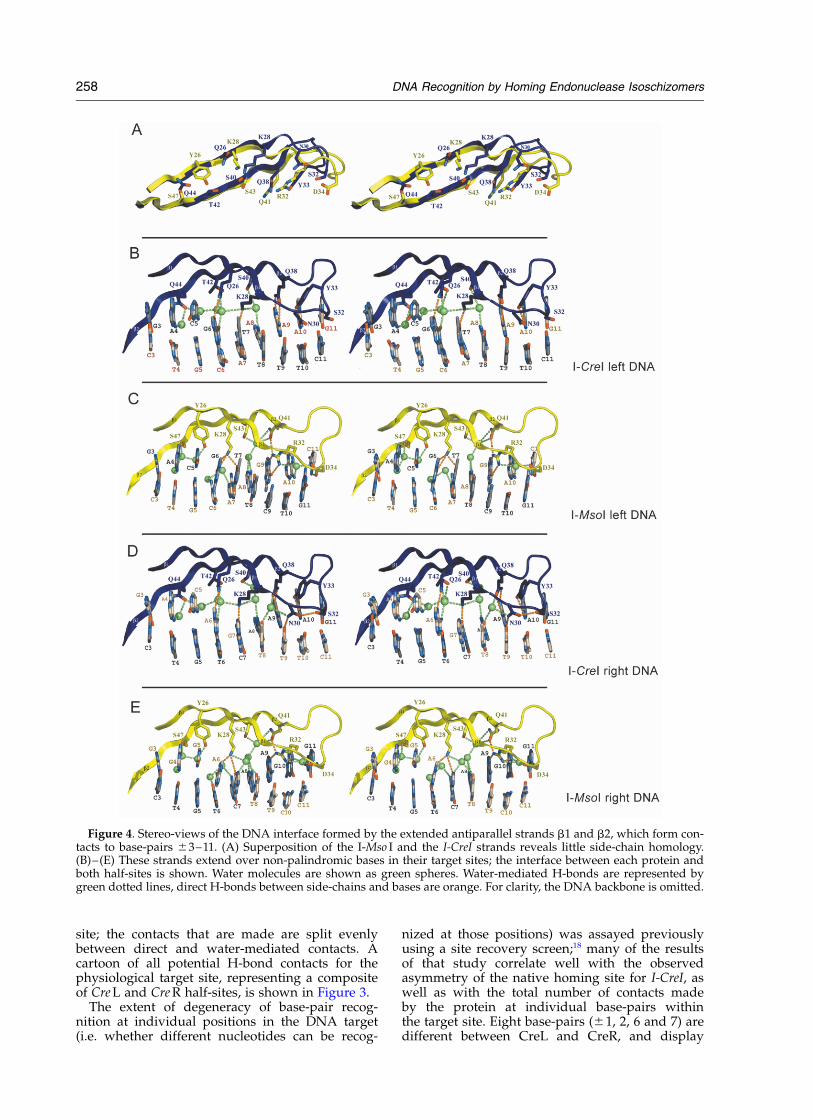
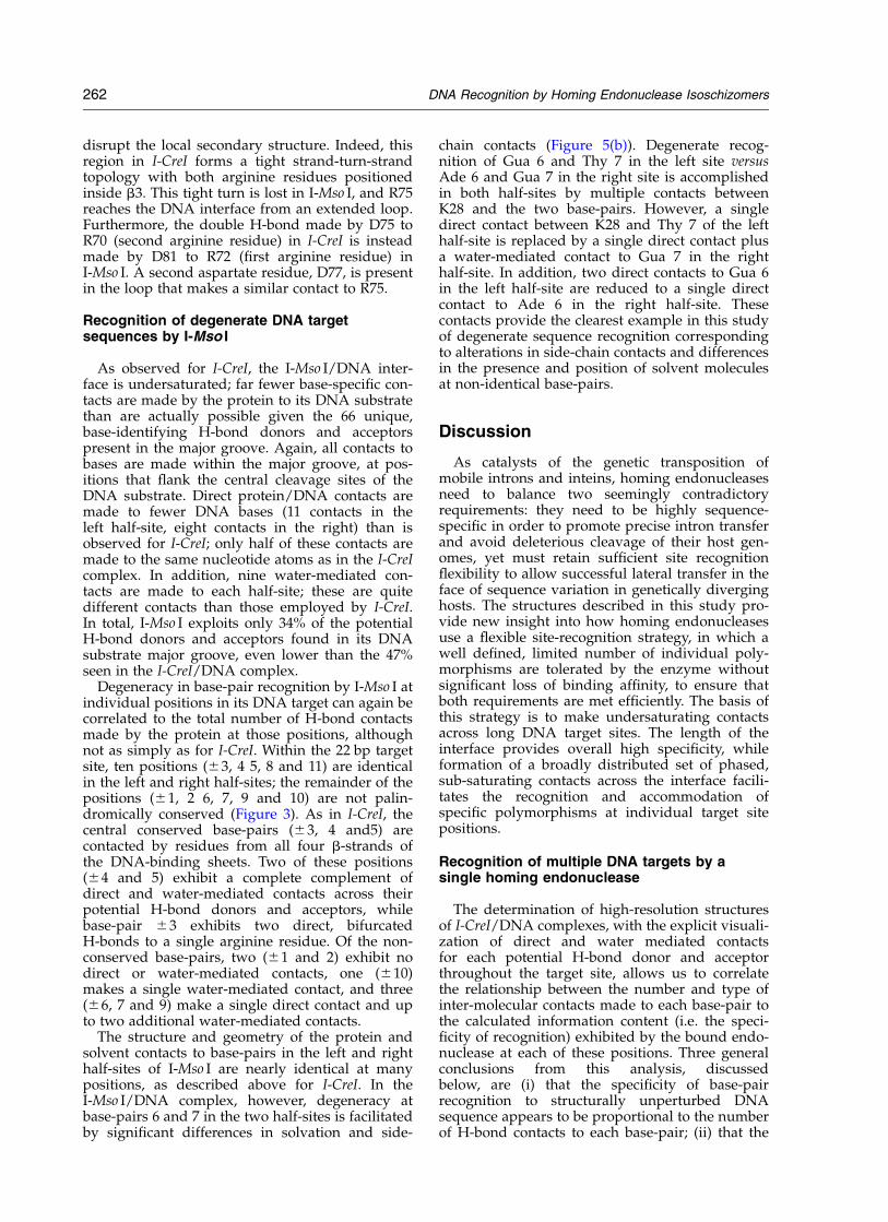
Figure 4. Stereo-views of the DNA interface formed by the extended antiparallel strands b1 and b2, which form con-tacts to base-pairs ^3–11. (A) Superposition of the I-Mso I and the I-CreI strands reveals little side-chain homology.(B)–(E) These strands extend over non-palindromic bases in their target sites; the interface between each protein andboth half-sites is shown. Water molecules are shown as green spheres. Water-mediated H-bonds are represented bygreen dotted lines, direct H-bonds between side-chains and bases are orange. For clarity, the DNA backbone is omitted.
258 DNA Recognition by Homing Endonuclease Isoschizomers

significant degeneracy in site recovery experi-ments. Four of these base-pairs (^1 and 2) makeno direct or water-mediated contact to proteinside-chains and are located between between thecleaved phosphate groups in the substrate. Theother four non-conserved base-pairs (^6 and ^7)exhibit one direct side-chain contact and oneadditional water-mediated contact. In contrast,the remaining base-pairs are palindromically con-served between CreL and CreR, and display sig-nificantly higher recognition specificity in siterecovery experiments. Of these, two base-pairs(^3 and ^10) display direct contacts to two pro-tein side-chain atoms; two (^4 and ^5) displaydirect contacts to two protein side-chain atomsand one additional water-mediated contact; onebase-pair (^ 9) displays three direct contacts tothe protein, and one base-pair (^8) displays threewater-mediated contacts. The correlation betweenthe structures of the DNA complexes and thesequence specificity displayed by these enzymes isdiscussed in detail below.
The contacts to base-pairs in Cre L and Cre R arenearly identical, both at positions that are con-served, and at positions that differ between thetwo DNA target sites. Protein strands b1 and b2,
comprising residues 21–48, form the most exten-sive part of the complex interface, extending fromthe central bases ^3 to ^11 (Figure 4(b) and (d)).From these strands, 16 direct contacts are made tothe DNA target (eight to each half-site) in eithercomplex. In each structure, Y33 and Q38 makeidentical bipartite contacts to conserved bases (toAde ^10 and Ade ^9, respectively) and Q44makes a single direct contact to Ade ^4. Thecontacts made to the non-palindromic bases atpositions ^6 and ^7 are structurally conserved(Figure 5(a) and (b)). In the Cre L structure, Q26contacts Gua ^6 and K28 contacts Thy ^7; in theCre R structure, these same residues make similarcontacts to Ade ^6 and Gua ^7. These contactsare appropriate for this particular nucleotide poly-morphism: alternate base-pairs at either positionwould fail to present suitable H-bond acceptors tothese same side-chain atoms. The positions andinteractions of solvent molecules to these non-conserved base-pairs are virtually identical.
The only observable, and quite subtle, differencein the protein/DNA interface between Cre L andCre R appears to be a contact made by N30 to Thy^9. Though strong density is evident in Cre R forthis residue, the corresponding density is not as
Figure 5. Stereo-views of contacts made by I-CreI and by I-Mso I to non-conserved base-pairs at positions ^6 and 7.Note that I-CreI uses a set of contacts that are structurally and chemically similar for the two different dinucleotidebase-pair sequences, whereas I-Mso I uses a set of contacts in which a water molecule is used in place of a thyminemethyl group.
DNA Recognition by Homing Endonuclease Isoschizomers 259

strong in Cre L and the residue appears to exhibithigher torsional mobility in that complex. Thisresidue is located directly adjacent to the region ofthe protein (a loop connecting strands b1 and b2)that undergoes the largest conformational changeupon DNA-binding.
Residues from the short strands b3 and b4 makeadditional contacts across the three most centralpalindromically conserved base-pairs (^3–5)(Figures 2, 3 and 6) and appear to greatly increasespecificity at these base-pairs. Bipartite contactsare made by R68 to Gua ^5 and R70 to Gua ^3.R70 forms an additional bond with Thy ^4 (acrossfrom Ade ^4, contacted by Q44). Furthermore,Q44, R68 and D75 are all in H-bond distance to asingle common water molecule that, in turn,makes a second specific contact to Ade ^4. This isthe most obvious sequence-specific, water-mediated contact in the protein/DNA interface.Identical contacts are made by each of these resi-dues to the palindromic bases in either half-site.
Binding of I-CreI to its target DNA involves sig-nificant conformational changes in both molecules(Figure 7). Superposition of the bound andunbound enzyme structures reveals differencesprimarily in two loop regions. Residues 29–37,which connect strands b1 and b2 in the protein/DNA interface, participate in DNA binding andadopt a significantly altered conformation. Thisloop donates three side-chains (Asn30, Ser32, andTyr33) to contacts to nucleotides ^ 10 and 11 atthe ends of the homing site and provides a
Figure 6. Stereo-views of theDNA interface formed by the b3and b4 protein strands. I-CreI withDNA bases is on the top; I-Mso Iwith bases is on the bottom. Theinterface of only one half-site isshown, since these bases are palin-dromic and the observed inter-actions are identical in both half-sites. Direct H-bonds between side-chains and bases are orange. Forclarity, the DNA backbone isomitted.
Figure 7. Conformational changes associated with I-CreI binding to DNA. (A) A superposition of boundand unbound protein backbones. The largest, and onlysignificant, motions are confined to loops connectingb-strands 1 and 2 at the distal ends of each subunit andtheir DNA half-sites, and related loops precedingb-strand 3 in each subunit. (B) The local overwinding(twist) and resulting reduction in roll values for DNAbase-pairs across the four base center of the cleavagetarget. These motions result in closure of the minorgroove at the sites of cleavage and a shallow bend inthe DNA around the enzyme surface. DNA bend para-meters were calculated with program 3DNA.47
260 DNA Recognition by Homing Endonuclease Isoschizomers

distinctive twist of the b-ribbon that allows it tomaintain contact over a long DNA site. A secondloop preceding strand b3 does not place side-chains in contact with nuceotide base-pairs, butdoes optimize contacts to the phosphate backbonein the region of basepairs ^3. The target DNA isgradually bent around the endonuclease bindingsurface, giving an overall curvature across theentire length of the site of approximately 458. TheDNA is locally overwound between bases 23 toþ3 (twist rising to ,508), with a correspondingdeformation in the base-pair propeller twist andbuckle angles for those same bases. The bendingof the DNA is symmetric, resulting from thecombination of two identical kinks in the DNAcaused by positive roll values of 188 locatedthree bases distant from each cleavage site.27 Asobserved for the DNA complex with the endo-nuclease domain of PI-SceI,25 the central 4 bpregion of the cleavage site displays negative rollvalues, which translate into a narrowing of theminor groove. As a result, the scissile phosphategroups are positioned 5 A apart and are locatednear the conserved acidic residues and boundmagnesium ions in the active sites.
Recognition of a similar DNA target by thedivergent isoschizomer I-Mso I
The I-Mso I/DNA complex contains the native,asymmetric DNA target site crystallized in spacegroup P1 with one complex per unit cell. Thesequences of the I-Mso I DNA target half-sitesused in his study are identical with the half-sitesof Cre L and Cre R with the exception of G:C at 29and G:C at þ10 (which are A:T base-pairs in Cre Land Cre R, respectively). The uncleaved, 24 bpDNA duplex in the crystal structure contains the22 bp I-Mso I target site flanked by one additionalbase-pair at either end (Figure 1(a)). Fortuitously,the orientation of the DNA in this structure wasnot averaged (i.e. the DNA was found in a singleunique orientation). This allowed a direct compari-son of unique nucleotide–protein–solvent contactsbetween half-sites for this enzyme to the contactsmade in the Cre L and Cre R structures describedabove.
All but the first two and last five residues in eachprotein chain are visible in the structure ofI-Mso I. Overall, the structures display 33%sequence identity22 (Figure 1(b)) and an r.m.s.d.between all conserved atoms of 1.29 A (Figure2(a)). As observed for I-CreI,5 three divalent cal-cium ions are present in the active sites and thereis a similar enzyme-imposed 108 bend of the DNAsubstrate. The three most critical active-siteresidues found in I-CreI are conserved: D20, Q47and K98 in I-CreI are D22, Q50, and K104 inI-Mso I. The DNA-binding saddle formed by theantiparallel b-sheets in I-Mso I superimposes wellwith the b-sheets of I-CreI, except for variation inthe peripheral loops connecting b1 to b2 and b3 tob4 (Figure 2(b) and (c)). The greatest structural
difference between the two proteins is in the loopconnecting a5 to a6; in I-Mso I, this loop does notfold as tightly to the body of the protein andmakes fewer contacts to the DNA backbone (Figure2(c)). I-Mso I is seven residues longer than I-CreI(170 versus 163). Two of these additional residuesare inserted near the N-terminal tail and onenear the C-terminal tail. An additional residue isinserted into the loop between b1 and b2, and theremaining three are inserted into or near thesequence spanning b3 and b4 (Figure 1(b)). Thelatter strands fold into part of the protein/DNAinterface and the insertions in this region disruptthe secondary structure as discussed below(Figures 1(b) and 2(b)).
The structures of I-CreI and I-Mso I revealedremarkable and unexpected differences in theirDNA interfaces (Figures 3–6). Despite nearly iden-tical DNA substrates, only five of the approxi-mately 25 residues of each subunit of I-CreIcontacting the DNA substrate are conserved inboth identity and function in I-Mso I. The moststriking differences are found throughout the b1and b2 strands that extend over 8 bp of each half-site (Figure 4). Except for S22 and S40 in I-CreIrelative to S24 and S43 in I-Mso I, respectively,which contact similar solvent and backbone atomsin the interface, no residue from these b-strands isconserved in structure or in binding function. Forexample, K28 of each protein appear to align in aninitial sequence alignment created prior to thestructure-determination of I-Mso I; however, theseside-chains make different contacts to the DNA:K28 in I-Mso I is actually functionally homologousto Q26 in I-CreI (Figures 3–5). Similarly, while theI-Mso I residue Q41 is equivalent to Q38 in I-CreIand shares a nearly identical Ca position, theirside-chains make entirely different contacts to theDNA. Q38 in I-CreI makes a pair of direct bipartitebonds to Ade ^9, whereas Q41 in I-Mso I is notdirectly involved in DNA binding. Yet again, Y33in I-CreI makes a bipartite contact to Ade ^10,while the homologous residue in I-Mso I, Y35,contacts a nearby backbone phosphate group.Thus, these enzymes bound to their respectiveDNA target sites reveal a much greater degree ofstructural divergence at the DNA interface thanwas predicted from primary sequence alignmentsand site similarities.
The direct protein/DNA contacts are more con-served between the final two strands, b3 and b4,that make up the rest of the DNA-binding saddlefor both enzymes (Figure 6). This region includesthree conserved residues: R68, R70 and D75 inI-CreI are R72, R75, and D81 in I-Mso I. In eachprotein, both arginine residues make highlyspecific bipartite bonds to conserved guaninebases. The conservation of contacts for these resi-dues is surprising, as this region of the proteinbackbone displays the greatest amount of struc-tural divergence between the two enzymes. I-Mso Ihas two additional residues inserted in this region,one of which is between R72 and R75, that severely
DNA Recognition by Homing Endonuclease Isoschizomers 261
disrupt the local secondary structure. Indeed, thisregion in I-CreI forms a tight strand-turn-strandtopology with both arginine residues positionedinside b3. This tight turn is lost in I-Mso I, and R75reaches the DNA interface from an extended loop.Furthermore, the double H-bond made by D75 toR70 (second arginine residue) in I-CreI is insteadmade by D81 to R72 (first arginine residue) inI-Mso I. A second aspartate residue, D77, is presentin the loop that makes a similar contact to R75.
Recognition of degenerate DNA targetsequences by I-Mso I
As observed for I-CreI, the I-Mso I/DNA inter-face is undersaturated; far fewer base-specific con-tacts are made by the protein to its DNA substratethan are actually possible given the 66 unique,base-identifying H-bond donors and acceptorspresent in the major groove. Again, all contacts tobases are made within the major groove, at pos-itions that flank the central cleavage sites of theDNA substrate. Direct protein/DNA contacts aremade to fewer DNA bases (11 contacts in theleft half-site, eight contacts in the right) than isobserved for I-CreI; only half of these contacts aremade to the same nucleotide atoms as in the I-CreIcomplex. In addition, nine water-mediated con-tacts are made to each half-site; these are quitedifferent contacts than those employed by I-CreI.In total, I-Mso I exploits only 34% of the potentialH-bond donors and acceptors found in its DNAsubstrate major groove, even lower than the 47%seen in the I-CreI/DNA complex.
Degeneracy in base-pair recognition by I-Mso I atindividual positions in its DNA target can again becorrelated to the total number of H-bond contactsmade by the protein at those positions, althoughnot as simply as for I-CreI. Within the 22 bp targetsite, ten positions (^3, 4 5, 8 and 11) are identicalin the left and right half-sites; the remainder of thepositions (^1, 2 6, 7, 9 and 10) are not palin-dromically conserved (Figure 3). As in I-CreI, thecentral conserved base-pairs (^3, 4 and5) arecontacted by residues from all four b-strands ofthe DNA-binding sheets. Two of these positions(^4 and 5) exhibit a complete complement ofdirect and water-mediated contacts across theirpotential H-bond donors and acceptors, whilebase-pair ^3 exhibits two direct, bifurcatedH-bonds to a single arginine residue. Of the non-conserved base-pairs, two (^1 and 2) exhibit nodirect or water-mediated contacts, one (^10)makes a single water-mediated contact, and three(^6, 7 and 9) make a single direct contact and upto two additional water-mediated contacts.
The structure and geometry of the protein andsolvent contacts to base-pairs in the left and righthalf-sites of I-Mso I are nearly identical at manypositions, as described above for I-CreI. In theI-Mso I/DNA complex, however, degeneracy atbase-pairs 6 and 7 in the two half-sites is facilitatedby significant differences in solvation and side-
chain contacts (Figure 5(b)). Degenerate recog-nition of Gua 6 and Thy 7 in the left site versusAde 6 and Gua 7 in the right site is accomplishedin both half-sites by multiple contacts betweenK28 and the two base-pairs. However, a singledirect contact between K28 and Thy 7 of the lefthalf-site is replaced by a single direct contact plusa water-mediated contact to Gua 7 in the righthalf-site. In addition, two direct contacts to Gua 6in the left half-site are reduced to a single directcontact to Ade 6 in the right half-site. Thesecontacts provide the clearest example in this studyof degenerate sequence recognition correspondingto alterations in side-chain contacts and differencesin the presence and position of solvent moleculesat non-identical base-pairs.
Discussion
As catalysts of the genetic transposition ofmobile introns and inteins, homing endonucleasesneed to balance two seemingly contradictoryrequirements: they need to be highly sequence-specific in order to promote precise intron transferand avoid deleterious cleavage of their host gen-omes, yet must retain sufficient site recognitionflexibility to allow successful lateral transfer in theface of sequence variation in genetically diverginghosts. The structures described in this study pro-vide new insight into how homing endonucleasesuse a flexible site-recognition strategy, in which awell defined, limited number of individual poly-morphisms are tolerated by the enzyme withoutsignificant loss of binding affinity, to ensure thatboth requirements are met efficiently. The basis ofthis strategy is to make undersaturating contactsacross long DNA target sites. The length of theinterface provides overall high specificity, whileformation of a broadly distributed set of phased,sub-saturating contacts across the interface facili-tates the recognition and accommodation ofspecific polymorphisms at individual target sitepositions.
Recognition of multiple DNA targets by asingle homing endonuclease
The determination of high-resolution structuresof I-CreI/DNA complexes, with the explicit visuali-zation of direct and water mediated contactsfor each potential H-bond donor and acceptorthroughout the target site, allows us to correlatethe relationship between the number and type ofinter-molecular contacts made to each base-pair tothe calculated information content (i.e. the speci-ficity of recognition) exhibited by the bound endo-nuclease at each of these positions. Three generalconclusions from this analysis, discussedbelow, are (i) that the specificity of base-pairrecognition to structurally unperturbed DNAsequence appears to be proportional to the numberof H-bond contacts to each base-pair; (ii) that the
262 DNA Recognition by Homing Endonuclease Isoschizomers

degree of specificity is not attenuated significantlyby the use of solvent molecules as direct bridgesbetween nucleotide atoms and protein side-chains;and (iii) information content (sequence specificity)is increased at individual base-pairs, particularlynear the center of the cleavage site, by indirectrecognition of DNA conformational preferences.
The distribution of related target site sequencesthat are recognized and cleaved by I-CreI has beendescribed using a site preference screen.18 In thoseexperiments, variant target site variants that werecleaved by the native enzyme were recoveredfrom a randomized homing site library. Usingthese data, the information content at each base-pair of the target site can be calculated using acomputational method that accounts for the proba-bility of each possible base being found at eachposition across the site, relative to the backgroundbase content expected at each position on the basisof genome composition or library design.28 Thedistribution and recovery of sequence variationsor polymorphisms among DNA sites recognizedby a single protein allows us to calculate the speci-ficity of the enzyme towards each individualbase-pair. This form of analysis, which can bedirected against collections of either naturallyoccurring sites that are recognized by a singleprotein, or sites recovered from a library screen, isthe accepted standard for describing site degener-acy for a DNA-binding protein and for quantifyingthe relative specificity observed at individualbase-pairs at each position in those sites. The infor-mation content or specificity exhibited towardsbase-pairs within the site are quantified as “bits”representing the probability of a specific base-pairat any given position. The units of bits as ameasure of specificity or information contentcorresponds to the units often shown on sequencelogo figures throughout the literature for aligned,homologous sites or of aligned homologous pro-tein signatures. Information content at individualbase-pairs in a collection of DNA recognition sitesrange from 0 bits, corresponding to completedegeneracy (25% probability of any base at a pos-ition) to two bits, corresponding to no degeneracy(100% chance of a unique base at that position).
The results of an information content analysisfor I-CreI (Figure 8) demonstrate several importantfeatures of site-recognition by that enzyme thatare presumably generalizable to the LAGLIDADGenzyme family. Information content (sequencespecificity) varies dramatically across the targetsite (Figure 8(a)). No single position in the site isdetermined absolutely, and no single position isdevoid of information content. With the exceptionof base-pairs ^1 and ^3 (discussed below), plotsof the information content versus the total numberof contacts made at each base-pair (Figure 8(b))give reasonable linear correlations of 0.84, 0.89and 0.86, corresponding to water-mediated con-tacts being counted as one-half, two-thirds or asfull equivalents of direct protein/DNA contacts,respectively. When only direct protein/DNA con-
tacts are counted for each base-pair (i.e. water-mediated contacts are not counted), the correlationis much lower (R 2 ¼ 0.57). When analyzedtogether, the structural and genetic analysessummarized above imply that water-mediatedcontacts impart nearly the same informationcontent to DNA target site recognition as directprotein/DNA contacts. However, sequence degen-eracy can be facilitated at some nucleotide pos-itions by the ability of water to reposition and/orreorient its dipole in response to sequence changes.This role for bound water is visualized directly atbase-pairs ^ 6 and 7 in the I-MsoI/DNA complex(Figure 5).
The observation that water-mediated contactscontribute to information content in protein/DNAbinding to a degree that roughly matches thecontribution of direct contacts is in agreementwith a variety of published studies, especiallythose describing site recognition by the Trp repres-sor and by Hox transcriptional regulators. Crystal-lographic analysis of the Trp repressor bound toits DNA operator sequence29 showed that sequencerecognition is accomplished entirely through acombination of water-mediated contacts to nucleo-tide bases and indirect readout of DNA geometry.This observation was subsequently validated bymutagenic30 and thermodynamic31 analyses of pro-tein binding. Similarly, a crystallographic analysis32
and subsequent mutagenesis study33 of a pairedclass homeodomain to its DNA site indicatedthat sequence-specific DNA recognition is drivenlargely by water-mediated contacts.
As mentioned above, base-pairs ^1 and ^3 bothdeviate from the linear correlation described abovefor the rest of the site (Figure 8(b)); in both casesthese positions display twice the information con-tent predicted by intermolecular contacts alone.One simple explanation for this is that these basesare found in the region of maximum DNA bend-ing, base twisting and unstacking in the complexnear the scissile phosphate groups (Figure 7).They may thus display conformational preferencesthat contribute favorably to binding energy andcould therefore be recognized indirectly as anadditional source of sequence specificity.
The overall information content of the entire22 bp I-CreI target site, consisting of a summationof individual contributions to specificity for eachbase-pair position (assuming independent recog-nition of individual base-pairs) is approximately24 bits, corresponding to an average of 1.09 bitsper base-pair. Information content is spreadunevenly over the target site, with no one base-pair displaying absolute sequence specificity forbinding (Figure 8). In contrast, the HincII restric-tion endonuclease, which displays strict recog-nition of sequences (G-T-(T/C)-(A/G)-A-C), has atotal information content of approximately 11,corresponding to an average of 1.83 bits per base-pair, and a predicted frequency of recognitionevery 1024 bases in a random genome sequence.28
The greater specificity per base-pair for a restriction
DNA Recognition by Homing Endonuclease Isoschizomers 263

enzyme is correlated directly to an extraordinarynumber of contacts between each target sitenucleotide and the protein. For example, the Mun Ienzyme, which recognizes a six base target site,forms 22 contacts to nucleotide bases in the majorand minor groove, and 18 contacts to the 14 phos-phate groups across the site;34 this corresponds toover double the number of contacts per base-pairformed by I-Cre I or I-Mso I.
The individual contacts between side-chains andnucleotide bases, in addition to exhibiting additivecontributions to information content, can be corre-lated with their contribution to the overall freeenergy of binding and the resulting associationconstants for the complex. For example, individual
DNA site mutations at position ^10 in the targetsite (corresponding to an inversion of an A:T base-pair that confounds the ability of the enzyme tomake its usual side-chain contacts at those pos-itions) causes an increase in KD of 100-fold (5 nM–560 nM), corresponding to an approximate DDGfor binding of 3–4 kcal/mol (1 cal ¼ 4.184 J).35 Incontrast, a similar base-pair inversion at base-pairs^11 causes a smaller tenfold increase in KD, corre-sponding to a more moderate DDG for binding of1–2 kcal/mol.35 The relative magnitude of theseeffects are in agreement with the correspondingvalues of information content in the protein–DNA complex for these two base-pairs (1.15 and0.65, respectively). Furthermore, the individual
Figure 8. Information content in the I-CreI/DNA complex. (a) Information content calculated at individual base-pairpositions across the target site. The positions with highest information content (^3, 4, 5, 8, 9 and 10) correspond topositions that are palindromically conserved in the native I-Cre I homing site. The average information content is 1.09bits per base-pair. Information content falls off to background beyond positions ^11. (b) Information content for indi-vidual positions in the I-CreI target half-site plotted against the number of contacts made to each base-pair. The linearcorrelation coefficient is calculated for bases 4–11 (filled diamonds), corresponding to positions outside the activesites and the region of significant DNA distortion. The plot and its linear correlation is shown for direct protein con-tacts only (left; R 2 ¼ 0.57) and for total direct and water-mediated contacts, with water-mediated contacts counted astwo-thirds of a direct contact (right; R 2 ¼ 0.89). The correlation is slightly lower if water-mediated contacts are countedas either half of a direct contact (R 2 ¼ 0.86) or as the equivalent of a full contact (R 2 ¼ 0.84). Note that base-pairs 1 and3 (grey ovals), which are located near the position of maximal DNA distortion and cleavage, display higher infor-mation content than expected solely on the basis of sequence-specific contact only (possibly indicating indirect readoutof conformational preferences at those positions), but that the overall trend is similar to bases outside this region.
264 DNA Recognition by Homing Endonuclease Isoschizomers
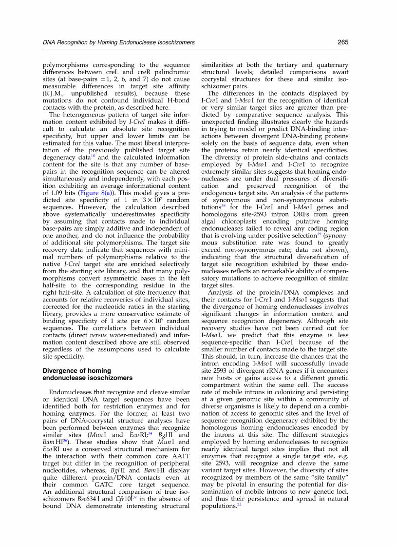
polymorphisms corresponding to the sequencedifferences between creL and creR palindromicsites (at base-pairs ^1, 2, 6, and 7) do not causemeasurable differences in target site affinity(R.J.M., unpublished results), because thesemutations do not confound individual H-bondcontacts with the protein, as described here.
The heterogeneous pattern of target site infor-mation content exhibited by I-CreI makes it diffi-cult to calculate an absolute site recognitionspecificity, but upper and lower limits can beestimated for this value. The most liberal interpre-tation of the previously published target sitedegeneracy data18 and the calculated informationcontent for the site is that any number of base-pairs in the recognition sequence can be alteredsimultaneously and independently, with each pos-ition exhibiting an average informational contentof 1.09 bits (Figure 8(a)). This model gives a pre-dicted site specificity of 1 in 3 £ 107 randomsequences. However, the calculation describedabove systematically underestimates specificityby assuming that contacts made to individualbase-pairs are simply additive and independent ofone another, and do not influence the probabilityof additional site polymorphisms. The target siterecovery data indicate that sequences with mini-mal numbers of polymorphisms relative to thenative I-CreI target site are enriched selectivelyfrom the starting site library, and that many poly-morphisms convert asymmetric bases in the lefthalf-site to the corresponding residue in theright half-site. A calculation of site frequency thataccounts for relative recoveries of individual sites,corrected for the nucleotide ratios in the startinglibrary, provides a more conservative estimate ofbinding specificity of 1 site per 6 £ 109 randomsequences. The correlations between individualcontacts (direct versus water-mediated) and infor-mation content described above are still observedregardless of the assumptions used to calculatesite specificity.
Divergence of homingendonuclease isoschizomers
Endonucleases that recognize and cleave similaror identical DNA target sequences have beenidentified both for restriction enzymes and forhoming enzymes. For the former, at least twopairs of DNA-cocrystal structure analyses havebeen performed between enzymes that recognizesimilar sites (Mun I and Eco RI;34 Bgl II andBam HI36). These studies show that Mun I andEco RI use a conserved structural mechanism forthe interaction with their common core AATTtarget but differ in the recognition of peripheralnucleotides, whereas, Bgl II and Bam HI displayquite different protein/DNA contacts even attheir common GATC core target sequence.An additional structural comparison of true iso-schizomers Bse634 l and Cfr10l37 in the absence ofbound DNA demonstrate interesting structural
similarities at both the tertiary and quaternarystructural levels; detailed comparisons awaitcocrystal structures for these and similar iso-schizomer pairs.
The differences in the contacts displayed byI-Cre I and I-Mso I for the recognition of identicalor very similar target sites are greater than pre-dicted by comparative sequence analysis. Thisunexpected finding illustrates clearly the hazardsin trying to model or predict DNA-binding inter-actions between divergent DNA-binding proteinssolely on the basis of sequence data, even whenthe proteins retain nearly identical specificities.The diversity of protein side-chains and contactsemployed by I-Mso I and I-Cre I to recognizeextremely similar sites suggests that homing endo-nucleases are under dual pressures of diversifi-cation and preserved recognition of theendogenous target site. An analysis of the patternsof synonymous and non-synonymous substi-tutions38 for the I-Cre I and I-Mso I genes andhomologous site-2593 intron ORFs from greenalgal chloroplasts encoding putative homingendonucleases failed to reveal any coding regionthat is evolving under positive selection39 (synony-mous substitution rate was found to greatlyexceed non-synonymous rate; data not shown),indicating that the structural diversification oftarget site recognition exhibited by these endo-nucleases reflects an remarkable ability of compen-satory mutations to achieve recognition of similartarget sites.
Analysis of the protein/DNA complexes andtheir contacts for I-Cre I and I-Mso I suggests thatthe divergence of homing endonucleases involvessignificant changes in information content andsequence recognition degeneracy. Although siterecovery studies have not been carried out forI-Mso I, we predict that this enzyme is lesssequence-specific than I-Cre I because of thesmaller number of contacts made to the target site.This should, in turn, increase the chances that theintron encoding I-Mso I will successfully invadesite 2593 of divergent rRNA genes if it encountersnew hosts or gains access to a different geneticcompartment within the same cell. The successrate of mobile introns in colonizing and persistingat a given genomic site within a community ofdiverse organisms is likely to depend on a combi-nation of access to genomic sites and the level ofsequence recognition degeneracy exhibited by thehomologous homing endonucleases encoded bythe introns at this site. The different strategiesemployed by homing endonucleases to recognizenearly identical target sites implies that not allenzymes that recognize a single target site, e.g.site 2593, will recognize and cleave the samevariant target sites. However, the diversity of sitesrecognized by members of the same “site family”may be pivotal in ensuring the potential for dis-semination of mobile introns to new genetic loci,and thus their persistence and spread in naturalpopulations.22
DNA Recognition by Homing Endonuclease Isoschizomers 265
Implication for engineering of homingendonucleases with novel specificities
Homing endonucleases (in particular theLAGLIDADG enzyme family) are highlysequence-specific, with DNA-binding domainsthat are highly modular, separable, and intimatelyassociated with catalytic active sites. As such,these endonucleases appear to be highly attractivetargets for the creation of gene-specific reagents,using a variety of tools for the engineering andselection of novel DNA-recognition activities.The importance of solvent-mediated contacts forDNA site recognition by these enzymes has clearimplications for the engineering variants withaltered specificity, in that any structure-based com-putational approaches to such a problem mustincorporate an accurate strategy for the explicitmodeling of water molecules in the protein/DNAinterface. At this time, successful strategies havebeen reported both for the selection of homingendonuclease point mutants with minor alterationsin site specificity35 and for the recombination andfusion of independent LAGLIDADG domains tocreate artificial chimeric enzymes with drasticallyaltered site-specificity.40 Future attempts to engi-neer artificial homing endonucleases withcompletely novel sequence specificities will pre-sumably exploit both of these types of strategies,but will be dependent also on the concertedrandomization and selection of large numbers ofresidues in the DNA-binding interface as part ofan overall redesign strategy. The results reportedhere seem to indicate that the accurate modelingand prediction of the structural and energeticfeatures of direct and water-mediated protein/DNA contacts, in combination with continueddevelopment of powerful selection methods forenzyme/DNA specificity, will be critical for thesuccess of such experiments.
In addition, the comparative analysis of bindingby I-Mso I and I-CreI to similar DNA target sitesimplies that a large number of diverse side-chainand solvent packing Scheme may allow the recog-nition of a given DNA target site, and that varia-tions within selected interfaces may be associatedwith dramatic differences in overall sequencespecificity. It should be possible to use additionalanalyses of DNA-binding properties of LAGLI-DADG homing endonucleases to clarify the rulesgoverning sequence-specific DNA recognition andthus facilitate the design of novel, gene-specificproteins.
Materials and Methods
Protein expression and purification
The I-Mso I ORF was PCR amplified from pET-I-Mso I22
and subcloned into the Nde I/Xho I sites of the pI-CreIvector41 to make pI-Mso I. I-CreI and I-Mso I were purifiedin identical fashion. Briefly, protein was overexpressed
by induction with 0.5 mM IPTG in BL21[DE3] E. colicells overnight at 15 8C. Cells were harvested by centrifu-gation and lysed by sonication in 50 mM Tris (pH 8.0),100 mM NaCl. Cell debris was removed by centrifu-gation at 40,000g for 45 minutes at 4 8C, then forcedthrough a 0.2 mm syringe filter and applied to a heparincolumn (Pharmacia). Protein eluted with an increasingsalt gradient as a single peak that was collected, dilutedwith an equal volume of 50 mM Tris (pH 8.0), andloaded onto the heparin column. After the secondelution, protein was .95% pure as determined bySDS-PAGE analysis. The protein solution was dialyzedovernight against storage buffer (50 mM Tris (pH 8.0),100 mM NaCl, 1 mM CaCl2) and concentrated to,4 mg/ml by centrifugation (Centriprep, Millipore),flash-frozen in liquid nitrogen and stored at 280 8C.
Crystallization
The design of DNA oligonucleotides for I-CreI andI-Mso I crystallizations is summarized in Results. Despitecrystallizing in different space groups, all I-CreI/DNAcrystals grew in previously described conditions(21–27% PEG 400, 100 mM Mes (pH 6.2–6.8), 10 mMCaCl2, 20 mM NaCl, 22 8C) at ,4 mg/ml protein with a1.2–1.7 molar excess of DNA target substrate. Cre Rcrystals grew readily in three to six days; Cre L crystalsgrew as thick needles in one to two weeks. Diffraction-quality I-Mso I/DNA crystals grew at 18 8C in 19–22%PEG 400, 100 mM Tris (pH 7.3–7.9), 10 mM CaCl2,20 mM NaCl at 3.5 mg/ml of protein with a 1.5–2.0molar excess of DNA substrate. Crystals appearedwithin one hour, and diffraction quality crystals wereobtained after two to six days.
Data collection
Crystals were removed directly from the drops inwhich they grew, suspended in a fiber loop, frozen inliquid nitrogen and maintained at 100 K during data col-lection. Data from crystals of Cre R and the I-Mso I/DNAcomplex were collected at the Advanced Light Source(beamline 5.0.2). Data from the crystals of Cre L were col-lected at the Advanced Photon Source (beamline BM-19)with 2u ¼ 88. Data were reduced using the DENZO/SCALEPACK crystallographic data reduction package(Table 1).
The structures were solved via molecular replacementusing EPMR† with the low-resolution I-CreI/DNA com-plex structures as an initial search model for Cre L andCre R and a polyalanine I-CreI model (lacking loops anda6) for the I-Mso I structure. In both Cre L and Cre R, twoI-CreI/DNA complexes were found in each asymmetricunit. Only one I-Mso I structure was found within its P1cell. All structures were modeled in XtalView42 and O43,then refined using CNS44 with 5% of the data set asidefor cross-validation45. The final refinement statistics(Table 1) for Cre L were Rwork/Rfree ¼ 0.219/0.246, forCre R Rwork/Rfree ¼ 0.195/0.229, and for the I-Mso Icomplex Rwork/Rfree ¼ 0.218/0.253. Geometric analysis ofthe structures using PROCHECK46 indicates no residue
† Kissinger, C. R. & Gehlhaar, D. K. (1997). EPMR: Aprogram for crystallographic molecular replacement byevolutionary search. Agouron Pharmaceuticals, La Jolla,CA.
266 DNA Recognition by Homing Endonuclease Isoschizomers
in any structure with generously allowed or unfavorablebackbone dihedral angles.
Protein Data Bank accession codes
The I-Mso I/DNA (RCSB accession code 1M5X), Cre L(IN3E) and Cre R (1N3F) structural models and datahave been deposited in the Protein Data Bank.
Acknowledgements
The work described here was funded by grantsfrom the NIH to B.S. (GM49857) and to R.M.(CA88942), and by a grant from the NaturalSciences and Engineering Research Council ofCanada (GP0002830) to C.L. and M.T. B.C. wassupported by an Interdisciplinary Training Grantin Cancer Research pre-doctoral fellowship (T32CA80416). We thank members of the FHCRCstructural biology program, particularly AdrianFerre D’Amare, for helpful criticisms and advice.
References
1. Dujon, B. (1989). Group I introns as mobile geneticelements: facts and mechanistic speculations—areview. Gene, 82, 91–114.
2. Lambowitz, A. M. & Belfort, M. (1993). Introns asmobile genetic elements. Annu. Rev. Biochem. 62,587–622.
3. Belfort, M. & Perlman, P. S. (1995). Mechanisms ofintron mobility. J. Biol. Chem. 270, 30237–30240.
4. Belfort, M. & Roberts, R. J. (1997). Homing endo-nucleases—keeping the house in order. Nucl. AcidsRes. 25, 3379–3388.
5. Chevalier, B., Monnatt, R. J. & Stoddard, B. L. (2001).The LAGLIDADG homing endonuclease I-CreIshares three divalent cations between two activesites. Nature Struct. Biol. 8, 312–316.
6. Jacquier, A. & Dujon, B. (1985). An intron-encodedprotein is active in a gene conversion process thatspreads an intron into a mitochondrial gene. Cell,41, 383–394.
7. Turmel, M., Cote, V., Otis, C., Mercier, J. P., Gray,M. W., Lonergan, K. M. & Lemieux, C. (1995). Evolu-tionary transfer of ORF-containing group I intronsbetween different subcellular compartments (chloro-plast and mitochondrion). Mol. Biol. Evol. 12,533–545.
8. Cho, Y., Qiu, Y.-L., Kuhlman, P. & Palmer, J. D.
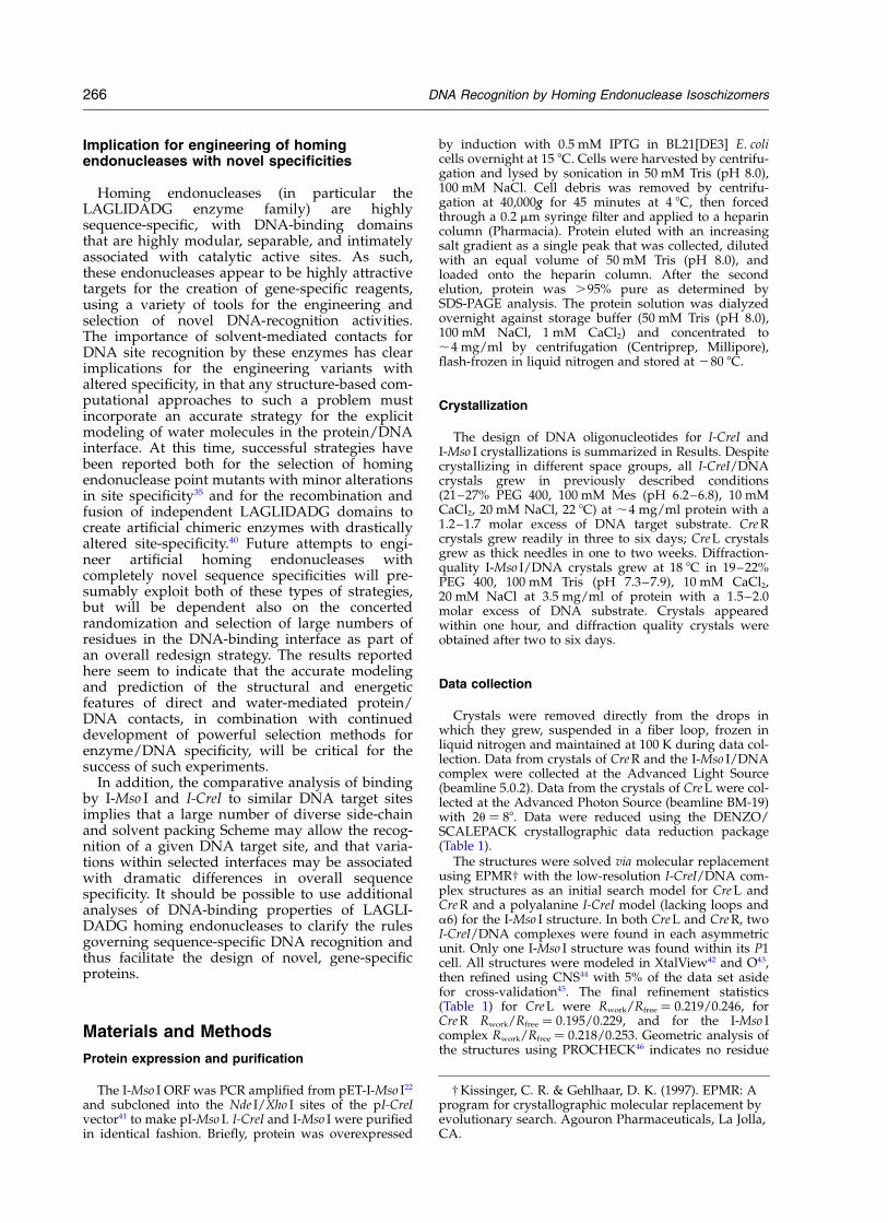
Table 1. Data and refinement statistics
A. DataStructure I-Cre I (left; CreL) I-Cre I (right; CreR) I-Mso ISource APS 5.0.2 ALS 5.0.2 ALSResolution limit (A) 2.50 2.00 2.25Wavelength (A) 1.00 1.10 1.10Space group P212121 P21 P1Unit cell parametersa, b, c (A) 46.7, 68.4, 301.9 78.4, 76.4, 81.1 41.5, 42.2, 71.3a, b, g (deg.) 90, 90, 90 90, 108.8, 90 73.3, 73.2, 71.1
Measured reflections 77,280 193,008 38,547Unique reflections 32,363 60,035 19,849Rmerge
a 5.4 (32.4) 4.0 (20.8) 3.0 (7.6)Competeness (%)1 93.5 (71.1) 99.1 (98.1) 97.5 (96.9)
B. RefinementRwork (%) 21.9 19.5 21.8Rfree (%)b 24.6 22.9 25.3Resolution(A) 50–2.50 50–2.00 50–2.25No. atoms 7102 7331 3824No. water molecules 233 513 229
r.m.s. deviationsBond length (A) 0.008 0.006 0.006Bond angles (deg.) 1.61 1.26 1.22
Ramachandran plotFavorable (%) 88.9 88.8 92.3Allowed (%) 11.1 11.2 7.7Generous (%) 0 0 0Unfavorable (%) 0 0 0
Mean B value (A2)Overall 41.5 39.9 38.3Protein 41.2 37.6 34.0DNA 42.7 41.4 48.9Solvent 38.9 42.3 40.6Cations 31.0 38.1 53.7
a The numbers in parentheses are statistics from the highest-resolution shell.b Rfree was calculated with 5% of the data that was not used for calculation of Rwork.
DNA Recognition by Homing Endonuclease Isoschizomers 267
(1998). Explosive invasion of plant mitochondria bya group I intron. Proc. Natl Acad. Sci. USA, 95,14244–14249.
9. Dalgaard, J. Z., Klar, A. J., Moser, M. J., Holley, W. R.,Chatterjee, A. & Mian, I. S. (1997). Statistical model-ing and analysis of the LAGLIDADG family of site-specific endonucleases and identification of an inteinthat encodes a site-specific endonuclease of the HNHfamily. Nucl. Acids Res. 25, 4626–4638.
10. Heath, P. J., Stephens, K. M., Monnat, R. J. &Stoddard, B. L. (1997). The structure of I-CreI, agroup I intron-encoded homing endonuclease.Nature Struct. Biol. 4, 468–476.
11. Duan, X., Gimble, F. S. & Quiocho, F. A. (1997).Crystal structure of PI-SceI, a homing endonucleasewith protein splicing activity. Cell, 89, 555–564.
12. Chevalier, B. S. & Stoddard, B. L. (2001). Homingendonucleases: structural and functional insight intothe catalysts of intron/intein mobility. Nucl. AcidsRes. 29, 3757–3774.
13. Colleaux, L., D’Auriol, L., Galibert, F. & Dujon, B.(1988). Recognition and cleavage site of the intron-encoded omega transposase. Proc. Natl Acad. Sci.USA, 85, 6022–6026.
14. Thompson, A. J., Yuan, X., Kudlicki, W. & Herrin,D. L. (1992). Cleavage and recognition patternof a double-strand-specific endonuclease (I-Cre I)encoded by the chloroplast 23S rRNA intron ofChlamydomonas reinhardtii. Gene, 119, 247–251.
15. Marshall, P. & Lemieux, C. (1991). Cleavage patternof the homing endonuclease encoded by the fifthintron in the chloroplast large subunit rRNA-enco-ding gene of Chlamydomonas eugametos. Gene, 104,241–245.
16. Dalgaard, J. Z., Garrett, R. A. & Belfort, M. (1993).A site-specific endonuclease encoded by a typicalarchaeal intron. Proc. Natl Acad. Sci. USA, 90,5414–5417.
17. Durrenberger, F. & Rochaix, J.-D. (1993). Characteri-zation of the cleavage site and the recognitionsequence of the I-Cre I DNA endonuclease encodedby the chloroplast ribosomal intron of Chlamydo-monas reinhardtii. Mol. Gen. Genet. 236, 409–414.
18. Argast, G. M. (1998). I-Ppo I and I-Cre I homing sitesequence degeneracy determined by random muta-genesis and sequential in vitro enrichment. J. Mol.Biol. 280, 345–353.
19. Jurica, M. S., Monnat, R. J. & Stoddard, B. L. (1998).DNA recognition and cleavage by the LAGLIDADGhoming endonuclease I-Cre I. Mol. Cell, 2, 469–476.
20. Koufopanou, V., Goddard, M. R. & Burt, A. (2002).Adaptation for horizontal transfer in a homing endo-nuclease. Mol. Biol. Evol. 19, 239–246.
21. Goddard, M. R. & Burt, A. (1999). Recurrent invasionand extinction of a selfish gene. Proc. Natl Acad Sci.USA, 96, 13880–13885.
22. Lucas, P., Otis, C., Mercier, J. P., Turmel, M. &Lemieux, C. (2001). Rapid evolution of the DNA-binding site in LAGLIDADG homing endonucleases.Nucl. Acids Res. 29, 960–969.
23. Ichiyanagi, K., Ishino, Y., Ariyoshi, M., Komori, K. &Morikawa, K. (2000). Crystal structure of an archaealintein-encoded homing endonuclease PI-PfuI. J. Mol.Biol. 300, 889–901.
24. Silva, G. H., Dalgaard, J. Z., Belfort, M. & Van Roey,P. (2003). Crystal structure of the thermostablearchaeal intron-encoded endonuclease I-Dmo I.J. Mol. Biol. 286, 1123–1136.
25. Moure, C. M., Gimble, F. S. & Quiocho, F. A. (2002).
Crystal structure of the intein homing endonucleasePI-SceI bound to its recognition sequence. NatureStruct. Biol. 9, 764–770.
26. Pingoud, A. & Jeltsch, A. (2001). Structure and func-tion of type II restriction endonucleases. Nucl. AcidsRes., 3705–3727.
27. Jurica, M. S., Monnat, R. J., Jr & Stoddard, B. L.(1998). DNA recognition and cleavage by the LAGLI-DADG homing endonuclease I-CreI. Mol. Cell, 2,469–476.
28. Schneider, T. D., Stormo, G. D., Gold, L. &Ehrenfeucht, A. (1986). Information content of bind-ing sites on nucleotide sequences. J. Mol. Biol. 188,415–431.
29. Otwinowski, Z., Schevitz, R. W., Zhang, R. G.,Lawson, C. L., Joachimiak, A., Marmorstein, R. Q.et al. (1988). Crystal structure of trp repressor/operator complex at atomic resolution. Nature, 335,321–329.
30. Joachimiak, A., Haran, T. E. & Sigler, P. B. (1994).Mutagenesis supports water mediated recognitionin the trp repressor–operator system. EMBO J. 13,367–372.
31. Ladbury, J. E., Wright, J. G., Sturtevant, J. M. & Sigler,P. B. (1994). A thermodynamic study of the trprepressor–operator interaction. J. Mol. Biol. 238,669–681.
32. Wilson, D. S., Guenther, B., Desplan, C. & Kuriyan, J.(1995). High resolution crystal structure of a paired(Pax) class cooperative homeodomain dimer onDNA. Cell, 82, 709–719.
33. Wilson, D. S., Sheng, G., Jun, S. & Desplan, C. (1996).Conservation and diversification in homeodomain–DNA interactions: a comparative genetic analysis.Proc. Natl Acad. Sci. USA, 93, 6886–6891.
34. Deibert, M., Grazulis, S., Janulaitis, A., Siksnys, V. &Huber, R. (1999). Crystal structure of MunI restric-tion endonuclease in complex with cognate DNA at1.7 A resolution. EMBO J. 18, 5805–5816.
35. Seligman, L., Chisholm, K. M., Chevalier, B. S.,Chadsey, M. S., Edwards, S. T., Savage, J. H. & Veil-let, A. L. (2002). Mutations altering the cleavagespecificity of a homing endonuclease. Nucl. AcidsRes. 30, 3870–3879.
36. Lukacs, C. M., Kucera, R., Schildkraut, I. &Aggarwal, A. K. (2000). Understanding the immuta-bility of restriction enzymes: crystal structure ofBglII and its DNA substrate at 1.5 A resolution.Nature Struct. Biol. 7, 134–140.
37. Grazulis, S., Deibert, M., Rimseliene, R., Skirgaila, R.,Sasnauskas, G., Lagunavicius, A. et al. (2002). Crystalstructure of the Bse634I restriction endonuclease:comparison of two enzymes recognizing the sameDNA sequence. Nucl. Acids Res. 30, 876–885.
38. Yang, Z. & Bielawaki, J. P. (2000). Statistical methodsfor detecting molecular adaptation. TREE, 15, 496–503.
39. Hughes, A. L. (1999). Adaptive Evolution of Genes andGenomes, Oxford University Press, Oxford.
40. Chevalier, B. S., Kortemme, T., Chadsey, M. S., Baker,D. R. J., Monnat, J. & Stoddard, B. L. (2002). Design,activity and structure of a highly specific artificialendonuclease. Molec. Cell, 10, 895–905.
41. Wang, J., Kim, H.-H., Yuan, X. & Herrin, D. L. (1997).Purification, biochemical characterization and pro-tein–DNA interactions of the I-Cre I endonucleaseproduced in Escherichia coli. Nucl. Acids Res. 25,3767–3776.
42. McRee, D. E. (1999). A versatile program for manipu-
268 DNA Recognition by Homing Endonuclease Isoschizomers
lating atomic coordinates and electron density.J. Struct. Biol. 125, 156–165.
43. Jones, T. A., Zou, J.-Y., Cowan, S. W. & Kjeldgaard,M. (1991). Improved methods for building proteinmodels in electron density maps and the location oferrors in these models. Acta Crystallog. sect. A, 47,110–119.
44. Brunger, A. T., Adams, P. D., Clore, G. M., DeLano,W. L., Gros, P., Grosse-Kunstleve, R. W. et al. (1998).Crystallography and NMR system: a new softwaresuite for macromolecular structure determination.Acta Crystallog. sect. D, 54, 905–921.
45. Brunger, A. (1993). Assessment of phase accuracy bycross validation: the free R value. Meth. Appl. ActaCrystallog. sect. D, 49, 24–36.
46. Laskowski, R. J., Macarthur, M. W., Moss, D. S. &Thornton, J. M. (1993). PROCHECK: a program tocheck the stereochemical quality of protein struc-tures. J. Appl. Crystallog. 26, 283–291.
47. Lu, X.-J., Zippora, S. & Olson, W. K. (2000). A DNAconformational motifs in ligand bound doublehelices. J. Mol. Biol. 300, 819–840.
Edited by K. Morikawa
(Received 11 December 2002; received in revised form 27 March 2003; accepted 27 March 2003)
DNA Recognition by Homing Endonuclease Isoschizomers 269