
First Report of Nocardia farcinica Bursitis in a Patient ... · diabetes mellitus. A 67-yr-old man...
4
ISSN 2234-3806 • eISSN 2234-3814 252 www.annlabmed.org http://dx.doi.org/10.3343/alm.2014.34.3.252 Ann Lab Med 2014;34:252-255 http://dx.doi.org/10.3343/alm.2014.34.3.252 Letter to the Editor Clinical Microbiology First Report of Nocardia farcinica Bursitis in a Patient with Diabetes Mellitus Soon-Deok Park, Ph.D. 1 , Han Jun Kim, M.D. 1 , In Ho Jang, Ph.D. 1 , Young Uh, M.D. 1 , Juwon Kim, M.D. 1 , Kap Joon Yoon, M.D. 1 , and Jin-Rok Oh, M.D. 2 Departments of Laboratory Medicine 1 and Orthopedic Surgery 2 , Yonsei University Wonju College of Medicine, Wonju, Korea Dear Editor Nocardia species of the family Nocardiaceae form a homoge- nous cluster within the order Corynebacteriales, formerly subor- der Corynebacteriaceae [1]. Nocardia, a genus of aerobic acti- nomycetes, is characterized by filamentous, branching, gram positive, partially acid-fast bacteria found worldwide as soil sap- rophytes [2]. The most common site of Nocardia infection is the respiratory tract, with subsequent dissemination to distant or- gans. Disseminated nocardiosis to the brain, kidneys, joints, or eyes can occur by hematogenous spread of infection [3]. Micro- biological diagnosis of nocardiosis and identification of Nocardia clinical isolates to the species level by conventional methods are difficult. However, identification to species level is important to characterize associated disease manifestations, to predict anti- microbial susceptibility, and for epidemiological and ecological purposes [2]. Various nucleic acid amplification methods target- ing conserved Nocardia gene regions have been proposed for accurate species-level identification [4-6]. Sequence analysis of the 16S ribosomal RNA (rRNA) gene has become the gold stan- dard for definitive species identification [2, 7]. N. farcinica is considered an opportunistic pathogen that usu- ally affects patients with impaired cell-mediated immunity and predisposing factors like hematologic malignances, treatment with corticosteroids, chronic pulmonary conditions, renal dis- eases, and other conditions leading to immunosuppression. The organism is increasingly recognized as a human pathogen in in- fections of the lungs, brain, skin, wounds, and kidneys [5]. Here, we report a case of bursitis due to N. farcinica infection, con- firmed by 16S rRNA sequencing, in a patient with long-standing diabetes mellitus. A 67-yr-old man was admitted to the hospital complaining of right ankle pain that had persisted for 4 months. Swelling with an external wound was observed on the lateral malleolus of his right ankle (Fig. 1). The patient had been prescribed antihyper- tensive drugs for 5 yr and an oral hypoglycemic agent for 2 yr for hypertension and diabetes mellitus, respectively. On admis- sion, his body temperature was 36.9°C, and his other vital signs were within normal limits. Hematological investigation revealed hemoglobin level of 13.6 g/dL, white blood cell count of 10.82 × 10 9 /L, and platelet count of 341 × 10 9 /L. Serum C-reactive pro- tein level (0.38 mg/dL, reference range: < 0.30 mg/dL) and erythrocyte sedimentation rate (31 mm/hr, reference range: 0-22 mm/hr) were slightly elevated. Prothrombin and activated partial thromboplastin times were within reference ranges. Re- nal and liver blood chemistry tests were also within reference ranges. The patient’s fasting blood glucose level and hemoglo- bin A1c concentration were 160 mg/dL and 6.0%, respectively. Simple chest radiograph and electrocardiograph findings were not remarkable. On the basis of these clinical features, the patient was diag- Received: July 11, 2013 Revision received: October 11, 2013 Accepted: January 22, 2014 Corresponding author: Young Uh Department of Laboratory Medicine, Yonsei University Wonju College of Medicine, 20 Ilsan-ro, Wonju 220-701, Korea Tel: +82-33-741-1592, Fax: +82-33-731-0506 E-mail: [email protected] © The Korean Society for Laboratory Medicine. This is an Open Access article distributed under the terms of the Creative Commons Attribution Non-Commercial License (http://creativecommons.org/licenses/by-nc/3.0) which permits unrestricted non-commercial use, distribution, and reproduction in any medium, provided the original work is properly cited.
Transcript of First Report of Nocardia farcinica Bursitis in a Patient ... · diabetes mellitus. A 67-yr-old man...

ISSN 2234-3806 • eISSN 2234-3814
252 www.annlabmed.org http://dx.doi.org/10.3343/alm.2014.34.3.252
Ann Lab Med 2014;34:252-255http://dx.doi.org/10.3343/alm.2014.34.3.252
Letter to the Editor Clinical Microbiology
First Report of Nocardia farcinica Bursitis in a Patient with Diabetes MellitusSoon-Deok Park, Ph.D.1, Han Jun Kim, M.D.1, In Ho Jang, Ph.D.1, Young Uh, M.D.1, Juwon Kim, M.D.1, Kap Joon Yoon, M.D.1, and Jin-Rok Oh, M.D.2
Departments of Laboratory Medicine1 and Orthopedic Surgery2, Yonsei University Wonju College of Medicine, Wonju, Korea
Dear Editor
Nocardia species of the family Nocardiaceae form a homoge-
nous cluster within the order Corynebacteriales, formerly subor-
der Corynebacteriaceae [1]. Nocardia, a genus of aerobic acti-
nomycetes, is characterized by filamentous, branching, gram
positive, partially acid-fast bacteria found worldwide as soil sap-
rophytes [2]. The most common site of Nocardia infection is the
respiratory tract, with subsequent dissemination to distant or-
gans. Disseminated nocardiosis to the brain, kidneys, joints, or
eyes can occur by hematogenous spread of infection [3]. Micro-
biological diagnosis of nocardiosis and identification of Nocardia
clinical isolates to the species level by conventional methods are
difficult. However, identification to species level is important to
characterize associated disease manifestations, to predict anti-
microbial susceptibility, and for epidemiological and ecological
purposes [2]. Various nucleic acid amplification methods target-
ing conserved Nocardia gene regions have been proposed for
accurate species-level identification [4-6]. Sequence analysis of
the 16S ribosomal RNA (rRNA) gene has become the gold stan-
dard for definitive species identification [2, 7].
N. farcinica is considered an opportunistic pathogen that usu-
ally affects patients with impaired cell-mediated immunity and
predisposing factors like hematologic malignances, treatment
with corticosteroids, chronic pulmonary conditions, renal dis-
eases, and other conditions leading to immunosuppression. The
organism is increasingly recognized as a human pathogen in in-
fections of the lungs, brain, skin, wounds, and kidneys [5]. Here,
we report a case of bursitis due to N. farcinica infection, con-
firmed by 16S rRNA sequencing, in a patient with long-standing
diabetes mellitus.
A 67-yr-old man was admitted to the hospital complaining of
right ankle pain that had persisted for 4 months. Swelling with
an external wound was observed on the lateral malleolus of his
right ankle (Fig. 1). The patient had been prescribed antihyper-
tensive drugs for 5 yr and an oral hypoglycemic agent for 2 yr
for hypertension and diabetes mellitus, respectively. On admis-
sion, his body temperature was 36.9°C, and his other vital signs
were within normal limits. Hematological investigation revealed
hemoglobin level of 13.6 g/dL, white blood cell count of 10.82×
109/L, and platelet count of 341×109/L. Serum C-reactive pro-
tein level (0.38 mg/dL, reference range: <0.30 mg/dL) and
erythrocyte sedimentation rate (31 mm/hr, reference range:
0-22 mm/hr) were slightly elevated. Prothrombin and activated
partial thromboplastin times were within reference ranges. Re-
nal and liver blood chemistry tests were also within reference
ranges. The patient’s fasting blood glucose level and hemoglo-
bin A1c concentration were 160 mg/dL and 6.0%, respectively.
Simple chest radiograph and electrocardiograph findings were
not remarkable.
On the basis of these clinical features, the patient was diag-
Received: July 11, 2013Revision received: October 11, 2013Accepted: January 22, 2014
Corresponding author: Young UhDepartment of Laboratory Medicine, Yonsei University Wonju College of Medicine, 20 Ilsan-ro, Wonju 220-701, KoreaTel: +82-33-741-1592, Fax: +82-33-731-0506 E-mail: [email protected]
© The Korean Society for Laboratory Medicine.This is an Open Access article distributed under the terms of the Creative Commons Attribution Non-Commercial License (http://creativecommons.org/licenses/by-nc/3.0) which permits unrestricted non-commercial use, distribution, and reproduction in any medium, provided the original work is properly cited.

Park S-D, et al.N. farcinica bursitis
http://dx.doi.org/10.3343/alm.2014.34.3.252 www.annlabmed.org 253
nosed as having lateral malleolar bursitis on the right ankle and
underwent emergency bursotomy. The resected bursa revealed
massive granulation tissue and fistula connecting the bursa to
the subtalar joint (Fig. 2).
The surgical specimen taken during the operation was sub-
jected to microscopy and culture analysis. The specimen was
plated onto 5% sheep blood agar (BD Diagnostic Systems,
Sparks, MD, USA) and MacConkey agar (BD Diagnostic Sys-
tems) for bacterial culture. After 24 hr of aerobic incubation at
35°C, chalky white colonies grew on 5% sheep blood agar (Fig.
3), but no colonies formed on MacConkey agar. The isolate was
found to be a gram-positive rod with branched filaments (Fig. 4)
and was catalase positive but oxidase negative. Ziehl-Neelsen
staining of the bacteria revealed partially acid-fast organisms ar-
ranged in branching filaments. The VITEK 2 ANC identification
system (bioMérieux, Marcy-l’Etoile, France) was used according
to the manufacturer’s recommendations for initial identification
of the isolate as Corynebacterium pseudobacterium and Cory-nebacterium urealyticum (bionumber: 2320020400105, confi-
dence level: low discrimination). Identity was confirmed by se-
quencing analysis of isolated colony 16S rRNA.
After PCR amplification of a region of the 16S rRNA by using
primers 518F (5’-CCA GCA GCC GCG GTA ATA CG-3’) and 800R
(5’-TAC CAG GGT ATC TAA TCC-3’), sequencing was conducted
using the Big Dye Terminator Cycle Sequencing kit (Applied Bio-
systems, Foster City, CA, USA) and ABI PRISM 3730 genetic
Fig. 1. Lateral malleolus swelling with external wound.
Fig. 2. Massive granulation tissue and fistula connecting bursa to subtalar joint.
Fig. 3. Small chalky white non-hemolytic colonies on sheep blood agar.
Fig. 4. Microscopic morphology of filamentous branching Gram-positive rods (×1,000).
Park S-D, et al.N. farcinica bursitis
254 www.annlabmed.org http://dx.doi.org/10.3343/alm.2014.34.3.252
analyzer (Applied Biosystems). All sequences were analyzed by
using the basic local alignment search tool (BLAST, a genome
database of the National Center for Biotechnology Information)
and ribosomal database project (RDP). The 1,428 bp 16S rRNA
gene sequence from our isolate showed 100% similarity to and
100% query coverage with several N. farcinica strains (GenBank
accession no. AB634920.1, KC478309.1, GQ217499.1). On the
basis of our 16S rRNA sequencing results, we concluded that
the isolate was N. farcinica.
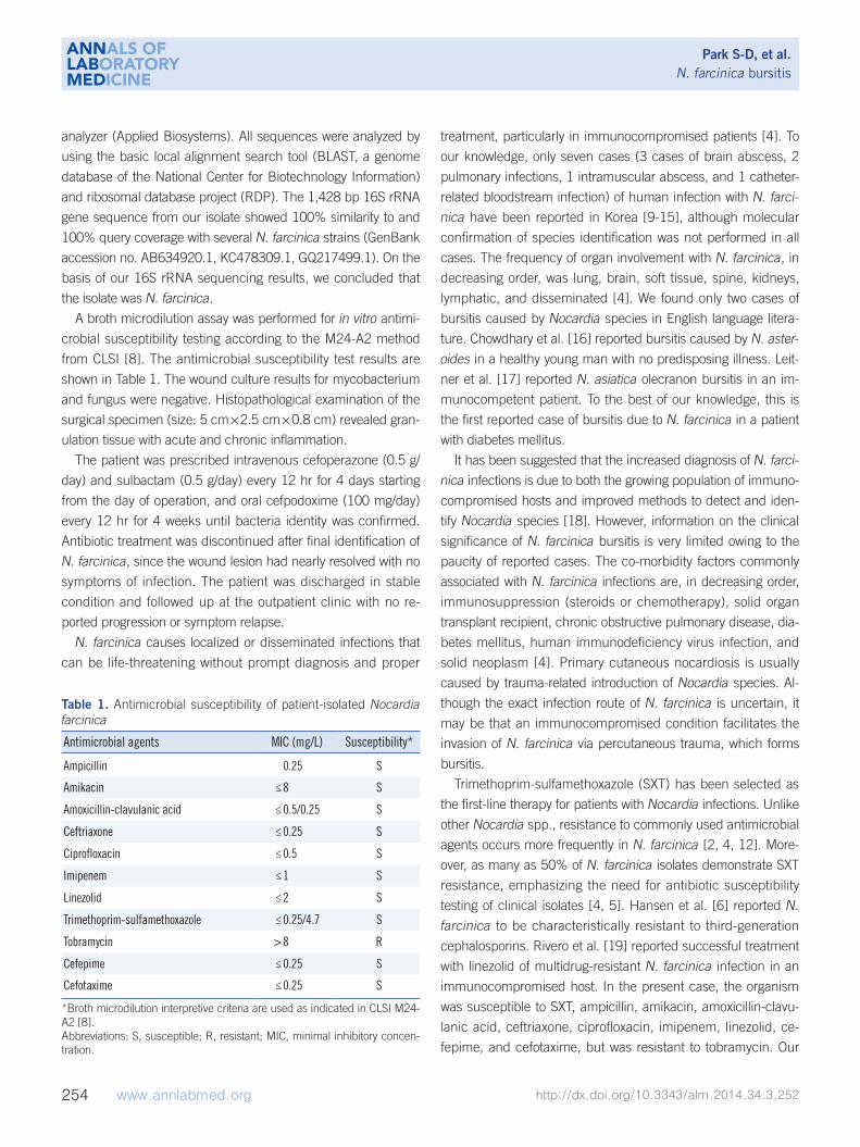
A broth microdilution assay was performed for in vitro antimi-
crobial susceptibility testing according to the M24-A2 method
from CLSI [8]. The antimicrobial susceptibility test results are
shown in Table 1. The wound culture results for mycobacterium
and fungus were negative. Histopathological examination of the
surgical specimen (size: 5 cm×2.5 cm×0.8 cm) revealed gran-
ulation tissue with acute and chronic inflammation.
The patient was prescribed intravenous cefoperazone (0.5 g/
day) and sulbactam (0.5 g/day) every 12 hr for 4 days starting
from the day of operation, and oral cefpodoxime (100 mg/day)
every 12 hr for 4 weeks until bacteria identity was confirmed.
Antibiotic treatment was discontinued after final identification of
N. farcinica, since the wound lesion had nearly resolved with no
symptoms of infection. The patient was discharged in stable
condition and followed up at the outpatient clinic with no re-
ported progression or symptom relapse.
N. farcinica causes localized or disseminated infections that
can be life-threatening without prompt diagnosis and proper
treatment, particularly in immunocompromised patients [4]. To
our knowledge, only seven cases (3 cases of brain abscess, 2
pulmonary infections, 1 intramuscular abscess, and 1 catheter-
related bloodstream infection) of human infection with N. farci-nica have been reported in Korea [9-15], although molecular
confirmation of species identification was not performed in all
cases. The frequency of organ involvement with N. farcinica, in
decreasing order, was lung, brain, soft tissue, spine, kidneys,
lymphatic, and disseminated [4]. We found only two cases of
bursitis caused by Nocardia species in English language litera-
ture. Chowdhary et al. [16] reported bursitis caused by N. aster-oides in a healthy young man with no predisposing illness. Leit-
ner et al. [17] reported N. asiatica olecranon bursitis in an im-
munocompetent patient. To the best of our knowledge, this is
the first reported case of bursitis due to N. farcinica in a patient
with diabetes mellitus.
It has been suggested that the increased diagnosis of N. farci-nica infections is due to both the growing population of immuno-
compromised hosts and improved methods to detect and iden-
tify Nocardia species [18]. However, information on the clinical
significance of N. farcinica bursitis is very limited owing to the
paucity of reported cases. The co-morbidity factors commonly
associated with N. farcinica infections are, in decreasing order,
immunosuppression (steroids or chemotherapy), solid organ
transplant recipient, chronic obstructive pulmonary disease, dia-
betes mellitus, human immunodeficiency virus infection, and
solid neoplasm [4]. Primary cutaneous nocardiosis is usually
caused by trauma-related introduction of Nocardia species. Al-
though the exact infection route of N. farcinica is uncertain, it
may be that an immunocompromised condition facilitates the
invasion of N. farcinica via percutaneous trauma, which forms
bursitis.
Trimethoprim-sulfamethoxazole (SXT) has been selected as
the first-line therapy for patients with Nocardia infections. Unlike
other Nocardia spp., resistance to commonly used antimicrobial
agents occurs more frequently in N. farcinica [2, 4, 12]. More-
over, as many as 50% of N. farcinica isolates demonstrate SXT
resistance, emphasizing the need for antibiotic susceptibility
testing of clinical isolates [4, 5]. Hansen et al. [6] reported N. farcinica to be characteristically resistant to third-generation
cephalosporins. Rivero et al. [19] reported successful treatment
with linezolid of multidrug-resistant N. farcinica infection in an
immunocompromised host. In the present case, the organism
was susceptible to SXT, ampicillin, amikacin, amoxicillin-clavu-
lanic acid, ceftriaxone, ciprofloxacin, imipenem, linezolid, ce-
fepime, and cefotaxime, but was resistant to tobramycin. Our
Table 1. Antimicrobial susceptibility of patient-isolated Nocardia farcinica
Antimicrobial agents MIC (mg/L) Susceptibility*
Ampicillin 0.25 S
Amikacin ≤8 S
Amoxicillin-clavulanic acid ≤0.5/0.25 S
Ceftriaxone ≤0.25 S
Ciprofloxacin ≤0.5 S
Imipenem ≤1 S
Linezolid ≤2 S
Trimethoprim-sulfamethoxazole ≤0.25/4.7 S
Tobramycin >8 R
Cefepime ≤0.25 S
Cefotaxime ≤0.25 S
*Broth microdilution interpretive criteria are used as indicated in CLSI M24-A2 [8].Abbreviations: S, susceptible; R, resistant; MIC, minimal inhibitory concen-tration.
Park S-D, et al.N. farcinica bursitis
http://dx.doi.org/10.3343/alm.2014.34.3.252 www.annlabmed.org 255
patient was successfully treated with antibiotics such as cefo-
perazone and cefpodoxime in addition to surgical debridement.
REFERENCES
1. Stackebrandt E, Rainey FA, Ward-Rainey NL. Proposal for a new hierar-chic classification system, Actinobacteria classis nov. Int J Syst Bacteriol 1997;47:479-91.
2. Brown-Elliott BA, Brown JM, Conville PS, Wallace RJ Jr. Clinical and laboratory features of the Nocardia spp. based on current molecular taxonomy. Clin Microbiol Rev 2006;19:259-82.
3. Beaman BL and Beaman L. Nocardia species: host-parasite relation-ships. Clin Microbiol Rev 1994;7:213-64.
4. Budzik JM, Hosseini M, Mackinnon AC Jr, Taxy JB. Disseminated No-cardia farcinica: literature review and fatal outcome in an immunocom-petent patient. Surg Infect (Larchmt) 2012;13:163-70.
5. Torres OH, Domingo P, Pericas R, Boiron P, Montiel JA, Vázquez G. In-fection caused by Nocardia farcinica: case report and review. Eur J Clin Microbiol Infect Dis 2000;19:205-12.
6. Hansen G, Swanzy S, Gupta R, Cookson B, Limaye AP. In vitro activity of fluoroquinolones against clinical isolates of Nocardia identified by partial 16S rRNA sequencing. Eur J Clin Microbiol Infect Dis 2008;27: 115-20.
7. Clinical and Laboratory Standards Institute. Interpretive criteria for iden-tification of bacteria and fungi by DNA target sequencing; approved guideline, MM18-A. Wayne, PA: Clinical and Laboratory Standards In-stitute, 2008.
8. Clinical and Laboratory Standards Institute. Susceptibility testing of my-cobacteria, nocardiae, and other aerobic actinomycetes; approved standard-second edition, M24-A2. Wayne, PA: Clinical and Laboratory Standards Institute, 2011.
9. Baek YH, Kim YJ, Lee HH, Youm JY, Kwon OW, Kim JH, et al. A case of intramuscular abscess caused by Nocardia farcinica in a patient with lu-
pus nephritis concurrent with pulmonary tuberculosis. J Korean Rheum Assoc 2006;13:327-32.
10. Sim SH, Park HC, Kim CJ, Jeon JH, Kim EC, Oh MD, et al. A case of Nocardia farcinica brain abscess in the patient receiving steroid treat-ment. Infect Chemother 2008;40:301-4.
11. Park I, Yim H, Kwan LS, Yu S, Cho J, Kim H, et al. A case of a kidney transplant recipient with pulmonary cytomegalovirus and Nocardia coinfection with cytomegalovirus nephropathy. Korean J Nephrol 2009; 28:161-5.
12. Heo ST, Ko KS, Kwon TK, Ryu SY, Bae IG, Oh WS, et al. The first case of catheter-related bloodstream infection caused by Nocardia farcinica. J Korean Med Sci 2010;25:1665-8.
13. Lee SH, Park BH, Son JY, Jung JY, Kim EY, Lim JE, et al. A case of pneumonia with septic shock due to Nocardia farcinia in liver transplant patient. Tuberc Respir Dis 2010;69:469-73.
14. Moon JH, Cho WS, Kang HS, Kim JE. Nocardia brain abscess in a liver transplant recipient. J Korean Neurosurg Soc 2011;50:396-8.
15. Han ST, Kim YS, Song SH, Uh Y, Han BG, Choi SO, et al. A case of No-cardia farcinica brain abscess in a focal segmental glomerulosclerosis patient after steroid treatment. Korean J Nephrol 2011;30:98-101.
16. Chowdhary G, Wormser GP, Mascarenhas BR. Nocardia bursitis. J Rheumatol 1988;15:139-40.
17. Leitner E, Valentin T, Hoenigl M, Lanz P, Flick H, Zollner-Schwetz I, et al. First report of Nocardia asiatica olecranon bursitis in an immunocompe-tent traveler returning to Austria. J Clin Microbiol 2013;51:2461-2.
18. De Nardo P, Giancola ML, Noto S, Gentilotti E, Ghirga P, Tommasi C, et al. Left thigh phlegmon caused by Nocardia farcinica identified by 16S rRNA sequencing in a patient with leprosy: a case report. BMC Infect Dis 2013;13:162.
19. Rivero A, García-Lázaro M, Pérez-Camacho I, Natera C, del Carmen Al-modovar M, Camacho A, et al. Successful long-term treatment with li-nezolid for disseminated infection with multiresistant Nocardia farcinica. Infection 2008;36:389-91.


![Nocardia Brain Abscess in an Immunocompetent Patient · Nocardia species are a rare cause of cerebral abscess [3]. Nocardia brain abscess appears in a gradually progressive mass lesion,](https://static.fdocuments.us/doc/165x107/5f9d9fa5c479af2f1c584bd9/nocardia-brain-abscess-in-an-immunocompetent-patient-nocardia-species-are-a-rare.jpg)


