
Filariasis
19
Filaria sis Dr. Mejbah Uddin Ahmed
-
Upload
api-19969058 -
Category
Documents
-
view
833 -
download
0
Transcript of Filariasis

Filariasis
Dr. Mejbah Uddin Ahmed
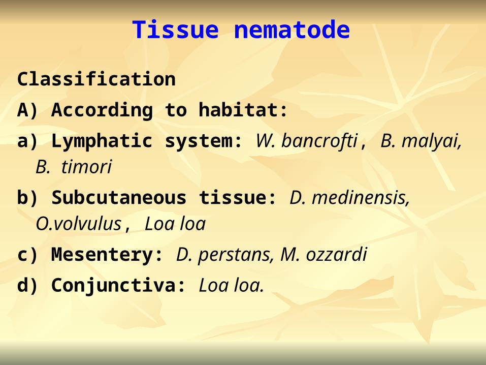
Tissue nematode
Classification
A) According to habitat:
a) Lymphatic system: W. bancrofti, B. malyai, B. timori
b) Subcutaneous tissue: D. medinensis, O.volvulus, Loa loa
c) Mesentery: D. perstans, M. ozzardi
d) Conjunctiva: Loa loa.
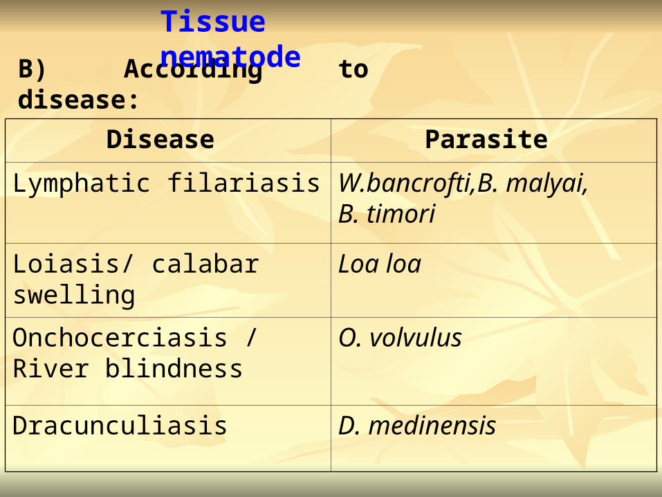
B) According to disease:
Disease Parasite
Lymphatic filariasis W.bancrofti,B. malyai, B. timori
Loiasis/ calabar swelling Loa loa
Onchocerciasis / River blindness
O. volvulus
Dracunculiasis D. medinensis
Tissue nematode
Filariasis
Filariasis are a group of parasitic diseases
caused by nematodes reside in the lymphatic and
connective tissue (W. bancrofti, B. malyai, B.
Timori).
Lymphatic filarisis is a debilitating and disfiguring
chronic disease (lyphoedema, elephantiasis and
hydrocele).
Lymphatic filarisis

Morphological forms:
Adult worms
Microfilaria
Adult worms:
The adult worms are white
Females are larger than males and
viviparous.
Wuchereria bancrofti

Microfilaria:
Average microfilaria measures 240–300 μm in
length. A thin delicate sheath surrounds the
organism. The head is blunt and round and the tail
is pointed. Except the tail tip numerous nuclei are
contained in the body.
Wuchereria bancrofti

Periodicity: The microfilarias are not constantly
found in the peripheral blood, but show nocturnal
periodicity. The exact mechanism is not fully
understood. It is determined by species and life
style of host and can be altered.
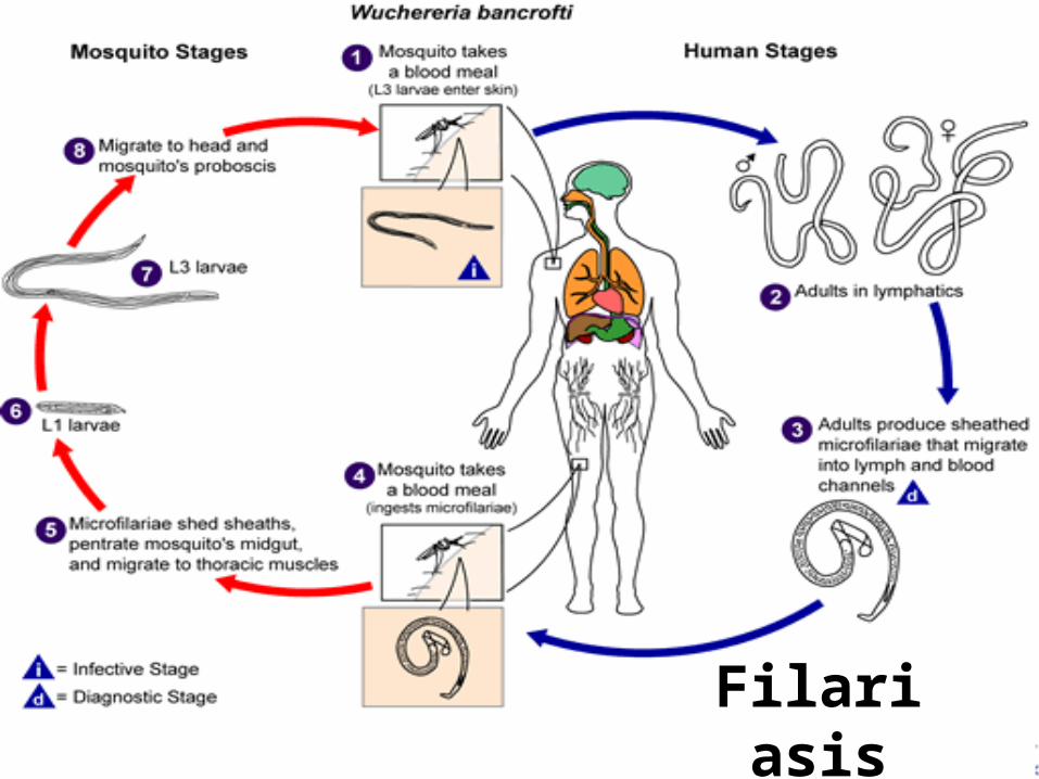
Wuchereria bancrofti

Life cycle:
Definitive host: Man.
Intermediate Host: Mosquito.
Infective form: Microfilaria.
Portal of entry: Skin, by mosquito bite.
Site of localization: Lymphatic system.
Wuchereria bancrofti
Filariasis

Pathology & Clinical feature :
May be asymptomatic
Adult and developing worm causes classical
filariasis
Microfilaria causes occult filariasis .
Wuchereria bancrofti

Classical filariasis:
Acute lymphatic filariasis: In the acute form
there are recurrent attacks of fever with
lymphadenitis and lymphangitis. The lymphatics
involved are those of the limbs, genital organs
(specially spermatic cord) and breasts.
Wuchereria bancrofti

Causes of lymphangitis: Mechanical irritation,
liberation of metabolites, bacterial infection.
Obstruction of lymphatics: Mechanical,
excessive fibrosis of lymph vessels and fibrosis of
afferent nodes.
Filariasis

Dilatation of lymphatics- Lymphangiovarix.
Rupture of lymphnagiovarix
Lymphorrhgia– Lymphscrotum, Lymphuria
Lymphocoele.
Chylorrhagia–Chylocoele, chyluria, chylorrhgia or
hematochyluria, chylus diarrhoea, chylus ascitis
and chylothorax. .
Filariasis
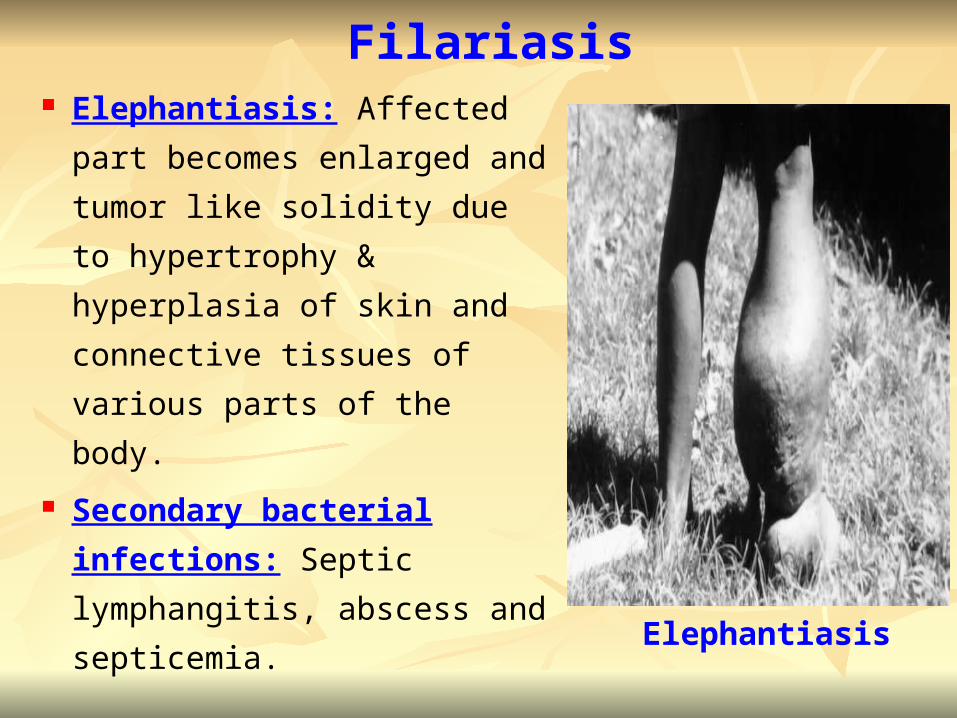
Elephantiasis: Affected part
becomes enlarged and
tumor like solidity due to
hypertrophy & hyperplasia of
skin and connective tissues
of various parts of the body.
Secondary bacterial
infections: Septic
lymphangitis, abscess and
septicemia.
Filariasis
Elephantiasis

Occult filariasis:
The condition when adult worm produces
microfilaria continuously but absent in the
peripheral blood because they are destroyed in
the tissues. This is characterized by massive
eosinophilia, hepatosplenomegali, generalized
lymphadenopathy & pulmonary symptoms..
Filariasis
Filariasis
Tropical pulmonary eosinophilia: A syndrome
of immunological hyper responsiveness of Mf in
the lung. It is particularly found in filaria endemic
areas. There is a marked eosinophilia, raised
ESR and high levels of filarial antibody including
high titers of IgE.

Laboratory Diagnosis:
Sample: Peripheral blood, chylous urine, hydrocele
fluid and exudates of lymph varix.
Microscopy: Detection of microfilaria stained with
Leishman, Giemsa or hematoxylin and eosin stain.
Antigen detection: Using an immunoassay for
circulating filarial antigens constitutes a useful
diagnostic approach.
Filariasis
Molecular diagnosis: Polymerase chain reaction
is available for W. bancrofti and B. malayi.
Identification of adult worms: is possible from
tissue samples collected from subcutaneous
biopsies.
Antibody detection: Is of limited value.
Complement fixation test.
Filariasis


![Filariasis by Cj[1]](https://static.fdocuments.us/doc/165x107/577cdbff1a28ab9e78a9973c/filariasis-by-cj1.jpg)


