Filariasis ( wuchereria bancrofti)
20
BANCROFT’S FILARIASIS SUBMITTED TO DEPARTMENT OF ZOOLOGY ISABELLA THOBURN COLLEGE LUCKNOW
-
Upload
richa-tiwari-it-college-lucknow-university -
Category
Science
-
view
258 -
download
12
Transcript of Filariasis ( wuchereria bancrofti)

BANCROFT’S
FILARIASIS
SUBMITTED TO
DEPARTMENT OF ZOOLOGY
ISABELLA THOBURN COLLEGE
LUCKNOW

INTRODUCTION
Filariasis is the term for a group of disease caused by
parasitic nematodes.
Filariasis caused by nematodes that live in the human
lymph system, Bancroftian filariasis or Lymphatic
filariasis.
Causative organism- filarial worm Wuchereria bancrofti
(Cobbold 1877)
Elephantiasis is the end result of disease.
• Cutaneous and subcutaneous tissue enlarge and harden in
areas where lymph has accumulated.
• Usually occurs in lower extremities.

Wuchereria bancrofti
Common name- Bancroft’s filaria
Bancroft (1876-77) demonstrated adult female and
Sibthorpe (1888) first found adult male.
Manson (1878) first demonstrated the culex mosquitoes
as the INTERMEDIATE HOST.
Geographical distribution• Very widespread and
important human parasite in
worm countries.
• In Africa (Mediterranean and
east west coastal areas)
• In Asia (coasts of Arabia,
India, Malaya and north to
china.
Habitat• Filarial worm inhabit the
lymphatic vessels, especially
the lymph nodes of man and
other vertebrates.
• Microfilariae are found in
peripheral blood (also found
in chylous urine or
hydrocele fluid)

Morphology
Adult worm
Third stage
larva
Microfilariae
(first stage larva)

ADULT WORM
Long hair like transparent nematodes
Creamy white in colour.
Filiform in shape, both ends are tapering
Male- 2.5-4 cm in length and 0.1mm thickness
Tail end is curved ventrally contain 2 spicules
Female- 8-10cm in length and 0.2-0.3mm
thickness
Tail end is narrow and abruptly pointed
Ovo-viviparous, though liberating active
embryos.
Male and female coiled together, can be
separated with difficulty
Life span is long, 5-10 years Wuchereria bancrofti
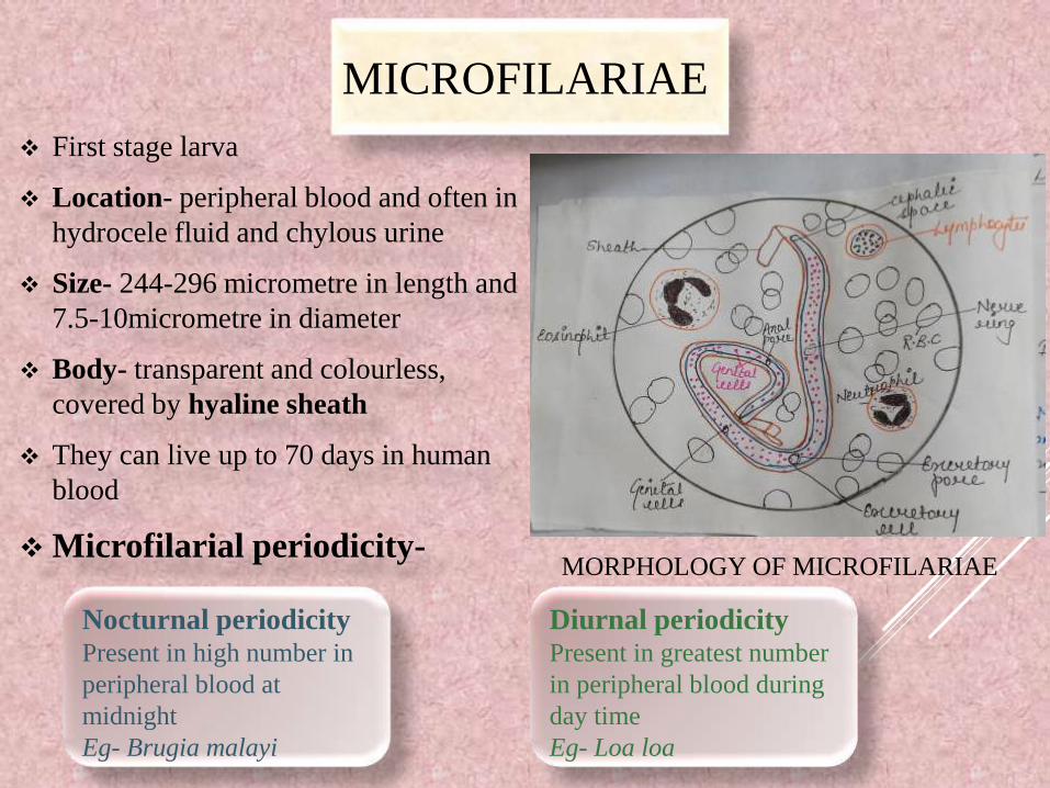
MICROFILARIAE
First stage larva
Location- peripheral blood and often in
hydrocele fluid and chylous urine
Size- 244-296 micrometre in length and
7.5-10micrometre in diameter
Body- transparent and colourless,
covered by hyaline sheath
They can live up to 70 days in human
blood
Microfilarial periodicity-
Nocturnal periodicityPresent in high number in
peripheral blood at
midnight
Eg- Brugia malayi
Diurnal periodicityPresent in greatest number
in peripheral blood during
day time
Eg- Loa loa
MORPHOLOGY OF MICROFILARIAE

THIRD STAGE LARVA
Infective form of the filarial worm in human.
Shape- elongated, Filiform
Size- 1.4-2 micrometre in length and 18-23
micrometre in breadth
MICROFILARIAE IN
BLOOD FILM

LIFE CYCLE
• DEFINITIVE HOST- Man
(In human lymphatic system)
• INTERMEDIATE HOST-
Mosquitoes
Culex quinquesfasciatus
Anopheles
Aedes
Culex
quinquesfasciatus
Aedes
Anopheles


PATHOGENICITY
INFECTION- Wuchereriasis (commonly called
filariasis)
MODE OF INFECTION- Inoculative method,
through the bite of mosquito
TRANSMITTING AGENT- Female mosquito(Culex,
Aedes, Anopheles)
INFECTIVE FORM-Third stage larva
PORTAL OF ENTRY-Skin
SITE OF LOCALIZATION-Lymphatic system of
superior or inferior extremities
INCUBATION PERIOD- About 1 to 2 years(3rd stage
infective larva grows to adult form)

CLINICAL MENIFESTATION
LYMPHATIC
FILARIASIS
OCCULT
FILARIASIS
endemic normal
Asymptomatic stage
Acute filariasis
Chronic filariasis
Non-filarial
elephantiasis
Tropical pulmonary
eosinophilia
(TPE)

LYMPHATIC FILARIASIS
ENDEMIC NORMAL
• In the area of
endemic filariasis, a
certain proportion of
the population living
in these area do not
show any overt
clinical manifestation
of disease
• Difficult to know,
people are infected or
not
ASYMPTOMATIC
STAGE
• Have microfilariae
in their blood but
do not show any
symptoms of
disease
• Remains
asymptomatic for
years or even life
ACUTE
FILARIASIS
• Filarial fever and
lymphadenitis
(major symptoms)
• May occur several
times a year

ELEPHANTIASIS

THANK YOU…

1. Hydrocele- obstruction of
the lymph vessels of
spermatic cord and
inflammation in testes
2. Elephantiasis- affected part
become enormously enlarged
(tumour like solidity)
• The surface of skin become
rough and papliomatous
• Hairs become rough and
sparse
CHRONIC
FILARIASIS
Hydrocele elephantiasis
1. Lymph varices-
varicosity of lymphatic
vessels (abdominal and
genital area)
2. Chyluria- escape of chyle
through the urine (rapture
of varicose chyle vessels)
• Microfilariae are detected
in chylous urine and
peripheral blood
NON- FILARIAL
ELEPHANTIASIS
Lymph
variceschyluria

OCCULT FILARIASIS
• Hypersensitivity reaction of the host to
Microfilarial antigen
• Microfilariae are not detected in peripheral blood
• Examples- tropical pulmonary eosinophilia
(TPE), Less frequently arthritis
TROPICAL PULMONARY EOSINOPHILIA (TPE)
Eosinophilic lung, Weingarten’s syndrome
• Characterised by low fever, loss of weight, paroxysmal
cough with scanty sputum, dyspnoea and splenomegaly
• Chest radiography shows increased branchiovascular
marking or military “mottling” in lung fields
• Microfilariae may b e demonstrated in tissue obtained by
lung biopsy

EPIDEMIOLOGY
GEOGRAPHICAL DISTRIBUTION- topics and
subtropics of Asia, Africa, South America
• Periodic nocturnal W. bancrofti is the most
widespread
• Endemic in India, China and other countries of
South-East Asia
RESERVOIR HOST AND TRANSMISSION OF
INFECTION
• Infected person with circulating microfilariae are the
chief source of reservoir and infection
• Man- to- man transmission by the bite of mosquito

DIAGNOSIS
DIRECT EVIDENCES
1. Microfilariae (sheathed having
tail- tip free from nucleus)
• In peripheral blood
• In chylous urine
• In hydrocele fluid
2. Adult worm
• In biopsied lymph nodes
• Calcified worm by X-ray
INDIRECT EVIDENCES
1. Allergic testa. Blood examination(eosinophilia 5 to
15%)
b. Intradermat test- an immediate
hypersensitivity reaction
2. Immunological test
(complement fixative test)
• A sensitive test for loiasis and occult
filariasis
Other tests-• Biopsy (for purely diagnostic purpose)
• Serological diagnosis (ELISA, RIA)
• Trop Bio Test (detection of adult worm infection not depends on
Microfilarial periodicity)
• PCR assay (detection of Microfilarial infection)
• X-ray examination ( shows calcified adult worm)

TREATMENT
Diethyl carbamazine (DEC)• Kills mainly microfilariae
• Most effective against 3rd and 4th stage larva
• Adenolymphangitis decreases significantly
• Cheap, effective and safe with few side effects
(fever, chill, headache etc.)
DOSE
1st day - 50mg after food
2nd day - 50mg three times daily
3rd day - 100mg three times daily
4th day – 21st day - 5mg/kg/day in three
divided dose
Other drugs • Iveemeciin (150ug/kg body wt., destroy microfilariae, no
mocrofilaricidal effect)
• Lavamisole
• Mebendazole
• Centprazine (CDRI Lucknow)

PREVENTION AND CONTROL
1. Mosquito control
• clinical control by spraying
insecticides (DDT, malathion)
• Biological control by the use of
carnivorous bacteria(Bacillus
sphaericus), carnivorous fishes
(Poecilia reticulata)
• Environmental control by efficient
drainage and sewage system
2. Chemotherapeutic control• Reduce morbidity du to filariasis by
treating clinical case
• Lower transmission by treating the
case of crofilaraemia
• Interrupt transmission of the
infection
• Based on mass or selective treatment
of the cases by administering DEC