Figure 5.0 Spider’s web made of protein. Figure 5.1 Building models to study the structure and...
48
Figure 5.0 Spider’s web made of protein
-
Upload
augusta-carson -
Category
Documents
-
view
216 -
download
0
Transcript of Figure 5.0 Spider’s web made of protein. Figure 5.1 Building models to study the structure and...

Figure 5.0 Spider’s web made of protein

Figure 5.1 Building models to study the structure and function of macromolecules
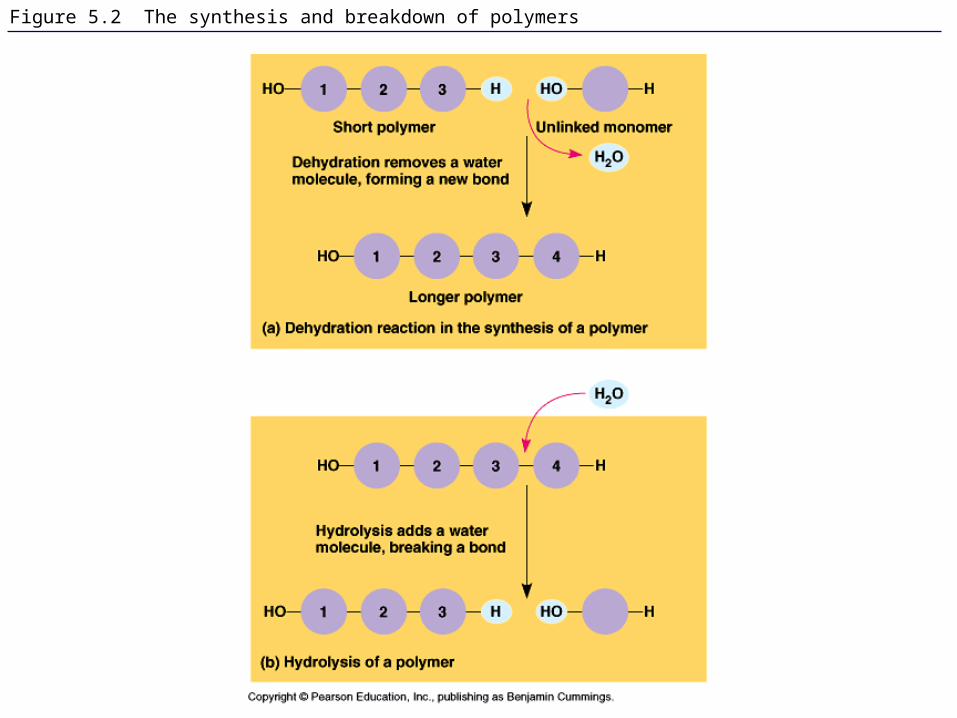
Figure 5.2 The synthesis and breakdown of polymers
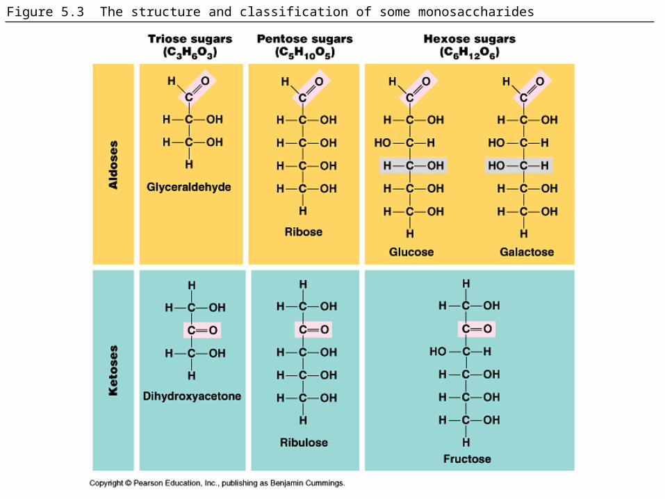
Figure 5.3 The structure and classification of some monosaccharides

Figure 5.29 The components of nucleic acids; differences between DNA and RNA

Figure 5.3x Hexose sugars
Glucose Galactose
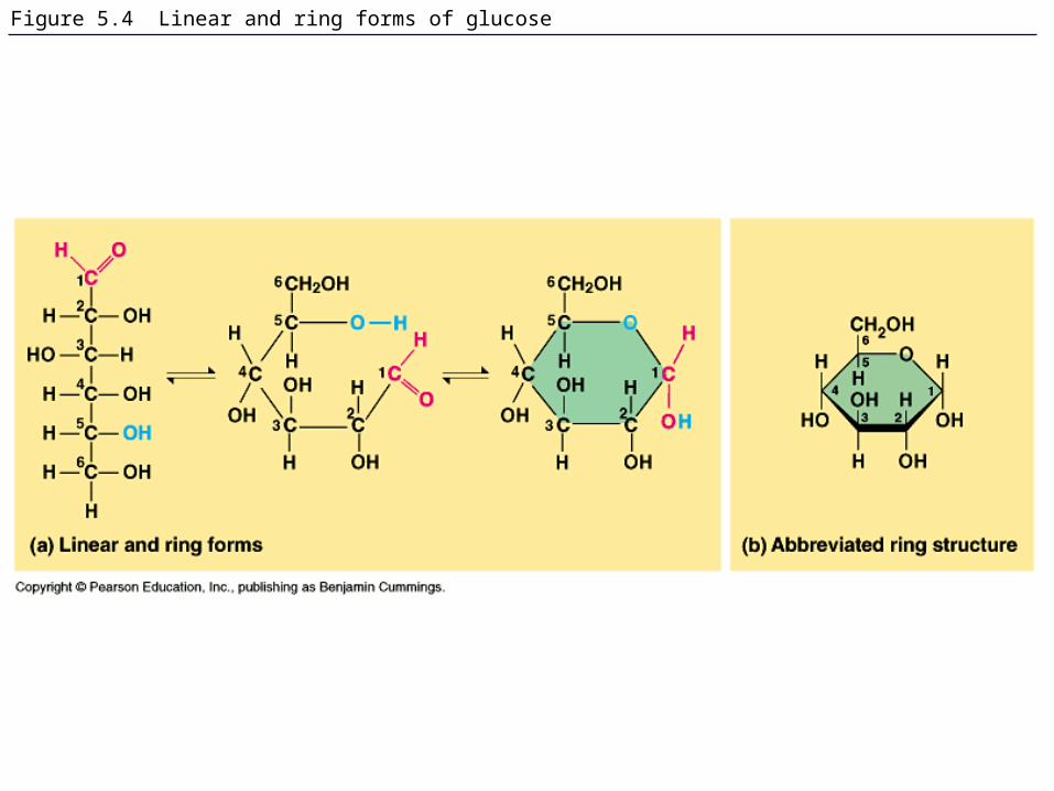
Figure 5.4 Linear and ring forms of glucose

Figure 5.5 Examples of disaccharide synthesis

Figure 5.5x Glucose monomer and disaccharides
Glucose monomer
Sucrose
Maltose
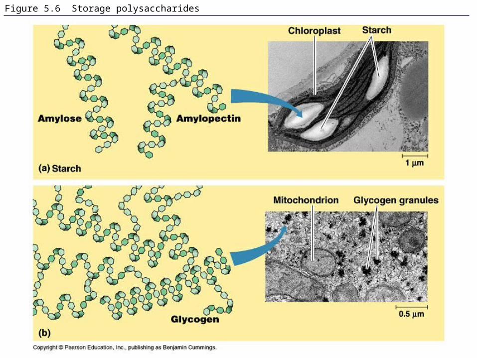
Figure 5.6 Storage polysaccharides
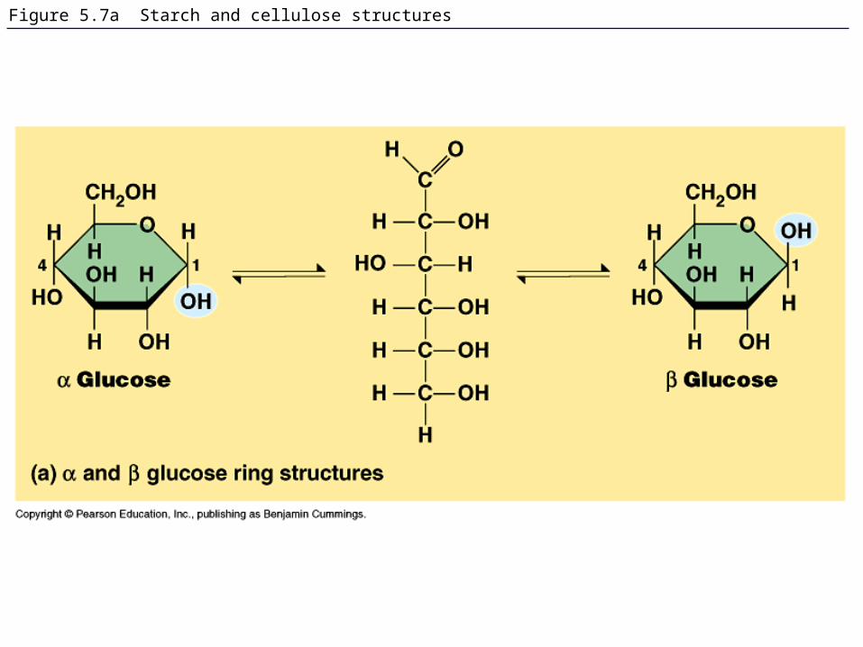
Figure 5.7a Starch and cellulose structures
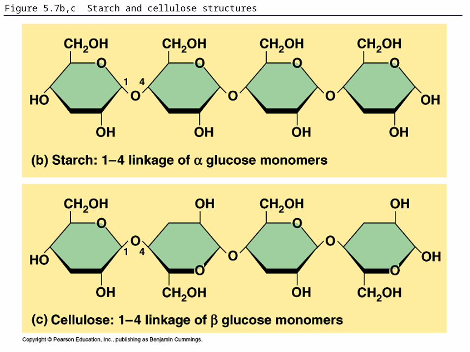
Figure 5.7b,c Starch and cellulose structures

Figure 5.7x Starch and cellulose molecular models
Glucose Glucose
Starch
Cellulose
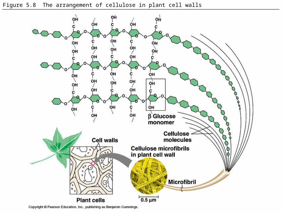
Figure 5.8 The arrangement of cellulose in plant cell walls
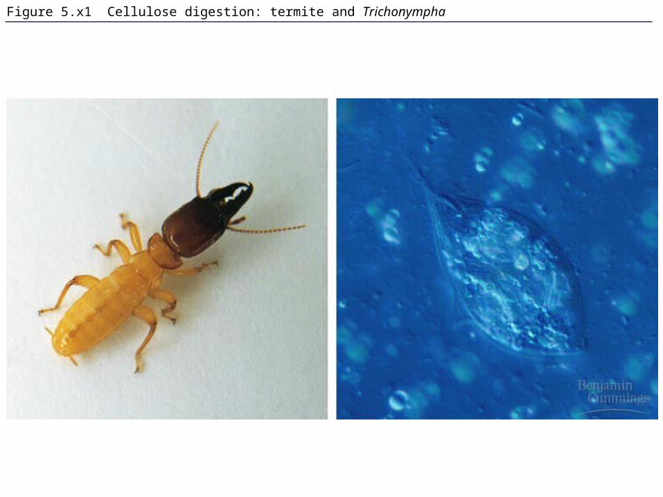
Figure 5.x1 Cellulose digestion: termite and Trichonympha
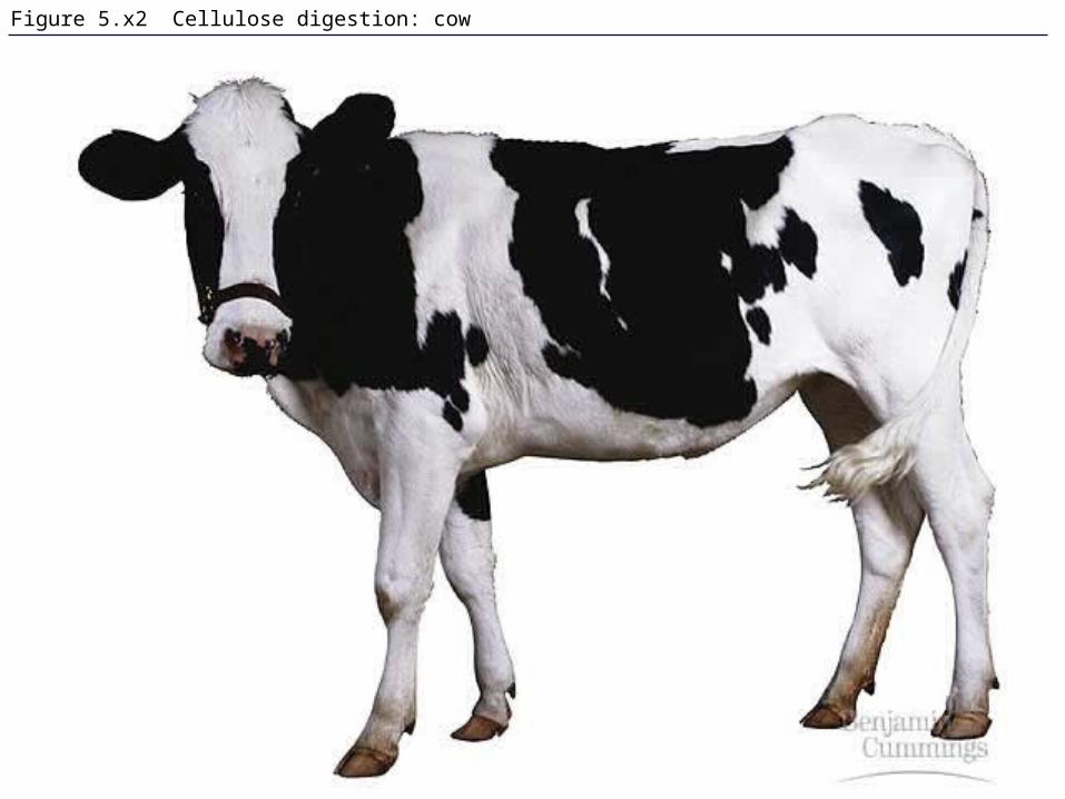
Figure 5.x2 Cellulose digestion: cow

Figure 5.9 Chitin, a structural polysaccharide: exoskeleton and surgical thread
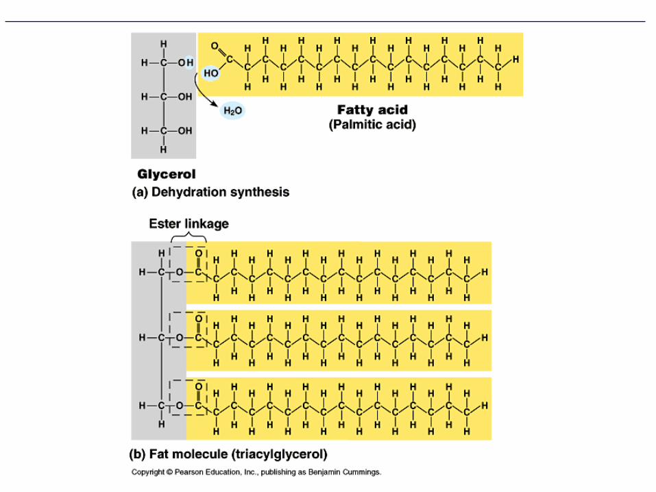

Figure 5.10 The synthesis and structure of a fat, or triacylglycerol
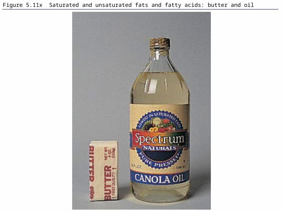
Figure 5.11x Saturated and unsaturated fats and fatty acids: butter and oil
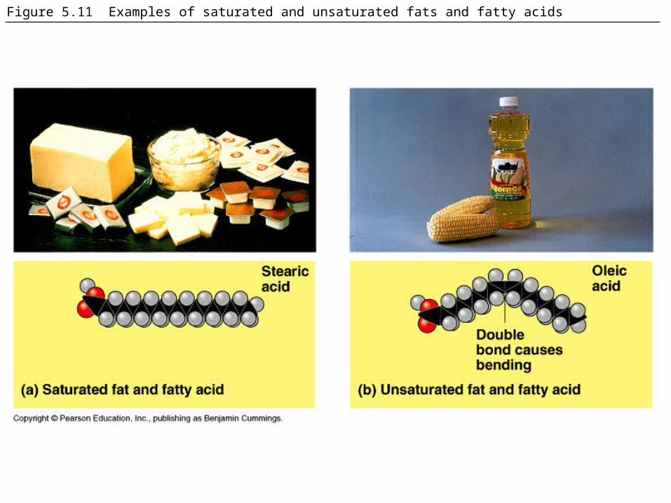
Figure 5.11 Examples of saturated and unsaturated fats and fatty acids

Figure 5.12 The structure of a phospholipid

Figure 5.13 Two structures formed by self-assembly of phospholipids in aqueous environments
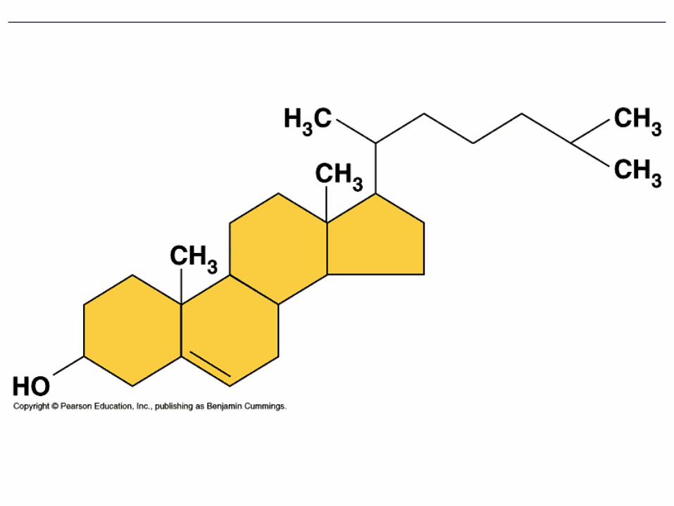
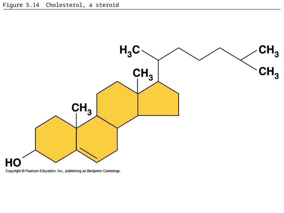
Figure 5.14 Cholesterol, a steroid

Figure 8.6 The detailed structure of an animal cell’s plasma membrane, in cross section

Figure 4.8 A comparison of functional groups of female (estradiol) and male (testosterone) sex hormones
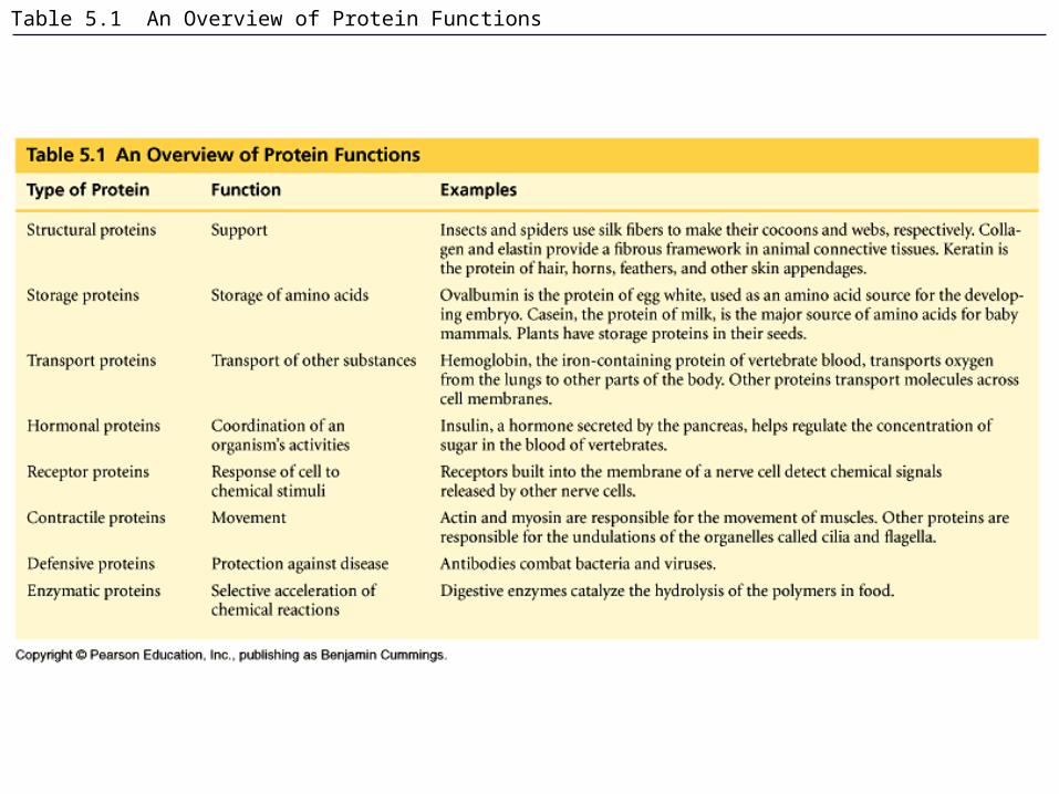
Table 5.1 An Overview of Protein Functions

Figure 5.0 Spider’s web made of protein

Figure 5.15 The 20 amino acids of proteins: nonpolar
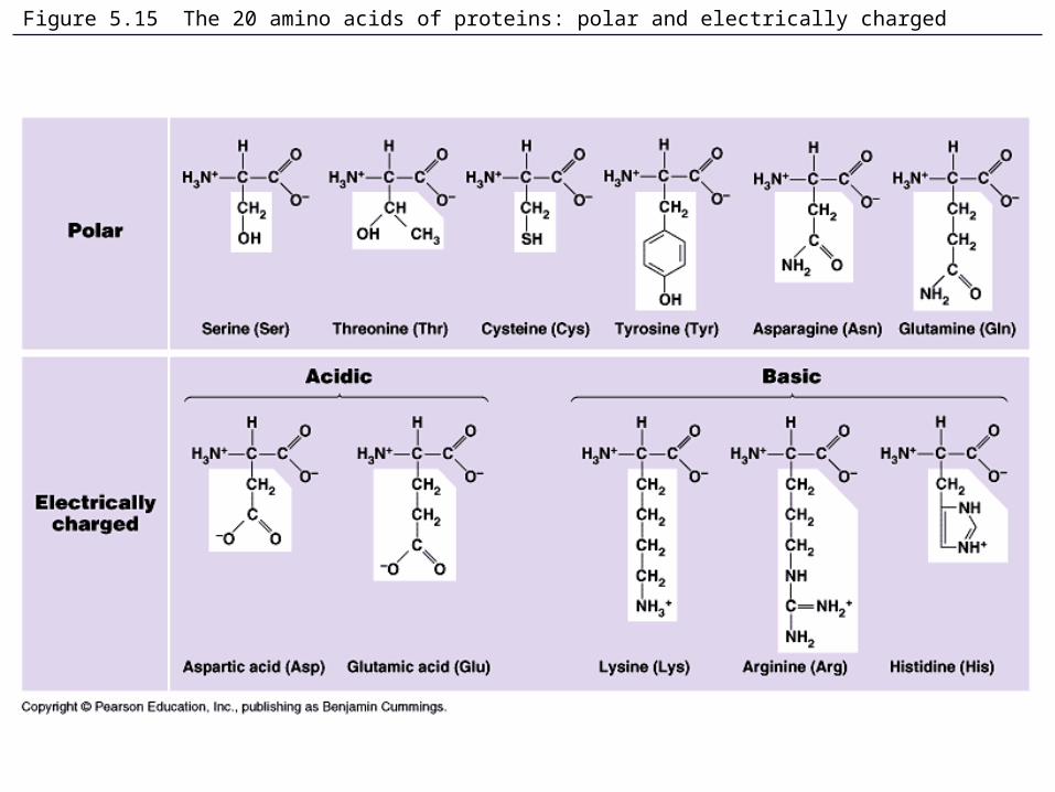
Figure 5.15 The 20 amino acids of proteins: polar and electrically charged

Figure 5.16 Making a polypeptide chain
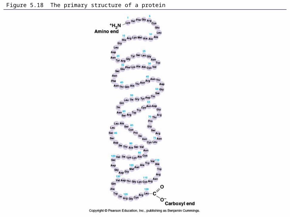
Figure 5.18 The primary structure of a protein

Figure 5.20 The secondary structure of a protein
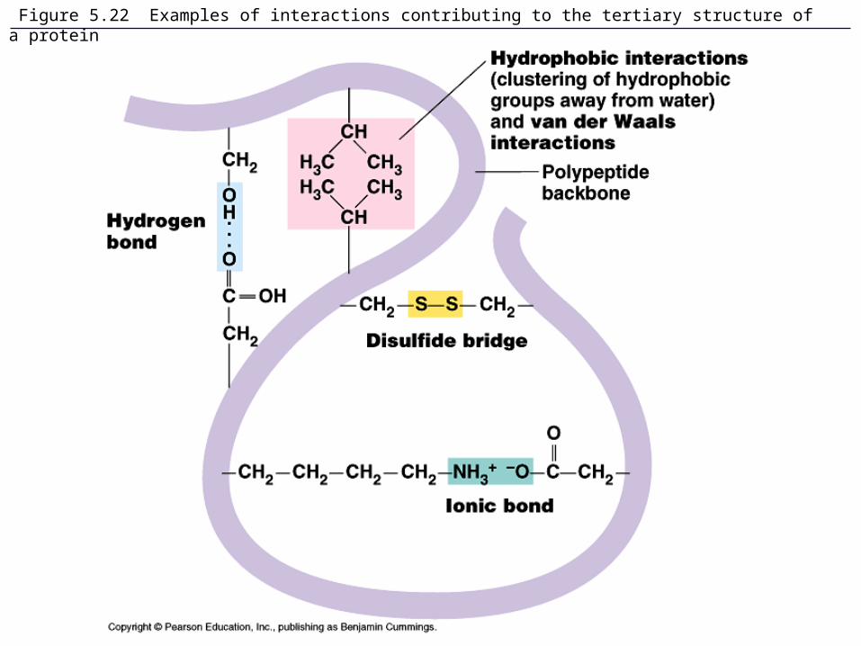
Figure 5.22 Examples of interactions contributing to the tertiary structure of a protein

Figure 5.17 Conformation of a protein, the enzyme lysozyme

Figure 5.23 The quaternary structure of proteins
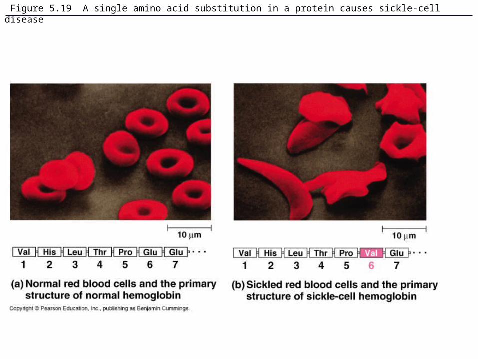
Figure 5.19 A single amino acid substitution in a protein causes sickle-cell disease
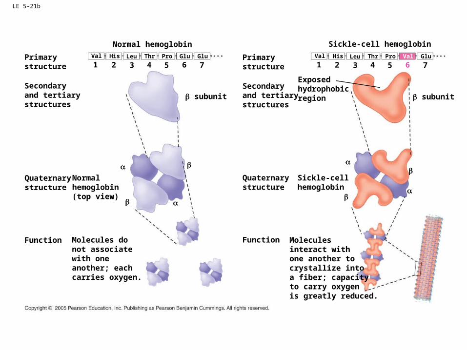
LE 5-21b
Primarystructure
Secondaryand tertiarystructures
1 2 3
Normal hemoglobin
Val His Leu
4Thr
5Pro
6Glu Glu
7Primarystructure
Secondaryand tertiarystructures
1 2 3
Sickle-cell hemoglobin
Val His Leu
4Thr
5Pro
6Val Glu
7
Quaternarystructure
Normalhemoglobin(top view)
Function Molecules donot associatewith oneanother; eachcarries oxygen.
Quaternarystructure
Sickle-cellhemoglobin
Function Molecules interact withone another tocrystallize intoa fiber; capacityto carry oxygenis greatly reduced.
Exposedhydrophobicregion subunit subunit
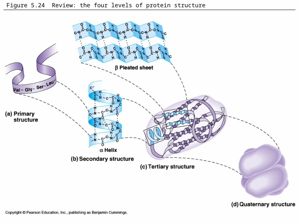
Figure 5.24 Review: the four levels of protein structure

Figure 5.25 Denaturation and renaturation of a protein
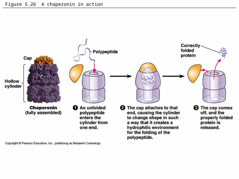
Figure 5.26 A chaperonin in action

Figure 5.x3 James Watson and Francis Crick

Figure 5.28 DNA RNA protein: a diagrammatic overview of information flow in a cell
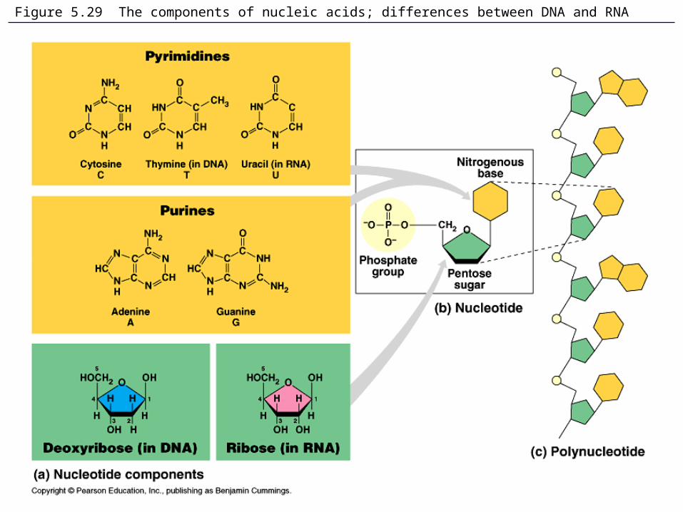
Figure 5.29 The components of nucleic acids; differences between DNA and RNA
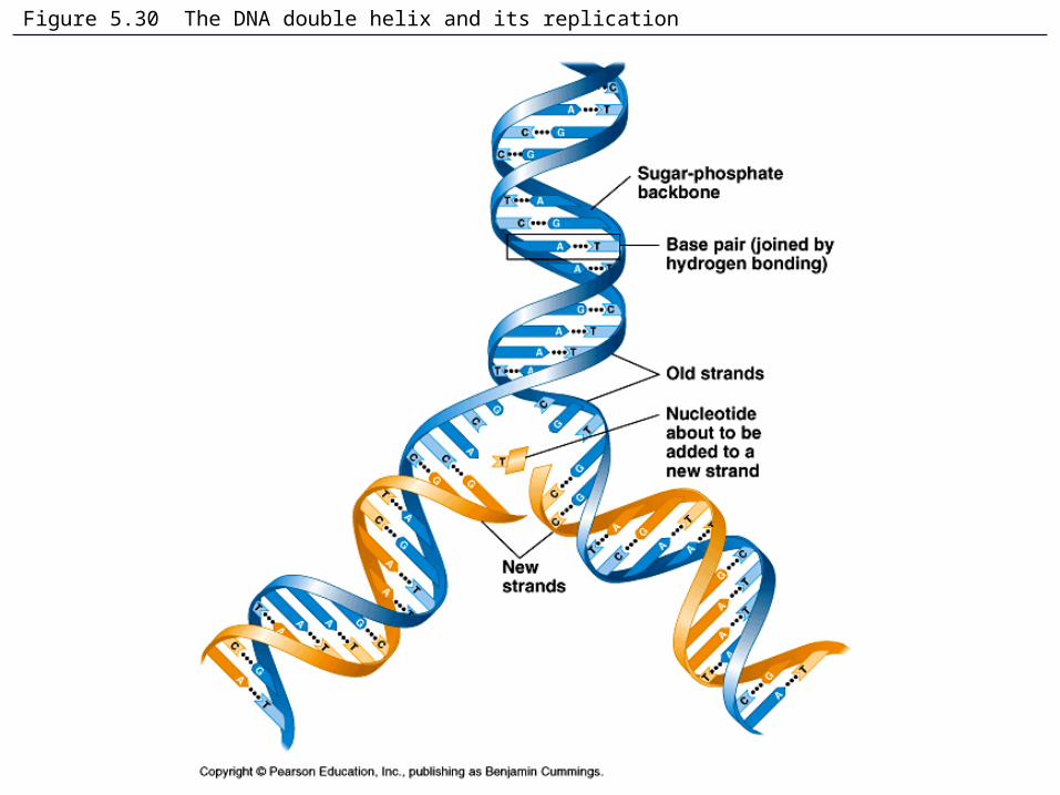
Figure 5.30 The DNA double helix and its replication
Figure 5.x4 Rosalind Franklin
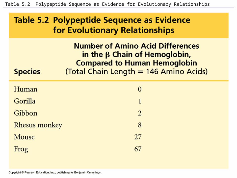
Table 5.2 Polypeptide Sequence as Evidence for Evolutionary Relationships