E-commerceEssentials Kenneth C. Laudon Carol Guercio Traver first edition.

Fifteenth Annual Louis R.M. Del Guercio Distinguished Visiting Professorship and Research Day
Presented at
New York Medical College 7 Dana Road Facility Valhalla, New York December 19, 2018
PROCEEDINGS
Program Committee:
JORGE CON, MD
THOMAS DIFLO, MD
RIFAT LATIFI, MD
KRIST NIKOLLA, MPH
JOHN A. SAVINO, MD
KATHRYN SPANKNEBEL, MD
THOMAS SULLIVAN
KEVIN WOLFE, PHD
1

Table of Contents:
Podium Presentations…...............................p.03
Moderated Oral Poster Presentations…......p.29
Poster Exhibits…….…................................p.59
2
Podium Presentations (In alphabetical order)
3

TITLE: PREOPERATIVE MENINGIOMA EMBOLIZATION IS SAFE BUT COSTS MORE THAN NON-EMBOLIZATION RESECTIONS: A MULTI-CENTER RETROSPECTIVE MATCHED CASE-CONTROL STUDY Authors: ANUBHAV G. AMIN MD1(PGY6), John V. Wainwright MD1 , Ilya Rybkin MS2, Hussam Abou Al-Shaar MD3, William T. Couldwell MD/PhD4, Fawaz Al-Mulfti MD1, Justin Santarelli MD1, Chirag D. Gandhi MD1, Meic H. Schmidt MD1, Christian Bowers MD1
1 Department of Neurosurgery, Westchester Medical Center, New York Medical College, Valhalla, NY 2 New York Medical College, Valhalla, NY 3 Department of Neurosurgery, Hofstra/Northwell, Manhasset, NY 4 Department of Neurosurgery, University of Utah, Salt Lake City, UT Background: The literature has been mixed regarding the potential benefit of reduced blood loss with preoperative meningioma embolization (ME). However, a comparison of embolization-associated costs with non-embolization meningioma (NE) patients has not been completed. Objective: To determine the potential benefits of ME in blood loss and its associated costs. Design/Methods: This is a retrospective case control study matched for tumor location, size, and radiographic appearance between two centers. We reviewed demographic and clinical data for 29 matched meningioma patients from each center. Studied variables included: EBL, Pre/Post-operative Hg/Hct, and perioperative complications. Meningiomas were analyzed as a group and subdivided into skull base and non-skull base locations. The additional ME cost was calculated. Results: Both groups had similar baseline characteristics. There was no significantly decreased blood loss based on EBL or Pre/Post-operative Hg/HCT comparisons, although there was a trend towards decreased blood loss in the ME cohort. This was true for all meningiomas and when subdivided by skull base location. Successful embolization of a feeding vessel occurred in 76% of the ME patients (22/29 pts) while the other patients had angiograms due to unfavorable feeding vessel anatomy. Importantly, there were no complications associated with angiography/embolization in the ME group. The mean additional charge per patient, which included procedural charges, embolic agent costs, as well as one night in the intensive care unit, was $88,767. Conclusions: Within the limitations of the small patient cohorts and the retrospective nature of this study, the ME group had a trend towards reduced blood loss but there was no blood-loss related morbidity in either group. Angiography/Embolization is safe, with no procedural-related complications, but the ME group accrued nearly an additional $90,000 in hospital charges. These findings were unchanged when meningiomas were further subdivided into skull and non-skull base locations.
4

TITLE: IN-HOSPITAL OUTCOMES AFTER OPERATIVE FIXATION OF MULTIPLE RIB FRACTURES WITH NON-FLAIL CHEST. A PROPENSITY MATCHED ANALYSIS Authors: ASAD AZIM (PGY2), Jorge Con, Faisal Jehan, Prabhakaran Kartik, Rifat Latifi *New York Medical College and Westchester Medical Center, Valhalla* Background: Rib fractures are associated with significant morbidity and mortality. Recent data suggests operative fixation of rib fractures with flail chest is associated with better outcomes. Objective: Aim of this study was to compare the in-hospital outcomes in patients with rib fracture with non-flail chest who underwent operative fixation vs non-operative management. Design/Methods: One-Year (2016) retrospective analysis of blunt trauma patients with greater than >5 rib fractures with non-flail chest in ACS-TQIP database was performed. Patients were divided into two groups: those who underwent Operative-Fixation (OF) and those who underwent Non-Operative (NOP) management. Propensity scoring matched patients in a 1:1 ratio for demographics, ISS, Head and Abdominal AIS, number or rib fractures and presence of Hemothorax(HTX) or Pneumothorax(PTX). Results: A total of 8,322 patients with >5 rib fractures were identified out of which 248 underwent Operative fixation. After PS score matching we included 474 patients (OF: 237, NOP: 237). Matched groups were similar in age (0.77), Gender (0.68), Head AIS (0.39), Abdominal AIS (0.18), Chest AIS (0.53), and presence of PTX/HTX (0.63). Patient underwent OF has significantly lesser ICU days (9.5 vs 12.1; p<0.002), Ventilator days (3.7 vs 5.1 p=0.01) and pneumonia rates (5.6 vs 7.4 % p=0.02). However, there was no difference in Hospital LOS (19.2 vs 20.4), and in-hospital mortality (13.8% vs 12.1%). Conclusions: Operative fixation is associated with improved outcomes in patients with of multiple rib fractures without flail chest. Additional studies are required to further identify subset of patients that can maximally benefit from this costly intervention.
5
TITLE: SARCOPENIA AND ITS ROLE IN PREDICTING POST-TIPS HEPATIC ENCEPHALOPATHY; A SINGLE-INSTITUTION REVIEW Authors: CHEN Y (MS3), Farkas Z, Rashid T, Siddiqui MT, Ahn C, Yandrapalli S, Frager S, Aronow WS, Bodin R, Maddineni S. Background: Transjugular intrahepatic portosystemic shunt (TIPS) is a common therapeutic procedure for cirrhotic patients with refractory ascites or variceal bleeding. Hepatic encephalopathy (HE) is an important complication in patients with cirrhosis who received TIPS and appropriate patient selection is key to achieving good outcomes. The conventional metric for peri-procedure risk stratification is the model for end-stage liver disease (MELD). However, it does not take into account factors such as nutritional status or functional disability. Malnutrition is the most common complication of cirrhosis and adversely affects morbidity, mortality and quality of life. Objective: Sarcopenia, or muscle wasting, can be used to quantify malnutrition in cirrhotic patients and is now being evaluated as a strong predictor of patient outcome post-TIPS. The purpose of our study was to investigate this relationship and the utility of incorporating such data into pre-procedure risk calculators. Design/Methods: In this single-institution retrospective study, a list of all patients who received TIPS at Westchester Medical Center from July 2015 to June 2017 was obtained and a total of thirty nine cases were evaluated for inclusion in the study. Of these, four cases were excluded due to lack of imaging studies within one year of their TIPS and an additional five were excluded due to complications unrelated to the procedure or lack of available data extending six months post TIPS. In total, data from 30 patients were analyzed using the electronic medical record system used by the institution. Basic demographic information was gathered including gender, age, body mass index and labs required for MELD calculation. Additional information collected included etiology of cirrhosis, indication for TIPS, trends in renal function and six-month mortality rates. Based on recent published data, the psoas muscle area (PMA) offers better accuracy than alternative metrics including L3 skeletal muscle index (L3SMI) in defining sarcopenia. Images from pre-procedure computed tomography of the abdomen were used to calculate the PMA for each patient. Results: Of the 30 patients included in the cohort, 19 were sarcopenic using the criteria of a PMA less than 1561 mm2 for men and less than 1464 mm2 for women. One third of patients who met criteria for sarcopenia developed HE within 6 months of their TIPS procedure. Conversely, of the 11 patients who were not sarcopenic, none developed HE. Statistical analysis was performed with IBM SPSS statistical software 25. An independent sample T test was performed. Mean PMA for HE and non-HE patients was 1046.6433 (SD: 318.54409) and 1419.4913 ( SD: 491.54777) respectively with p = 0.043 and 95% CI 14.34676 to 731.34908. Conclusions: Although mortality rates between these two group were similar, there was a statistically significant difference in the incidence of HE post-TIPS between patients that are sarcopenic and those who are not. Consistent with recently published data, it appears that sarcopenia may be an important prognostic indicator of long term morbidity and mortality in cirrhotic patients and should therefore be considered prior to proceeding with TIPS.
6

TITLE: EXTRACELLULAR MATRIX FOR THE MANAGEMENT OF MID-DERMAL TO DEEP-DERMAL PARTIAL THICKNESS BURN INJURIES: A RETROSPECTIVE REVIEW AND ESTABLISHMENT OF PROTOCOL Authors: VASU CHIRUMAMILLA M.D. (PGY3), Francis Winski M.D., Joseph R. Turkowski M.D. Background: Mid-to-Deep burn injuries often require a decision period of time. Burn injury is allowed to evolve and so Determination of need for surgery can have large impact on overall patient care (Length of stay, Dressing change requirements, Pain management and patient discomfort). Rapid coverage of injury can have short and long-term benefits. Extracellular matrix (ECM) technology is significantly impacting the ability to manage these complex injuries till remains a need to establish best practices for these ECM’s Objective: From December 2013 to October 2016, we identified 354 patients with mid-to-deep partial thickness burns managed with MatriStem Burn Matrix Goal to retrospectively summarize our experiences and to describe appropriate protocol for its application Design/Methods: ECM patients Burns limited to mid-dermal to indeterminate depth,Wounds debrided/excised MatriStem ECM applied, Covered w/hydrogel coated non-stick gauze/Bulky dressings,Large burns irrigated Q shift w/ sulfamlyon solution, Patients d/c when clinically ready / insurance carrier pre-approval OP return Dressing take down POD#5-7, ECM re-applied Q5-7 days until wounds healed Standard of care Patients Daily wound debridement BID,Application of Silvadene Results: Mean (All patients) LOS ECM11.8 ± 7.6 days SOC14.2 ± 11.5 days Mean (all patients) Dressing Changes ECM11 ± 7*P<0.05 SOC 27 ± 24 This resulted in a per case savings of $15,436 and $1,219,400 in total for the 79 cases. Conclusions: Length of Stay increases with increasing %TBSA, but no significant difference between treatment groups for each TBSA.MatriStem use allowed for significantly fewer dressing changes on average compared to Standard of Care.Utilization of ECM was associated with decreased LOS.MatriStem decreased the total cost of care Decreased nursing work load / number of dressing changes.Non-inferior wound healing results compared to SoC. Limitations : Retrospective .Depth of burn injury based on clinical judgment and Laser Doppler Imaging (LDI ) not utilized. Normalize sample size.
7

TITLE: USE OF MULTIPLE PIPELINE EMBOLIZATION DEVICES ALLOWS FOR EARLY ANGIOGRAPHIC OCCLUSION OF ICA ANEURYSMS, WITHOUT INCREASED RISK OF PERI-PROCEDURAL COMPLICATION Authors: COOPER JB, MS, MD1 (PGY3), Cohen A, BS2; Gandhi CD, MD1; Santarelli JG, MD1
1Department of Neurosurgery, New York Medical College, Westchester Medical Center, Valhalla, NY, USA 2School of Medicine, New York Medical College, Valhalla, NY, USA Background: The Pipeline Embolization Device (PED) is a low-porosity stent used for treatment of cerebral aneurysms that promotes regional changes in circulation allowing blood flow past cerebral aneurysms leading to intra-aneurysmal stasis and thrombosis. While use of a single PED has up to 35% surface area coverage, multiple devices may also be utilized in a telescoping technique, thus increasing the surface area covered in the parent vessel. The ideal number of pipeline devices used to treat an aneurysm of the internal carotid artery remains a subject of debate. Objective: Assess aneurysmal occlusion using single versus multiple pipeline embolization devices at initial follow up. Design/Methods: We reviewed the results of pipeline flow-diversion for ICA aneurysms from the cavernous through supraclinoid segments performed by a single surgeon from 2016 through 2017. Results: 44 ICA aneurysms were treated, and all patients underwent angiographic follow-up (mean=6.8 months). 72.7% of patients were treated with multiple devices (mean=2.4). Occlusion rate at initial follow-up for aneurysms treated with multiple devices was significantly greater than in those treated with a single device (90.6% v. 66.7%, p=0.05). Complication rate did not differ between groups (8.3% v. 3.2%, p=0.46). No neurologic complications noted in either group at 30-day follow-up. Of aneurysms treated, 36 measured less than 12mm (mean=5.9mm) and 8 measured 12mm or greater (mean=13.4mm). 92% of aneurysms <12mm treated with multiple devices were occluded at first follow-up; 0% were unprotected. In the single device group, 72.7% of aneurysms <12mm were occluded at first follow-up. Among large aneurysms, 87.5% were treated with multiple devices (mean=2.4), with 85.7% achieving occlusion at 6 months. Only one patient with an aneurysm >12mm was treated with a single device and had persistent filling at 6-month follow-up. The peri-procedural complication rate did not differ between subgroups. Conclusions: ICA aneurysms treated with multiple Pipeline devices are more likely to be cured at 6-month follow-up compared to those treated with a single device, without an increased risk of peri-operative complication. For small and large aneurysms, those treated with multiple devices tended to have a greater rate of occlusion at initial follow-up, however these did not reach statistical significance. Complication rates did not differ between these groups. Neurosurgeons may consider placing multiple devices in ICA aneurysms to achieve more rapid and more likely cure. This technique may result in reduced need for subsequent angiography and re-treatment.
8

TITLE: INSURANCE STATUS AND OTHER FACTORS ASSOCIATED WITH HOSPITAL LENGTH OF STAY IN PATIENTS UNDERGOING PRIMARY LUMBAR SPINE SURGERY Authors: JOSE DOMINGUEZ, MD (PGY2); Piyush Kalakoti, MD; Xintong Chen, MD, PhD, MPH; Kaisen Yao, BS; Nam K. Lee, BS; Meich Schmidt MD, MBA; Chad Cole, MD, MSc; Chirag Gandhi, MD; Fawaz Al-Mufti, MD; Christian Bowers, MD Background: The Medicaid patient population and health care costs for spine surgeries among these patients have increased since 2010. Length of stay (LOS) contributes appreciably to costs for patients undergoing primary lumbar spine surgery (PLSS). Objective: The aim of this study was to identify independent risk factors for increased hospital LOS in patients undergoing PLSS. Design/Methods: This was a single-center retrospective study. We reviewed demographic and clinical date from the electronic medical records for 181conseutive adult patients who underwent PLSS from July 2014 to July 2017. We performed regression analyses to identify risk factors for increased LOS and to quantify their effects as percent changes in LOS. Results: Among 181 patients who underwent PLSS, the mean LOS was 3.57 days. Based on the Charlson comorbidity index (CCI), the patients with Medicaid insurance were healthier than non-Medicaid patients (Mean CCI: 0.34 versus 0.65; p = 0.041) yet Medicaid patients had a longer LOS compared with non-Medicaid patients (mean LOS: 4.03 versus 3.30 days; p = 0.047). Multivariate regression modeling identified risk factors positively associated with increased LOS as age (+1.0% per year; p = 0.007), Medicaid insurance status (+28.7%; p = 0.07), and CCI (101% per increment in CCI; p -= 0.030). Fusion surgery also was an independent risk factor for increased LOS when compared with laminectomy (-54%; p < 0.001) or discectomy (-51.3%; p < 0.001). Conclusions: Increasing Age, Medicaid insurance, higher CCI, and fusion surgery were independently associated with increased LOS after PLSS. This information is useful for preoperative patient counseling, shared decision-making, and risk stratification and may help to further ongoing discussion regarding contributors to rising health care costs. Findings of increased LOS among Medicaid patients may exacerbate existing reluctance among providers and hospitals to serve this population.
9
TITLE: EARLY-ONSET VS. LATE-ONSET INGUINAL HERNIA MESH INFECTIONS: A RETROSPECTIVE COHORT STUDY Authors: GACHABAYOV M (RESEARCH FELLOW), Latifi R. Background: Inguinal hernia mesh infection (IHMI) although rare it is significant problem. Objective: The aim of this study was to analyze the possible differences between early-onset and late-onset IHMI. Design/Methods: This was a retrospective cohort study with prospective data collection of patients operated in 3 teaching hospitals from January 2013 to December 2015. Patients were grouped into early-onset (within 1 year) (Group 1) and late-onset > 1 year after index surgery (Group 2). Age, gender, ASA score, BMI, time from index surgery, isolated infectious agents and possible pathogenetic mechanisms were analyzed. Results: Of 1438 patients who underwent hernia surgery 1.1% (16) had IHMI, of which 9 were early-onset and 7 late-onset. The groups were comparable for age (p=0.54), gender (p=1.0), ASA score (p=1.0) and BMI (p=0.79). The most common infectious agent in Group 1 was St. aureus (77% vs 28.5%, p=0.05) while Group 2 intestinal flora prevailed (22% vs 71%, p=0.04). In 2/5 patients in Group 1 St.aureus was methicillin-resistant. The possible pathogenesis of IHMI in 7/9 patients in Group 1 was primary exogenous infection, whereas in the Group 2 the pathogenesis might be hematogenous or contact spread. All patients with IHMI underwent surgery – explantation of hernia mesh. In 2 patients (one from each group) partial explantation was performed previously and IHMI recurred. Conclusions: Early-onset hernia mesh infection is mostly caused by St.aureus by exogenous contamination while late-onset infection is may be a result of hematogenous or contact spread of intestinal bacteria. Proper preventive measures should be taken to minimize the occurrence of mesh infection. Further experimental studies are needed to confirm these findings.
10

TITLE: OUTCOMES OF 1,327 PATIENTS OPERATED ON THROUGH TWELVE MULTISPECIALTY SURGICAL VOLUNTEERISM MISSIONS: A RETROSPECTIVE COHORT STUDY Authors: GACHABAYOV M (RESEARCH FELLOW), Rivera R, Tilley E, Samson D, El-Menyar A, Latifi R. Background: Surgical volunteer missions (SVMs) have become a popular approach for reducing the burden of surgical disease worldwide. Objective: The aim of this study was to evaluate the outcomes of 12 surgical missions entitled “Operation Giving Back Bohol” Tagbilaran, Philippines held between 2006 and 2018. Design/Methods: This was a retrospective descriptive study of prospectively collected data on all patients treated during one SVM. The data collected included gender, age, diagnosis, types of surgeries performed, and perioperative adverse events. Results: During the study period 1,327 operations were performed (842 females (63.4%) and 485 males (36.6%); (male-to-female ratio 0.59); mean age 37±18 years. The majority of operations were for thyroid disease (31.6%), followed by hernia (17.3%), hysterectomies/salpingo-oophorectomies (12.2%), soft tissue tumors (9.9%), cleft lip/palate repairs (7.2%), breast (6.4%), gallbladder disease (4.7%), cataract (2.9%), parotid masses (1.4%) and others (6.4%). For each mission, there were an average 5.5 days of operating, performing a median of 105.5 (80-148) cases per mission. There were 27 complications (2%), of which, 22 were postoperative bleeding and two temporary tracheostomies. The mortality rate was 0.15% (2/1327). In one patient, the family withdrew care following compassionate last ditch effort thyroidectomy for advanced cancer and one patient died as a result of intracranial bleeding from a brain tumor, which was unrecognized before mastectomy. There was increasing trend in impact of surgical missions (goodness of fit: R2 = 0.526; p = 0.007) (Figure 1). Conclusions: Surgical volunteerism missions are safe and valuable in lessening the burden of surgical disease globally when performed in an organized fashion and with continuity of care. However, there is need for standardization of surgical care provided during SVMs and creation of a world-wide database of all SVMs, and each surgeon and others who participate in SVMs should be familiar with critical elements and challenges for the successful mission. Figure 1. A scatter plot depicting increasing trends in impact of surgical missions.
11
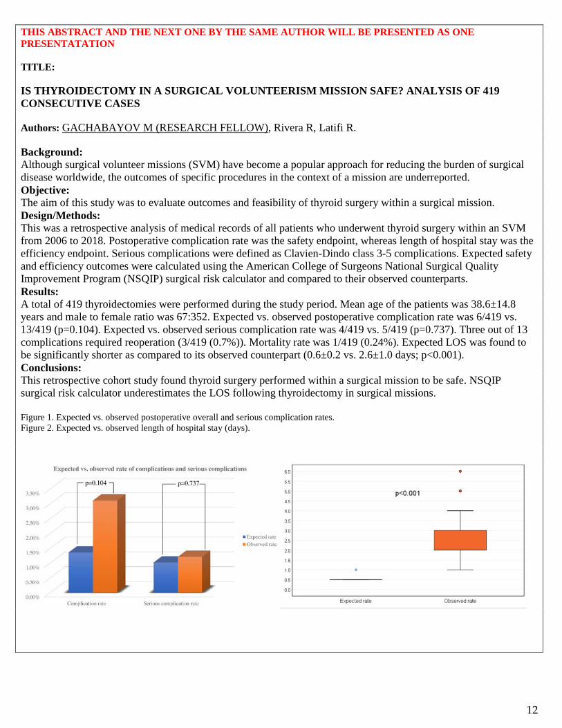
THIS ABSTRACT AND THE NEXT ONE BY THE SAME AUTHOR WILL BE PRESENTED AS ONE PRESENTATATION TITLE: IS THYROIDECTOMY IN A SURGICAL VOLUNTEERISM MISSION SAFE? ANALYSIS OF 419 CONSECUTIVE CASES Authors: GACHABAYOV M (RESEARCH FELLOW), Rivera R, Latifi R. Background: Although surgical volunteer missions (SVM) have become a popular approach for reducing the burden of surgical disease worldwide, the outcomes of specific procedures in the context of a mission are underreported. Objective: The aim of this study was to evaluate outcomes and feasibility of thyroid surgery within a surgical mission. Design/Methods: This was a retrospective analysis of medical records of all patients who underwent thyroid surgery within an SVM from 2006 to 2018. Postoperative complication rate was the safety endpoint, whereas length of hospital stay was the efficiency endpoint. Serious complications were defined as Clavien-Dindo class 3-5 complications. Expected safety and efficiency outcomes were calculated using the American College of Surgeons National Surgical Quality Improvement Program (NSQIP) surgical risk calculator and compared to their observed counterparts. Results: A total of 419 thyroidectomies were performed during the study period. Mean age of the patients was 38.6±14.8 years and male to female ratio was 67:352. Expected vs. observed postoperative complication rate was 6/419 vs. 13/419 (p=0.104). Expected vs. observed serious complication rate was 4/419 vs. 5/419 (p=0.737). Three out of 13 complications required reoperation (3/419 (0.7%)). Mortality rate was 1/419 (0.24%). Expected LOS was found to be significantly shorter as compared to its observed counterpart (0.6±0.2 vs. 2.6±1.0 days; p<0.001). Conclusions: This retrospective cohort study found thyroid surgery performed within a surgical mission to be safe. NSQIP surgical risk calculator underestimates the LOS following thyroidectomy in surgical missions. Figure 1. Expected vs. observed postoperative overall and serious complication rates. Figure 2. Expected vs. observed length of hospital stay (days).
12
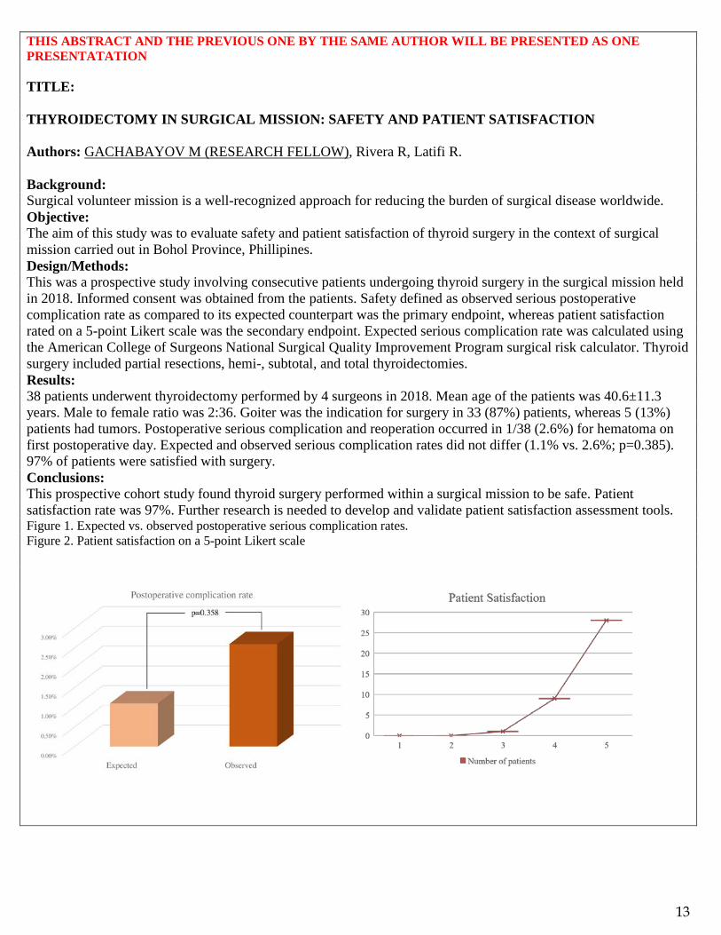
THIS ABSTRACT AND THE PREVIOUS ONE BY THE SAME AUTHOR WILL BE PRESENTED AS ONE PRESENTATATION TITLE: THYROIDECTOMY IN SURGICAL MISSION: SAFETY AND PATIENT SATISFACTION Authors: GACHABAYOV M (RESEARCH FELLOW), Rivera R, Latifi R. Background: Surgical volunteer mission is a well-recognized approach for reducing the burden of surgical disease worldwide. Objective: The aim of this study was to evaluate safety and patient satisfaction of thyroid surgery in the context of surgical mission carried out in Bohol Province, Phillipines. Design/Methods: This was a prospective study involving consecutive patients undergoing thyroid surgery in the surgical mission held in 2018. Informed consent was obtained from the patients. Safety defined as observed serious postoperative complication rate as compared to its expected counterpart was the primary endpoint, whereas patient satisfaction rated on a 5-point Likert scale was the secondary endpoint. Expected serious complication rate was calculated using the American College of Surgeons National Surgical Quality Improvement Program surgical risk calculator. Thyroid surgery included partial resections, hemi-, subtotal, and total thyroidectomies. Results: 38 patients underwent thyroidectomy performed by 4 surgeons in 2018. Mean age of the patients was 40.6±11.3 years. Male to female ratio was 2:36. Goiter was the indication for surgery in 33 (87%) patients, whereas 5 (13%) patients had tumors. Postoperative serious complication and reoperation occurred in 1/38 (2.6%) for hematoma on first postoperative day. Expected and observed serious complication rates did not differ (1.1% vs. 2.6%; p=0.385). 97% of patients were satisfied with surgery. Conclusions: This prospective cohort study found thyroid surgery performed within a surgical mission to be safe. Patient satisfaction rate was 97%. Further research is needed to develop and validate patient satisfaction assessment tools. Figure 1. Expected vs. observed postoperative serious complication rates. Figure 2. Patient satisfaction on a 5-point Likert scale
13

TITLE: EVALUATING THE SAFETY OF LOW MOLECULAR HEPARIN USE FOR THROMBOPROPHYLAXIS IN TRAUMATIC BRAIN INJURY Authors: ANSAB A. HAIDER, MD (PGY3), Jorge Con, MD, Kartik Prabhakaran, MD, Anthony Policastro, MD, Rifat Latifi, MD Background: Low molecular weight heparin (LMWH) has been established as the gold standard for chemical thromboprophylaxis in trauma patients and is considered superior to unfractionated heparin in preventing thromboembolic events. Its use however is controversial in traumatic brain injury patients due to fears of progression of intracranial hemorrhage. Objective: The aim of our study was to evaluate the safety of LMWH use of VTE prophylaxis in patients with TBI. Design/Methods: We queried 2015 ACS TQIP database for all patients ≥16 years of age with blunt head trauma who either received LMWH or UFH for thromboprophylaxis. Outcome measures were mortality, rates of deep venous thrombosis (DVT) and pulmonary embolism (PE), and neurosurgical intervention (craniotomy/craniectomy). Multivariate regression analysis was performed. Results: A total of 117,832 patients were included. Mean age was 52±21 years, median ISS [IQR] was 10 [9 - 17], and 5.6% population had severe TBI. 76.5% patients received LMWH and 23.5% received UFH. Overall mortality rate was 1.5%. Patients who received LMWH had lower rates of unadjusted mortality (p<0.001), DVT (p<0.001), and PE (p<0.001). Patients who received LMWH also had significantly lower rates of neurosurgical intervention (p<0.001). On multivariate logistic regression, LMWH use was independently associated with lower odds of mortality (OR [95% CI]: 0.38 [0.35-0.42]), DVT (OR [95% CI]: 0.56 [0.51-0.62]), and PE (OR [95% CI]: 0.59 [0.51-0.67]). Similarly, LMWH was independently associated with lower odds of neurosurgical intervention (OR [95% CI]: 0.30 [0.24-0.37]). On subgroup analysis of patients with GCS<8, LMWH use was still associated with lower odds of mortality (OR [95% CI]: 0.41 [0.28-0.61]) and neurosurgical intervention (OR [95% CI]: 0.39 [0.20-0.76]). Conclusions: The use of LMWH in patients with TBI is safe and is associated with lower mortality, neurosurgical intervention, and thromboembolic events when compared with UFH. LMWH should be used as the agent of choice for thromboprophylaxis in trauma patients with TBI.
14
TITLE: AGE SHOCK INDEX: A RELIABLE PREDICTOR OF MORTALITY IN GERIATRIC TRAUMA PATIENTS Authors: ANSAB A. HAIDER, MD (PGY3), Jorge Con, MD, Kartik Prabhakaran, MD, Gary Lombardo, MD, Anthony Policastro, MD, Rifat Latifi, MD Background: Shock index and age are both well-known predictors of mortality in trauma patients. However, it is often postulated that due to the dampened physiologic responses in elderly trauma patients, traditional vital signs including SI may have higher false negative rate for predicting mortality. Objective: The aim of our study was to evaluate if age shock index (ASI= age x SI) may be a better predictor of early mortality in geriatric trauma patients. Design/Methods: We abstracted two years of NTDB for all patients ≥65 years of age and ISS >15 with complete data. Patient demographics, injury parameters, and traditional vital signs were recorded and SI, and ASI were calculated. Our outcome measure was early mortality (≤24 hours). Area under receiver operating curve (AUROC) was calculated for each vital signs and index and compared. Results: A total of 18,736 patients were included. Mean age was 74±6 years, 58.7% were male, median ISS [IQR] was 21 [17 - 26], and the early mortality rate was 4.1%. HR, SBP, SI, and ASI were all significant predictors of early mortality (p<0.001). AUROC [95% CI] for SBP and HR was 0.43 [0.42-0.45] and 0.58 [0.56-0.60] respectively. Highest AUROC was noted for ASI 0.62 [0.60-0.64] followed by SI AUROC of 0.61 [0.59-0.63]. Even in the subgroup of patients with normal traditional vital signs, patients with ASI>50 had 30% higher odds of early mortality (OR [95% CI] : 1.3 [1.0-1.6]). SI was unable to predict mortality in this subgroup of patients. Conclusions: A traditional vital sign significantly underperforms in predicting early mortality in geriatric trauma patients. ASI has the highest predictive power followed by SI. ASI may be a better tool for effective triage of seriously injured geriatric trauma patients.
15

TITLE: INDIVIDUALIZED PREDICTION OF INCONTINENCE AFTER ROBOT-ASSISTED RADICAL PROSTATECTOMY: DEVELOPMENT AND VALIDATION OF PROSTATECTOMY INCONTINENCE NOMOGRAM Authors: HUANG R. (PGY1), Lee T., Dodge N., Pinkhasov A., Pinkhasov R., Attwood K., Pop E., and Mohler J. Background: Even though a number of risk factors for incontinence after robot-assisted radical prostatectomy (RARP) are known, there is a paucity of data that integrates them. Therefore, we sought to develop and validate a prostatectomy incontinence nomogram (PIN) that predicts probability of incontinence at 6- and 24-months after RARP. Objective: The purpose of this study is twofold: 1. To evaluate predictors of early return to social and perfect continence and 2. Create a risk calculator that accurately predicts time to return to social and perfect continence after robot-assisted radical prostatectomy (RARP). Design/Methods: Data from 1,033 men with prostate cancer that underwent RARP from January, 2008 to December, 2015 at our institution was queried. After applying exclusion criteria, a total of 680 men were divided into nomogram: 1) model development cohort (n = 544, 80%), and 2) model validation cohort (n = 136, 20%). Logistic regression was used for univariate/multivariate analyses, and to build a nomogram. Reduced model selection was performed using backward step-down selection process. Calibration plots and receiver operating curves (ROC) were used for quantifying the nomogram accuracy. Internal validation was performed by bootstrapping and the reduced nomogram model was calibrated. Using UCLA-PCI-Short Form-v2 Urinary Function questionnaire, perfect continence was defined as 0 pads, social continence was defined as 1 or 2 pads, and incontinence was defined as ≥ 3 pads used after RARP. Results: Variables found to be predictive on univariate and multivariate analysis used in the model development cohort included age, race, body mass index (BMI), surgical margins status, and pre-operative erectile function. The initial model has moderate to good predictability with a 6- and 24-month AUC of 0.64 and 0.80, respectively. The recalibrated model has modest to reasonably good predictability with a 6- and 24-month AUC of 0.52 and 0.76, respectively. Using the developed calibrated nomogram, the overall predicted probability of incontinence is 26% (95% CI: 0.23-0.30) by 6 months, and 3% (95% CI: 0.02 – 0.04) by 24 months. Conclusions: We devised and validated a modest 6-month and a reasonably strong 24-month nomogram that is superior to any single clinical variable for predicting 6- and 24-month risk of incontinence after RARP.
16

TITLE: METABOLIC SYNDROME EXPONENTIALLY INCREASES THE RISK OF ADVERSE OUTCOMES AFTER SURGERY FOR DIVERTICULITIS Authors: FAISAL JEHAN, MD (PGY1), Jorge Con, MD, Muhammad Khan, MD, Thomas Diflo, MD, Kartik Prabhakaran, MD, Anthony Policastro, MD, Patrice Anderson, MD, Herminio Diaz, MD, Gary Lombardo, MD, Rifat Latifi, MD Background: Metabolic syndrome is defined as a cluster of high blood pressure, blood sugar, and a high BMI. It is associated with increased risk of heart disease, stroke and numerous other diseases. Although the effect of obesity has been study in patients undergoing surgery, the effect of metabolic syndrome on outcomes in surgery patients remains unclear. Objective: The aim of our study was to evaluate the impact of metabolic syndrome on the risk of morbidity and mortality in patients who underwent surgery for acute diverticulitis. Design/Methods: We analyzed the National Surgical Quality Improvement Program (NSQIP) database from 2012-2015. Patients with acute diverticulitis who underwent operative intervention were included in our study. Metabolic syndrome was defined as presence of BMI > 30kg/m2, hypertension and diabetes. Our primary outcome measure was risk of any adverse event (complications, 30-day readmission, and mortality). Secondary outcome measures were complications following the procedure; hospital length of stay, 30-day readmission and mortality rate. Regression and ROC analysis was performed. Results: A total of 4,572 patients who underwent surgery for acute diverticulitis were identified. The mean body mass index was 28.8 ± 10 kg/m2; 41.2% (1883) patients were obese (>30 kg/m2) while 9% patients had BMI > 40. Of the patients who were obese, 14.6% (275) had metabolic syndrome. The adjusted risk of any adverse effects was exponentially higher in patients with metabolic syndrome than the obese alone group and the obese and hypertensive group (OR, 8.1, p<0.001). Patients with metabolic syndrome had a greater risk for reintubation (OR 1.9, p=0.03), >48 hours ventilator dependence (OR 3.5, p=0.01), myocardial infarction (OR 2.3, p=0.03) and superficial and deep surgical-site infections (OR 2.1; p=0.01) compared with patients who did not have metabolic syndrome. In addition, they also had a longer length of hospital stay (b= 1.23, p=0.02), higher 30-days readmission (OR 1.7, p<0.01) and mortality rate (OR 2.1, p<0.01). On ROC curve analysis, AUROC for predicting adverse outcomes by metabolic syndrome was 0.797 which was higher than the AUROC for BMI (0.58), hypertension (0.51) or diabetes (0.64) alone. Conclusions: The risk of any adverse event in patients with metabolic syndrome after surgery for diverticulitis is exponentially higher than the BMI, hypertension or diabetes alone. Patients with metabolic syndrome tend to take longer to recover after surgery for acute diverticulitis and have higher rates of complications, readmissions and mortality. This is a simple tool and will help in risk stratification and prognosis discussion with patient and family.
17

TITLE: NOVEL ORAL ANTICOAGULANTS VS. LOW MOLECULAR WEIGHT HEPARIN FOR THROMBOPROPHYLAXIS IN OPERATIVE PELVIC FRACTURES Authors: MUHAMMAD KHAN, MD (PGY1), Jorge Con, MD, Faisal Jehan, MD, Kartik Prabhakaran, MD, Asad Azim, MD, Anthony Policastro, MD, Rifat Latifi, MD Background: Pelvic fractures have been identified as a risk factor for venous thromboembolic (VTE) complications. Recent literature shows the superiority of novel oral anticoagulants (NOACs) over low molecular weight heparin (LMWH) for thromboprophylaxis in orthopedic patients. Objective: The aim of our study was to evaluate the impact of NOACs vs. LMWH on outcomes in patients with operative pelvic fractures. Design/Methods: We performed a 2-year (2015-16) analysis of the ACS-TQIP database. We included all adult patients with isolated blunt pelvic fractures who were managed operatively and received post-operative thromboprophylaxis with either LMWH or NOACs (Factor Xa inhibitor and direct thrombin inhibitor). Patients were stratified into two groups based on the type of thromboprophylactic agent (NOACs vs. LMWH) and were matched in a 1:2 ratio for demographics, admission vitals, injury parameters, hospital stay, facility, and timing of initiation of thromboprophylaxis. Primary outcomes were rates of DVT and/or PE. Secondary outcomes were pRBC transfusions, and intervention for hemorrhage control after initiation of thromboprophylaxis. Results: We analyzed 11,219 patients with pelvic fractures. A total of 3,529 patients with isolated pelvic fractures were included of which 708 patients were matched (NOACs: 236; LMWH: 472). Mean age was 61±12 and median ISS was 12[10-16]. Matched groups were similar in demographics, vitals and injury parameters, hospital stay, and timing of initiation of thromboprophylaxis. Overall 5.8% of patients had DVT, and 1.8% PE%. Patients who received NOACs were less likely to develop DVT (2.9% vs. 7.2%, p=0.01) compared to LMWH. There was no difference in PE (1.6% vs. 1.9%, p=0.28) between the two groups. Similarly there was no difference in post prophylaxis blood products transfusion, and post-prophylaxis intervention for hemorrhage control(Table 1.). Conclusions: In patients with operative pelvic fracture, thromboprophylaxis with novel oral anticoagulant is associated with lower rates of DVT. There was no association between type of thromboprophylactic agent with PE. Further prospective clinical trials should evaluate the role of NOACs for thromboprophylaxis in high-risk trauma patients.
Table 1. Primary and Secondary Outcomes Outcomes NOACs (n=236) LMWH (n=472) P-Value VTE Complications, % (n)
DVT 2.9% 7.2% 0.01 PE 1.6% 1.9% 0.28
Secondary Outcomes, % (n)
Post-prophylaxis Blood Products Transfusion 5.5% 3.7% 0.43
Intervention for hemorrhage control 1.7% 1.2% 0.57
18
TITLE: BLOOD ALCOHOL CONTENT IS ASSOCIATED WITH LOWER INJURY SEVERITY SCORES AND SPINAL INJURIES AMONG INTOXICATED TRAUMA PATIENTS Authors: NIKATHAN S. KUMAR, (MS3), Matthew K. McIntyre, BA, Elizabeth H. Tilley, PhD, David J. Samson, MS, Rifat Latifi, MD, FACS. Background: Intoxicated patients represent 10% of trauma patients presenting to the emergency department (ED). Previous studies show varied reports regarding the protective effects of alcohol intoxication against injury and mortality. Objective: Our study aimed to determine the effects of blood alcohol content (BAC) on injury type and severity. Design/Methods: This 4-year retrospective study (2013-2017) considered all Level 1 and 2 trauma patients with a BAC > 10 mg/dL on admission (≥14 years) presenting to a Level 1 trauma center emergency department. Patients were stratified by BAC < 200 (Low BAC) and BAC > 200 (High BAC). Patients’ BAC was compared to their calculated injury severity scores (ISS) and abbreviated injury scale (AIS). T tests and ANOVAs with Tukey’s post-hoc analysis were used for continuous variables, while Fisher’s exact test was used for binary variables. Results: We identified 437 intoxicated patients with Level 1 or 2 trauma activations (average BAC: 212.3±89.2 mg/dL; 71.9% male; median age 35; range: 16-90). The main mechanisms of injury were motor vehicle crashes (55.5%) and falls (23.2%). Overall, 336 (76.8%) patients had an average ISS of 9.86 (range: 1-48), and 88 (26.2%) patients had ISS ≥ 16. The High BAC group (n = 178) had lower ISS (8.8±0.66) compared to the Low BAC group (n = 158; ISS = 11.1±0.79; p = 0.0268) and fewer spinal injuries (7.19% vs 11.38%; OR = 0.50); however, there were no significant differences in injury frequency of other body regions. Additionally, Low BAC patients in motor vehicle collisions had increased head AIS scores than the High BAC MVC group (1.36±0.16 vs 0.84±0.16; p = 0.0244). Finally, High BAC patients had shorter hospital length of stay (4.1±0.54 vs 2.8±0.35 days; p = 0.0441), but no significant differences in ICU length of stay, ventilator days, or GCS. Conclusions: Trauma patients with higher BAC have lower ISS, less spinal injury, and shorter stays in the hospital. Patients in MVCs with higher BAC had less severe head injury. These findings further substantiate the protective role of alcohol in trauma patients, although this mechanism is still poorly understood.
19

TITLE: PURSESTRING VERSUS LINEAR SKIN CLOSURE AT LOOP ILEOSTOMY REVERSAL: A SYSTEMATIC REVIEW AND META-ANALYSIS Authors: LEE H (PGY4), Gachabayov M, Bergamaschi R. Background: There is no level 1a evidence regarding the best technique for skin closure at loop ileostomy reversal. Objective: The aim of this study was to evaluate whether purse string skin closure (PSC) is associated with lower surgical site infection (SSI) rates as compared to linear skin closure (LC). Design/Methods: EMBASE, MEDLINE, Pubmed, Cochrane Library, Web of Science, and CINAHL databases were systematically searched. PSC was defined as a circumferential subcuticular suture leaving a small circular skin defect allowing for free drainage, granulation, and epithelialization. In LC, the wound edges were approximated side to side with or without drainage. The primary endpoint was SSI rate. Secondary endpoints included operating time, length of hospital stay, wound healing time, and incisional hernia rates. Inclusion criterion was any observational or experimental study comparing PSC to LC in patients undergoing ostomy reversal. Results: Twenty studies (6 experimental and 14 observational) totaling 1,812 patients (826 PSC and 986 LC) were included. SSI rates were statistically and clinically significantly lower in patients with PSC [OR (95%CI) = 0.14 (0.09, 0.21); p < 0.0001; NNT = 6] in the meta-analysis of all studies (Figure 1). The subgroup analysis of randomized trials [OR (95%CI) = 0.10 (0.04, 0.21); p < 0.0001; NNT = 6] (Figure 2) as well as the analysis of randomized trials including patients with loop ileostomy only [OR (95%CI) = 0.12 (0.05, 0.28); p < 0.0001; NNT = 5] confirmed this finding (Figure 3). Conclusions: This meta-analysis found that PSC was associated with significantly decreased rates of SSI in patients undergoing loop ileostomy reversal.
Figure 1: Meta-analysis of all studies: SSI rate in PSC vs LC.
20

TITLE: LACTATED RINGERS ATTENUATES GUT MICROBIOME AND INTESTINAL CHANGES AFTER 40% TOTAL BODY SURFACE AREA BURN INJURY IN SWINE Authors: MATTHEW MCINTYRE, (MS3)1, Charlotte J. Winkler2, Belinda I. Gómez, PhD3, Tony Chao, PhD3, Joshua S. Little3, Susannah Nicholson, MD4, Michael A. Dubick, PhD3, David M. Burmeister, PhD3
1School of Medicine, New York Medical College, Valhalla, NY 10598 2Yale University, New Haven, CT 06520 3Damage Control Resuscitation, US Army Institute of Surgical Research, JBSA Fort Sam Houston, TX 78234 4Department of Surgery, University of Texas Health Science Center San Antonio, San Antonio, TX 78229 Background: Extensive burns induce systemic inflammation and increased intestinal permeability which can put the patient at risk for infectious complications such as sepsis. While recent reports underscore the importance of the gut microbiome in health and disease, the role of the microbiome in burn outcomes remains unclear. We describe the changes in the gut microbiome following a 40% Total body surface area (TBSA) burn in swine, and determine the effect of different intravenous (IV) resuscitation volumes on the microbiome and intestinal integrity. Methods: Anesthetized Yorkshire swine sustained 40% TBSA full-thickness burns and were randomized to different volumes of IV Lactated Ringers’: none (n=3), 15mL/kg/day (Low; n=6), or 2mL/kg/%TBSA/day (High; n=6). All animals received 15mL/TBSA/kg/day of oral rehydration solution to control for enteral hydration. At baseline and days 1 and 2 post-burn, fecal swabs were collected for 16s rDNA sequencing. Ileum was collected immediately after euthanasia (day 2) for western blot, histopathology, and cytokine analyses. Results: Following burn injury there is a significant shift in the gut microbiome community in terms of β-diversity, regardless of treatment. This was accompanied by a non-significant reduction in α-diversity and evenness that did not recover by day 2 regardless of treatment group. We also found a significant increase in Proteobacteria following injury that was attenuated by IV fluids in a dose-dependent manner and was restored to normal levels by day 2 high and none, but not low, groups. Increased proteobacteria was also significantly correlated with decreased alpha diversity (P<0.014). Interestingly, we found greater Hsp70 levels (p=0.0464) in the high fluid group, as well as higher concentrations of SGLT1 (p=0.0213) and caspase (p=0.0139) in the no-fluid group. IL1a and IL12 levels were elevated in the low group and other pro-inflammatory cytokines were generally associated with decreased fluid levels. Bacteroidetes levels correlated with elevated IL18 levels (p=0.0166, R2=0.4201). Conclusions: Aggressive fluid resuscitation is critical for improving outcomes following a large burn injury. Despite specific differences in certain species, we show a marked shift in the gut microbiome community regardless of IV fluid. However, we found that high levels of fluids are anti-apoptotic, anti-inflammatory, and alter critical protein expression in the ileum. This research highlights the need to explore the microbiome for diagnostic and therapeutic purposes to improve burn outcomes.
21
TITLE: INCREASING FRAILTY IS ASSOCIATED WITH WORSE OUTCOMES FOLLOWING ANGIOGRAM-NEGATIVE SUBARACHNOID HEMORRHAGES Authors: MATTHEW MCINTYRE BA (MS3)1, James Dragonette BS MS31, Fawaz Al-Mufti MD2, Chirag Ghandi MD2, Chad Cole MD MSc2, Meic Schmidt MD MBA2, Justin Santarelli MD2, Christian Bowers MD2 New York Medical College1 and Department of Neurological Surgery2, Valhalla, New York 10595 Corresponding author: Christian Bowers, MD Introduction: Angiogram negative subarachnoid hemorrhage (ANSAH) comprises a substantial portion of intracranial hemorrhage. Neurological ICU admissions occurs frequently in the elderly and the overall number of elderly neurosurgical patients is increasing significantly. The etiology of ANSAH is often undetermined but multiple studies have demonstrated that these patients have distinct clinical courses from patients with subarachnoid hemorrhage due to trauma or aneurysm rupture. Increasing frailty is associated with increasing rates of morbidity, mortality, complications, and other worse outcomes in a variety of surgical disciplines. There is no existing study examining the impact of increasing frailty on ANSAH but we predicted that increased frailty would correlate with worse outcomes in this patient group as well. Methods: This is a retrospective cohort study conducted between June, 2014 and July, 2018 at a tertiary care center. We identified all non-traumatic subarachnoid patients who underwent diagnostic cerebral angiogram (DSA) without identifying an underlying aneurysm or vascular lesion. We divided the cohort into a non-frail [modified frailty index (mFI) of 0] and a frail group (mFI) ≥1. The primary outcomes were discharge Glasgow Coma Score (GCS), mortality rate, hospital length of stay (LOS) and ICU LOS. Secondary endpoints were extra-ventricular drain (EVD) placement (clinical hydrocephalus), vasospasm (clinical and radiographic), and ventriculomegaly (no external ventricular drain required). Groups were compared using Student’s T-test for continuous variables and Fisher’s exact test for binary values using Graphpad Prism 8. Results: We identified 93 ANSAH patients during the study period. The mean age was 56.6±1.6 years and 40 (43.5%) were male. The average Hunt-Hess (HH) and Fisher scores were 1.9 and 3.0, respectively. The mean mFI was 1.0 with almost half the patients (45 pts/48.4%) having a mFI ≥1. We found that a mFI≥1 was significantly associated with higher presenting HH (P<0.0001) and Fisher (P=0.0006) scores, lower admission GCS scores (P=0.0008), and lower discharge GCS (P=0.0119). Interestingly, mFI≥1 was associated with an increased likelihood of clinical hydrocephalus requiring an EVD (OR=5.2; 95% CI: 1.7-13.8; P=0.0029) but not the presence of ventriculomegaly (not requiring CSF diversion) (OR 2.3; 95% CI: 0.9-6.4; P=0.1412). Frailty (mFI≥1) was not associated with increased rates of radiographic vasospasm (P>0.05). Frailty (mFI≥1) was associated with an increased mortality rate (5.4% in mFI≥1 vs 0% in mFI of 0) (P=0.0235), increased hospital LOS (P<0.0001; R2=0.3611), and increased ICU LOS (P<0.0001; R2=0.4111). Conclusions: Frailty (mFI≥1) was associated with worse clinical presentations and worse outcomes in patients with ANSAH. Frail patients presented with worse HH and Fisher Scores, lower admit and discharge GCS scores, increased rates of clinical hydrocephalus, increased hospital LOS, increased ICU LOS, and increased mortality rates when compared to non-frail ANSAH patients (mFI of 0). This study provides the first initial evidence that frail ANSAH patients have worse presentations and worse outcomes compared to the non-frail (mFI=0) ANSAH cohorts. This information has important prognostic and treatment significance and needs to be studied further.
22
TITLE: CD5+ DENDRITIC CELLS ARE A POTENT IMMUNOSTIMULATORY MEDIATOR IN GVHD Authors: ADIEL MUNK (MS3), Daniel Korenfeld, Laurent Gorvel, Joshua Man, Andras Schaffer, Thomas Tung, Caroline Mann, Eynav Klechevsky Background: Graft vs. Host Disease is a feared complication of transplantation and has led to significant mortality and morbidity. Recently, Stenger et al (Blood, 2012) have shown that while traditional therapies have targeted T cells, immunostimulatory dendritic cells (DCs) are critical in the pathogenesis of GVHD. Conventional immunosuppressant continue to be the mainstay of treatment for GVHD, yet they often fail and carry a significant risk for infection. The role of DCs in immune tolerance and stimulation has begun to be elucidated, and its role in GVHD pathogenesis is vital to the understanding and therapeutic approach to transplantation. Objective: The objective of this study was to elucidate the role of dendritic cells in GVHD. Additionally, we sought to identify the dendritic cell subset responsible for the potent immunostimulatory capacity of dendritic cells in regards to GVHD, and how this subset may elicit this response. Design/Methods: Briefly, healthy human skin was obtained from donors who underwent cosmetic and plastic surgeries. Psoriatic plaque biopsy samples, lupus, LC histocytosis and GVHD disease skin specimens were obtained. The samples were immunofluorescently stained with antibodies or isotype controls. Images were taken using a Leica microscope. Skin DC subsets were isolated from healthy skin, by using an enzymatic reaction, followed by cell sorting using a BD FacsARIA II. Results: We observed that CD5+ epidermal Langerhan cells (LCs) were the only epidermal DCs in acute graft-versus-host disease patient skin, suggesting their involvement in this inflammatory disease. Additionally, the CD5+ LCs were potent immunostimulatory DCs ,capable of eliciting a stronger immune response than any other DC subset in the epidermis or dermis. Additionally, the terminally differentiated CD5+ DCs were particularly potent at eliciting a Th22 response, characterized by increased expression of IL-22. Conclusions: Although transplantation has significantly improved medical outcomes, GVHD still remains a complication that has not been completely defined. In our study, we showed the importance of identifying a distinct CD5+ DC subset that is a potent immunostimulator, which was the only DC subset found in the epidermis. Further studies characterizing the CD5 subset, including its’ complete cytokine profile will be useful in helping to further understand the pathogenesis of GVHD. Genome-wide association studies have shown a pathogenic relevance for CD5 in many inflammatory conditions, including psoriasis and rheumatoid arthritis (Nature, 2011), and a treatment specifically targeting CD5+ DCs may serve a purpose in improving transplantation outcomes. Strategies to modify the CD5+ DC composition and function may represent an innovative approach for the treatment of GVHD and other immune-mediated disorders.
23
TITLE: OPIOID-FREE ANALGESIA IN ELECTIVE BOWEL RESECTION: CHANGES OVER TIME. Authors: PATEL AH (MS3), Fakas S, Samson D, Xu J, Bergamaschi R. Background: Lack of research showing opioid-free strategies in post-operative surgical patients, specifically those undergoing bowel resection. Objective: The aim of this study was to review the use of different strategies for postoperative analgesia in elective bowel resection. Design/Methods: This was a retrospective cohort study including patients having undergone elective bowel resection between February 2012 and June 2018 at one center. This study was approved by the Institutional Review Board at New York Medical College. Trend analysis was conducted using Joinpoint regression, (National Cancer Institute, Bethesda Maryland) employing 9-month intervals. The primary outcome for each interval was the proportion of patients receiving postoperative opioid-free analgesia (OFA), defined as forgoing all opioid analgesics after the day of surgery. Statistical tests performed on non-overlapping segments of intervals were performed using Student’s t-test for continuous variables and Pearson’s chi-squared test for categorical variables. These latter tests were performed using SPSS version 25 (IBM, Inc., Armonk New York). We considered p values <0.05 to be statistically significant. Results: 125 patients met selection criteria. Females comprised 52.0% and mean age was 58.0±15.5 years (range 19-86). Mean BMI was 29.1±8.3 kg/m2. The distribution by ASA class was: class 1, 0.8%; class 2, 22.4%; class 3, 69.6%; and class 4, 7.2%. Opioid tolerance was reported by 14 patients (11.3%). Indications for surgery included: colon or rectal cancer (23.2%), diverticulitis (20.0%), other cancer (11.2%), hernia (10.4%), obesity (9.6%), ulcerative colitis (8.0%), fistula (7.2%), small bowel obstruction (4.8%), Crohn’s disease (3.2%), gastrointestinal bleeding (2.4%) and polyps (2.4%). Preexisting comorbidities included: hypertension (45.6%), cancer (28.8%), diabetes mellitus (20.8%), hyperlipidemia (20.0%), gastroesophageal reflux disease (16.8%), obesity (12.0%) and depression (10.4%). Surgical access was laparotomy in 44.0%, laparoscopic in 46.0%, laparoscopic converted to laparotomy in 3%, robotic in 7%. Post-anesthesia care unit (PACU) visual analog scale (VAS, 0-10) pain at postoperative interval 1 was 4.58±2.45, compared with 4.09±2.65 at interval 2 and 3.51±2.82 at interval 3. Mean hospital length of stay was 11.2±15.5 days. Mean operative time was 325.1±133.6 minutes. Mean estimated blood loss was 242.5±333.8 mL. Trend analysis was conducted using nine 9-month intervals. The proportion of patients receiving OFA during interval 1 was 20%, falling steadily to 0% at interval 4. The OFA proportion then rose back to 20% at interval 6, declined to 10% at interval 7, then rebounded to 15.4% at interval 8, and rose further to 42.9% at interval 9. Joinpoint regression found that the slope of the line between intervals 1 and 4 (segment 0) was not different statistically from zero. However, modeling detected a joinpoint at interval 4 marking the beginning of segment 1. The slope of segment 1 was positive and statistically different from zero, with an average percent change (APC) of 42.6 with a 95% confidence interval of 1.8 to 99.8 and a p value of 0.043. Thus, since interval 4, there has been a positive increasing trend of use of OFA in this patient population at our institution. This change coincides with the arrival of an anesthesiologist co-author who implemented a new analgesics usage policy favoring non-opioid agents. Conclusions: This study showed a significant increasing trend in opioid-free analgesia in elective bowel resection from 0 to 42.9% over 4.5 years.
24
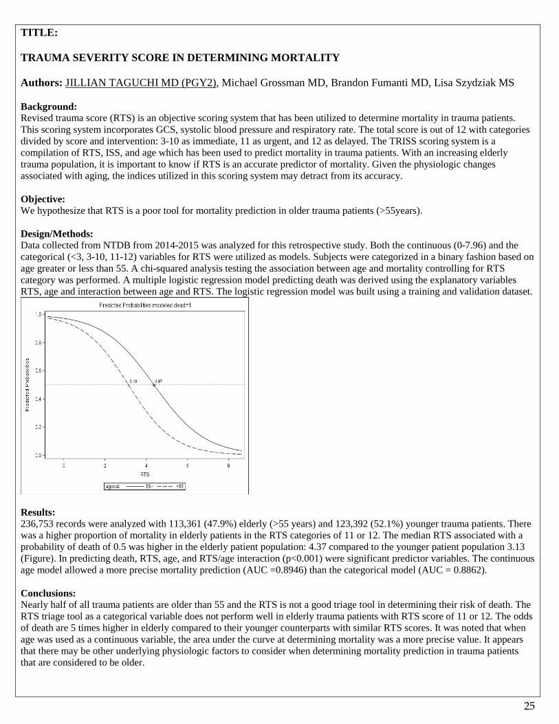
TITLE: TRAUMA SEVERITY SCORE IN DETERMINING MORTALITY Authors: JILLIAN TAGUCHI MD (PGY2), Michael Grossman MD, Brandon Fumanti MD, Lisa Szydziak MS Background: Revised trauma score (RTS) is an objective scoring system that has been utilized to determine mortality in trauma patients. This scoring system incorporates GCS, systolic blood pressure and respiratory rate. The total score is out of 12 with categories divided by score and intervention: 3-10 as immediate, 11 as urgent, and 12 as delayed. The TRISS scoring system is a compilation of RTS, ISS, and age which has been used to predict mortality in trauma patients. With an increasing elderly trauma population, it is important to know if RTS is an accurate predictor of mortality. Given the physiologic changes associated with aging, the indices utilized in this scoring system may detract from its accuracy. Objective: We hypothesize that RTS is a poor tool for mortality prediction in older trauma patients (>55years). Design/Methods: Data collected from NTDB from 2014-2015 was analyzed for this retrospective study. Both the continuous (0-7.96) and the categorical (<3, 3-10, 11-12) variables for RTS were utilized as models. Subjects were categorized in a binary fashion based on age greater or less than 55. A chi-squared analysis testing the association between age and mortality controlling for RTS category was performed. A multiple logistic regression model predicting death was derived using the explanatory variables RTS, age and interaction between age and RTS. The logistic regression model was built using a training and validation dataset.
Results: 236,753 records were analyzed with 113,361 (47.9%) elderly (>55 years) and 123,392 (52.1%) younger trauma patients. There was a higher proportion of mortality in elderly patients in the RTS categories of 11 or 12. The median RTS associated with a probability of death of 0.5 was higher in the elderly patient population: 4.37 compared to the younger patient population 3.13 (Figure). In predicting death, RTS, age, and RTS/age interaction (p<0.001) were significant predictor variables. The continuous age model allowed a more precise mortality prediction (AUC =0.8946) than the categorical model (AUC = 0.8862). Conclusions: Nearly half of all trauma patients are older than 55 and the RTS is not a good triage tool in determining their risk of death. The RTS triage tool as a categorical variable does not perform well in elderly trauma patients with RTS score of 11 or 12. The odds of death are 5 times higher in elderly compared to their younger counterparts with similar RTS scores. It was noted that when age was used as a continuous variable, the area under the curve at determining mortality was a more precise value. It appears that there may be other underlying physiologic factors to consider when determining mortality prediction in trauma patients that are considered to be older.
25

TITLE: WHAT DOES IT MEAN FOR A SURGEON TO “RUN TWO ROOMS”? A COMPREHENSIVE LITERATURE REVIEW OF OVERLAPPING AND CONCURRENT SURGERY POLICIES Authors: BRIANNA THERIAULT*, BS, PHD2 (MS3); JULIA PAZNIOKAS*, BS2; Abhitini Mittal, BS2; Meic Schmidt, MD, MBA1 ; Chad Cole, MD, MSc1 ; Chirag Gandhi, MD1 ; Patrice Anderson, MD3 ; Christian Bowers, MD1 *The authors contributed equally. 1 Department of Neurosurgery, Westchester Medical Center, New York Medical College, Valhalla, NY 10595 2 New York Medical College, Valhalla, NY 10595 3 Department of Surgery, Westchester Medical Center, New York Medical College, Valhalla, NY 10595 Background: The concurrent and overlapping surgery literature has grown exponentially over the past two years. Prior to this, there were no significant publications addressing this topic. There is an extremely wide variance on how “running two rooms” is defined and whether it should be permitted. These differences affect our patients’ perception of this practice. The literature lacks any comprehensive review of the topic and terminology. Objective: The aim of this study was to review and analyze all of the “concurrent” and “overlapping” surgery literature with the goal of: standardizing terminology, defining discrepancies in the literature and proposing solutions for the current challenges of regulating surgery to achieve maximal safety and efficiency. Design/Methods: We performed a PubMed search to identify studies that considered the issue of overlapping surgery (OS). The terms “overlapping surgery”, “concurrent surgery”(CS), and “simultaneous surgery”(SS) were used in the query. We then analyzed the publications identified. Results: The literature contained 18 published studies analyzing OS safety between November 2016 and June 2018. Eight were neurosurgical studies, three were orthopedic, and the remaining seven papers were in other surgical specialties. A total of 1,207,155 surgical cases (range 250 to >500,000 patients) were analyzed among the 18 studies. There were 57,880 (5.04%) OS cases. The OS rates in the individual studies ranged from 1.2% to 68%. Neurosurgical studies had the highest average OS rate of 54% (range 37–68%) while the average OS rate in orthopedic surgery was 43% (range 2.7–68%). Approximately one third of the studies were multicenter investigations (27.7%). The studies measured more than 20 distinct outcomes but there were only 5 outcomes that were included in the majority of the studies: mortality rates, reoperation rates, procedure length of time, readmission rates, and hospital length of stay (LOS). Conclusions: The current body of literature repeatedly demonstrates that OS is a safe and effective option when undertaken by experienced surgeons who practice it frequently. For successful OS, the Mandatory Attending Portion (MAP) for two surgeries must not overlap and Unnecessary Anesthesia Time (UAT) must be prohibited. Hospitals and surgical specialty organizations must implement policies to assure the safe practice of OS.
26
TITLE: TRANSPLANTATION OF NEURAL PRECURSORS DERIVED FROM SPINAL PROGENITOR CELLS IMPROVES FUNCTIONAL RECOVERY FROM SPINAL CORD INJURY BY REDUCED INFLAMMATION VIA INHIBITION OF THE NF-KB PATHWAY IN A RAT MODEL OF SPINAL CORD INJURY. Authors: JOHN V. WAINWRIGHT1 (PGY5), Kristyna Karova2, Lucia Machova-Urdzikova2, Rishikaysh Prasad2, Chirag D. Gandhi1, Meic H. Schmidt1, Pavla Jendelova2, 3, Meena Jhanwar-Uniyal1
1Westchester Medical Center / New York Medical College, Department of Neurosurgery, Valhalla, NY 10595, USA. 2Institute of Experimental Medicine, The Czech Academy of Sciences, Vídenská, 1083, 142 20, Prague, Czech Republic. 3 2nd Faculty of Medicine, Charles University, Prague, Czech Republic Background: Traumatic spinal cord injury (SCI) triggers a chain of events that hinders recovery characterized by an inflammatory cascade leading to necrotic cell death at the core injury site with astrogliosis and apoptotic cell death in surrounding areas. Activation of the nuclear factor-kappa-B (NF-kappa-B) signaling pathway is associated with inflammation in SCI. Here, we elucidate the activation pattern of NF-kappa-B in SCI while investigating the effect of transplantation of spinal neural precursors (SPC-01) on its activity, related astrogliosis, and functional recovery in a rat model. Objective: We elucidate the activation pattern of NF-kappa-B in SCI while investigating the effect of transplantation of spinal neural precursors (SPC-01) on its activity, related astrogliosis, and functional recovery in a rat model. Design/Methods: Using a balloon compression rat model of SCI, injury was induced at T8 and SPC-01 cells or saline was injected into the lesion 7-days post-injury. Rats were followed for functional recovery (Basso, Beattie, Bresnahan (BBB), Rotarod, Plantar Thermal Nociception, and Flat Beam tests) and sacrificed to retrieve spinal cord sections at multiple time points. The course of SPC-01 cells was determined by immunofluorescence response to stem cell and neuronal markers. NF-kappa-B activity, phosphorylated-p65, secretory levels of cytokines (e.g. TNF-alpha), extent of glial scaring, white and gray matter preservation, and cavity size were determined. Results: Functional recovery was enhanced in SPC-01-treated rats as confirmed by BBB score. A bimodal activation pattern of the NF-kappa-B signaling pathway was seen, with peaks at 3- and 28-days. Transplantation of SPC-01 cells resulted in significant downregulation of TNF-alpha at 10- and 14-days and inhibition of NF-kappa-B activity at 28 days after SCI, mainly in gray matter. Transplanted rats exhibited gray matter preservation and reduced glial scar and cavity size. Conclusions: We demonstrate immunomodulatory properties of SPC-01 cells based on inhibition of the canonical NF-kappa-B pathway. This effect occurs prior to maturation of SPC-01 cells, implying that the observed results are due to paracrine mechanisms rather than cell replacement. Reduced inflammation may result in tissue sparing, reduced glial scar, and improved functional recovery.
27

TITLE: PLACEMENT OF OMMAYA RESERVOIRS USING LECTROMAGNETIC NEURONAVIGATION AND NEUROENDOSCOPY: A RETROSPECTIVE STUDY WITH COST-BENEFIT ANALYSIS Authors: ARTHUR WANG, M.D. (PGY6), 1Michael S. Tenner, M.D.2Meic Schmidt, M.D.3, Christian Bowers, M.D.4 Background: Placement of intraventricular catheters in oncology patients is associated with high complication rates. Placing Ommaya reservoirs with the Zero-Error Precision Protocol (ZEPP), a combination of neuronavigation (AxiEM stealth frameless neuronavigation system) and direct verification of catheter tip placement with a flexible neuroendoscope is associated with decreased complication rates as a result of increased catheter placement accuracy. Objective: ZEPP costs more than traditional methods of catheter placement and the question of whether this increased accuracy with ZEPP is cost-effective has yet to be determined. Design/Methods: We performed a single-center retrospective chart review of 50 consecutive Ommaya reservoir patient placements between 2010 and 2017. Twenty-five ventricular catheters were placed using the ZEPP protocol and 25 ventricular catheters were placed using only AxiEM stealth navigation. Postoperative catheter accuracy and complication rates were assessed. A cost-benefit analysis was then conducted to determine if the overall cost for placing ommayas with ZEPP was effective compared to the alternative method of using neuronavigation alone. Results: In the non-ZEPP cohort, 10/25 catheters were placed within the optimal location compared to 25/25 catheters placed in with the ZEPP cohort. Three complications occurred in the non-ZEPP cohort: two malpositioned catheters requiring surgical revision and one catheter related hemorrhage resulting in a prolonged stay in the intensive care unit. No complications occurred in the ZEPP cohort. A Cost-benefit analysis showed $4,784 savings (US dollars) per patient with ZEPP utilization due to the high complication-associated costs. Conclusions: Implementation of a combined neuronavigation and neuroendoscopic protocol (ZEPP) for verifying ventricular catheter placement in Ommaya reservoirs improved catheter tip accuracy, resulted in lower complication rates, and was more cost-effective when compared to the non-ZEPP cohort who used only neuronavigation. ZEPP can be used for ventricular shunt catheter placement in order to decrease complications and verify catheter tip accuracy in Ommaya or standard ventriculoperitoneal shunts.
28
Oral Poster Presentations
(In alphabetical order)
29

TITLE: POST-TRANSPLANT SURVIVAL IN LIVING DONOR VS. DECEASED DONOR LIVER TRANSPLANTATION FOR HEPATOCELLULAR CARCINOMA Authors: NIKITA AHMAD, M.S. (MS3), David J. Samson, M.S., Hiroshi Sogawa, M.D. Background: Although liver transplantation (LT) remains the curative treatment of choice for hepatocellular carcinoma (HCC) in patients meeting the Milan criteria, debate remains ongoing about whether there is a survival advantage offered by either deceased donor liver transplantation (DDLT) or living donor liver transplantation (LDLT). Given the overwhelming trend of the United States to perform DDLT over LDLT, there are relatively few studies comparing patient and graft survival outcomes between the two treatment modalities using clinical data from the United States. Objective: To assess and compare post-transplant outcomes as well as baseline characteristics of LDLT and DDLT patients with HCC. Design/Methods: Patients who underwent LT for HCC (either LDLT or DDLT) between 2/2002 and 3/2017, regardless of Milan status, were identified from the Scientific Registry of Transplant Recipients (SRTR) database. For each patient, data collection began at the date of listing for organ transplant and continued beyond the date of LT to a maximum of 15 years post-transplant follow-up. Preliminary patient and graft survival outcomes were calculated using Kaplan-Meier estimates. A Cox proportional hazards regression analysis was then used to compare patient and graft survival outcomes between LDLT and DDLT using both a univariable and multivariable-adjusted model. The Cox proportional hazards assumptions were tested on each putative covariate in the multivariable-adjusted model by regressing the outcome for each covariate on a time-by-covariate interaction term. Baseline characteristics of LDLT and DDLT patients, including socioeconomic and demographic information, were compared using Student’s t-test for continuous variables and Pearson Chi-squared analyses for categorical variables. Results: Of the 22,529 patients who underwent liver transplant for HCC during the study period, 326 underwent LDLT and 22,203 underwent DDLT. Unadjusted patient survival outcomes were similar between LDLT and DDLT (73% vs. 72% at 5 years and 55.8% vs. 56.9% at 10 years). Graft survival outcomes followed a similar trend, (73.2% vs. 72% at 5 years and 55.8 vs. 56.9% at 10 years). Log-rank (Mantel-Cox) tests showed no significant difference in overall survival (patient or graft) between the two groups (p=0.71). The following factors retained significance on multivariable analysis of patient survival: recipient U.S. Citizenship Status [Hazard Ratio (HR) 0.78, 95% Confidence Interval (CI) 0.64-0.95; P= 0.02, recipient ethnicity (latino/a vs. non-latino/a) [Hazard Ratio (HR) 1.15, 95% Confidence Interval (CI) 1.03-1.27; P= 0.01, first MELD score [Hazard Ratio (HR) 0.99, 95% Confidence Interval (CI) 0.98-1; P= 0.01, last serum albumin [Hazard Ratio (HR) 0.93, 95% Confidence Interval (CI) 0.88-0.98; P= 0.01, Milan status [Hazard Ratio (HR) 1.22, 95% Confidence Interval (CI) 1.02-1.46; P= 0.03, and donor history of hypertension [Hazard Ratio (HR) 0.87, 95% Confidence Interval (CI) 0.81-0.93; P<0.01. Of note, donor type (living vs. deceased) did not perform significantly in the model [Hazard Ratio (HR) 1.04, 95% Confidence Interval (CI) 0.83-1.3. An omnibus test of coefficients using Chi-squared tests showed that the multivariable-adjusted model performed significantly better than the baseline model (p<0.001).
Conclusions: LDLT and DDLT outcomes in the US were comparable for HCC patients in this recent cohort. Socioeconomic factors were unfavored for transplant outcomes besides the degree of illness of the patients with HCC. Further analysis for intent-to-treat is necessary for the future study.
30
TITLE: HARD TO SWALLOW: EXTRALUMINAL CAUSES OF CERVICAL DYSPHAGIA Authors: CLARA ANGELES, MD (PGY2), Seungwhan Pee, MD, Tracey Weigel, MD, FACS Background/Introduction: Swallowing disorders are a common problem and have been recognized by the World Health Organization (WHO) as a medical disability. Dysphagia has the potential to result in severe complications such as malnutrition and aspiration. Oropharyngeal or cervical dysphagia refers to a disturbance in the oral, pharyngeal and/or upper esophageal swallowing phases. It is usually multifactorial, resulting from a variety of causes including neurologic disorders, immunologic disturbances, structural abnormalities, and congenital disorders. Surgeons are frequently involved in the treatment of structural and congenital anomalies causing dysphagia; they usually require surgical resection, repair, and/or reconstruction. These can be broadly subdivided into intrinsic causes, such as tumors, malformations, and foreign bodies; and extraluminal or extrinsic causes that result in compression. Extraluminal causes of cervical dysphagia are rare and unique. We present two completely different pathologies, treated successfully with the same surgical approach. Case Description #1: 72 year-old male with no significant past medical history, transferred from an outside facility, where he had been admitted complaining of progressive dysphagia to both solids and liquids for two months, which had recently advanced to complete aphagia. He also reported a 30-pound weight-loss during that time. He was found to have severe anemia, hypernatremia, chronic kidney disease and atrial fibrillation. A CT abdomen and chest was notable for an abnormally thickened lower cervical and upper thoracic esophagus with an air-fluid level, highly suspicious for malignancy, with no evidence of metastatic disease. The patient was admitted on 1/11/2018 and underwent an EGD, which revealed a large Zenker's diverticulum (approximately 6cm) causing complete esophageal obstruction, and inability to pass a feeding tube, he was started on TPN. He was also found to have a large left ventricular (LV) thrombus and reduced ejection fraction to 25%. A cardiac catheterization revealed 100% right coronary artery occlusion and severe left main disease. He underwent 3-vessel coronary artery bypass grafting, LV thrombectomy, and mitral valve repair on 1/26/2018. He tolerated the procedure well, remained on TPN and therapeutic anticoagulation. On February 5th, decision was made to proceed with cricopharyngeal myotomy and excision of Zenker's diverticulum through a left neck exploration. On postoperative day 1, patient underwent a non-barium esophagram, which showed passage of contrast and no leakage. He was started on a diet and advanced to puree with no complications. Patient was discharged on 2/22/2018 to a rehab facility in stable conditions. Case Description #2: 35 year-old male with history of dysphagia most of his life, who was worked up by gastroenterology and found to have a nonenhancing cyst in the left tracheoesophageal groove 2.9x2.4 cm at the level of c7-t1, causing extrinsic compression. He underwent an esophagram, which showed mass effect and luminal narrowing at the level of t1. An EGD/EUS with FNA was consistent with a duplication cyst, at 23cm from the incisors. He was admitted on 9/25/2018 for left neck exploration via a 3cm neck incision along the anterior sternocleidomastoid muscle border. The cyst was excised intact using two Kittners to gently tease it out of its position adjacent and slightly posterior to his esophagus. He tolerated the procedure well; the entire mass was excised without injury to the esophagus or surrounding structures. No intraoperative or postoperative complications. He was discharged home on postoperative day 1. Pathology was consistent with a congenital bronchogenic duplication cyst. To our knowledge, this is the first report of a cervical congenital bronchogenic duplication cyst. Conclusions: Extrinsic compression of the cervical esophagus can cause dysphagia and result in severe complications such as malnutrition and aspiration pneumonia. The cause should be systematically investigated with CT/MRI, esophagram, and endoscopy with EUS if malignancy is suspected. Surgical management is the first-line treatment choice. Exploration through the left neck, when feasible, is an excellent and low-risk procedure.
31
TITLE: RESUSCITATIVE THORACOTOMY IN ISOLATED PENETRATING TRAUMA TO THORAX, DOES TRAUMA CENTER DESIGNATION MAKE A DIFFERENCE? Authors: ASAD AZIM (PGY2), Jorge Con, Ansab Haider, Gary Lombardo, Kartik Prabhakaran, Rifat Latifi *New York Medical College and Westchester Medical Center, Valhalla* Background: Patients with isolated penetrating trauma to the thorax who arrive with signs of life (SOL) and lose pulse in Emergency Department (ED) are most likely to benefit from Resuscitative Thoracotomy (RT). Objective: The aim of our study was to determine the differences in RT attempt rate and survival rate after RT between various levels of trauma center (TC) designation in patients with penetrating trauma to the thorax. Design/Methods: We performed 5-year (2011-2015) analysis of National Trauma Databank. All patients >18 years of age were included. Patients with isolated penetrating trauma to thorax who arrived ED with SOL were identified. Patients who lost vitals in ED were defined as those who died in ED or underwent RT. RT was defined as patients who underwent Exploratory Thoracotomy (PCODE: 34.02) within 1-hour of ED arrival. RT attempt rate was calculated as a percentage of patients who underwent RT out of all patients who lost vitals in ED. RT attempt rate and survival rate were compared. Results: A total of 54,780 patients with isolated penetrating trauma to the thorax were identified. Mean ± SD age was 32±15, 82% were male and 62% were white. Mechanism of injury (Gunshot: 72.3%, Stab: 27.7%). 90.4% (49,521) of the patients arrived at the ED with signs of life of which 11.9% (5917) lost vitals in ED (Died in ED: 2265, Underwent RT: 3562). Overall RT attempt rate was 60.2% which has increased from 45.2% in 2011 to 68.7% in 2015 (p=0.002). RT attempt rate was highest in Level-1 TC at 74.4% followed by Level-II at 61.1% and was lowest in Level-III TC at 48.2% (p<0.001). Level-1 TC had better survival rate after RT as compared to level II and III TC (42.4% vs 31.1% vs 29.2%; p=0.013). Conclusions: Our analysis demonstrates a significant increase in RT attempt rate over 5 years on patients with an isolated penetrating chest injury. Level-1 trauma centers had the highest RT attempt rates and the highest survival rates. Further studies are required to identify the factors influencing the decision to perform RT and to identify the factors associated with better survival in this cohort of patients.
32
TITLE: PREDICTING THE NEED FOR EARLY TRACHEOSTOMY IN TRAUMA PATIENTS WITHOUT SEVERE HEAD INJURY: ANALYSIS FROM TRAUMA QUALITY IMPROVEMENT PROGRAM (TQIP) Authors: ASAD AZIM (PGY2), Kartik Prabhakaran, Muhammad Khan, Gary Lombardo, Jorge Con, Rifat Latifi *New York Medical College and Westchester Medical Center, Valhalla* Background: Multiple studies have shown that early tracheostomy may be associated with improved resource utilization and outcomes in trauma patients requiring mechanical ventilation. Although well established in patients with severe traumatic brain injury (TBI), predicting the need for tracheostomy in trauma patients during the early hospital course can be challenging. Objective: The aim of this study was to study clinical characteristics that may help in early identification of trauma patients that require tracheostomy. Design/Methods: A one year 2016 retrospective analysis of all trauma patients who were admitted to intensive Care Unit for > 7 days was performed, using the ACS-TQIP database . Patients who were dead on arrival, and those with severe TBI (GCS ≤ 8) were excluded. Data points including age, gender, race, admission vitals, injury severity score (ISS), abbreviated injury score for chest (AIS-C), ICU admission, comorbidities, number of ventilator days and major operative intervention (thoracotomy or laparotomy) were collected. Univariate and Multivariate regression analysis was performed. Results: A total of 21,663 trauma patient who were admitted ICU for >7 days without severe head injury were identified. Mean age was 44.5 +/- 34, 72.2% were white, 67.9% were male, mean ISS was 18.82 +/- 31, mean SBP on admission was 102 +/- 36.3, and mean HR was 110.7 +/- 28.2. The overall tracheostomy rate was 18.3%. On univariate regression analysis Age >70, major operative intervention, presence of flail chest, need for thoracostomy tube, bilateral rib fractures, ventilator days > 5, and underlying COPD were significant factors associated with need of tracheostomy. On multivariate regression analysis, age > 70 OR:1.84 [CI: 1.76 – 1.93], flail chest OR: 2.6 [1.8 – 3.2], major operative intervention OR 8.3 [6.2 – 9.9], ventilator days > 5 days OR: 6.6 [5.3 – 7.8] and underlying COPD OR:1.4[1.2 – 1.6] were factors independently associated with need of tracheostomy. Conclusions: Our data analysis show that age >70, presence of flail chest, need for major operative intervention, ventilator days >5 and underlying COPD are independent predictors of need for tracheostomy in trauma patients without severe TBI. Prospective studies are required to further validate predictive models in identifying trauma patients requiring early tracheostomy.
33

TITLE: ECMO SUPPORT FOR POLYTRAUMA PATIENTS: A CASE SERIES Authors: BRONSTEIN M (PGY5), Lombardo G. Background: The use of extracorporeal membrane oxygenation (ECMO) in critically ill polytrauma patients with ARDS has been debated among trauma surgeons. Most large level 1 trauma centers are using ECMO as rescue therapy for severe ARDS once conventional treatments have failed. The need for systemic heparinization is often a relative contraindication in the polytrauma patient. We report our recent experience treating three polytrauma patients with severe Acute Respiratory Distress Syndrome (ARDS) who were placed on ECMO. Design/Methods: This is a case series of three consecutive patients placed on ECMO at our level 1 trauma center between July 2018 and November 2018. We included only adult trauma patients who developed severe ARDS in the trauma intensive care unit (TICU) and were placed on venovenous (VV) - ECMO. We performed a literature review of the available literature to investigate the safety and efficacy of ECMO in the poly trauma patients, focusing on the use of early ECMO initiation vs late (as rescue therapy). Results: Three patients all initially presented as level 1 traumas with severe traumatic injuries all with ISS >18. Two patients mechanisms were motor vehicle collisions and the remaining patient was involved in fall of >20 feet. All patients had blunt chest trauma requiring at least one tube thoracostomy. All 3 patients developed severe ARDS (PF < 100), hypoxemia resulting in initiation of V-V ECMO. Average time on ECMO was 20.7 days. All patients (100%) were successfully decanulated, 2 have been discharged awake and alert to rehab and the 3rd patient remains admitted to the floor, GCS 11T awake and alert. Conclusions: ECMO can be lifesaving in respiratory failure. Our experience as well as the review of the literature suggests that vv-ECMO should definitely be considered as a rescue treatment for the management of severe ARDS in polytrauma patients when conventional methods have failed. Several large level 1 centers are placing patients on ECMO early in their hospitalization when the diagnosis of ARDS is made. Promising outcomes such as decreased mortality and morbidity have been demonstrated with the early initiation of ECMO.
34

TITLE: ROBOTIC APPROACH TO A RARE COMBINATION OF ACHALASIA AND DIAPHRAGMATIC PARALYSIS Authors: VASU CHIRUMAMILLA, MD (PGY3), Ansab Haider, MD, Tracey Weigel, MD Background: Achalasia is an uncommon esophageal motility disorder with a prevalence of 0.01%. It is characterized by aperistalsis, increased resting pressure of the lower esophageal sphincter (LES), and impaired relaxation of the LES. Chicago classification subdivides achalasia into 3 subtypes based on manometry. All subtypes have clinical symptoms that include dysphagia, weight loss, and chest pain. Diaphragmatic paralysis is a completely unrelated condition to Achalasia characterized by diaphragmatic elevation. We present a rare combined case of type I achalasia and idiopathic left diaphragmatic paralysis treated with a robotic approach. Case presentation: Patient is an 81 year old male with a long standing history of gastroesophageal reflux symptoms who recently developed new symptoms of dysphagia, epigastric pain and discomfort, and weight loss presented to our clinic. He underwent workup with upper endoscopy and that revealed a severely dilated esophagus and biopsies confirmed no malignancy. Workup with manometry revealed complete aperistalsis and no pressurizations consistent with type I achalasia. Preoperative CT scan showed an elevated left hemi-diaphragm. Fluoroscopic sniff test confirmed paradoxical movement of diaphragm consistent with diaphragmatic paralysis. Patient underwent a robotic assisted Heller myotomy through an abdominal approach. The stomach was noted to be significantly displaced superiorly causing acute angulation of the gastro-esophageal junction secondary to paralysis of left hemi-diaphragm. Therefore, we proceeded with robotic plication of the left hemi-diaphragm by creating a small hole in the diaphragm to allow for equalization of pressure between left chest and abdomen to create a floppy diaphragm and then running multiple simple interrupted pledgeted sutures of Ethibond from front to the back. Post operatively patient had an unremarkable course and was discharged home within 48 hours. On post-operative follow up visit he had complete resolution of symptoms and was tolerating diet well. Restoration of the left hemi-diaphragm to more normal position likely improved transfer of food bolus from the aperistaltic esophageal body to the stomach. Conclusions: We report a rare combination of two uncommon conditions contributing to severe dysphagia in a patient. Our robotic laparoscopic approach allowed for precise, safe performance of the Heller myotomy as well as plication of the left hemidiaphragm using the same port sites without repositioning the patient or utilization of a double lumen endotracheal tube. To our knowledge, this case is the first description of a robotic laparoscopic plication of the diaphragm combined with robotic Heller myotomy.
35

TITLE: UTILIZING MESS AND MECHANISM OF INJURY TO PREDICT LIMB SALVAGE VS. AMPUTATION IN TRAUMATIC LOWER EXTREMITY INJURIES Authors: CHOI, JAMES (PGY2), Morse, Ashlyn, Sethuraman, Saranya, Shaner, Adam, Asprinio, David, Lombardo, Gary Background: The Mangled Extremity Severity Score (MESS) is a standardized tool used to classify the severity of limb injury in trauma patients and predict the need for amputation. Developed in 1987, MESS does not account for recent achievements in modern surgical technique. Recent studies including the Lower Extremity Assessment Project (LEAP) trial and outcomes from the PROspective Vascular Injury Treatment (PROOVIT) registry place the MESS scoring system under scrutiny, both championing the need for more timely predictors of amputation versus limb salavage following extremity injury. Objective: To explore associations between mechanism of injury, MESS score, and whether an extremity was amputated or salvaged in the population served by Westchester Medical Center. Design/Methods: Internal retrospective chart review of 11 cases managed by the general surgical and orthopaedic trauma services from November 2017 to October 2018 who presented with lower extremity injury. Inclusion criteria included the presence of at least one mangled extremity (e.g., any extremity sustaining sufficiently severe injury to a combination of vascular, bony, soft tissue and/or nerve structure that results in subsequent concern for viability of the limb). The cases were further subdivided by mechanism, injuries sustained, MESS score, and whether the limb was salvaged vs. amputated. Results: The average patient age was 48.8 years old with an average MESS score of 7.8. Of the eleven cases, five were involved in motor vehicle accidents (MVA), three were in motorcycle crashes (MCC), two were pedestrians struck by vehicles at moderate to high speed, and one suffered an explosion. There were three amputations and nine salvaged limbs. Those requiring amputations had an average MESS score of 8.3. Of the three patients requiring amputation, all had different mechanisms (pedestrian struck, MVA, and MCC respectively). The average MESS score of patients who underwent limb salvage was 7.6. Two of the three patients requiring amputation had already sustained traumatic partial amputation of the affected limb on initial presentation. Conclusions: Although the small sample size of this review limits the power of association in our findings, we find that the MESS tool and mechanism of injury are poor predictors of amputation after traumatic lower extremity injury. Although mechanism of injury and MESS are not correlated with limb salvage or amputation in our study, we believe other variables deserve further review as possible predictors of limb amputation versus salvage, including location of bony injury (femur vs. tibia/fibula) or type of vessel injured. Other standardized scoring tools such as the abbreviated injury score (AIS) or injury severity score (ISS) may serve as potential adjuncts along with the MESS score in predicting limb salvage vs. amputation and also warrant further investigation.
36

TITLE: DESCRIPTIVE ANALYSIS OF NECROTIZING SOFT TISSUE INFECTIONS IN PEDIATRIC INPATIENT POPULATION Authors: DIAMOND CLOUGH (MS1), Ashraf Elzanie, Leah Harburg, Rifat Latifi, and Elizabeth Zellner Background: Pediatric necrotizing soft tissue infections (NSTI) are rare, but can have devastating outcomes. Given that these infections are very rare in the pediatric populations, the is minimal research beyond small case reports and case studies. Similarly, statistical analysis of multicenter data can be difficult since necrotizing soft tissue infections may be coded as various entities including: necrotizing fasciitis, Fournier’s gangrene and gas gangrene. We analyzed the Kids’ Inpatient Database, a subset of the Healthcare Cost and Utilization and Project, which includes comprehensive pediatric inpatient data for over 3 million admissions a year to collect demographics, clinical variables and outcomes of a large pediatric population with NSTI. In the existing literature, the mortality rate of pediatric NSTIs has been reported as 5% in the pediatric population and there is significant morbidity including amputation and numerous surgeries. Prompt surgical debridement and initiation of antibiotics are important for optimal outcome given the severity and rapid progression of these infections. Delays in diagnosis may extend the time to surgical and medical therapy which may result in poor outcome. Objective: Our objective is to analyze NSTI and the outcomes in pediatric inpatient populations. Design/Methods: There is a large body of research surrounding adult NSTI, but little data exists about pediatric infections given the small sample sizes. The Healthcare Cost and Utilization and Project data was used to gather a large sample population data for this analysis. This project consists of many inpatient databases that can be used to analyze nationwide hospital data. For this project, we specifically used the Kids’ Inpatient Databases for the years 2006, 2009 and 2012. The data was queried for any ICD 9 or 10 code related to necrotizing soft tissue infections including: necrotizing fasciitis, Fournier’s gangrene and gas gangrene. Results: For the three years assessed (2009, 2012 and 2016), the sample population of inpatient pediatric admissions were 3,117,413, 3,195,789 and 3,407,146 respectively. The yearly incidence of NSTI was 490 in 2009 (0.016%), 401 in 2012 (0.013%) and 291 in 2016 (0.0085%). Over the three years sampled, the overall sample population was 9,720,348 with 1,182 overall cases of pediatric NSTI, giving an average rate of 0.012%. The overall mortality for the three years sampled was 3.5% (3.5% in 2009, 3% in 2012 and 4.1% in 2016) and the amputation rate was 3.6% overall (3.9% in 2009, 3.7% in 2012 and 3.1% in 2016). The mean age for all three years was 12.6 years old with an average M/F ratio of 64.3% to 35.7%. The average overall length of stay was 16.1 days. Conclusions: Based on this large sample size, the overall incidence of pediatric necrotizing soft tissue infections is 0.012% with a mortality rate of 3.5%. Interestingly, there is a predominance of these infections in the male population. This could be attributed to increased risk taking behavior in young males which may results in injuries allowing bacteria to enter the body. The amputation rate is 3.6%, almost equal to the mortality rate. Combined mortality and amputation morbidity rate is 7.1%, which does not account for other long term sequelae. The overall mortality and significant morbidity rate is likely significantly higher.
37

TITLE: PIPELINE EMBOLIZATION OF RUPTURED, PREVIOUSLY COILED POSTERIOR COMMUNICATING ARTERY ANEURYSMS: CASE SERIES AND CONSIDERATIONS FOR MANAGEMENT Authors: JARED B. COOPER, MD1 (PGY3), John V. Wainwright, MD1; Christian Bowers, MD1; Chirag D. Gandhi, MD1; Justin G. Santarelli, MD1 1Department of Neurosurgery, New York Medical College, Westchester Medical Center, Valhalla, NY, USA Background: Coiled aneurysms have a high recurrence rate, ranging from 13-49%. Posterior communicating artery aneurysms have been shown to have a particularly high rate of recurrence after coiling with prior studies reporting recurrence in up to 37% of cases. In general, only 15% of aneurysms are completely occluded at time of initial coiling, and the presence of residual aneurysm is associated with a higher risk of post-treatment rupture in previously unruptured aneurysm. Previously ruptured aneurysms have an even higher rate of rupture when incompletely coiled, which is a critical factor when evaluating treatment strategies for intracranial aneurysms. The Pipeline Embolization Device (PED) has been effectively used to treat recurrent, previously coiled aneurysms. Objective: We assessed the efficacy and safety of the PED in the treatment of primarily coiled, ruptured aneurysms, with a focus on aneurysms of the posterior communicating artery. Design/Methods: We performed a retrospective analysis of 19 patients who underwent PED treatment of a recurrent, previously coiled, ruptured aneurysm. The most recent cerebral angiogram was reviewed to assess efficacy with regards to recurrence and retreatment rates after PED placement. Safety was evaluated by assessing complications, morbidity, and mortality. Results: The average patient age at time of initial rupture was 56 years. Of the 19 aneurysms that recurred, 13 (68%) were Posterior Communicating Artery (PCoA) aneurysms. Of those patients who have received follow-up angiograms (53%) to date, aneurysm obliteration rate is 100%. There were no PED procedural complications or treatment related morbidity or mortalities. Conclusions: PED as a second-line treatment is a safe and effective modality for achieving aneurysm occlusion in recurrent, primarily coiled, ruptured PCoA aneurysms. We propose that a staged coil-to-PED approach be considered for management of ruptured PCoA aneurysms to achieve aneurysmal obliteration.
38
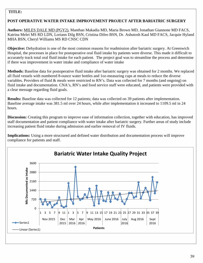
TITLE: POST OPERATIVE WATER INTAKE IMPROVEMENT PROJECT AFTER BARIATRIC SURGERY Authors: MILES DALE MD (PGY2), Manthan Makadia MD, Maria Brown MD, Jonathan Giannone MD FACS, Katrina Melei MS RD LDN, Loriann Dilg BSN, Cristina Difeo BSN, Dr. Ashutosh Kaul MD FACS, Jacquie Hyland MHA BSN, Cheryl Williams MS RD CNSC CDN Objective: Dehydration is one of the most common reasons for readmission after bariatric surgery. At Greenwich Hospital, the processes in place for postoperative oral fluid intake by patients were diverse. This made it difficult to accurately track total oral fluid intake for each patient. The project goal was to streamline the process and determine if there was improvement in water intake and compliance of water intake Methods: Baseline data for postoperative fluid intake after bariatric surgery was obtained for 2 months. We replaced all fluid vessels with numbered 8-ounce water bottles and 1oz-measuring cups at meals to reduce the diverse variables. Providers of fluid & meals were restricted to RN’s. Data was collected for 7 months (and ongoing) on fluid intake and documentation. CNA’s, RN’s and food service staff were educated, and patients were provided with a clear message regarding fluid goals. Results: Baseline data was collected for 12 patients; data was collected on 39 patients after implementation. Baseline average intake was 381.5 ml over 24 hours, while after implementation it increased to 1109.5 ml in 24 hours. Discussion: Creating this program to improve ease of information collection, together with education, has improved staff documentation and patient compliance with water intake after bariatric surgery. Further areas of study include increasing patient fluid intake during admission and earlier removal of IV fluids. Implications: Using a more structured and defined water distribution and documentation process will improve compliance for patients and staff.
0
720
1440
2160
2880
3600
1 3 5 7 9 11 1 3 5 7 9 11 13 15 17 19 21 23 25 27 29 31 33 35 37 39
Nov 2015 Dec2015
Mar2016
Apr2016
May 2016 June 2016 July2016
Aug 2016 Sept2016
Mls
Doc
umen
ted
as In
take
Patients
Bariatric Water Intake Quality Project
Series1
Linear (Series1)
39
TITLE: SPINAL EPIDURAL ABSCESS PATIENTS HAVE HIGHER MODIFIED FRAILTY INDICES THAN BACK PAIN PATIENTS UPON EMERGENCY ROOM PRESENTATION: A RETROSPECTIVE CASE-CONTROL STUDY Authors: JOSE DOMINGUEZ, MD (PGY2); Xintong Chen MD, PhD MPH; Donna Koo, BS; Bahwneet Chadha, BS; Joo Lee, BS; Akshitha Yarrabothula, BS; Naina Rao, BS; Anusha Adkoli, BS; Chad Cole, MD, MSc; Chirag Gandhi, MD; Fawz Al-Mufti, MD; Ivan Miller, MD FACEP; Christian Bowers, MD; Meic Schmidt, MD, MBa Background: Frailty is a clinical syndrome in older patients who have less physiologic reserve, resulting in multi-system impairments. Increasing frailty is frequently associated with worse outcomes in medical and surgical subspecialties. Spinal Epidural Abscess (SEA) patients have increased medical comorbidities and increased risk factors for infections when compared to control patients. Objective: To study the association of frailty as assessed by the modified frailty index (MFI) in SEA patients as compared to control patients. Design/Methods: We reviewed demographic and clinical data for 46 adult SEA patients and paired control patients (non-SEA but matched by age and sex) who presented to the ER with back pain from 2012-2017. We performed regression analysis to identify independent risk factors associated with SEA and to quantify their effects as percent changes in MFI. Results: Among the 46 SEA patients, the mean MFI was 1.89, significantly higher than mean MFI for the age paired control group (1.20; p = 0.023) and the hospital length of stay (LOS) in the SEA group was also significantly higher than the control patients (mean LOS: 22.89 vs 1.72; p = < 0.001). As expected, SEA patients had a much higher rate of previous invasive spinal procedures (surgery, steroid injections, etc.) compared to control patients (43.48% vs 23.91%; p < 0.001), as invasive spine procedures are known risk factors for SEA> The odds ratio for SEA is 1.08 per MFI increment. Multivariate regression modeling identified independent risk factors associated with increased MFI as age (+3.07 per year; p = 0.004) and SEA (+102.36%; p = 0.023). A factor negatively associated with increased MFI was IV drug use (-66.20%; p = 0.0028) Conclusions: Increasing age and SEA were independently associated with increased MFI and there is a significantly increased odds ratio for SEA with increased MFI. Thus, increased MFI may be a useful risk factor to look for with patients presenting with concern for SEA.
40

TITLE: POST-HOC ANALYSIS OF POOLED PATIENT-LEVEL DATA EVALUATING THE IMPACT OF SPECIFIC INTERVENTIONS WITHIN PRIMARY ANASTOMOSIS FOR PERFORATED DIVERTICULITIS WITH PERITONITIS ON OUTCOMES Authors: GACHABAYOV M (RESEARCH FELLOW), Bergamaschi R. Background: There is controversy regarding the impact of specific interventions within resection with primary anastomosis (PRA) on outcomes. Objective: The aim of this pooled analysis was to determine whether any specific interventions performed during resection with primary anastomosis in patients with perforated diverticulitis with peritonitis influenced the outcomes. Design/Methods: A post-hoc analysis of pooled data including 254 patients enrolled to the three randomized trials comparing PRA to nonrestorative resection (NRR) (NCT01239927; NCT01233713; NCT00692393) was carried out. The primary endpoint was postoperative complication rate. All included patients were adults older than 18 years with perforated diverticulitis with purulent or fecal peritonitis. Specific interventions performed within PRA included intraoperative on-table colonic lavage, anastomosing technique (hand-sewn or stapled), diverting ostomy, and type of ostomy (colostomy or ileostomy). Independent continuous variables were compared using Student’s t and Wilcoxon rank-sum tests, whereas Chi-squared and Fisher’s exact tests were utilized to compare categorical variables. Results: There were 116 PRA patients and 138 NRR patients. Postoperative outcomes did not differ, except for organ/space SSI rate which was significantly lower in PRA as compared to NRR (3.5% vs. 11.6%; p=0.017). Significant negative correlation was found between colonic lavage and postoperative complication rate (rs=-0.482; p=0.011). Positive correlation with postoperative complication rate was found in stapled anastomosis (rs=0.224; p=0.019) and creation of ostomy (rs=0.327; p<0.001). Type of ostomy showed no significant correlation with postoperative complication rates. Conclusions: While intraoperative colonic lavage was associated with decreased complication rates, stapling the primary anastomosis and adding an ostomy were correlated with increased complication rates.
41

TITLE: NATIONAL TRENDS IN PULMONARY EMBOLISM MANAGEMENT AND OUTCOMES: SHIFTING PARADIGMS Authors: ANSAB A. HAIDER, MD (PGY3), Joshua Goldberg, MD Background: Pulmonary embolism remains a major cause or morbidity and mortality. Data regarding the incidence of pulmonary embolism, its management and outcomes remains limited over the last decade. Objective: The aim of our study was to review the national rates of pulmonary embolism and its outcomes. Design/Methods: We performed a 12 year (2004-2015) analysis of the National Inpatient Sample database. Patients with a diagnosis of acute pulmonary embolism (PE) were identified. Major PE was defined as PE with associated mechanical ventilation, vasopressor need, or non-septic shock. Outcomes measures were surgical embolectomy, catheter directed thrombolysis (CDT), and overall mortality. Time trend analysis was performed using linear regression. Results: A total of 713,083 patients were diagnosed with acute PE during the study period of which 8.6% were major PE. Mean age was 62.9±17.4 years and 47.0% were male. The rate of acute PE increased from 5.3 per 1000 hospital admissions in 2004 to 9.7 per 1000 hospital admissions in 2015 (p<0.001). The rate of major PE increased from 7.9% to 9.7% (p<0.001). The rate of surgical embolectomy remained unchanged (0.2% in 2004 vs. 1.0% in 2015; p=0.47), and there was a 5-fold increase in the rate of CDT during the study period (0.2% in 2004 vs. 1.0% in 2015; p<0.001). There was a significant decrease in the overall mortality for PE (8.9% in 2004 vs. 6.4% in 2015; p<0.001) as well as surgical mortality associated with embolectomy (32.4% in 2004 vs. 14.0% in 2015; p<0.001).
Conclusions: The overall incidence of acute PE and major PE has been increasing during the last decade. The rate of surgical embolectomy has remained unchanged and there has been an increase in the use of CDT for PE. However, the mortality associated with acute PE as well that associated with surgical embolectomy has significantly improved during this time period.
8.9 8.1 7.8 7.7 7.5 7 6.6 6.6 6.4 6.6 6.6 6.4
5.4 6.5 6.5 6.9
7.8 8.2 8.5 9 9 9.2 9.5 9.7
32.4 31.3
22.6
30.6
23.4
16
23.3
17
22.3 19.2 19.5
14
0
5
10
15
20
25
30
35
-1
1
3
5
7
9
11
13
15
2004 2005 2006 2007 2008 2009 2010 2011 2012 2013 2014 2015
National Trends in PE
Overall mortality PE Surgical mortality
42
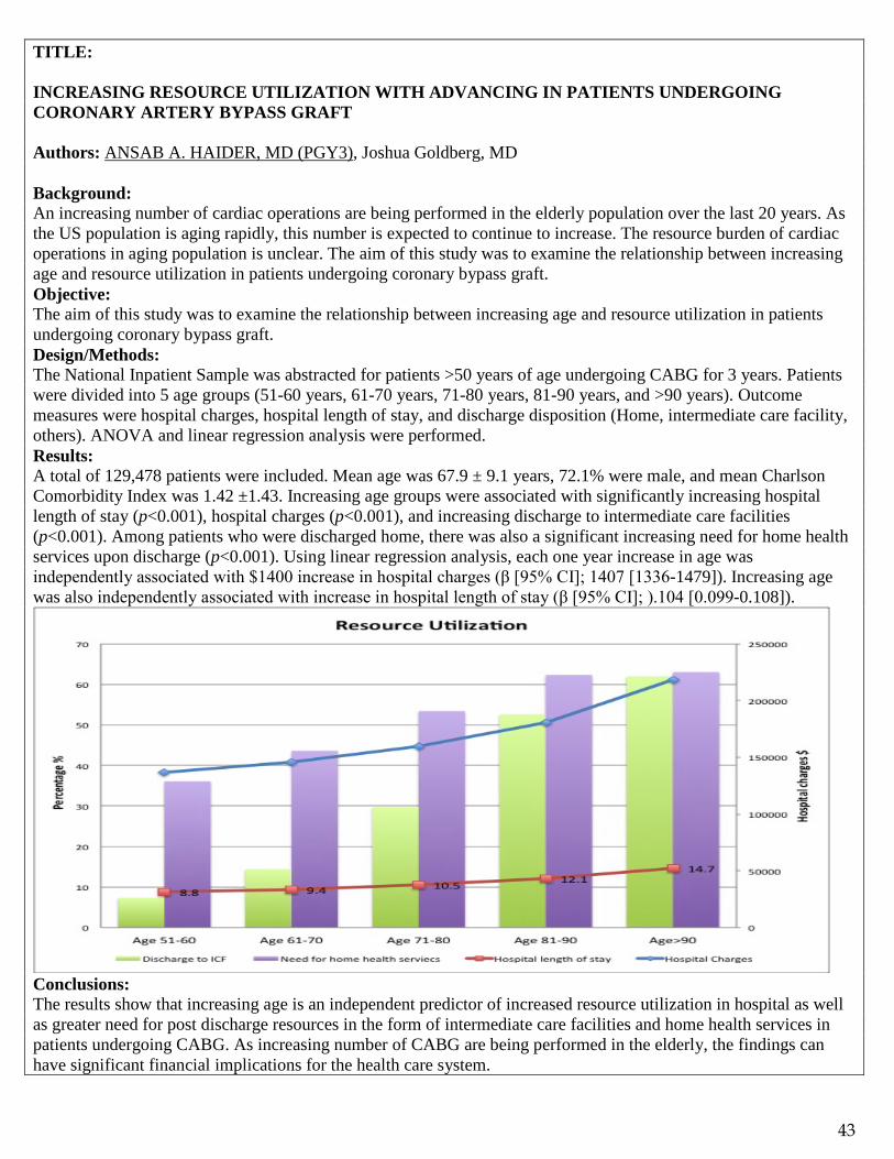
TITLE: INCREASING RESOURCE UTILIZATION WITH ADVANCING IN PATIENTS UNDERGOING CORONARY ARTERY BYPASS GRAFT Authors: ANSAB A. HAIDER, MD (PGY3), Joshua Goldberg, MD Background: An increasing number of cardiac operations are being performed in the elderly population over the last 20 years. As the US population is aging rapidly, this number is expected to continue to increase. The resource burden of cardiac operations in aging population is unclear. The aim of this study was to examine the relationship between increasing age and resource utilization in patients undergoing coronary bypass graft. Objective: The aim of this study was to examine the relationship between increasing age and resource utilization in patients undergoing coronary bypass graft. Design/Methods: The National Inpatient Sample was abstracted for patients >50 years of age undergoing CABG for 3 years. Patients were divided into 5 age groups (51-60 years, 61-70 years, 71-80 years, 81-90 years, and >90 years). Outcome measures were hospital charges, hospital length of stay, and discharge disposition (Home, intermediate care facility, others). ANOVA and linear regression analysis were performed. Results: A total of 129,478 patients were included. Mean age was 67.9 ± 9.1 years, 72.1% were male, and mean Charlson Comorbidity Index was 1.42 ±1.43. Increasing age groups were associated with significantly increasing hospital length of stay (p<0.001), hospital charges (p<0.001), and increasing discharge to intermediate care facilities (p<0.001). Among patients who were discharged home, there was also a significant increasing need for home health services upon discharge (p<0.001). Using linear regression analysis, each one year increase in age was independently associated with $1400 increase in hospital charges (β [95% CI]; 1407 [1336-1479]). Increasing age was also independently associated with increase in hospital length of stay (β [95% CI]; ).104 [0.099-0.108]).
Conclusions: The results show that increasing age is an independent predictor of increased resource utilization in hospital as well as greater need for post discharge resources in the form of intermediate care facilities and home health services in patients undergoing CABG. As increasing number of CABG are being performed in the elderly, the findings can have significant financial implications for the health care system.
43

TITLE: RELATIONSHIP BETWEEN SLEEP-DISORDERED BREATHING AND OUTCOMES AFTER TRAUMA: A NATIONWIDE ANALYSIS Authors: FAISAL JEHAN, MD (PGY1), Jorge Con, MD, Muhammad Khan, MD, Thomas Diflo, MD, Kartik Prabhakaran, MD, Anthony Policastro, MD, Patrice Anderson, MD, Herminio Diaz, MD, Gary Lombardo, MD, Rifat Latifi, MD Background: Sleep-disordered breathing (SDA) also known as obstructive sleep apnea is feared to be associated with respiratory complications especially in surgical patients. Trauma patients with SDA may have increased risk of these complications usually due to complex nature of injuries, increase use of opioids/ sedative medications and decreased consciousness levels. However, the association between SDA and outcomes in trauma patients has not been evaluated. Objective: The aim of our study was to evaluate the relationship between sleep-disordered breathing and outcomes after trauma. Design/Methods: We performed a 2-year (2011-2012) analysis of the Nationwide Inpatient Sample (NIS) and included all adult (>18 year) trauma patients. Patients were stratified into those with history of SDA and those without history of SDA. Primary outcomes were complications; respiratory and cardiac; the need for non-invasive ventilation and tracheostomy. Secondary outcomes were hospital length of stay, and mortality. Multivariate regression analysis was performed. Results: A total of 63,284 trauma patients were included in the study. Mean age was 43±17 years and 60% were males. 7.5%(4746) of patients had a SDB. Overall 16.7% patients developed a complication and overall mortality rate was 5.1%.The unadjusted rate of complications between SDA and non-SDA group was (26% vs. 16%, p=0.01) while the unadjusted mortality was (7.6% vs. 4.9%, p=0.02). After performing regression analysis and controlling for all the possible confounders, trauma patients with SDA had higher adjusted rates of developing any complication (OR: 1.5[1.2-2.5], p=0.03), cardiac complications (OR: 1.7[1.3-2.4], p=0.02), respiratory complication [OR: 3.1[2.1-3.9], p<0.01], the need for non-invasive ventilation (OR: 2.5[1.9-.3.2], p<0.01) and tracheostomy (OR: 1.8[1.3-.2.2], p=0.02). The adjusted hospital length of stay was higher (3 days vs. 2 days, p=0.02) in the SDA group compared to the non-SDA group. However, there was no difference in the adjusted mortality between the two groups. Conclusions: Trauma patients with sleep-disordered breathing are associated with higher risk of cardiac and respiratory complications, the need for non-invasive ventilation, and tracheostomy rates. Patients with SDA spend longer time in the hospital; however, there was no difference between the mortality compared to patients without SDA. These effects of SDA might be attributed to Use of screening criteria including the STOP BANG, will lead to early identification of these patients, and allocation of resources to prevent these complications.
44

TITLE: PRE-EXISTING MAJOR PSYCHIATRIC ILLNESS IN PATIENTS WITH TRAUMATIC BRAIN INJURY INCREASES THE RISK OF POST-TBI SEIZURES Authors: FAISAL JEHAN, MD (PGY1), Jorge Con, MD, Muhammad Khan, MD, Thomas Diflo, MD, Kartik Prabhakaran, MD, Anthony Policastro, MD, Patrice Anderson, MD, Herminio Diaz, MD, Gary Lombardo, MD, Rifat Latifi, MD Background: Traumatic brain injury (TBI) is leading cause of mortality and morbidity in trauma. One of the problems that can occur after a TBI is seizures. The risk of seizures in TBI patients with pre-existing major psychiatric illness has never been studied. Objective: The aim of our study was to evaluate the incidence of seizure and the overall outcomes in TBI patients with pre-existing major psychiatric illness. Design/Methods: We performed a 2-year analysis (2013-2014) of the American College of Surgeons, Trauma Quality Improvement Program (ACS-TQIP) database. We included all adult patients (age > 18) with traumatic brain injury. Patients were divided into two groups those with a pre-existing major psychiatric illness and those with no psychiatric illness. Our primary outcome measure was the incidence of post-traumatic seizures. Secondary outcome measures were ICU admission, hospital length of stay and mortality. We performed regression analysis to control for confounding variables. Results: A total of 1,87,472 patients with TBI were included in our analysis. Mean age was 46 + 13 years and 61% were male. The most common race was White. 8.6% (16034) had a pre-existing major psychiatric illness. Patients with pre-existing psychiatric illness had higher incidence of post traumatic seizures (6.1% vs. 1.1%, p<0.01) compared to patients with no major psychiatric illness. Similarly, the ICU admission rate (64% vs. 42%, p=0.02), the median [IQR] ICU length of stay (3[1-3] days vs. 1[1-2], p=0.04) and mortality (8.4% vs. 6.4%, p=0.01) was higher in patients with pre-existing psychiatric illness compared to those who did not have any major psychiatric illness. On regression analysis after controlling for age, gender, race, ISS, head-AIS, ED parameters (HR, SBP, GCS), the presence/type of surgical intervention, other body region injuries and comorbidities, TBI patients with pre-existing major psychiatric illness had higher rates of post traumatic seizures (OR: 6, CI[3-8], p<0.01), ICU admission (OR: 3, CI[1-6], p=0.02) and mortality (OR: 3, CI[2-5], p<0.01). On sub analysis of patient with mild TBI (GCS 13-15), patient with pre-existing major psychiatric illness had still 8 (OR: 8, CI[5-10], p<0. 01) times higher odds of developing a post-traumatic seizure compared to patients with no history of pre-existing psychiatric illness. Conclusions: Results of this nationwide study suggest that patients with pre-existing psychiatric illness have worse outcomes after TBI, including higher rates of post-traumatic seizures, ICU admission and mortality. The higher rates of seizure also persist in patient with mild TBI. Seizure prophylaxis may be warranted even in mild TBI patients who have a history of major-psychiatric illness.
45

TITLE: PREDICTORS OF ACUTE KIDNEY INJURY IN TRAUMA PATIENTS Authors: JEHAN F (PGY1), Mcintyre M, Azim A, Con J, Lombardo G, Latifi R, Prabhakaran K Background: Acute Kidney Injury (AKI) is a common cause of morbidity in trauma patients. The aim of our study was to identify factors that predict AKI in trauma patients. Objective: The aim of our study was to identify factors that predict AKI in trauma patients. Design/Methods: A 2-year (2013-14) analysis of all trauma patients in the Trauma Quality Improvement Program (TQIP) was performed. Patients who were dead on arrival were excluded. Primary outcome measure was predictors of AKI after trauma laparotomy. Secondary outcome measures were mortality. Multivariate-regression analysis was performed to control for demographics, admission vitals and injury parameters. Results: A total of 5,23,256 trauma patients were included in our analysis. Mean age was 41.6±18.3 years, 74.7% males, and 58% white. Overall prevalence of AKI in the analysis was 9.6% and mortality rate was 4.5%. On regression-analysis, independent predictors of AKI were female gender, age>60, African-American race, ISS>18, frailty, diabetes mellitus, and shock index>1. (Table 1) Demonstrates independent predictors of AKI in trauma patients. Patients with AKI had higher mortality-rate (10.2% vs 3.1%, p<0.001) as compared to the other group. On regression-analysis for mortality, AKI (OR:2.3, [1.6-2.7] was independently associated with in-hospital mortality. Conclusions: AKI is a common cause of morbidity in trauma patient and is associated with higher mortality rate. Appropriate intervention and resuscitation should be administered to prevent AKI. African American race, obesity and female gender are independent predictors and further studies are required to elucidate underlying mechanism. Interestingly enough age >60 and frailty were also associated with increased AKI.
Table 1. Regression Analysis of Acute Kidney Injury Covariates OR 95% CI P Diabetes Mellitus 2.4 1.7-3.3 <0.001 Shock Index >1 2.16 1.7-2.6 0.01 Female Gender 1.48 1.2-1.8 0.002 Age > 60 years 2.6 2.1-3.3 <0.001 Obesity 2.3 1.8-2.9 0.015 ISS > 18 2.5 1.2-4.5 0.02 Frailty 3.1 1.7-4.1 0.01 African-American 2.1 1.6-2.5 0.002
46
TITLE: MODIFIED FRAILTY INDEX AS PREDICTOR OF MORTALITY IN INTRACEREBRAL HEMORRHAGE Authors: MICHAEL KIM MD (PGY5), Ida Azizhkhanian MS, Abhiniti Mittal BS, Benjamin Epstein BS, Nam Lee BS, Justin Santarelli MD, Chirag Gandhi MD, Meic Schmidt MD MBA, Christian Bowers MD All authors are affiliated with New York Medical College Background: Intracerebral hemorrhages (ICH) carry the highest rates of morbidity and mortality of the stroke subtypes. Many current grading scales such as the ICH score only take into account factors such as size of hemorrhage, neurologic exam, and age. They do not consider the patient's pre-hemorrhage medical comorbidities. One model of frailty suggests that there is decreased reserve in multiple organ systems due to accumulating deficits. The modified frailty index (mFI) consists of 11 items and has been shown to correlate with increased morbidity and mortality in multiple surgical subspecialties. Objective: This study aims to analyze whether there is a similar correlation between mFI and ICH mortality rates. Design/Methods: We performed a retrospective chart review of all patients admitted with intracerebral hemorrhage between September 2015 and September 2017. The electronic medical records were analyzed to identify mFI components, in-hospital mortality rates, and other standard ICH variables (e.g. hematoma size, neurologic outcomes, etc.). Results: Our preliminary data on 137 of 282 overall patients shows that increasing frailty is associated with worse morbidity and mortality. 19.8% (27/137) had an mFI of 0, 23.4% (32/137) an mFI of 1, 20.4% (28/137) an mFI of 2, 13.1% (18/137) an MFI of 3, 15.3% (21/137) an mFI of 4, 7.3% (10/137) an mFI of 5, and 0.7% an mFI of 6 (1/137). mFI of 0 had a mortality rate of 29.6%, mFI 1 a rate of 25%, mFI 2 a rate of 25%, mFI 3 a rate of 61.1%, mFI 4 a rate of 21%, mFI 5 a rate of 40%, and mFI 6 a rate of 0%. When grouped into robust (mFI of 0), pre-frail (mFI 1-2), and frail (mFI ≥3), there was an association with increasing mortality rates. There was a mortality rate of 29.6% (8/27) in robust patients, 25% (15/60) in prefrail patients, and 44% in frail patients (22/50). There were 20 patients with an ICH score of 0 with a mortality rate of 0%, 35 patients with an ICH score of 1 with a mortality rate of 8.5%, 31 patients with an ICH score of 2 with a mortality rate of 16.1%, 24 patients with an ICH score of 3 with a mortality rate of 54.2%, 22 patients with an ICH score of 4 with a mortality rate of 86.4%, and 5 patients with an ICH score of 5 with a 100% mortality rate. Conclusions: There are numerous scoring systems that predict morbidity and mortality after spontaneous intracerebral hemorrhages. However, these scoring schemas are influenced principally by the hemorrhage size and the presenting neurologic exam. They do not factor in the patient’s frailty or underlying medical comorbidities. The mFI adds another element that is useful in counseling spontaneous ICH patients by predicting their mortality rates based on their medical history.
47
TITLE: PROPOFOL INFUSION SYNDROME: A RARE COMPLICATION OF A COMMON SEDATIVE Authors: JOSHUA KLEIN DO (PGY6), Ida Molavi MD, Patrice Anderson MD Introduction: Propofol is an intravenous sedative-hypnotic agent that has widespread use in operating rooms as an anesthetic induction agent, as well as in Intensive Care Units to provide sedation for critically ill patients. It is a short acting medication with both a rapid onset and rapid recovery. This makes it particularly beneficial in use with traumatic brain injured patients. Propofol Infusion Syndrome (PRIS) is a potentially lethal complication that can be seen with long-term Propofol infusions or at high dosages. This case-report looks at a poly-trauma patient, on a Propofol infusion, who developed signs of PRIS. We then discuss the characteristics and management of this rare syndrome. Case Report: 27-year-old intoxicated male presented to the trauma center following a motor vehicle crash. Upon arrival the patient had a GCS 9, however due to combativeness in the trauma bay he required intubation. Injuries included a parietal subdural hematoma, left rib fractures, and bilateral pulmonary contusions. He was subsequently placed on Propofol and Fentanyl infusions and transferred to the ICU where an intracranial pressure monitor was placed. On arrival to our facility the patient had an elevated alcohol level and his toxicology screen was positive for cocaine and cannabis. Most likely because of his utilization of illicit substances the patient had a very high tolerance to sedative medications. Over the course of the hospitalization, the patient had intermittent episodes of severe agitation and it was imperative to keep his intracranial pressures at a low level. The patient ultimately required high levels of Propofol and Dexmedetomidine. On hospital day 3 the patient’s urine was noted to be significantly darker. Laboratory testing revealed an elevating creatinine kinase (3406 U/L) and elevated triglycerides (740mg/dL). The patient additionally began having premature ventricular contractions noted on cardiac rhythm strips. The Propofol drip was immediately discontinued and the patient was transitioned to a midazolam infusion. The patient’s creatinine kinase and triglyceride levels decreased to normal levels with intravenous hydration, and his new onset cardiac arrhythmia resolved. Discussion: PRIS is a rare entity, with an incidence of less than 1% in patients receiving a Propofol infusion. Mediated on a cellular level by impairment of mitochondrial activity, the mortality rate of PRIS can be high varying widely from 18-53% in published reports and case series. While high dosages (>4mg/kg/hr) and prolonged infusion times (>48hrs) have previously been implicated as the chief risk factors for developing PRIS, there have been several reports of patients developing this syndrome at lower dosages and shorter infusion times. Common manifestations of PRIS include: metabolic acidosis, rhabdomyolysis, hepatomegaly, and cardiac arrhythmias. Patients can rapidly develop renal and myocardial failure ultimately leading to death. Laboratory monitoring can help indicate impending PRIS. For example, increasing triglyceride levels, myoglobinuria, and elevating creatinine kinase in the setting of no other muscle pathologies is the most frequent laboratory finding. Management involves immediate cessation of Propofol and supportive measures. Patients may require renal replacement therapy for metabolic acidosis and/or cardiovascular support. PRIS is a highly morbid complication of Propofol that can lead to death. Every practitioner utilizing this medication should be aware of this possible side effect and they must be cognizant of early signs.
48

TITLE: COMPLEX ABDOMINAL WALL RECONSTRUCTION FOR LOSS OF ABDOMINAL DOMAIN Authors: M KOBRITZ, BS (MS4); N Zhang, MD (PGY5); R Latifi, MD *Westchester Medical Center, New York Medical College, Valhalla, New York, USA (all authors) Background: Complex abdominal wall defects are defined as wounds that involve several tissues, develop after severe injuries and their surgical management, do not heal in a timely manner, or fail to heal completely. The etiologies of such wounds are diverse and include acute trauma, infectious processes, damage control surgery, recurrent incisional hernias, and poor wound healing in the setting of multiple comorbidities. Complex abdominal wall defects pose a difficult surgical problem, and the approaches to surgical treatment are variable depending on the patient, and the anatomy and etiology of the defect itself. In this case report we present a patient with a large ventral hernia with loss of abdominal domain as the result of multiple abdominal surgeries and infectious processes. Design/Methods: We present a case of abdominal wall reconstruction for a complex ventral hernia with complete loss of abdominal domain. A 30-year-old female with an extensive surgical history was referred by an outside institution to Westchester Medical Center for management of a symptomatic large ventral hernia with loss of abdominal domain. Five years prior to presentation she suffered from acute diverticulitis complicated by sigmoid perforation and abscess formation, for which she underwent exploratory laparotomy and Hartmann procedure with colostomy. Subsequently she required emergent laparotomy for strangulated parastomal hernia, at which point the colostomy was reversed. She later developed an incarcerated ventral hernia requiring emergent laparotomy, and the hernia was repaired with mesh. This repair was complicated by infection, so the mesh was removed, leaving her with a large ventral hernia with loss of abdominal domain. At the time of presentation, she complained of abdominal pain and constipation. Due to the extent of her hernia she had no Valsalva, and required application of manual pressure to her abdomen in order to defecate. Imaging revealed a large abdominal wall defect with herniation of >50% of the abdominal contents. Intraoperatively there was a huge ventral hernia of approximately 40 cm by 20 cm in size, no left abdominal wall, adhesions and prior mesh granulomas present. Intraoperative indocyanine green fluorescence angiography (ICG-FA) was used to guide panniculectomy and creation of skin flaps. Excess skin and subcutaneous tissue with poor perfusion was excised. Extensive adhesions were lysed. The abdominal wall was reconstructed using a 45 cm by 25 cm Strattice biologic mesh placed in an interposition fashion. The fascia was closed primarily from the xyphoid process to approximately the level of the umbilicus. Cultures of intra-operative specimens revealed evidence of MRSA infection and the patient was treated with antibiotics. Results: This patient recovered well prior to discharge and was discharged to an inpatient rehabilitation facility. Four weeks post-operatively, she presented to our hospital with right lower quadrant pain and overlying erythema. Imaging revealed two abdominal wall abscesses. The abscesses were drained and antibiotics were administered. Discussion: This case highlights the difficulty of managing complex abdominal wall defects and the individualized approach they require. The use of intraoperative ICG-FA in the setting of complex abdominal wall reconstruction has been shown to identify patients at risk for wound-related complications, however its use to guide panniculectomy has not yet been studied. The patient’s postoperative complication of abscess formation was likely a result of poor drain management. This operation significantly improved the patient’s symptoms, and overall quality of life. Conclusions: Many factors influence the surgical approach to complex abdominal wall reconstruction. Because of the diversity of etiology and symptoms, the surgeon must take into careful consideration the patient’s surgical history, presenting symptoms, comorbidities, and individual anatomy.
49

TITLE: PREDICTIVE FACTORS OF GOOD OUTCOMES IN CIRRHOTICS UNDERGOING SURGERY: AGGRESSIVE AND CAREFUL RESUSCITATION LEAD TO BETTER OUTCOMES Authors: DANNY LASCANO (PGY3), David Cohen MS3, Angelo Ciaraglia MS4, Zahava Farkas PGY 4, and Gregory Veillette MD Objective: To investigate what factors lead to good post-operative outcomes in cirrhotics undergoing elective and emergent surgery. We hypothesize, multidisciplinary optimization , short surgeries, and albumin drips on those with significant ascites post-operatively may optimize outcomes decreasing morbidity, death, and reoperation. Design/Methods: A single surgeon’s experience was queried for any cirrhotic patients from 2014-2018. Outcomes measured included pre-operative factors, intraoperative evaluation, and post-operative. Results: A total of 73 cases in 66 unique patients with cirrhosis were performed which included 7 cholecystectomies, 2 Whipples, 1 renal transplant , 9 hepatectomies, and the rest hernia repairs including inguinal and umbilical/ventral hernia repairs. There were 2 deaths (3.3%), 11 Clavien Dindo III or greater complications (13%). Pre-operative factors determined to decrease reoperation and improve mortality included albumin drips post-operatively instead of normal saline (p= 0.033, B = 0.071) while length of surgery did not impact outcomes (p= 0.989) when correcting for Child scores. Conclusions: Aggressive resuscitation with albumin in those patients putting out increased ascites may lead to better outcomes while shorter surgeries may not impact reoperation rates. More research into the intraoperative factors affecting outcomes in cirrhotics undergoing surgery is warranted but central tenets include multidisciplinary approach and optimization pre-operatively. Weaknesses of this study include single surgeon experience, repeat surgeries , and case mix of both elective and emergent procedures, the latter of which do not permit optimization of patients.
50

TITLE: MANAGEMENT OF ESOPHAGEAL PERFORATIONS AND LEAKS IN THE MODERN ERA: THE PERVASIVENESS OF NON-OPERATIVE MANAGEMENT Authors: DANNY LASCANO, MD (PGY3) and Tracey Weigel, MD Introduction/Background: Esophageal perforation is a morbid complication that results from either blunt injury, iatrogenic, or from particular disease processes such as traction diverticuli. We discuss the non-operative management of esophageal perforations and leaks. Case Series: Case 1/2: The first case patient presented with blunt trauma and delayed presentation over 24 hours with esophageal perforation. An intercostal flap was performed to repair the defect but patient had a leak . This was managed non-operatively with a negative vacuum suctioning and patient was discharged with no esophageal leaks. Another patient presented with blunt trauma and had an esophageal perforation, which was small with a small abscess. This was drained with a negative vacuum suctioning device and patient was kept NPO and on antibiotics. Patient eventually did well, had a gastrostomy tube placed and was advanced to clears with no leak or abscess. Case 3: A patient presented with iatrogenic injury to his esophagus from EGD and had a clip placed. Patient was admitted trialed on PO meals, had non-barium followed by barium esophagram confirming no leak and was discharged. Case 4: A patient presented with traction diverticuli with an esophageal perforation with mediastinal collection, was managed non-operatively with negative vacuum suctioning along the esophageal perforation, had a gastric tube placed and kept NPO and on antibiotics and although left against medical advice, presented with healing of his esophagus with no collection 1 month after discharge. Conclusion: Esophageal perforations and leaks in the modern era may be managed non-operatively with variable success. In our case series, we should good outcomes although success hinges on close monitoring for any clinical deterioration, antibiotics, cautions advancement of diet, repeat imaging to evaluate progress, and a multidisciplinary approach including working with gastroenterology for advanced endoscopic techniques.
51

TITLE: BREAST REDUCTION FOLLOWING HORMONAL THERAPY IN A TRANSGENDER FEMALE PATIENT Authors: ILANA G. MARGULIES MS (MS3), Hope Xu BA, Aki Kozato BS, Alexander Facque MD, and Peter J. Taub, MD, MS Background: Hormone therapy is widely used by male-to-female transgender individuals to promote feminizing characteristics including breast development. Surgical breast augmentation to supplement unsatisfying breast growth is a common subsequent course of management. In departure from convention, the authors present the first reported case of symptomatic macromastia requiring surgical breast reduction in a transgender woman following 24 years of hormone therapy and illicit silicone injections. Objective: The objective of this study is to present the first reported case of symptomatic macromastia requiring surgical breast reduction in a transgender woman following 24 years of hormone therapy and illicit silicone injections. Design/Methods: Chart review was conducted for a 39-year-old transgender female who presented with a chief complaint of back and neck pain secondary to macromastia. Nipple to IMF distance was 38cm on the right and 36cm on the left. Nipple to sternal notch was 16cm on the right and 16cm on the left. Breast width was 20cm. Preoperative mammogram showed extensive silicone granulomas, but otherwise benign findings. The patient underwent a Wise pattern skin resection with superior pedicle reduction mammoplasty bilaterally, with 1316 grams of tissue and silicone removed from the right breast and 916 grams from the left breast. Results: No complications were noted at one- and four-month postoperative follow-up, and her nipples remained viable with complete take of each graft. The patient was happy with the aesthetic result and reported complete resolution of her neck and back pain. Conclusions: This appears to be the first report describing breast reduction in the management of macromastia secondary to hormone therapy in the transgender patient. As hormone therapy is being increasingly used by transgender individuals, further investigation is needed to elucidate its varied effects on breast development.
52
TITLE: PROGNOSTIC UTILITY OF TRENDING TROPONIN LEVELS IN TRAUMA PATIENTS ADMITTED TO CRITICAL CARE UNIT. Authors: M FARHAN NADEEM (PGY5), Charles LaPunzina Background: Troponin T levels are used frequently to diagnose and manage cardiac ischemia and mild elevation (> 0.012 to 1.000) of screening troponin T levels is frequently seen in ICU patients. Objective: We sought to understand if there is any prognostic significance of this mild elevation in Troponin levels especially if it trends up on serial testing. Design/Methods: This was an observational case series with retrospective chart review of Trauma patients who were admitted to surgical ICU of a community level 2 trauma center. Two groups were created, one in which the Troponin levels remained < 1. The primary endpoint was mortality and secondary end points were LOS and LOSICU. LOS and LOS-ICU were compared using student t test. For comparison, Injury Severity Score was calculated, and compared for both groups. Results: One hundred and forty-six patients were included. Thirty patients had troponin levels that trended up to greater than 1 versus one hundred and sixteen patients in the group where troponin levels stayed < 1 on serial testing. Of the patients in which troponin levels trended up, 6 out of 30 experienced death vs 9 out of 116 in the comparison group. LOS in patients with higher troponins was 32.97 days (CI 1.702) vs 21.32 days (CI 4.18) [p = 0.038] in the other group. ICU-LOS was 13.9 days (CI 4.47) in the elevated troponin group vs 8.14 days (CI 1.62) [p = 0.021]. Of note, both groups had similar ISS score of 21.9 (CI 4.45) vs 16.06 (CI 1.86). Conclusions: Troponin level trending up to a level > 1 in trauma patients admitted in the SICU does hold a prognostic significance in determining both overall length of stay and ICU length of stay.
53

TITLE: PEDIATRIC FACIAL FRACTURES, CHARACTERISTICS AND PATTERNS IN THE UNITED STATES: A SURVEY OF RECENT TRAUMA QUALITY IMPROVEMENT PROJECT (TQIP) DATA Authors: KITAE E. PARK, BA1 (MS3), Ashraf S. Elzanie, MD2; Kaveh Alizadeh, MD2; Asad Azim, MD2; Rifat Latifi, MD2; Elizabeth Zellner, MD2 1 School of Medicine, New York Medical College 2 Department of General Surgery, New York Medical College and Westchester Medical Center Background: Trauma involving the facial bones has been shown to be associated with high severity in previous studies. Characteristics of facial fractures in adults have been well described in the adult population, less so in the pediatric literature. Our investigation further aims to define these epidemiological measures using the most recent data. Objective: To characterize epidemiological measures of pediatric facial fractures. Design/Methods: Characteristics of facial fractures among children and adolescents (ages 0-18 years old) has been previously examined using the National Trauma Data Bank between the years of 2001-2005. Our investigation uses 2016 data of the Trauma Quality Improvement project (TQIP) Databank to study facial fracture pattern, mechanism of injury, and demographic descriptive data to characterize pediatric trauma patients. Results: A total of 51,168 pediatric trauma patients were identified. Among these patients 2,878 (5.6%) presented with facial fractures. Among patients with facial fractures, fracture pattern differed by age. Mandibular fractures were more common in the 0-1 age category while nasal bone fractures were most common among 15-18 category. When stratified by mechanism, MVA was the most common among patients with facial fractures. The most common type of facial fracture identified was Maxillary/Malar fractures (35.1%) followed by Mandibular (34.4%) and Nasal bone (26.6%). Conclusions: Facial fracture patterns in pediatric patients differ by various age group and mechanism of injury. Contrary to previous studies showing central facial fractures are more common among younger groups, our investigation suggests this may not be the case.
54
TITLE: MOREL LAVALLEE LESIONS – NOT JUST AN ORTHOPEDIC INJURY Authors: SEUNGWHAN PEE MD (PGY6), Ida Molavi MD, Kartik Prabhkaran MD, FACS, Patrice Anderson MD Background: Morel Lavallee (ML) lesions are closed degloving injuries that occur in the deep subcutaneous tissue due to a shearing force and disruption of capillaries. Initially described by a French physician Dr. Maurice Morel Lavallee in 1853, these rare lesions are often the cause of significant morbidity. They can result in large cavities that do not heal secondary to an effusion containing hemolymph and necrotic fat. Often times, because the overlying skin is devascularized, the skin can become necrotic making tissue coverage difficult. In review of the literature, most publications are in the Orthopedic and Radiologic journals and they concentrate on ML lesions of the hips and the extremities. The Mellado-Bencardino classification is a radiologic tool used to evaluate the severity of these lesions and guide treatment. We have recently noted that ML lesions of the anterior abdominal wall, due to blunt abdominal trauma, are an increasing challenge faced by trauma surgeons. Extrapolating how the orthopedists treat ML lesions in the extremities may not be adequate. In this paper we discuss one of the ML cases we took care of in the abdominal wall caused by seatbelt shearing. Since we have had very good success with our current approach to treating these injuries we will share our treatment algorithm. Design/Methods: The patient was a 27 year female, who was involved in a motor vehicle collision. She presented to the trauma bay with multiple orthopedic injuries and blunt abdominal trauma. Radiologic imaging revealed hemoperitoneum and the patient was taken to the operating room for an exploratory laparotomy. The patient was found to have colon and small bowel injuries for which she underwent an extended right hemicolectomy and a proximal small bowel resection. When making the midline incision the patient was found to have separation of the superficial and deep abdominal wall structures, with a large hematoma. After the patient’s intra-abdominal injuries were addressed the abdominal wall was carefully evaluated. The skin over the degloved area was opened. All the nonviable tissue was debrided and washed out. In some areas the subcutaneous tissue was destroyed down to fascia. The wound was initially treated with wet to dry dressing changes and locally debrided at the bedside. Once viable granulation tissue was noted the patient had a negative pressure dressing placed for approximately one week. Ultimately, the wound was then closed in the operating room. Conclusions: Morel Lavallee lesions can be inconspicuous injuries that may lead to significant morbidity if not identified and aggressively treated. These wounds often require multiple wound explorations and prolonged wound care. There is a paucity of data regarding treatment for these lesions when they occur in the abdominal wall. As a division we have recently had 4 abdominal wall MLs admitted to our service. We currently recommend the following treatment algorithm: 1. Opening the area that is sheared to allow for early debridement of necrotic tissue and evacuation of any fluid
collection 2. Return to the operating room for re-debridement of nonviable tissue as needed 3. Temporarily utilizing wet to dry dressing until the wound is clean enough to apply a negative pressure
dressing 4. Closure of the skin after the underlying tissue is granulating well Since publications about ML lesions of the anterior abdominal wall are limited we plan to follow our patients and continue to improve upon our current protocol as we learn more.
55

TITLE: AN ENDOVASCULAR JOURNEY IN 3D Authors: CHRISTY STOLLER MD (PGY3), Sateesh Babu MD, Igor Laskowski MD Introduction: The endovascular aneurysm repair (EVAR) has replaced open surgical treatment of the abdominal aortic aneurysm in the United States. Recently, major technical advancements in intraoperative imaging have pushed the EVAR further. These developments have made image fusion techniques possible. It creates a 3D patient-specific vascular roadmap based on preoperative imaging which aligns with intraoperative fluoroscopy. In its current beginning stages of use, such technology has already proven effective and leads to decreased radiation dose, less iodinated contrast use, and shorter procedure times. We would like to present a case in which this image fusion technology provided yet another advantage to the surgeon. Presentation of Case: Our patient is a 79 year old lady with past medical history significant for hypertension, nephrolithiasis, uterine cancer, kidney disease, atherosclerosis with claudication, 5cm abdominal aortic aneurysm, bilateral renal artery stenosis and severe iliac disease who was previously admitted to the hospital for complete cardiologic work up due to the extent of her vascular disease. As part of pre-operative planning, the patient had undergone a CTA (thoracoabdominal with 1mm thickness of slices). The measurements of the arterial diameters and angulations were computed with the angulation of renal arteries measured as a caudal angle between the aortic centerline and the horizontal axis of the renal artery. This information was then processed using the software Vessel Navigator®, (Philips) making the marking of relevant side branches of the aorta possible (including the exact position, size, and best angles to demonstrate the ostia). After extensive work up the patient was taken to the OR and underwent placement of two stent grafts in each renal artery from the left axillary artery approach (snorkel technique), then placement of aortouniliac stent graft via left iliac approach, coil embolization of the diseased, nearly occlusive right common iliac artery, endarterectomy of the left external iliac and bilateral femoral arteries with pericardial patch repair of the left femoral artery and finally a cross over fem-fem graft from left to right. The procedure was performed under fluoroscopic guidance in our hybrid OR. After bony alignment, the vessels of interest (as selected from the CTA) were overlaid on top of the fluoroscopic image stream. In our patient, these were the renal arteries. By using the angulation of the vessels as illustrated by the Vessel Navigator software, we were able to place our C-arm in equal position and cannulate the bilateral stenotic renal arteries with ease whereas without the guidance the combination of left axillary artery approach and the tight angle of the left renal artery would have been difficult leading to extensive use of radiation for the patient. Conclusions: This new technological advancement of multimodality image fusion guidance using the 2D-3D registration method is sufficiently accurate to delineate the vessels of interest and does help the execution of the EVAR procedures as exampled here by our case of a very complicated case navigated successfully. We agree that the use of imaging fusion could allow for reduced contrast usage, a reduced radiation dose and a shorter procedural duration for the EVAR. References: Ahmad W, Obeidi Y, Majd P, et al., The 2D-3D Registration Method in Image Fusion Is Accurate and Helps to Reduce the Used Contrast Medium, Radiation, and Procedural Time in Standard EVAR Procedures. Ann Vasc Surg 2018; 51:177-186. Jones DW, Stangenberg L, Swerdlow NJ, et al., Image Fusion and 3-Dimensional Roadmapping in Endovascular Surgery. Ann Vasc Surg 2018; 52: 302-311.
56

TITLE: SOMETHING FISHY: INGESTED FISH BONES IN TWO PATIENTS LEADING TO MARKEDLY VARIED SURGICAL SEQUELAE Authors: MICHAEL WINTER BA (MS4), Dimitra Lotakis MD, Ana Machado Hopkins MD, Joshua D. Stephens MD, Robert Prichep MD, Michael Grossman MD, Cristian D. Valenzuela MD. Background: Ingested fish bone is likely a common benign occurrence in the general population. However it may cause serious surgical sequelae by eroding through a hollow viscus and potentially causing damage to the surrounding structures. This entity is poorly described with only a handful of published reports describing this phenomenon in the surgical and radiological literature. The morbidity associated with it is variable. Objective: Here, we report two patients (Patient A) and (Patient B) who presented with sequelae from this ultra-rare entity: both patients accidentally ingested a fish bone, which completely exited through the upper gastrointestinal tract. Design/Methods: This was a retrospective medical record review of two patients from two affiliated hospitals. Both patients presented within a few months of each other in 2018, and had no relationship to each other. Patient A is a 55-year old African male, and Patient B is a 64-year old Hispanic male. Written consent for publication was obtained from both patients. Results: During initial work up of Patient A for possible acute cholecystitis, a foreign body was seen on imaging traversing the gastric/duodenal wall with intra-abdominal involvement. Retrieval of the foreign body was attempted unsuccessfully via endoscopy. The patient was taken for a diagnostic laparoscopy where the bone was seen exiting into the peritoneal cavity adjacent to the liver, resulting in a localized perihepatic intraperitoneal abscess adjacent to the gallbladder (Figure 1). Laparoscopic abdominal washout and drainage of the abscess were performed, with foreign body removal (Figure 2). The patient was discharged home on hospital day 8 after tolerating diet and return of normal bowel function. In Patient B, initial imaging showed a left hepatic abscess and contained gastric perforation (Figure 3) secondary to a foreign body. The patient was given antibiotics and the hepatic abscess was drained percutaneously under CT-guidance. The hospital course was complicated by left hepatic vein thrombosis requiring anticoagulation, and evolution of the hepatic abscess requiring additional percutaneous drain placement. The patient was discharged home on hospital day 16 on oral antibiotics. Approximately 4 weeks after discharge, the patient was readmitted to the hospital due to worsening abdominal pain. The patient underwent a diagnostic laparoscopy with foreign body removal and unroofing of the hepatic abscess. An additional hepatic collection developed postoperatively requiring an additional percutaneous drain. The patient was discharged on hospital day 14, and is doing well at 1 and 3 months follow-up after discharge. Ultimately, both patients were treated successfully with minimally-invasive modalities. Both patients survived and are in good health today. Conclusions: (1) The difference in morbidity between these two patients is remarkable in this ultra-rare event, due to the difference of only a few centimeters of distance between where the fish bones lodged. (2) Endoscopy may potentially play a role in similar cases: the physician may attempt to extract the bone intraluminally if it is found early enough to be partially-eroded through the stomach or duodenum. (3) We conclude that for this ultra-rare entity, in the entirety of its spectrum of presentation, sequelae can be managed in a minimally-invasive fashion, with laparoscopy and percutaneous drainage.
57
TITLE: INTRACORPOREAL VERSUS EXTRACORPOREAL ILEOCOLIC ANASTOMOSIS IN RIGHT COLON RESECTION: A SYSTEMATIC REVIEW AND META-ANALYSIS Authors: NIU ZHANG (PGY5), Mahir Gachabayov, Roberto Bergamaschi. Background: There is no level 1a evidence regarding the best technique for ileocolic anastomosis following laparoscopic right colon resection. Objective: The aim of this meta-analysis was to evaluate the impact of intracorporeal (IA) and extracorporeal (EA) ileocolic anastomoses on the clinical outcomes of patients undergoing laparoscopic right colon resection. Design/Methods: The Pubmed, EMBASE, Cochrane Library, MEDLINE via Ovid, CINAHL, and Web of Science databases were systematically searched. Laparoscopic right colon resection was defined as either an ileocolic resection anastomosing proximal ileum to the ascending colon sparing ileocolic vessels or right colectomy transecting ileocolic vessels and the right branch of mid-colic vessels anastomosing the ileum to the transverse colon. Intracorporeal ileocolic anastomosis was defined as anastomosis of the ileum to the colon performed hand-sewn, stapled, or both. Extracorporeal anastomosis entailed externalizing the intestine through a small laparotomy, resecting the intestine, and anastomosing the ileum to the colon hand-sewn, stapled, or both. The primary endpoint was anastomotic leak rate. Secondary endpoints included intraoperative, short- and long-term postoperative, and pathologic outcomes. Results: Twenty-one studies (2 experimental and 19 observational) totaling 2,558 patients (1,271 IA and 1,287 EA) were included. Anastomotic leak rates did not differ [OR (95%CI) = 0.69 (0.40, 1.20); p =0.19] with low heterogeneity (I2=0%) (Figure 1). Incisional hernia rates were significantly decreased in IA [OR (95%CI) = 0.32 (0.13, 0.76); p =0.01] with low heterogeneity (I2=7%) (Figure 2). No publication bias was found. Conclusions: This meta-analysis found that although IA was associated with similar anastomotic leak rates, it decreased incisional hernia rates at long-term.
58
Poster Exhibits
(In alphabetical order)
59

TITLE: IMPACT OF ANTIBIOTIC USE ON TYPE 2 DIABETES OUTCOMES 1 YEAR AFTER SLEEVE GASTRECTOMY Authors: YEDIDYA BEN-AVIE (MS2)1; Keyvan Heshmati, MD2; Eric Sheu, MD, PhD2; Ali Tavakkoli, MD2 1: New York Medical College, School of Medicine, Valhalla, NY 2: Brigham and Women’s Hospital, Harvard Medical School, Boston, MA Background: Although sleeve gastrectomy is one of the most common bariatric procedures for the treatment of obesity and associated comorbidities including type 2 diabetes (T2D), there is variation in outcomes with some patients not achieving durable weight loss or diabetes remission. Recent research has pointed to the negative effects of antibiotic use on weight loss and blood sugar control. Objective: The aim of the study was to evaluate whether there was an association between antibiotic use before and after sleeve gastrectomy surgery and Type 2 Diabetes outcomes one year after surgery. Design/Methods: We analyzed 187 patients (61 male, mean age 50.1) with T2D who underwent sleeve gastrectomy between 2010-2015. All patients received one dose of IV prophylactic antibiotics before surgery. Patients were grouped by whether they used antibiotics in the three months before surgery, the 12 months after, both before and after, or neither before nor after. The groups’ one year T2D outcomes were then compared. Results: For pre-op use (n = 11), post-op use (n = 51), both (n = 15), and neither (n = 110) the results were as follows: BMI change, -7.64 + 4.94 kg/m2, -8.92 + 5.20 kg/m2, -11.18 + 4.10 kg/m2, -9.51 + 4.98 kg/m2; for HbA1c change, -1.08 + 0.70%, -0.46 + 1.50%, -0.01 + 1.12%, -0.89 + 1.60%; and for the percent of each group able to discontinue T2D medications, 77.8%, 51.0%, 57.1%, and 55.7%. Conclusions: There were no significant differences in terms of outcomes between groups. Overall, this shows that antibiotic use did not influence the one-year outcomes of sleeve gastrectomy. Although not statistically significant, antibiotic use after surgery did suggest a worsening in T2D outcomes (less reduction in HbA1c), and an interesting observation that fewer patients required T2D medications in the group that received antibiotics pre-operatively. The study is likely underpowered to detect such differences but warrants further studies using larger cohorts. Impact of antibiotic use on sleeve gastrectomy is complex and appears to depend on timing of use in relation to surgery.
60
TITLE: FALSE PET POSITIVE AXILLARY LYMPH NODES IN PATIENTS WITH SUSPECTED BRONCHOGENIC CARCINOMA Authors: MICHELLE BRAVO MD (PGY4), Jermyn Addy BS, Francis X. Carroll MD FACS, Kassem Harris MD FCCP Background: PET is a valuable tool in the evaluation of patients with suspected lung cancer for both diagnostic and staging purposes. Metastasis to axillary lymph nodes in this disease is unusual, and in the absence of mediastinal or distant metastatic disease, is rare. We present two cases of suspected lung cancer with PET positive axillary adenopathy; proven histologically negative by ultrasound guided core biopsy. Objective: To clarify the role of histologic confirmation of PET positive axillary lymph nodes in cases of suspected lung cancer. Design/Methods: In both cases, there was PET positivity in both the suspicious lung lesion and in axillary lymph nodes. In both cases, the nodes were proven to be negative for metastatic disease by tissue confirmation. Results: One patient had confirmation of a diagnosis of adenocarcinoma of the right upper lobe, had negative staging of the mediastinum by EBUS, and then resection. In that case the PET finding was later attributed to recent flu shot in the ipsilateral arm. The other patient had intraoperative wedge biopsy of the PET positive left upper lobe lesion which proved to be aspergillus. Interestingly, he had undergone VAT right middle lobectomy for early stage NSCLS 5 years earlier. In that case PET positivity was later ascribed to extravasation of the radioactive isoptope at the injection site with uptake in the ipsilateral axillary nodal basin. Conclusions: Armed with the knowledge that axillary lymph node metastasis is uncommon in otherwise resectable bronchogenic carcinoma, histologic confirmation of suspected falsely PET positive axillary nodes maybe unnecessary, particularly in the absence of mediastinal or distant metastasis. In both cases a consensus opinion at tumor board conference favored biopsy, implying a general uneasiness with clinical staging alone. We must reassure patients, families and colleagues in these cases, such that an added procedure, ultrasound guided nodal biopsy, with its attendant added time, cost and risk, (including risk of a non-diagnostic biopsy) can safely be avoided.
61
TITLE: AN INTERESTING CASE OF A LARGE RENAL MASS CAUSING DIET INTOLERANCE AND RESECTION RESULTING IN HYPONATREMIA AND LIKELY SALT WASTING SYNDROME Authors: J BRUCKNER (PGY1), N Zhang, G Veillette. Westchester Medical Center, Valhalla, NY. Background: Malignant renal masses represent over 2% of all cancer incidence and mortality in the United States, and sarcomas in that area represent a small portion of these cancers. Importantly, it is very difficult to determine the origin of these masses, whether they arise primarily from the kidney or from the retroperitoneum. These large abdominal masses, when symptomatic, commonly present with back pain, bowel obstruction, anemia, and palpable abdominal masses. Objective: To describe our technique of en bloc resection including radical nephrectomy, adrenalectomy, distal pancreatectomy, and splenectomy, of this large renal mass and workup and treatment of the resulting hyponatremia in the setting of a non-descript pathological setting. Design/Methods: A 75 year old female with medical history significant for hypertension, asthma, gastroesophageal reflux disease, celiac disease, depression, anxiety, and known renal mass. In addition, she had a surgical history of cholecystectomy, hysterectomy, and appendectomy. The patient presented after a fall from standing that resulted in no significant injuries, however the patient was noted to have a history of nausea and vomiting. CT imaging from an outpatient facility had documented the renal mass, noting that it was abutting the abdominal aorta, superior mesenteric artery, and celiac trunk, as well as being closely adjacent to the pancreas and large bowel. Pheochromocytoma workup had been done in the patient and returned negative, as plasma metanephrines were in the normal range, and workup also revealed that it was unlikely for the tumor to be cortisol secreting. After admission, the patient was booked for surgical resection of the mass and a left radical nephrectomy, left adrenalectomy, distal pancreatectomy, and splenectomy was performed. The patient was admitted to the ICU post-operatively, where she did well and was transferred to the floor. After the surgery, on post-operative day 8, the patient was noted to have hyponatremia with a sodium level of 124. Despite treatment with salt tabs, steroids, and management of medications to restrict salt loss, the patient’s hyponatremia persisted, with levels ranging from 120-127. After approximately 10 days, the patients sodium level returned to normal and the patient was discharged. On follow-up, the patient’s sodium level was at her baseline. Results: Pathology of the resected specimen revealed a high grade pleomorphic sarcoma, unclassifiable, possibly arising from the retroperitoneum. Samples were sent to Brigham Women’s Hospital, where it was also stated to be a high grade unclassified pleomorphic sarcoma. Immunostains of the specimen revealed no specific line of differentiation. Workup for resulting hyponatremia revealed unremarkable ACTH stimulation test, aldosterone and renin activity. High levels of sodium were found in urine revealing likely salt wasting syndrome as cause of hyponatremia. Conclusions: Large renal masses of unknown origin present a difficult surgical problem, as they are often asymptomatic in the early stages of their growth and can be difficult to pinpoint the origin, such as in this case of a likely retroperitoneal mass. In addition, in such a setting where pathology is difficult and the patient’s post-operative course is complicated by electrolyte imbalances, it is critical to evaluate all possible mechanisms of these imbalances to rule out an underlying endocrine pathology of the renal mass.
62
TITLE: HETEROTOPIC PANCREAS IN THE CYSTIC DUCT: A CASE REPORT Authors: ERIN CARAHER, (MS3)1, Cynthia Sulzbach, MD, PGY52, Venkat R. Modukuru, MD 2 1New York Medical College, School of Medicine 2Metropolitan Hospital Center, Department of Surgery Background: Heterotopic pancreas is defined as pancreatic tissue lacking anatomic communication with the normal body of the pancreas. It is believed to be a congenital abnormality, and is most commonly found in the stomach, duodenum, proximal jejunum, Meckel diverticulum or ileum. Rarely, heterotopic pancreas may also be found in the esophagus or biliary tree. The majority of cases of heterotopic pancreas are asymptomatic and found either incidentally or on autopsy, making the actual incidence of heterotopic pancreas difficult to estimate. Preoperative diagnosis is difficult, and pathology is necessary to definitively distinguish the heterotopic pancreatic tissue from malignancy. Case Details: Patient is a 40 year old male who presented to the Emergency Department with a three day history of right upper quadrant pain. The pain was sharp, non-radiating, intermittent, 10/10 at its onset and 5/10 at time of presentation. Pain was associated with nausea but no vomiting, with no change to the patient’s bowel habits. The patient noted the pain worsened with eating and he was therefore afraid to eat for three days prior to admission. On physical exam the patient was afebrile and hemodynamically stable. No scleral icterus was noted. The abdomen was minimally tender in the right upper quadrant with no guarding, rigidity, rebound tenderness or Murphy’s sign. Labs were significant for leukocytosis with left shift (12.32; 71.7%). Basic metabolic panel, hepatic function tests and lipase were within normal limits. Right upper quadrant ultrasound showed multiple stones in the gallbladder with trace pericholecystic fluid and a positive sonographic Murphy’s sign. Abdominal ultrasound additionally showed gallbladder wall thickening (measured up to 6mm) and a normal common bile duct (4mm). The patient was started on IV antibiotics and scheduled for laparoscopic cholecystectomy the next day. During the laparoscopic cholecystectomy there was noted to be significant pericholecystic fluid and inflammation as well as adhesions to the duodenum. Pathological exam showed a gallbladder measuring 10.5cm in length and 4.2cm in width with a tan-pink serosa congested with diffuse hemorrhagic areas. The cystic duct measured 0.3cm x0.2cm and showed no grossly visible lesions. Tissue sectioning showed acute on chronic cholecystitis with cholesterolosis and heterotopic pancreatic tissue in the wall of the cystic duct. Patient tolerated the procedure well and was discharged to home one day after surgery. Patient followed up in surgery clinic with no post-op complications. Conclusion: In summary, this is a case of a 40 year old man presenting with a three day history of right upper quadrant pain and sonographic evidence of acute cholecystitis. On pathology he was found to have heterotopic pancreatic tissue in the cystic duct. Heterotopic pancreas is rare, and hepatobiliary heterotopic pancreas accounts for 1% of all documented cases. Twenty-nine cases of heterotopic pancreas in the gallbladder have been reported since the first documented case in 1916. While the majority of cases are asymptomatic, the ectopic tissue is susceptible to the same pathology as typical pancreatic tissue including local pancreatitis, pseudocyst formation and benign and malignant neoplasms. Additionally there are complications specific to heterotopic pancreas include gastrointestinal bleeding, bowel obstruction and intussusception. Knowledge of the typical anatomic locations and unique complications of heterotopic pancreas is important as it presents similarly to other more common pathologies and is difficult to distinguish on current imaging studies.
63

TITLE: DIAPHRAGMATIC METASTASIS FROM PRIMARY THYMOMA: A CASE REPORT Authors: CHOI J (PGY2), Haider A, Chudner A, Carroll F, Weigel T Background: Thymomas are the most common neoplasm arising from the thymus with a current incidence rate of 0.13 cases per 100,000 persons per year in the U.S. It accounts for less than 1% of all adult malignancies. Rarely seen in the pediatric and young adult population, the incidence generally peaks between 40 to 60 years of age. Most metastasis from thymoma occur within the pleura and pericardium. Metastasis to distal structures are incredibly rare, but have been reported in the liver, spine, kidney, and retroperitoneum. The following is a case report of a Stage IV thymoma that has spread to the diaphragm Case Report: The patient is a 60 year old male diagnosed in April 2018 with a type B2 thymoma (Stage IV thymoma) with known left lung parenchymal metastasis, status post chemotherapy. Subsequent PET scan showed marked interval response the chemotherapy and presents to WMC for definitive resection. At the time of his diagnosis, he had a left VATS and wedge resection of couple of pulmonary metastasis, but no attempt at complete resection. The patient had no history of thymectomy. On August 15, 2018, the patient underwent robotic-assisted left video-assisted thoracoscopic resection of anterior mediastinal mass, en block wedge resection of left lower lobe, en bloc wedge resection left upper lobe, diaphragmatic resection of mass with primary closure. Pathology of diaphragmatic mass showed 2.8 x 2.0 thymoma, non-encapsulated involving the visceral pleural and pushing border compressing the adjoining lung parenchyma. The patient was subsequently discharged on post-operative day two. Discussion: Although thymic cancers are incredibly rare, rarer still is metastasis to distal structures such as the diaphragm. If possible, complete resection (R0) is favorable. In this case, the diaphragmatic implants were removed with histologically negative margins and the patient tolerated primary repair well. There were no respiratory sequelae from this procedure. However, it is still important to note that the overall 10 year survival rate in Stage IV disease is 47%. In contrast, the 10 year survival rate of stage III disease is 83%. Conclusion: Despite the overall mortality of stage IV thymoma, this case report shows that it is feasible to remove metastatic implants to the diaphragm with few complications and a length of stay of only two hospital days. Even though this was accomplished with the aid of adjuvant chemotherapy, the importance of resectability can not be understated. Although a rare occurrence, thymic metastasis to the diaphragm should be resected if feasible. References: 1. Engels, EA. Epidemiology of thymoma and associated malignancies. J Thorac Oncol. 2010 Oct; 5(10 Suppl 4): S260–S265. 2. Gajendra, S, Sahoo, MK. 18F FDG-PET/CT of malignant thymoma with pleural and diaphragmatic metastases. Lung India. 2016 Jan-Feb; 33(1): 116–117. 3. Song Z, Xiangyu J, Zhang Y. Treatment and Prognosis of type B2 thymoma. World Journal of Surgical Oncology201412:291
64

TITLE: SUBTOTAL COLECTOMY FOR COLONIC TUBERCULOSIS Authors: ALEXANDRA CHUDNER, MD, (PGY4), Alexandra Nee MS3, Jorge Con, MD, FACS, Kartik Prabakharan, MD, FACS, Gary Lombardo, MD, FACS Background: According to WHO, there were 10 million new cases of tuberculosis (TB) in 2017.1 In the United States in 2017, there were 9105 new cases of TB , 70% of which occurred among foreign born people.2 Cases of extrapulmonary tuberculosis (EPTB) are often difficult to diagnose and manage because of the nonspecific presentation3. TB of the gastrointestinal tract is the sixth most frequent form of EPTB after lymphatic, genitourinary, bone, miliary, and CNS tuberculosis.4 There are several routes of acquisition of this type of TB: swallowing of respiratory tract secretions, hematogenous spread, invasion from adjacent infected tissues and ingestion of contaminated unpasteurized dairy products.4
Case Description: 36F from Ecuador presented with weakness and inability to walk in the setting of 70lbs weight loss, nausea, and diarrhea over the past 2 years. She was cachectic with lower extremity edema. Imaging showed bilateral pulmonary nodules (<8mm), left pleural effusion, and wall thickening of the stomach, small and large bowel. A colonoscopy showed atrophic mucosa and a segment of circumferential friable ulcerated mucosa. A week later, the patient developed fevers, lactic acidosis and worsening abdominal pain prompting a diagnostic laparoscopy, which only showed ascites. Results of her sputum and stool AFB (acid fast bacilli) returned positive shortly thereafter and she was started on rifampin, isoniazid, pyrazinamide, ethambutol (RIPE) therapy. She then required heparin drip for a catheter-associated deep venous thrombosis. A week later, she developed hypotension, tachycardia, and GI bleeding. CT showed large bowel obstruction. Nasogastric tube was placed for decompression, but she began to bleed again and was taken for subtotal colectomy, end-ileostomy and skin closure. Pathology revealed necrotizing granulomatous inflammation with transmural colonic wall thickening, luminal stricture and lymph node involvement. The specimen was negative for acid fast bacilli. Her hospital course was further complicated by pulmonary embolism, requiring re-initiating of anticoagulation and midline abdominal wound infection. After stabilization and 3 negative sputum AFB tests, patient was discharged to rehab. Discussion: Tuberculous enteritis occurs in about 2% of patients with pulmonary tuberculosis.3 Of the abdominal manifestations, only 2-3 %o f patients have isolated colonic involvement and peritoneal tuberculosis and tuberculous enteritis are the most common presentation in patients under 45 years old.4 Ileocecal involvement is prevalent due to the high concentrations of lymphoid tissue or Peyer’s patches, which are susceptible to invasion. A mass in the right lower quadrant is palpable in 25 -50% of patients.3 The most common signs and symptoms include ascites (73%), abdominal pain (65%), weight loss (61%), fever (59%), diarrhea (21.4%), and constipation (11%).3 Standard therapy for TB consists of two months of RIPE therapy followed by four months of two drug therapy (rifampin, isoniazid).4 Majority of the ulcers (87.2%), nodules (84.6%), polypoid lesions (85.7%), luminal narrowing (76.2%), and ileo-cecal valve deformities (76.5%) resolved with anti-TB therapy.5 Strictureplasty or colonic balloon dilation can be used for short segment obstruction. 4 However, for late onset obstruction and/or perforation surgical intervention is required. Ascites may occur, but rarely test positive for AFP (2%). Resected specimens often show caseating granulomas (93%), but only 58% had AFB positivity.6 Conclusion: A high index of suspicion is essential for early diagnosis of gastrointestinal TB. Sole reliance on culture of ascitic fluid may delay the diagnosis, whereas biopsy or resection results in appropriate treatment. In the US, while TB is often addressed in its early stages, the rise of multi-drug resistance strains results in more extra-pulmonary infection and the complications of disseminated disease.7
References: 1. Global tuberculosis report. 2018. Geneva: World Health Organization. 2. Tuberculosis. Centers for Disease Control and Prevention. 3. Golden MP, Vikram HR “Extrapulmonary Tuberculosis: An Overview” American Family Physician. 2005 Nov 1; 72(9):1761-1768 4. Malikowski T, Mahmood M, Smyrk T, Raffals L, Nehra V “ Tuberculosis of the gastrointestinal tract and associated viscera” Journal of Clinical Tuberculosis and Other
Mycobacterial Diseases. 2018 April 9; 12: 1-8 5. Mukewar Sa, Mukewar Sh, Ravi R, Prasad A, Dua K “Colon Tuberculosis: Endoscopic Features and Prospective Endoscopic Follow-up After Anti-Tuberculosis
Treatment” Clinical Translational gastroenterology 2021 Oct; 3 (10): e24; 1-9 6. Khan FY, AL-Muzrakchi AM, Elbedawi MM, AL-Muzrakchi AA, Al Tabeb A “Peritoneal tuberculosis in Qatar: A five-year hospital-based study from 2005 to 2009”
Travel Medicine and Infectious Disease. 2012 Dec 29;10(1): 25-31 7. Fry DE “Extra-Pulmonary Tuberculosis and Its Surgical Treatment” Surg Infect (Larchmt). 2016 Aug;17(4):394-401
65

Figure 1: Figure A shows the abrupt cutoff and distal re-constitution of the arteries below the popliteal artery at the knee. Figure B. shows renal infarction of the left lower pole with acute demarcation. Figure C shows left lateral rectus enlargement with hypodensity changes concerning for inflammatory versus necrotic tissue initially and confirmed on pathology to be myositis.
TITLE: THE CRITICAL CARE MANAGEMENT OF A PATIENT WITH SERONEGATIVE CATASTROPHIC ANTIPHOSPHOLIPID SYNDROME IN MULTISYSTEM ORGAN FAILURE Authors: ANGELO CIARAGLIA (MS4), Danny Lascano, Asad Azim, Pee Seungwhan, and Gary Lombardo Introduction/Background: Seronegative Catastrophic Antiphospholipid Syndrome (CAPS) is a rare, life-threatening presentation of antiphospholipid syndrome characterized by widespread intravascular thrombosis resulting in multiorgan system failure and very low titer of antibodies classically associated with anti-phospholipid syndrome Case Description: We present a case report of a 44-year-old male who presented with multiorgan system failure after having initial acute limb ischemia who subsequently developed seronegative CAPS with resulting acute kidney injury with evidence of lower pole kidney ischemia, lower extremity myositis with rhabdomyolysis, and non-ST wave elevated myocardial infarction. His course was complicated by a duodenal ulcer bleed and left sided pleural effusion. We will discuss the coordination of care, different specialists involved, the surgical management for the complications, and the unique situations critical to this patient’s survival. Conclusions: Optimization of anticoagulation and long-term management following any surgical intervention is necessary to limit associated complications. A. B.
C.
66
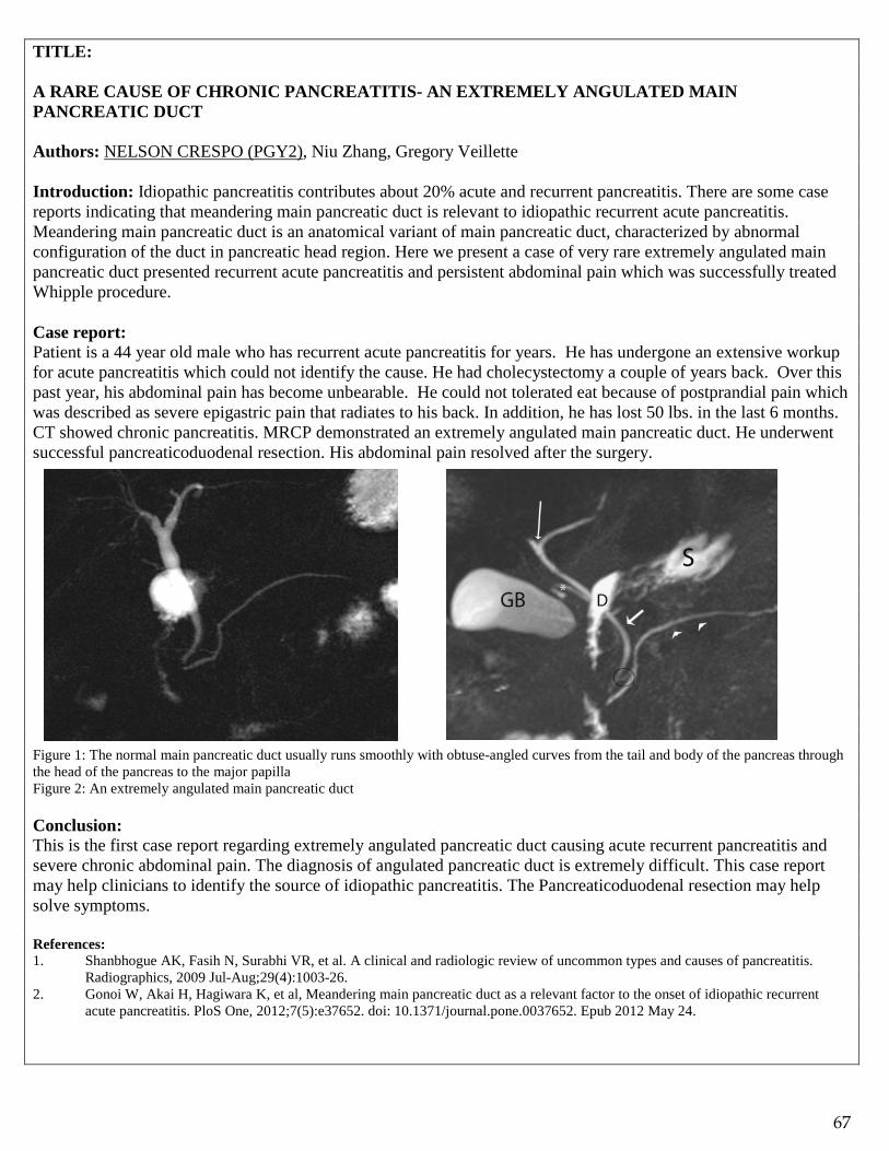
TITLE: A RARE CAUSE OF CHRONIC PANCREATITIS- AN EXTREMELY ANGULATED MAIN PANCREATIC DUCT Authors: NELSON CRESPO (PGY2), Niu Zhang, Gregory Veillette Introduction: Idiopathic pancreatitis contributes about 20% acute and recurrent pancreatitis. There are some case reports indicating that meandering main pancreatic duct is relevant to idiopathic recurrent acute pancreatitis. Meandering main pancreatic duct is an anatomical variant of main pancreatic duct, characterized by abnormal configuration of the duct in pancreatic head region. Here we present a case of very rare extremely angulated main pancreatic duct presented recurrent acute pancreatitis and persistent abdominal pain which was successfully treated Whipple procedure. Case report: Patient is a 44 year old male who has recurrent acute pancreatitis for years. He has undergone an extensive workup for acute pancreatitis which could not identify the cause. He had cholecystectomy a couple of years back. Over this past year, his abdominal pain has become unbearable. He could not tolerated eat because of postprandial pain which was described as severe epigastric pain that radiates to his back. In addition, he has lost 50 lbs. in the last 6 months. CT showed chronic pancreatitis. MRCP demonstrated an extremely angulated main pancreatic duct. He underwent successful pancreaticoduodenal resection. His abdominal pain resolved after the surgery. Figure 1: The normal main pancreatic duct usually runs smoothly with obtuse-angled curves from the tail and body of the pancreas through the head of the pancreas to the major papilla Figure 2: An extremely angulated main pancreatic duct Conclusion: This is the first case report regarding extremely angulated pancreatic duct causing acute recurrent pancreatitis and severe chronic abdominal pain. The diagnosis of angulated pancreatic duct is extremely difficult. This case report may help clinicians to identify the source of idiopathic pancreatitis. The Pancreaticoduodenal resection may help solve symptoms. References: 1. Shanbhogue AK, Fasih N, Surabhi VR, et al. A clinical and radiologic review of uncommon types and causes of pancreatitis.
Radiographics, 2009 Jul-Aug;29(4):1003-26. 2. Gonoi W, Akai H, Hagiwara K, et al, Meandering main pancreatic duct as a relevant factor to the onset of idiopathic recurrent
acute pancreatitis. PloS One, 2012;7(5):e37652. doi: 10.1371/journal.pone.0037652. Epub 2012 May 24.
67

TITLE: LAPAROSCOPIC APPROACH AS THE SURGICAL TREATMENT OF A LARGE APPENDICEAL MUCOCELE Authors: DR. MILES DALE (PGY2); Dr. Roman Kremen, Dr. Anthony Maffei Background: Appendiceal mucocele is a mucinous intraluminal distension of the appendix that is usually asymptomatic. The four most likely etiologies are: mucosal hyperplasia, retention cyst, mucinous cystadenocarcinoma, or mucinous cystadenoma (5,6). Appendiceal mucoceles are often identified incidentally, during another diagnostic test or surgical intervention, with an incidence between 0.2-0.7% (1,3,4,6,7). The diagnosis can be confirmed with CT abdomen, revealing a well-encapsulated cystic mass in Right lower quadrant (6). When identified the goal is to remove the appendiceal mucocele as it can be malignant or if ruptures mucinous material into peritoneal cavity it can result in Pseudomyxoma Peritonei (1,3,4,6,7). Excision has typically been performed open (6), however, with the continued advancement of Laparoscopic technique, Laparoscopic surgery has become a viable option. (3,5). The current treatment standard is a laparotomy, due to the perceived reduction in rupture risk and subsequently less inadvertent dissemination of possible malignant cells (6). Laparoscopic surgery has been shown to be an effective method for removal of an appendiceal mucocele (3,5). Case Description: 66-year-old male who presented with symptoms of partial small-bowel obstruction. Patient was found to have a cystic mass of the appendix. There was no gross evidence of cyst rupture or lymphadenopathy. Patient with a large 7 x 3 x 5 cm neoplasm of the appendix on CT imaging. Patient was taken to the OR for laparoscopic excision. Conclusions: The removal of the large appendiceal mucocele was successful laparoscopically without rupture. There has been a contraindication to a laparoscopic approach reported in the literature for an appendiceal mucocele due to the risk of rupture (2). However, laparoscopic techniques and tools have improved since 1998 when Gonzalez and colleagues published their work. Laparoscopic surgery is now an appropriate surgical management of appendiceal mucocele, as the risk of rupture or complication has been examined to be not significantly different than that of open appendectomy, when comparing feasibility and safety as demonstrated in Park and colleagues (5) and Kim and colleagues (3).
Resources: 1. Dhage-Ivatury, Shubhada, and Paul H. Sugarbaker. “Update on the Surgical Approach to Mucocele of the Appendix.” Journal of the American College of Surgeons, vol. 202, no. 4, 2006, pp. 680–684., doi:10.1016/j.jamcollsurg.2005.12.003. 2. Gonzalez Moreno S, Shmookler BM, Sugarbaker PH (1998) Appendiceal mucocele. Contraindication to laparoscopic appendectomy. Surg Endosc 12:1177–1179 3. Kim, Tae Kyu, et al. “Safety and Feasibility of Laparoscopic Surgery for Appendiceal Mucocele: a Multicenter Study.” Surgical Endoscopy, 2018, doi:10.1007/s00464-018-6182-4. 4. Marudanayagam R, Williams GT, Rees BI (2006) Review of thepathological results of 2660 appendicectomy specimens. J Gastroenterol 41:745–749 5. Park, Ki-Jae, et al. “Laparoscopic Approach to Mucocele of Appendiceal Mucinous Cystadenoma: Feasibility and Short-Term Outcomes in 24 Consecutive Cases.” Surgical Endoscopy, vol. 29, no. 11, 2015, pp. 3179–3183., doi:10.1007/s00464-014-4050-4. 6. Stocchi, Luca, and Bruce G. Wolff. “Surgical Treatment of Appendiceal Mucocele.” JAMA Internal Medicine, American Medical Association, 1 June 2003, jamanetwork.com/journals/jamasurgery/fullarticle/394931. 7. Ruiz-Tovar, J., et al. “Mucocele of the Appendix.” World Journal of Surgery, vol. 31, no. 3, 2007, pp. 542–548., doi:10.1007/s00268-006-0454-1.
68

TITLE: COMPLETE TREATMENT OF LOCALLY ADVANCED PANCREATIC ADENOCARCINOMA: A CASE REPORT Authors: KRISTINA DZEBA (PGY5), Gregory Veillette Background: Patient RP was a 51 year old male. MR 1629029. He had initially presented to WCMC in 2015 with complaint of abdominal pain resulting in inability to tolerate food and nausea. At that time, he was found upon CT imaging to have a pancreatic head mass. On EUS FNA, pancreatic mass was found to be consistent with adenocarcinoma. The patient was taken to the OR in August of 2015 with plan for a Whipple procedure. However, intraoperatively, the patient was found to have tumor extending through the transverse mesocolon and extending along the root of the mesentery as it coursed anterior to the duodenum and at the base of the transverse mesocolon. It appeared locally advanced and unresectable at this time, therefore the case was aborted and the patient’s abdomen was closed. He then underwent neoadjuvant chemotherapy and radiation. Objective: To share the unique case of complete treatment of locally advanced pancreatic cancer Design/Methods: Case report Results: The patient returned to the operating room in August of 2018 and underwent successful Whipple operation with negative margins, negative lymph nodes, as well as main specimen demonstrating no residual carcinoma. Conclusions: This is the unique case of complete treatment of locally advanced pancreatic adenocarcinoma with chemotherapy and radiation resulting in a wonderful outcome for this patient. Pancreatic adenocarcinoma, especially a case this locally advanced, typically portends a much poorer prognosis. Therefore this patient’s excellent outcome is a unique case.
69
TITLE: AN INTERESTING CASE OF INCIDENTALLY FOUND SPLENOSIS IN A COMPLEX PATIENT: A CASE REPORT Authors: KRISTINA DZEBA (PGY5), Anthony Policastro Background: Patient MM was a 56-year-old female (MR 1685126) who initially presented to WCMC on March 25, 2018 as a transfer with a chief complaint of right lower quadrant abdominal pain. She had a medical history significant for hypertension, hypercholesterolemia, and aortic dissection for which she underwent a Bentall procedure in 2016 and a carotid sub-clavian bypass in January of 2018 with a reoperation of stage one of elephant trunk procedure in February. An outside hospital CT scan revealed type b aortic dissection, so she was transferred to WCMC for further care. Objective: To share the unique case of splenosis found incidentally in a complex patient Design/Methods: Case report Results: Throughout the patient’s hospitalization, she was suspected to have acute cholecystitis but was not deemed stable enough for cholecystectomy, and thus underwent placement of a percutaneous cholecystostomy tube. Unfortunately, she subsequently developed massive hemoperitoneum from the tube with a large subcapsular liver hematoma. She developed hemodynamic instability from this and thus was taken to the operating room for exploration. Hemostasis was achieved, and the patient’s gallbladder was removed, but she was left with an open abdomen, and thus a second and third look operation were planned. On the third look operation, the entire abdomen was explored, including the entire length of the small bowel. Interestingly, several masses were noted on the small bowel just distal to the ligament of Treitz and also in the mid-small bowel. Out of concern for possible malignant process, a representative frozen section of these masses was sent for pathology. It was found to be splenic tissue, and in fact, the patient had had a trauma splenectomy performed as a child. Conclusions: Splenosis is the heterotopic autotransplantation of splenic tissue, usually after a traumatic splenic rupture, as in the case of our patient. It is important to recognize possible splenosis and also to nonetheless maintain a high threshold of suspicion for abdominal neoplasm when evaluating areas of splenosis. In the case of our patient, appropriate action was taken to rule out malignancy and the patient was found to have splenosis. She later recovered and was discharged to an acute rehab facility for further rehabilitation.
70
TITLE: OMENTAL FLAP HARVESTING FOR CLOSURE OF OPEN STERNAL WOUND FOLLOWING AORTIC GRAFT INFECTION Authors: ASHRAF ELZANIE MD (PGY3), Maria Brown MD, Manthan Makadia MD, Francis Winski MD, Ashutosh Kaul MD Background: Open sternal wounds for management of postoperative mediastinitis has been shown to be difficult in the setting of active infection. Furthermore, this condition is associated with high rates of morbidity and mortality. In cases where the sternal wounds must be left open to manage infection, tissue coverage of vital structures in a timely fashion has been shown to decrease length of hospital stay, risk of chronic infection and risk of ventricular rupture. Large sternal defects where surrounding bone is not viable often necessitate omental and/or muscular flap to achieve successful anterior chest wall closure. Objective: To present successful surgical management of open sternal wound following explanation of infected thoracic aortic graft. Design/Methods: We performed an omental flap with pectoralis major flap in a 53 year old male for closure of open sternal wound following explanation of a MRSA infected aortic graft with reconstruction of ascending aortic hemiarch. Prior to flap reconstruction patient was being managed with negative pressure wound therapy due to need for serial debridement. Results: Omental flap in conjunction with pectoralis major flap provided ample tissue coverage for large sternal defect resulting from serial debridement. The patient tolerated the procedure well and was subsequently discharged to rehab following a one month hospital stay. Conclusions: Following adequate debridement and source control, omental flap in conjunction with local muscular reconstruction remains a viable option for successful management of open sternal wound following complex cases of postoperative mediastinitis.
71

TITLE: GASTROSTOMY TUBE COMPLICATIONS: A REVIEW OF THE CURRENT LITERATURE AND INTERESTING CASE PRESENTATION. Authors: ESCANO, LUIS MD (PGY2); Lombardo, Gary MD Background: Around 100,000 to 125,000 gastrostomies are created in the united states every year. Percutaneous, open and laparoscopic gastrostomy tubes continue to be the treatment of choice for patients that are not able to tolerate oral nutrition as a mean of providing appropriate enteral feeding. Generally considered relatively safe and straight forward procedures, minor and major complications have been described. There is a wide range of reported complications, going from wound infections to peritonitis and even death in some cases, the reported incidence can go from as low as 0-1% in the case of leakage of gastric content into the peritoneal space to 30% for the most common complications like wound infection. It is primordial to be aware and familiar with these issues as we are bound to come across some of them giving how common these procedures are. Objective: The final purpose of this literature review is to raise awareness of the potential complications that could arise after gastrostomy tube placements as a mean to increase early detection and improve the quality of the care we provide for these patients. Design/Methods: An extensive literature review was conducted from multiple sources including PubMed, UpToDate, JAMA in order to compile the most important aspects of these dreadful complications. In addition, in hopes exemplifying how morbid these seemingly rare complications can become, a case of peritonitis and subsequent fascial dehiscence seen in Westchester Medical Center was also added to the discussion Results: Complications following a gastrostomy tube placement have been widely described in the past, the incidence rate varies widely due to the fact that the population included is different from study to study. A fact that has been widely recognized is that these complications tend to arise in elderly patients with multiple comorbidities especially those with a concomitant infectious process that may jeopardize the tube. They can be classified on regards of the severity as minor and major, or by taking on account the timeframe on which they tend to develop. Tube infection (5.4-30%), wound infection, necrotizing fasciitis (<1%), bleeding (0-2.5%), peristomal leakage(1-2%), deterioration of gastrostomy site, buried bumper syndrome (0.3-2.4%), persistent gastric fistula following gastrostomy tube removal and tumor seeding are among the best examples. This review also provides additional information about the current management for these complications and in addition presents an interesting case seen in our institution as a mean of putting into perspective what has been discussed. Conclusions: Open/Laparoscopic Gastrostomies and PEG tubes are accepted as safe techniques for providing enteral feedings in patients that are not able to properly take oral nutrition, this put them amongst the most common surgical procedures in the United states. It is important to be appropriately educated about the potential complications of these interventions as we are bound to come across some them given how often these procedures are being done. In addition, given how severe and even fatal some of these complications could be, prompt recognition and management of these issues can significantly reduce overall morbidity and mortality in our patients. Westchester Medical Center is no stranger to this topic as we have experienced some of these complications first hand in the last few months, which further strengthen the need of addressing the topic.
72

TITLE: RETROPERITONEAL LYMPHANGIOMA PRESENTING AS MESENTERIC INCLUSION CYST Authors: D. FERNANDEZ (MS3), A. Safaya MD, G. Veillette MD Background: Lymphangiomas are a rare benign cystic tumor of the lymphatic system characterized by proliferating lymphatic vessels and typically contain non-miscible serous and/or chylous fluid. The majority of the lymphangiomas occur in the pediatric population and approximately 90% are diagnosed before the age of two. Within the adult population, lymphangioma is an uncommon occurrence. The most common sites of the tumor are regions of the neck, head and axillae while those localized to the abdomen are rare and even then, more commonly occur in the mesentery followed by omentum, mesocolon and retroperitoneum. Retroperitoneal lymphangioma occur in less than 1% of the cases. Therefore, being a rare case that we have been involved in, reporting it will be helpful in expanding our knowledge and that of others. Lesions are usually asymptomatic but can become symptomatic via mass effect of a large lesion that encroaches on surrounding structures. Diagnostic imagining includes computer tomography (CT), magnetic resonance imagining (MRI) and endoscopic ultrasound with cyst fluid fine needle aspiration. However, a definitive diagnosis of cystic lymphangioma is typically achieved by histological examination subsequent to surgery to exploratory laparotomy. The present study reports a case of a retroperitoneal cystic lymphangioma that was treated by surgical resection. Objective: In the form of this Case Report—we want to discuss the rare incidence of retroperitoneal lymphangiomas in order to further develop our knowledge and that of others. Design/Methods: 47-year-old Caucasian female presenting with abdominal pain, abdominal distention, and nausea for several weeks. There was no blood in stools, dysuria, shortness of breath or vomiting. The abdominal pain was diffuse, 10/10 on a pain scale and constant. The patient noted nothing to alleviate the pain. The abdominal distention was moderate, and no mass was palpable. The bowel sounds were faint in all quadrants. Her past medical history was unremarkable. The patient is a 1 pack per week smoker and had been for the past 15 years. She denied any recreational drug use or alcohol intake. Differential diagnoses included a mesenteric inclusion cyst, pancreatic cyst, duodenal cyst, cancer of the duodenum and pancreatic carcinoma. A computer tomography (CT) scan of the abdomen and pelvis showed adjacent to the pancreatic head and neck, a large cystic mass measuring 10 cm transversely, 5.5 cm antero-posteriorly and 14 cm craniocaudally. Results: The patient underwent an exploratory laparotomy with drainage and excision of the retroperitoneal cyst. Upon laparotomy, a large, approximately 14 cm retroperitoneal cyst attached to the head of the pancreas was found. The small bowel was not involved but was adherent to the cyst. The cyst was first drained of approximately 500cc serous fluid and removed using blunt dissection. The portion of the wall of the cyst that was sent for frozen examination was diagnosed as retroperitoneal mass. The final histological examination confirmed a lymphangioma consisting of a large lymphatic channel with some luminal lymphocytes and a wall of varying thickness of smooth muscle bundle embedded in fibro-adipose tissue. Conclusions: Retroperitoneal lymphangioma is a rare, slowly growing lesion that is typically asymptomatic and presents incidentally. Rarely does it present in an adult population and can sometimes present similarly to a mesenteric inclusion cyst. Therefore, surgeons should consider retroperitoneal lymphangioma as possible a possible diagnosis. In order to properly diagnose a lymphangioma, thorough radiological imaging studies as well as histological examination must be done. These benign tumors have an excellent prognosis with total resolution of symptoms with a complete surgical resection.
73

TITLE: SARCOMATOID MESOTHELIOMA OF RETROPERITONEUM – ‘SHINING LIGHT ON A VERY UNIQUE CASE IN LITERATURE’ Authors: SHEKHAR GOGNA (RESEARCH FELLOW), Sam Barasch, Gregory Veillette Background: Malignant peritoneal mesothelioma is classified into three histological types, epithelial, sarcomatoid, and biphasic type. The sarcomatoid type is the most aggressive disease and has worse prognosis. [1] We have elucidated a very rare pathology of retroperitoneal mesothelioma with sarcamatoid differentiation and there is no description of sarcomatoid mesothelioma in retroperitoneum in literature. Our aim is to describe the presentation, diagnosis and management of this pathology. Case description The patient was initially diagnosed at outside hospital with a retroperitoneal mass on imaging which was originating from upper pole of right kidney. He underwent CT guided needle biopsy which showed high grade liposarcoma. Decision was made by Urologist to perform Laparoscopic Retroperitoneal exploration and possible right nephrectomy. The procedure was attempted, upper pole renal mass was adherent to the liver. Surgery was aborted and decision was made to transfer the patient for operative treatment in conjunction with HPB surgery. After extensive review of physical examination, radiological examination (figure I) and discussion with patient and family it was decided to proceed with surgery. On laparotomy the operative findings showed; Solitary omental metastatic nodule, and hard mass on upper pole of right kidney involving segment V, VI and VII of liver, serosa of 2nd part of duodenum and the lateral wall of IVC. We performed En-bloc Right radical nephrectomy with Right Partial hepatectomy (segment 5, 6, 7), portal lymph node dissection, cholecystectomy mesenteric nodule excision, Partial duodenectomy with primary repair of 2nd part of duodenum. Final histopathology showed malignant neoplasm consistent with a sarcomatoid mesothelioma. The IHC was positive for calretinin and cytokeratin. Discussion: In most patients with malignant peritoneal mesothelioma the survival is limited in months. This disease has wide spectrum of biologic behavior. Studies have shown that of the three histological subtypes of MPM, the epithelioid subtype is the most common and associated with the best prognosis. A panel of immunohistochemical antibodies against calretinin, cytokeratin 5/6, and vimentin must be employed to definitively establish the diagnosis. Nuclear/ nucleolar size as histomorphologic parameters carry prognostic significance in predicting the survival of patients with diffuse peritoneal mesothelioma. [2] Based on the diffuse nature of this tumor and its propensity to remain confined to and progress within the abdominal cavity, a traditional TNM staging system has limited utility. The recommended treatment is cytoreductive surgery (CRS)/HIPEC. [3] Conclusions: Sarcomatoid mesotheliomas are associated with poor prognosis, Surgical resection remains curative option in operable cases. References: 1. Henderson DW, Reid G, Kao SC, et al. Challenges and controversies in the diagnosis of malignant mesothelioma: Part 2. Malignant mesothelioma subtypes, pleural synovial sarcoma, molecular and prognostic aspects of mesothelioma, BAP1, aquaporin-1 and microRNA. Journal of Clinical Pathology 2013;66:854-861. 2. Cerruto CA, Brun EA, Chang D, Sugarbaker PH. Prognostic significance of histomorphologic parameters in diffuse malignant peritoneal mesothelioma. Arch Pathol Lab Med. 2006;130:1654–61. 6. Verma, V., Sleightholm, R.L., Rusthoven, C.G. et al. Ann Surg Oncol (2018) 25: 2018. Figure I
74
TITLE: ADRENAL CORTICAL CARCINOMA: A RARE ENTITY Authors: ANSAB A. HAIDER, MD (PGY3), Xiang Da Dong, MD Background: Adrenocortical carcinoma (ACC) is a rare and aggressive tumor that affects only 2 person per one million population. Open surgical resection remains the standard treatment for these tumors. Due to the extent of the tumor, complete resection frequently warrants en-bloc resection of surrounding organs. Objective: We present a case of a massive ACC with the intent to highlight the clinical presentation of these rare tumors. Case presentation: Patient is 51-year-old female who presented to our clinic with left upper quadrant discomfort. Laboratory workup revealed erythrocytosis and elevated hematocrit. She underwent a CT scan and MRI of the abdomen that showed a large 18 x 20 cm mass in the left upper quadrant arising from the left adrenal gland involving the pancreatic tail as well as the left kidney with internal enhancement. She was also noted to have elevated DHEA level of 2200mcg/dl (range: 8-188) and an elevated erythropoietin level of 31.4mIU/ml (range: 2.6-18.5). With the suspicion of an adrenal-cortical tumor despite the unusual finding of elevated erythropoietin levels, the patient was planned for a radial adrenalectomy. We approached the tumor through a midline open approach with initial mobilization of the left colon. We mobilized the tumor from medial to lateral side and ligated its blood supply which appeared to be arising from the medial aspect from the aorta and renal vessels. Due to dense adhesions of the tumor to the pancreatic tail, splenic vessels, and the left kidney we deemed it necessary to perform an en-bloc left nephrectomy, splenectomy, and distal pancreatectomy. Pathological examination of the tumor revealed multi-nucleated cells with atypia and necrosis arising from the adrenal cortex suggestive of ACC. The patient recovered uneventfully after the surgery and was seen in the office post-operatively. Her follow up DHEA levels normalized and erythrocytosis resolved completely. She is currently undergoing mitotane therapy as part of her oncologic treatment. Conclusions: ACC should be considered as a differential in patients with large unilateral rapidly enlarging adrenal masses which show inhomogeneous enhancement and delayed contrast washout on CT scan or MRI. Although steroid hormone excess is a frequent finding in patients with ACC, other hormonal excess such as hyperreninemic hyperaldosteronism, erythropoietin-associated polycythemia, and leukocytosis are occasionally encountered as well. Where surgical removal is feasible, radical surgical resection followed by mitotane therapy should be undertaken for these patients.
75

TITLE: BREAST RECONSTRUCTION: MICROMASTIA COMPLICATED BY SEVERE TUBEROUS BREAST DEFORMITY IN AN ADOLESCENT Authors: SIU-YUAN HUANG (MS-4), Danny Lascano, MD (PGY-3), Kaveh Alizadeh, MD Introduction/Background: Treatment of hypoplastic and tuberous breast abnormalities involve addressing the cosmetic and psychosocial aspects simultaneously, as these disorders affect women at a vulnerable time point in their life: adolescence. Hypoplastic deformities such as amazia and micromastia arise due to interruptions of blood supply and other abnormalities in embryogenesis. Potential comorbidities with hypoplastic breast abnormalities include tuberous breast deformities, lack of a nipple-areolar complex, and chest wall deformities. Case Description: Our 17-year-old female patient had a grade IV Von Heimberg tuberous right breast deformity with severe breast constriction and deficiency of breast volume, no breast base, and elevation of the inframammary fold. This was complicated by a grade II ptosis in the contralateral breast (Figure 1). The least invasive option for congenital hypoplastic tuberous breast deformity involves tissue expansion of the hypoplastic breast followed by breast prosthesis implantation. Two months after release of the tuberous breast and placement of the tissue expander on the right, the tissue expander was removed and a textured round silicone implant was placed on the right. Additionally, a new inframammary fold was created via a lower thoracic advancement flap (Ryan Flap). The decision was made to address the asymmetry from the left breast ptosis by performing a left mastopexy at the same time as the right breast reconstruction to spare the patient from requiring a third surgery. On the left, the nipple-areolar complex was raised 3 cm and a silicone implant was also placed. Conclusions: An aesthetically acceptable outcome (Figure 2) was achieved at 1 year post-operation with no complications and minor morbidity. Following her experience, the patient became actively involved with the non-profit organization Mission: Restore, which aims to educate surgeons across the world, focusing on areas with particularly low doctor:patient ratios. She created a Young Professionals board and raised thousands of dollars for Mission: Restore as a high school and college student. The patient is living proof that the traumatic experience of growing up with a congenital deformity can be transformed into a powerful positive experience of being a role model and effecting change.
Figure 1: Anterior and lateral preoperative views. Patient had severe hypoplastic right breast and a 2nd degree ptosis of the left breast.
Figure 2: Anterior, lateral, superior, and anterolateral post-operative views
76

TITLE: APPENDICEAL CYSTADENOMA DIAGNOSED AFTER LAPAROSCOPIC APPENDECTOMY Authors: KEITH, BRITNY L (MS4), Adunbarin A, White A, Simpson T Background: We present a rare case of mucinous cystadenoma in a 38-year-old male who underwent laparoscopic appendectomy. Design/Methods: 38-year-old male with a past medical history of hypothyroidism presented to the emergency department with a compliant of right lower quadrant abdominal pain that began 12 hours prior to arrival. He states the pain does not radiate anywhere else and denies any previous similar episodes. He denies any fevers, chills, nausea or vomiting at home. He denies any change in bowel function. Admits to anorexia. He has a past surgical history of inguinal hernia repair as a child, tonsillectomy and adenoidectomy. Patient has an allergy to penicillin. He admits to being 1 pack a day tobacco use, admits to marijuana use and denies alcohol use. Initial vitals showed that patient was afebrile at a temperature of 98 degrees Fahrenheit, had a heart rate of 71, a blood pressure of 126/83 and saturating at 99 percent on room air. Physical exam of abdomen revealed the abdomen was soft, with moderate to severe tenderness in the right lower quadrant, non-distended, positive for guarding and positive Rovsing sign. Initial labs on presentation indicated a white blood cell count of 11.4 and neutrophils of 70.2%. Urinalysis was negative for any abnormalities and urine toxicology was positive for amphetamines and cannabinoids. Patient underwent a CT scan with IV contrast which showed and enlarged appendix measuring 1.1cm with equivocal minimal stranding around the appendix. It also showed incidental bilateral renal cysts. With an Alvarado score of 6 out of 10 treatment which is compatible with possible appendicitis patient was admitted and treated for acute appendicitis. Results/Conclusions: Initial management for patient including nil per os, maintenance fluids using sodium chloride 0.9% and initiation of antibiotics using Ciprofloxacin, Metronidazole. Patient was also taken to the operating room for a laparoscopic appendectomy. During the case, an enlarged appendix was found with minimal surrounding inflammation. Upon entry to the abdomen an umbilical hernia was noted and also repaired. Both the appendix and hernia sac were sent to pathology. Pathology of the hernia sac showed lobulated adipose tissue with patchy mild congestion. Pathology of the appendix showed mucinous cystadenoma which occupied the distal half of the appendix. The resected margin was clear of cystadenoma. The serosal surface showed congestion and the muscularis wall showed very minimal congestion with very few scattered eosinophils, lymphocytes and rare neutrophils. The mesoappendix was unremarkable.
77

TITLE: PERFORATED MECKEL’S DIVERTICULUM: A CASE REPORT Authors: KEITH, BRITNY L (MS4), Adunbarin A, White A, Simpson T Background: We present a rare case of Meckel’s Diverticulum in a 53-year-old man who presented to the emergency room with abdominal pain, nausea, vomiting and change in bowel movement quality. Design/Methods: 53-year-old male with a past medical history of Hypertension and Gout presents to the emergency department with abdominal pain. Patient reports that symptoms of abdominal pain started five days prior. The abdominal pain was initially localized to the left lower quadrant, then moved to the peri-umbilical region and right lower quadrant. He describes the pain as crampy and sharp in nature. The patient also reported seven episodes of loose stools which were dark black in color at the time the pain began 5 days prior. He denied any frank blood per rectum. On the day he presented to the emergency department, he had new symptoms including chills, dry heaving, two episodes of non-bloody, non-bilious vomiting and lack of flatus. He denied history of similar episodes. On presentation patient was hemodynamically stable, vital signs: temperature 98.0F, heart rate 71 beats per minute, respiratory rate 16 breaths per minute, blood pressure 122/76mmHg and pulse oximeter measured 99 percent). On physical exam, abdomen was mildly firm, tender to palpation in the peri-umbilical/ right lower quadrant, moderately distended, non-rigid, no guarding, no rebound tenderness. Rectal exam revealed no mass and no gross blood per rectum. Initially, white blood cell count was 9.0 K/µL and hemoglobin was 12.9 g/dL. Fecal occult blood test was negative. Abdominal CT scan revealed fatty infiltration, peripherally enhancing tubular structure along distal small bowel within lower abdomen/upper pelvis, roughly 5cm in length with mild-to-moderate stranding within adjacent fat compatible with Meckel’s diverticulitis, mild bladder wall thickening with stranding within adjacent fat. It also revealed trace free fluid within pelvis. Repeat labs in the early morning revealed an increase in white blood cell count. Results/Conclusions: Patient was immediately booked for an operation and under a laparoscopic small bowel resection with primary anastomosis. Resection included the portion of the bowel affected by the Meckel’s diverticulitis. Operative findings included perforated Meckel’s diverticulitis and fibrinous exudates which were spread within the pelvis. Pathology report of the specimen stated the Meckel’s diverticulum had small bowel-type mucosa and muscularis. It also reports acute peritonitis and micro abscesses.
78
TITLE: COMPLEX ABDOMINAL WALL RECONSTRUCTION FOR LOSS OF ABDOMINAL DOMAIN Authors: M Kobritz, BS (MS4); N Zhang, MD (PGY5); R Latifi, MD *Westchester Medical Center, New York Medical College, Valhalla, New York, USA (all authors) Background: Complex abdominal wall defects are defined as wounds that involve several tissues, develop after severe injuries and their surgical management, do not heal in a timely manner, or fail to heal completely. The etiologies of such wounds are diverse and include acute trauma, infectious processes, damage control surgery, recurrent incisional hernias, and poor wound healing in the setting of multiple comorbidities. Complex abdominal wall defects pose a difficult surgical problem, and the approaches to surgical treatment are variable depending on the patient, and the anatomy and etiology of the defect itself. In this case report we present a patient with a large ventral hernia with loss of abdominal domain as the result of multiple abdominal surgeries and infectious processes. Design/Methods: We present a case of abdominal wall reconstruction for a complex ventral hernia with complete loss of abdominal domain. A 30-year-old female with an extensive surgical history was referred by an outside institution to Westchester Medical Center for management of a symptomatic large ventral hernia with loss of abdominal domain. Five years prior to presentation she suffered from acute diverticulitis complicated by sigmoid perforation and abscess formation, for which she underwent exploratory laparotomy and Hartmann procedure with colostomy. Subsequently she required emergent laparotomy for strangulated parastomal hernia, at which point the colostomy was reversed. She later developed an incarcerated ventral hernia requiring emergent laparotomy, and the hernia was repaired with mesh. This repair was complicated by infection, so the mesh was removed, leaving her with a large ventral hernia with loss of abdominal domain. At the time of presentation, she complained of abdominal pain and constipation. Due to the extent of her hernia she had no Valsalva, and required application of manual pressure to her abdomen in order to defecate. Imaging revealed a large abdominal wall defect with herniation of >50% of the abdominal contents. Intraoperatively there was a huge ventral hernia of approximately 40 cm by 20 cm in size, no left abdominal wall, adhesions and prior mesh granulomas present. Intraoperative indocyanine green fluorescence angiography (ICG-FA) was used to guide panniculectomy and creation of skin flaps. Excess skin and subcutaneous tissue with poor perfusion was excised. Extensive adhesions were lysed. The abdominal wall was reconstructed using a 45 cm by 25 cm Strattice biologic mesh placed in an interposition fashion. The fascia was closed primarily from the xyphoid process to approximately the level of the umbilicus. Cultures of intra-operative specimens revealed evidence of MRSA infection and the patient was treated with antibiotics. Results: This patient recovered well prior to discharge and was discharged to an inpatient rehabilitation facility. Four weeks post-operatively, she presented to our hospital with right lower quadrant pain and overlying erythema. Imaging revealed two abdominal wall abscesses. The abscesses were drained and antibiotics were administered. Discussion: This case highlights the difficulty of managing complex abdominal wall defects and the individualized approach they require. The use of intraoperative ICG-FA in the setting of complex abdominal wall reconstruction has been shown to identify patients at risk for wound-related complications, however its use to guide panniculectomy has not yet been studied. The patient’s postoperative complication of abscess formation was likely a result of poor drain management. This operation significantly improved the patient’s symptoms, and overall quality of life. Conclusions: Many factors influence the surgical approach to complex abdominal wall reconstruction. Because of the diversity of etiology and symptoms, the surgeon must take into careful consideration the patient’s surgical history, presenting symptoms, comorbidities, and individual anatomy.
79

TITLE: THERE IS NO ASSOCIATION BETWEEN DAY OF ADMISSION AND LENGTH OF STAY FOR ACUTE APPENDICITIS Authors: DANNY LASCANO (PGY3) and Samir Pandya Background: Prior research in urologic and orthopedic urgent cases show that length of stay is impacted by when a patient presents to a hospital with weekend admissions leading to increased length of stay. No study has looked at this relationship for patients presenting with acute appendicitis in the pediatric population under the age of 17. Objective: To investigate whether a weekend admission led to longer length of stays in an administrative database. Design/Methods: The Statewide Planning and Research Cooperative System (SPARCS) database was queried for patients undergoing an appendectomy in New York State between 2009-2012. Results: 18, 177 underwent appendectomy for acute appendicitis in NYS between 2009-12: 61% males (n= 11125), 81% (n=14885) with private insurance. No difference between groups was noted based on the day of admission and length of stay (Independent Samples Kruskal Wallis p= 0.366). A multivariate binary logistic regression model showed rural hospitals and programs with pediatrics residency program were associated with increased length of stay after adjusting for disease severity, gender, race/ethnicity, and insurance; no association was seen with weekend admission or emergent/urgent admission (Table 1, Hosmer and Lemeshow p = 0.246). Linear regression model showed insurance status (β = 0.016, p= 0. 45, p< 0.005), race (β = 0.074, p < 0.005), emergent admission (β = 0.24), association with pediatrics residency programs (β = 0.114, p< 0.005), risk of mortality (β = 0.189, p< 0.005), and severity of disease (β = 0.259, p< 0.005) were associated with increased hospital costs while rural hospital intervention (β = -0.073, p= 0.037) was associated with decreased costs. Conclusions: No association was seen between the day of admission and length of stay. Rural hospitals have decreased costs while urban hospitals and those with affiliated pediatrics residency programs have increased cost and length of stay possibly due to referral of complex cases to pediatric hospitals. Limitations include the use of billing codes to assess disease severity and race/ethnicity; also the inability to assess cases of perforated appendicitis. More research in terms of optimization of resources for pediatric management of acute appendicitis is needed.
Variable Odds Ratio P- value Lower CI Upper CI Geography: Rural Urban
- 1.584
- < 0.005
- 1.384
- 1.813
Race: White African American Other
- 1.971 1.234
- < 0.005 < 0.005
- 1.752 1.144
- 2.218 1.331
Ethnicity: Non – Hispanic Hispanic Other
- 1.492 1.001
- < 0.005 0.986
- 1.380 .880
- 1.612 1.139
Gender: Male Female
- 1.206
- < 0.005
- 1.133
- 1.283
Insurance: Private Federal/State Self-pay
- 1.173 0.967
- 0.001 0. 670
- 1.071 .0.827
- 1.286 1.130
Hospital type: General Hospital Pediatrics Residency
- 1.421
- <0.005
- 1.330
- 1.518
Day of admission: Weekday Saturday/Sunday
- 1.041
- 0.255
- 0.972
- 1.115
Admission type: Emergent Urgent
- 0.923
- 0.355
- 0.779
- 1.094
Risk of Mortality: Level 1-2 Level 3-4
- 3.171
- 0.121
- .737
- 13.644
Severity of Disease: Level 1-2 Level 3-4
- 8.234
- <0.005
- 5.506
- 12.312
80
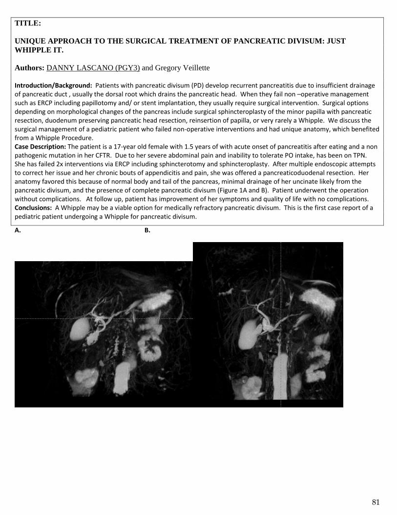
Figure 1A and B: Anatomy shows the separation of the pancreatic duct with the dorsal root draining the uncinate process and the ventral root draining the body and tail of the pancreas.
TITLE: UNIQUE APPROACH TO THE SURGICAL TREATMENT OF PANCREATIC DIVISUM: JUST WHIPPLE IT. Authors: DANNY LASCANO (PGY3) and Gregory Veillette Introduction/Background: Patients with pancreatic divisum (PD) develop recurrent pancreatitis due to insufficient drainage of pancreatic duct , usually the dorsal root which drains the pancreatic head. When they fail non –operative management such as ERCP including papillotomy and/ or stent implantation, they usually require surgical intervention. Surgical options depending on morphological changes of the pancreas include surgical sphincteroplasty of the minor papilla with pancreatic resection, duodenum preserving pancreatic head resection, reinsertion of papilla, or very rarely a Whipple. We discuss the surgical management of a pediatric patient who failed non-operative interventions and had unique anatomy, which benefited from a Whipple Procedure. Case Description: The patient is a 17-year old female with 1.5 years of with acute onset of pancreatitis after eating and a non pathogenic mutation in her CFTR. Due to her severe abdominal pain and inability to tolerate PO intake, has been on TPN. She has failed 2x interventions via ERCP including sphincterotomy and sphincteroplasty. After multiple endoscopic attempts to correct her issue and her chronic bouts of appendicitis and pain, she was offered a pancreaticoduodenal resection. Her anatomy favored this because of normal body and tail of the pancreas, minimal drainage of her uncinate likely from the pancreatic divisum, and the presence of complete pancreatic divisum (Figure 1A and B). Patient underwent the operation without complications. At follow up, patient has improvement of her symptoms and quality of life with no complications. Conclusions: A Whipple may be a viable option for medically refractory pancreatic divisum. This is the first case report of a pediatric patient undergoing a Whipple for pancreatic divisum.
A. B.
81
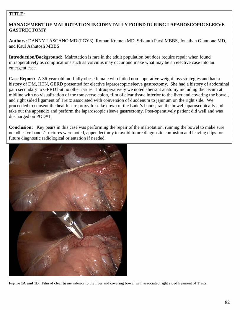
TITLE: MANAGEMENT OF MALROTATION INCIDENTALLY FOUND DURING LAPAROSCOPIC SLEEVE GASTRECTOMY Authors: DANNY LASCANO MD (PGY3), Roman Kremen MD, Srikanth Parsi MBBS, Jonathan Giannone MD, and Kaul Ashutosh MBBS Introduction/Background: Malrotation is rare in the adult population but does require repair when found intraoperatively as complications such as volvulus may occur and make what may be an elective case into an emergent case. Case Report: A 36-year-old morbidly obese female who failed non –operative weight loss strategies and had a history of DM, HTN, GERD presented for elective laparoscopic sleeve gastrectomy. She had a history of abdominal pain secondary to GERD but no other issues. Intraoperatively we noted aberrant anatomy including the cecum at midline with no visualization of the transverse colon, film of clear tissue inferior to the liver and covering the bowel, and right sided ligament of Treitz associated with conversion of duodenum to jejunum on the right side. We proceeded to consent the health care proxy for take down of the Ladd’s bands, ran the bowel laparoscopically and take out the appendix and perform the laparoscopic sleeve gastrectomy. Post-operatively patient did well and was discharged on POD#1. Conclusion: Key pears in this case was performing the repair of the malrotation, running the bowel to make sure no adhesive bands/strictures were noted, appendectomy to avoid future diagnostic confusion and leaving clips for future diagnostic radiological orientation if needed.
Figure 1A and 1B. Film of clear tissue inferior to the liver and covering bowel with associated right sided ligament of Treitz.
82

TITLE: SUCCESSFUL LAPAROSCOPIC EXCISION OF A FOREGUT DUPLICATION CYST IN A YOUNG ADULT FEMALE Authors: JOSEPH LOPEZ (PGY4), Ashutosh Kaul, Marc Arkovitz Background: Foregut duplication cysts are uncommon in the adolescent and young adult population. Objective: We present a video presentation of a laparoscopic excision of a subdiaphragmatic foregut duplication cyst in a young adult female. Design/Methods: A 20 year-old female with no significant past medical or surgical history was found to have a left paraspinal mass in the lesser sac on computerized tomography. A video was obtained with the patient’s informed consent of the laparoscopic excision of this mass. Four 5 mm trocars were used. Results: The video shows the successful complete laparoscopic excision of the paraspinal mass located in the lesser sac. Pathological evaluation of this mass revealed a benign cyst with ciliated epithelium which is consistent with a foregut duplication cyst. This is a rare finding in this patient population. Conclusions: Successful laparoscopic excision of a foregut duplication cyst may be performed in the adolescent and young adult population.
83

TITLE: AN INTERESTING CASE OF GIANT ESOPHAGEAL DIVERTICULUM CAUSING DYSPHAGIA AND SUCCESSFUL MANAGEMENT BY LAPAROSCOPIC RESECTION WITH MYOTOMY AND FUNDOPLICATION: CASE REPORT AND PRESENTATION OF SURGICAL TECHNIQUE Authors: M MAKADIA (PGY6), M Brown, A Elzanie, M Mcguirk, J Bruckner, J Giannone, A Maffei, T Cerabona, A Kaul. Westchester Medical Center, Valhalla, NY. Background: Epiphrenic diverticulum is a rare type of esophageal diverticulum. It is a pulson diverticulum located in last 10 cm of esophagus and assosciated with esophageal motility disorder that causes increased lower esophageal sphincter pressure such as achalasia. It can present with chronic symptoms of dysphagia, regurgitation, reflux, and aspiration. It is successfully treated with heller myotomy with or without diverticulum resection with minimally invasive techniques. Objective: To describe our technique of laparoscopic esophageal diverticulum resection with heller myotomy and Toupet fundoplication in an unique case of giant epiphrenic diverticulum causing dysphagia and weight loss. Design/Methods: Case presentation, discussion of essential surgical steps and review of literature. A 71 year old male patient with past medical history of hypertension, chronic obstructive pulmonary disease, hyperlipidemia along with a surgical history of open abdominal aortic aneurysm repair complicated by aortojejunal fistula approximately 10 years ago. He had also undergone amputation above the knee on his left leg and axillary bifemoral grafts with multiple revisions. The patient presented with dysphagia and weight loss and upon further investigation, he was found to have esophageal diverticulum on preoperative barium esophagogram and endoscopy. After an in-depth discussion, decision was made to perform laparoscopic esophageal diverticulum resection under the guidance of an endoscopy. We opted for Toupet fundoplication as diverticula was at 3’clock position. 100 units of botulinum toxin was injected into pylorus to help with gastric emptying. Patient was transferred to Intensive Care unit and extubated the next morning. Patient underwent gastrograffin swallow study on postoperative day 3 which was negative for any leaks and hence was started on a clear liquid diet and discharged to a short-term rehabilitation facility on day 4. Results: Two weeks after surgery, patient was examined in an out-patient setting with a notable gain of 6 pounds and complete resolution of dysphagia. Diet was advanced to soft diet in the next step. Conclusions: Esophageal diverticula are rare cause of dysphagia and weight loss. Epiphrenic diverticulum is usually false diverticulum in distal 10 cm esophagus caused by pulson mechanism associated with motility disorder. It can be successfully managed by myotomy +/- resection of diverticulum. With advances in minimally invasive surgery, laparoscopic or thoracosopic approach have been used as preferred option.
84

TITLE: CASE REPORT: OPEN CYSTGASTROSTOMY FOR A SYMPTOMATIC GIANT INFECTED PANCREATIC PSEUDOCYST WITH NECROSIS Authors: MATTHEW MCGUIRK, MD (PGY1), Reid Goodman (MS3), Rifat Latifi, MD Background: Pancreatic pseudocyst is an encapsulated collection of fluid with a well-defined fibrous or granulation tissue wall usually occurring 4 or more weeks after the onset of interstitial edematous pancreatitis.1 Severe acute pancreatitis with pancreatic necrosis is also referred to as necrotizing pancreatitis. Pancreatic necrosis becomes infected in 40-70% of cases1. External surgical drainage is indicated for patients with symptomatic or infected mature cysts that cannot tolerate or have failed percutaneous or endoscopic drainage.2 We present the case of an open cystgastrostomy for a symptomatic giant infected pancreatic pseudocyst with necrosis. Case: 71-year-old male presented to an outside hospital after having severe abdominal pain while at rehab, where he was recovering from a fall. He was diagnosed with acute pancreatitis with a cystic collection and underwent an IR drainage. He began to develop a poor appetite and was placed on peripheral parenteral nutrition (PPN). A CT of the abdomen was done on 6/22/18, revealing a 20.8 cm x 8.4 cm pancreatic pseudocyst replacing the majority of the pancreatic parenchyma. This then increased in size to 21.6 cm x 8.4 cm on 7/9/18. Due to his extremely poor nutritional status, with an albumin of 2.2, a prealbumin of 4.0, and a weight loss 10.5 kg the decision was made to attempt nutritional optimization before operating. His hospital course was also complicated by pneumonia with a pleural effusion, for which he had a left thoracostomy tube and a pseudomonas and vancomycin-resistant enterococcus positive urinary tract infection that lead to urosepsis, for which he was transferred to the ICU. After continuing to have poor nutritional status, with little to no appetite, a repeat CT of the abdomen was obtained to evaluate if the pseudocyst was amenable to cystgastrostomy. The CT done on 7/23/18 revealed a 20.6 cm x 6.7 cm cyst, inseparable from the body and tail of the pancreas and extending to the head. The cyst contained new air pockets representing a likely infection. The patient was taken emergently to the operating room for an open cystgastrostomy with pancreatic necrosectomy, cholecystectomy, and drain placement on 7/24/18. Operative cultures showed staphylococcus haemolyticus. He was extubated on post-op day 2 and begun on tube feeds via nasogastric tube. His appetite improved and he was able to tolerate a regular diet. His cortrak was removed on 8/14/18 and his JP drain was removed on 8/19/18 after drainage became minimal. He was discharged to an inpatient rehab facility on regular diet. Conclusions: Open cystgastrostomy and necrosectomy is indicated for giant infected pancreatic pseudocyst with necrosis and should be considered. 1. Swaroop, Vege Santhi, Suresh T. Chari, and Jonathan E. Clain. "Severe acute pancreatitis." Jama 291.23 (2004): 2865-2868. 2. Habashi, Samir, and Peter V. Draganov. "Pancreatic pseudocyst." World journal of gastroenterology: WJG 15.1 (2009): 38.
85
TITLE: BLUNT ABDOMINAL TRAUMA AND SMALL BOWEL PERFORATION IN A PATIENT WITH CROHN’S DISEASE Authors: IDA MOLAVI, MD (PGY6), Gary Lombardo, MD Introduction: In the setting of blunt abdominal trauma, small bowel injury (SBI) is an infrequent diagnosis, occurring in less than 1.1% of all cases. Furthermore, although small bowel perforation has been reported to be the third most common injury in blunt abdominal trauma, its absolute rate of occurrence is a rare phenomenon at 0.3%. Diagnostic approaches, such as FAST and CT scans, lack the sensitivity in diagnosis of SBI and cannot reliably predict the need for emergent laparotomy. It is therefore essential to combine the mechanism of injury, vital signs, physical findings, and underlying medical conditions in the decision making algorithm. There is very little in the literature on small bowel perforation in patients with a history of inflammatory bowel disease following blunt abdominal trauma. This case-report looks at a high-speed trauma patient with a history of crohn’s disease, presenting with small bowel perforation, and discusses the diagnosis and management of this rare presentation. Case Report: 45-year-old female presented as a level 2 trauma activation following a high speed motor vehicle crash. Patient was in the front seat, restrained passenger. On arrival, she was a GCS 15, mainly complaining of abdominal pain and nausea. On examination, she was tachycardic but responsive to fluid resuscitation. Her abdomen was soft and non-distended, with diffuse tenderness on deep palpation. There was slight bruising around the umbilicus, concerning for a seatbelt sign. Patient’s medical history was significant for a 25 year history of crohn’s disease, in remission for years and on no current medications. The patient was subsequently intubated in the trauma bay for airway protection from persistent nausea and active emesis. She remained hemodynamically stable. FAST was negative. A CT scan of the abdomen and pelvis was performed as part of her trauma workup, revealing pneumoperitoneum, diffuse segmental small bowel wall thickening with areas of mucosal hyperemia suggesting active inflammatory bowel disease. Patient was taken to the operating room for an exploratory laparotomy, and found to have evidence of significant crohn’s disease involving the ileum and cecum. There was severe ileal mesenteric thickening and tethering, involving 76 cm of adherent loops of ileum, and a 6.5 cm transected segment of the involved ileum found in the pelvis. We performed an ileocecetomy with side to side ileocolic anastomosis of healthy viable margins. Pathology revealed inflammatory bowel disease, favoring crohn’s, in remission with no acute activity. Patient recovered well postoperatively, and was referred to gastroenterology for management and follow-up of her crohn’s disease. Discussion: Small bowel perforation in the setting of blunt abdominal trauma, is a rare diagnosis, and only in 0.3% of patients admitted to major trauma centers in the United States. In patients with blunt abdominal trauma and underlying Crohn's disease, we propose a low threshold for CT imaging. It is quite possible that the underlying inflammation and edema make the bowel more susceptible to perforation in blunt trauma. In our case, physical examination was difficult to interpret as despite the tenderness on deep palpation, she was not an acute surgical abdomen. Second, her history of crohn’s disease in remission, without any active signs or current medications, seemed to indicate minimum severity of her disease. However, intraoperatively, she was found to have extensive disease with a transected segment of the affected ileum resulting in small bowel perforation. This case emphasizes the need to approach patients with even a remote history of inflammatory bowel disease cautiously. In our case, the CT scan of the abdomen did reveal the small bowel perforation; however, it is important to note that diagnostic tests, such as FAST and CT, lack the sensitivity in diagnosis of small bowel perforation and hence cannot reliably predict the need for emergent laparotomy. Our patient was also found to have an L1 chance fracture on the final CT reads, and though this was noted after the laparotomy, one should note that 89% of chance fractures are associated with some form of SBI. Presence of a seatbelt signs has also been shown to be the single most significant risk factor for small bowel injury, carrying an increase in relative risk of 2.4. Therefore, when it comes to assessing our patients for small bowel injury post blunt trauma, it is essential to consider the mechanism along with other information, such as hemodynamic status, clinical findings, imaging (ie FAST and CT), as well as patient’s underlying medical conditions.
86

TITLE: REPAIRING A JUXTARENAL AORTA ANEURYSM USING A SNORKEL GRAFT IN A COMPLEX VASCULAR DISEASE PATIENT Authors: BINYAMIN ROTHSCHILD (PGY1), David Cohen, Dr. Laskowski Background: Conventional endovascular aneurysm repair EVAR have become in recent years first line treatment in abdominal aorta aneurysm. However, there are many patients who are unsuitable for conventional EVAR due to anatomical challenges or difficult access points. Juxtarenal aorta aneurysm are challenging due to need for renal arteries stenting in addition to the aneurysm repair. With advanced in endovascular technologies and operator experience a rapidly expanding strategies have evolved to address these challenges. Objective: We are presenting a case a juxtarenal aneurysm that was repaired using a ‘snorkel’ graft that enables blood flow to branch arteries which otherwise would be blocked by a the graft Case: 79-year-old woman with history of PAD,HTN, CAD who presented with bilateral leg claudication. CTA Showed She was found to have bilateral ilio-femoral disease and a 5cm juxtarenal AAA with left and right renal artery stenosis on CTA which was also seen on angiogram. The aneurysm was repaired using a snorkel graft that was inserted through the axillary artery. I-Cast stents were placed in each renal artery. An aorto-uni-iliac graft was deployed. Angiogram showed good position and both profusions of renal arteries. The surgery was completed with a Left iliac enterectomy and a fem- fem by pass. Pos op initially patient was recovering well. On POD3 patient developed respiratory distress and was transferred to the ICU. The patient was found to have a 3 vessel MI. Patient stayed in the ICU for 10 days and was transferred to the floor upon improvement of respiratory and cardiac state. Currently patient is pending discharge to rehab. Discussion: The snorkel graft offers an endovascular approach to an otherwise open approach in complicated aneurysm patients. EVAR offers a better short-term morbidity and mortality compared to an open approached as shown in the DREAM and EVAR 1 clinical trials. Long term morbidity and mortality are currently investigated. Of note this patient was not a good canidate for a fenestrated graft due to lack of infra renal neck and poor access of iliac vessels.
87
TITLE: “INFLAMMATORY BOWEL DISEASE- NOT JUST THE BOWELS BANE.” ARTERIAL AND VENOUS THROMBOSIS IN A PATIENT WITH CROHNS DISEASE. Authors: A. SAFAYA MD (PGY4), S. Babu MD, R. Mateo MD, F. Carroll MD, A. Goyal MD, I. Laskowski MD Background: Inflammatory bowel diseases (IBD) that include the spectrum of Crohn’s disease and ulcerative colitis are primarily inflammatory diseases of the bowel associated sometimes with a myriad of extra-intestinal manifestations. Systemic inflammatory states with its extra-intestinal manifestations are well known and sought after signs in patients with IBD. However, hypercoagulability and the prothrombotic nature of the disease is lesser known and often overlooked. These vascular manifestations, affecting both the arterial and venous systems, contribute significantly in increasing the morbidity and mortality in IBD patients. We highlight this serious condition through a case of a 63 year-old man, who presented with catastrophic manifestations of his untreated crohn’s disease in the form of arterial and venous thrombosis. Objective: In the form of this Case Report – we want to show the emphasis of recognizing a common gastro-intestinal disease like inflammatory bowel disease as a harbinger of severe thrombo-embolic events. Design/Methods: We present here a case of a Sixty-three year old male with past medical history significant for Crohn’s disease that was left untreated for 5-10 years. The patient presented with critical limb ischemia of the right upper extremity. The patient had previous vascular history significant for severe peripheral arterial disease that had rendered him with a right sided below knee amputation and lower extremity deep venous thrombosis for which the patient had undergone an inferior vena-cava filter. Results: Patient underwent emergent thrombectomy of the right upper extremity vasculature with initial resolution of symptoms and flow. However, around post-operative day 5 the patient had to be re-explored for worsening flow dynamics and symptoms with similar outcome of initial relief with worsening of the flow within 2-3 days. After a well-informed and involved family decision a third operative intervention was withheld and patient was treated conservatively with intra-venous anticoagulation. At, this time within hours the patient developed tense edematous left lower extremity with erythema spreading towards the left flank and abdomen. The disease progressed to multi-system organ failure despite operative intervention leading to the demise of the patient. Conclusions: Intra-operative findings from the last procedure showed viable bulging muscles with no evidence of necrotizing soft tissue pathology. However, the left common femoral vein was found to be extensively fibrosed and unable to be cannulated. The mottling of the skin around the left lower extremity and the swift spread towards the left abdominal wall and flanks related more towards a severe vascular thrombotic event. Given the history of untreated IBD and the fact that the patient has had venous and arterial thrombosis in the past, it is likely that the patient’s current manifestation may have association with his underlying crohn’s disease. Thrombotic complications of IBD are often overlooked and hence through this case we would like to emphasize the importance of keeping a very high index of suspicion of this association for our routine vascular patients that have the diagnosis of IBD and are not under treatment. Further studies are needed to understand the pathophysiology of this condition and determine clinical implications in terms of need for anticoagulation both therapeutic and prophylactic in hospitalized patients with IBD to prevent catastrophic thrombotic events.
88
TITLE: VENO-VENOUS EXTRACOPOREAL MEMBRANE OXYGENATION IN A PATIENT WITH ACUTE T CELL LEUKEMIA/LYMPHOMA Authors: TIAN YUE SONG M.S. (MS3), Michelle Bravo M.D., Tracey Weigel M.D. Background: Acute respiratory failure (ARF) in patients with hematologic malignancy (HM) due to extrinsic airway compression is associated with mortality > 50% when they require invasive mechanical ventilation (MV). Extracoporeal membrane oxygenation (ECMO) is a well-known rescue therapy in cardiopulmonary failure. However, literature regarding ECMO in adult patients with ARF secondary to acute airway compression from a rapidly enlarging anterior mediastinal mass associated with an HM is limited. Specifically, veno-venous (VV) ECMO can supplement oxygenation when upper airway obstruction drastically limits conventional ventilator therapy. Another benefit of ECMO is that it can minimize ventilator related lung injury and may decrease MV associated mortality. Additionally, prompt diagnosis and rapid initiation of chemotherapy can provide dramatic improvement and minimize duration of ECMO. Herein, we report a case of respiratory management in an adult patient with T cell acute lymphoblastic leukemia/lymphoma presenting with ARF. Objective: To highlight management of acute airway and ventilatory compromise using VV ECMO in adult patients with massive mediastinal mass, and the importance of rapid diagnosis and treatment of HM to minimize the duration and potential morbidities of ECMO. Design/Methods: We report a recent case from the cardiothoracic service, discussing acute presentation, multidisciplinary planning and coordination of treatment and rapid improvement of the patient’s status. Results: A 26-year-old female with no pertinent medical history presented to an outside institution due to rapid onset of difficulty breathing and progressive facial swelling. She endorsed one month of vague exertional dyspnea, cough, facial and neck swelling not severe enough for her to seek medical care. She denied fever, headache, abdominal pain or recent weight loss. On presentation to outside hospital, she had severe edema involving the face, neck and upper extremities consistent with superior vena cava (SVC) syndrome. A large mediastinal mass was seen on chest x-ray. She developed respiratory distress while in supine position for Computed Tomography (CT), was emergently intubated and transferred directly to WMC operating room (OR). Given her age, finding of mediastinal mass and acuity of presentation, there was high clinical suspicion for HM. On arrival, she went directly to OR with cardiothoracic surgery team. Bronchoscopy revealed a near complete collapse of the trachea with patient desaturating to 60%. Emergent veno-venous (VV) ECMO was initiated and immediately improved oxygenation and ventilation. Thoracic team performed a chamberlain procedure as venous catheters were being inserted by cardiac team. Intraoperative pathology confirmed lymphoid malignancy. The patient was transferred to the cardiothoracic intensive care unit (CTICU) post operatively and chemotherapy was initiated within 12 hours. Subsequent sternal bone marrow biopsy confirmed T cell acute lymphoblastic leukemia. Within 1week of initiating chemotherapy, plain CXR showed near complete resolution of the mediastinal mass. She was successfully weaned from ECMO and extubated on hospital day 6 and 7. Patient improved and was later discharged without severe complications. Conclusions: VV ECMO is a novel way to salvage severe, life threatening ARF in HM patients that present with a large mediastinal mass. It can be initiated concurrently with a chamberlain procedure affording rapid diagnosis and initiation of treatment. Currently ECMO is not a standardized procedure in acute HM airway management but should be considered if resources are readily available and rapid response to therapy is considered highly likely, thus minimizing the morbidity associated with VV ECMO.
89

TITLE: ANATOMIC DELINEATION OF FAILED COLONIC INTERPOSITION Authors: MICHAEL WINTER (MS4), Dr. Miles Dale, Dr. Patrice Anderson Background: The patient was a 52 year old male who was transferred to WMC for vent-dependent respiratory failure and poor nutritional status. Patient originally suffered polytrauma secondary to MVA in 1979 resulting in: esophageal resection, gastric construction, colonic interpositional graft, partial bowel resection, and splenectomy. Prior to transfer to WMC, there was a concern the patient had a tracheoesophageal fistula so the patient underwent endoscopy. During endoscopy patient sustained a perforation to the colonic interposition resulting in a colo-tracheal fistula (between the interposition graft and trachea). After endoscopy the patient developed aspiration pneumonia, a bronchopleural fistula, and multiple other organ to pleural fistula. He then underwent an exploratory laparotomy with thoracotomy for lysis of adhesions and repair of fistula as well as excision of colonic conduit. This resulted in an esophageal stump that was closed with drain placement and creation of right end colostomy. A chest tube was placed near the gastric stump and LUQ which obliterated the left hemidiaphragm. Patient then had placement of an open J-tube. At this point the patient was admitted to WMC with failure to thrive, cachexia with a weight of 31.7 kg and ventilator dependent respiratory failure. When at WMC, bile was found to be coming out from his tracheal secretions. He underwent bronchoscopy to evaluate the source but none could be found. At this point there was a need for his anatomy to be delineated before any surgical intervention could be offered. Objective: Case report of the anatomic delineation using novel methods of a patient with a failed colonic interposition and failure to thrive on ventilator. Results: Patient’s anatomy as we know it after delineation. The proximal Esophagus is in discontinuity and open with a Chest tube track. The stomach is left in discontinuity as well and open with a Malecot/Chest tube draining the output. There is a bronchoespohageal fistula from the gastric remnant and esophageal junction that is draining into the left bronchus. This is what allowed bile to reflux into the tracheal secretions. The Malecot is located in the left thoracic cavity between stomach and left lower lobe of lung. No or minimal left diaphragm remains. There is a J tube in Jejunum. Patient also has a right end colostomy. Conclusions: Delineation of anatomy is a necessary process in any surgical planning. By delineating the anatomy surgeons can offer patients the best surgical options and have safe outcomes. The delineation of this patient in particular has detailed the extensive nature of the surgical repair they would require to be adequately re-anastomosed. The patient would first require extensive supplemental nutrition and ventilator weaning to the point they were no longer cachectic and could physically withstand the procedure and adequately heal from the procedure. At the moment it has unanimously been determined the patient would not survive surgical intervention. The patient will require coordination of a Thoracic and general surgery experts to make a surgical plan, as it cannot be simple repaired with a Gastric Pull Up or Jejunum Interposition, as the distance from distal esophagus to proximal stomach is too long. After appropriate strength building and weight gain the patient can undergo reconstruction and have his alimentary system back in continuity.
90

TITLE: ROBOTIC RADICAL CHOLECYSTECTOMY – A CASE REPORT Authors: NIU ZHANG (PGY5), MD, Xiang Da Dong, MD Background: Gallbladder cancer represents one of the most aggressive malignancy of the biliary tract and is characterized by an extremely poor prognosis. For resectable gallbladder cancer, radical cholecystectomy (RC) is the only potential curative approach, which consists of liver resection (liver bed resection or segment 4b-5 bisegmentectomy) with locoregional lymphadenectomy. The use of laparoscopic surgery is seldom considered previously in the management of gallbladder cancer due to the fear of dissemination during laparoscopy and the risk of incomplete resection. Recently, robotic surgery is gaining in popularity. Several small case series of robotic radical cholecystectomy have been published. Herein we are present a case of robotic En-Bloc resection of liver segment 4b/5, gallbladder and portal lymph nodes for gallbladder cancer. Case description: Patient is an 83 years old female of Asian descent with known history of gallbladder mass suspicious for malignancy. She had previously undergone CT and MRI which were concerning of gallbladder cancer. Her preoperative workup also showed an elevated AFP level concerning for malignancy. The patient was consented and underwent a robotic radical cholecystectomy including segment 4b and 5 partial liver resection and portal lymph node sampling. Intraoperative fluorescent imaging cholangiogram with Firefly® was performed with indocyanine-green (ICG). Surgery underwent uneventfully. Patient was discharged on postop day 5. Pathology report demonstrated moderately differentiated carcinoma invading muscular layer (pT1b), and lymphovascular invasion identified. Conclusions: Robotic radical cholecystectomy is technically feasible and safe in elderly patients. Fluorescent cholangiography can be a useful tool for intra-operative visualization of the biliary tree during robotic radical cholecystectomies. A large randomized clinical study with long term follow-up is still needed to identify the oncology results of robotic radical cholecystectomy for gallbladder cancer.
91