Fibrous Dysplasia by Matthew R. DiCaprio, and William F. Enneking J Bone Joint Surg Am Volume...
16
Fibrous Dysplasia by Matthew R. DiCaprio, and William F. Enneking J Bone Joint Surg Am Volume 87(8):1848-1864 August 1, 2005 ©2005 by The Journal of Bone and Joint Surgery, Inc.
-
Upload
lena-standage -
Category
Documents
-
view
221 -
download
0
Transcript of Fibrous Dysplasia by Matthew R. DiCaprio, and William F. Enneking J Bone Joint Surg Am Volume...

Fibrous Dysplasia
by Matthew R. DiCaprio, and William F. Enneking
J Bone Joint Surg AmVolume 87(8):1848-1864
August 1, 2005
©2005 by The Journal of Bone and Joint Surgery, Inc.
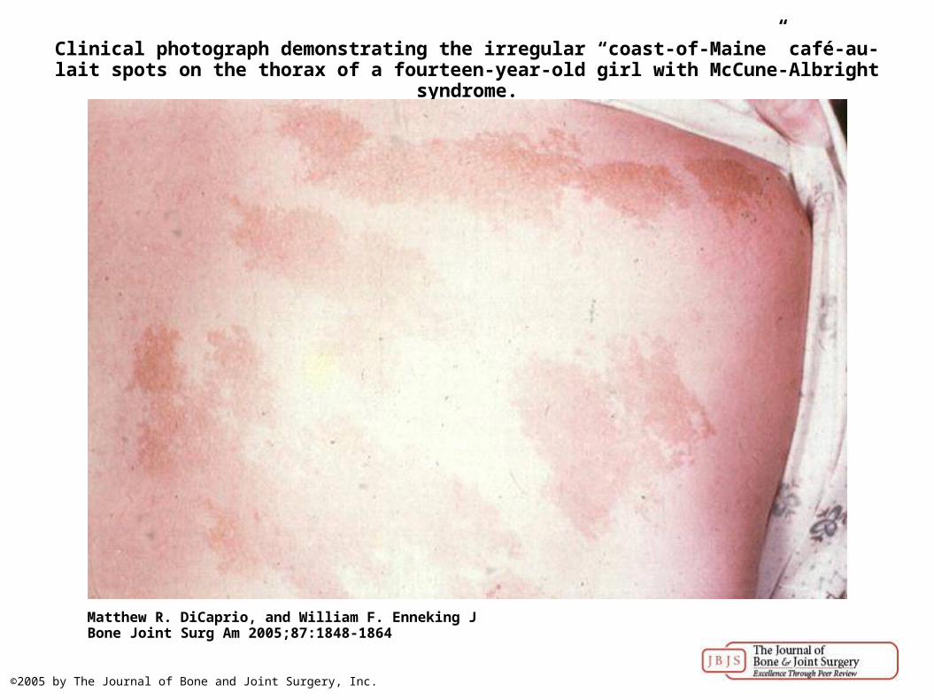
Clinical photograph demonstrating the irregular “coast-of-Maine” café-au-lait spots on the thorax of a fourteen-year-old girl with McCune-Albright syndrome.
Matthew R. DiCaprio, and William F. Enneking J Bone Joint Surg Am 2005;87:1848-1864
©2005 by The Journal of Bone and Joint Surgery, Inc.
Matthew R. DiCaprio, and William F. Enneking J Bone Joint Surg Am 2005;87:1848-1864
©2005 by The Journal of Bone and Joint Surgery, Inc.

Matthew R. DiCaprio, and William F. Enneking J Bone Joint Surg Am 2005;87:1848-1864
©2005 by The Journal of Bone and Joint Surgery, Inc.

Matthew R. DiCaprio, and William F. Enneking J Bone Joint Surg Am 2005;87:1848-1864
©2005 by The Journal of Bone and Joint Surgery, Inc.

Matthew R. DiCaprio, and William F. Enneking J Bone Joint Surg Am 2005;87:1848-1864
©2005 by The Journal of Bone and Joint Surgery, Inc.
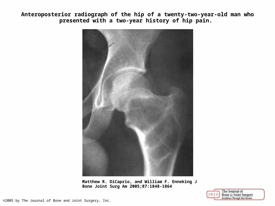
Anteroposterior radiograph of the hip of a twenty-two-year-old man who presented with a two-year history of hip pain.
Matthew R. DiCaprio, and William F. Enneking J Bone Joint Surg Am 2005;87:1848-1864
©2005 by The Journal of Bone and Joint Surgery, Inc.

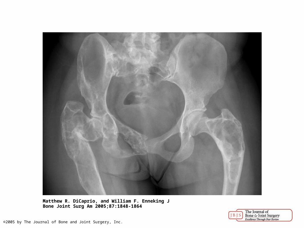
Radiographic features of fibrous dysplasia.
Matthew R. DiCaprio, and William F. Enneking J Bone Joint Surg Am 2005;87:1848-1864
©2005 by The Journal of Bone and Joint Surgery, Inc.
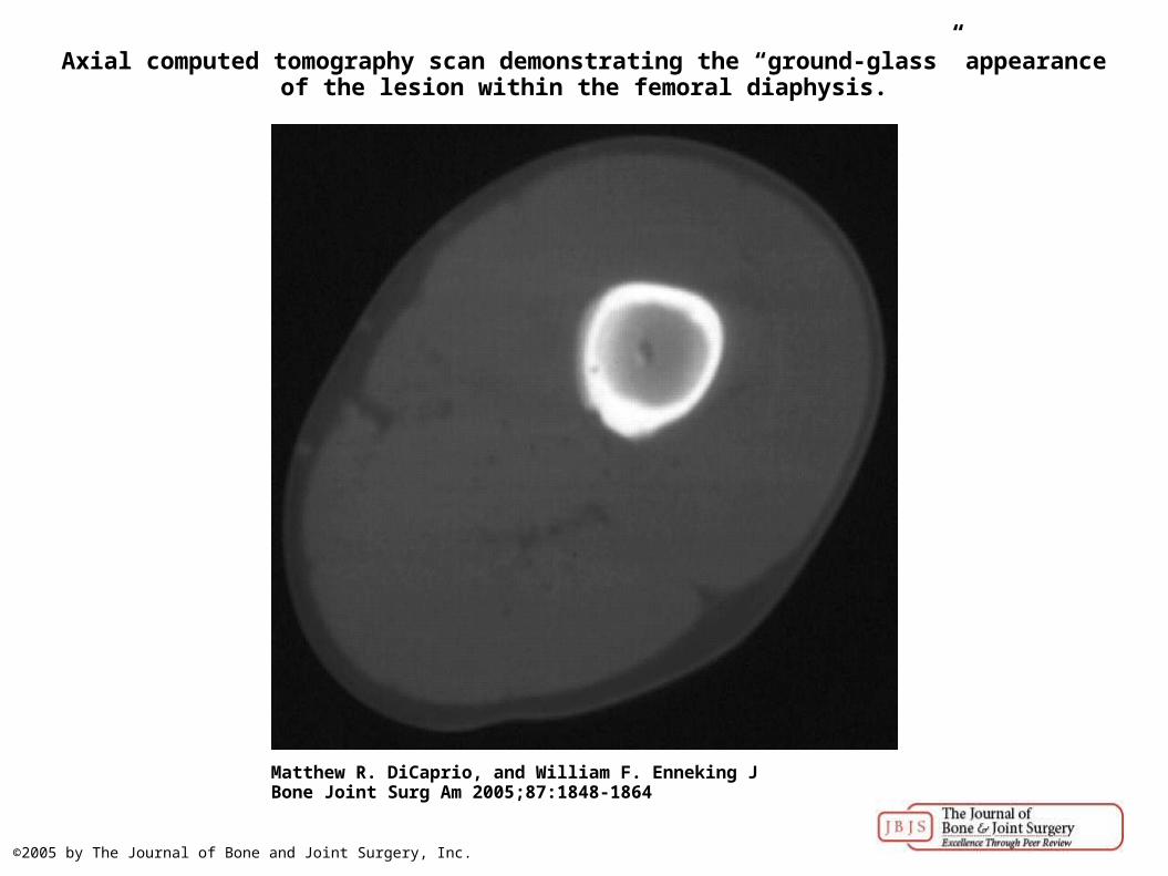
Axial computed tomography scan demonstrating the “ground-glass” appearance of the lesion within the femoral diaphysis.
Matthew R. DiCaprio, and William F. Enneking J Bone Joint Surg Am 2005;87:1848-1864
©2005 by The Journal of Bone and Joint Surgery, Inc.

Sagittal T1-weighted magnetic resonance image of the same lesion, showing primarily a low signal intensity.
Matthew R. DiCaprio, and William F. Enneking J Bone Joint Surg Am 2005;87:1848-1864
©2005 by The Journal of Bone and Joint Surgery, Inc.

Histologic features of fibrous dysplasia.
Matthew R. DiCaprio, and William F. Enneking J Bone Joint Surg Am 2005;87:1848-1864
©2005 by The Journal of Bone and Joint Surgery, Inc.

The mesenchymal stroma surrounding the dysplastic trabeculae is relatively hypocellular.
Matthew R. DiCaprio, and William F. Enneking J Bone Joint Surg Am 2005;87:1848-1864
©2005 by The Journal of Bone and Joint Surgery, Inc.
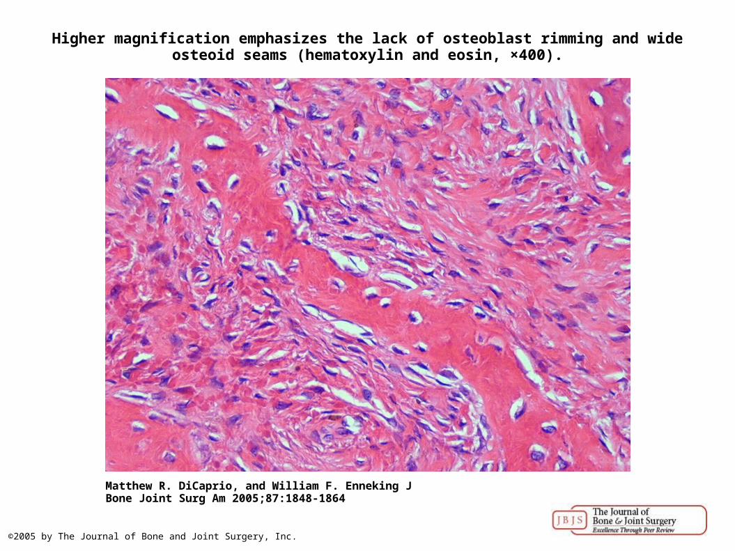
Higher magnification emphasizes the lack of osteoblast rimming and wide osteoid seams (hematoxylin and eosin, ×400).
Matthew R. DiCaprio, and William F. Enneking J Bone Joint Surg Am 2005;87:1848-1864
©2005 by The Journal of Bone and Joint Surgery, Inc.

A field of mature hyaline cartilage is shown adjacent to a bland fibrous stroma (hematoxylin and eosin, ×100).
Matthew R. DiCaprio, and William F. Enneking J Bone Joint Surg Am 2005;87:1848-1864
©2005 by The Journal of Bone and Joint Surgery, Inc.

Examples of fibrous dysplasia of the femoral neck treated with fibular grafting.
Matthew R. DiCaprio, and William F. Enneking J Bone Joint Surg Am 2005;87:1848-1864
©2005 by The Journal of Bone and Joint Surgery, Inc.

Fifteen-year follow-up radiograph of the proximal part of the femur in a patient treated with a fibular allograft for monostotic fibrous dysplasia.
Matthew R. DiCaprio, and William F. Enneking J Bone Joint Surg Am 2005;87:1848-1864
©2005 by The Journal of Bone and Joint Surgery, Inc.