Fe-S cofactors in the SARS-CoV-2 RNA-dependent RNA ...
12
Cite as: N. Maio et al., Science 10.1126/science.abi5224 (2021). REPORTS First release: 3 June 2021 www.sciencemag.org (Page numbers not final at time of first release) 1 The novel coronavirus, severe acute respiratory syndrome- coronavirus 2 (SARS-CoV-2), has caused a global pandemic known as coronavirus disease (COVID-19) (1–3), which can be prevented by vaccines, but for which antiviral treatments are much needed. Coronaviruses (CoVs) employ a multisubunit machinery for replication and transcription. A set of nonstructural proteins (nsps) produced as cleavage products of the ORF1a and ORF1ab polyproteins (4) assembles to facilitate viral replication and transcription. The core component of this complex is the catalytic subunit (nsp12) of an RNA-dependent RNA polymerase (RdRp) (5), which catalyzes the synthesis of viral RNA and thus plays a central role in the replication and transcription cycle of SARS-CoV- 2, with the assistance of nsp7 and nsp8 as accessory factors (6, 7). Structures of the RdRp (nsp12-nsp7-nsp8 complex) alone and in complex with the helicase have been determined by cryo-electron microscopy (8–11); in all of them the RdRp of SARS-CoV-2 was proposed to contain zinc ions ligated in the same locations as those observed in SARS-CoV (7) in highly conserved metal binding motifs composed of H295- C301-C306-C310 and C487-H642-C645-C646 (Fig. S1). These zinc ions have been proposed to serve a structural role in maintaining the integrity of the RdRp architecture (7–11) (see supplementary text). Zinc has long been known to have the capability to replace endogenous iron-sulfur (Fe-S) metal cofactors during standard aerobic purification of proteins (12–15), because Fe-S clusters are inherently susceptible to destabilization and degradation by oxidants, including oxygen, superoxide (O2 − ), and nitric oxide (16). Notably, Fe-S clusters, inorganic cofactors often associated with biological redox reactions (17, 18), have been identified in numerous proteins involved in DNA and RNA metabolism, where they play a variety of critical functional roles (12, 13, 19–26). Having recently demonstrated that we are able to predict the presence of Fe-S cofactors in candidate proteins based on the identification of specific amino acid sequence motifs (27), we analyzed the primary sequences of SARS-CoV-2 proteins to investigate whether any might incorporate Fe-S clusters. We identified two highly conserved LYR (leucine-arginine-ty- rosine)-like motifs (fig. S2A) in nsp12 that have been previ- ously characterized as potential binding sites for the cochaperone HSC20 (aka HSCB) of the Fe-S biogenesis ma- chinery (27–30), which facilitates Fe-S cluster transfer from the main scaffold protein, ISCU, to recipient proteins (fig. S2B). To assess whether the LYR-like motifs were involved in direct binding of nsp12 to HSC20, we incubated full-length SARS-CoV-2 nsp12 wild type or variants wherein either or both LYR motifs were replaced by alanines (fig. S2C) with pu- rified HSC20. Nsp12 wild type (WT) bound HSC20, indicating that the RdRp subunit interacts directly with the cochaper- one (Fig. 1A). Substitution of either of the two LYR motifs with alanines decreased the amount of bound HSC20 (Fig. 1A), which was even more profoundly diminished by loss of both motifs in nsp12 VYR/LYR-AAA (Fig. 1A). Co- Fe-S cofactors in the SARS-CoV-2 RNA-dependent RNA polymerase are potential antiviral targets Nunziata Maio 1 , Bernard A. P. Lafont 2 , Debangsu Sil 3 , Yan Li 4 , J. Martin Bollinger Jr. 3,5 , Carsten Krebs 3,5 , Theodore C. Pierson 6 , W. Marston Linehan 7 , Tracey A. Rouault 1 * 1 Eunice Kennedy Shriver National Institute of Child Health and Human Development, National Institutes of Health, Bethesda, MD 20892, USA. 2 SARS-CoV-2 Virology Core, Laboratory of Viral Diseases, Division of Intramural Research, National Institute of Allergy and Infectious Diseases, National Institutes of Health, Bethesda, MD 20892, USA. 3 Department of Chemistry, The Pennsylvania State University, University Park, PA 16802, USA. 4 Proteomics Core Facility, National Institute of Neurological Disorders and Stroke, National Institutes of Health, Bethesda, MD 20892, USA. 5 Department of Biochemistry and Molecular Biology, The Pennsylvania State University, University Park, PA 16802, USA. 6 Laboratory of Viral Diseases, Division of Intramural Research, National Institute of Allergy and Infectious Diseases, National Institutes of Health, Bethesda, MD 20892, USA. 7 Urologic Oncology Branch, Center for Cancer Research, National Cancer Institute, Bethesda, MD 20892, USA. *Corresponding author. Email: [email protected] Severe acute respiratory syndrome coronavirus 2 (SARS-CoV-2), the causal agent of coronavirus disease 2019 (COVID-19), uses an RNA-dependent RNA polymerase (RdRp) for the replication of its genome and the transcription of its genes. We found that the catalytic subunit of the RdRp, nsp12, ligates two iron-sulfur metal cofactors in sites that were modeled as zinc centers in the available cryo-electron microscopy structures of the RdRp complex. These metal binding sites are essential for replication and for interaction with the viral helicase. Oxidation of the clusters by the stable nitroxide TEMPOL caused their disassembly, potently inhibited the RdRp, and blocked SARS-CoV-2 replication in cell culture. These iron-sulfur clusters thus serve as cofactors for the SARS-CoV-2 RdRp and are targets for therapy of COVID-19. on June 16, 2021 http://science.sciencemag.org/ Downloaded from
Transcript of Fe-S cofactors in the SARS-CoV-2 RNA-dependent RNA ...

Cite as: N. Maio et al., Science 10.1126/science.abi5224 (2021).
REPORTS
First release: 3 June 2021 www.sciencemag.org (Page numbers not final at time of first release) 1
The novel coronavirus, severe acute respiratory syndrome-coronavirus 2 (SARS-CoV-2), has caused a global pandemic known as coronavirus disease (COVID-19) (1–3), which can be prevented by vaccines, but for which antiviral treatments are much needed. Coronaviruses (CoVs) employ a multisubunit machinery for replication and transcription. A set of nonstructural proteins (nsps) produced as cleavage products of the ORF1a and ORF1ab polyproteins (4) assembles to facilitate viral replication and transcription. The core component of this complex is the catalytic subunit (nsp12) of an RNA-dependent RNA polymerase (RdRp) (5), which catalyzes the synthesis of viral RNA and thus plays a central role in the replication and transcription cycle of SARS-CoV-2, with the assistance of nsp7 and nsp8 as accessory factors (6, 7). Structures of the RdRp (nsp12-nsp7-nsp8 complex) alone and in complex with the helicase have been determined by cryo-electron microscopy (8–11); in all of them the RdRp of SARS-CoV-2 was proposed to contain zinc ions ligated in the same locations as those observed in SARS-CoV (7) in highly conserved metal binding motifs composed of H295-C301-C306-C310 and C487-H642-C645-C646 (Fig. S1). These zinc ions have been proposed to serve a structural role in maintaining the integrity of the RdRp architecture (7–11) (see supplementary text). Zinc has long been known to have the capability to replace endogenous iron-sulfur (Fe-S) metal cofactors during standard aerobic purification of proteins (12–15), because Fe-S clusters are inherently susceptible to
destabilization and degradation by oxidants, including oxygen, superoxide (O2
−), and nitric oxide (16). Notably, Fe-S clusters, inorganic cofactors often associated with biological redox reactions (17, 18), have been identified in numerous proteins involved in DNA and RNA metabolism, where they play a variety of critical functional roles (12, 13, 19–26).
Having recently demonstrated that we are able to predict the presence of Fe-S cofactors in candidate proteins based on the identification of specific amino acid sequence motifs (27), we analyzed the primary sequences of SARS-CoV-2 proteins to investigate whether any might incorporate Fe-S clusters. We identified two highly conserved LYR (leucine-arginine-ty-rosine)-like motifs (fig. S2A) in nsp12 that have been previ-ously characterized as potential binding sites for the cochaperone HSC20 (aka HSCB) of the Fe-S biogenesis ma-chinery (27–30), which facilitates Fe-S cluster transfer from the main scaffold protein, ISCU, to recipient proteins (fig. S2B). To assess whether the LYR-like motifs were involved in direct binding of nsp12 to HSC20, we incubated full-length SARS-CoV-2 nsp12 wild type or variants wherein either or both LYR motifs were replaced by alanines (fig. S2C) with pu-rified HSC20. Nsp12 wild type (WT) bound HSC20, indicating that the RdRp subunit interacts directly with the cochaper-one (Fig. 1A). Substitution of either of the two LYR motifs with alanines decreased the amount of bound HSC20 (Fig. 1A), which was even more profoundly diminished by loss of both motifs in nsp12VYR/LYR-AAA (Fig. 1A). Co-
Fe-S cofactors in the SARS-CoV-2 RNA-dependent RNA polymerase are potential antiviral targets Nunziata Maio1, Bernard A. P. Lafont2, Debangsu Sil3, Yan Li4, J. Martin Bollinger Jr.3,5, Carsten Krebs3,5, Theodore C. Pierson6, W. Marston Linehan7, Tracey A. Rouault1* 1Eunice Kennedy Shriver National Institute of Child Health and Human Development, National Institutes of Health, Bethesda, MD 20892, USA. 2SARS-CoV-2 Virology Core, Laboratory of Viral Diseases, Division of Intramural Research, National Institute of Allergy and Infectious Diseases, National Institutes of Health, Bethesda, MD 20892, USA. 3Department of Chemistry, The Pennsylvania State University, University Park, PA 16802, USA. 4Proteomics Core Facility, National Institute of Neurological Disorders and Stroke, National Institutes of Health, Bethesda, MD 20892, USA. 5Department of Biochemistry and Molecular Biology, The Pennsylvania State University, University Park, PA 16802, USA. 6Laboratory of Viral Diseases, Division of Intramural Research, National Institute of Allergy and Infectious Diseases, National Institutes of Health, Bethesda, MD 20892, USA. 7Urologic Oncology Branch, Center for Cancer Research, National Cancer Institute, Bethesda, MD 20892, USA.
*Corresponding author. Email: [email protected]
Severe acute respiratory syndrome coronavirus 2 (SARS-CoV-2), the causal agent of coronavirus disease 2019 (COVID-19), uses an RNA-dependent RNA polymerase (RdRp) for the replication of its genome and the transcription of its genes. We found that the catalytic subunit of the RdRp, nsp12, ligates two iron-sulfur metal cofactors in sites that were modeled as zinc centers in the available cryo-electron microscopy structures of the RdRp complex. These metal binding sites are essential for replication and for interaction with the viral helicase. Oxidation of the clusters by the stable nitroxide TEMPOL caused their disassembly, potently inhibited the RdRp, and blocked SARS-CoV-2 replication in cell culture. These iron-sulfur clusters thus serve as cofactors for the SARS-CoV-2 RdRp and are targets for therapy of COVID-19.
on June 16, 2021
http://science.sciencemag.org/
Dow
nloaded from
First release: 3 June 2021 www.sciencemag.org (Page numbers not final at time of first release) 2
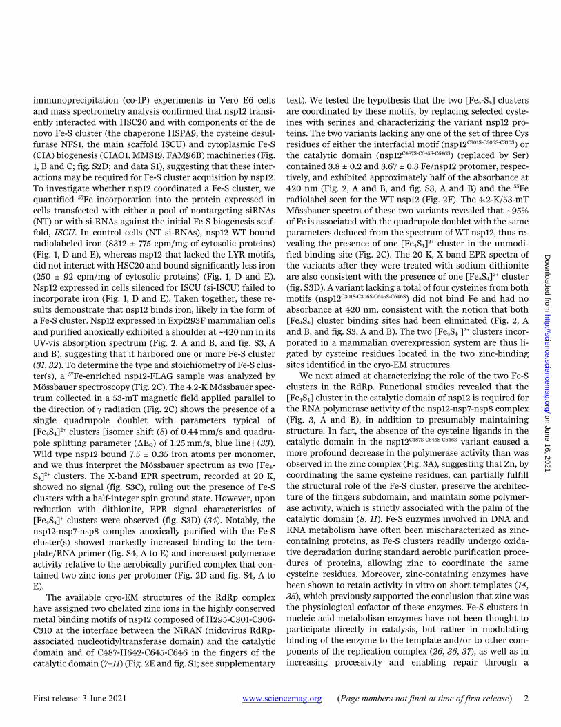
immunoprecipitation (co-IP) experiments in Vero E6 cells and mass spectrometry analysis confirmed that nsp12 transi-ently interacted with HSC20 and with components of the de novo Fe-S cluster (the chaperone HSPA9, the cysteine desul-furase NFS1, the main scaffold ISCU) and cytoplasmic Fe-S (CIA) biogenesis (CIAO1, MMS19, FAM96B) machineries (Fig. 1, B and C; fig. S2D; and data S1), suggesting that these inter-actions may be required for Fe-S cluster acquisition by nsp12. To investigate whether nsp12 coordinated a Fe-S cluster, we quantified 55Fe incorporation into the protein expressed in cells transfected with either a pool of nontargeting siRNAs (NT) or with si-RNAs against the initial Fe-S biogenesis scaf-fold, ISCU. In control cells (NT si-RNAs), nsp12 WT bound radiolabeled iron (8312 ± 775 cpm/mg of cytosolic proteins) (Fig. 1, D and E), whereas nsp12 that lacked the LYR motifs, did not interact with HSC20 and bound significantly less iron (250 ± 92 cpm/mg of cytosolic proteins) (Fig. 1, D and E). Nsp12 expressed in cells silenced for ISCU (si-ISCU) failed to incorporate iron (Fig. 1, D and E). Taken together, these re-sults demonstrate that nsp12 binds iron, likely in the form of a Fe-S cluster. Nsp12 expressed in Expi293F mammalian cells and purified anoxically exhibited a shoulder at ~420 nm in its UV-vis absorption spectrum (Fig. 2, A and B, and fig. S3, A and B), suggesting that it harbored one or more Fe-S cluster (31, 32). To determine the type and stoichiometry of Fe-S clus-ter(s), a 57Fe-enriched nsp12-FLAG sample was analyzed by Mössbauer spectroscopy (Fig. 2C). The 4.2-K Mössbauer spec-trum collected in a 53-mT magnetic field applied parallel to the direction of γ radiation (Fig. 2C) shows the presence of a single quadrupole doublet with parameters typical of [Fe4S4]2+ clusters [isomer shift (δ) of 0.44 mm/s and quadru-pole splitting parameter (ΔEQ) of 1.25 mm/s, blue line] (33). Wild type nsp12 bound 7.5 ± 0.35 iron atoms per monomer, and we thus interpret the Mössbauer spectrum as two [Fe4-S4]2+ clusters. The X-band EPR spectrum, recorded at 20 K, showed no signal (fig. S3C), ruling out the presence of Fe-S clusters with a half-integer spin ground state. However, upon reduction with dithionite, EPR signal characteristics of [Fe4S4]+ clusters were observed (fig. S3D) (34). Notably, the nsp12-nsp7-nsp8 complex anoxically purified with the Fe-S cluster(s) showed markedly increased binding to the tem-plate/RNA primer (fig. S4, A to E) and increased polymerase activity relative to the aerobically purified complex that con-tained two zinc ions per protomer (Fig. 2D and fig. S4, A to E).
The available cryo-EM structures of the RdRp complex have assigned two chelated zinc ions in the highly conserved metal binding motifs of nsp12 composed of H295-C301-C306-C310 at the interface between the NiRAN (nidovirus RdRp-associated nucleotidyltransferase domain) and the catalytic domain and of C487-H642-C645-C646 in the fingers of the catalytic domain (7–11) (Fig. 2E and fig. S1; see supplementary
text). We tested the hypothesis that the two [Fe4-S4] clusters are coordinated by these motifs, by replacing selected cyste-ines with serines and characterizing the variant nsp12 pro-teins. The two variants lacking any one of the set of three Cys residues of either the interfacial motif (nsp12C301S-C306S-C310S) or the catalytic domain (nsp12C487S-C645S-C646S) (replaced by Ser) contained 3.8 ± 0.2 and 3.67 ± 0.3 Fe/nsp12 protomer, respec-tively, and exhibited approximately half of the absorbance at 420 nm (Fig. 2, A and B, and fig. S3, A and B) and the 55Fe radiolabel seen for the WT nsp12 (Fig. 2F). The 4.2-K/53-mT Mössbauer spectra of these two variants revealed that ~95% of Fe is associated with the quadrupole doublet with the same parameters deduced from the spectrum of WT nsp12, thus re-vealing the presence of one [Fe4S4]2+ cluster in the unmodi-fied binding site (Fig. 2C). The 20 K, X-band EPR spectra of the variants after they were treated with sodium dithionite are also consistent with the presence of one [Fe4S4]2+ cluster (fig. S3D). A variant lacking a total of four cysteines from both motifs (nsp12C301S-C306S-C645S-C646S) did not bind Fe and had no absorbance at 420 nm, consistent with the notion that both [Fe4S4] cluster binding sites had been eliminated (Fig. 2, A and B, and fig. S3, A and B). The two [Fe4S4 ]2+ clusters incor-porated in a mammalian overexpression system are thus li-gated by cysteine residues located in the two zinc-binding sites identified in the cryo-EM structures.
We next aimed at characterizing the role of the two Fe-S clusters in the RdRp. Functional studies revealed that the [Fe4S4] cluster in the catalytic domain of nsp12 is required for the RNA polymerase activity of the nsp12-nsp7-nsp8 complex (Fig. 3, A and B), in addition to presumably maintaining structure. In fact, the absence of the cysteine ligands in the catalytic domain in the nsp12C487S-C645S-C646S variant caused a more profound decrease in the polymerase activity than was observed in the zinc complex (Fig. 3A), suggesting that Zn, by coordinating the same cysteine residues, can partially fulfill the structural role of the Fe-S cluster, preserve the architec-ture of the fingers subdomain, and maintain some polymer-ase activity, which is strictly associated with the palm of the catalytic domain (8, 11). Fe-S enzymes involved in DNA and RNA metabolism have often been mischaracterized as zinc-containing proteins, as Fe-S clusters readily undergo oxida-tive degradation during standard aerobic purification proce-dures of proteins, allowing zinc to coordinate the same cysteine residues. Moreover, zinc-containing enzymes have been shown to retain activity in vitro on short templates (14, 35), which previously supported the conclusion that zinc was the physiological cofactor of these enzymes. Fe-S clusters in nucleic acid metabolism enzymes have not been thought to participate directly in catalysis, but rather in modulating binding of the enzyme to the template and/or to other com-ponents of the replication complex (26, 36, 37), as well as in increasing processivity and enabling repair through a
on June 16, 2021
http://science.sciencemag.org/
Dow
nloaded from

First release: 3 June 2021 www.sciencemag.org (Page numbers not final at time of first release) 3
proposed charge transfer mechanism (38, 39). Consistent with the notion that zinc is likely not the physiological cofac-tor in several viral replicases that have so far been crystallized with chelated zinc ions, supplementation with zinc has been reported to inhibit replication in several cell culture models of viral infection (40–42). Loss of the [Fe4-S4] cluster ligated by H295-C301-C306-C310, which is located at the interface between the NiRAN and the catalytic domain of nsp12, had minimal effect on the RNA polymerase activity (Fig. 3, A and B). However, loss of this cluster profoundly diminished the interaction with the helicase nsp13 (Fig. 3, B and C), which is an essential component of the replication complex.
We attempted to exploit the sensitivity of Fe-S clusters to oxidative degradation (43) to prevent coronavirus replication in cell culture models. Previous studies have shown that a stable nitroxide, TEMPOL, was beneficial in two different an-imal models of human conditions through its ability to oxi-dize and disassemble the Fe-S cluster of cytosolic aconitase (IRP1), thereby converting it into the IRE-binding apo-form (44–46). RdRp isolated from Expi293F cells that had been treated with TEMPOL (Fig. 4A) had diminished absorbance at 420 nm relative to the complex isolated from untreated cells, indicative of loss of the Fe-S clusters of nsp12. Likewise, treatment with TEMPOL of the Fe-S cluster-containing pro-tein in vitro caused loss of absorbance in the same region (Fig. 4B). Either treatment resulted in loss of polymerase ac-tivity (Fig. 4, C to E). The TEMPOL treatment of cells did not impact the activities of several mitochondrial Fe-S enzymes, including the respiratory complexes and mitochondrial aco-nitase (ACO2), and the cytosolic Fe-S enzyme, DPYD (figs. S5, A to F, and S6, A to F), nor it caused any cytotoxicity at doses up to 5 mM (fig. S6G). TEMPOL treatment also did not affect the interactions of nsp12 with the components of the Fe-S and CIA biogenesis machinery from which nsp12 acquires its Fe-S clusters (fig. S7, A to D). We thus infer that TEMPOL di-rectly reacts with Fe-S clusters in RdRp, leading to their deg-radation.
In support of this mechanism of action, DEA/NO, a nitric oxide donor that readily reacts with Fe-S clusters to form di-nitrosyl complexes with diminished absorbance (47, 48), also inhibited the RdRp (Fig. 4E and fig. S8, A and B), although less effectively than TEMPOL. We found that TEMPOL was both a more potent RdRp inhibitor (fig. S9) and synergized with remdesivir (RDV) (fig. S10), a nucleoside analog that has been used to target the replication of SARS-CoV-2 (49). RDV was notably less effective against the Fe-S-RdRp than the zinc-RdRp (fig. S11).
Having demonstrated a strong inhibitory effect of TEMPOL on the activity of the RdRp of SARS-CoV-2, we asked whether TEMPOL might exhibit antiviral activity against live virus replication. Vero E6 cells were infected with the SARS-CoV-2 USA-WA1/2020 isolate in the presence of
increasing concentrations of TEMPOL (range 0.1 μM to 1 mM). TEMPOL exhibited a strong antiviral activity at concen-trations above 0.2 mM. Viral titers were reduced by more than 5 log10 in the presence of 0.4 mM TEMPOL, which is reported to have a CC50 greater than 100 mM (50). With its low cytotoxicity and known access to tissues relevant for COVID-19 infection (51, 52), our studies present a molecular basis for pursuing TEMPOL and other related stable nitrox-ides as potential SARS-CoV-2 therapies during active viral in-fection.
REFERENCES AND NOTES 1. J. F. Chan, S. Yuan, K.-H. Kok, K. K.-W. To, H. Chu, J. Yang, F. Xing, J. Liu, C. C.-Y. Yip,
R. W.-S. Poon, H.-W. Tsoi, S. K.-F. Lo, K.-H. Chan, V. K.-M. Poon, W.-M. Chan, J. D. Ip, J.-P. Cai, V. C.-C. Cheng, H. Chen, C. K.-M. Hui, K.-Y. Yuen, A familial cluster of pneumonia associated with the 2019 novel coronavirus indicating person-to-person transmission: A study of a family cluster. Lancet 395, 514–523 (2020). doi:10.1016/S0140-6736(20)30154-9 Medline
2. N. Chen, M. Zhou, X. Dong, J. Qu, F. Gong, Y. Han, Y. Qiu, J. Wang, Y. Liu, Y. Wei, J. Xia, T. Yu, X. Zhang, L. Zhang, Epidemiological and clinical characteristics of 99 cases of 2019 novel coronavirus pneumonia in Wuhan, China: A descriptive study. Lancet 395, 507–513 (2020). doi:10.1016/S0140-6736(20)30211-7 Medline
3. F. Wu, S. Zhao, B. Yu, Y.-M. Chen, W. Wang, Z.-G. Song, Y. Hu, Z.-W. Tao, J.-H. Tian, Y.-Y. Pei, M.-L. Yuan, Y.-L. Zhang, F.-H. Dai, Y. Liu, Q.-M. Wang, J.-J. Zheng, L. Xu, E. C. Holmes, Y.-Z. Zhang, A new coronavirus associated with human respiratory disease in China. Nature 579, 265–269 (2020). doi:10.1038/s41586-020-2008-3 Medline
4. P. V’kovski, A. Kratzel, S. Steiner, H. Stalder, V. Thiel, Coronavirus biology and replication: Implications for SARS-CoV-2. Nat. Rev. Microbiol. 19, 155–170 (2021). doi:10.1038/s41579-020-00468-6 Medline
5. D. G. Ahn, J. K. Choi, D. R. Taylor, J. W. Oh, Biochemical characterization of a recombinant SARS coronavirus nsp12 RNA-dependent RNA polymerase capable of copying viral RNA templates. Arch. Virol. 157, 2095–2104 (2012). doi:10.1007/s00705-012-1404-x Medline
6. L. Subissi, C. C. Posthuma, A. Collet, J. C. Zevenhoven-Dobbe, A. E. Gorbalenya, E. Decroly, E. J. Snijder, B. Canard, I. Imbert, One severe acute respiratory syndrome coronavirus protein complex integrates processive RNA polymerase and exonuclease activities. Proc. Natl. Acad. Sci. U.S.A. 111, E3900–E3909 (2014). doi:10.1073/pnas.1323705111 Medline
7. R. N. Kirchdoerfer, A. B. Ward, Structure of the SARS-CoV nsp12 polymerase bound to nsp7 and nsp8 co-factors. Nat. Commun. 10, 2342 (2019). doi:10.1038/s41467-019-10280-3 Medline
8. Y. Gao, L. Yan, Y. Huang, F. Liu, Y. Zhao, L. Cao, T. Wang, Q. Sun, Z. Ming, L. Zhang, J. Ge, L. Zheng, Y. Zhang, H. Wang, Y. Zhu, C. Zhu, T. Hu, T. Hua, B. Zhang, X. Yang, J. Li, H. Yang, Z. Liu, W. Xu, L. W. Guddat, Q. Wang, Z. Lou, Z. Rao, Structure of the RNA-dependent RNA polymerase from COVID-19 virus. Science 368, 779–782 (2020). doi:10.1126/science.abb7498 Medline
9. H. S. Hillen, G. Kokic, L. Farnung, C. Dienemann, D. Tegunov, P. Cramer, Structure of replicating SARS-CoV-2 polymerase. Nature 584, 154–156 (2020). doi:10.1038/s41586-020-2368-8 Medline
10. J. Chen, B. Malone, E. Llewellyn, M. Grasso, P. M. M. Shelton, P. D. B. Olinares, K. Maruthi, E. T. Eng, H. Vatandaslar, B. T. Chait, T. M. Kapoor, S. A. Darst, E. A. Campbell, Structural basis for helicase-polymerase coupling in the SARS-CoV-2 replication-transcription complex. Cell 182, 1560–1573.e13 (2020). doi:10.1016/j.cell.2020.07.033 Medline
11. W. Yin, C. Mao, X. Luan, D.-D. Shen, Q. Shen, H. Su, X. Wang, F. Zhou, W. Zhao, M. Gao, S. Chang, Y.-C. Xie, G. Tian, H.-W. Jiang, S.-C. Tao, J. Shen, Y. Jiang, H. Jiang, Y. Xu, S. Zhang, Y. Zhang, H. E. Xu, Structural basis for inhibition of the RNA-dependent RNA polymerase from SARS-CoV-2 by remdesivir. Science 368, 1499–1504 (2020). doi:10.1126/science.abc1560 Medline
12. G. D. Shimberg, J. L. Michalek, A. A. Oluyadi, A. V. Rodrigues, B. E. Zucconi, H. M. Neu, S. Ghosh, K. Sureschandra, G. M. Wilson, T. L. Stemmler, S. L. J. Michel,
on June 16, 2021
http://science.sciencemag.org/
Dow
nloaded from

First release: 3 June 2021 www.sciencemag.org (Page numbers not final at time of first release) 4
Cleavage and polyadenylation specificity factor 30: An RNA-binding zinc-finger protein with an unexpected 2Fe–2S cluster. Proc. Natl. Acad. Sci. U.S.A. 113, 4700–4705 (2016). doi:10.1073/pnas.1517620113 Medline
13. D. J. Netz, C. M. Stith, M. Stümpfig, G. Köpf, D. Vogel, H. M. Genau, J. L. Stodola, R. Lill, P. M. J. Burgers, A. J. Pierik, Eukaryotic DNA polymerases require an iron-sulfur cluster for the formation of active complexes. Nat. Chem. Biol. 8, 125–132 (2011). doi:10.1038/nchembio.721 Medline
14. G. D. Shimberg, J. D. Pritts, S. L. J. Michel, Iron-sulfur clusters in zinc finger proteins. Methods Enzymol. 599, 101–137 (2018). doi:10.1016/bs.mie.2017.09.005 Medline
15. A. R. Conlan, H. L. Axelrod, A. E. Cohen, E. C. Abresch, J. Zuris, D. Yee, R. Nechushtai, P. A. Jennings, M. L. Paddock, Crystal structure of Miner1: The redox-active 2Fe-2S protein causative in Wolfram Syndrome 2. J. Mol. Biol. 392, 143–153 (2009). doi:10.1016/j.jmb.2009.06.079 Medline
16. J. A. Imlay, Iron-sulphur clusters and the problem with oxygen. Mol. Microbiol. 59, 1073–1082 (2006). doi:10.1111/j.1365-2958.2006.05028.x Medline
17. N. Maio, T. A. Rouault, Outlining the complex pathway of mammalian Fe-S cluster biogenesis. Trends Biochem. Sci. 45, 411–426 (2020). doi:10.1016/j.tibs.2020.02.001 Medline
18. N. Maio, T. A. Rouault, “Delivery of iron-sulfur clusters to recipient proteins: the role of chaperone and cochaperone proteins” in Biochemistry, Biosynthesis and Human Diseases, vol. 2 of Iron-Sulfur Clusters in Chemistry and Biology, T. A. Rouault, Ed. (De Gruyter, ed. 2, 2017).
19. S. Klinge, J. Hirst, J. D. Maman, T. Krude, L. Pellegrini, An iron-sulfur domain of the eukaryotic primase is essential for RNA primer synthesis. Nat. Struct. Mol. Biol. 14, 875–877 (2007). doi:10.1038/nsmb1288 Medline
20. B. E. Weiner, H. Huang, B. M. Dattilo, M. J. Nilges, E. Fanning, W. J. Chazin, An iron-sulfur cluster in the C-terminal domain of the p58 subunit of human DNA primase. J. Biol. Chem. 282, 33444–33451 (2007). doi:10.1074/jbc.M705826200 Medline
21. S. Vaithiyalingam, E. M. Warren, B. F. Eichman, W. J. Chazin, Insights into eukaryotic DNA priming from the structure and functional interactions of the 4Fe-4S cluster domain of human DNA primase. Proc. Natl. Acad. Sci. U.S.A. 107, 13684–13689 (2010). doi:10.1073/pnas.1002009107 Medline
22. L. Sauguet, S. Klinge, R. L. Perera, J. D. Maman, L. Pellegrini, Shared active site architecture between the large subunit of eukaryotic primase and DNA photolyase. PLOS ONE 5, e10083 (2010). doi:10.1371/journal.pone.0010083 Medline
23. V. B. Agarkar, N. D. Babayeva, Y. I. Pavlov, T. H. Tahirov, Crystal structure of the C-terminal domain of human DNA primase large subunit: Implications for the mechanism of the primase-polymerase α switch. Cell Cycle 10, 926–931 (2011). doi:10.4161/cc.10.6.15010 Medline
24. A. G. Baranovskiy, Y. Zhang, Y. Suwa, N. D. Babayeva, J. Gu, Y. I. Pavlov, T. H. Tahirov, Crystal structure of the human primase. J. Biol. Chem. 290, 5635–5646 (2015). doi:10.1074/jbc.M114.624742 Medline
25. M. L. Kilkenny, G. De Piccoli, R. L. Perera, K. Labib, L. Pellegrini, A conserved motif in the C-terminal tail of DNA polymerase α tethers primase to the eukaryotic replisome. J. Biol. Chem. 287, 23740–23747 (2012). doi:10.1074/jbc.M112.368951 Medline
26. M. Girbig, A. D. Misiaszek, M. K. Vorländer, A. Lafita, H. Grötsch, F. Baudin, A. Bateman, C. W. Müller, Cryo-EM structures of human RNA polymerase III in its unbound and transcribing states. Nat. Struct. Mol. Biol. 28, 210–219 (2021). doi:10.1038/s41594-020-00555-5 Medline
27. G. Liu, D. Sil, N. Maio, W.-H. Tong, J. M. Bollinger Jr., C. Krebs, T. A. Rouault, Heme biosynthesis depends on previously unrecognized acquisition of iron-sulfur cofactors in human amino-levulinic acid dehydratase. Nat. Commun. 11, 6310 (2020). doi:10.1038/s41467-020-20145-9 Medline
28. N. Maio, A. Singh, H. Uhrigshardt, N. Saxena, W.-H. Tong, T. A. Rouault, Cochaperone binding to LYR motifs confers specificity of iron sulfur cluster delivery. Cell Metab. 19, 445–457 (2014). doi:10.1016/j.cmet.2014.01.015 Medline
29. N. Maio, T. A. Rouault, Iron-sulfur cluster biogenesis in mammalian cells: New insights into the molecular mechanisms of cluster delivery. Biochim. Biophys. Acta 1853, 1493–1512 (2015). doi:10.1016/j.bbamcr.2014.09.009 Medline
30. K. S. Kim, N. Maio, A. Singh, T. A. Rouault, Cytosolic HSC20 integrates de novo iron–sulfur cluster biogenesis with the CIAO1-mediated transfer to recipients.
Hum. Mol. Genet. 27, 837–852 (2018). doi:10.1093/hmg/ddy004 Medline 31. E. C. Duin, M. E. Lafferty, B. R. Crouse, R. M. Allen, I. Sanyal, D. H. Flint, M. K.
Johnson, [2Fe-2S] to [4Fe-4S] cluster conversion in Escherichia coli biotin synthase. Biochemistry 36, 11811–11820 (1997). doi:10.1021/bi9706430 Medline
32. S. Arragain, O. Bimai, P. Legrand, S. Caillat, J.-L. Ravanat, N. Touati, L. Binet, M. Atta, M. Fontecave, B. Golinelli-Pimpaneau, Nonredox thiolation in tRNA occurring via sulfur activation by a [4Fe-4S] cluster. Proc. Natl. Acad. Sci. U.S.A. 114, 7355–7360 (2017). doi:10.1073/pnas.1700902114 Medline
33. M. E. Pandelia, N. D. Lanz, S. J. Booker, C. Krebs, Mössbauer spectroscopy of Fe/S proteins. Biochim. Biophys. Acta 1853, 1395–1405 (2015). doi:10.1016/j.bbamcr.2014.12.005 Medline
34. W. R. Hagen, Biomolecular EPR Spectroscopy (CRC Press, 2020). 35. J. K. Barton, R. M. B. Silva, E. O’Brien, Redox chemistry in the genome: Emergence
of the [4Fe4S] cofactor in repair and replication. Annu. Rev. Biochem. 88, 163–190 (2019). doi:10.1146/annurev-biochem-013118-110644 Medline
36. D. Martin, A. Charpilienne, A. Parent, A. Boussac, B. D’Autreaux, J. Poupon, D. Poncet, The rotavirus nonstructural protein NSP5 coordinates a [2Fe-2S] iron-sulfur cluster that modulates interaction to RNA. FASEB J. 27, 1074–1083 (2013). doi:10.1096/fj.12-217182 Medline
37. J. Ter Beek, V. Parkash, G. O. Bylund, P. Osterman, A. E. Sauer-Eriksson, E. Johansson, Structural evidence for an essential Fe–S cluster in the catalytic core domain of DNA polymerase ϵ. Nucleic Acids Res. 47, 5712–5722 (2019). doi:10.1093/nar/gkz248 Medline
38. E. M. Boon, A. L. Livingston, N. H. Chmiel, S. S. David, J. K. Barton, DNA-mediated charge transport for DNA repair. Proc. Natl. Acad. Sci. U.S.A. 100, 12543–12547 (2003). doi:10.1073/pnas.2035257100 Medline
39. E. O’Brien, M. E. Holt, M. K. Thompson, L. E. Salay, A. C. Ehlinger, W. J. Chazin, J. K. Barton, The [4Fe4S] cluster of human DNA primase functions as a redox switch using DNA charge transport. Science 355, eaag1789 (2017). doi:10.1126/science.aag1789 Medline
40. A. J. te Velthuis, S. H. E. van den Worm, A. C. Sims, R. S. Baric, E. J. Snijder, M. J. van Hemert, Zn2+ inhibits coronavirus and arterivirus RNA polymerase activity in vitro and zinc ionophores block the replication of these viruses in cell culture. PLOS Pathog. 6, e1001176 (2010). doi:10.1371/journal.ppat.1001176 Medline
41. N. Kaushik, C. Subramani, S. Anang, R. Muthumohan, B. Shalimar, B. Nayak, C. T. Ranjith-Kumar, M. Surjit, Zinc salts block hepatitis E virus replication by inhibiting the activity of viral RNA-dependent RNA polymerase. J. Virol. 91, 91 (2017). doi:10.1128/JVI.00754-17 Medline
42. R. O. Suara, J. E. Crowe Jr., Effect of zinc salts on respiratory syncytial virus replication. Antimicrob. Agents Chemother. 48, 783–790 (2004). doi:10.1128/AAC.48.3.783-790.2004 Medline
43. T. A. Rouault, N. Maio, How oxidation of a unique iron-sulfur cluster in FBXL5 regulates IRP2 levels and promotes regulation of iron metabolism proteins. Mol. Cell 78, 1–3 (2020). doi:10.1016/j.molcel.2020.03.020 Medline
44. M. C. Ghosh, W.-H. Tong, D. Zhang, H. Ollivierre-Wilson, A. Singh, M. C. Krishna, J. B. Mitchell, T. A. Rouault, Tempol-mediated activation of latent iron regulatory protein activity prevents symptoms of neurodegenerative disease in IRP2 knockout mice. Proc. Natl. Acad. Sci. U.S.A. 105, 12028–12033 (2008). doi:10.1073/pnas.0805361105 Medline
45. M. C. Ghosh, D. L. Zhang, H. Ollivierre, M. A. Eckhaus, T. A. Rouault, Translational repression of HIF2α expression in mice with Chuvash polycythemia reverses polycythemia. J. Clin. Invest. 128, 1317–1325 (2018). doi:10.1172/JCI97684 Medline
46. G. Costain, M. C. Ghosh, N. Maio, A. Carnevale, Y. C. Si, T. A. Rouault, G. Yoon, Absence of iron-responsive element-binding protein 2 causes a novel neurodegenerative syndrome. Brain 142, 1195–1202 (2019). doi:10.1093/brain/awz072 Medline
47. S. R. Jaffrey, N. A. Cohen, T. A. Rouault, R. D. Klausner, S. H. Snyder, The iron-responsive element binding protein: A target for synaptic actions of nitric oxide. Proc. Natl. Acad. Sci. U.S.A. 91, 12994–12998 (1994). doi:10.1073/pnas.91.26.12994 Medline
48. E. M. Palmieri, M. Gonzalez-Cotto, W. A. Baseler, L. C. Davies, B. Ghesquière, N. Maio, C. M. Rice, T. A. Rouault, T. Cassel, R. M. Higashi, A. N. Lane, T. W.-M. Fan, D. A. Wink, D. W. McVicar, Nitric oxide orchestrates metabolic rewiring in M1 macrophages by targeting aconitase 2 and pyruvate dehydrogenase. Nat.
on June 16, 2021
http://science.sciencemag.org/
Dow
nloaded from
http://www.ncbi.nlm.nih.gov/entrez/query.fcgi?cmd=Retrieve&db=PubMed&list_uids=9305972&dopt=Abstract

First release: 3 June 2021 www.sciencemag.org (Page numbers not final at time of first release) 5
Commun. 11, 698 (2020). doi:10.1038/s41467-020-14433-7 Medline 49. M. Wang, R. Cao, L. Zhang, X. Yang, J. Liu, M. Xu, Z. Shi, Z. Hu, W. Zhong, G. Xiao,
Remdesivir and chloroquine effectively inhibit the recently emerged novel coronavirus (2019-nCoV) in vitro. Cell Res. 30, 269–271 (2020). doi:10.1038/s41422-020-0282-0 Medline
50. L. B. Oliveira, F. S. Celes, C. N. Paiva, C. I. de Oliveira, The paradoxical leishmanicidal effects of superoxide dismutase (SOD)-mimetic tempol in Leishmania braziliensis infection in vitro. Front. Cell. Infect. Microbiol. 9, 237 (2019). doi:10.3389/fcimb.2019.00237 Medline
51. A. P. Cotrim, F. Hyodo, K. Matsumoto, A. L. Sowers, J. A. Cook, B. J. Baum, M. C. Krishna, J. B. Mitchell, Differential radiation protection of salivary glands versus tumor by Tempol with accompanying tissue assessment of Tempol by magnetic resonance imaging. Clin. Cancer Res. 13, 4928–4933 (2007). doi:10.1158/1078-0432.CCR-07-0662 Medline
52. Y. Wang, B. Hai, L. Ai, Y. Cao, R. Li, H. Li, Y. Li, Tempol relieves lung injury in a rat model of chronic intermittent hypoxia via suppression of inflammation and oxidative stress. Iran. J. Basic Med. Sci. 21, 1238–1244 (2018). Medline
53. N. Maio, B. A. P. Lafont, D. Sil, Y. Li, J. M. Bollinger Jr., C. Krebs, T. C. Pierson, W. M. Linehan, T. A. Rouault, Supplementary data for “Fe-S cofactors in the SARS-CoV-2 RNA-dependent RNA polymerase are potential antiviral targets,” MassIVE (2021); https://doi.org/10.25345/C5MV60.
54. N. Maio, D. Ghezzi, D. Verrigni, T. Rizza, E. Bertini, D. Martinelli, M. Zeviani, A. Singh, R. Carrozzo, T. A. Rouault, Disease-causing SDHAF1 mutations impair transfer of Fe-S clusters to SDHB. Cell Metab. 23, 292–302 (2016). doi:10.1016/j.cmet.2015.12.005 Medline
55. N. Maio, K. S. Kim, A. Singh, T. A. Rouault, A single adaptable cochaperone-scaffold complex delivers nascent iron-sulfur clusters to mammalian respiratory chain complexes I–III. Cell Metab. 25, 945–953.e6 (2017). doi:10.1016/j.cmet.2017.03.010 Medline
56. N. D. Lanz, T. L. Grove, C. B. Gogonea, K.-H. Lee, C. Krebs, S. J. Booker, RlmN and AtsB as models for the overproduction and characterization of radical SAM proteins. Methods Enzymol. 516, 125–152 (2012). doi:10.1016/B978-0-12-394291-3.00030-7 Medline
57. O. Stehling, A. A. Vashisht, J. Mascarenhas, Z. O. Jonsson, T. Sharma, D. J. A. Netz, A. J. Pierik, J. A. Wohlschlegel, R. Lill, MMS19 assembles iron-sulfur proteins required for DNA metabolism and genomic integrity. Science 337, 195–199 (2012). doi:10.1126/science.1219723 Medline
58. S. Stoll, A. Schweiger, EasySpin, a comprehensive software package for spectral simulation and analysis in EPR. J. Magn. Reson. 178, 42–55 (2006). doi:10.1016/j.jmr.2005.08.013 Medline
59. R. A. Love, K. A. Maegley, X. Yu, R. A. Ferre, L. K. Lingardo, W. Diehl, H. E. Parge, P. S. Dragovich, S. A. Fuhrman, The crystal structure of the RNA-dependent RNA polymerase from human rhinovirus: A dual function target for common cold antiviral therapy. Structure 12, 1533–1544 (2004). doi:10.1016/j.str.2004.05.024 Medline
60. R. P. Cunningham, H. Asahara, J. F. Bank, C. P. Scholes, J. C. Salerno, K. Surerus, E. Münck, J. McCracken, J. Peisach, M. H. Emptage, Endonuclease III is an iron-sulfur protein. Biochemistry 28, 4450–4455 (1989). doi:10.1021/bi00436a049 Medline
61. S. J. Vollmer, R. L. Switzer, P. G. Debrunner, Oxidation-reduction properties of the iron-sulfur cluster in Bacillus subtilis glutamine phosphoribosylpyrophosphate amidotransferase. J. Biol. Chem. 258, 14284–14293 (1983). doi:10.1016/S0021-9258(17)43858-0 Medline
62. C. D. Stout, Crystal structures of oxidized and reduced Azotobacter vinelandii ferredoxin at pH 8 and 6. J. Biol. Chem. 268, 25920–25927 (1993). doi:10.1016/S0021-9258(19)74475-5 Medline
63. D. E. Gordon, G. M. Jang, M. Bouhaddou, J. Xu, K. Obernier, K. M. White, M. J. O’Meara, V. V. Rezelj, J. Z. Guo, D. L. Swaney, T. A. Tummino, R. Hüttenhain, R. M. Kaake, A. L. Richards, B. Tutuncuoglu, H. Foussard, J. Batra, K. Haas, M. Modak, M. Kim, P. Haas, B. J. Polacco, H. Braberg, J. M. Fabius, M. Eckhardt, M. Soucheray, M. J. Bennett, M. Cakir, M. J. McGregor, Q. Li, B. Meyer, F. Roesch, T. Vallet, A. Mac Kain, L. Miorin, E. Moreno, Z. Z. C. Naing, Y. Zhou, S. Peng, Y. Shi, Z. Zhang, W. Shen, I. T. Kirby, J. E. Melnyk, J. S. Chorba, K. Lou, S. A. Dai, I. Barrio-Hernandez, D. Memon, C. Hernandez-Armenta, J. Lyu, C. J. P. Mathy, T. Perica, K. B. Pilla, S. J. Ganesan, D. J. Saltzberg, R. Rakesh, X. Liu, S. B. Rosenthal, L.
Calviello, S. Venkataramanan, J. Liboy-Lugo, Y. Lin, X.-P. Huang, Y. Liu, S. A. Wankowicz, M. Bohn, M. Safari, F. S. Ugur, C. Koh, N. S. Savar, Q. D. Tran, D. Shengjuler, S. J. Fletcher, M. C. O’Neal, Y. Cai, J. C. J. Chang, D. J. Broadhurst, S. Klippsten, P. P. Sharp, N. A. Wenzell, D. Kuzuoglu-Ozturk, H.-Y. Wang, R. Trenker, J. M. Young, D. A. Cavero, J. Hiatt, T. L. Roth, U. Rathore, A. Subramanian, J. Noack, M. Hubert, R. M. Stroud, A. D. Frankel, O. S. Rosenberg, K. A. Verba, D. A. Agard, M. Ott, M. Emerman, N. Jura, M. von Zastrow, E. Verdin, A. Ashworth, O. Schwartz, C. d’Enfert, S. Mukherjee, M. Jacobson, H. S. Malik, D. G. Fujimori, T. Ideker, C. S. Craik, S. N. Floor, J. S. Fraser, J. D. Gross, A. Sali, B. L. Roth, D. Ruggero, J. Taunton, T. Kortemme, P. Beltrao, M. Vignuzzi, A. García-Sastre, K. M. Shokat, B. K. Shoichet, N. J. Krogan, A SARS-CoV-2 protein interaction map reveals targets for drug repurposing. Nature 583, 459–468 (2020). doi:10.1038/s41586-020-2286-9 Medline
64. A. K. Banerjee, M. R. Blanco, E. A. Bruce, D. D. Honson, L. M. Chen, A. Chow, P. Bhat, N. Ollikainen, S. A. Quinodoz, C. Loney, J. Thai, Z. D. Miller, A. E. Lin, M. M. Schmidt, D. G. Stewart, D. Goldfarb, G. De Lorenzo, S. J. Rihn, R. M. Voorhees, J. W. Botten, D. Majumdar, M. Guttman, SARS-CoV-2 disrupts splicing, translation, and protein trafficking to suppress host defenses. Cell 183, 1325–1339.e21 (2020). doi:10.1016/j.cell.2020.10.004 Medline
65. N. Schmidt, C. A. Lareau, H. Keshishian, S. Ganskih, C. Schneider, T. Hennig, R. Melanson, S. Werner, Y. Wei, M. Zimmer, J. Ade, L. Kirschner, S. Zielinski, L. Dölken, E. S. Lander, N. Caliskan, U. Fischer, J. Vogel, S. A. Carr, J. Bodem, M. Munschauer, The SARS-CoV-2 RNA-protein interactome in infected human cells. Nat. Microbiol. 6, 339–353 (2021). doi:10.1038/s41564-020-00846-z Medline
66. A. Fabregat, K. Sidiropoulos, G. Viteri, O. Forner, P. Marin-Garcia, V. Arnau, P. D’Eustachio, L. Stein, H. Hermjakob, Reactome pathway analysis: A high-performance in-memory approach. BMC Bioinformatics 18, 142 (2017). doi:10.1186/s12859-017-1559-2 Medline
ACKNOWLEDGMENTS
The authors are grateful to Dr. Steve Holland (NIAID) for insightful discussions and guidance, to NIAID for access to live viral testing, to Anamika Singh (NICHD) and Boma Fubara (NINDS) for technical assistance, to all members of the Rouault lab for feedback that greatly improved the quality of this work, to the Eunice Kennedy Shriver National Institute of Child Health and Human Development for support. Funding: This work was supported by the Intramural Research Program of the National Institutes of Health (to T.A.R.), by the Center for Cancer Research, National Cancer Institute (W.M.L), Division of Intramural Research, NIAID (T.C.P.), and award R35 GM-127079 from the National Institutes of Health (to C.K.). Author contributions: N.M., T.A.R. and W.M.L. conceived the project. N.M. designed the project, wrote the manuscript, and designed and performed most of the experiments, except as follows: SARS-CoV-2 viral infection assays (B.A.P.L.); EPR and Mossbauer spectroscopies (D.S.); mass spectrometry sample preparation and analysis (Y.L.). N.M., T.A.R., W.M.L., B.A.P.L., D.S., Y.L., J.M.B. Jr., T.C.P. and C.K. analyzed the data. T.A.R. supervised the study and wrote the manuscript. All authors revised the manuscript. Competing interests: Based on the implications of the discoveries reported here, N.M., T.A.R. and W.M.L. have filed a patent (application No. 63/193656). Data and materials availability: The mass spectrometry data have been deposited to MassIVE (53). All other data needed to evaluate the conclusions of the paper are present in the main text and supplementary materials. This work is licensed under a Creative Commons Attribution 4.0 International (CC BY 4.0) license, which permits unrestricted use, distribution, and reproduction in any medium, provided the original work is properly cited. To view a copy of this license, visit https://creativecommons.org/licenses/by/4.0/. This license does not apply to figures/photos/artwork or other content included in the article that is credited to a third party; obtain authorization from the rights holder before using such material.
on June 16, 2021
http://science.sciencemag.org/
Dow
nloaded from
http://www.ncbi.nlm.nih.gov/entrez/query.fcgi?cmd=Retrieve&db=PubMed&list_uids=2548577&dopt=Abstract
http://www.ncbi.nlm.nih.gov/entrez/query.fcgi?cmd=Retrieve&db=PubMed&list_uids=6315725&dopt=Abstract

First release: 3 June 2021 www.sciencemag.org (Page numbers not final at time of first release) 6
SUPPLEMENTARY MATERIALS science.sciencemag.org/cgi/content/full/science.abi5224/DC1 Materials and Methods Supplementary Text Figs. S1 to S11 References (54–66) MDAR Reproducibility Checklist Data S1 20 March 2021; accepted 28 May 2021 Published online 3 June 2021 10.1126/science.abi5224
on June 16, 2021
http://science.sciencemag.org/
Dow
nloaded from

First release: 3 June 2021 www.sciencemag.org (Page numbers not final at time of first release) 7
on June 16, 2021
http://science.sciencemag.org/
Dow
nloaded from
First release: 3 June 2021 www.sciencemag.org (Page numbers not final at time of first release) 8
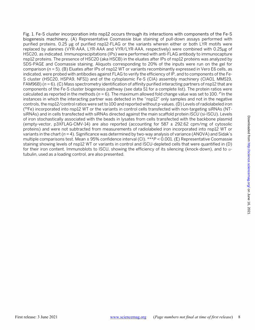
Fig. 1. Fe-S cluster incorporation into nsp12 occurs through its interactions with components of the Fe-S biogenesis machinery. (A) Representative Coomassie blue staining of pull-down assays performed with purified proteins. 0.25 μg of purified nsp12-FLAG or the variants wherein either or both LYR motifs were replaced by alanines (VYR-AAA, LYR-AAA and VYR/LYR-AAA, respectively) were combined with 0.25μg of HSC20, as indicated. Immunoprecipitations (IPs) were performed with anti-FLAG antibody to immunocapture nsp12 proteins. The presence of HSC20 (aka HSCB) in the eluates after IPs of nsp12 proteins was analyzed by SDS-PAGE and Coomassie staining. Aliquots corresponding to 20% of the inputs were run on the gel for comparison (n = 5). (B) Eluates after IPs of nsp12 WT or variants recombinantly expressed in Vero E6 cells, as indicated, were probed with antibodies against FLAG to verify the efficiency of IP, and to components of the Fe-S cluster (HSC20, HSPA9, NFS1) and of the cytoplasmic Fe-S (CIA) assembly machinery (CIAO1, MMS19, FAM96B) (n = 6). (C) Mass spectrometry identification of affinity purified interacting partners of nsp12 that are components of the Fe-S cluster biogenesis pathway (see data S1 for a complete list). The protein ratios were calculated as reported in the methods (n = 6). The maximum allowed fold change value was set to 100. P In the instances in which the interacting partner was detected in the “nsp12” only samples and not in the negative controls, the nsp12/control ratios were set to 100 and reported without p-values. (D) Levels of radiolabeled iron (55Fe) incorporated into nsp12 WT or the variants in control cells transfected with non-targeting siRNAs (NT-siRNAs) and in cells transfected with siRNAs directed against the main scaffold protein ISCU (si-ISCU). Levels of iron stochastically associated with the beads in lysates from cells transfected with the backbone plasmid (empty-vector, p3XFLAG-CMV-14) are also reported (accounting for 587 ± 292.62 cpm/mg of cytosolic proteins) and were not subtracted from measurements of radiolabeled iron incorporated into nsp12 WT or variants in the chart (n = 4). Significance was determined by two-way analysis of variance (ANOVA) and Sidak’s multiple comparisons test. Mean ± 95% confidence interval (CI). ***P < 0.001. (E) Representative Coomassie staining showing levels of nsp12 WT or variants in control and ISCU-depleted cells that were quantified in (D) for their iron content. Immunoblots to ISCU, showing the efficiency of its silencing (knock-down), and to α-tubulin, used as a loading control, are also presented.
on June 16, 2021
http://science.sciencemag.org/
Dow
nloaded from

First release: 3 June 2021 www.sciencemag.org (Page numbers not final at time of first release) 9
Fig. 2. Evidence for ligation of two Fe-S metal cofactors by nsp12. (A) UV-vis spectra of nsp12 WT or variants of the cysteine residues in the two metal ligating centers. (B) Representative Coomassie blue staining of purified nsp12 WT or variants analyzed in (A). (C) Mössbauer spectra of nsp12 WT and variants exhibited the parameters typical of [Fe4S4] clusters. For each of the two nsp12 cys-to-ser variants, approximately 95% of iron was still associated with a quadrupole doublet that matched parameters of WT nsp12. (D) RNA polymerase activity of anoxically purified RdRp ([Fe-S]-RdRp at 1 μM) and aerobically purified RdRp reconstituted with zinc and containing 2 zinc ions/protomer (Zn-RdRp at 1 μM) (n = 4). (E) Conserved zinc-binding motifs in SARS-CoV-2 nsp12 (PDB ID 7BTF) (8) rendered in the ribbon representation. H295-C301-C306-C310 ligate zinc at the interface between the NiRAN and the catalytic domain, whereas the C487-H642-C645-C646 residues ligate zinc in the catalytic domain. (F) Levels of radiolabeled iron (55Fe) incorporated into nsp12 WT or variants, as indicated. 55Fe content of nsp12 treated with the chelator EDTA is also reported to provide a control for the complete loss of 55Fe in the protein (n = 4). Significance was determined by two-way analysis of variance (ANOVA) and Sidak’s multiple comparisons test. Mean ± 95% CI. ***P < 0.001.
on June 16, 2021
http://science.sciencemag.org/
Dow
nloaded from

First release: 3 June 2021 www.sciencemag.org (Page numbers not final at time of first release) 10
Fig. 3. Fe-S cluster sites in nsp12 are important for activity and interactions with nsp13. (A) RNA polymerase activity of anoxically purified RdRp (all lanes except Zn-nsp12) (RdRp at 1 μM) and of aerobically purified and Zn-reconstituted RdRp containing 2 zinc ions/ protomer (3 technical replicates are shown, n = 4). (B) Schematic of the complex required for coronaviral replication (10), in which the two Fe-S clusters and their coordination spheres are highlighted. (C) Co-IP of nsp12 WT or variants recombinantly expressed in Vero E6 cells co-transfected with helicase nsp13 and accessory factors nsp7 and nsp8 (Strep II tagged) probed with antibodies against FLAG, Strep II or nsp13 (3 technical replicates are shown, n = 4).
on June 16, 2021
http://science.sciencemag.org/
Dow
nloaded from

First release: 3 June 2021 www.sciencemag.org (Page numbers not final at time of first release) 11
Fig. 4. The stable nitroxide TEMPOL potently inhibited the RdRp by causing disassembly of its Fe-S clusters and blocked viral replication in cell culture models of SARS-CoV-2 infection. (A) UV-vis spectra of nsp12 anoxically purified from Expi293F control cells and from cells treated with TEMPOL. (B) UV-vis spectra of as purified nsp12 and of purified nsp12 incubated with TEMPOL (1:2 ratio nsp12:TEMPOL) for 10 min. (C) RNA polymerase activity of the RdRp complexes anoxically purified from control and TEMPOL-treated Expi293F cells. (D) Representative Coomassie staining of the RdRp complexes analyzed for the activity in (C). (E) RNA polymerase assay of the RdRp complexes (at 1μM) anoxically purified from control, DEA/NO- or TEMPOL-treated Vero E6 cells, as indicated (n = 4). (F) Titer of infectious virus produced at 48 hours measured by TCID50 assay in Vero E6 cells infected with SARS-CoV-2 at a multiplicity of infection (moi) of 0.1 or 0.01 (n = 3).
on June 16, 2021
http://science.sciencemag.org/
Dow
nloaded from

Fe-S cofactors in the SARS-CoV-2 RNA-dependent RNA polymerase are potential antiviral targets
Linehan and Tracey A. RouaultNunziata Maio, Bernard A. P. Lafont, Debangsu Sil, Yan Li, J. Martin Bollinger Jr., Carsten Krebs, Theodore C. Pierson, W. Marston
published online June 3, 2021
ARTICLE TOOLS http://science.sciencemag.org/content/early/2021/06/02/science.abi5224
MATERIALSSUPPLEMENTARY http://science.sciencemag.org/content/suppl/2021/06/02/science.abi5224.DC1
REFERENCES
http://science.sciencemag.org/content/early/2021/06/02/science.abi5224#BIBLThis article cites 63 articles, 19 of which you can access for free
PERMISSIONS http://www.sciencemag.org/help/reprints-and-permissions
Terms of ServiceUse of this article is subject to the
is a registered trademark of AAAS.ScienceScience, 1200 New York Avenue NW, Washington, DC 20005. The title (print ISSN 0036-8075; online ISSN 1095-9203) is published by the American Association for the Advancement ofScience
No claim to original U.S. Government Works. Distributed under a Creative Commons Attribution License 4.0 (CC BY).Copyright © 2021 The Authors, some rights reserved; exclusive licensee American Association for the Advancement of Science.
on June 16, 2021
http://science.sciencemag.org/
Dow
nloaded from