Faculty of Resource Science and Technology Rapid ... Amplification of cDNA End of...ditambah pula...
24
Faculty of Resource Science and Technology Rapid Amplification of cDNA End of Metallothionein Gene during Necrosis of Plant Cells from Morinda citrifolia Lee Jong Jen Bachelor of Science with Honours (Biotechnology Resource) 2008
Transcript of Faculty of Resource Science and Technology Rapid ... Amplification of cDNA End of...ditambah pula...

Faculty of Resource Science and Technology
Rapid Amplification of cDNA End of Metallothionein Gene during Necrosis of Plant Cells from
Morinda citrifolia
Lee Jong Jen
Bachelor of Science with Honours
(Biotechnology Resource)
2008

Rapid Amplification of cDNA End of Metallothionein Gene during Necrosis of Plant Cells from
Morinda citrifolia
Lee Jong Jen
This Project is Submitted in Partial Fulfillment of the Requirements for the Degree of Bachelor
of Sciences with Honors
(Resource Biotechnology)
Faculty of Resource Science and Technology
UNIVERSITI MALAYSIA SARAWAK
2008

i
AKNOWLEDGEMENT
During the time when I was conducting my final year project, I had been supported and
encouraged by many people. Here, I would like to express my deepest gratitude to all those
that gave me precious help and support. Firstly, I would like to thank my supervisor, Dr.
Hairul Azman Roslan, who gave me guidance, support and encouragement on every part of
the project. He showed a great patience being the light on my way during the experiment and
dedicated his time to analyse problem faced during the time of facing difficulty. I wish him a
wonderful life. I am grateful when my beloved parents always stand by my side and
encouraging me of not giving up during my desperate moment.
I would like to send my thanks to all the master students in Genetic Engineering Lab
for giving me great help and advice during the my lab work. Working in lab with them was an
invaluable moment. I am also thankful to lab assistants for their kind cooperation throughout
the experimentation. Last but not least, I would also like to express my gratitude to my friends
for their kind help, support and advice for accomplishing my project.

ii
Rapid Amplification of cDNA End of Metallothionein Gene during Necrosis of Plant
Cells from Morinda citrifolia
Lee Jong Jen
Resource Biotechnology
Faculty of Resource Science and Technology
Universiti Malaysia Sarawak
ABSTRACT
The objective is to determine the presence of cysteine protease gene in the cDNA fragment being synthesized
and determining the efficiency of the 3’RACE PCR approach for amplification of cDNA end. Morinda citrifolia
is from the family Rubiaceae. It is the plant with great medicinal values. The property of this plant has not been
characterized thoroughly in previous time. Cysteine protease play important role in plant growth and
development in addition to protein degradation process in plant. Better understanding of the biochemical
properties will be attained through the study of cysteine protease gene. The amino acids sequences of this
enzyme govern the functional characteristic of this enzyme. Therefore, the termini of the cDNA encoding
cysteine protease are amplified through Rapid Amplification of cDNA ends (RACE). This experiment was to
amplify the 3’ terminal of the cDNA nucleotide sequence. The RNA extraction was the preliminary step of the
reverse transcription reaction to get the coding RNA fragment. First strand cDNA constructed was used for
further step of 3’ RACE by using gene specific primer and primers that enabled amplification 3’terminal
nucleotides. The PCR products were gel extracted and sent for sequencing. Purified PCR products were cloned
for further characterization and analysis of the isolated gene. The gene isolated was found to be similar to
metallothionein-like gene and drought stress-induced gene when analysed through Blast.
Key words: RACE, cDNA, RNA, PCR
ABSTRAK
Objektif eksperimen ini adalah menentukan kehadiran gen cysteine protease dalam fragmen cDNA yang
disintesiskan dan menentukan keberkesanan pendekatan 3’RACE yang diaplikasikan dalam mengamplifikasikan
terminal cDNA. Morinda citrifolia berasal daripada famili Rubiaceae. Ia merupakan tumbuhan yang
mempunyai nilai perubatan yang tinggi. Tumbuhan ini mempunyai banyak potensi yang belum ditemui lagi.
Cysteine protease memainkan peranan yang penting dalam pertumbuhan dan perkembangan tumbuhan
ditambah pula kepentingan enzim ini dalam prosess pemecahan protein. Pemahaman yang lebih mendalam
tentang ciri-ciri biokimia tumbuhan akan diperolehi dalam kajian terhadap cysteine protease. Enzim ini
mempunyai jujukan amino asid yang menentukan fungsinya. Oleh itu, bahagian terminal cDNA yang
merekodkan protein cysteine protease diamplifikasikan melalui teknik khas yang dipanggil RACE. Eksperimen
ini cuma mengamplifikasikan 3’ nukleotida terminal cDNA itu. Pemencilan RNA merupakan teknik pertama
sebelum langkah transkripsi berbalik untuk mendapatkan jujukan yang merekodkan gen berfungsi sahaja. cDNA
yang disintesiskan itu digunakan dalam 3’RACE dengan menggunakan primer yang spesifik kepada gen sasaran
dan primer yang membolehkan 3’nukleotida terminal cDNA itu diamplifikasikan. Hasil PCR akan dipencilkan
dan kemudian untuk penjujukan. Hasil PCR yang tulen diklonkan untuk pengelasan and analisis gen itu. Gen
fragmen itu didapati seiras dengan gen metallothionein dan gen berkaitan dengan tekanan kekurangan air
apabila dianalysiskan dengan Blast.
Kata kunci: RACE, cDNA, RNA, PCR

iii
TABLE OF CONTENTS
ACKNOWLEDGEMENT i
ABSTRACT ii
TABLE OF CONTENTS iii
LIST OF ABBREVIATIONS vi
LIST OF TABLES viii
LIST OF FIGURES ix
CHAPTER ONE INTRODUCTION
1
CHAPTER TWO LITERTURE REVIEW
2.1 Morinda citrifolia 3
2.1.1 Introduction of the plant 3
2.12 Researches on Morinda citrifolia 4
2.2 Leaf senescence 5
2.3 Cysteine protease 6
2.3.1 Roles of cysteine protease 6
2.3.2 Cysteine protease related researches 8
2.4 Metallothionein (Cysteine rich protein) 10
2.5 Drought stress-induced Gene 12
2.6 Rapid Amplification of cDNA Ends (RACE)
13
CHAPTER THREE MATERIALS AND METHODS
3.1 Plant leaf sampling 15
3.2 Isolation of RNA using CTAB method 15
3.3 Quantification of total RNA by spectrophotometer 16
3.4 DNase I treatment 16
3.5 Primer design 16
3.6 3’-Rapid Amplification of cDNA Ends 17
3.6.1 Reverse transcription to generate first strand 17
iv
cDNA templates
3.6.2 First round of the amplification process 18
3.6.3 Second round of the amplification process 18
3.7 Analysis of the 3’RACE PCR products 18
3.7.1 Gel electrophoresis of the PCR products 18
3.7.2 Gel extraction and sequencing 19
3.8 DNA sequence analysis 19
3.9 Cloning ligation 19
3.10 Competent cells preparation 20
3.11 Transformation of competent cells 20
3.12 DNA insert determination
21
CHAPTER FOUR RESULTS
4.1 Green healthy leaf sample 22
4.1.1 RNA extraction without the use of liquid
nitrogen
22
4.1.2 Quantitative and qualitative estimation of RNA
sample
23
4.1.3 PCR with elf 1α primer to determine the validity
of the RNA sample
24
4.1.4 First round PCR with gene specific primers 24
4.1.5 Second round PCR (partial nested PCR) 26
4.1.6 Gel extraction 26
4.1.7 PCR confirmation test for the presence of DNA
insert in the pGEMT vector
27
4.2 Necrosis leaf sample of Morinda citrifolia 28
4.2.1 RNA extraction with liquid nitrogen 28
4.2.2 Quantitative and qualitative quantification of
RNA sample
29
4.2.3 PCR with elf 1α primer to determine
the validity of RNA extracted
30

v
4.2.4 3’-Rapid amplification of cDNA ends 30
4.2.5 PCR reamplification 31
4.2.6 Gel excised of DNA fragment generated through
PCR
32
4.3 Sequencing result for green healthy leaf and Blast result 32
4.4 Sequencing result for necrosis leaf and Blast result 33
4.5 Comparison of the nucleotide sequences in DNA
fragment isolated from green healthy and necrosis leaves
34
CHAPTER FIVE DISCUSSIONS
5.1 RNA isolation 35
5.2 RNA gel analysis 36
5.2.1 Green healthy leaf sample 36
5.2.2 Necrosis leaf sample 37
5.3 Reverse transcription for first strand cDNA construction 37
5.4 Rapid Amplification of cDNA at the 3’end 38
5.5 Blast analysis of PCR amplicons 40
5.6 Cloning ligation and transformation process 41
5.7 PCR confirmation test for the inserted DNA 42
5.8 Metallothionein 43
5.9 Factors contributing to the isolation of non-target genes
from the plant sample
44
CHAPTER SIX CONCLUSIONS AND RECOMMENDATIONS 47
REFERENCES 48
APPENDICES
55

vi
LIST OF ABBREVIATIONS
oC Degree Celsius
% Percentage
bps Base pairs
cDNA Complementary DNA
CTAB Hexadecyltrimethylammonium bomide
DEPC Diethylpyrocarbonate
DMSO Dimethyl sulfoxide
DNA Deoxyribonucleic Acid
dNTP Deoxynucleotide triphosphates
dT Deoxythymidine
EDTA Ethylenediaminetretraacetate
GSP Gene specific primer
IPTG Isopropyl-β-D-thiogalactoside
kb Kilobase pairs
KCl Potassium chloride
LB Luria-Bertani Broth
LiCl Lithium chloride
µl Microlitre
µg Microgram
MgCl2 Magnesium Chloride
Min Minutes
ml Millilitre
mM MilliMolars
MMuLV-RT Moloney Murine Leukemia Virus Reverse
Transcriptase
mRNA Messenger Ribonucleic Acid
NCBI National Center for Biotechnology Information
OD Optical density
PCR Polymerase chain reaction
vii
PEG Polyethylene glycol
pmol Picomole
Poly (A) Poly adenosine
PVP Polyvinylpyrrolidone
RACE Rapid Amplifcation of cDNA Ends
RNase Ribonuclease
RNasin Ribonuclease Inhibitor
rpm Revolution per time
rRNA Ribosomal ribonucleic acid
RT Reverse transcription
RT PCR Reverse transcription polymerase chain reaction
TAE Tris-acetate EDTA electrophoresis buffer
U Unit
UV Ultraviolet
V Volts
X-gal 5-bromo-4-chloro-3-indoxyl-β-D-
galactopyranoside

viii
LIST OF TABLES
Table Page
1 The types of primers used for the experiments 17
2 Universal primers used in PCR confirmation test 21
3 Results of the measurement of extracted RNA from green healthy leaf
using spectrophotometer
23
4 Results of the measurement of extracted RNA from necrosis leaf using
spectrophotometer
29

ix
LIST OF FIGURES
Figure Page
1 RNA extraction using green healthy leaf 23
2 Electrophoresis of PCR product using ef1α primers 24
3 First round PCR using 3’cysprot and outer primers 25
4 Electrophoresis of the repeated PCR product using 3’cysprot and outer
primer
25
5 Second round PCR using 3’cysprot and inner primer 26
6 Gel extraction of PCR product from 3’cysprot and inner primer PCR 27
7 PCR confirmation tests using primers SP6 and T7 27
8 RNA extraction from necrosis leaf sample with 2 % of ß-mercaptoethanol 28
9 RNA extraction from necrosis leaf sample with 2.5 % ß-mercaptoethanol 29
10 Electrophoresis of PCR product using ef1α primers 30
11 PCR with 3’cysprot and inner primer using first strand cDNA as the
template
31
12 Reamplification of the PCR products 31
13 Gel extracted fragment from PCR products 32
14 Nucleotide sequence of the isolated gene using 3’cysprot primer from
green healthy leaf sample
33
15 Nucleotide sequence of the isolated gene using 3’cysprot primer from
necrosis leaf sample
33
16 Blast analysis result for the gene isolated from green healthy leaf sample 55
17 Blast analysis result for the gene isolated from green healthy leaf sample 55
18 Blast analysis for isolated gene from necrosis leaf sample 56
19 Blast analysis for isolated gene from necrosis leaf sample 56
20 Pairwise alignment result 57

1
CHAPTER 1
INTRODUCTION
Morinda citrifolia plant is from the family Rubiaceae or its subfamily is Rubioideae (Nelson,
2006). It has been called Noni in Hawaii, and Mengkudu in Malaysia (Zin et al., 2004). Most
of the plant parts have medicinal values such as roots, flowers, seeds, leaves, barks and fruits
(Wang et al., 2002). Since M. citrifolia has a wide range of medicinal properties, this plant
species is beneficial for used as research purposes. Most of the traditional and modern
applications of this plant in vast number of ailments have yet to be scientifically proven.
However, there were still several researches that had been done on the Noni fruit and root
extracts that demonstrated the medicinal value of the plant. Besides, Noni has gained
significance economic importance in the world today during recent years from the various
types of health and cosmetic products which had been produced from leaves and fruits
particularly (Nelson, 2006).
The study was conducted to have a more in depth understanding of Noni plant, by
constructing cDNA ends encoding cysteine protease through Rapid Amplification of cDNA
ends (RACE) method. There is still no published paper on the construction of cDNA encoding
cysteine protease enzyme from M. citrifolia. Research had found that cysteine protease has
multifunctional role. Cysteine protease also known as thiol proteases has important role
during plant growth and development as well as programmed cell death (Grudkowska and
Zagdanska, 2004). Since cysteine protease is very important in governing plant metabolism
from stress respond, senescing, causing cell death, production of new proteins and even
pathogenic resistant of plant, isolation of cysteine protease gene from M. citrifolia can be used
for further research such as recombinant DNA technology. Complete sequence of cysteine

2
protease must be obtained through RACE before other research work related to cysteine
protease can be performed.
RNA extraction was done since RNA only contains the coding regions of the gene
encoding cysteine protease protein. Modified CTAB method from the method developed by
Zeng and Yang (2002) was practised suitable for M. citrifolia RNA isolation (Tan and Roslan,
2006). The modifications being made include using chloroform to remove all the
polysaccharides and secondary metabolites contaminants instead of using chloroform-isoamyl
alcohol solution and the incubation period for precipitation of nucleic acid was four hours and
not overnight incubation as in Zeng and Yang (2002) method.
RACE method is the common method applied to obtain complete sequence of specific
gene by constructing the complete cDNA ends as traditional method applied such as the
common process of RT-PCR usually result in only amplification of partial cDNA fragments.
Besides, traditional method of obtaining gene specific sequence from the cDNA libraries
through hybridization with radioisotope-labeled probes was unsuccessful and often includes
intron sequence. RACE protocols can be used to generate specific gene in unlimited number
of clones in short time compared to the traditional method that the researchers had to screen
libraries with large number of clones which even caused more tedious and laborious work
(Wang and Young, 2003).
The objectives of the study were to determine the presence of cysteine protease gene
in the cDNA fragments constructed, determining the efficiency of the PCR approach for
amplification of cDNA ends and extract of RNA from M. citrifolia leaf.

3
CHAPTER 2
LITERATURE REVIEW
2.1 Morinda citriflolia
2.1.1 Introduction of the plant
Morinda citriflolia plant is native to Southeast Asia (Indonesia) and Australia. The
distributions of the plant are Eastern Polynesia such as Hawaii, Melanesia, and Western
Polynesia such as Tonga, Indonesia, Australia and Malaysia. Noni plant can tolerate for an
extremely wide range of environmental conditions including harsh environments such as
basaltic lava flows. The plant also grows well in infertile, acidic, alkaline soils and relatively
dry areas or low areas close to shorelines. It is the first plant colonizes harsh areas or lava
flows. M. citrifolia plant can be found in brackish tide pools near coast, in limestone soils,
coral atolls, coconut plantation, waste areas or native forest of 0-300m (Nelson, 2006).
The plant is generally an evergreen tree or shrub and will usually grow from 3 to 10
meter in heights when achieved maturity. The corolla of the flower is white in colour and
lobed which is about 7-9mm long. The leaves grow in apposite direction and pinnately veined
(Wang et al., 2002). Different varieties of Noni plant exist due to different leaf morphology.
The leaves can be rounded, elliptic or long and strap-like (McClatchey, 2002). Noni plant has
fruit which is yellowish in colour and will become soft and fetid when ripe (Nelson, 2006)
with some varieties are odorless and some varieties have strong butyric acid smell
(McClatchey, 2002). The surface of the fruit is lumpy covered by polygonal-shaped sections
(Wang et al., 2002). The fruits contain seed which are triangular in shape, reddish brown and
have air chamber (Wang et al., 2002) and enable the seeds to retain viability even after

4
floating in water surface for months (Nelson, 2006). The root or bark part of the plant has
been used by the Hawaiian as dye in painting on clothes (McClatchey, 2002).
Olden healers such as the Rotuman and Hawaiian gave treatment and prescription
based on the M. citrifolia immature fruits or mature leaves as the main ingredients
(McClatchey, 2002). According to the traditional treatments and scientific researches over the
years, Noni is the plant with great botanical medication value. Malaysians used heated leaves
of Noni plant to treat coughs, splenomegaly, nausea, fever and bacterial infection by placing
them onto the infected area (Zaidan et al., 2005).
2.1.2 Researches on Morinda citrifolia
According to Liu et al. (2001), two novel glycosides known as 6-O-(b-D-glucopyranosyl)-1-
O-octanoyl-b-D-glucopyranose and asperusidic acid which was extracted from Noni fruit
juice had chemopreventive effect. This indicated that the noni juice involved in the
mechanism of tumor suppression. Noni juice had blocked AP-1 transactivation activity which
had important role in tumorigenesis.
Zin et al. (2004) reported that chromatographic fractions for extraction from the root,
fruit and leaf demonstrated considerably high antioxidative activity. Phenolic compounds with
antioxidative activity were potent scavengers of free radicals and useful for prevention of
arteriosclerosis, cancer, diabetis and arthritis. There was a possibility that the Noni associated
arthritis and diabetis diseases relieving was caused by the existence of certain compounds
such as scopoletin, nitric oxide, alkaloids and sterols in plant (Blanco et al., 2005).
Research had been done by Komaraiah et al. (2005) on suitability of certain elicitors,
ultrasonication and sucrose feeding to enhance the accumulation of anthraquinones in M.
citrifolia suspension culture. Anthraquinones were stored in root part of the plant species as

5
glycosides and aglycones. The use of root in herbal preparation for various disease treatments
showed the importance of the anthraquinone compounds production in M. citrifolia.
Polyunsaturated fatty acids such as linoleic acid, α-linolenic acid, arachidonic acid, methyl
jasmonate, salicylate, and sodium nitroprusside had shown to be effective to stimulate
anthraquinones production in M. citrifolia. The combination of sucrose and methyl jasmonate
gave even higher yield to anthraquinone production which was synergistic. In addition,
treatment of the suspension culture with ultrasonocation had increased the accumulation of
anthraquinones by four-fold. The anthraquinones in fruit was not enough in terms of quantity
to cause liver tissue damage (West et al., 2006).
In addition, study done by Zaidan et al. (2005) showed that M. citrifolia leaves had
antibacterial activity towards gram positive Staphylococcus aureus and Methicillin Resistant
Staphylococcus (MRSA) but not to Escherichia coli and Klebsiella pneumoniae. Acute
toxicity test on the Noni fruit crude extracts was found to be greater than the minimum criteria
for nontoxic status.
2.2 Leaf senescence
Leaf senescence is the condition occurred when there are changes in cell structure,
metabolism, and gene expression (Gan and Amasino, 1997). Senescence that occurs in higher
plants gone through programmed cell death (Lim et al., 2003). Examples of enzymes involved
in senescence are cysteine protease (Beyene et al., 2006), metallothionein (Buchanan-
Wolleston, 1994), ubiquitin specific protease, kinases (Navapbour et al., 2007) and lipases
(Ryu and Wong, 1995 cited in Gan and Amasino, 1997). There are two types of senescence
which are replicative senescence and post-mitotic senescence. Replicative senescence is the
loss of plant capacity for further cell division during aging while post-mitotic senescence
6
refers to a degenerative process that occurs after cell differentiation and which leads to cell
death (Lim et al., 2003).
Leaf senescence is regulated by environmental factors such as extremes temperature,
drought stress, ozone, nutrient deficiency, pathogen infection, wounding and shading as well
as internal factors such as age, reproductive development and levels of phytohormone. The
most significant change in the cell which cause symptom of leaf yellowing is breakdown of
chloroplast whereby chlorophyll and macromolecules catabolism occurred replacing carbon
assimilation caused the photosynthetic rate drops below certain threshold. These changes are
caused by changes in gene expression including nuclear gene expression. Nutrients released
from the macromolecules are translocated to other young leaves, developing plant organs,
reproductive organs or storage tissues. A group of senescence-associated genes that are up-
regulated during senescence were found and the regulation of the genes may be involving
multiple pathways that form a regulatory network (Gan and Amasino, 1997).
2.3 Cysteine protease
2.3.1 Roles of cysteine protease
Vierstra (1989) reported that cysteine protease is important for protein degradation during
seed germination in seed storage protein. Hydrolysis of the protein in seed storage will
provide amino acids for new protein synthesis in the growing seedling. Callis (1995) found
that selective degradation of protein in non-senescing organ in turn will serve to reduce the
toxic effects of inactivated, denatured, unassembled and abnormal proteins. Furthermore,
cysteine protease is involved in senescence and programmed cell death. Programmed cell
death is executed by metacaspases during plant embryogenesis and depended upon its
7
cysteine-dependent arginine-specific proteolytic activity (Bozhkov et al., 2005). Programmed
cell death is involved in the elimination of proembryogenentic masses and embryo suspensor.
Cysteine protease participates in both anabolic and catabolic processes. Cysteine
protease is synthesized at polyribosomes as large precursor that consists of short N-terminal
and much longer C-terminal propeptides. It is produced in necrosis leaves that contain dead
cells and in seed undergo germination. The plant cysteine proteases are located in vacuoles
and in the cell wall. It is endopeptidase with a cysteine residue in their active site. Iodoacetate,
iodoacetamide, and E64 are the active site inhibitors used to identify cysteine protease.
Activation of the enzymes by thiol compounds is also used to identify this enzyme
(Grudkowska and Zagdanska, 2004). Most of the cysteine protease that had been studied
showed acidic pH optima. Most plant cysteine protease belongs to the papain-like (C1) and
legumain (C3) family (Ghosh et al., 2007). Five other members of the cysteine proteases that
have been found are caspases (family C14), calpains, the calcium-dependent proteases (family
C2), ubiquitin C-terminal hydrolases (family C12) and ubiquitin-specific proteinases (Vierstra,
2003 cited in Grudkowska and Zagdanska, 2004).
According to Grudkowska and Zagdanska (2004), cysteine protease has
multifunctional roles with its activity in mature non-senescing organs up to 30% of total
proteolytic activity. However, different internal and external stimuli will cause the enzyme’s
activity rise up to 90% of the total proteolytic activity. Proteolysis in plant by cysteine
protease is a highly regulated process. The ability of the plant to modulate protein levels is
necessary for the plant to adapt to changes in environment and to internal developmental
signals. Besides, the enzymes also concern with the storage protein deposited in the cotyledon
mesophyll of dicotyledonous plants and cereals endosperm tissue. Water deficit stress had
8
caused changes in proteolytic activity of ten genotypes of spring wheat of Triticum aestivum L.
(Folinova et al., 2000).
2.3.2 Cysteine protease related researches
Cysteine protease has been found in plants, animals, bacteria and eukaryotic microorganisms.
Early resistant responses of potato towards Phytophthora infestans had enabled the isolation
of cDNA clones encoding cysteine protease from this plant. Both the nucleotide and deduced
amino acid sequences showed high homology to tomato cysteine protease and cathepsins in
animals particularly cathepsin K. Comparison of the cysteine protease domains in potato and
tomato cysteine protease as well as rice oryzain α uncovered that amino acids important for
structural and functional activity of cysteine protease were conserved among them (Avrova et
al., 1999).
Harrak et al. (2001) had done research on elucidating the nature of the protein (TDI-
65) resulted from drought-stresses tomato (Lycopersicon esculentum) plants. A cDNA clone
(tdi-65) was isolated which had high homology with known cysteine protease such as
actinidin and papain. Amino acids important for the structure and function of the cysteine
proteases are conserved in tdi-65 protein. The size of the tdi-65 protein was approximately the
same as TDI-65. The protein was found to be located in nuclei, chloroplasts and some leaf
cytoplasmic regions. Results from Northern blot analysis showed that tdi-65 mRNA is 10-fold
higher in drought-stressed plants when compared to control and rewatered plants. The result
suggested the role of tdi-65 in protein degradation localized in chloroplast and nuclei during
drought stress condition.
According to Kusaka et al. (2004), cysteine protease is the important component in
causing the changes in morphological character of isolated nuclei in plants by using oat

9
(Avena sativa L.) for the research. In this experiment, cysteine protease was shown to be
involved in executive phase of oat apotosis by inducing DNA laddering during programmed
cell death. Moreover, cysteine protease was indicated to be essential for nuclear DNA
fragmentation in the presence of nuclease.
According to Asp et al. (2004), three groups of genes comprise of two full length and
three partial cysteine protease genes from white clover (Trifolium repens) were expressed in
nodules, primarily the senescence zone but not in roots, leaves, petioles and internodes. The
identified cysteine protease genes from Trifolium repens were shown to be highly
homologous to cysteine protease gene from Astragalus sinicus, Alnus glutinosa and Medicago
truncatula. On the other hands, the expression patterns of cysteine protease in sweet potato
(Ipomoea batatas) storage roots have been reported by Dong et al. (2005). The expression of
cysteine protease was found to be highest in storage roots compared to sprouted roots, green
leaves and flowers.
Sheokand et al. (2005) demonstrated the important function and localization of
cysteine protease (Cyp15a) in plant by modifying the gene encoding cysteine protease
resulted in the production of non-functional protein. The transgenic lines being used is
Medicago truncatula (R108-1). There was strong expression of promoter Cyp15 in
cotyledonary leaves, senescent leaves and root nodules and its increased expression in the
present of 0.6M mannitol and increased concentration of NaCl which indicated the role of
Cyp15 as stress-inducible gene. Suppression of cysteine protease expression demonstrated no
regeneration of plant and no seed germination occurred. Besides, plant range from slow
growing mode and fatal phenotype to leafy and late senenscing kinds which demonstrated
function of Cyp15a cysteine protease in seed germination and stress adaptation of plant.

10
Cysteine protease was induced to be expressed in rice (Oryza sativa L.) infected with
blast fungus, Magnaporthe grisea. Oryzain α-A induced gene was not involved in rice
resistant towards blast fungus but may be a factor for pathogen-induced necrosis. Furthermore,
expression of oryzain α-A can be induced through wounding, ultraviolet radiation and
treatment with salicylic acid (Fu et al., 2007).
According to Yamauchi (2007), gibberellins (GA) and brassinosteroids (BR)
overcome the repression of abscisic acid (ABA) on the expression of germination-induced
cysteine protease genes (GICPs) in cotyledons of common bean seeds. Result showed that the
mRNAs levels for all the cDNAs except two of the cDNAs code for papain-like proteases
could not be detected in cotyledons that had been treated with 10µM abscisic acids. The
mRNA levels increased after treatment with gibberellins or brassinosteroids. Result from the
experiment suggested that gibberellins and brassinosteroids are essential for the expression of
germination-induced cysteine protease genes in bean seeds or cereal seeds.
2.4 Metallothionein (Cysteine rich protein)
Metallothionein is a low molecular weight cysteine-rich protein. Metallothionein in plants and
animals has metal binding properties within the cells with the potential function in metabolism
and homeostasis of different metal ions. The metal binding properties play essential role in
homeostasis of metal ions such as zinc and copper ions and detoxification of metal ions such
as cadmium, zinc and copper ions (Jin et al., 2006). There was also report claim that the
binding with metal ion protected protein from proteolytic degradation. Presently, there are 3
classes of metallothionein-like proteins found in terms of Cys residues locations which are
class I, II and III and Class II metallothionein can be further categorized into four types which
are type 1, 2, 3 and Ec (early cystein labeled) protein according to the amino acid sequence in

11
cysteine domains (Cobbett and Goldbrough, 2002). Different plants with metallothionein
proteins from different gene family expressed in different organs. For example,
metallothionein expressed in soya bean roots and abundantly in leaves while in pea plants, the
metallothionein was abundant during growth of the root organs in hydroponic culture
(Robinson et al., 1993).
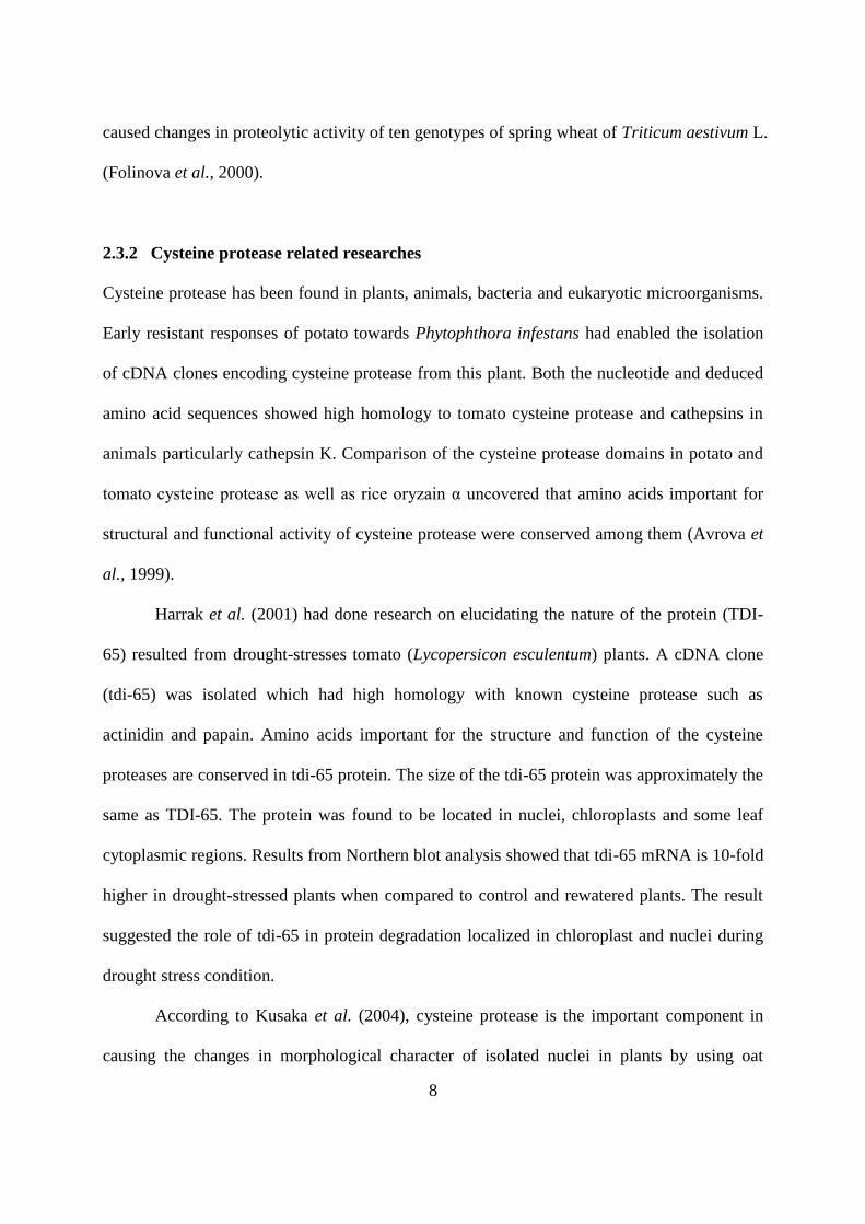
According to Robinson et al. (1993), there had been different theories regarding the
biosynthesis of Class III metallothionein. However, the theories were mainly on the synthesis
of Class III metallothionein from glutathione. Structural similarities between glutathione and
Class III metallothionein had gave rise to the possibility that Class III metallothionein
synthesized from glutathione. Synthesis of the metallothionein from glutathione by enzyme γ-
glutamylcysteine dipeptidyl transpeptidase had been reported in Silence cucubalus cell
suspension. This biosynthesis mechanism needed two glutathione molecules or one
glutathione with previously produced metallothionein. There was also theory regarding the
inhibition of glutathione biosynthesis by cadmium ions in maize caused accumulation of γ-
glutamylcysteine which led to the synthesis of Class III metallothionein. Another alternative
pathway for the biosynthesis was the polymerization of γ-glutamylcysteine by the transfer of
γ-glutamylcysteine from glutathione to another (γ-glutamylcysteine)n to produce
(glutamylcysteine)n+1 and glycine. Figure below shows the illustration of this pathway.
12
Adapted from Robinson et al. (1993). Class III metallothionein shown in
the figure above contain multiple polypeptide molecules and some of the
complexes contain inorganic S2-
in the metal core. HMTl gene located in
the vacouler membrane which is required for accumulation of
metallothionein in vacuoles. Red circles, cysteine residues; black circles,
glutamate residues; white circles, glycine residues
2.5 Drought stress-induced gene
The decreased content of water in the soil had caused changes in gene expression patterns
which increased the rate of transcription of a specific gene in respond to drought stress (Bray,
2002). Drought stress is the condition when the water potential around the cell lowered and
there is reduced cell growth (Cramer and Bowman, 1994). The gene expressed during water
deficit condition might function to promote the survival of the plant (Bray, 2002). Examples
of drought-stress induced gene are cysteine protease (Harrak et al., 2001), metallothionein-
like protein (Jin et al., 2006) and glutathione-S-transferase (Reymond et al., 2000). The gene
expression was induced by a complex series of signal transduction events not fully defined.
Abscisic acid is one of the signal molecules that might confer adaptation function. There are
11 functional categories of genes induced by drought stress such as cell defence, cell death,
ageing, protein metabolism and ionic homeostasis (Bray, 2002). Other categories of drought
stress induced gene have protein of unknown function. Water deficit condition has decreased
13
the availability of the mineral ions in soil (Alam, 1994). The plant adapt to the drought stress
by a few modifications in its structures and processes such as stomatal response and osmotic
adjustments (Pugnaire et al., 1994).
There are three mechanisms of drought resistance which are drought avoidance,
drought escape and drought tolerance depending on the plant response to drought (Mitra,
2001). Plant showed drought avoidance when it is able to maintain high water potential in
water depletion soil (Mitra, 2001) or when the plant restrict its activity until water content
return to normal (Pugnaire et al., 1994). Drought escape is demonstrated when the plant
complete its life cycle before serious water deficit condition of the soil occur whereas drought
tolerance is the ability of the plant to survive in water depletion soil by maintaining low water
potential in the tissues (Mitra, 2001) through decreasing osmotic potential (Pugnaire et al.,
1994).
2.6 Rapid Amplification of cDNA ends (RACE)
Rapid Amplification of cDNA Ends (RACE) encoding cysteine protease is a type of
polymerase chain reaction (PCR) based technique which was developed to enable the cloning
of full-length cDNA 5'- and 3'-ends after obtaining partial cDNA fragment (Schaefer, 1995).
There are two general RACE strategies that are 3’RACE which amplifies 3’ cDNA ends and
5’RACE which amplifies 5’cDNA end sequences (Wang and Young, 2003). This method has
been used widely to determine the 5’ and 3’ terminal nucleotide sequences of the genes (Shi
and Jarvis, 2006).
First round of the RACE would usually produced a high background of nonspecific
RT-PCR products which necessitates the second round of RACE with another set of nested
gene specific primers to enable the construction of cDNA encoding cysteine protease with