FACTORS ASSOCIATED WITH LATE …wiredspace.wits.ac.za/jspui/bitstream/10539/14374/1...FACTORS...
56
FACTORS ASSOCIATED WITH LATE PRESENTATION OF GLAUCOMA: A STUDY IN A BLACK SOUTH AFRICAN POPULATION. Dr Philip Kraukamp A research report submitted to the Faculty of Health Sciences, University of the Witwatersrand, Johannesburg, in partial fulfillment of the requirements for the degree of Master of Medicine in the branch of Ophthalmology. Johannesburg, 2013
Transcript of FACTORS ASSOCIATED WITH LATE …wiredspace.wits.ac.za/jspui/bitstream/10539/14374/1...FACTORS...
FACTORS ASSOCIATED WITH LATE PRESENTATION OF
GLAUCOMA: A STUDY IN A BLACK SOUTH AFRICAN
POPULATION.
Dr Philip Kraukamp
A research report submitted to the Faculty of Health Sciences, University of the Witwatersrand,
Johannesburg, in partial fulfillment of the requirements for the degree of Master of Medicine in the
branch of Ophthalmology.
Johannesburg, 2013


ii
DECLARATION
I, Philip Kraukamp, hereby declare that this research is my own unaided work. It is being
submitted for the degree of Master of Medicine in the branch of Ophthalmology at the University
of the Witwatersrand, Johannesburg. It has not been submitted before for any degree or
examination at this or any other University.
…………………………………….
……………………Day of ………………………2013

iii
DEDICATION
To my wife, Angela, for her love and support throughout my career.
To my mom and dad, Marietjie and Johan, for supporting me throughout my life.
To my son Xavier, and daughter Liana, for being the light in their parents’ lives.

iv
PRESENTATIONS
The Ophthalmological Society of South Africa congress
Port Elizabeth
South Africa 2011
Presenter: Philip Kraukamp
The South African Glaucoma Society congress
Stellenbosch
South Africa 2011
Presenter: Philip Kraukamp

v
ABSTRACT
OBJECTIVES: To determine the association between various ocular and non-ocular factors and
late presentation of glaucoma.
DESIGN & METHOD: This study represents a comparative study between patients who at their
first clinic presentation were assessed as having advanced chronic glaucoma and glaucoma
patients who presented early in the course of their disease. The study adhered to the tenets of
the declaration of Helsinki. 133 Glaucoma patients who attended the St John Eye Hospital in
Soweto from December 2008 to October 2009 consented to participate in the study. They
completed a questionnaire, underwent a complete ophthalmological examination and their
hospital records were reviewed. 68 of the patients were assessed to be 'late presenters' with
typical glaucomatous field loss and a mean deviation (MD) of greater than -14 dB in the better
seeing eye, as well as typical glaucomatous cupping of 0.8 or more. 65 of the patients were
assessed to be 'early presenters' with typical glaucomatous field loss and a MD between 0 dB
and -11 dB in the worse eye as well as a cup to disc ratio of 0.5 or more or an interocular
difference of 0.2 or greater between the discs.
RESULTS: Presenting IOP in 'late presenters' (OD 33.56mmHg +/- 9.61: OS 33.46mmHg +/-
10.37) was significantly higher (P<0001) than those of 'early presenters' (OD 18.37mmHg +/-
5.95 : OS 19.24mmHg +/- 7.21). Other factors associated with late presentation include poor
English language ability (52.9% vs. 16.9%)(P<0001), previous/current rural inhabitants (44.1%
vs. 27.7%)(P=0.048), informal housing (17.6% vs. 6.1%)(P=0.041), smoking (16.1% vs.
4.6%)(P=0.029) and smaller optic discs [(OD 1.98mm+/-0.27 : OS 1.97mm+/-0.27) vs.(OD
2.11mm+/-0.35 : OS 2.10mm+/-0.37)](P<0.05). A history of previous eye trauma was more
common in 'early presenters' (18.4% vs 5.8%)(P=0.025) as was a medical history of diabetes
mellitus (33.8% vs 16.1%)(P=0.018). The following factors were found to be statistically

vi
insignificant: age, sex, hypertension, primary vascular dysregulation, patient education, a positive
family history of glaucoma and the presence of pseudocapsular exfoliation.
CONCLUSION: In this patient cohort we found that the IOP at presentation was significantly
higher in patients presenting with advanced glaucoma than in those presenting earlier in the
disease process. Our data has also identified other factors which may be associated with late
presentation.

vii
ACKNOWLEGDEMENTS
1. To my supervisor, Professor Grant McLaren, for his encouragement and continuous support.
2. Professor T R Carmichael for his help with my statistical analysis.
3. All the staff at St John Eye Hospital for their neverending help and support.

viii
TABLE OF CONTENTS
DECLARATION...............................................................................................................................ii
DEDICATION..................................................................................................................................iii
PRESENTATIONS.........................................................................................................................iv
ABSTRACT.....................................................................................................................................v
AKNOWLEDGEMENTS................................................................................................................vii
TABLE OF CONTENTS...............................................................................................................viii
LIST OF FIGURES..........................................................................................................................x
LIST OF TABLES...........................................................................................................................xi
CHAPTER 1 – LITERATURE REVIEW..........................................................................................1
1.1 GLAUCOMA – THE THIEF OF SIGHT …………………………………………….1
1.2 GLAUCOMA IN AFRICA……………………………………………………………….2
1.3 GLAUCOMA AND LATE PRESENTATION………………………………………….3 CHAPTER 2 – AIM & OBJECTIVES……………………………………………………….……….…...8
2.1 AIM………………………………………………………………………………………..8
2.2 OBJECTIVES…………………………………………………………………………....8 CHAPTER 3 – METHODS……………………………………………………………………………....10
3.1 STUDY DESIGN……………………………………………………………………...…10
3.2 STUDY PROCEDURE………………………………………………………………….10
3.3 STUDY CRITERIA………………………………………………………………………11
3.4 DATA COLLECTION AND STATISTICAL ANALYSIS……………………………...12
3.5 ETHICAL CONSIDERATIONS…………………………………………………...……12

ix
CHAPTER 4 – RESULTS………………………………………………………………………………….13
4.1 PRESENTING INTRAOCULAR PRESSURE………………………………..............13 4.2 AGE……...………………………………………………………………………………..14 4.3 GENDER……...……….………………………………………………………………….15
4.4 ENGLISH LANGUAGE ABILITY ……………………………………………………….16
4.5 LEVEL OF EDUCATION………………………………………………………………..17
4.6 KNOWLEDGE OF GLAUCOMA AS A SIGHT THREATENING DISEASE……….18
4.7 FAMILY HISTORY OF GLAUCOMA…………………………………………………..18
4.8 ANNUAL INCOME……………………………………………………………………….19
4.9 HOUSING…………………………………………………………………………………20
4.10 PREVIOUS OCULAR TRAUMA………………………………………………………..21
4.11 DIABETES MELLITUS…………………………………………………………………..22
4.12 HYPERTENSION………………………………………………………………………...22
4.13 PRIMARY VASCULAR DYSREGULATION…………………………………………..23
4.14 SMOKING…………………………………………………………………………………24
4.15 ALCOHOL USE…………………………………………………………………………..24
4.16 CORNEAL THICKNESS………………………………………………………………...25
4.17 VERTICAL DISC HEIGHT………………………………………………………………26
4.18 PSEUDOCAPSULAR EXFOLIATION…………………………………………………27
CHAPTER 5 – DISCUSSION………………………………………………………...……………………28 CHAPTER 6 – CONCLUSION AND RECOMMENDATIONS………………………………………….34 CHAPTER 7 – APPENDIX …………………………………………………………………………..……35
7.1 PATIENT QUESTIONAIRE………………………………………………..……………35 7.2 DATA CAPTURING SHEET…………………………………..………………………..37
CHAPTER 8 – REFERENCES……………..………………………………………………………..……39

x
LIST OF FIGURES
Figure 4.1 Presenting intraocular pressures
Figure 4.2 Age distribution amongst early and late presenters
Figure 4.3 Gender distribution
Figure 4.4 Highest level of education reached by participants
Figure 4.5 Average monthly income per patient
Figure 4.6 Incidence of previous ocular trauma
Figure 4.7 Percentage of patients with diabetes mellitus
Figure 4.8 Pie charts showing the proportion of patients that smoke
Figure 4.9 Median box plot of central corneal thickness
Figure 4.10 Histogram of vertical disc height
Figure 4.11 Presence of pseudocapsular exfoliation

xi
LIST OF TABLES
Table 4.1 Glaucoma knowledge
Table 4.2 Income comparison
Table 4.3 Formal vs. Informal housing
Table 4.4 Average corneal thickness
Table 3.5 Average vertical disc height

xii

1
CHAPTER 1 – LITERATURE REVIEW
1.1 GLAUCOMA – THE THIEF OF SIGHT Glaucoma refers to a group of diseases that have in common a characteristic optic neuropathy
with associated visual field loss for which elevated intraocular pressure is one of the primary risk
factors.
The diagnosis of glaucoma is often delayed due to the lack of early symptoms. It is often
irreversible, slowly progressive, remains asymptomatic until late and thus represents a significant
public health problem.
There will be 60.5 million people with Open angle glaucoma (OAG) and angle closure glaucoma
(ACG) in 2010, increasing to 79.6 million by 2020, and of these, 74% will have OAG. Bilateral
blindness will be present in 4.5 million people with OAG and 3.9 million people with ACG in 2010,
rising to 5.9 and 5.3 million people in 2020, respectively.1 There is still no definition of blindness
which is universally accepted. The 1965 International Classification of Diseases of the World
Health Organization (WHO) includes blindness believed to be congenital, but excludes impaired
vision due to refractive error. Specifically defined, it refers to a central visual acuity of 3/60 or
worse with the best correcting lens, or a field defect in which the field has contracted to such an
extent that the widest diameter of visual fields subtends an angular distance no greater than 10
degrees around fixation or 20 degrees in diameter.2

2
1.2 GLAUCOMA IN AFRICA Research has shown that most African people living with glaucoma are not aware of it and at
least half of eyes are already blind at the time of presentation to an ophthalmologist.3,4,5A study
conducted by Salmon et al6 in the mid 1990’s reported that open angle glaucoma had a
prevalence of 1,5% in the western Cape. However, the population in this study included a
distinctive ethnic mix of mainly South East Asian ancestry mixed with east African and European.
Rotchford and Johnson7 studied the prevalence of glaucoma in an indigenous population in rural
Zululand. They found that 4,5% of subjects studied had glaucoma. It was also found that 41% of
people with POAG were blind, compared to 75% of those with secondary open angle glaucoma.
Overall, glaucoma ranked as the second most common cause of blindness, the most common
being age-related cataract. With considerable emphasis now being placed on the provision of
cataract surgery among the indigenous people of South Africa, and with the population ageing at
a rapid rate, the prevalence and proportion of blindness due to glaucoma are likely to increase
even further.7

3
1.3 GLAUCOMA AND LATE PRESENTATION
Earlier studies have shown late presentation to be the major cause of blindness in 29% to 41% of
patients registered as blind from glaucoma.8,9,10
Oliver et al found that patients at greatest risk of blindness had visual field loss at the time of
diagnosis of glaucoma9. Kwon et al10 concurred. They stated that eyes which start out with more
glaucomatous damage will become blind more quickly.
Relatively few studies have looked at the factors associated with late presentation of glaucoma.
However, studies have looked at the risk factors for glaucoma progression, which include
structural, vascular and genetic factors.11 In their pilot study, conducted in England, Fraser et al
identified some basic characteristics of people who present with late glaucoma. These included
being female, age over 40 and intraocular pressures of greater than 31 mmHg.12 Socioeconomic
status was also shown to be strongly associated with the risk of late attendance, with those of
highest socioeconomic status at lowest risk.13 Those with lower socioeconomic status and lower
education levels were more likely to present with more advanced field loss.14
Those with a family history of glaucoma were found to be about one third as likely to present with
advanced field loss as those with no family history. People referred from any source other than an
optometrist with the correct diagnosis (of glaucoma) were four and a half times more likely to be
late attenders.
People of African-Caribbean origin were found to be four and a half times more likely to present
with late glaucoma compared to Caucasians.12,13,14 To date, only one study has looked at the
risks of late presentation in an African population. Ntim-Amponsah et al15 conducted their
research in Ghana where referrals to hospital eye services are usually from mass screening
exercises by ophthalmic nurses and a few practicing optometrists. While the study from England13
showed an average age difference of approximately 9 years between early and late presenters,
the Ghanaian study pointed out that their average age difference between cases and controls

4
was only 3 years. This difference in average age at presentation for the early stage versus the
late stage of glaucoma, may provide an estimate of the average rate of progression of field loss
before diagnosis and treatment.15,16 This viewpoint supports the general conception that primary
open angle glaucoma in people of African origin runs a more aggressive course than in
Caucasians15. In addition an intraocular pressure of greater than 31mmHg was another important
factor for glaucomatous optic neuropathy in the Ghanaian African.
Since impaired optic nerve perfusion may be responsible for glaucomatous optic nerve damage, it
would be important to know the role of systemic vasculopathies (including diabetes mellitus and
blood pressure) as risk factors in advanced glaucomatous damage.15 In a recent report of the
“Blue Mountains Eye study”, systemic hypertension was found to be modestly, but significantly,
associated with an increased risk of POAG.17 However, in the Barbados Eye Studies it was found
that higher blood pressure parameters were consistently related to lower risk of developing
POAG. Leske et al reported a low systolic blood pressure to be a risk factor for progression of
glaucoma amongst patients with a lower baseline IOP (< 21 mmHG).18
It seems logical that factors associated with glaucoma progression might also predispose patients
to present late. Various factors have been mentioned as risk factors for glaucoma progression.
Exfoliation has been shown to be a major factor predicting glaucoma progression. The presence
of it more than doubles the risk of progression. Vascular factors may be the possible mechanisms
contributing to this increased progression, given the reports of altered haemodynamics in patients
with exfoliative glaucoma.19 A study by Rotchford et al confirmed the high prevalence of
exfoliation across several different Bantu tribal groups in South Africa. The rates of 16,1% and
12,5% for those older than 60 in Hlabisa and Temba, respectively, are not far below the highest
published figures from Scandinavia, where the syndrome was first reported.20 Interestingly, in
their study population, nearly one fourth of cases with open angle glaucoma were associated with
exfoliation.

5
In an investigation of progression factors, which was restricted to patients in the untreated arm of
the Collaborative Normal Tension Glaucoma Study (CNTGS), progression was related to female
gender, migraine and Raynauds disease. However, none of those factors were significant in the
Early Manifest Glaucoma Trial, a divergence that could be due to the difference in study
populations.19 The presence of disc haemorrhages during follow up has also been confirmed as
conferring a worse prognosis for the patient.19
IOP fluctuation, blood pressure dips, and disturbed ocular blood flow (OBF) autoregulation have
all clearly been linked to progression of glaucomatous damage. The main cause for the disturbed
autoregulation of OBF is the primary vascular dysregulation syndrome.21Vascular dysregulation is
known to contribute to cellular oxidative stress at the mitochondrial level and it may increase
susceptibility to intraocular pressure in patients with glaucoma.22 Vascular dysregulation can be
primary or secondary. Secondary vascular dysregulation (SVD) is caused by a number of
conditions including autoimmune and infectious diseases. Patients with primary vascular
dysregulation (PVD) have an inborn tendency to respond differently with their vascular system to
various stimuli. Among the most prominent pathological reactions are the vasoconstrictions,
leading to the previously used term vasospastic syndrome.22 SVD reduces baseline OBF without
having a major impact on autoregulation. In contrast, PVD, although only mildly influencing
baseline OBF, has a major impact on autoregulation. Therefore fluctuating perfusion pressure
leads to a fluctuation of OBF in patients with PVD but less so in patients with SVD. This explains
why PVD is a major risk factor for glaucomatous optic neuropathy whereas SVD remains a minor
risk factor.21 There is no gold standard for the diagnosis of PVD although cold provocation in
nailfold capillaromicroscopy is the most often used diagnostic test. There are, however, many
clinical signs that points towards PVD: these subjects often have cold extremities, like cold hands
or feet, they tend to have normal or low body mass index, the feeling of thirst is often reduced
(they drink because they know they have to drink and not so much because they are thirsty), they
tend to have low blood pressure especially when they are young, and they suffer more often from
migraines than non-PVD subjects.21 They also, on average have a longer sleep-onset time and

6
their sleep is more often interrupted. The sleep-onset time depends on the body temperature;
warm feet are a prerequisite for falling asleep. Patients with PVD, on average, have colder feet
and therefore need longer to warm them up, explaining the prolonged sleep-onset time.
As mentioned, increased IOP is one of the main risk factors for glaucomatous optic neuropathy.
Flammer and Mozaffarieh states that all factors known to be risk factors for atherosclerosis are
also risk factors for an increase in IOP. These include age, smoking, dislipidaemia, diabetes
mellitus, systemic hypertension, male sex and obesity. Why does this happen? On one hand
ischaemia (from atherosclerosis) can damage the outflow system, in particular the trabecular
meshwork, and thereby increase IOP. On the other hand, changes brought about at the
molecular level in the trabecular meshwork of glaucoma patients have similarities to changes in
the vessel walls of atherosclerotic patients.21
The influences of healthy lifestyle choices on glaucoma risk or progression are also important.
Aerobic exercise may lead to decreases in IOP. It has also been found that oxidative stress at the
mitochondrial level (thought to be important in the pathogenesis of glaucoma) can be reduced by
an intake of the polyphenolic flavenoids found in certain food and drinks, eg. resveratrol which is
found in red wine and grapes.23
Large optic disc size in combination with the reportedly higher glaucoma susceptibility in Afro-
American population compared to whites has led to the hypothesis that eyes with large optic
discs may be more prone to glaucomatous damage. However, others have found that optic disc
size is not associated with the progression of glaucomatous visual field defects.24
It is well known that African derived populations have thinner central corneal thicknesses
compared to other populations.25 Although thinner central corneal thickness (CCT) is an important
independent risk factor for the development of primary open angle glaucoma, its role in disease
progression is less well understood.11 Leske et al reported that thinner CTT was independently

7
related to POAG progression. This was only seen in patients with higher baseline intraocular
pressures (>25mmHg).18 It goes without saying that these patients would have a high risk of
presenting late.

8
CHAPTER 2 – AIM & OBJECTIVES
2.1 AIM
To determine the factors associated with late presentation of glaucoma in black patients at St.
John Eye Hospital, Soweto.
2.2 OBJECTIVES
Primary Objective: To determine if a higher intraocular pressure at presentation is associated
with more advanced glaucoma (late presentation).
Secondary Objectives: To examine the association between late presentation and the following
factors:
• Age
• Gender
• English language ability
• Level of education
• Knowledge about glaucoma as a sight threatening disease
• Family history of glaucoma
• Annual income
• Formal/Informal housing
• Previous ocular trauma
• Systemic vasculopathies (including diabetes and hypertension)
• Possible presence of Primary vascular dysregulation
• Smoking
• Alcohol consumption

9
• Corneal thickness
• Optic disc size (vertical disc height)
• Presence of pseudocapsular exfoliation
10
CHAPTER 3 – METHOD
3.1 STUDY DESIGN
Case control study with some cross sectional data collection. 3.2 STUDY PROCEDURE
Consecutive patients presenting to the St. John Eye hospital who qualified for inclusion were
approached. All initial hospital files were studied by Dr. Philip Kraukamp to confirm that standard
examination protocols were adhered to.
During their initial visit, all participants had visual acuity, blood pressure and random blood
glucose tests done by nursing staff in the department. This was followed by a general ophthalmic
examination by registrars and/or medical officers in the hospital’s outpatient department. A
Perkins applanation tonometer was used for all initial intraocular pressure measurements and
ophthalmoscopy was performed using a handheld direct ophthalmoscope. Gonioscopy was
performed at a slitlamp biomicroscope. Following the examination, a visual field test was done on
all patients using an Oculus centerfield 30-2 test. The visual fields were obtained by an ocular
technician working in the department. Scanning laser polarimetry was also performed by the
technician using a GDx VCC Scanning laser polarimeter. Due to technical difficulties and financial
constraints this machine was not readily available for use.
132 patients were recruited. A sample size of 66 cases and 66 controls was calculated to provide
the power of 80% to detect a significant difference (α = 0.05) in the proportions of patients who
presented with an intraocular pressure higher than 25 mmHg.

11
Cases were defined as typical glaucomatous field loss with a mean deviation (MD) of greater than -14 dB in the better seeing eye. Optic discs had to show typical glaucomatous cupping of more than 0,8. Fields were excluded when visual loss was so advanced that field testing was not possible. They were also excluded if more than 20% fixation losses or more than 33% false positive errors existed. If GDX was available, it had to confirm nerve fiber layer thinning. Controls were defined as having typical glaucomatous field loss and a MD between 0 dB and -11dB in the worst eye. Fields were excluded if there were greater than 20% fixation losses or false positive errors were more than 33%.A cup to disc ratio of more than 0,5 , or a difference of more than 0,2 had to be noted. Where possible, damage was confirmed by looking at the Gdx.
Dr. Philip Kraukamp extracted the necessary relevant data from the patients initial hospital file. A
data capturing sheet was completed by Dr. Kraukamp and a questionnaire filled out by the patient
with the aid of Dr. Kraukamp and if necessary a qualified nurse (for translation purposes only). Dr.
Kraukamp performed indirect ophthalmoscopy on all patients using a 60 diopter Volk lens to
measure vertical disk height. This was followed by central corneal thickness measurements using
a Heidelberg engineering IOPac pachymeter.
Due to the extreme workloads and regular understaffing, the visual acuity measurements done by
nursing staff were often found to be irregular and unreliable. It was thus not included in the study
parameters.
3.3 STUDY CRITERIA
Patients were included if they had been diagnosed with any chronic open angle glaucoma, were of African descent and 20 years of age or older. Patients were excluded from the study if the initial hospital file was not available or if they were unable to comply with BOTH visual field testing and scanning laser polarimetry.

12
3.4 DATA COLLECTION AND STATISTICAL ANALYSIS
Patients’ hospital files were used to obtain the necessary information as noted on the initial
presentation. A data capturing sheet was completed by Dr. Philip Kraukamp and a questionnaire
filled out by the patient with the aid of Dr. Kraukamp and if necessary a qualified nurse (for
translation purposes). All patients were examined by Dr. Kraukamp to obtain central corneal
thickness and optic disc size.
Data was transcribed from the data collection sheets into an Excel spreadsheet and then
imported into the Stata 9 statistical program for analysis.
Descriptive statistics were used to compare the cases and controls and univariate analysis were performed using t tests (continuous data), chi square (categorical data) or non-parametric tests as indicated, to assess significant differences between the two groups. 3.5 ETHICAL CONSIDERATIONS The study was passed unconditionally by the Ethics Committee of the University of the Witwatersrand on the 29th of August 2008. Ethics no: 080815.

13
CHAPTER 4 – RESULTS Data analysis of 66 cases and 66 controls revealed the following:
4.1 PRESENTING INTRAOCULAR PRESSURE
Late presenters had average intraocular pressures of 33.56 mmHG (SD 9.62) and 33.46 mmHG
(SD 10.37) of the right and left eyes respectively. Early presenters had pressure of 18.37 mmHG
(SD 5.95) and 19.24 mmHG (SD 7.21).
Mean IOP (mmHG)
Figure 4.1 Presenting intraocular pressures There was a very statistically significant difference in pressures between the two study groups. (t-Test p-value < 0.001)
0
5
10
15
20
25
30
35
R L
LateEarly
14
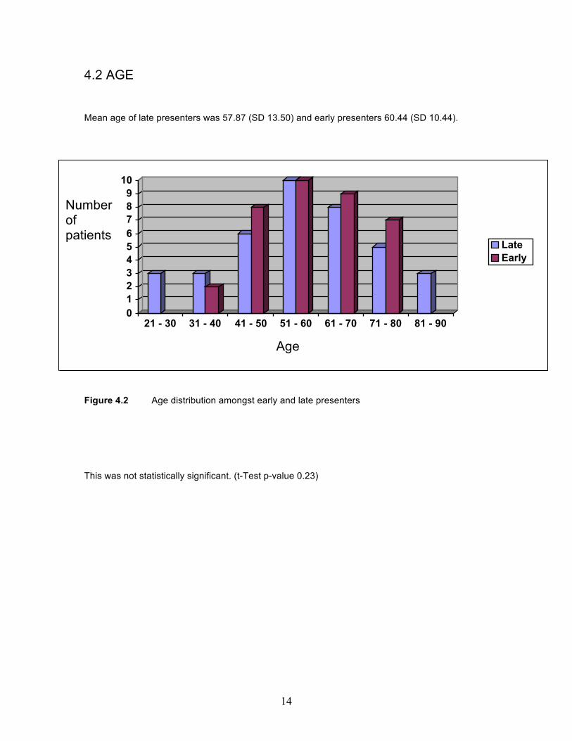
4.2 AGE
Mean age of late presenters was 57.87 (SD 13.50) and early presenters 60.44 (SD 10.44).
0123456789
10
21 - 30 31 - 40 41 - 50 51 - 60 61 - 70 71 - 80 81 - 90
LateEarly
Figure 4.2 Age distribution amongst early and late presenters This was not statistically significant. (t-Test p-value 0.23)
Number of patients
Age

15
4.3 GENDER
Overall there were 68 females and 65 males (n=133). 33 females and 35 males presented late.
Of the early presenters 35 were female compared to 30 males.
0
10
20
30
40
50
60
70
Overall Early Late
MaleFemale
Figure 4.3 Gender distribution Gender was not statistically significant. (p-value 0.54)
Number of patients

16
4.4 ENGLISH LANGUAGE ABILITY When analyzing English language skills I used a modified form of the international English language testing system (IELTS) criteria. Patients were classified into five different groups.
5. Good (Has operational command of the language. May have occasional inaccuracies,
inappropriacies and misunderstandings in some situations. Generally handles complex language well.)
4. Modest (Partial command of the language. Can cope with overall meaning in most
situations. Likely to make many mistakes.)
3. Poor (Conveys and understands only general meaning in very familiar situations.
Frequent breakdowns in communication occurs.)
2. Very poor (No real communication is possible. Has great difficulty understanding spoken
and written English)
1. No English language ability 53% (n = 36) of late presenters and 17% (n = 11) of early presenters had poor, very poor or no English language ability. This was statistically significant (p < 0.001)

17
4.5 LEVEL OF EDUCATION
0 5 10 15 20
None
Grade 3
Grade 4
Grade 5
Grade 6
Grade 7
Grade 8
Grade 9
Grade 10
Grade 11
Grade 12
Tertiary
LateEarly
Figure 4.4 Highest level of education reached by participants There was no statistical significant difference between the groups (p=0.82) with late presenters having 57% (n = 39) and early presenters 55% (n = 36) of participants not completing grade 9.

18
4.6 KNOWLEDGE OF GLAUCOMA AS A SIGHT THREATENING DISEASE Table 4.1 Glaucoma knowledge YES NO
Early Presenters 15% (n = 10) 85% (n = 55)
Late Presenters 19% (n = 13) 81% (n = 55)
There was no statistical significant difference between the groups. (p=0.41)
4.7 FAMILY HISTORY OF GLAUCOMA
There was no statistical significant difference between the groups. (p=0.33) 86% (n = 58) of late presenters and 91% (n = 59) of early presenters had no relatives that suffered from glaucoma

19
4.8 ANNUAL INCOME
0102030405060708090
100
%
0 R1 -R1200
R1201 -R4999
>R5000
LateEarly
Figure 4.5 Average monthly income per patient Table 4.2 Income comparison
There was a statistically significant difference between groups when looking at people earning more than R5000 per month. (p = 0.02) The overwhelming majority of people were earning less than R1200 per month and a two-sample test of proportions showed no statistical significant difference. (see Table 4.2)
Late Presenters Early Presenters p-value R0 18% (n = 12) 12% (n = 8) 0.39 R1 – R1200 59% (n = 40) 65% (n = 42) 0.49 R1201 – R4999 22% (n = 15) 12 % (n = 8) 0.14 > R5000 1% (n = 1) 11% (n = 7) 0.02

20
4.9 HOUSING Table 4.3 Formal vs. Informal housing Late Presenters Early Presenters p-value Formal Housing 82% (n = 56) 94% (n = 61) Informal housing 18% (n = 12) 6% (n = 4) 0.04 More late presenters (18%) lived in informal settlements compared to early presenters (6%). This was a statistically significant difference. (p = 0.04)

21
4.10 PREVIOUS OCULAR TRAUMA
0
20
40
60
80
100
%
Yes No
LateEarly
Figure 4.6 Incidence of previous ocular trauma More early presenters - 18% (n = 12) had previous ocular trauma compared to that of late presenters – 6% (n = 4). This was statistically significant. ( p = 0.03)
22
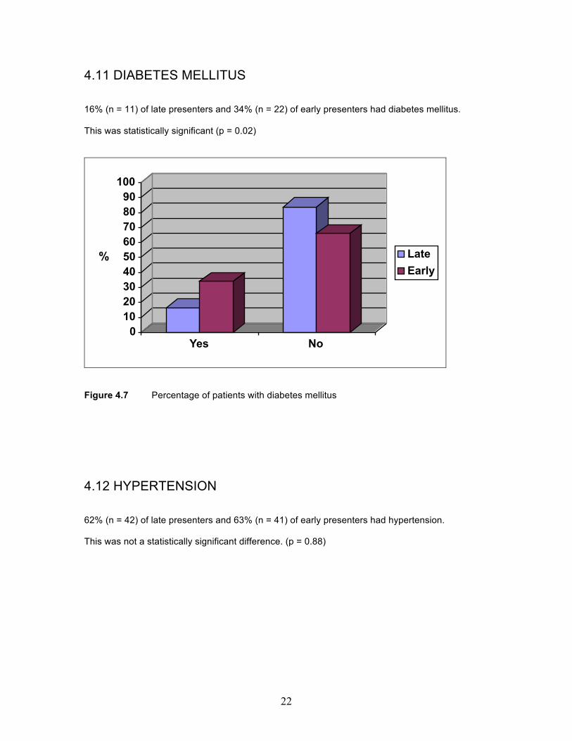
4.11 DIABETES MELLITUS 16% (n = 11) of late presenters and 34% (n = 22) of early presenters had diabetes mellitus. This was statistically significant (p = 0.02)
0102030405060708090100
%
Yes No
LateEarly
Figure 4.7 Percentage of patients with diabetes mellitus 4.12 HYPERTENSION 62% (n = 42) of late presenters and 63% (n = 41) of early presenters had hypertension. This was not a statistically significant difference. (p = 0.88)

23
4.13 PRIMARY VASCULAR DYSREGULATION Since we were unable to do nailfold capillaromicroscopy, it was felt that patients had to have at least 4 out of 5 typical symptoms of vascular dysregulation including:
• Cold extremities
• Reduced feeling of thirst
• Migraines
• Difficulty in falling asleep (longer sleep onset time)
• History of low blood pressure
in order to qualify as a possible vascular dysregulation suspect. There was no statistical significant difference (p = 0.29) between late (1% n = 1) and early presenters (5% n = 3) when looking at patients with 4 or more symptoms of primary vascular dysregulation.

24
4.14 SMOKING
Late Presenters
16%
84%
Yes
No
Early Presenters
5%
95%
Figure 4.8 Pie charts showing the proportion of patients that smoke Cigarette smoking was more common in late presenters. 16% (n = 11) of late presenters and 5% (n = 3) of early presenters were smokers. This was a statistically significant difference. (p = 0.03) 4.15 ALCOHOL USE 69% (n = 47) of late presenters denied using any alcohol. This was echoed in the early presenters with 74% (n = 48) claiming they don’t use alcohol. This was not a statistical significant difference. (p = 0.55)

25
4.16 CORNEAL THICKNESS
The average corneal thickness was thin in both groups.
Table 4.4 Average corneal thickness
Figure 4.9 Median box plot of central corneal thickness There was no statistical significant difference between the groups when looking at right eyes (p- value 0.74) or left eyes (p-value 0.99).
Right Eye Left Eye
Early Presenters 506µm (SD 45µm) 507µm (SD 37µm)
Late Presenters 508µm (SD 39µm) 507µm (SD 40µm)
400
450
500
550
600 Late Early
Right Left
Corneal thickness (µm)

26
4.17 VERTICAL OPTIC DISC HEIGHT
Average optic disc size is shown below.
Table 4.5 Average vertical disc height
Right Eye Left Eye
Late Presenters 1.98mm (SD 0.27mm) 1.98mm (SD 0.27mm)
Early Presenters 2.11mm (SD 0.36mm) 2.10mm (SD 0.37mm)
Figure 4.10 Histogram of vertical disc height
A statistical significant difference between the two groups were observed with late presenters having smaller optic nerves. (p-value 0.03)
0
10
1 1.5 2 2.5 3 1 1.5 2 2.5 3
Late Early
%
Left eye vertical disc height
20
0
10
20
1 1.5 2 2.5 3 1 1.5 2 2.5 3
Late Early
%
Right eye vertical disc height

27
4.18 PSEUDOCAPSULAR EXFOLIATION
The majority of patients had no pseudocapsular exfoliation, accounting for 84% (n=57) of late
presenters and 89% (n=58) of early presenters.
0
10
20
30
40
50
60
Yes No
LateEarly
Figure 4.11 Presence of pseudocapsular exfoliation There was no statistical significant difference between the groups. (p=0.36)

28
CHAPTER 5 – DISCUSSION Our results indicate that a high intraocular pressure at initial presentation is strongly associated
with advanced glaucomatous optic neuropathy. The findings were consistent with studies done in
England 12 and Ghana15 and we agree that higher intraocular pressures would likely lead to more
rapid visual field loss and hence an increased likelihood of delayed attendance. The relationship
between IOP and visual field loss have been evaluated in many clinical trials in recent years.
Results from the Early Manifest Glaucoma Trial showed that each 1-mmHg higher mean IOP
during follow-up was associated with a 12% increase in the chance of developing progressive
visual field loss over time.18 Researchers in the Advanced Glaucoma Intervention Study found
that patients with advanced glaucoma who were kept under control with mean IOP levels of 12.3
mmHg had a mean change in visual field scores close to zero.26 Higher levels of IOP during
follow-up were also significantly related to higher rates of progressive retinal nerve fiber layer
loss, as seen on the GDx scanning laser polarimeter.27
Results from Ghana showed that age greater than 60 years was associated with advanced
glaucoma at presentation.15 This seems plausible since both the prevalence and incidence of
glaucoma rise with age.12 Our results showed no statistical significant difference when looking at
patient age. It is possible that older patients in our society might be hindered from attending our
hospital due to social isolation or reduced mobility. Hence a relatively smaller number of older
patients will attend for vision testing.
Our results showed that gender was not associated with late presentation, while other studies
suggest that males are more likely to have advanced glaucomatous change at initial
presentation.12,15 Fraser et al postulated that these results were due to higher rates of sight
testing in woman leading to earlier glaucoma detection. I believe that these differences do not
occur in our society. Hence our findings reflect my assumption that men and women in our
society have similar opportunities and rates of vision testing and thus an equal chance of being
detected at an earlier stage.

29
Our data indicates that overall level of education was very poor. Once again there was no
statistical significant difference between groups with more than 50% of cases and controls not
completing grade 9.
When studying the English language ability of patients it was noted that early presenters were
able to understand and communicate much better than late presenters. This statistically
significant finding emphasizes the notion that language plays a major role in preventing glaucoma
blindness. When looking at this result one might assume that early presenters should also have a
better knowledge of glaucoma – hence their early clinic attendance. Sadly, the majority of our
patients lacked glaucoma knowledge. Our results show that more than 80% of both cases and
controls had no knowledge of glaucoma as a sight threatening disease. Due to the poor level of
education, it is possible that general information regarding glaucoma and other sight threatening
diseases might be too complicated for these patients to understand
Of particular interest is the fact that most of these patients had been seen by ophthalmologists in
our clinic prior to the study – an indication that our attempts to educate our patients might not be
sufficient. As English is the chosen language of most doctors at the time of this study, one has to
consider that a language barrier between doctor-patient might foil our attempts at educating non-
English speaking patients. In addition, ever increasing patient loads and work stress could
hamper our ability to explain patient pathology in an easy, patient friendly manner. Attempts
should be directed at improving our communication with patients – preferably in a language of
their choice.

30
It has been shown that a family history contributes to the risk of developing glaucoma.25 Having a
close relative with glaucoma cuts down the risk of late presentation by half in a Ghanaian study.15
Similar results were found in England.12 The fact that our results show no association between a
positive family history and the degree of glaucomatous damage on presentation might be related
to the poor level of education that we encountered. Another possibility is that the majority of
patients had no knowledge of glaucoma, which would almost certainly lead to some form of recall
bias.
Several reports have noted that glaucoma patients from lower socioeconomic groups have more
severe visual field loss at the time of diagnosis, which might increase the risk of becoming
blind.14,15,28,29 Our results support these findings with a statistically significant difference between
early and late presenters, especially when looking at people earning more than R5000 per month.
(p = 0.02). Early presenters earned more than their late presenter counterparts. In addition, it
came as no surprise when our data showed that late presenters were more likely to live in poor,
informal settlements.
Hoevenaar et al.28 found that patients from low socioeconomic groups less often knew that the
likelihood of getting glaucoma is higher if intraocular pressure is increased, that it is possible to
have glaucoma without knowing it and that early detection and treatment will slow down the
course of glaucoma. Because they are also less aware of the fact that a family predisposition is a
risk factor for glaucoma, they do not encourage their family members to check for glaucoma,
leading to more avoidable visual impairment in lower socioeconomic groups.
We found a significant association between previous ocular trauma and early presentation.
Trauma would increase the number of eye examinations a patient will have in his/her lifetime and
hence increase the likelihood of detecting glaucoma at an early stage.

31
While some researchers have found that cigarette smoking could be an important risk factor in
the occurrence of Glaucoma and Ocular hypertension30, others dispute this association.31 Our
results indicate that late presenters are more likely to smoke cigarettes. In our view, this once
again is an indication of the socioeconomic factors mentioned above, as smoking has been found
to be more prevalent in lower income groups.32
There is little evidence of an association between alcohol consumption and glaucoma.33 As for
socioeconomic status and alcohol use, epidemiological evidence indicates a positive relationship
between income and the prevalence of alcohol abuse in the general population, but an inverse
relationship between income and alcohol dependence.34 We found no association between
alcohol use and late presentation glaucoma. It is possible that our data was influenced by a
general unwillingness of participants to declare alcohol usage as there is a social stigma attached
to it – i.e. response bias.
What about systemic vasculopathies? Diabetes mellitus is known to cause microvascular damage
and may affect vascular autoregulation of the retina and optic nerve. Diabetes has been found to
be associated with elevated intraocular pressure (IOP)35 and has therefore been suggested as a
possible risk factor for glaucoma, particularly primary open-angle glaucoma (POAG). However,
the current evidence to support this relationship remains conflicting.36 Our results indicate that
patients with diabetes is more likely to present to an ophthalmologist with early glaucomatous
changes. It is our feeling that these patients are more likely to have regular medical examinations
which provide an opportunity for physicians to examine the patients’ eyes for diabetic retinopathy,
and hence the opportunity to detect glaucoma at an early stage. When looking at hypertension,
results from the Blue Mountains Eye study showed a modest, but significant, association with an
increased risk of POAG.17 However, in the Barbados Eye Studies it was found that higher blood
pressure parameters were consistently related to lower risk of developing POAG.25 Whatever the
association, it would seem logical that regular attendance for hypertension treatment would also
provide an opportunity for early glaucoma detection. Our results showed no statistical significant
32
difference between early and late presenters. This was a surprising result and one could argue
that these patients are less likely to have their eyes examined compared to those patients with
diabetes mellitus. It is possible that diabetic patients are more likely to complain of ocular
disturbances compared to patients with hypertension.
Another factor associated with glaucoma progression is disturbed ocular blood flow (OBF). We
were interested to see if there could be any association with this factor and late presentation. As
mentioned before the main cause for the disturbed autoregulation of OBF is the primary vascular
dysregulation syndrome.21 We found no significant difference between the two groups in our
study.
Central corneal thickness and its association with glaucomatous damage has been the subject of
numerous written reports. A study by Herndon et al.37 looked at central corneal thickness and its
relationship to the level of glaucoma severity at the initial ophthalmic examination. They found
that lower central corneal thickness measurements correlated significantly and inversely with the
stage of glaucomatous optic neuropathy. Numerous studies have supported these findings.38,39
While our findings are in keeping with reports that African derived populations have thinner
central corneal thicknesses compared to other populations25, we found no statistical difference
between cases and controls. It is well known that high intraocular pressures might give rise to
endothelial decompensation and hence interfere with the accuracy of corneal thickness
measurements. However, no patients in our study had corneal oedema at the time of
examination.
A larger optic disc size in combination with the reportedly higher glaucoma susceptibility in the
African derived populations compared to whites has led to the hypothesis that eyes with large
optic discs may be more prone to glaucomatous optic nerve fiber loss than eyes with small optic
discs.40,41 Our findings are not consistent with this notion as late presenters had a statistically
significantly smaller vertical disc height compared to early presenters. One possible explanation

33
for this is that patients with larger optic discs (and hence larger physiological cup to disc ratios)
are referred to specialists at an earlier stage than those who have physiologically small optic
discs.
The Early manifest glaucoma trials have shown that pseudoexfoliation more than doubles the risk
of glaucoma progression.19 When one considers the more severe clinical course of
pseudoexfoliative glaucoma, one might assume that these patients have a higher risk of
presenting with advanced field loss. It is also reasonable to hypothesize that a higher incidence of
associated eye pathology 42(eg. cataracts) might lead to these patients been seen earlier
compared to primary open angle glaucoma. However, our results showed neither. No statistical
significant difference was found between the incidence of pseudoexfoliation in cases and
controls.
This study has several weaknesses. Being a single centre trial it lacks the benefits that a
multicenter trial provides. It included a fairly small number of patients, all from the same
geographic location. This obviously decreases the generalizability of the study. Because
information was taken from initial hospital files, we rely on the accuracy of different examiners.
Some of this data might have been inaccurately recorded and might thus be a potential source of
bias. In addition, because patients had to answer questionnaires, it introduces the possibility of
recall bias. Another weakness is the fact that we only performed univariate data analysis. A
multivariate data analysis is needed in order to control for confounding.

34
CHAPTER 6 – CONCLUSION AND RECOMMENDATIONS
Our findings confirm that raised intraocular pressure is a major factor in patients presenting with
advanced glaucomatous optic neuropathy. Being our only modifiable risk factor in the
pathogenesis of glaucoma, efforts should be directed at early detection. However, there are
several factors that make this a near impossible task. Poverty seems to be a major factor
responsible for our failure to detect glaucoma patients at an earlier stage. Unfortunately this is a
significant problem in most developing countries and South Africa is no different. As
opthalmologists, the scope of this problem is out of our hands. However, efforts could be focused
at improving our glaucoma education and screening programs in the community. Our results also
suggest that even at tertiary care level we are failing to educate our patients properly.
Considering the fact that most of our patients have a very poor level of education, we should
direct our attention at simplifying our communication with these patients in a language of their
choice. Our results further indicate that there are several factors which might contribute to earlier
or more regular medical examinations and hence earlier glaucoma detection. This highlights the
fact that although it will be difficult, it may be possible to reduce the number of people with
glaucoma related blindness in South Africa.

35
CHAPTER 7 – APPENDIX
In the following chapter there are examples of the documents that were used in the completion of the study. 7.1 PATIENT QUESTIONNAIRE
• Participant number:……………………………………………………….. • Who told (referred) you to come see us at our hospital? ……………………
……………………………………………………………………………….. Did they diagnose you with glaucoma? Y / N
• Have you ever had your eyes examined by an optometrist or a doctor? Y / N
If Yes: How many times?.............................................................. By whom?…………………………………………..........
When?................................................................................
• Have you seen a doctor before? Y / N
If yes: When was the last time?..................................................... How often do you see the doctor?......................................
• Has anyone in your family got glaucoma? Y / N
If Yes: Who?...................................................................................
Has anyone ever advised you to have your eyes checked for glaucoma? Y / N
Who?.....................
• Did you go to school? Y / N If Yes: What is the highest grade you completed? ………………
• Did you go to university? Y / N
If yes: What did you study? ……………………………………. Did you complete your studies? Y / N
• Do you know what glaucoma is? Y / N If yes: Explain what you know about it………………………… ………………………………………………………………………… How did you learn about glaucoma?................................. …………………………………………………………………………
• How much money do you earn / receive per month? ……………………….
How do you obtain this money? (eg what job? Grant?)…………………….... ………………………………………………………………………………...

36
• Where do you live? …………………………………………………………..
• How long have you lived there?.......................................................................
• In what structure do you live?(eg house, shack, caravan, apartment etc)……
………………………………………………………………………………...
• Have you ever lived in a rural area?................................................................ When? (eg. 1970 – 1993) ……………………………………………..
• Who do you stay with? ……………………………………………………….
• What language do you speak at home?……………………………………
• Have you ever been diagnosed with Diabetes (high blood sugar?) Y / N
If Yes: How long have you had it? ……………………………..
• Have you ever been diagnosed with high blood pressure? Y / N
If Yes: How long have you had it? ……………………………..
• Do you smoke? Y / N If Yes: How many cigarettes per day? …………………………. For how long? ………………………………………….. If No: Have you ever smoked? ………………………………. For how long? (eg 1980 – 1992) ………………………
• Do you drink alcohol? Y / N If Yes: How often, how much? ………………………………..
• Do you suffer from any of the following?
Cold hands and feet? Y / N Migraines? Y / N Difficulty in falling asleep at night? Y / N Low blood pressure? Y / N Would you consider yourself to have a reduced feeling of thirst? I.e. do you seldom get thirsty? Y / N
• Have you ever had any accident / injury to you eyes? Y / N
If yes: Explain…………………………………………… ………………………………………………………………………...
Thank you for taking the time to complete this questionnaire.

37
7.2 DATA CAPTURING SHEET 1. Patients Participant Number:………………………………………. 2. EARLY: Typical glaucomatous field loss and a MD between 0 dB and -11 dB in the worst eye. A
cup to disc ratio of more than 0,5 must be present or a difference of more than 0,2 must have been noted.
or
LATE: Typical glaucomatous field loss with a mean deviation (MD) of greater than -14 dB in the better seeing eye. Optic discs must show typical glaucomatous cupping of more than 0,8. Fields will be excluded if visual loss is so advanced that field testing is not possible.
3. Visual Acuity OD……… OS…… 4. Presenting intraocular pressure ………………….. 5. Central corneal thickness ………………….. 6. Optic disk size (vertical disk height)…………………. 7. Pseudocapsular exfoliation Y / N 8. Age at first presentation: ………. 9. Sex: M / F 10. English language ability: Can patient communicate in English Y / N
If Y: Good ( Has operational command of the language. May have occasional inaccuracies,
inappropriaceis and misunderstandings in some situations. Generally handles complex language well.)
Modest (Partial command of the language. Can cope with overall meaning in most situations. Likely to make many mistakes.) Poor (Conveys and understands only general meaning in very familiar situations. Frequent breakdowns in communication occurs.)
Very poor: (No real communication is possible. Has great difficulty understanding spoken and written English)
11. Method of referral:
a) None
b) Optometrist Glaucoma suspect / diagnosis? Y / N c) Medical Practitioner Glaucoma suspect / diagnosis? Y / N d) Local Clinic Glaucoma suspect / diagnosis? Y / N

38
12. Previous eye examinations Y / N How many? ……. When? ……………… 13. Previous medical examinations Y / N Frequency ………………….. Last visit to doctor …………. 14. Family history of glaucoma Y / N Relation to patient ………………………….. 15. Level of education a) No formal education
b) Primary school -grade ………………… c) Secondary school - grade ………………… d) Tertiary institution - degree/diploma ………………………….. 16. Knowledge about glaucoma: as an eye disease? Y / N as a cause of blindness? Y / N 17. Average annual income ………………………………… Source of income …………………………………… 18. Formal / Informal housing? 19. Ever lived in rural area? Y / N When? (eg 1970 – 1989 )………………………… 20. Home language ………………………………….. 21. Diabetes Mellitus Y / N HGT …………. 22. Hypertension Y / N BP ……/……. 23. Smoker Y / N If Y: Pack year history ……………………. If N: Previous Smoker? Y / N 24. Alcohol consumption Y / N If Y: Average daily consumption …………………. 25. Symptoms of Primary Vascular Disease Cold extremities? Y / N Reduced feeling of thirst Y / N Migraines Y / N Low Blood pressure Y / N Difficulty in falling asleep Y / N / 5 26. Previous ocular trauma Y / N

39
CHAPTER 8 – REFERENCES
1. Quigley HA, Broman AT. The number of people with glaucoma worldwide in 2010 and 2020. Br
J Ophthalmol 2006: 90: 262 - 7.
2. Lim K, Vision 2020 and Prevention of Blindness: Is it Relevant or Achievable in the Modern
Era? Ann Acad Med Singapore 2006; 35: 215 – 22.
3. Cunningham ET. Blindness in Africa: present situation and future needs. Br J Ophthalmol 2001;
85: 897 – 903. 4. Verrey JD, Foster A, Wormald R, et al. Chronic glaucoma in Northern Ghana – a retrospective
study of 397 patents. Eye 1990; 4: 115 – 120. 5. Buhrmann RR, Quigley HA, Barron Y, et al. Prevalence of glaucoma in a rural east African
population. Invest Ophthalmol Vis Sci 2000; 41: 40 – 48. 6. Salmon JF, Mermoud A, Ivey A, et al. The prevalence of primary angle closure glaucoma and
open angle glaucoma in Mamre, Western Cape, South Africa. Arch Ophthalmol 1993; 111: 1263 – 1269.
7. Rotchford AP, Johnson GJ. Glaucoma in Zulus. Arch Ophthalmol 2002; 120: 471 – 478. 8. Chen PP. Risk and risk factors for blindness from glaucoma. Curr opin Ophthalmol 2004; 15:
107 – 111. 9. Oliver JE, Hattenhauer MG, Herman D, et al. Blindness and glaucoma: a comparison of
patients progressing to blindness from glaucoma with patients maintaining vision. Am J
Ophthalmol 2002; 133: 764 – 772. 10. Kwon YH, Chang-sik K, Zimmerman B, et al. Rate of visual field loss and long term visual
outcome in primary open-angle glaucoma. Am J Ophthalmol 2001; 132(1); 47 – 56.

40
11. Jorge LR, Nicholas PB, Feldman RM. Risk factors for primary open angle glaucoma
progression: what we know and what we need to know. Curr opin Ophthalmol 2008; 19: 102 – 106.
12. Fraser S, Bunce C, Wormald R. Retrospective analysis of risk factors for late presentation of
chronic glaucoma. Br J Ophthalmol 1999; 83: 24 – 28.
13. Fraser S, Bunce C, Wormald R. Risk factors for late presentation in chronic glaucoma. Invest
Ophthalmol Vis Sci 1999; 40(10): 2251 – 2257.
14. Fraser S, Bunce C, Wormald R, et al. Deprivation and late presentation of glaucoma: case-
control study. British Medical Journal 2001; 322: 639 – 643. 15. Ntim-Amponsah CT, Amoaku WMK, Ewusi RK et al. Evaluation of risk factors for advanced
glaucoma in Ghanaian patients. Eye 2005; 19: 528 – 534. 16. Jay JL, Murdoch JR. The rate of visual field loss in untreated primary open angle glaucoma.
Br J Ophthalmol 1993; 77: 176 – 178. 17. Mitchell P, Lee AJ, Rochtchina E, et al. Open-angle glaucoma and hypertension. The Blue
Mountains Eye Study. J Glaucoma 2004; 13: 319 – 326. 18. Leske MC, Heijl A, Hyman L, et al. Predictors of long-term progression in the early manifest
glaucoma trial. Ophthalmology 2007; 114: 1965 – 1972. 19. Leske MC, Heijl A, Hussein M, et al. Factors for glaucoma progression and the effect of
treatment. Arch Ophthalmol 2003; 121: 48 – 56. 20. Rotchford AP, Kirwan JF, Johnson GJ, et al. Exfoliation Syndrome in black South Africans.
Arch Ophthalmol 2003; 121: 863 – 870. 21. Flammer J, Mozaffarieh M. What is the present pathogenic concept of glaucomatous optic
neuropathy? Surv Ophthalmol 2007; 52(Suppl 2): S162 – S173.

41
22. Grieshaber MC, Mozaffarieh M, Flammer J. What is the link between vascular dysregulation
and glaucoma? Surv Ophthalmol 2007; 52(Suppl 2): S144 – S154. 23. Tsai JC. Influencing ocular blood flow in glaucoma patients: the cardiovascular system and
healthy lifestyle choices. Can J Ophthalmol 2008; 43: 347 – 350. 24. Jonas JB, Martus P, Horn FK et al. Predictive factors of the optic nerve head for
development or progression of glaucomatous visual field loss. Invest Ophthalmol Vis Sci 2004; 45(8): 2613 – 2618.
25. Leske MC, Wu S, Hennis A, et al. Risk factors for incident open-angle glaucoma. The
Barbados eye studies. Ophthalmology 2008; 115: 85 – 93.
26. The Advanced Glaucoma Intervention Study (AGIS): 7. The relationship between control of
intraocular pressure and visual field deterioration. The AGIS Investigators. Am J Ophthalmol. 2000; 130:429 – 40.
27. Medeiros FA, Alencar LM, Zangwill LM, et al. The Relationship between intraocular pressure
and progressive retinal nerve fiber layer loss in glaucoma. Ophthalmology 2009; 116(6): 1125 – 33.
28. Hoevenaars JG, Schouten JS, Van den Borne B, et al. Socioeconomic differences in
glaucoma patients’ knowledge, need for information and expectations of treatments. Acta Ophthalmol. Scand 2006: 84; 84 – 91.
29. Oliver JE, Hattenhauer MG, Herman D, et al. Blindness and glaucoma: a comparison of
patients progressing to blindness from glaucoma with patients maintaining vision. Am J Ophthalmol. 2002: 133; 764 – 772.
30. Afshan A, Bhutkar MV, Reddy R, et.al. Effect of chronic cigarette smoking on intraocular
pressure and audio-visual reaction time. Int J Biol Med Res. 2012: 3(2); 1760 – 1763.

42
31. Ramdas WD, Wolfs RC, Hofman A, et al. Lifestyle and Risk of Developing Open-Angle
Glaucoma. Arch Ophthalmol. 2011:129(6); 767 – 772.
32. Hiscock R, Bauld L, Amos A, et al. Socioeconomic status and smoking: a review. Ann N Y
Acad Sci. 2012: 1248; 107 – 123. 33. Wang S, Wang JJ, Wong TY. Alcohol and eye diseases. Surv Ophthalmol. 2008: 53(5): 512 –
525. 34. Keyes KM, Hasin DS. Socio-economic status and problem alcohol use: the positive
relationship between income and the DSM-IV alcohol abuse diagnosis. Addiction. 2008: 103; 1120 – 1130.
35. Wu SY, Leske MC. Associations with intraocular pressure in the Barbados Eye
Study. Arch Ophthalmol. 1997;115(12):1572-1576. 36. Tan GS, Wong TY, Fong CW, et al. Diabetes, Metabolic Abnormalities, and Glaucoma: The
Singapore Malay Eye Study. Arch Ophthalmol. 2009;127(10):1354 – 1361. 37. Herndon LW, Weizer JS, Stinnett SS. Central corneal thickness as a risk factor for advanced
glaucoma damage. Arch Ophthalmol. 2004; 122:17–21. 38. Jonas JB, Stroux A, Velten I, et al. Central Corneal Thickness Correlated with Glaucoma
Damage and Rate of Progression. Invest Ophthalmol Vis Sci 2005; 46(4): 1269 – 1274.
39. Fernandez J, Roman C, Fernandez M. Central Corneal Thickness as a Predictor of Visual
Field Loss in Primary Open Angle Glaucoma for a Hispanic Population. Semin Ophthalmol.
2011; 26(1): 28–32.
40. Chi T, Ritch R, Stickler D, et al. Racial differences in optic nerve head parameters. Arch
Ophthalmol. 1989; 107:836–839.

43
41. Varma R, Tielsch JM, Quigley HA, et al. Race-, age-, gender-, and refractive error-related
differences in the normal optic disc. Arch Ophthalmol. 1994: 112; 1068–1076.
42. Ritch R, Schlotzer-Schrehardt U. Exfoliation syndrome. Surv Ophthalmol. 2001; 45: 265 –
315.