Extraoral imaging The Orthophos family - Dentsply Sirona...for extraoral imaging As versatile as...
21
Extraoral imaging The Orthophos family dentsplysirona.com/ orthophos
Transcript of Extraoral imaging The Orthophos family - Dentsply Sirona...for extraoral imaging As versatile as...
Extraoral imaging
The Orthophos familydentsplysirona.com/orthophos
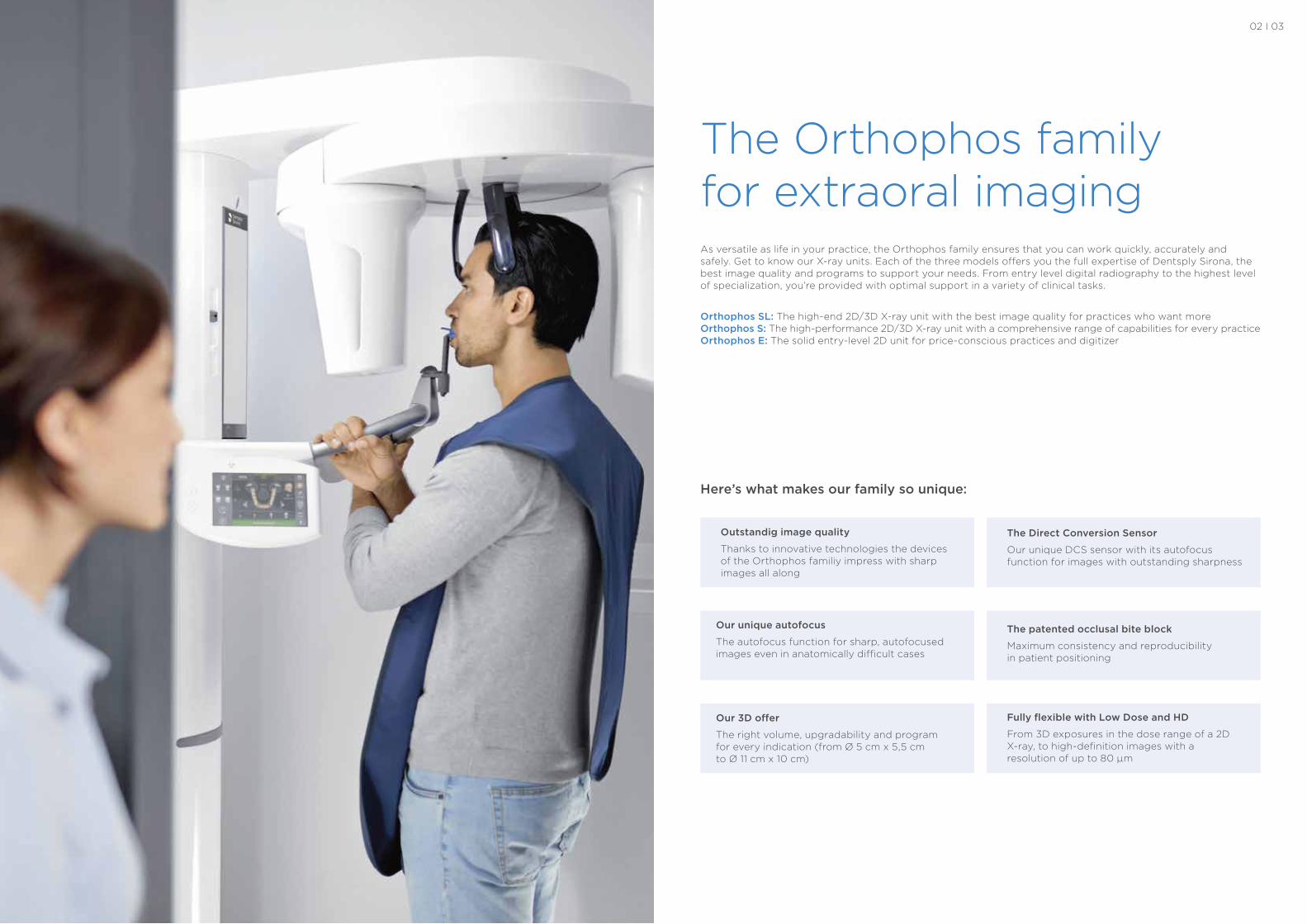
The Orthophos family for extraoral imagingAs versatile as life in your practice, the Orthophos family ensures that you can work quickly, accurately and safely. Get to know our X-ray units. Each of the three models offers you the full expertise of Dentsply Sirona, the best image quality and programs to support your needs. From entry level digital radiography to the highest level of specialization, you’re provided with optimal support in a variety of clinical tasks.
Orthophos SL: The high-end 2D/3D X-ray unit with the best image quality for practices who want moreOrthophos S: The high-performance 2D/3D X-ray unit with a comprehensive range of capabilities for every practiceOrthophos E: The solid entry-level 2D unit for price-conscious practices and digitizer
Here’s what makes our family so unique:
Our 3D offer
The right volume, upgradability and program for every indication (from Ø 5 cm x 5,5 cm to Ø 11 cm x 10 cm)
The patented occlusal bite block
Maximum consistency and reproducibility in patient positioning
The Direct Conversion Sensor
Our unique DCS sensor with its autofocus function for images with outstanding sharpness
Fully flexible with Low Dose and HD
From 3D exposures in the dose range of a 2D X-ray, to high-definition images with a resolution of up to 80 μm
Our unique autofocus
The autofocus function for sharp, autofocused images even in anatomically difficult cases
Outstandig image quality
Thanks to innovative technologies the devices of the Orthophos familiy impress with sharp images all along
02 I 03
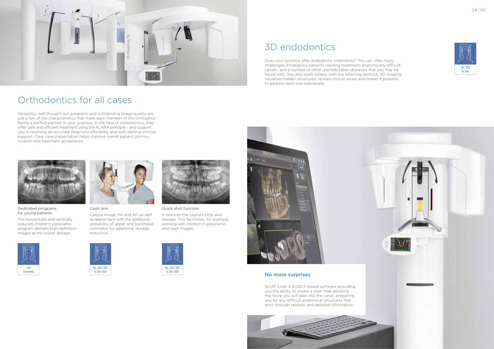
Versatility, well thought-out programs and outstanding image quality are just a few of the characteristics that make each member of the Orthophos family a perfect partner in your practice. In the field of orthodontics, they offer safe and efficient treatment using the ALARA principle – and support you in reaching an accurate diagnosis efficiently and with optimal clinical support. Clear case presentation helps improve overall patient commu- nication and treatment acceptance.
Orthodontics for all cases
SL 2D /3D S 2D /3D
All models
SL 2D /3D S 2D /3D
Quick shot function
It reduces the capture time and dosage. This facilitates, for example, working with children in panoramic and ceph images.
Dedicated programs for young patients
The horizontally and vertically reduced children’s panoramic program delivers high-definition images at the lowest dosage.
Ceph arm
Carpus image, PA and AP, as well as lateral ceph with the additional possibility of upper and backhead collimator for additional dosage reduction.
Does your practice offer endodontic treatments? This can offer many challenges. Emergency patients needing treatment, anatomically difficult canals , and a number of other unpredictable obstacles that you may be faced with. You also work closely with the referring dentists. 3D imaging visualizes hidden structures, reveals clinical issues and makes it possible to address each one individually.
3D endodontics
SL 3D S 3D
No more surprises
SICAT Endo is a CBCT-based software providing you the ability to create a clear map detailing the route you will take into the canal, preparing you for any difficult anatomical structures that exist through realistic and detailed information.
04 I 05
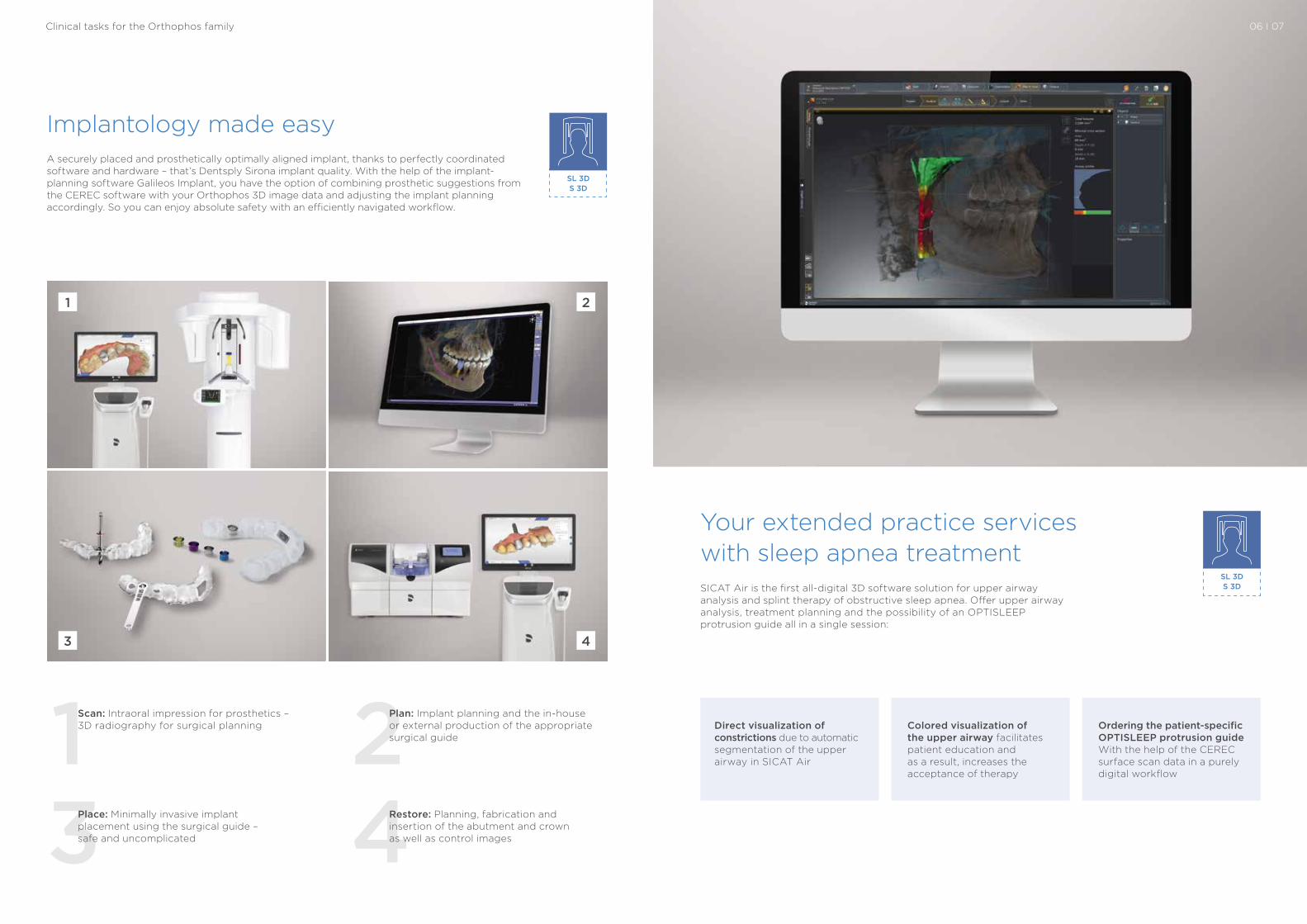
A securely placed and prosthetically optimally aligned implant, thanks to perfectly coordinated software and hardware – that’s Dentsply Sirona implant quality. With the help of the implant- planning software Galileos Implant, you have the option of combining prosthetic suggestions from the CEREC software with your Orthophos 3D image data and adjusting the implant planning accordingly. So you can enjoy absolute safety with an efficiently navigated workflow.
Implantology made easy
SL 3D S 3D
1 23 4
Scan: Intraoral impression for prosthetics – 3D radiography for surgical planning
Place: Minimally invasive implant placement using the surgical guide – safe and uncomplicated
Plan: Implant planning and the in-house or external production of the appropriate surgical guide
Restore: Planning, fabrication and insertion of the abutment and crown as well as control images
1
3 4
2
SICAT Air is the first all-digital 3D software solution for upper airway analysis and splint therapy of obstructive sleep apnea. Offer upper airway analysis, treatment planning and the possibility of an OPTISLEEP protrusion guide all in a single session:
Your extended practice services with sleep apnea treatment
Direct visualization of constrictions due to automatic segmentation of the upper airway in SICAT Air
Colored visualization of the upper airway facilitates patient education and as a result, increases the acceptance of therapy
Ordering the patient-specific OPTISLEEP protrusion guide With the help of the CEREC surface scan data in a purely digital workflow
SL 3D S 3D
Clinical tasks for the Orthophos family 06 I 07
08 I 09
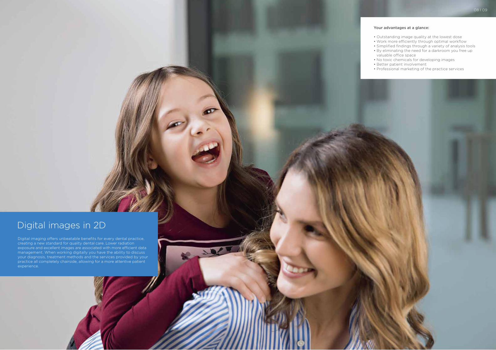
Digital images in 2DDigital imaging offers unbeatable benefits for every dental practice, creating a new standard for quality dental care. Lower radiation exposure and excellent images are associated with more efficient data management. When working digitally you have the ability to discuss your diagnosis, treatment methods and the services provided by your practice all completely chairside, allowing for a more attentive patient experience.
Your advantages at a glance:
• Outstanding image quality at the lowest dose• Work more efficiently through optimal workflow• Simplified findings through a variety of analysis tools• By eliminating the need for a darkroom you free up
valuable office space• No toxic chemicals for developing images• Better patient involvement• Professional marketing of the practice services
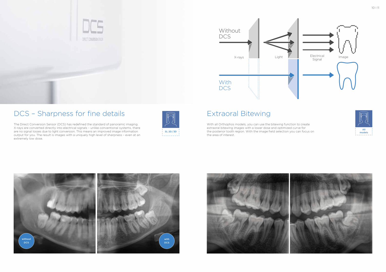
DCS – Sharpness for fine detailsThe Direct Conversion Sensor (DCS) has redefined the standard of panoramic imaging. X-rays are converted directly into electrical signals – unlike conventional systems, there are no signal losses due to light conversion. This means an improved image information output for you. The result is images with a uniquely high level of sharpness – even at an extremely low dose.
10 I 11
without DCS
with DCS
With all Orthophos models, you can use the bitewing function to create extraoral bitewing images with a lower dose and optimized curve for the posterior tooth region. With the image field selection you can focus on the area of interest.
Extraoral Bitewing
All modelsSL 2D / 3D
Without DCS
With DCS
X-rays Light ImageElectrical Signal
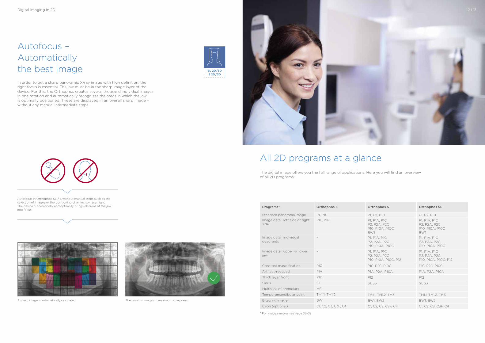
The digital image offers you the full range of applications. Here you will find an overview of all 2D programs:
* For image samples see page 38–39
In order to get a sharp panoramic X-ray image with high definition, the right focus is essential. The jaw must be in the sharp image layer of the device. For this, the Orthophos creates several thousand individual images in one rotation and automatically recognizes the areas in which the jaw is optimally positioned. These are displayed in an overall sharp image – without any manual intermediate steps.
Autofocus – Automatically the best image SL 2D /3D
S 2D /3D
12 I 13
All 2D programs at a glance
Digital imaging in 2D
Programs* Orthophos E Orthophos S Orthophos SL
Standard panorama image P1, P10 P1, P2, P10 P1, P2, P10
Image detail left side or right side
P1L, P1R P1, P1A, P1C P2, P2A, P2CP10, P10A, P10C BW1
P1, P1A, P1C P2, P2A, P2CP10, P10A, P10C BW1
Image detail individual quadrants
– P1, P1A, P1C P2, P2A, P2CP10, P10A, P10C
P1, P1A, P1C P2, P2A, P2CP10, P10A, P10C
Image detail upper or lower jaw
– P1, P1A, P1C P2, P2A, P2CP10, P10A, P10C, P12
P1, P1A, P1C P2, P2A, P2CP10, P10A, P10C, P12
Constant magnification P1C P1C, P2C, P10C P1C, P2C, P10C
Artifact-reduced P1A P1A, P2A, P10A P1A, P2A, P10A
Thick layer front P12 P12 P12
Sinus S1 S1, S3 S1, S3
Multislice of premolars MS1 – –
Temporomandibular Joint TM1.1, TM1.2 TM1.1, TM1.2, TM3 TM1.1, TM1.2, TM3
Bitewing image BW1 BW1, BW2 BW1, BW2
Ceph (optional) C1, C2, C3, C3F, C4 C1, C2, C3, C3F, C4 C1, C2, C3, C3F, C4
A sharp image is automatically calculated The result is images in maximum sharpness
Autofocus in Orthophos SL / S without manual steps such as the selection of images or the positioning of an incisor laser light. The device automatically and optimally brings all areas of the jaw into focus.
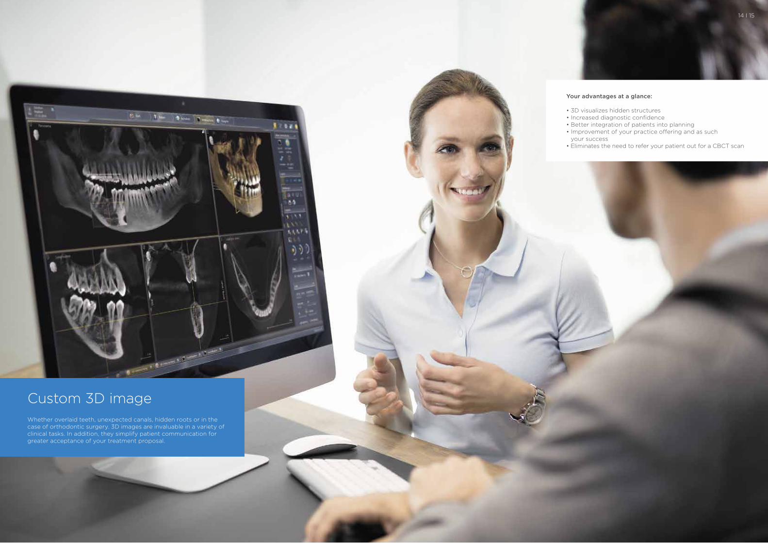
Custom 3D imageWhether overlaid teeth, unexpected canals, hidden roots or in the case of orthodontic surgery. 3D images are invaluable in a variety of clinical tasks. In addition, they simplify patient communication for greater acceptance of your treatment proposal.
Your advantages at a glance:
• 3D visualizes hidden structures• Increased diagnostic confidence• Better integration of patients into planning• Improvement of your practice offering and as such
your success• Eliminates the need to refer your patient out for a CBCT scan
14 I 15
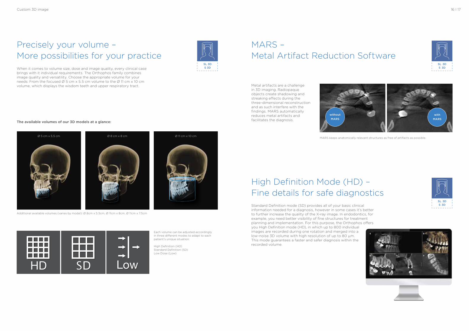
The available volumes of our 3D models at a glance:
Each volume can be adjusted accordingly in three different modes to adapt to each patient’s unique situation:
High Definition (HD) Standard Definition (SD) Low Dose (Low)
Additional available volumes (varies by model): Ø 8cm x 5.5cm, Ø 11cm x 8cm, Ø 11cm x 7.5cm
SL 3D S 3D
SL 3D S 3D
High Definition Mode (HD) – Fine details for safe diagnosticsStandard Definition mode (SD) provides all of your basic clinical information needed for a diagnosis, however in some cases it’s better to further increase the quality of the X-ray image. In endodontics, for example, you need better visibility of fine structures for treatment planning and implementation. For this purpose, the Orthophos offers you High Definition mode (HD), in which up to 800 individual images are recorded during one rotation and merged into a low-noise 3D volume with high resolution of up to 80 μm. This mode guarantees a faster and safer diagnosis within the recorded volume.
Metal artifacts are a challenge in 3D imaging. Radiopaque objects create shadowing and streaking effects during the three-dimensional reconstruction and as such interfere with the findings. MARS automatically reduces metal artifacts and facilitates the diagnosis.
MARS – Metal Artifact Reduction Software
MARS keeps anatomically relevant structures as free of artifacts as possible.
withoutMARS
withMARS
Ø 5 cm x 5,5 cm Ø 11 cm x 10 cmØ 8 cm x 8 cm
16 I 17
SL 3D S 3D When it comes to volume size, dose and image quality, every clinical case
brings with it individual requirements. The Orthophos family combines image quality and versatility. Choose the appropriate volume for your needs: From the focused Ø 5 cm x 5.5 cm volume to the Ø 11 cm x 10 cm volume, which displays the wisdom teeth and upper respiratory tract.
Precisely your volume – More possibilities for your practice
Custom 3D image
SL 3D S 3D
Low Dose for a variety of clinical tasks Program selection for
the case-based applica-tion using the ALARA (As Low As Reasonable Achievable) principle
Sleep apnea therapy with SICAT Air and OPTISLEEP
Implant control in 3D in the dose range of an intraoral X-ray
Tooth position determi-nation in 3D at low dose, especially for young, radiation-sensitive patients
18 I 19
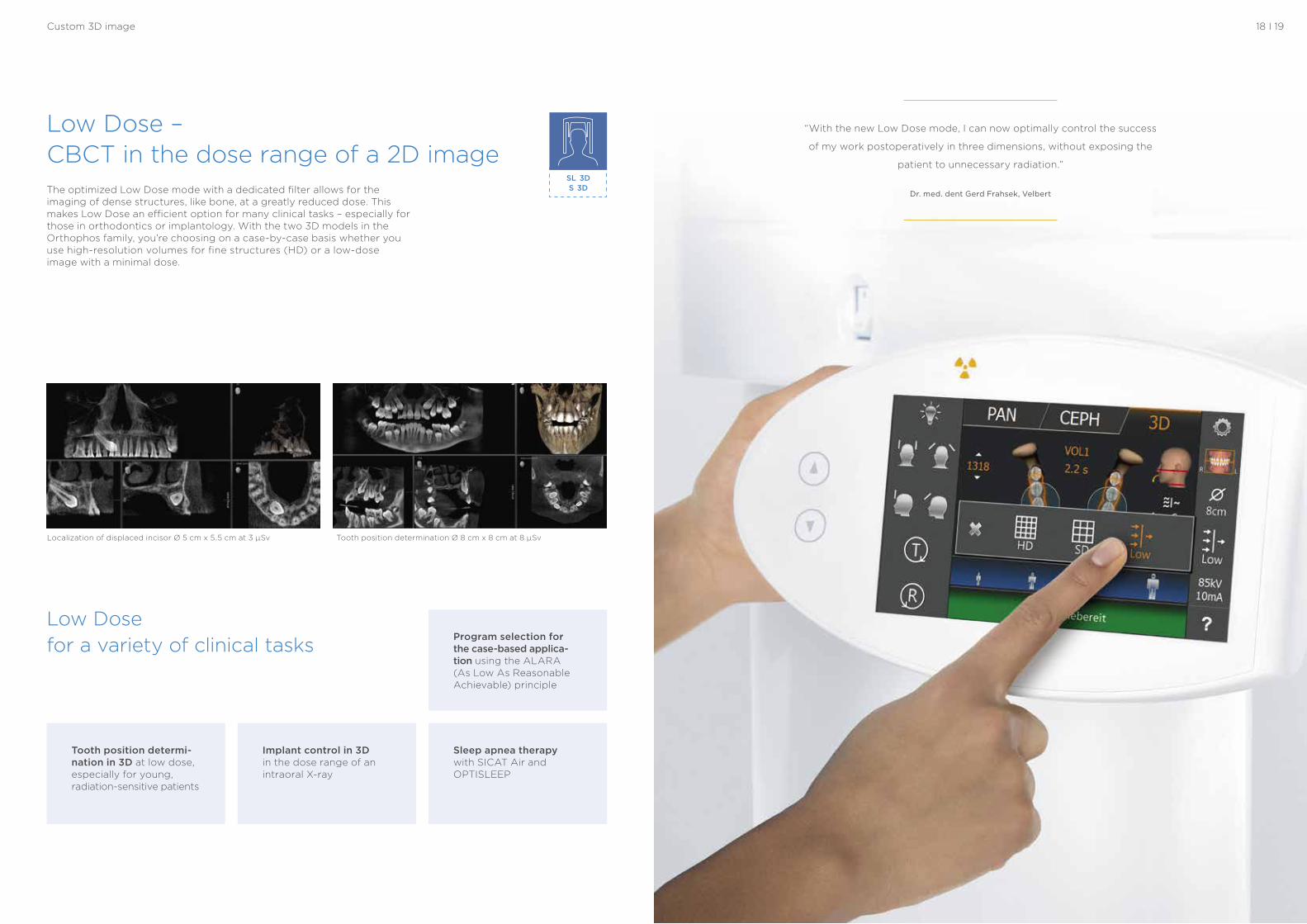
Low Dose – CBCT in the dose range of a 2D imageThe optimized Low Dose mode with a dedicated filter allows for the imaging of dense structures, like bone, at a greatly reduced dose. This makes Low Dose an efficient option for many clinical tasks – especially for those in orthodontics or implantology. With the two 3D models in the Orthophos family, you’re choosing on a case-by-case basis whether you use high-resolution volumes for fine structures (HD) or a low-dose image with a minimal dose.
Localization of displaced incisor Ø 5 cm x 5.5 cm at 3 μSv Tooth position determination Ø 8 cm x 8 cm at 8 μSv
Custom 3D image
“With the new Low Dose mode, I can now optimally control the success
of my work postoperatively in three dimensions, without exposing the
patient to unnecessary radiation.”
Dr. med. dent Gerd Frahsek, Velbert

For you, choosing the Orthophos family is about two things: getting the best possible image to support your diagnosis and having your patient feel comfortable. For both, our models offer unique, patented solutions. Optimize your practice’s workflow with intuitive user interfaces and automatic positioning aids to avoid unnecessary secondary exposures.
Easy to operate, secure positioning
SL 3D S 3D
SL 3D S 3D
All models
1 32Patented occlusal bite block
Position the patient with the patented occlusal bite block. The Orthophos intuitively deter mines the correct tilt of the head for optimal positioning and informs you through cor relating symbols and colors how to adjust accordingly with just the press of the up or down arrow.
Intuitive use
The EasyPad, which can be swiveled and tilted to your desired position, offers you absolute flexibility and opti-mal use. In addition to clear user options on the innovative touchpad your workflow is supported, no matter how your X-ray room is set up.
Stable patient positioning
Stable patient positioning prevents motion blur. The motorized 3-point head fixation and sturdy handles give your patient the neces-sary support. The integrated temple width measurement automatically ensures a patient- specific orbit. Unnecessary downtime can be reduced by the automatic opening of the temple support for a success-ful X-ray outcome.
20 I 21
1
3
2
Everything for the best image
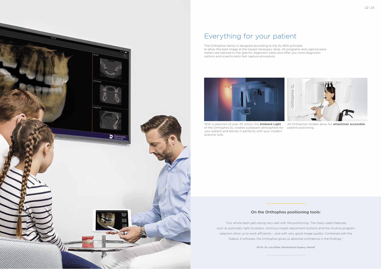
With a selection of over 30 colors, the Ambient Light of the Orthophos SL creates a pleasant atmosphere for your patient and blends in perfectly with your modern practice look.
All Orthophos models allow for wheelchair accessible patient postioning.
Everything for your patientThe Orthophos family is designed according to the ALARA principle to allow the best image at the lowest necessary dose. All programs and capture para-meters are tailored to the specific diagnostic tasks and offer you more diagnostic options and a particularly fast capture procedure.
On the Orthophos positioning tools:
“Our whole team gets along very well with the positioning. The many useful features
such as automatic light localizers, luminous height adjustment buttons and the intuitive program
selection allow us to work efficiently – and with very good image quality. Combined with the
Sidexis 4 software, the Orthophos gives us absolute confidence in the findings.”
PD Dr. Dr. Lutz Ritter, Maxillofacial Surgery, Hennef
22 I 23
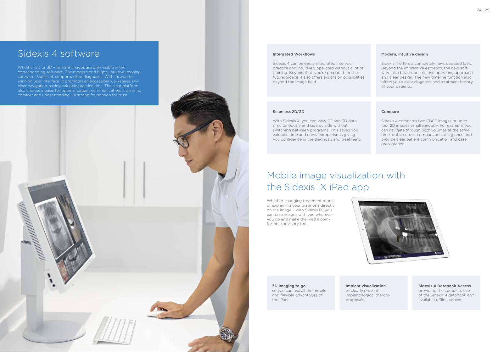
Modern, intuitive design
Sidexis 4 offers a completely new, updated look. Beyond the impressive esthetics, the new soft-ware also boasts an intuitive operating approach and clear design. The new timeline function also offers you a clear diagnosis and treatment history of your patients.
Integrated Workflows
Sidexis 4 can be easily integrated into your practice and intuitively operated without a lot of training. Beyond that, you’re prepared for the future: Sidexis 4 also offers expansion possibilities beyond the image field.
Seamless 2D/3D
With Sidexis 4, you can view 2D and 3D data simultaneously and side by side without switching between programs. This saves you valuable time and cross-comparisons giving you confidence in the diagnosis and treatment.
Compare
Sidexis 4 compares two CBCT images or up to four 2D images simultaneously. For example, you can navigate through both volumes at the same time, obtain cross-comparisons at a glance and provide clear patient communication and case presentation.
Sidexis 4 softwareWhether 2D or 3D – brilliant images are only visible in the corresponding software. The modern and highly intuitive imaging software, Sidexis 4, supports clear diagnoses. With its award- winning user interface, it promotes an accessible workspace and clear navigation, saving valuable practice time. The clear platform also creates a basis for optimal patient communication, increasing comfort and understanding – a strong foundation for trust.
Mobile image visualization with the Sidexis iX iPad appWhether changing treatment rooms or explaining your diagnosis directly on the image – with Sidexis iX, you can take images with you wherever you go and make the iPad a com-fortable advisory tool.
3D imaging to go so you can use all the mobile and flexible advantages of the iPad
Implant visualization to clearly present implantological therapy proposals
Sidexis 4 Databank Access providing the complete use of the Sidexis 4 databank and available offline copies
24 I 25
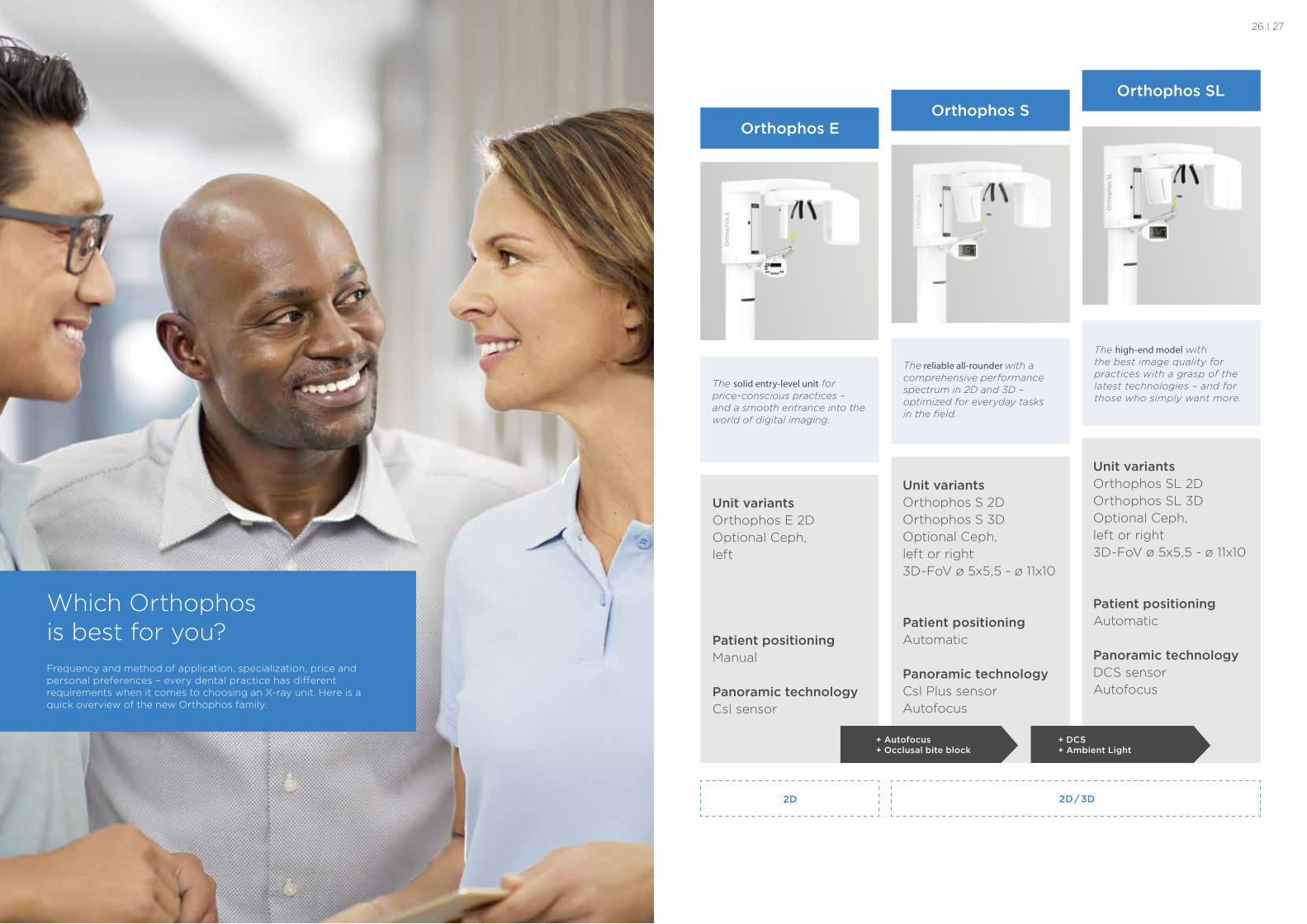
Unit variants Orthophos E 2DOptional Ceph, left
Patient positioning Manual
Panoramic technology CsI sensor
Orthophos E
Unit variants Orthophos S 2D Orthophos S 3DOptional Ceph, left or right 3D-FoV ø 5x5,5 - ø 11x10
Patient positioning Automatic Panoramic technology CsI Plus sensorAutofocus
Orthophos S
Unit variants Orthophos SL 2D Orthophos SL 3DOptional Ceph, left or right 3D-FoV ø 5x5,5 - ø 11x10
Patient positioning Automatic
Panoramic technology DCS sensorAutofocus
Orthophos SL
+ Autofocus + Occlusal bite block
+ DCS + Ambient Light
2D 2D / 3D
The solid entry-level unit for price-conscious practices – and a smooth entrance into the world of digital imaging.
The reliable all-rounder with a comprehensive performance spectrum in 2D and 3D – optimized for everyday tasks in the field.
The high-end model with the best image quality for practices with a grasp of the latest technologies – and for those who simply want more.
Which Orthophos is best for you?Frequency and method of application, specialization, price and personal preferences – every dental practice has different requirements when it comes to choosing an X-ray unit. Here is a quick overview of the new Orthophos family.
26 I 27
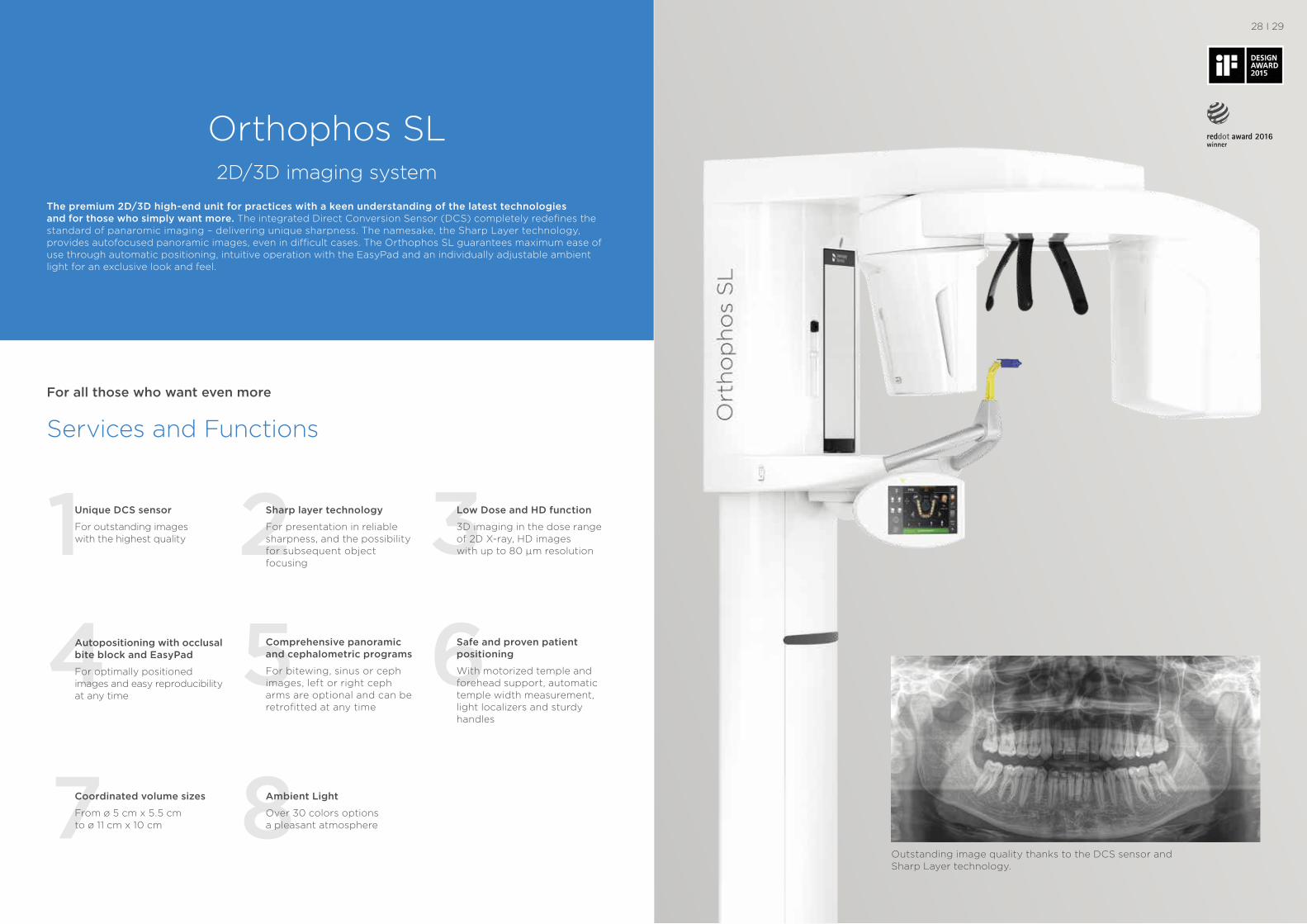
Orthophos SL 2D/3D imaging system
Services and Functions
For all those who want even more
The premium 2D/3D high-end unit for practices with a keen understanding of the latest technologies and for those who simply want more. The integrated Direct Conversion Sensor (DCS) completely redefines the standard of panaromic imaging – delivering unique sharpness. The namesake, the Sharp Layer technology, provides autofocused panoramic images, even in difficult cases. The Orthophos SL guarantees maximum ease of use through automatic positioning, intuitive operation with the EasyPad and an individually adjustable ambient light for an exclusive look and feel.
1 2 3Unique DCS sensor
For outstanding images with the highest quality
Low Dose and HD function
3D imaging in the dose range of 2D X-ray, HD images with up to 80 μm resolution
Sharp layer technology
For presentation in reliable sharpness, and the possibility for subsequent object focusing
4
87
5 6Comprehensive panoramic and cephalometric programs
For bitewing, sinus or ceph images, left or right ceph arms are optional and can be retrofitted at any time
Safe and proven patient positioning
With motorized temple and forehead support, automatic temple width measurement, light localizers and sturdy handles
Autopositioning with occlusal bite block and EasyPad
For optimally positioned images and easy reproducibility at any time
Ambient Light
Over 30 colors options a pleasant atmosphere
Coordinated volume sizes
From ø 5 cm x 5.5 cm to ø 11 cm x 10 cm
28 I 29
Outstanding image quality thanks to the DCS sensor and Sharp Layer technology.
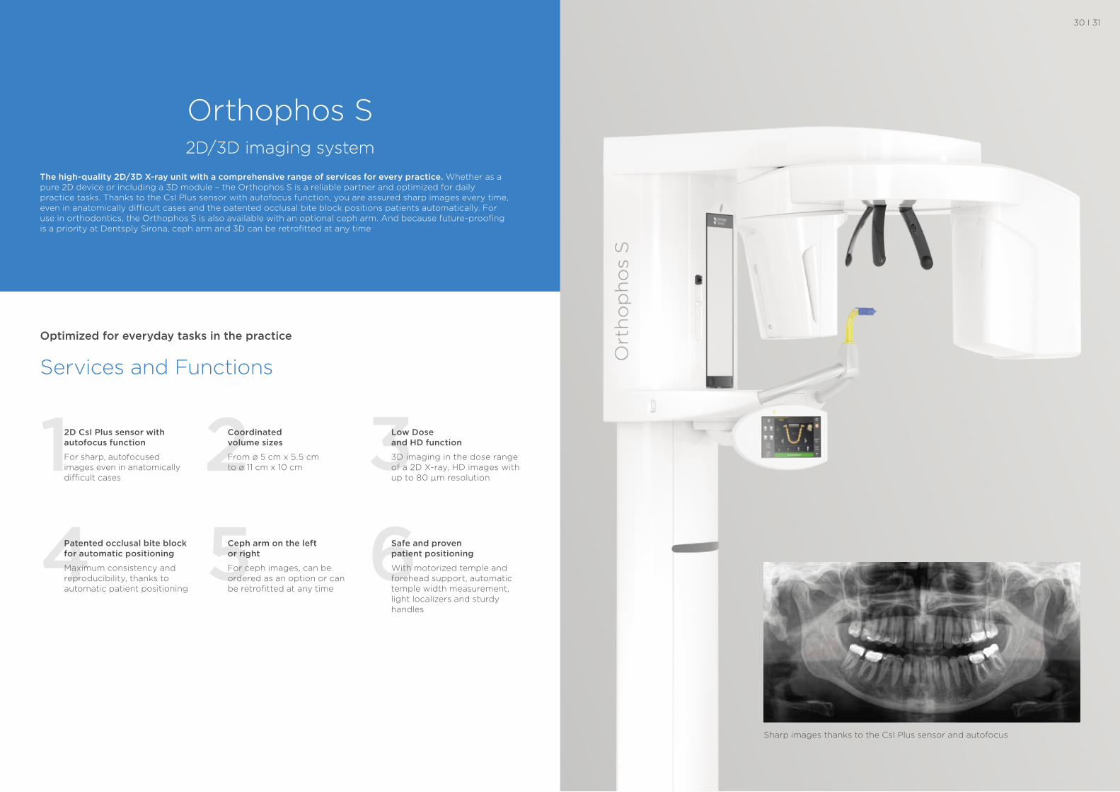
Orthophos S 2D/3D imaging system
The high-quality 2D/3D X-ray unit with a comprehensive range of services for every practice. Whether as a pure 2D device or including a 3D module – the Orthophos S is a reliable partner and optimized for daily practice tasks. Thanks to the CsI Plus sensor with autofocus function, you are assured sharp images every time, even in anatomically difficult cases and the patented occlusal bite block positions patients automatically. For use in orthodontics, the Orthophos S is also available with an optional ceph arm. And because future-proofing is a priority at Dentsply Sirona, ceph arm and 3D can be retrofitted at any time
1 2 32D CsI Plus sensor with autofocus function
For sharp, autofocused images even in anatomically difficult cases
Low Dose and HD function
3D imaging in the dose range of a 2D X-ray, HD images with up to 80 µm resolution
Coordinated volume sizes
From ø 5 cm x 5.5 cm to ø 11 cm x 10 cm
4 5 6Ceph arm on the left or right
For ceph images, can be ordered as an option or can be retrofitted at any time
Safe and proven patient positioning
With motorized temple and forehead support, automatic temple width measurement, light localizers and sturdy handles
Patented occlusal bite block for automatic positioning
Maximum consistency and reproducibility, thanks to automatic patient positioning
Services and Functions
Optimized for everyday tasks in the practice
30 I 31
Sharp images thanks to the CsI Plus sensor and autofocus
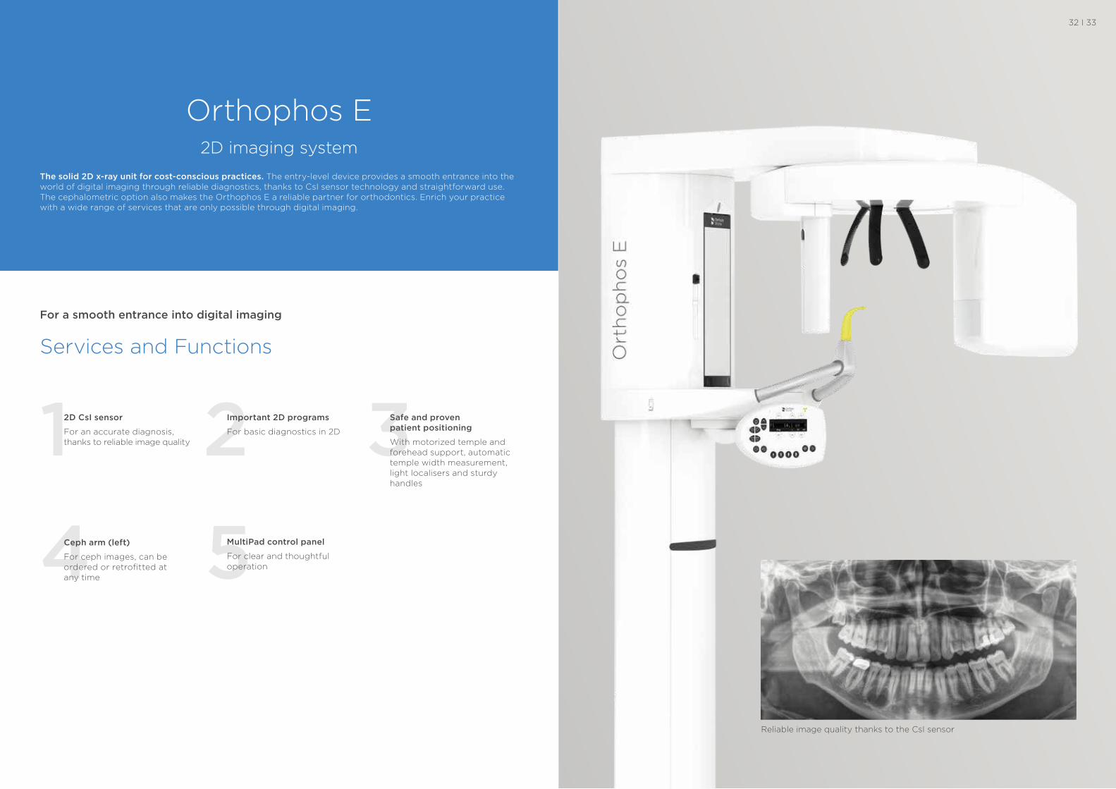
Orthophos E 2D imaging system
The solid 2D x-ray unit for cost-conscious practices. The entry-level device provides a smooth entrance into the world of digital imaging through reliable diagnostics, thanks to CsI sensor technology and straightforward use. The cephalometric option also makes the Orthophos E a reliable partner for orthodontics. Enrich your practice with a wide range of services that are only possible through digital imaging.
1 2 32D CsI sensor
For an accurate diagnosis, thanks to reliable image quality
Safe and proven patient positioning
With motorized temple and forehead support, automatic temple width measurement, light localisers and sturdy handles
Important 2D programs
For basic diagnostics in 2D
4 5MultiPad control panel
For clear and thoughtful operation
Ceph arm (left)
For ceph images, can be ordered or retrofitted at any time
Services and Functions
For a smooth entrance into digital imaging
32 I 33
Reliable image quality thanks to the CsI sensor
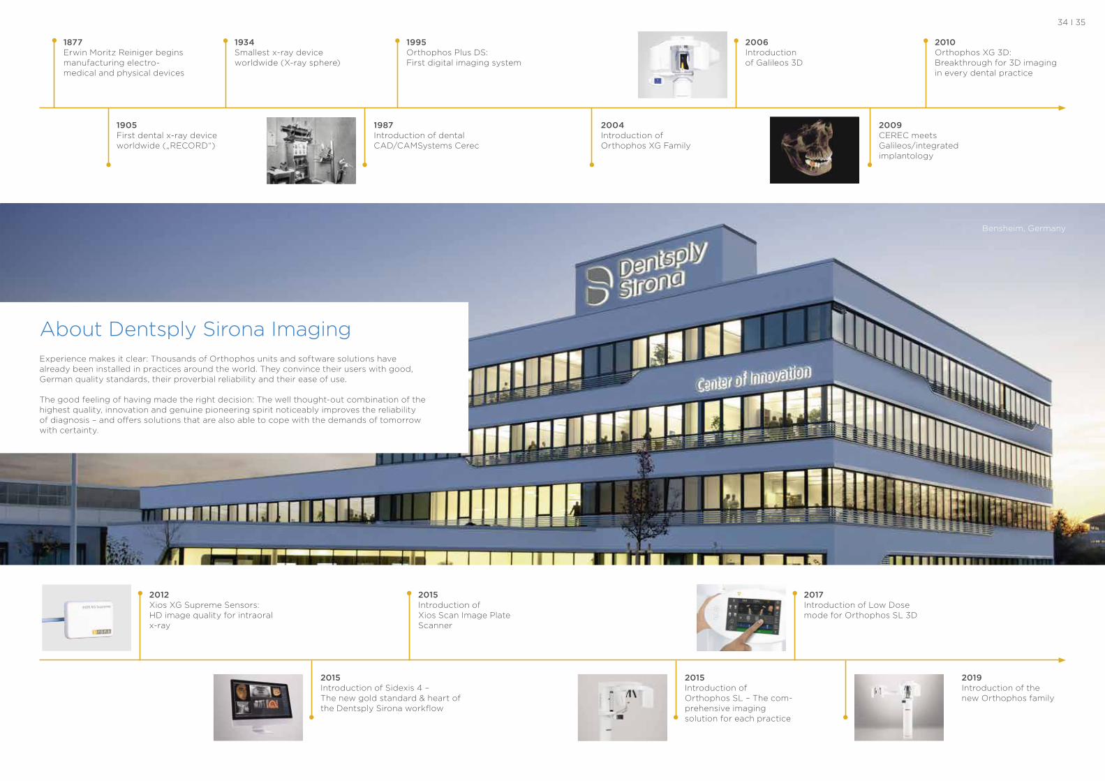
About Dentsply Sirona Imaging Experience makes it clear: Thousands of Orthophos units and software solutions have already been installed in practices around the world. They convince their users with good, German quality standards, their proverbial reliability and their ease of use.
The good feeling of having made the right decision: The well thought-out combination of the highest quality, innovation and genuine pioneering spirit noticeably improves the reliability of diagnosis – and offers solutions that are also able to cope with the demands of tomorrow with certainty.
Bensheim, Germany
34 I 35
1877 Erwin Moritz Reiniger begins manufacturing electro- medical and physical devices
1934 Smallest x-ray device worldwide (X-ray sphere)
1995 Orthophos Plus DS: First digital imaging system
1905 First dental x-ray device worldwide („RECORD“)
2004 Introduction of Orthophos XG Family
2006 Introduction of Galileos 3D
2009 CEREC meets Galileos/integrated implantology
1987 Introduction of dental CAD/CAMSystems Cerec
2015 Introduction of Xios Scan Image Plate Scanner
2010 Orthophos XG 3D: Breakthrough for 3D imaging in every dental practice
2017 Introduction of Low Dose mode for Orthophos SL 3D
2015 Introduction of Sidexis 4 – The new gold standard & heart of the Dentsply Sirona workflow
2015 Introduction of Orthophos SL – The com-prehensive imaging solution for each practice
2012 Xios XG Supreme Sensors: HD image quality for intraoral x-ray
2019 Introduction of the new Orthophos family
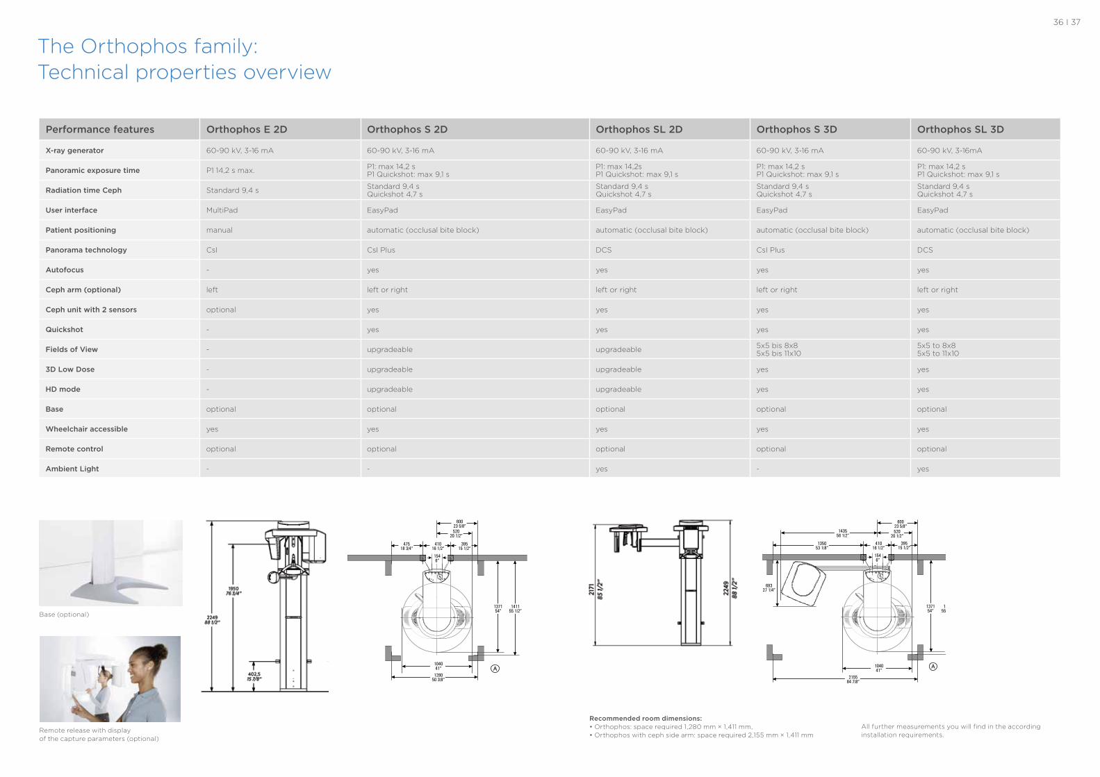
The Orthophos family:Technical properties overview
Base (optional)
Remote release with display of the capture parameters (optional)
Performance features Orthophos E 2D Orthophos S 2D Orthophos SL 2D Orthophos S 3D Orthophos SL 3D
X-ray generator 60-90 kV, 3-16 mA 60-90 kV, 3-16 mA 60-90 kV, 3-16 mA 60-90 kV, 3-16 mA 60-90 kV, 3-16mA
Panoramic exposure time P1 14,2 s max. P1: max 14,2 s P1 Quickshot: max 9,1 s
P1: max 14,2s P1 Quickshot: max 9,1 s
P1: max 14,2 s P1 Quickshot: max 9,1 s
P1: max 14,2 s P1 Quickshot: max 9,1 s
Radiation time Ceph Standard 9,4 s Standard 9,4 s Quickshot 4,7 s
Standard 9,4 s Quickshot 4,7 s
Standard 9,4 s Quickshot 4,7 s
Standard 9,4 s Quickshot 4,7 s
User interface MultiPad EasyPad EasyPad EasyPad EasyPad
Patient positioning manual automatic (occlusal bite block) automatic (occlusal bite block) automatic (occlusal bite block) automatic (occlusal bite block)
Panorama technology CsI CsI Plus DCS CsI Plus DCS
Autofocus - yes yes yes yes
Ceph arm (optional) left left or right left or right left or right left or right
Ceph unit with 2 sensors optional yes yes yes yes
Quickshot - yes yes yes yes
Fields of View - upgradeable upgradeable 5x5 bis 8x85x5 bis 11x10
5x5 to 8x85x5 to 11x10
3D Low Dose - upgradeable upgradeable yes yes
HD mode - upgradeable upgradeable yes yes
Base optional optional optional optional optional
Wheelchair accessible yes yes yes yes yes
Remote control optional optional optional optional optional
Ambient Light - - yes - yes
Recommended room dimensions:• Orthophos: space required 1,280 mm × 1,411 mm, • Orthophos with ceph side arm: space required 2,155 mm × 1,411 mm
All further measurements you will find in the according installation requirements.
36 I 37
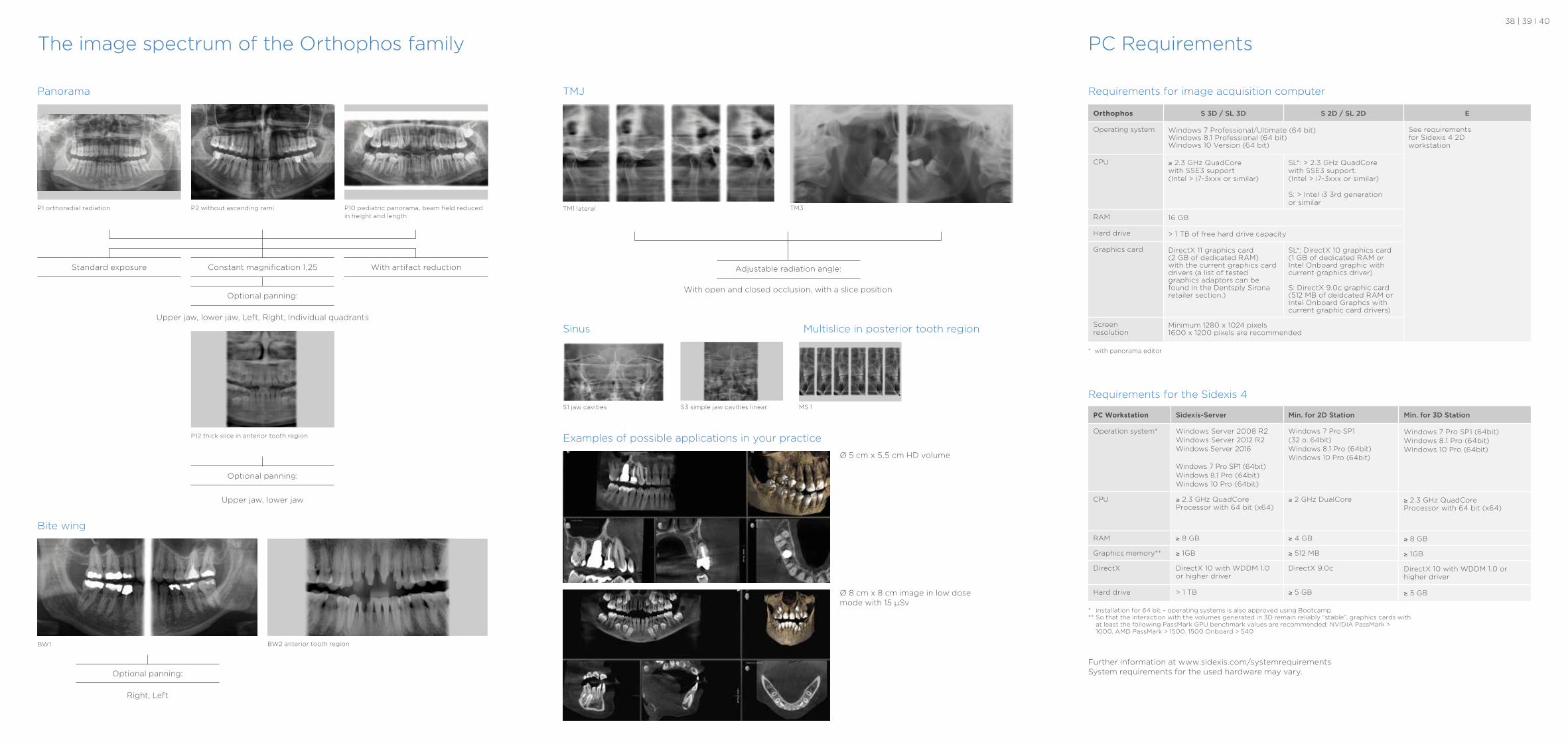
The image spectrum of the Orthophos family
TMJ
TM3
Sinus Multislice in posterior tooth region
Examples of possible applications in your practice
TM1 lateral
With open and closed occlusion, with a slice position
Adjustable radiation angle:
S1 jaw cavities S3 simple jaw cavities linear MS 1
PC Requirements
Requirements for image acquisition computer
Orthophos S 3D / SL 3D S 2D / SL 2D E
Operating system Windows 7 Professional/Ultimate (64 bit)Windows 8.1 Professional (64 bit)Windows 10 Version (64 bit)
See requirementsfor Sidexis 4 2Dworkstation
CPU ≥ 2.3 GHz QuadCorewith SSE3 support(Intel > i7-3xxx or similar)
SL*: > 2.3 GHz QuadCorewith SSE3 support. (Intel > i7-3xxx or similar)
S: > Intel i3 3rd generation or similar
RAM 16 GB
Hard drive > 1 TB of free hard drive capacity
Graphics card DirectX 11 graphics card (2 GB of dedicated RAM) with the current graphics card drivers (a list of tested graphics adaptors can befound in the Dentsply Sironaretailer section.)
SL*: DirectX 10 graphics card (1 GB of dedicated RAM orIntel Onboard graphic with current graphics driver)
S: DirectX 9.0c graphic card(512 MB of deidcated RAM orIntel Onboard Graphcs with current graphic card drivers)
Screen resolution
Minimum 1280 x 1024 pixels1600 x 1200 pixels are recommended
PC Workstation Sidexis-Server Min. for 2D Station Min. for 3D Station
Operation system* Windows Server 2008 R2 Windows Server 2012 R2 Windows Server 2016
Windows 7 Pro SP1 (64bit) Windows 8.1 Pro (64bit) Windows 10 Pro (64bit)
Windows 7 Pro SP1 (32 o. 64bit)Windows 8.1 Pro (64bit)Windows 10 Pro (64bit)
Windows 7 Pro SP1 (64bit)Windows 8.1 Pro (64bit)Windows 10 Pro (64bit)
CPU ≥ 2.3 GHz QuadCore Processor with 64 bit (x64)
≥ 2 GHz DualCore ≥ 2.3 GHz QuadCore Processor with 64 bit (x64)
RAM ≥ 8 GB ≥ 4 GB ≥ 8 GB
Graphics memory** ≥ 1GB ≥ 512 MB ≥ 1GB
DirectX DirectX 10 with WDDM 1.0 or higher driver
DirectX 9.0c DirectX 10 with WDDM 1.0 or higher driver
Hard drive > 1 TB ≥ 5 GB ≥ 5 GB
Requirements for the Sidexis 4
* installation for 64 bit – operating systems is also approved using Bootcamp ** So that the interaction with the volumes generated in 3D remain reliably “stable”, graphics cards with
at least the following PassMark GPU benchmark values are recommended: NVIDIA PassMark > 1000. AMD PassMark > 1500. 1500 Onboard > 540
* with panorama editor
Further information at www.sidexis.com/systemrequirementsSystem requirements for the used hardware may vary.
Ø 5 cm x 5.5 cm HD volume
Ø 8 cm x 8 cm image in low dose mode with 15 μSv
38 | 39 I 40
Panorama
P2 without ascending rami P1 orthoradial radiation P10 pediatric panorama, beam field reducedin height and length
Upper jaw, lower jaw, Left, Right, Individual quadrants
Upper jaw, lower jaw
Optional panning:
Standard exposure Constant magnification 1,25 With artifact reduction
Optional panning:
P12 thick slice in anterior tooth region
Bite wing
BW1 BW2 anterior tooth region
Right, Left
Optional panning:
Clinical Procedures
PreventiveRestorativeOrthodonticsEndodonticsImplantsProsthetics
Platform Technologies
CAD/CAMImagingTreatment CentersInstruments
Dentsply Sirona
Sirona Dental Systems GmbH Fabrikstraße 31, 64625 Bensheim, Germany dentsplysirona.com
Su
bje
ct t
o t
ech
nic
al c
han
ge
s an
d e
rro
rs in
th
e te
xt, O
rde
r N
o A
911
00
-M4
7-C
20
6, P
rin
ted
in G
erm
any,
Dis
po
No
. 04
60
2, O
EW
19 0
319
V0