Exfoliative cytopathology of human lip neoplasm. · Exfoliative cytopathology of human lip...
6
J Med Oncl Ther 2016; 1 (2): 41-46 Journal of Medical Oncology and Therapeutics J Med Oncl Ther 2016 Volume 1 Issue 2 41 Exfoliative cytopathology of human lip neoplasm. Abhimanyu Mohanta 1 , Prafulla K Mohanty 2 1 Research Scholar, Post-Graduate Department of Zoology, Utkal University, Bhubaneswar, Odisha, India. 2 Professor, Post-Graduate Department of Zoology, Utkal University, Bhubaneswar, Odisha, India. Objective: The objective of the present study is to investigate the cytopathology of human lip neoplasms, pattern of cervical lymph node (CLN) metastasis and to analyse the probable etiological risk factors associated with it. Methodology: In this hospital based case-control study, 22 subjects (11 cases of lip neoplasm and 11 healthy individuals as Control group) were included in this study. Scraped exfoliated cytosmears were collected from the affected site of the lip and smearing was done in the pre- cleaned-coded glass-slides. Two such slides were prepared from each subject. The cytosmears were immediately fixed in aceto-alcohol (1part of glacial acetic acid:3 part of absolute ethyl alcohol) fixative. One set of the slide was stained with Papanicolaou’s stain and the other set wascounter-stained with Giemsa’s Solution for cytopathological analysis.The TNM (Tumor- Node-Metastasis) System for staging of lip cancer formulated by American Joint Committee for Cancer (AJCC) Staging and End Results Reporting-2010 was followed. Results: Lower lip is found to be the most common site in the genesis of lip neoplasm. Pleomorphic cytological atypiassuch as Micronucleated cell (MNC), Plump keratinized squamous cell (PKSC), Keratinized spindle cell (KSC), Keratinized tadpole cell (KTC), Keratinized strap (Anitschkow) cell (KSC-A), Keratinized fiber cell (KFC), Keratinized round cell (KRC) and Non-keratinized malignant squamous cell (NMSC) with drastic modification were frequently observed in lip neoplasm. Unpredictable metastasis is a common feature in lip neoplasm cases. Conclusion: Excluding PKSC and MNC, presence of any other atypias in the cytosmears of lip neoplasm indicate the state of malignancy. Thus, exfoliative cytopathology has a potentiality in early detection of lip carcinoma in particular and oral cancer in general. Abstract Keywords: Exfoliated cytosmear, Lip neoplasm, Pleomorphism, Cytopathology, Cytological atypia. Accepted September 01, 2016 Introduction Lips, the superior and inferior ones are anatomically occupied an important position and primarily act as door of the gateway to the oral cavity. Intelligently designed lips play important roles in manipulation of food and phonetics. Due to excessive exposure to sun light and tobacco-burnt smoke, the melanin-free delicate lips are generally affected by a wide variety of diseases with diagnostic dilemma in most of cases. Lip carcinoma is relatively common among malignancies of the head and neck region, accounting for 12% of all head and neck cancers, excluding non- melanoma skin cancer and accounts for approximately one quarter of oral cavity cancers [1]. Lower lip is more commonly affected than the upper lip. The aetiological and predisposing factors of lip cancer include excessive exposure to sunlight, tobacco usage, viral infections, racial factors, a genetic predisposition, immunosuppression, immunodeficiency, certain occupations and familial factors [2-4]. Onset of lip carcinoma occurs frequently on premalignant lesions, especially on chronic keratotic cheilitis, pointing out the importance of early diagnosis and appropriate treatment for preblastomatous cheilitis. Squamous cell carcinoma (SCC) of the lip is an infiltrating and destructive malignant epithelial tumor, with high potential for lymphatic and/or haematogenic metastasis [5]. Inspite of various technological advancement and implementation of sophisticated methods, exfoliative cytopathology has primarily been accepted as an important tool for early detection of oral cancer. Due to its simplicity,
Transcript of Exfoliative cytopathology of human lip neoplasm. · Exfoliative cytopathology of human lip...

J Med Oncl Ther 2016; 1 (2): 41-46 Journal of Medical Oncology and Therapeutics
J Med Oncl Ther 2016 Volume 1 Issue 241
Exfoliative cytopathology of human lip neoplasm.
Abhimanyu Mohanta1, Prafulla K Mohanty2
1Research Scholar, Post-Graduate Department of Zoology, Utkal University, Bhubaneswar, Odisha, India.2Professor, Post-Graduate Department of Zoology, Utkal University, Bhubaneswar, Odisha, India.
Objective: The objective of the present study is to investigate the cytopathology of human lip neoplasms, pattern of cervical lymph node (CLN) metastasis and to analyse the probable etiological risk factors associated with it.
Methodology: In this hospital based case-control study, 22 subjects (11 cases of lip neoplasm and 11 healthy individuals as Control group) were included in this study. Scraped exfoliated cytosmears were collected from the affected site of the lip and smearing was done in the pre-cleaned-coded glass-slides. Two such slides were prepared from each subject. The cytosmears were immediately fixed in aceto-alcohol (1part of glacial acetic acid:3 part of absolute ethyl alcohol) fixative. One set of the slide was stained with Papanicolaou’s stain and the other set wascounter-stained with Giemsa’s Solution for cytopathological analysis.The TNM (Tumor-Node-Metastasis) System for staging of lip cancer formulated by American Joint Committee for Cancer (AJCC) Staging and End Results Reporting-2010 was followed.
Results: Lower lip is found to be the most common site in the genesis of lip neoplasm. Pleomorphic cytological atypiassuch as Micronucleated cell (MNC), Plump keratinized squamous cell (PKSC), Keratinized spindle cell (KSC), Keratinized tadpole cell (KTC), Keratinized strap (Anitschkow) cell (KSC-A), Keratinized fiber cell (KFC), Keratinized round cell (KRC) and Non-keratinized malignant squamous cell (NMSC) with drastic modification were frequently observed in lip neoplasm. Unpredictable metastasis is a common feature in lip neoplasm cases.
Conclusion: Excluding PKSC and MNC, presence of any other atypias in the cytosmears of lip neoplasm indicate the state of malignancy. Thus, exfoliative cytopathology has a potentiality in early detection of lip carcinoma in particular and oral cancer in general.
Abstract
Keywords: Exfoliated cytosmear, Lip neoplasm, Pleomorphism, Cytopathology, Cytological atypia.
Accepted September 01, 2016
Introduction Lips, the superior and inferior ones are anatomically occupied an important position and primarily act as door of the gateway to the oral cavity. Intelligently designed lips play important roles in manipulation of food and phonetics. Due to excessive exposure to sun light and tobacco-burnt smoke, the melanin-free delicate lips are generally affected by a wide variety of diseases with diagnostic dilemma in most of cases. Lip carcinoma is relatively common among malignancies of the head and neck region, accounting for 12% of all head and neck cancers, excluding non-melanoma skin cancer and accounts for approximately one quarter of oral cavity cancers [1]. Lower lip is more commonly affected than the upper lip. The aetiological and predisposing factors of lip cancer include excessive
exposure to sunlight, tobacco usage, viral infections, racial factors, a genetic predisposition, immunosuppression, immunodeficiency, certain occupations and familial factors [2-4]. Onset of lip carcinoma occurs frequently on premalignant lesions, especially on chronic keratotic cheilitis, pointing out the importance of early diagnosis and appropriate treatment for preblastomatous cheilitis. Squamous cell carcinoma (SCC) of the lip is an infiltrating and destructive malignant epithelial tumor, with high potential for lymphatic and/or haematogenic metastasis [5].
Inspite of various technological advancement and implementation of sophisticated methods, exfoliative cytopathology has primarily been accepted as an important tool for early detection of oral cancer. Due to its simplicity,

Exfoliative cytopathology of human lip neoplasm.
J Med Oncl Ther 2016 Volume 1 Issue 242
reliability and economically affordability (with respect to time and money), there is a growing interest on oral exfoliative cytopathology world-wide. To our knowledge, none of the published paper reported on exfoliative cytopathology of human lip carcinoma in detailed so far. Therefore, a detailed account of exfoliative cytopathology of premalignant and malignant neoplasm of the lip was analysed and various etiological factors responsible for the genesis of lip neoplasm was discussed in this study.
Materials and MethodsThe Subjects
In a hospital-based study, out of 136 oral cases, 11 (8.08%) lip neoplasm cases (5 males and 6 females) registered at the Out-patient Department (OPD), Acharya Harihar Regional Cancer Centre (AHRCC), Cuttack, Odisha, India during May 2007 to May 2009 were included in this study. Detailed case-history including the nature and types of addiction of each individual was recorded prior to the collection of samples. Addicted individuals were habituated with different forms of tobacco and alcohol for more than 15 years. Age-group and sex matched non-addicted 11 healthy individuals were also included in this study as Control group. Thus, a total of 22 lip cases were taken into account for this study.
Collection of Samples and Staining
Prior to the collection of sample, written consent of the respective subject was obtained. Two scalpel -scraped exfoliated cytosmears were collected from the affected site of the lip on the pre cleaned-coded glass-slides. Collected cytosmears were fixed in 1:3 aceto-alcohol (1 part of glacial acetic acid and 3 parts of ethyl alcohol) immediately. A set of smears was stained with Papanicolaou’s stain and the other set was counterstained with Giemsa’s stain for cytopathological analysis. Photomicrographs were taken out as the supporting evidences.
Statistical Analysis
Out of 1000 observed cells, the cytological atypias were scored. Z-test was carried out at 1% (p<0.01) level of significance.
Ethical considerations
This study was approved (Reference No EC/UU-38832/2007) by the Subject Research Committee (SRC) of Utkal University, Bhubaneswar, Odisha, India and necessary permission from the Director, AHRCC, Cuttack, Odisha, India was also obtained for the same purpose.
ResultsThe Cases: Clinical Aspects
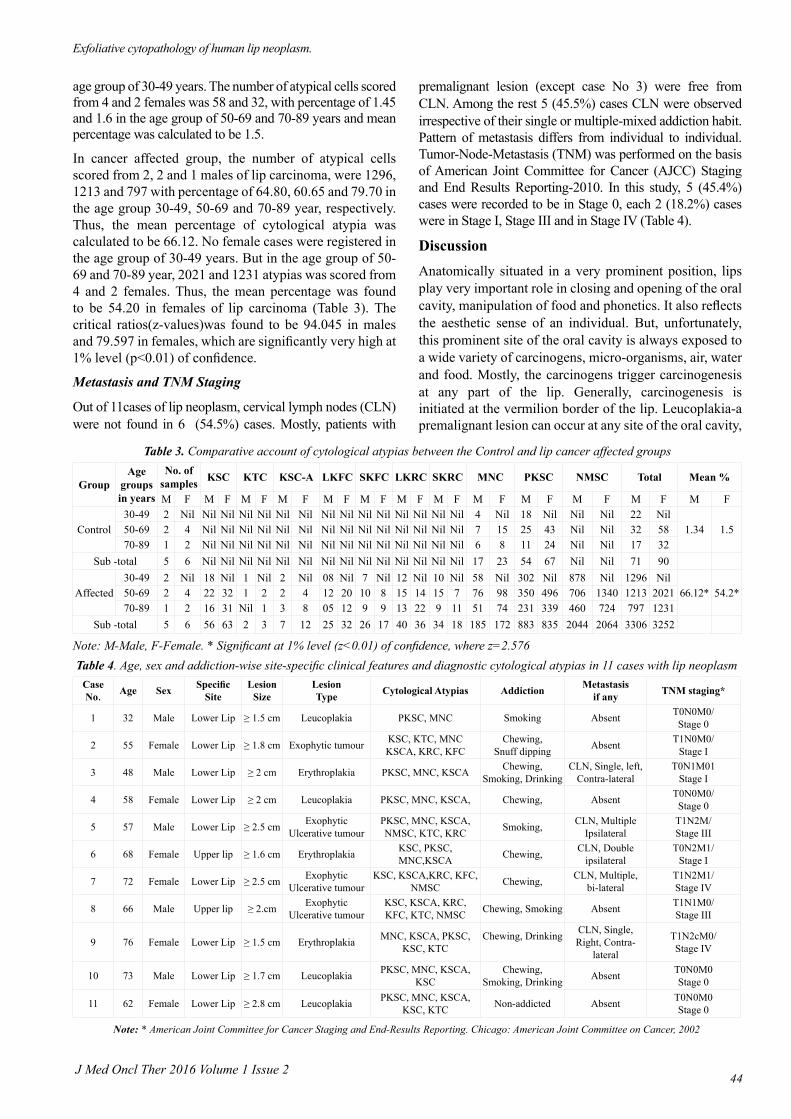
Out of 136 collected samples, a total of 11 (8.08%) cases of lip neoplasm were recorded in our study (Figure 1). Lower lip was found to be more prone (81.8%) than the upper lip (18.2%). Among them 5 (45.5%) male and 6 (54.5%) were females. Seven (63.6%) cases were with
premalignant lesion and 4 (36.4%) were malignant. Two male and 2 female were suffering from leucoplakia. One male and 2 female were with erythroplakia. Ten Among 11 patients, 10 (90.9%) addicted to different forms of tobacco and alcohol and only one (9.1%) female (Case No 11) was refrained from any type of addiction in her life. A brief general attributes of the subjects is summarized in Table 1.
Cytopathology
During this investigation a number of cytological atypias
Figure 1. Lip neoplasm; a: Leucoplakia in the lower lip, b: A tumor towards inner side of the lower lip
Attributes Male Female Total (%)No of Collected SamplesControl 5 6 11 (50)Affected 5 6 11 (50)Age groups in years30-49 2 Nil 2 (18.2)50-69 2 4 6 (54.5)70-89 1 2 3 (27.3)PathogenicityLeucoplakia 2 2 4 (36.35)Erythroplakia 1 2 3 (27.4)Malignant 2 2 4 (36.35)Addiction PatternChewers/Snuff dippers 1 3 4 (36.3)Smokers 2 Nil 2 (18.2)Alcoholics Nil 1 1 (9.1)Mixed 2 1 3 (27.3)Non-addicted Nil 1 1 (9.1)OccupationLabour 3 4 7 (63.6)Service 2 2 4 (36.4)
Table 1. General attributes of the subjects
Mohanta/Mohanty
J Med Oncl Ther 2016 Volume 1 Issue 243
were observed exhibiting pleomorphism. Normal epidermal cells of the lip were found to be more or less polyhedral with well-defined cell boundary, non-keratinized cytoplasm and centrally located rounded or oval nucleus (Figure 2). In due course of carcinogenesis, the normal cells of the lip were metamorphosed into Micronucleated cell (MNC), Plump keratinized squamous cell (PKSC), Keratinized spindle cell (KSC), Keratinized tadpole cell (KTC), Keratinized strap (Anitschkow) cell (KSC-A), Keratinized fiber cell (KFC), Keratinized round cell (KRC) and Non-keratinized malignant squamous cell (NMSC) with drastic modification (Figure 3). KFC and KRC were observed to be two different forms- large and small. Thus, large keratinized fiber cell (LKFC), small keratinized fiber cell (SKFC), large keratinized round cell (LKRC) and small keratinized round cell (SKRC) have their own identity so far as cytological pleomorphism is concerned.MNC and PKSC were observed to be well differentiated; KSC, KTC, KSC-A, KFC and KRC were moderately differentiated; whereas NMSC was reported to be absolutely poorly differentiated. Universal occurrence of MNCs in all exfoliated cytosmear with gradual increase in its frequency from normal to malignant cases proves itself to be an onco-indicator as well as a biomarker of carcinogenesis. PKSC were chiefly found in premalignant lesions (leucoplakia and erythroplakia) and less in number, if present, in benign and malignant cases. The moderately differentiated KSC, KTC, KSC-A, KFC and KRC as well as poorly differentiated NMSC were observed in malignant neoplasm of human lip. In most of the premalignant cases
(as in case No 6, 9, 10, 11) KSC, KSC-A and/ KTC were observed. It is important to note that except PKSC and MNC, other detected pleomorphic cells may or may not be found in all cases, but presence of any type of these cells indicates the state of malignancy.
Cytological differentiation indicates that all the normal exfoliated cells were well differentiated squamous cells (WDSC).However, well differentiated and moderately differentiated squamous cells (MDSC).were recorded to be 27.3% each and the rest 45.4% were of poorly differentiated squamous cells (PDSC) type in the lip neoplasm cases (Table 2).
Statistical Analysis
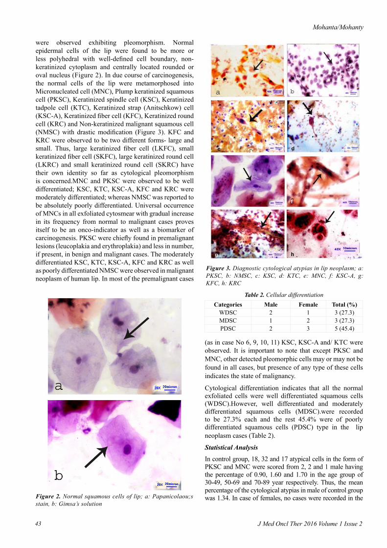
In control group, 18, 32 and 17 atypical cells in the form of PKSC and MNC were scored from 2, 2 and 1 male having the percentage of 0.90, 1.60 and 1.70 in the age group of 30-49, 50-69 and 70-89 year respectively. Thus, the mean percentage of the cytological atypias in male of control group was 1.34. In case of females, no cases were recorded in the Figure 2. Normal squamous cells of lip; a: Papanicolaou;s
stain, b: Gimsa’s solution
Figure 3. Diagnostic cytological atypias in lip neoplasm; a: PKSC, b: NMSC, c: KSC, d: KTC, e: MNC, f: KSC-A, g: KFC, h: KRC
Categories Male Female Total (%)WDSC 2 1 3 (27.3)MDSC 1 2 3 (27.3)PDSC 2 3 5 (45.4)
Table 2. Cellular differentiation
Exfoliative cytopathology of human lip neoplasm.
J Med Oncl Ther 2016 Volume 1 Issue 244
age group of 30-49 years. The number of atypical cells scored from 4 and 2 females was 58 and 32, with percentage of 1.45 and 1.6 in the age group of 50-69 and 70-89 years and mean percentage was calculated to be 1.5.
In cancer affected group, the number of atypical cells scored from 2, 2 and 1 males of lip carcinoma, were 1296, 1213 and 797 with percentage of 64.80, 60.65 and 79.70 in the age group 30-49, 50-69 and 70-89 year, respectively. Thus, the mean percentage of cytological atypia was calculated to be 66.12. No female cases were registered in the age group of 30-49 years. But in the age group of 50-69 and 70-89 year, 2021 and 1231 atypias was scored from 4 and 2 females. Thus, the mean percentage was found to be 54.20 in females of lip carcinoma (Table 3). The critical ratios(z-values)was found to be 94.045 in males and 79.597 in females, which are significantly very high at 1% level (p<0.01) of confidence.
Metastasis and TNM Staging
Out of 11cases of lip neoplasm, cervical lymph nodes (CLN) were not found in 6 (54.5%) cases. Mostly, patients with
premalignant lesion (except case No 3) were free from CLN. Among the rest 5 (45.5%) cases CLN were observed irrespective of their single or multiple-mixed addiction habit. Pattern of metastasis differs from individual to individual. Tumor-Node-Metastasis (TNM) was performed on the basis of American Joint Committee for Cancer (AJCC) Staging and End Results Reporting-2010. In this study, 5 (45.4%) cases were recorded to be in Stage 0, each 2 (18.2%) cases were in Stage I, Stage III and in Stage IV (Table 4).
DiscussionAnatomically situated in a very prominent po sition, lips play very important role in closing and opening of the oral cavity, manipulation of food and phonetics. It also reflects the aesthetic sense of an individual. But, unfortunately, this prominent site of the oral cavity is always exposed to a wide variety of carcinogens, micro-organisms, air, water and food. Mostly, the carcinogens trigger carcinogenesis at any part of the lip. Generally, carcinogenesis is initiated at the vermilion border of the lip. Leucoplakia-a premalignant lesion can occur at any site of the oral cavity,
GroupAge
groups in years
No. of samples KSC KTC KSC-A LKFC SKFC LKRC SKRC MNC PKSC NMSC Total Mean %
M F M F M F M F M F M F M F M F M F M F M F M F M F
Control30-49 2 Nil Nil Nil Nil Nil Nil Nil Nil Nil Nil Nil Nil Nil Nil Nil 4 Nil 18 Nil Nil Nil 22 Nil
1.34 1.550-69 2 4 Nil Nil Nil Nil Nil Nil Nil Nil Nil Nil Nil Nil Nil Nil 7 15 25 43 Nil Nil 32 5870-89 1 2 Nil Nil Nil Nil Nil Nil Nil Nil Nil Nil Nil Nil Nil Nil 6 8 11 24 Nil Nil 17 32
Sub -total 5 6 Nil Nil Nil Nil Nil Nil Nil Nil Nil Nil Nil Nil Nil Nil 17 23 54 67 Nil Nil 71 90
Affected30-49 2 Nil 18 Nil 1 Nil 2 Nil 08 Nil 7 Nil 12 Nil 10 Nil 58 Nil 302 Nil 878 Nil 1296 Nil
66.12* 54.2*50-69 2 4 22 32 1 2 2 4 12 20 10 8 15 14 15 7 76 98 350 496 706 1340 1213 202170-89 1 2 16 31 Nil 1 3 8 05 12 9 9 13 22 9 11 51 74 231 339 460 724 797 1231
Sub -total 5 6 56 63 2 3 7 12 25 32 26 17 40 36 34 18 185 172 883 835 2044 2064 3306 3252
Note: M-Male, F-Female. * Significant at 1% level (z<0.01) of confidence, where z=2.576
Table 3. Comparative account of cytological atypias between the Control and lip cancer affected groups
Case No. Age Sex Specific
SiteLesion
SizeLesionType Cytological Atypias Addiction Metastasis
if any TNM staging*
1 32 Male Lower Lip ≥ 1.5 cm Leucoplakia PKSC, MNC Smoking Absent T0N0M0/Stage 0
2 55 Female Lower Lip ≥ 1.8 cm Exophytic tumour KSC, KTC, MNCKSCA, KRC, KFC
Chewing,Snuff dipping Absent T1N0M0/
Stage I
3 48 Male Lower Lip ≥ 2 cm Erythroplakia PKSC, MNC, KSCA Chewing, Smoking, Drinking
CLN, Single, left, Contra-lateral
T0N1M01Stage I
4 58 Female Lower Lip ≥ 2 cm Leucoplakia PKSC, MNC, KSCA, Chewing, Absent T0N0M0/Stage 0
5 57 Male Lower Lip ≥ 2.5 cm Exophytic Ulcerative tumour
PKSC, MNC, KSCA, NMSC, KTC, KRC Smoking, CLN, Multiple
IpsilateralT1N2M/Stage III
6 68 Female Upper lip ≥ 1.6 cm Erythroplakia KSC, PKSC, MNC,KSCA Chewing, CLN, Double
ipsilateralT0N2M1/
Stage I
7 72 Female Lower Lip ≥ 2.5 cm Exophytic Ulcerative tumour
KSC, KSCA,KRC, KFC, NMSC Chewing, CLN, Multiple,
bi-lateralT1N2M1/Stage IV
8 66 Male Upper lip ≥ 2.cm Exophytic Ulcerative tumour
KSC, KSCA, KRC, KFC, KTC, NMSC Chewing, Smoking Absent T1N1M0/
Stage III
9 76 Female Lower Lip ≥ 1.5 cm Erythroplakia MNC, KSCA, PKSC, KSC, KTC
Chewing, Drinking CLN, Single, Right, Contra-
lateral
T1N2cM0/Stage IV
10 73 Male Lower Lip ≥ 1.7 cm Leucoplakia PKSC, MNC, KSCA, KSC
Chewing, Smoking, Drinking Absent T0N0M0
Stage 0
11 62 Female Lower Lip ≥ 2.8 cm Leucoplakia PKSC, MNC, KSCA, KSC, KTC Non-addicted Absent T0N0M0
Stage 0
Note: * American Joint Committee for Cancer Staging and End-Results Reporting. Chicago: American Joint Committee on Cancer, 2002
Table 4. Age, sex and addiction-wise site-specific clinical features and diagnostic cytological atypias in 11 cases with lip neoplasm
Mohanta/Mohanty
J Med Oncl Ther 2016 Volume 1 Issue 245
including the vermillion border of the lip, which is the most frequently involved site of the lip. It has a relatively high rate of malignant transformation due to its strong correlation with tobacco and alcohol use [6,7].
Patil and Maheshwari have reported that the prevalence of lip lesions was 18.8%. The most commonly diagnosed lesions were those due to infections, which affected 32.6% of the population, followed by mucocele (29.8%) and premalignant lesions and conditions which were observed in 20.6% of the population. Males were more commonly affected than females [8]. In the present study only 8.08% of lip neoplasm cases were recorded in which females (54.5%) more commonly are affected than males (44.5%) which partially contradicts with the report of Patil and Maheshwari.
Most commonly, lip carcinoma appears as an ulcer or wart-like growth that does not heal. Sometimes, the premalignant lesions like leucoplakia and erythroplakia (an abnormal velvety redness and thickening of the lip) become the precursor and potentially malignant transformer. In our present investigation, KSC, KSC-A like moderately differentiated cytological atypias were observed in most of the smears from the leucoplakia and erythroplakia cases. Presence of these atypias indicates the state of malignancy. The detected cytological atypias are also observed in the exfoliated cytosmearsoforal cancer patients. Thus, in terms of cytopathology, it has also been confirmed that lip cancer is more closely related to cancer of the oral cavity than to that of the skin.
Dufresne and Curlin have reported that lip squamous cell carcinoma (LSCC) may develop from a potentially malignant disorder known as Actinic cheilitis (AC), cheilitis exfoliativa, solar cheilosis, solar keratosis, and actinic keratosis of the lips [9]. AC comprises clinical and histological changes of the lower lip vermilion that occurs almost exclusively in fair-skinned due to chronic exposure to the sun and harmful UV rays [10,11].
Lip cancer (LC) has been related to sun exposure [12] in different descriptive studies of migrants [13,14] and in several case-control studies, measured with proxies like outdoor activities/work [15-18]. LC is more frequently seen among the profession groups who are more exposed to sun light [19,20]. After a 14-year long period of retrospective study at a single Cancer Institution in Mexico City, Luna-Ortiz et al. have presumed that antecedents of ultraviolet (UV) light and tobacco exposure were found in 20 (33.9%) and 16 cases (27%), respectively [21]. International Agency for Research on Cancer (1992) has confirmed that there was a close relation of tobacco and alcohol with lip cancer [22]. Other factors that have been related to LC are low socioeconomic status [23,24], viral infections, family predisposition and immunosuppression [25]. Findings of Adriana Demathe et al. showed that the presence of HPV DNA was detected in 43.33% of lip SCC samples. The presence of viral particles of HPV DNA had no correlation with age, sex, smoking and consumption of alcohol, solar
radiation, clinical staging, histological grade or survival; nevertheless more studies are necessary to understand the real HPV role in lip carcinogenesis [26]. Besides these, some naturally occurring and synthetic food components, food additives and lip-sticks are supposed to be the etiologic culprit of lip neoplasm [27]. Lo´pez et al. concluded that LC is related to phenotype, skin reaction to sun exposure, cumulative and early sun (UV) light exposure, as well as tobacco and alcohol consumption. Leaving the cigarette smoke over the lip surface is predictive of LC risk irrespective of cumulative tobacco consumption [28].
In our study, it has been observed that occupationally a total of 63.6% cases were belong to labourer class and were working mostly in agricultural fields. Rest 36.4% were service holders. Again, habitually 90.9% were addicted to different forms of tobacco and alcohol. Only one (9.1%) female case was reported to be non-addicted. Hence it is presumed that continuous exposure to solar light play an important role in lip carcinogenesis; whereas both tobacco and alcohol triggers the tumor progression followed by regional metastasis.
Probably, due to lack of protective pigment layer, prolonged exposure to sunlight and cumulative reaction of various carcinogens, DNA of lip mucosal cells get mutated and the cells become abnormal. Increased entropy in DNA brings about a successful failure in repairing the mutational alteration in the cell which leads to the event of tumorigenesis. Jenkins et al. have opined that defective DNA repair generates chromosomal derangement that can cause subsequent alterations in gene expression and is a hallmark of progression toward carcinoma [29].Bockm¨uhl et al. have reported that chromosomal aberrations associated with late disease and metastasis are less well characterised in head and neck cancer. It has been found that metastatic deposits mostly retain clonality with their primary cancers [30].
ConclusionExfoliative cytopathology of lip neoplasm exhibits diagnostic cytological pleomorphism and has a close similarity with that of oral cavity cancer. Presence of various cytological atypias including KSC, KSCA, KTC, KFC, KRC, MNC and NMSC in the exfoliated-scraped cytosmears of lip neoplasm indicate the state of malignancy. Also, cumulative exposure to sunlight along with tobacco and alcohol may be considered as real etiologic culprits of lip carcinogenesis. Thus, the present study has a practical utility in early detection and diagnosis of lip neoplasm cases and also it help to understand the drastic impact of tobacco and alcohol on lip mucosa so far as individual aesthetic sense is concerned.
AcknowledgementThe authors are thankful to Prof. Gadadhar Parida, M.D, formerly Professor and Head, Department of Oncopathology, Acharya Harihar Regional Cancer Centre (AHRCC), Cuttack, Odisha, India for his guidance and supervision during cytopathological analysis. We are also indebted to

Exfoliative cytopathology of human lip neoplasm.
J Med Oncl Ther 2016 Volume 1 Issue 246
the Head P.G. Department of Zoology, Utkal University, Vani Vihar, Bhubaneshwar, and Odisha, India and to the Director, AHRCC, Cuttack, and Odisha, India for permitting us to collect samples from oral cancer patients and also for providing library and laboratory facilities. One of us (AM) is grateful to the University Grants Commission (UGC), New Delhi, India for awarding UGC Meritorious Research Fellowship to carry out the research work.
References1. Zitsch RP, Park CW, Renner GJ, Rea JL. Outcome analysis
for lip carcinoma. Otolaryngol Head Neck Surg 1995; 113: 589-596.
2. Muir C, Weiland L. Upper aerodigestive tract cancers. Cancer 1995; 75: 147-53.
3. De Visscher JGAM, Van Der Waal I. Etiology of cancer of the lip, A review. Int J Oral Maxillofac Surg 1998; 27: 199-203.
4. Çankayaa H, Garçaa MF, Bozana N, Işıkb D, Kıroğlua AF. Epidemiological features of the lip cancers and it’s relation with smoking. Eastern Journal of Medicine 2013; 18: 64-67.
5. Pătraşcu V, Ciurea R. Lip squamous carcinoma-epidemiologic, clinical, evolutive and therapeutical aspects. Current Health Sciences Journal 2013; 39: 84-92.
6. Bentley JM, Barankin B, Lauzon GJ. Paying more than lip service to lip lesions. Can Fam Physician 2003; 49: 1111-1116.
7. Bouquot JE, Gundlach KKH. Odd lips: The prevalence of common lip lesions in 23,616 white Americans over 35 years of age. Quintessence Int 1987; 18: 277-284.
8. Patil S, Maheshwari S. Prevalence of lip lesions in an Indian population. J Clin Exp Dent 2014; 6: e374-e378.
9. Dufresne RG, Curlin MU. Actinic cheilitis: A treatment review. Dermatol Surg 1997; 23: 15-21.
10. Schwartz RA, Bridges TM, Butani AK, Ehrlich A. Actinic keratosis: An occupational and environmental disorder. J Eur Acad Dermatol Venereol 2008; 22: 606-615.
11. Menta S, Nico M, Rivitti EA, Lourenço SV. Actinic cheilitis: Histologic study of the entire vermilion and comparison with previous biopsy. J Cutan Pathol 2007; 34: 309-314.
12. International Agency for Research on Cancer. Some naturally occurring and synthetic food components, furocumarins and ultraviolet radiation. IARC Monographs on the Evaluation of the Carcinogenic Risk of Chemicals to Humans 1986; 40.
13. McCredie M, Coates MS. Cancer Incidence in Migrants to New South Wales from 1972 to 1984, New South Wales Central Cancer Registry. Woolloomooloo: NSW Cancer Council 1989.
14. Steinitz R, Parkin DM, Young JL, Bieber CA, Katz L. Cancer incidence in Jewish migrants to Israel, 1961-1981, IARC Scientific Publications No. 98, Lyon: International Agency for Research on Cancer 1989.
15. Keller AZ. Cellular types, survival, race, nativity, occupations, habits and associated diseases in the pathogenesis of lip cancers. Am J Epidemiol 1970; 91: 486-499.
16. Spitzer WO, Hill GB, Chambers LW, Helliwell BE, Murphy
HB. The occupation of fishing as a risk factor in cancer of the lip. N Engl J Med 1975; 293: 419-424.
17. Lindqvist C. Risk factors in lip cancer: A questionnaire survey. Am J Epidemiol 1979; 109: 521-530.
18. Dardanoni L, Gafa´ L, Paterno R, Pavone G. A case control study on LC risk factors in Ragusa (Sicily). Int J Cancer 1984; 34: 335-337.
19. Baker SR. Malignancy of the lip. In Paparella MM, Shumrick DA, Gluckman JL, Meyerhoff WL(Eds) Otolaryngology 3rd (eds). Philadelphia: WB Saunders Company. 1991; 3: 2021-2039.
20. Erdibil HH, Yazıcı MF, Yücel Z, Bozkurt ER. Journal of Lip Cancers, ENT and Head-Neck Surgery 1995; 3: 231-35.
21. Luna OK, Güemes MA, Villavicencio VV, Mosqueda T. Upper lip malignant neoplasms. A study of 59 cases. Med Oral Patol Oral Cir Bucal 2012; 17: e371-e376.
22. International Agency for Research on Cancer Solar and ultraviolet radiation. IARC Monographs on the Evaluation of Carcinogenic Risks to Humans 1992.
23. Williams RR, Horm JW. Association of cancer sites with tobacco and alcohol consumption and socieconomic status of patients: interview study from the Third National Cancer Survey. J Natl Cancer Inst 1977; 58: 525-547.
24. Lindqvist C, Teppo L, Pukkala E. Occupations with low risk of lip cancer showing high risk of skin cancer of the head. Comm Unity Dent Oral Epidemiol 1981; 9: 247-250.
25. de Visser JGAM, van der Waal I. Etiology of cancer of the lip: A review. Int J Oral Maxillofac Surg 1998; 27: 199-203.
26. Adriana D, José FG, Neivio JM, et al. Human papilloma virus (HPV) detection in lip squamous cell carcinoma: Correlation with clinical aspects and risk factors. Rev Bras Epidemiol 2011; 14: 98-105.
27. Liu S, Hammond SK, Rojas-Cheatham. A concentrations and potential health risks of metals in lip products. Environmental Health Perspectives 2013; 121: 705-710.
28. Perea-Milla Lo´pez E, Min˜ arro-del Mora RM, Martı´nez-Garcı´a C, et al. Lifestyles, environmental and phenotypic factors associated with lip cancer: A case-control study in southern Spain. Br J Cancer 2003; 88: 1702.
29. Jenkins G, O’Byrne KJ, Panizza B, Richard DJ. Genome stability pathways in head and neck cancers. International Journal of Genomics 201: 1-19.
30. Bockm¨uhl U, Petersen S, Schmidt S, et al. Patterns of chromosomal alterations in metastasizing and no metastasizing primary head and neck carcinomas. Cancer Research 1997; 57: 5213-5216.
Correspondence to:Abhimanyu Mohanta,Research Scholar,Post-Graduate Department of Zoology, Utkal University, Bhubaneshwar,Odisha, India. Tel: +919937575859E-mail: [email protected]